Chapter XVII
Reading edition
POLYZOA
INTRODUCTION—GENERAL CHARACTERS AND TERMINOLOGY—BROWN BODIES—HISTORY—OUTLINES OF CLASSIFICATION—MARINE POLYZOA—OCCURRENCE—FORMS OF COLONY AND OF ZOOECIA—OVICELLS—AVICULARIA—VIBRACULA—ENTOPROCTA.
The following pages[500] deal with animals whose very existence is hardly known to those who are not professed naturalists. There are but few Polyzoa which have earned the distinction of possessing a popular name, and most of such names as do exist cannot be found outside treatises on Natural History. It is true that many of the members of this group have been vaguely termed "Zoophytes"; but this term implies no more than that they possess a superficial resemblance to certain plants, and it must be remembered that this habit of growth is assumed by many animals which have nothing to do with the Polyzoa. The term "Coralline" is sometimes applied to those calcareous Polyzoa which grow into coral-like forms; and the Tertiary deposit known as the "Coralline Crag" is so called from the large number of fossil Polyzoa which it contains.
The Polyzoa are none the less a most attractive group. Let any one examine a dry piece of a brown paper-like substance (Fig. 232, A), which may be found thrown up on the beach on many parts of our coasts. Of this species (Flustra foliacea), the so-called "sea-mat," an old writer says: "For curiosity and beauty, I have not, among all the plants or vegetables I have yet observed, seen any one comparable to this seaweed."[501] Viewed with the microscope, the frond is seen to consist of two layers, placed back to back, of oblong chambers, each of which is the dried body-wall of a single individual. The whole is obviously a colony, and to this fact the term Polyzoa refers.
The chambers just noticed are termed "zooecia." Each is rounded at one end, near which is the "orifice," through which the tentacles of the living animal can be pushed out. Two short, stiff spines usually occur on each side of the orifice; and the symmetry of this forest of spines fully justifies the above-quoted remark.
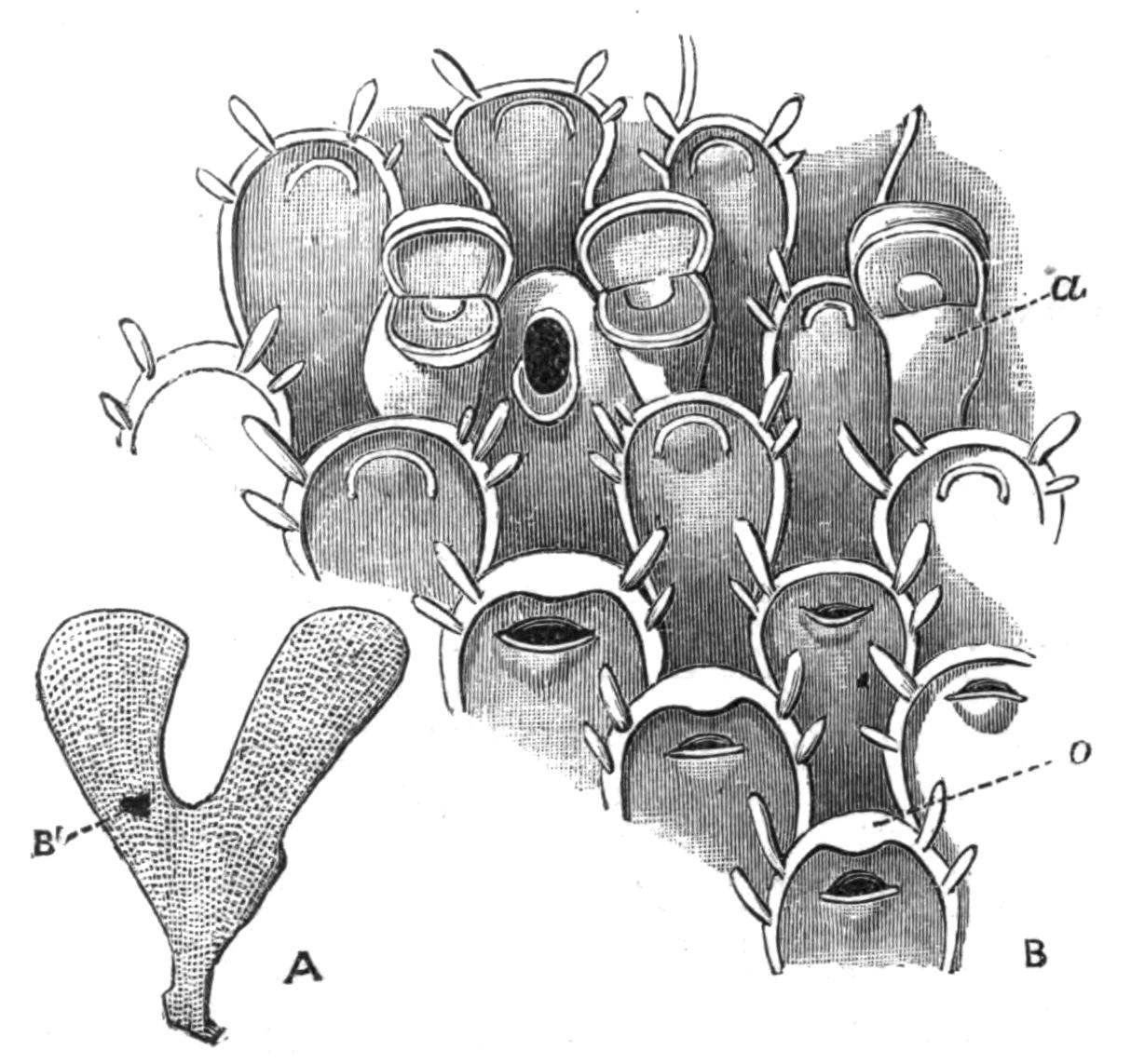
Fig. 232.—Flustra foliacea L., Cromer. A, Natural size, B' indicating the portion magnified in B (× 30): a, avicularium with closed mouth, to the left of which are seen two avicularia with open months; o, ovicell, forming the upper part of a zooecium. Ovicells are seen on three consecutive zooecia. The operculum, which closes the orifice of the zooecium, is seen in different positions in the individuals figured.
The upper part of some of the zooecia is somewhat swollen, these swellings representing the conspicuous "ovicells" of many other genera. In the early part of the year each ovicell protects an orange-coloured egg or embryo, and the larvae are readily liberated if the fresh colony be placed in clean sea-water. "At least ten thousand" were hatched out in three hours from a colony placed in a glass by Sir John Dalyell.[502] The larva swims freely in the water for a short time, and should it find a suitable resting-place, it fixes itself and forms the starting-point of a colony, the number of whose individuals is continually increased by the production of buds at the growing edge. The "avicularia" of this species will be alluded to later (see p. 482).
F. foliacea has long been known to possess in the fresh state a remarkable odour, which is described, according to the fancy of the observer, as a strong odour of fish, or as the smell of violets after a shower. Others have compared it to that of the orange or verbena, or to that of a mixture of roses and geranium.
Flustrella hispida, another of our commonest Polyzoa, which may be found between tide-marks on the stalks of Fucus, consists of a softish brown encrustation, about one-sixteenth of an inch thick, covered by numerous spines. If examined undisturbed in a rock-pool, or transferred to a glass of sea-water, the brown mass will be seen to become surrounded by a delicate bluish halo, which is about as thick as the encrusting mass itself, and consists of the tentacles of the numerous individuals of the colony. The microscope shows that each individual is provided with a circlet of some thirty or more long, delicate tentacles, which together form a graceful funnel (as in Fig. 233). At the bottom of the funnel is the mouth, to which Diatoms or other minute organic particles are conveyed by the cilia which fringe the tentacles. If the tentacles be touched with a needle, the whole funnel is retracted with great rapidity, and in this retracted condition we see no more than the body-walls of the animals. After an interval the tips of the tentacles are cautiously protruded; the tentacles are gradually pushed out, at first in a close bundle, but finally separating from one another to form the funnel which we have already noticed.
There is hardly a more surprising spectacle in the whole animal kingdom than a living fragment of the genus Bugula. The colony grows in the shape of a small tree, whose height may amount to several inches; and is characterised, in many species, by a spiral arrangement of the branches, which makes the genus easy to recognise at first sight (Fig. 233, A). The stem and branches are composed of a single layer of zooecia, arranged two or more abreast. Each zooecium bears, on its outer side, a most singular body termed an avicularium, from its resemblance to a bird's head. Imagine a minute eagle's head attached by a short but flexible neck to the zooecium. Suppose further that this structure moves backwards and forwards in a deliberate but determined fashion, its lower jaw usually widely open so as to be nearly 180° distant from its position when closed. Suppose that the lower jaw is moved by powerful muscles which can be distinctly seen inside the transparent head of the avicularium, and that every now and then it closes with a snap, seizing any unfortunate worm which may happen to be within reach with a grasp of iron. The above gives a very faint idea of the appearance of a living Bugula colony, with its hundreds of swaying avicularia, and with its tentacular funnels protruding from their zooecia, and withdrawing themselves capriciously from time to time.
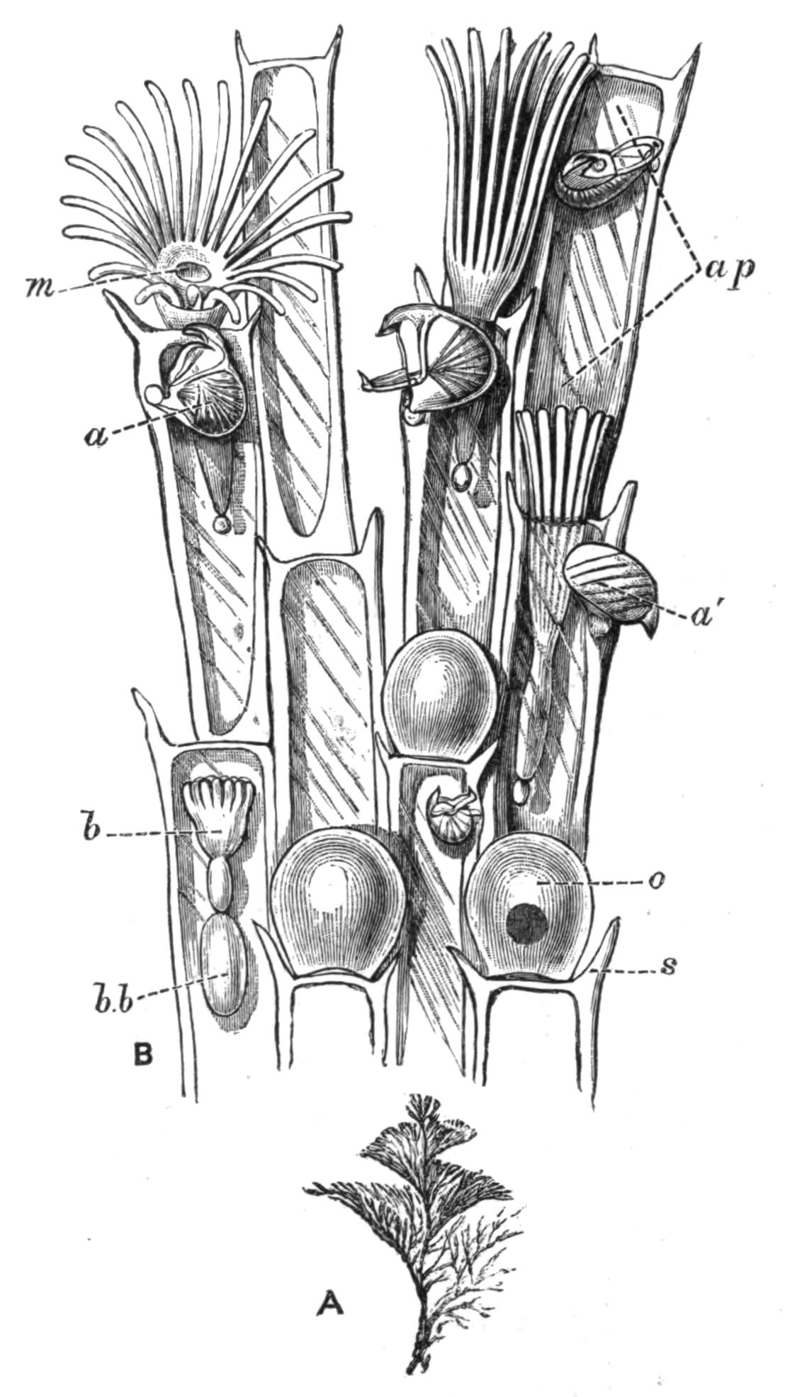
Fig. 233.—Bugula turbinata Alder, Plymouth. A, A small colony (natural size); B, portion of a branch (× 50): a, a', avicularia, in different positions; ap, "aperture" (see p. 524); b, polypide-bud, attached by its stomach to b.b, brown body; m, mouth, surrounded by the circle of tentacles; two individuals to the right show the tentacles partially expanded; o, ovicell; s, marginal spine. The avicularia of some of the zooecia have been omitted in B.
General Characters.—The Polyzoa are colonies, leaf-like or tree-like in form, and often strongly resembling seaweeds, or forming encrustations on the surface of stones and water-plants, or taking on other shapes. The units of the colony are complete individuals (Fig. 234). The zooecium or body-wall encloses a body-cavity, in which lies a digestive canal, with which are closely connected the central nervous system and the retractile, ciliated tentacles. The structures other than the zooecium constitute the "polypide." The mouth (m) leads into the ciliated pharynx (ph) which is followed by the oesophagus (oe) which again passes into the stomach (s), whose walls are coloured by a characteristic yellowish pigment. The stomach gives off the intestine (in), which is lined by strong cilia, by means of which a rotatory movement is given to the faeces contained in it. This communicates by a narrow passage with the rectum (r), which opens by means of the anus (a).
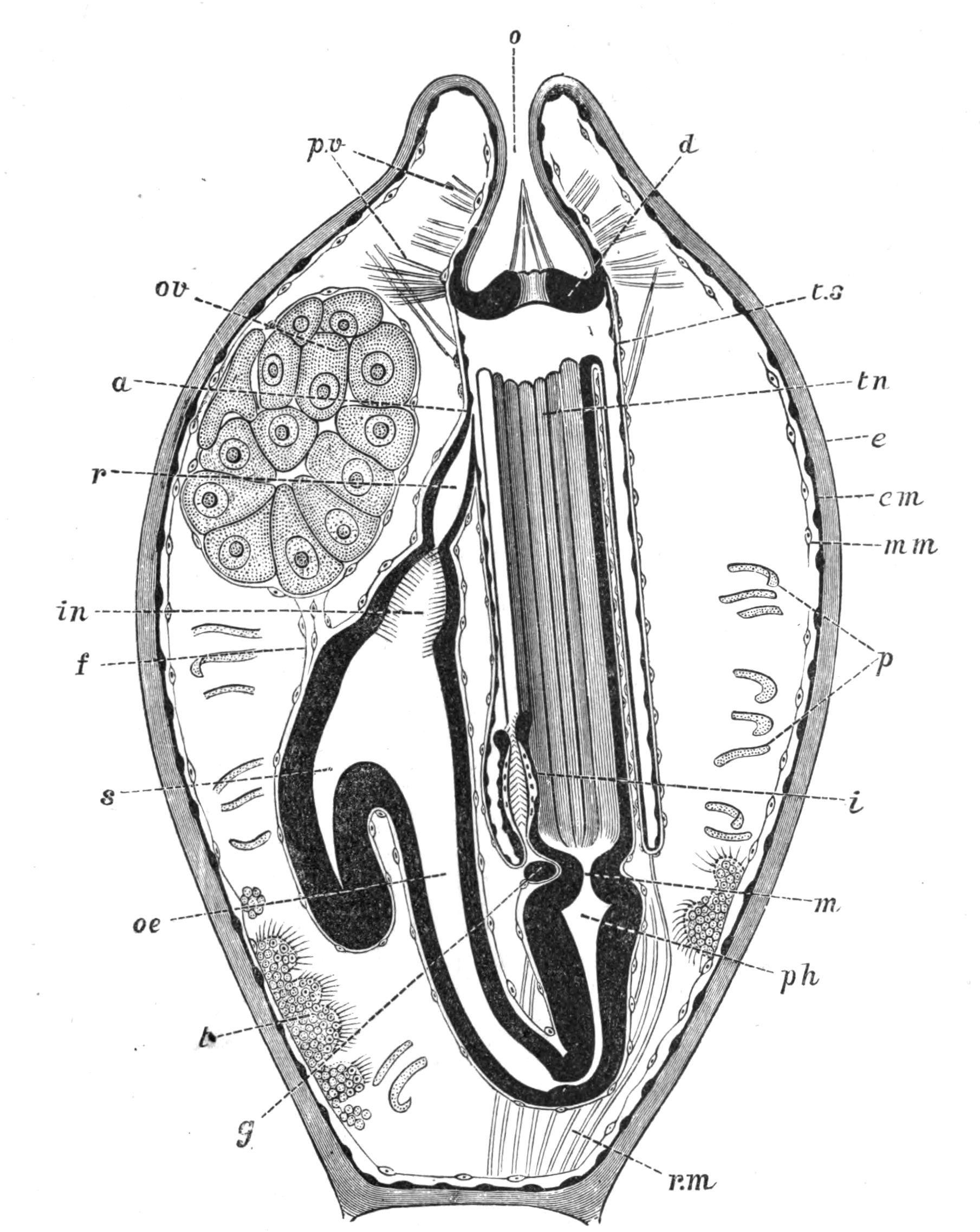
Fig. 234.—Alcyonidium albidum Alder, Banyuls-sur-Mer. Diagram showing the structure of a single zooecium with its polypide retracted: a, anus; d, diaphragm; e, ectocyst; em, ectoderm; f, funiculus; g, ganglion; i, intertentacular organ; in, intestine; m, mouth; mm, mesoderm of body-wall; o, orifice; oe, oesophagus; ov, ovary; p, parietal muscles; ph, pharynx; p.v, parieto-vaginal muscles; r, rectum; r.m, retractor muscles (contracted); s, stomach; t, testis; tn, tentacles; t.s, tentacle-sheath or kamptoderm. (After Prouho.[503])
In the retracted condition the tentacles (tn) lie in a cavity which opens to the exterior by the orifice (o). The cavity is bounded by a thin membrane termed the "tentacle-sheath" (ts), and it is incompletely subdivided, near its upper end, by a diaphragm (d), perforated by a circular hole through which the tentacles can be protruded. The diaphragm bears the thin folded collar characteristic of the Ctenostomata, the group to which the species figured belongs (see p. 477).
Fig. 238, B, shows the tentacles of Bowerbankia in their fully expanded and partially expanded condition. Comparing this with Fig. 234, it will be clear that when protrusion is taking place, the tentacles are forced in a bundle, tips first, through the diaphragm and next through the orifice of the zooecium, the alimentary canal offering no resistance to this movement, owing to the length of the oesophagus. A moment's consideration will show that the bases of the tentacles, in passing through the orifice, will carry with them that part of the flexible tentacle-sheath to which they are attached; and it will further be clear that so much of the tentacle-sheath as is thus protruded will be turned inside out. This process of "evagination" continues until its further progress is stopped by the retractor-muscles (r.m), and by the parieto-vaginal muscles (p.v), which pass from the interior of the body-wall to the upper part of the tentacle-sheath. The latter has now become the delicate layer which connects the expanded tentacles with the zooecium; and the anus (Fig. 238, C, a) opens directly to the exterior. Since the name "tentacle-sheath" is thus descriptive of the condition of retraction only, the term "kamptoderm"[504] has been suggested as an alternative name.
The presence of a complete digestive canal and the ciliation of the tentacles in Polyzoa are conspicuous differences between these animals and the Hydroids, with some of which the Polyzoa may have a marked external similarity.
The outermost[505] layer of the body-wall is known as the "ectocyst" (Fig. 234, e). This may be densely calcareous, in which case the dried Polyzoon differs little in appearance from the living animal with its tentacles retracted; or it may be partially calcified, or it may consist entirely of a flexible cuticle, as in Fig. 234. The ectocyst is prolonged through the orifice (o) as far as the diaphragm (d).
Forms with a calcareous ectocyst are commonly ornamented with ridges or other patterns, which are often of great beauty. The ectocyst in these cases is commonly interrupted at intervals by pores (Fig. 239, C), into which processes of the "endocyst"—the living, internal part of the body-wall—extend. These may appear as superficial pores, which apparently open to the exterior in the dried condition, or they may perforate the septa between adjacent individuals. This may be strikingly demonstrated by decalcifying a branch of Crisia (Fig. 237), in which the zooecia then appear connected by numerous strands of tissue. In many marine forms the communications between the individuals are in the form of small sieve-like plates known as "rosette-plates."
The endocyst may consist of definite layers of ectoderm (em) and mesoderm (mm), as in Fig. 234, but the mesoderm is commonly in the form of a loose network, some of which is attached to the body-wall, some to the alimentary canal, some forming connecting strands between these two layers, and other cells floating about freely in the body-cavity. These mesodermic structures are often spoken of as the "funicular tissue," since one or more strands of it commonly take on the form of a definite "funiculus" (f). This structure may bear the ovary (ov), while the testes (t) are found, commonly in the same zooecium, attached to various parts of the body-wall. The eggs and spermatozoa, when ripe, break off and float freely in the body-cavity.
The funicular tissue was at one time described as a "colonial nervous system." The idea expressed by this term must be considered erroneous from the fact that no nervous co-ordination of the individuals is known to exist, in the vast majority of cases. The actual nervous system consists of a ganglion (g) placed between the mouth and anus of each polypide, and lying in a small circular canal (not shown in Fig. 234) which immediately surrounds the oesophagus. This canal is developed in the bud as a part of the body-cavity, from which it becomes completely separated in marine forms. The Polyzoa have no vascular system.
Brown Bodies.—In the majority of cases, an extraordinary process of regeneration takes place periodically during the life of each zooecium. The tentacles, alimentary canal, and nervous system break down, and the tentacles cease to be capable of being protruded (Fig. 235, 1). The degenerating organs become compacted into a rounded mass (Fig. 235, 2 and 3, b.b), known from its colour as the "brown body." This structure may readily be seen in a large proportion of the zooecia of transparent species. In active parts of the colony the body-wall next develops an internal bud-like structure (Fig. 235, 1, b), which rapidly acquires the form of a new polypide (Fig. 235, 2 and 3). This takes the place originally occupied by the old polypide, while the latter may either remain in the zooecium in the permanent form of a "brown body," or pass to the exterior. In Flustra the young polypide-bud becomes connected with the "brown body" by a funiculus (Fig. 235, 1, 2). The apex of the blind pouch or "caecum" of the young stomach is guided by this strand to the "brown body," which it partially surrounds (3). The "brown body" then breaks up, and its fragments pass into the cavity of the stomach, from which they reach the exterior by means of the anus.
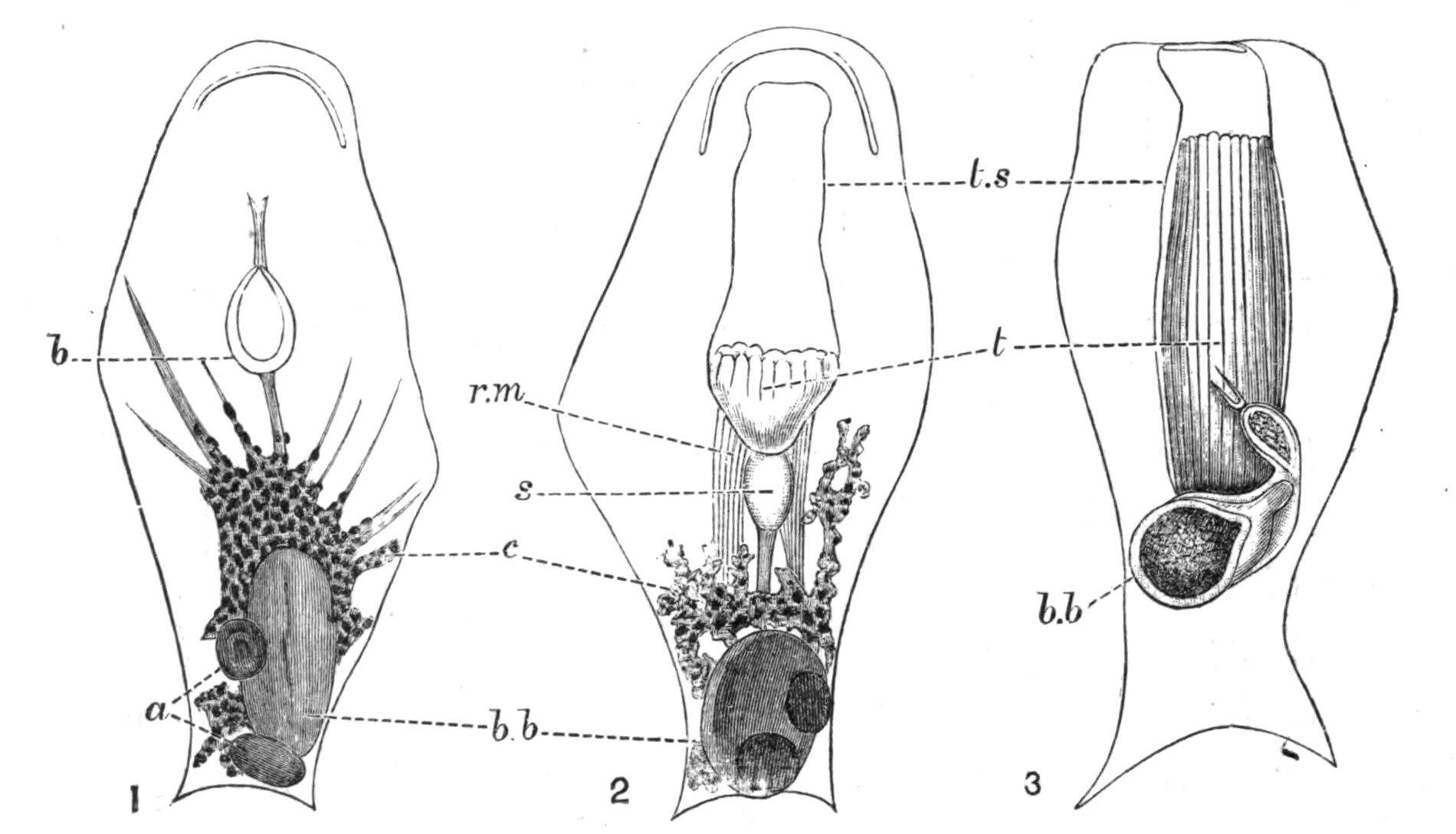
Fig. 235.—Flustra papyrea Pall. Naples. × 50. Illustrating the development of a new polypide after the formation of a "brown body." In 1, a, two masses formed from the alimentary canal; b, young polypide-bud; b.b, degenerating tentacles; c, connective tissue: 2, another zooecium, later stage; b.b, brown body; r.m, retractor muscles; s, stomach; t, tentacles of new polypide; t.s, tentacle-sheath: 3, the same zooecium, 191 hours later; letters as in 2. 1 and 2 are seen from the front, 3 from the back.[506]
There is some reason to believe[507] that these remarkable processes are connected with the removal of waste nitrogenous matters. The Marine Polyzoa are not known to be, in most cases, provided with definite excretory organs, although it is possible that the intertentacular organ (Fig. 234, i) described on p. 508 may in some cases perform excretory functions. There can, however, be little doubt that some kind of excretion takes place in the Polyzoa; and in considering what organs could possibly perform this work, our attention is arrested by the alimentary canal. The digestive organs of the young bud are perfectly colourless. As growth proceeds, certain parts acquire a yellowish, and later a brown colour. The degeneration of the polypide is followed by the grouping of large numbers of the free cells of the body-cavity into a mass which closely surrounds the incipient "brown body." Under their action, the latter becomes considerably smaller, probably as the result of the absorption of matters of nutritive value into other tissues. The final result is the formation of the compact "brown body," whose colour is principally derived from the pigment formerly present in the alimentary canal. Experiments made by introducing into the tissues of the Polyzoa certain artificial pigments which are known to be excreted by the kidneys when injected into the bodies of other animals, have given some reason for believing that the appearance of the brown pigment in the wall of the digestive organs is, in part, a normal process of excretion; although that process is not entirely carried out by the organs in question.
Little is known with regard to the duration of life of a single polypide; but some information bearing on this question may be obtained from a set of observations made on Flustra papyrea.[508] The table gives the number of days from the time at which the polypides were noticed to commence their degeneration:—
Days.
2 "Brown body" partially formed, the parts of the polypide being still easily recognisable.
5 Tentacles still just recognisable: appearance of new polypide-bud.
8 Stage shown in Fig. 235, 2.
11 Union of apex of stomach with "brown body."
16 "Brown body" half surrounded by stomach, and preparing to break up (Fig. 235, 3).
21 "Brown body" broken up into numerous fragments, contained in the alimentary canal of the new polypide.
35 "Brown body" almost completely absorbed.[509]
These results did not hold good for all the zooecia in a single colony. In some, the "brown body" was not completely got rid of at the end of sixty-eight days, the conclusion of the experiment.
So striking are the facts relating to the "brown bodies" that it has been believed[510] that what we have above described as the individual really consists of two kinds of individuals: firstly, the "polypide" or complex of tentacles and digestive organs; and secondly, the "zooecium," or house of the zooid or polypide, corresponding with what has been described above as the body-wall. The one individual, the zooecium, is on this view provided with successive generations of the second kind of individual, the polypide; and these latter function as the digestive organs of the two-fold organism. This view, though fascinating at first sight, is not borne out by an examination of all the facts of the case, especially when the Entoprocta are taken into account.
History.—The history of the Polyzoa, as far as 1856, has been fully treated by Allman in his great work on the Fresh-water Polyzoa;[511] but a few words may be said on this subject.
The Polyzoa attracted comparatively little attention before the beginning of the present century. Originally passed over as seaweeds, their real nature was established in connexion with the discovery of the animal nature of corals. So great a revolution could hardly be accepted without a struggle, and even Linnaeus went no further in this direction than to place them in a kind of half-way group of "zoophytes," whose nature was partly animal and partly vegetable. It is hardly necessary to point out that this view has now been abandoned by common consent; and indeed there is no more reason for regarding an animal as showing an approach to the plants because it grows in the external semblance of a seaweed than there would be for supposing a bee-orchid to be allied to the animal kingdom because of the form of its flowers.
But the claims of the Polyzoa to rank as a separate class were by no means admitted with the discovery that they were animals. They were still confounded with Hydroids, Alcyonarians, or Corals until their possession of a complete alimentary canal was recognised as a feature distinguishing them from those animals. This was principally due to the observations of J. V. Thompson[512] in Ireland, who introduced the term Polyzoa; and of C. G. Ehrenberg[513] in Germany, who proposed the class-name Bryozoa, or moss-like animals.
It is impossible to avoid all mention of the controversy which has raged with regard to these two rival terms. The controversy is for the present at rest, the name Polyzoa being employed by the majority of English writers, amongst whom must be mentioned Allman, Busk, Hincks, and Norman, admittedly authorities of the first rank; while Bryozoa is employed by practically all the Continental writers.
The priority of Thompson's name is unquestioned. While Ehrenberg, however, definitely introduced Bryozoa as the name of a group, Thompson was less precise in this respect, although he states[514] that his discovery "must be the cause of extensive alterations and dismemberments in the class with which they [the Polyzoa] have hitherto been associated." Thompson, in fact, clearly understood that the Polyzoa could no longer rank with the Hydroids. The controversy has been summarised by Hincks, in his History of the British Marine Polyzoa,[515] where references to other papers on the same subject are given.
The Polyzoa were associated by H. Milne-Edwards with the Tunicata in the group Molluscoidea (Molluscoïdes[516]), to which the Brachiopoda were afterwards added by Huxley.[517] A knowledge of the development of the Tunicata has, however, shown that these animals must be withdrawn from any association with the other two groups; while there is little real evidence that even the Brachiopods have anything to do with the Polyzoa.
Classification.—The Polyzoa are divided into two sub-classes:—I, the Entoprocta; and II, the Ectoprocta.[518] Although the character referred to by these terms is merely the position of the anus with relation to the tentacles,[519] there can be no doubt that the two groups differ widely from one another in many important respects. I do not, however, accept the view, maintained by some authors, that the Entoprocta and the Ectoprocta are two separate classes which are not nearly related.
The base from which the whole set of tentacles springs is known as the "lophophore."[520] In the Entoprocta (Fig. 236, 1) the lophophore is circular; the mouth is situated near the margin of the area surrounded by the tentacles; and the anus is found within the circlet, near the end opposite to the mouth.
In (2) and (3), representing the Ectoprocta, the anus is outside the series of tentacles. In the majority of cases, including all the marine Ectoprocta and one or two of the fresh-water forms, the lophophore is circular (2), the mouth occurring at the centre of the circle, and not being provided with a lip. These forms of Ectoprocta constitute the Order Gymnolaemata,[521] the dominant group of the Polyzoa in respect of number of genera and species. The remaining Ectoprocta belong to the exclusively fresh-water Order Phylactolaemata,[522] in which the mouth is protected by an overhanging lip or "epistome"; the ground-plan of the tentacles is, except in Fredericella, horse-shoe shaped (Fig. 236, 3), and the tentacles themselves are usually much more numerous than in the other cases.
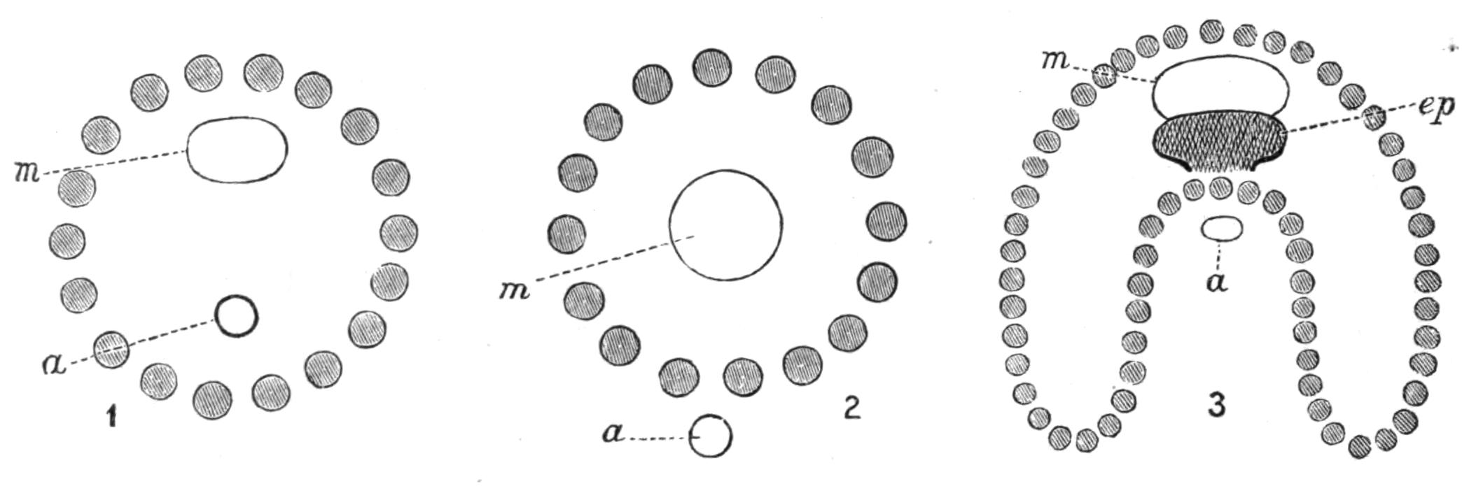
Fig. 236.—Ground-plan of the lophophore in (1) Entoprocta, (2) Gymnolaemata, (3) Phylactolaemata: a, anus; ep, epistome; m, mouth. The tentacles are represented by shaded circles.
The general characters of these divisions will be more easily understood by referring to the figures given of living representatives of the groups. The Entoprocta are illustrated by Figs. 243-245; the Gymnolaemata by Figs. 238, 240; and the Phylactolaemata by Figs. 247, 248.
The Gymnolaemata include three Sub-Orders:—
1. Cyclostomata.[523]—Body-wall densely calcareous, the zooecia being more or less tubular, usually with a circular orifice (Fig. 237).
2. Cheilostomata.[524]—Body-wall of varying consistency. The orifice is closed, in the retracted state of the polypide, by a chitinous lip or "operculum," which is more or less semicircular (Figs. 239, 241).
3. Ctenostomata.[525]—Body-wall always soft. The cavity into which the tentacles are retracted is closed by a frill-like membrane, the edges of whose folds have some resemblance to the teeth of a comb. This membrane, the "collar," is seen in different conditions of protrusion or retraction in Figs. 234, 238. The stomach may, in this group, be preceded by a muscular gizzard (Fig. 238, C, g).
Occurrence.—By far the larger number of the Polyzoa are inhabitants of the sea. A recently published catalogue[526] of marine Polyzoa includes nearly 1700 living species; and of these, the great majority belong to the Gymnolaemata. This group is further known to include an enormous number of fossil forms. Not only do we find that in living Polyzoa the members of a single Order largely outnumber the remainder of the Polyzoa, but we may further notice that the Cheilostomata, one of the sub-Orders of the dominant group, are at present largely in excess of the whole of the rest of the Polyzoa taken together.
Polyzoa may be collected with ease on almost any part of our coasts. The fronds of the "sea-mat" (Flustra foliacea) are thrown up by the waves in thousands in places where the bottom is shallow and sandy. The bases of the larger seaweeds growing on rocks between tide-marks are nearly always thickly covered with encrustations of Flustrella hispida or of species of Alcyonidium, in places where they are kept moist by being covered with a sufficiently thick layer of other algae. Rocks which are protected from the sun may be coated with calcareous Cheilostomes; and these are also found, in company with branching Polyzoa of various kinds, on the bases of the Laminaria thrown up by gales or exposed at spring tides. The graceful spirals of Bugula turbinata (Fig. 233, A) may be found hanging from the rocks at extreme low water; while colonies of Scrupocellaria, remarkable for their vibracula (see p. 484), are common in many places between tide-marks. Certain species affect the mouths of estuaries.
Membranipora membranacea commonly covers many square inches of the frond of Laminaria with its delicate lace-like encrustation. Nitsche[527] has shown that this species has its calcareous matter deposited in plates, separated by intervals of uncalcified ectocyst. The effect of this arrangement is to make the colony flexible, and to enable it to adapt its shape to the movements of the Laminaria, which is swayed to and fro by the action of the waves. Many of the calcareous forms growing on Laminaria have no special arrangement of this kind, and they accordingly grow in colonies whose area is so small that the greatest movements to which the seaweed is liable are not sufficient to crack or break the colony.
Many species show a decided, or even exclusive, preference for particular situations; as, for instance, species of Triticella, which are only found on certain Crustacea. Many encrusting forms prefer the inside of dead shells of Pecten, Cyprina, etc., to any other habitat. Terebripora[528] excavates tubular cavities in the substance of the shells of Molluscs. Hypophorella[529] inhabits passages which it forms in the walls of the tubes of the Polychaets, Lanice and Chaetopterus. Lepralia foliacea, one of the Cheilostomata, forms masses which may reach a circumference of several feet, simulating a small coral-reef. Its contorted plates are a regular museum of Polyzoa, so numerous are the species which delight to find shelter in the quiet interstices of the colony. The exquisite little colonies of Crisia eburnea are commonly found on red seaweeds, or on the branches of the Hydroid Sertularia.
The Polyzoa are found at all depths, certain Cheilostomes having been recorded from 3000 fathoms. The Cyclostomes dredged by the "Challenger" were all found in depths of 1600 fathoms or less, while the Ctenostomes are a distinctly shallow water group, most having been found at less than 40 fathoms, and only three at so great a depth as 150 fathoms.[530]
A few forms (Membranipora pilosa, Scrupocellaria reptans, etc.) are known to be phosphorescent;[531] but it is not known what is the purpose of this phenomenon.
External Form.—The Polyzoa may be roughly divided into (1) encrusting forms, usually calcareous, but sometimes soft; and (2) erect forms, which are either rigid or flexible. This flexibility can coexist with a highly calcified ectocyst, as in Crisia (Fig. 237), Cellaria, and others in which the branches are interrupted at intervals by chitinous joints. The coral-like forms may assume the most exquisite shapes, pre-eminent among which are the lovely net-like colonies of Retepora. Polyzoa of this type are seldom found between tide-marks, where their brittle branches would be liable to be snapped off by the waves. The erect species which occur in such positions are flexible, although flexible species are by no means restricted to the zone between tide-marks.
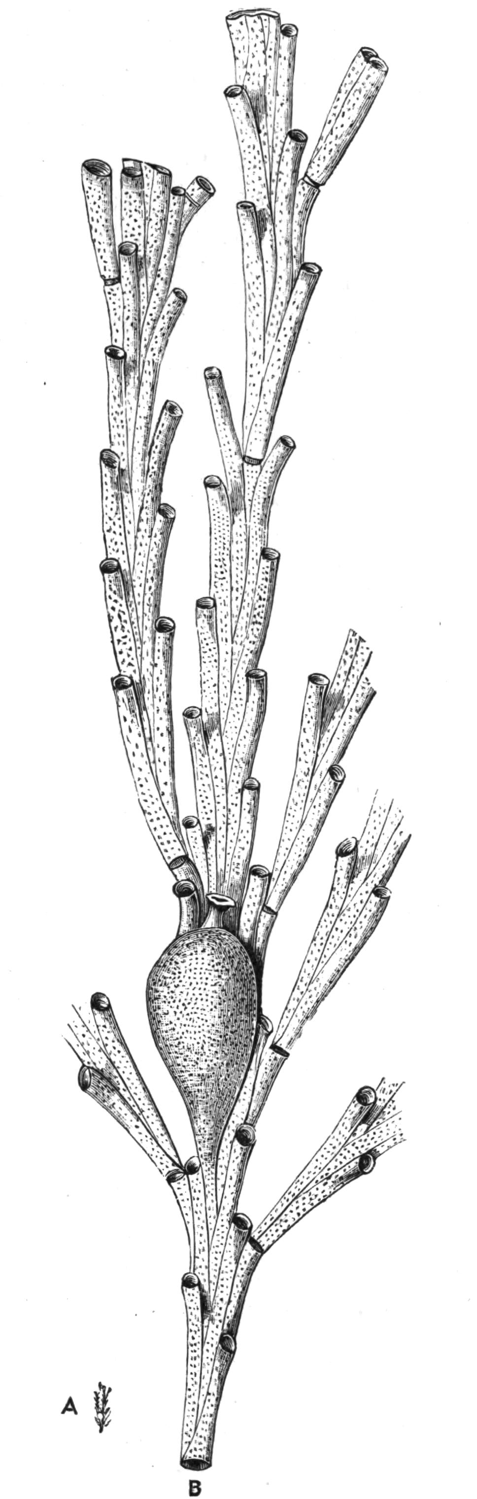
Fig. 237.—Crisia ramosa Harmer, Plymouth. A, End of a branch, × 1; B, another branch, × 20, showing the chitinous joints, the tubular zooecia characteristic of Cyclostomata, and the pear-shaped ovicell with a funnel-shaped orifice at its upper end.
Although the form of the colony is very different in different Polyzoa, a pocket-lens will usually show whether a given specimen belongs to the group or not. The surface is nearly always more or less distinctly composed of zooecia, or at least shows their orifices. The entire colony may be built up of these zooecia; and this is by far the commonest arrangement, both in encrusting and in erect forms. In certain genera, however, and particularly in some Ctenostomes (Fig. 238), and in most of the Entoprocta, the individuals grow out at intervals from a cylindrical stem or "stolon" (st), which is not composed of zooecia.
The Cyclostomata may assume an erect or encrusting habit. Their zooecia are always more or less cylindrical; the upper ends being often completely free, although in many cases the whole zooecium is closely adnate to its neighbours. In the breeding season the forms which belong to this group are provided with curious "ovicells," which contain the embryos. These may either be pear-shaped swellings on the branches (Crisia, Fig. 237), or they may form inflations of the surface, between the zooecia. The mature ovicell is provided with one or more openings, through which the larvae escape.
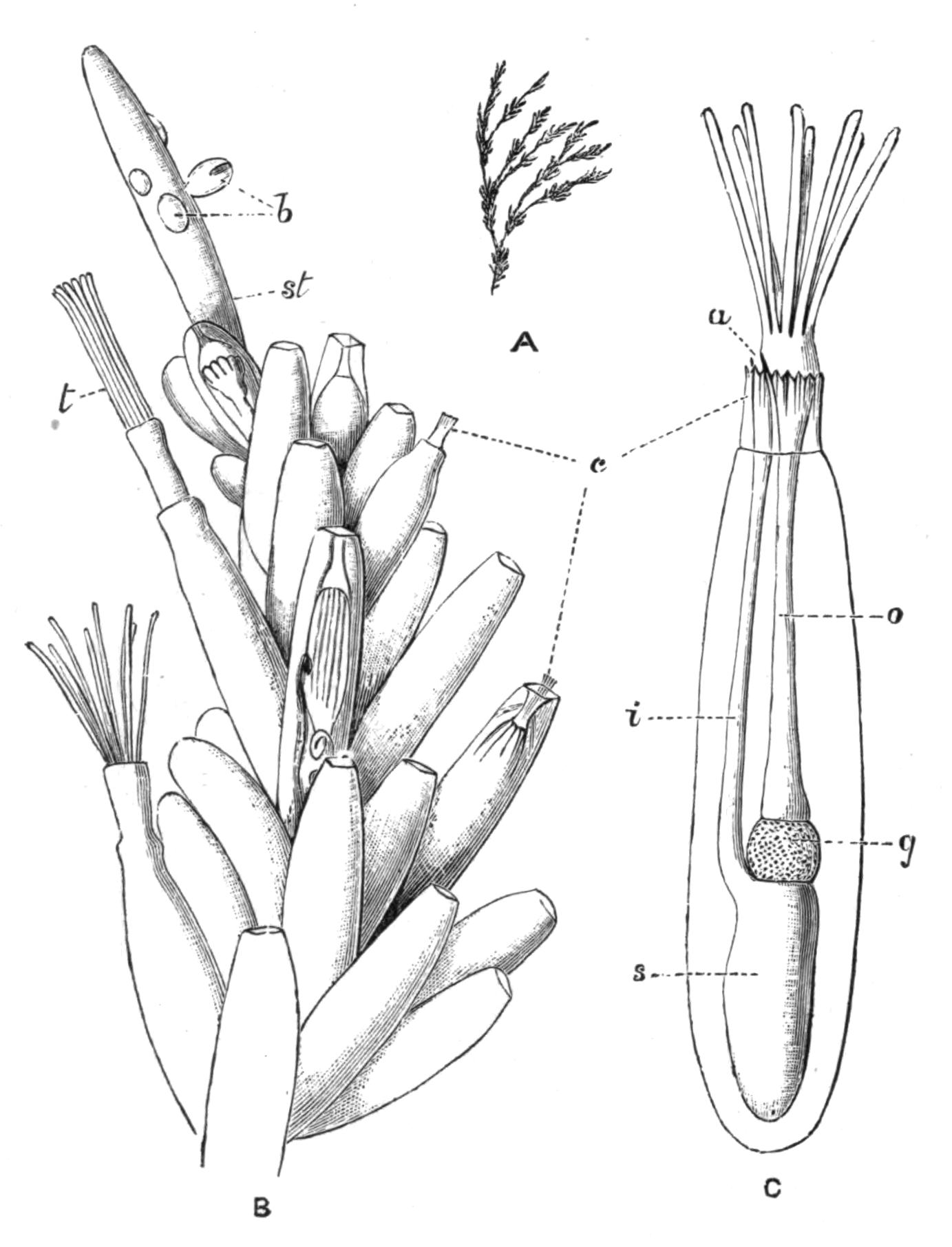
Fig. 238.—Bowerbankia pustulosa Ell. and Sol., Plymouth. A, Fragment of a colony, natural size, showing the branching stem, bearing tufts of zooecia: B, one of these tufts, with the growing apex of the stem (st), × 27; b, young zooecia (buds); c, the "collar" characteristic of Ctenostomata; t, tentacles; C, a single zooecium, with expanded tentacles, more highly magnified; a, anus; c, collar; g, gizzard; i, intestine; o, oesophagus; s, stomach.
The Ctenostomata rarely have even the slightest trace of calcareous matter. Alcyonidium and its allies form soft encrustations, or may even grow into erect masses six inches or more in height (A. gelatinosum). In this type the zooecia are often so closely united that it may be difficult or impossible to make out their limits in the living colony. Many of the dendritic or branching Ctenostomes (Fig. 238) are characterised by an extreme delicacy of habit. The zooecia in these cases are sharply marked off from the stem. They are either cylindrical or ovoid, being commonly attached by a very narrow base, so that in some species they readily fall off, and may thus be completely absent in certain parts of the colony. In such forms as Vesicularia spinosa, it requires considerable experience to recognise a stem which has lost its zooecia as being part of a Polyzoon. In Mimosella the zooecia possess a remarkable power of movement on the stem, similar to that possessed by the leaflets of the Sensitive Plant.[532] In certain forms (Bowerbankia, Amathia) the zooecia occur in groups separated by intervals which are devoid of zooecia, but in other cases they may have a more irregular arrangement. The collar to which this group owes its name is by no means a conspicuous feature. Its position when retracted has been shown in Fig. 234, while Fig. 238 further illustrates its relations.
The Cheilostomata grow in a great variety of forms, and also show a wide range of character in their zooecia. The orifice is commonly surrounded by stiff spines (Fig. 257, p. 524), which perhaps have the function of protecting the delicate polypides from the sudden impact of foreign bodies. These spines may attain an enormous development, as in Bicellaria ciliata, and some forms of Electra (Membranipora) pilosa (Fig. 256, A).
The operculum is usually, though by no means always, a conspicuous feature of the Cheilostome zooecium. It is invariably of chitinous consistency, and is more or less semicircular in outline, the straight edge forming a hinge on which the operculum opens. In some cases the orifice is surrounded by a raised margin or "peristome" (Fig. 255, B, C); the operculum is then situated at the bottom of a depression of the surface, and may be concealed from view. In others, in which the front wall of the zooecium is membranous (Bugula, Fig. 233), the operculum is merely a part of this membrane, and so is quite inconspicuous; and in cases of this kind the membranous wall may be protected by an arched spine, the "fornix," developed from one side of the zooecium (Fig. 254, f). The ovicells are commonly a conspicuous feature of this group, although they are believed to differ fundamentally from those of Cyclostomata. They have the form of a helmet-like covering overhanging the orifice (Figs. 240, 241), and may be either prominent or more or less concealed by the growth of adjacent parts of the zooecia. The presence of ovicells of this description is perfectly distinctive of the Cheilostomata.
Avicularia and Vibracula.—Most singular of the external appendages of the Cheilostomata are the extraordinary "avicularia" and "vibracula" of some genera.[533] By the comparison of a carefully selected series of genera, it has been established that the avicularium is a special modification of a zooecium. One of its least modified forms is found in Flustra foliacea (Fig. 232), where the avicularia (a) are small zooecia with a conspicuously large operculum ("mandible"). Avicularia of a similar type occur in Cellaria (Fig. 239, A), Schizotheca, etc., the avicularium occupying the place of an ordinary zooecium. These are the "vicarious" avicularia of Mr. Busk.[534]
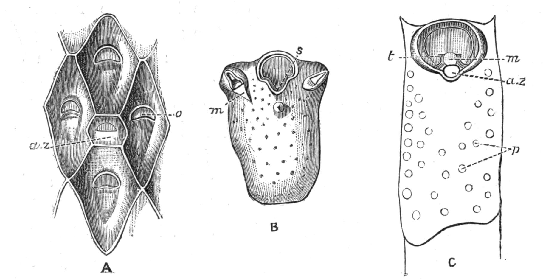
Fig. 239.—Forms of avicularia. A, Cellaria fistulosa L., Plymouth, × 43; a.z, avicularian zooecium, with closed mandible; o, operculum of zooecium: B, Schizoporella unicornis Johnst., Scilly Is., × 43; zooecium bearing two avicularia; m, opened mandible of avicularium; s, sinus of orifice: C, zooecium of Smittia landsborovii Johnst., Plymouth, × 43; the operculum is seen at the bottom of a depression surrounded by a thin collar or "peristome," in an emargination of which is seen an avicularian zooecium (a.z); m, mandible (opened); p, pores; t, tooth.
In the next stage (Figs. 239, B, 256, B) the avicularian zooecium is further reduced; it has in most cases lost its place in the series of individuals, and is found instead seated on some part of an ordinary zooecium ("adventitious" avicularia). The avicularium now consists of a much reduced zooecium, bearing the well-developed operculum or mandible.
Having arrived at this point, the avicularia seem to lose all sense of the propriety of remaining in the positions once occupied by zooecia. They have become degraded to the rank of appendages of the zooecia, and as such they may occur in an astonishing variety of positions. Sometimes one occurs on each zooecium in the middle line, or asymmetrically, or even on the top of the ovicell; in other cases the orifice is flanked by an avicularium on each side (Fig. 239, B). Sometimes (Cellepora) the avicularia are of more than one kind, some being large and some small, some having a pointed mandible and others a mandible with a rounded spoon-like end.
In the cases so far considered, the body of the avicularium is fixed. The highest differentiation acquired by these structures occurs in cases like Bugula, where they are borne on flexible stalks, which may even exceed the avicularia in length.[535]
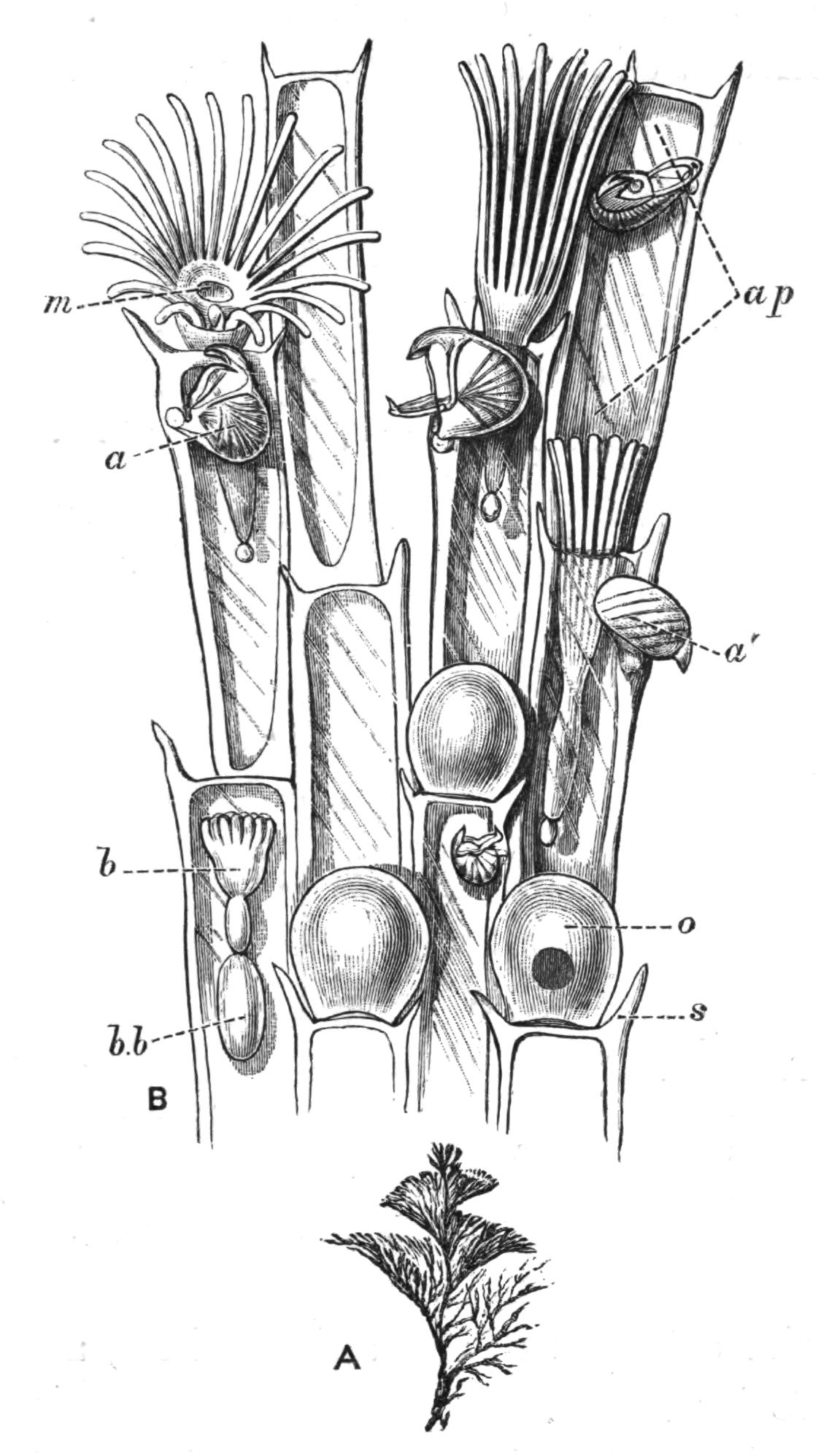
Fig. 240.—Bugula turbinata, showing avicularia (a, a'). The figure is explained on p. 468.
In Bugula turbinata (Fig. 240) each zooecium is provided with one of these appendages, attached to the base of the outer of the two spines which border its orifice. The avicularia of the two edges of the flattened branch are much larger than those of the more internal zooecia. The upper jaw is strengthened by a kind of buttress, or thickening of the ectocyst, which passes on each side across the avicularium to the hinge-line of its mandible. The upper part of the beak is strongly hooked, while the tip of the mandible bears a prominent spike, which fits inside the upper beak when the jaw snaps. A great part of the head is filled with a strong muscle, whose fibres exhibit a distinct transverse striation, and converge into a median tendon. The latter is inserted into the middle of the mandible. The muscle serves to close the jaws, and is the representative of the muscles by which the operculum is closed in an ordinary zooecium. The lower jaw is opened by means of a pair of muscles which are situated immediately under the ectocyst of the avicularium, and pass into the mandible close to its hinge.
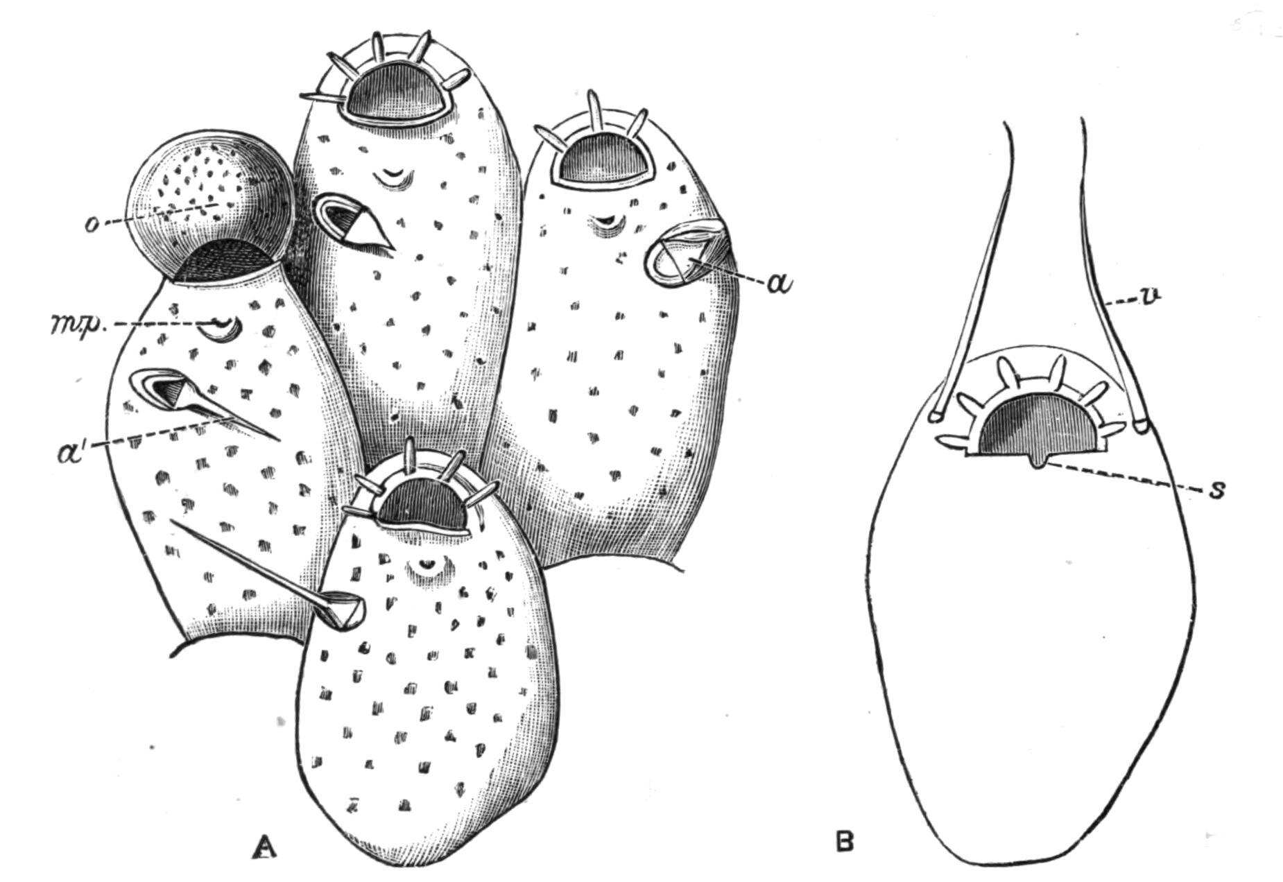
Fig. 241.—Illustrating the transition from avicularia to vibracula. A, Microporella ciliata Pall., Scilly Is., × 62; a, avicularium with short mandible (closed); a', avicularium with vibraculoid mandible (open); m.p, median pore; o, ovicell: B, Mastigophora dutertrei Aud., Shetland Is., × 47; s, sinus of orifice; v, seta of vibraculum (or vibraculoid avicularium).
Within the jaws, in the region which we may term the palate, is a rounded knob, which bears a tuft of delicate sensory hairs, which doubtless enable the avicularium to recognise the presence of any foreign body. The closure of the mouth may, indeed, be instantaneously induced by touching it with the point of a needle. It has been suggested that a small mass of cells which bears these hairs may represent the rudiment of the polypide.
The "vibraculum" (Fig. 242) is regarded as an avicularium in which the mandible has become elongated, so as to form a thin, chitinous "seta," which from time to time moves through the water. The part of the vibraculum which represents the zooecium commonly bears a tubular rootlet, used for attaching the colony to the substance on which it is growing (Fig. 254, p. 517).
In Microporella ciliata (Fig. 241, A) the avicularia are very variable, and in some cases take on a "vibraculoid" character. But in the fully-developed vibraculum (Fig. 242) there is usually no such compromise of characters. It may, however, be noted that Scrupocellaria scabra (Fig. 254), which belongs to a genus characterised by its highly differentiated vibracula, possesses structures (v.z) which could hardly be distinguished from avicularia were it not for the presence of the rootlet (r).
In the course of some observations which I had the opportunity of making on Bugula calathus at Naples, a fine hair offered to a small colony was seized with such force by the avicularia that the entire colony was lifted out of the water by the hair. The same colony had captured (1) a small Nereis, which it held with several of its avicularia; (2) an Anisopod Crustacean, 2½ mm. long; and (3) a small Amphipod, which was held by one of its antennae. The Anisopod was held by the tip of one leg with one avicularium, and by the penultimate joint of one of its chelae with an avicularium of another branch. It was captured in such a way that its chela, the "hand" of which was about half as long as the avicularium, actually closed on to the avicularium without being able to effect its escape. A little later the other chela was caught by another avicularium. Curiously enough, however, an avicularium did not necessarily close even when part of a captured animal was actually in its mouth. The avicularia made no attempt to place themselves in an advantageous position for catching fresh parts of the Nereis, which they might easily have done. The avicularia which had captured prey remained motionless. The others moved backwards and forwards (cf. the various positions of the avicularia shown in Fig. 240) ten times in ¾ to 1 minute, snapping their jaws perhaps once in that time. The two Crustacea were still retained by the avicularia two days later. On the next day they had both disappeared; but the colony had again caught the Nereis, which had previously effected its escape with the loss of nearly all its tentacular cirri.
These observations, and others which have been recorded, do not, unfortunately, give any information as to the purpose of the movements of the avicularia and vibracula. It is obvious that they may be defensive in character; and it cannot be doubted that the avicularia can prevent inquisitive worms from straying at will over the surface of the colony. There is no evidence to show that animals are discouraged from interfering with a Bugula owing to the presence of its defensive weapons.
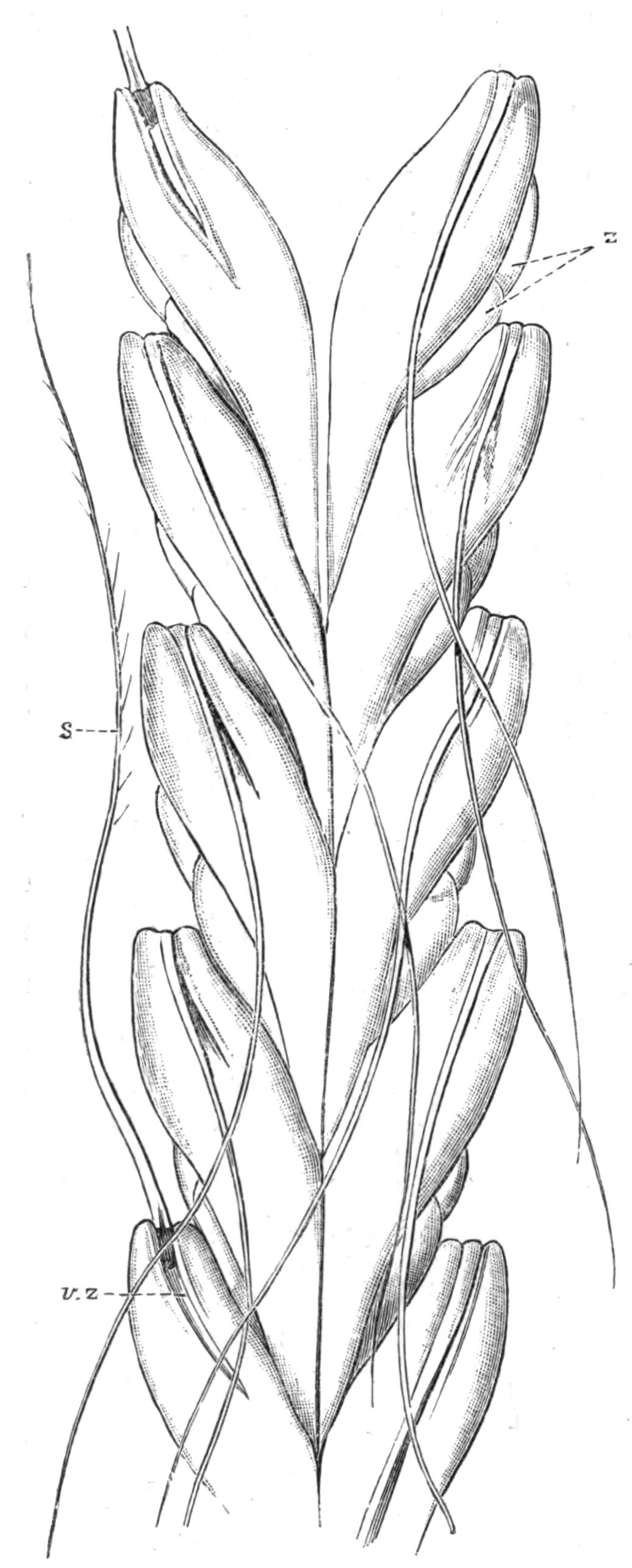
Fig. 242.—Caberea ellisii Flem., Norway. × 40. Back view of part of a branch. The large vibracular zooecia (v.z) occupy nearly the whole of the surface. s, Seta of vibraculum; z, zooecia.
It is not, indeed, certain what are the enemies against which the Polyzoa have specially to guard. Sea-urchins and certain Molluscs are known to browse on Polyzoa. Fresh-water Polyzoa, in which avicularia and vibracula are absent, are attacked by the larvae of Insects, and by Triclad Planarians. I have found the latter with their long pharynx everted and completely buried in a Cristatella colony. It is possible that some marine Cheilostomes may be saved from attacks of this kind owing to the existence of their armoury of avicularia and vibracula. It is also possible that these structures are of service by removing foreign particles which might otherwise settle on the colony, and tend to block up its orifices. It has further been suggested that animals seized by the avicularia may be held until they die, and that their disintegrating particles may then be carried to the mouths of the polypides by the ciliary currents of the tentacles; but proofs of this suggestion are wanting, and it must be admitted that the subject needs further elucidation.
The vibracula ordinarily remain stationary for some little time, every now and then giving a sweep through the water. In the majority of cases these structures, like the avicularia, act perfectly independently of one another, so far as can be made out; but in Caberea (Fig. 242) the vibracula move in unison, the simultaneous action of the whole series, after a period of quiet, being described as "positively startling."[536]
It has been stated by Busk[537] that the entire colony in Selenaria and Lunulites may be moved from place to place by the large vibracula which these forms possess.
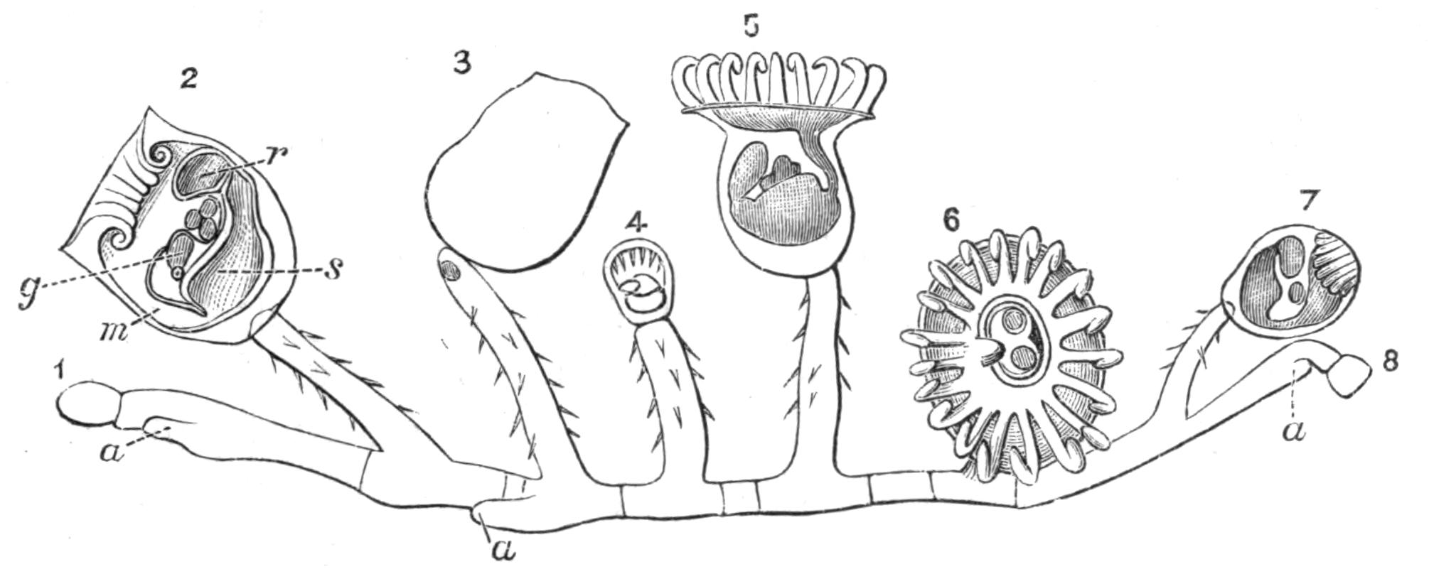
Fig. 243.—Pedicellina cernua Pall., Guernsey. Entire colony. × 27. The colony has three growing ends, a; 1-8, individuals of colony; 1 and 8 are quite immature; and 7 (tentacles retracted) is still young; 2, is seen in longitudinal section; g, generative organ, and below it the ganglion; m, mouth; r, rectum; s, stomach; between g and r are three embryos in the brood-pouch; the tentacles are retracted; in 5 and 6 the tentacles are expanded; in 6 two embryos are seen within the circle of the tentacles, to the left of them is the rectum, and to the right the mouth; 3 is in the act of losing its calyx, and has already developed the beginning of a new polypide-bud; in 4 the primary calyx has been lost, and the new calyx is clearly marked off from the stalk.
Entoprocta.—The Entoprocta, although a very small sub-class, deserve special consideration, if for no other reason, from the fact that many writers regard them as the most primitive group of Polyzoa, and consequently as the forms which show most affinity to other classes of animals.
Their most obvious characteristic is, as we have already seen,[538] the position of the anus within the circle of tentacles. The individuals formed by budding always remain more separate from one another than those of most Ectoprocta.
The commonest Entoproctous genus is Pedicellina, a graceful little animal, which occurs on many parts of our coast. It may often be discovered by looking carefully on the pink, jointed, calcareous alga, Corallina, which may be found growing at the edges of deep and cool rock-pools not too far above low-water mark. Its creeping stem or "stolon" is firmly attached to the surface of the seaweed, and sends off vertical stems here and there.[539] Each stem bears a "calyx," which is practically an individual of the colony. The stolon terminates, at one or both ends, in a growing-point (a), from which new individuals are budded off. The stalks bend from time to time in a curious spasmodic manner, by which means the calyces are moved about with an irritable and angry air. A good idea of the way in which the tentacles are folded away when the animal is disturbed may be obtained by putting the two wrists together, with the fingers spread out to represent the tentacles, the retraction of which would be represented by turning the tips of the fingers down into the space, the "vestibule," between the two palms. A delicate fold of skin growing from the edge of the calyx closes over the retracted tentacles, owing to the contraction of a sphincter muscle present in its circular edge. The body-wall is not separated from the alimentary canal by a definite body-cavity, so that there is no obvious distinction between the polypide and the zooecium. The existence of the Entoprocta is in fact a strong reason for refusing to admit that these two terms correspond with two different kinds of individuals.
Let us now imagine the condition we should have if a large and continuous cavity were developed between the alimentary canal and the body-wall. The body-wall would clearly have the general relations of a zooecium, while the alimentary canal and tentacles would obviously correspond with the polypide. The existence of the body-cavity would make it possible for the animal to retract its tentacles instead of merely turning them in. Regarded in this way, there is but little difficulty in comparing the Ectoprocta with the Entoprocta.
The calyces are deciduous, i.e. they are lost from time to time, the end of the stalk then producing a polypide-bud, which forms the vestibule and alimentary canal of a new calyx. Hence the phenomenon which may so commonly be noticed in Pedicellina of a "young head on old shoulders." The loss of the calyces may have some relation to the formation of the "brown bodies" in the Ectoprocta.
Another Entoproct, Loxosoma (Fig. 245) is remarkable for being the only Polyzoon which is not colonial. The buds, which are formed in two lateral series, break off as soon as they are mature, and at once begin to lead an independent existence. Loxosoma is further remarkable for being almost invariably found commensally with other animals, where it may occur in enormous numbers. L. phascolosomatum, common in the Channel Islands, is only found on the tip of the tail of Phascolosoma (see p. 428), which inhabits the mud of Zostera-beds. Other species are found on the external surface of certain sponges (Tethya, Euspongia, Cacospongia); or on the outside of a compound Ascidian, Leptoclinum, which may itself be carried about as a detachable covering on the back of a crab (Dromia). Another species is found on the ventral surface of the Polychaet Aphrodite, and of its ally Hermione.
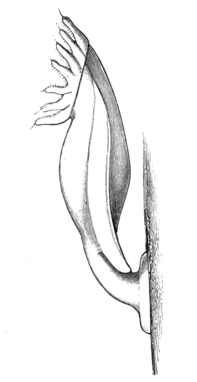
Fig. 244.—Side view of Loxosoma annelidicola Van Ben. and Hesse. × 50. (From Prouho.)
L. annelidicola, an interesting species recently investigated by Prouho,[540] was originally described in 1863 as a Trematode, under the name of Cyclatella. It escaped further notice until it was again found in the neighbourhood of Roscoff, in Brittany, on certain Polychaets belonging to the family Maldanidae (see p. 332). The calyx has a very flattened form, and is borne on a short stalk, which terminates in a large attaching disc, formerly mistaken for the sucker of a Trematode. The features in which this species differs from other members of the genus are shown by M. Prouho to be correlated with its mode of life. The animal has the habit of lying flat on its back, the disc at the end of its stalk being firmly attached to the skin of the worm, and its short stalk being bent round into a curve so as to bring the calyx into a supine position, with its lophophore directed upwards. This habit, together with its flattened form, prevents it from being crushed between the worm and its tube. But without some further provision its position might be merely a source of danger. For supposing the calyx to be directed backwards in relation to the worm, a sudden backward movement of the latter into its tube might bring the Loxosoma into fatal contact with the inner surface of the tube. There would obviously not be sufficient room to turn round in a vertical plane, so as to bring the body into a position of safety, i.e. into a position in which it moves stalk first. But by a beautiful arrangement of the muscles of its stalk this movement is effected in a horizontal plane; on touching the Loxosoma with the point of a needle it would swing round in this way through 180° with "une rapidité qui étonne."
Urnatella[541] is a beautiful form with a segmented stalk, the stalks usually arising in pairs from a common base. It has at present only been found in fresh water in the United States.
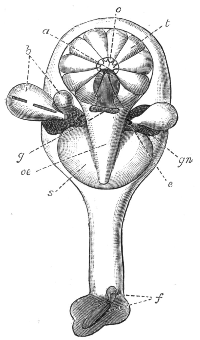
Fig. 245.—Diagram of the structure of Loxosoma, seen from the oesophageal side. × about 70. a, Anus; b, buds; e, excretory organ; f, foot-gland; g, ganglion; gn, generative organs; o, orifice of vestibule; oe, oesophagus; s, stomach; t, retracted tentacles.
In Pedicellina the plane of the lophophore is at right angles to the stalk, which is separated from its calyx by a marked constriction. In Loxosoma the lophophore is set obliquely,[542] and there is no constriction at the base of the calyx. In Urnatella we find an intermediate condition, the lophophore resembling that of Loxosoma, while the constriction at the base of the calyx is similar to that of Pedicellina. Since the latter is known to pass in its development[543] through a stage with an oblique lophophore, it may be presumed that Loxosoma is a more archaic form than Pedicellina. In other respects, the structure of the Entoprocta is very constant, whatever the genus.
A pair of ciliated excretory tubes open into the vestibule. These are similar in structure to the "head-kidneys" of the larvae of Polychaet worms, or to the excretory organs of adult Rotifers. Flame-cells have been described by Davenport in the stalk of Urnatella, but it is not known whether they are connected with the excretory tubes of the calyx. The animals are either hermaphrodite or have separate sexes, and the generative organs open by ducts of their own into the vestibule. The nervous system consists of a ganglion placed between the mouth and the anus, giving off a set of nerves, many of which end in delicate tactile hairs placed on the tentacles or other parts of the body.[544]