Chapter I
Reading edition
TURBELLARIA
INTRODUCTION: DESCRIPTION OF THE POLYCLAD LEPTOPLANA TREMELLARIS—APPEARANCE—HABITS—STRUCTURE: POLYCLADIDA—CLASSIFICATION—HABITS—ANATOMY—DEVELOPMENT: TRICLADIDA—OCCURRENCE—STRUCTURE—CLASSIFICATION: RHABDOCOELIDA—OCCURRENCE—HABITS—REPRODUCTION—CLASSIFICATION.
The Platyhelminthes, or Flat Worms, form a natural assemblage of animals, the members of which, however widely they may differ in appearance, habits, or life-history, exhibit a fundamental similarity of organisation which justifies their separation from other classes of worms, and their union into a distinct phylum. Excluding the leeches (Hirudinea), and the long sea-worms (Nemertinea)—which, though formerly included, are now treated independently—the Platyhelminthes may be divided into three branches: (1) Turbellaria (including the Planarians), (2) Trematoda (including the liver-flukes), and (3) Cestoda (tape-worms). The Mesozoa will be treated as an appendix to the Platyhelminthes.
The Turbellaria were so called by Ehrenberg[1] (1831) on account of the cilia or vibratile processes with which these aquatic animals are covered, causing by their incessant action, tiny currents ("turbellae," disturbances) in the surrounding water. The ciliary covering distinguishes this free-living group from the parasitic Trematodes and Cestodes, some of which possess such an investment, but only during their early free larval stage, for the short period when they have left the parental host and are seeking another (Figs. 26, 27, 42).
Some Turbellaria (Rhabdocoelida) resemble Infusoria in their minute size, shape, and movements. Nevertheless they possess an organisation of considerable complexity. The fresh-water Planarians (Fig. 14), abounding in ponds and streams, vary from a quarter to half an inch in length, and are elongated and flattened. Their body is soft, and progresses by a characteristic, even, gliding motion like a snail. The marine Planarians or Polyclads (Fig. 8) are usually broad and leaf-like, sometimes attaining a length of six inches, and swim or creep in a most graceful way. Land Planarians occur in this country (Fig. 15), but far more abundantly in tropical and sub-tropical districts, in moist places, venturing abroad at night in pursuit of prey. They are elongated and cylindrical, in some cases measuring, when fully extended, a foot or more in length, and are often ornamented with brilliantly coloured, longitudinal bands.
Turbellaria are carnivorous, overpowering their prey by peculiar cutaneous offensive weapons, and then sucking out the contents of the victim by the "pharynx." Land Planarians feed on earthworms, molluscs, and wood-lice; fresh-water Planarians on Oligochaet worms, water-snails, and water-beetles; marine forms devour Polychaet worms and molluscs. Some Turbellaria seem to prefer freshly-killed or weakly examples of animals too large to be overpowered when fully active. Certain Rhabdocoelida are messmates of Molluscs and Echinoderms, and a few others are truly parasitic—a mode of life adopted by all Trematodes save Temnocephala.
The Trematodes[2] may be divided into those living on the outer surface of various aquatic animals, usually fish (Ectoparasites); and those which penetrate more or less deeply into the alimentary canal or the associated organs of the host (Endoparasites). They are oval, flattened Platyhelminthes ranging from a microscopic size to a length of three feet (Nematobothrium, Fig. 22), and are provided with organs of adhesion by which they cling to the outer surface, or to the interior, of the animals they inhabit. Trematodes occur parasitically in all groups of Vertebrates, but, with the exception of the liver-flukes of the sheep (Distomum hepaticum and D. magnum), and of Bilharzia haematobia found in man (in the blood-vessels of the urinary bladder) over the greater part of Africa, their attacks are not usually of a serious nature. Ectoparasitic Trematodes are Monogenetic; that is, their larvae grow up directly into mature forms. The Endoparasitic species, however, are usually Digenetic. Their larvae enter an Invertebrate and produce a new generation of different larvae, and these another. The last are immature flukes. They enter a second host, which is swallowed by the final Vertebrate host in which they become mature.
The Cestodes or Tape-worms have undergone more profound modifications both in structure and in mode of development. They are all endoparasitic, and, with one exception (Archigetes), attain maturity solely within the alimentary canal of Vertebrates. In length they range from a few millimetres to several metres, but this great size is attained from the need for the rapid production and accumulation of enormous numbers of eggs. The "head" or "scolex" is attached to the mucous membrane of the host by suckers or hooks, but there is no mouth nor any certain trace of a digestive tract at any stage of the life-history of Cestodes. For nourishment they absorb, through the skin, the previously-digested food (of the host) that bathes them. In a few Cestodes the body is simple and not divided into "proglottides" or generative segments, but in most cases it is jointed in such a way that the last segment is the oldest, and each contains a set of reproductive organs. The life-histories of Cestodes are most remarkable. The proglottides containing the eggs pass out of the final host along with the faeces and enter the intermediate host with the food. The larvae hatch, and boring their way into the blood-vessels, are carried by the circulation to various internal organs. Here they usually become "bladder-worms," and develop the "head" of the future sexual form. Then, if, as is usually the case, the intermediate host is preyed upon by the final host, the larval Cestodes enter the alimentary canal of the latter. The head of the larva alone survives digestion, and from it the mature worm is formed.
Of these three branches of the phylum Platyhelminthes, the Turbellaria possess features of special interest and importance. Not only do they furnish the explanation of the structure of the two parasitic groups (which have probably arisen from Turbellarian-like ancestors), but they occupy the lowest position in the whole group of worms. There are reasons for thinking that this is the simplest group of bilateral animals which adopt the habit of creeping. The Turbellaria are most closely allied to that great extinct group from which they, the Nemertinea, Rotifera, and even the Annelids, offer increasingly convincing evidence of having been derived. Many questions relating to the affinities of, or the origin of organs in, the Annelids, resolve themselves into similar questions about the Turbellaria. For these reasons, this group is here dealt with at greater length than the others, the interest of which is of a more special nature.
The history of our knowledge of the Cestodes dates back to ancient times, as the presence and effects of tape-worms early attracted the attention of physicians. Trematodes are first distinctly referred to in the sixteenth century, while Turbellaria first figure in Trembley's memoir on Hydra (1744).[3] The whole subject of the increase in our knowledge of parasitic Platyhelminthes is dealt with in the standard work, The Parasites of Man, by Leuckart,[4] and a complete list of references in zoological literature to Cestodes and Trematodes is to be found in Bronn's Thierreich.[5] O. F. Müller[6] and Ehrenberg founded our knowledge of the Turbellaria, but for a long time the group remained in a most neglected condition. In this country Montagu, G. Johnston, and in Ireland, William Thompson, discovered several marine species, one of which, Planocera folium (from Berwick), has not again been met with on British shores. Dalyell[7] conducted classical researches on the habits of Planarians, and Faraday[8] made interesting experiments on their power of regenerating lost parts. The credit of assigning the correct interpretation to most of the various organs of fresh-water Planarians belongs to von Baer[9] and Dugès,[10] while Mertens[11] effected a similar service for the marine forms, or Polyclads. The minute Rhabdocoels were first successfully investigated and classified by Oscar Schmidt.[12] The great work on this group is, however, the monograph by von Graff.[13] A similarly comprehensive and indispensable treatise by Lang, on the Polycladida,[14] contains references to all previous publications on the group, among which the papers by Quatrefages, Johannes Müller, Keferstein, Minot, and Hallez stand out conspicuously. Moseley's work[15] on the Land Planarians of Ceylon is undoubtedly the most revolutionary paper referring to this group, and the best contribution towards elucidating the structure of the Tricladida at a time when the subject was very obscure. A monograph on Land Planarians is being prepared by von Graff.
The Turbellaria are divided into: (1) Polycladida, marine forms with multiple intestinal branches; (2) Tricladida, marine, fresh-water, and terrestrial Planarians with three main intestinal branches; (3) the Rhabdocoelida, as varied in habit as the Triclads, but possessing a straight and simple or slightly lobed, intestine. A detailed description of an example of the Polyclads, and then a comparative account of each division, will now be given.
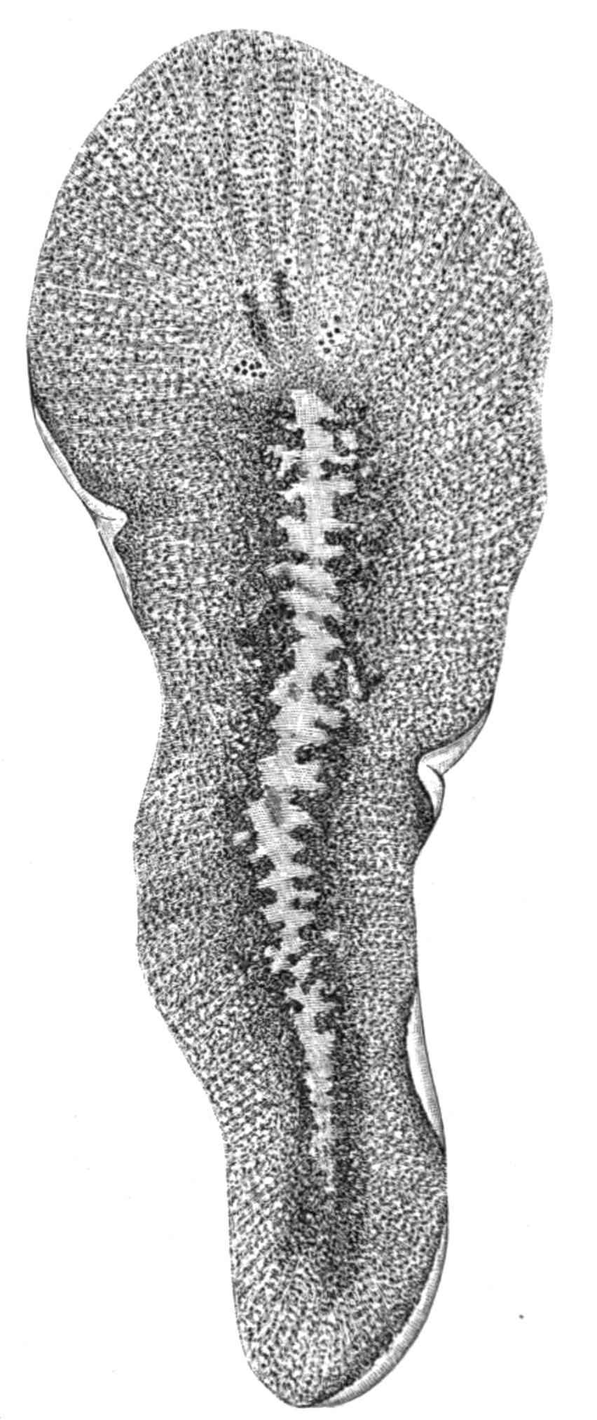
Fig. 1.—Leptoplana tremellaris O. F. M. Seen from the dorsal surface. The alimentary canal runs down the middle line and sends branches to the margin of the body. × 6.
Turbellaria. I. Polycladida.
Description of Leptoplana tremellaris.
Appearance and Habits.—An account of the Polyclad Turbellaria may be fitly prefaced by a description of a very common representative, Leptoplana tremellaris, so called on account of the thin, flat body which executes when disturbed, quivering or tremulous swimming movements.
Like all Polyclads, Leptoplana is marine. It is probably found on all European shores, northwards to Greenland and southwards to the Red Sea, while vertically it ranges from the littoral zone down to fifty fathoms. There is, however, an apparently well-marked difference between the littoral specimens, which vary from three-quarters to one inch in length, are brownish in colour and firm in consistency, and the more delicate examples half an inch long, white with a brown tinge, which occur in deeper water.
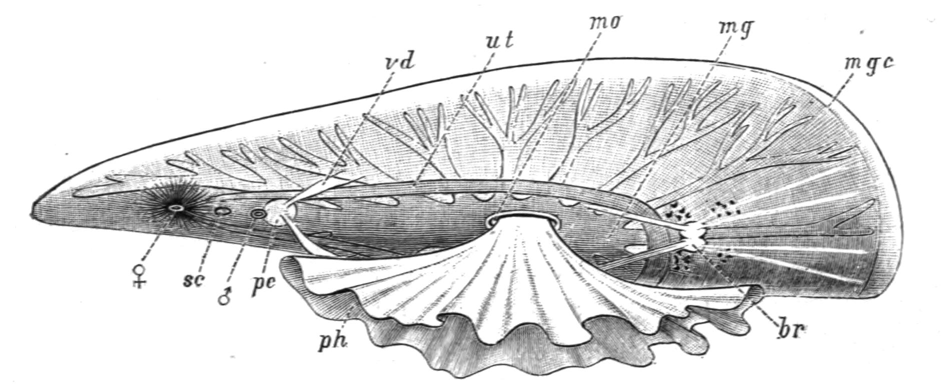
Fig. 2.—Leptoplana tremellaris. Three-quarters view from the ventral surface. The pharynx (ph) is widely protruded through the month (mo) as in the act of attacking prey. br, Brain with nerves, close to which are the four groups of eyes; mg, stomach; mgc, "marginal groove"; pe, penis; sc, sucker; ut, uterus; vd, vasa deferentia; ♀, female genital aperture surrounded by the shell-gland; ♂, male aperture. (Semi-diagrammatic, and × 6.)
At low water Leptoplana may be found buried in mud or on the under surface of stones, in pools where darkness and dampness may be ensured till the return of the tide. It is, however, by no means easy to detect and remove it from the encrusting Polyzoa, Ascidians, or Sponges with which it is usually associated. The flat, soft, unsegmented body is so closely appressed to the substratum that its presence is usually only betrayed by its movement, an even gliding motion of the mobile body, which suggested the apt name "la pellicule animée" to Dicquemare. The creeping surface is called ventral, the upper one dorsal, and as the broader end of the body always goes first, it is anterior as opposed to the more pointed posterior extremity. With a lens the characters shown in Figs. 1 and 2 may be observed. The eyes are seen as black dots near the anterior end, and are placed at the sides of a clear oval space, the brain. Along the transparent margin of the body, the ends of the intestinal branches may be seen. These ramify from a lobed stomach or main-gut, and should the specimen be mature, the "uterus" loaded with eggs forms a dark margin round the latter (Figs. 1 and 2, ut). The ventral surface is whitish, and through it the "pharynx," a frilled protrusible structure, may be dimly observed. The "mouth,"[16] through which the pharynx at the time of feeding is thrust out (Fig. 2, mo), is almost in the centre of the ventral surface. Behind this, a white, V-shaped mark (vd) indicates the ducts of the male reproductive organs, and still further back is the irregular opaque mark of the "shell-gland," by which the egg-shells are formed (Fig. 2, ♀).
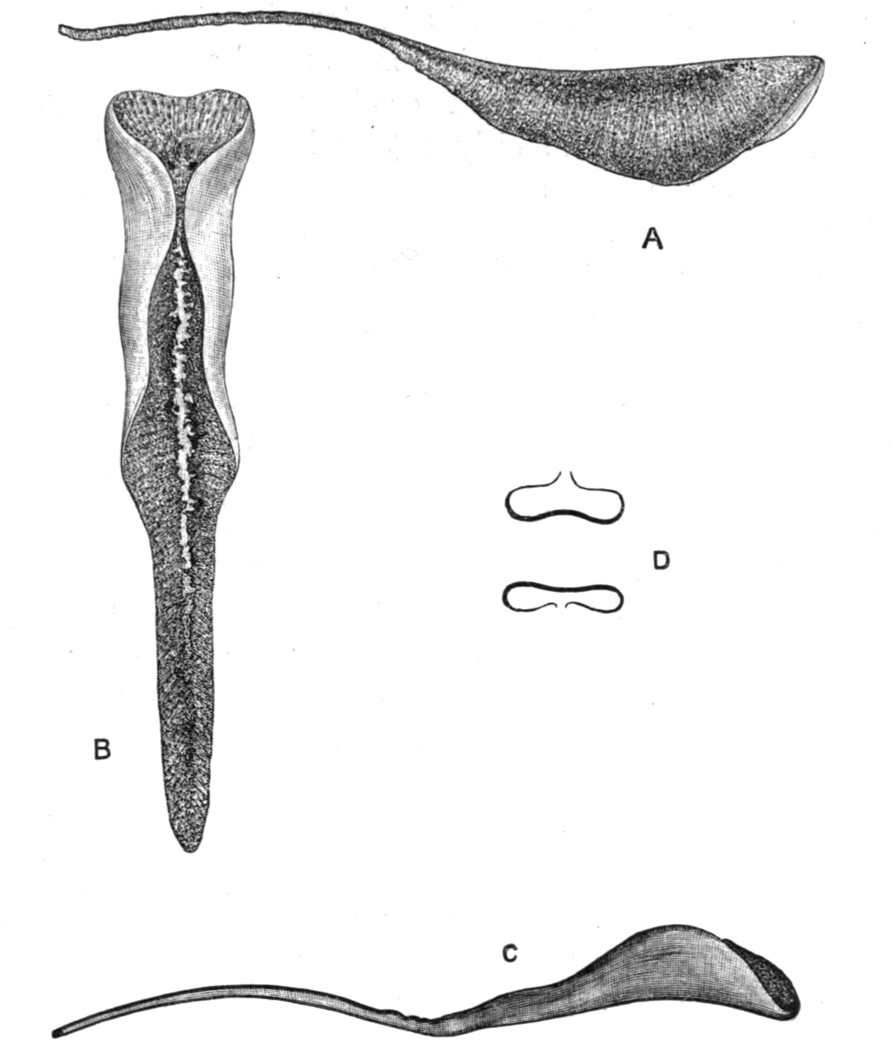
Fig. 3.—Leptoplana tremellaris in the act of swimming. A, Seen from the right side during the downward stroke (the resemblance to a skate is striking); B, from above, showing the upward stroke and longitudinal undulations of the swimming lobes; C, side view during the upward stroke; D, transverse sections of the body during the strokes. × 5.
Leptoplana employs two kinds of movement, creeping and swimming. Creeping is a uniform gliding movement, caused by the cilia of the ventral surface, aided perhaps by the longitudinal muscular layers of this surface, and is effected on the under side of the "surface-film" of water almost as well as on a solid substratum. Swimming is a more rapid and elegant movement, employed when alarmed or in pursuit of prey. The expanded fore-parts of the body act as lobes, which are flapped rapidly up over the body and then down beneath it, undulations running rapidly down them from before backwards. The action in fact is somewhat similar to that by which a skate swims, a resemblance pointed out long ago by Dugès[17] (Fig. 3).
We have few direct observations on the nature of the food of Leptoplana, or the exact mode by which it is obtained. Dalyell,[18] who observed this species very carefully, noticed that it was nocturnal and fed upon a Nereis, becoming greatly distended and of a green colour after the meal, but pale after a long fast. Keferstein[19] noticed a specimen in the act of devouring a Lumbriconereis longer than itself, and also found the radulae of Chiton and Taenioglossate Molluscs in the intestine. That such an apparently weak and defenceless animal does overpower large and healthy Annelids and Mollusca, has not hitherto been definitely proved. Weak or diseased examples may be chiefly selected. The flexible Leptoplana adheres firmly to its prey, and the rapid action of the salivary glands of its mobile pharynx quickly softens and disintegrates the internal parts of the victim. The food passes into the stomach (Fig. 2, mg), and is there digested. It is then transferred to the lateral branches of the intestine, and, after all the nutritious matters have been absorbed, the faeces are ejected with a sudden contraction of the whole body through the pharynx into the water.
Leptoplana probably does not live more than a year. In the spring or summer, batches of eggs are laid and fixed to algae or stones by one individual, after having been fertilised by another. Young Leptoplana hatch out in two to three weeks, and lead a pelagic existence till they are three or four millimetres in length. In late summer, numbers of such immature examples may be found among sea-weeds and Corallina in tide pools. In the succeeding spring they develop first the male and then the female reproductive organs.
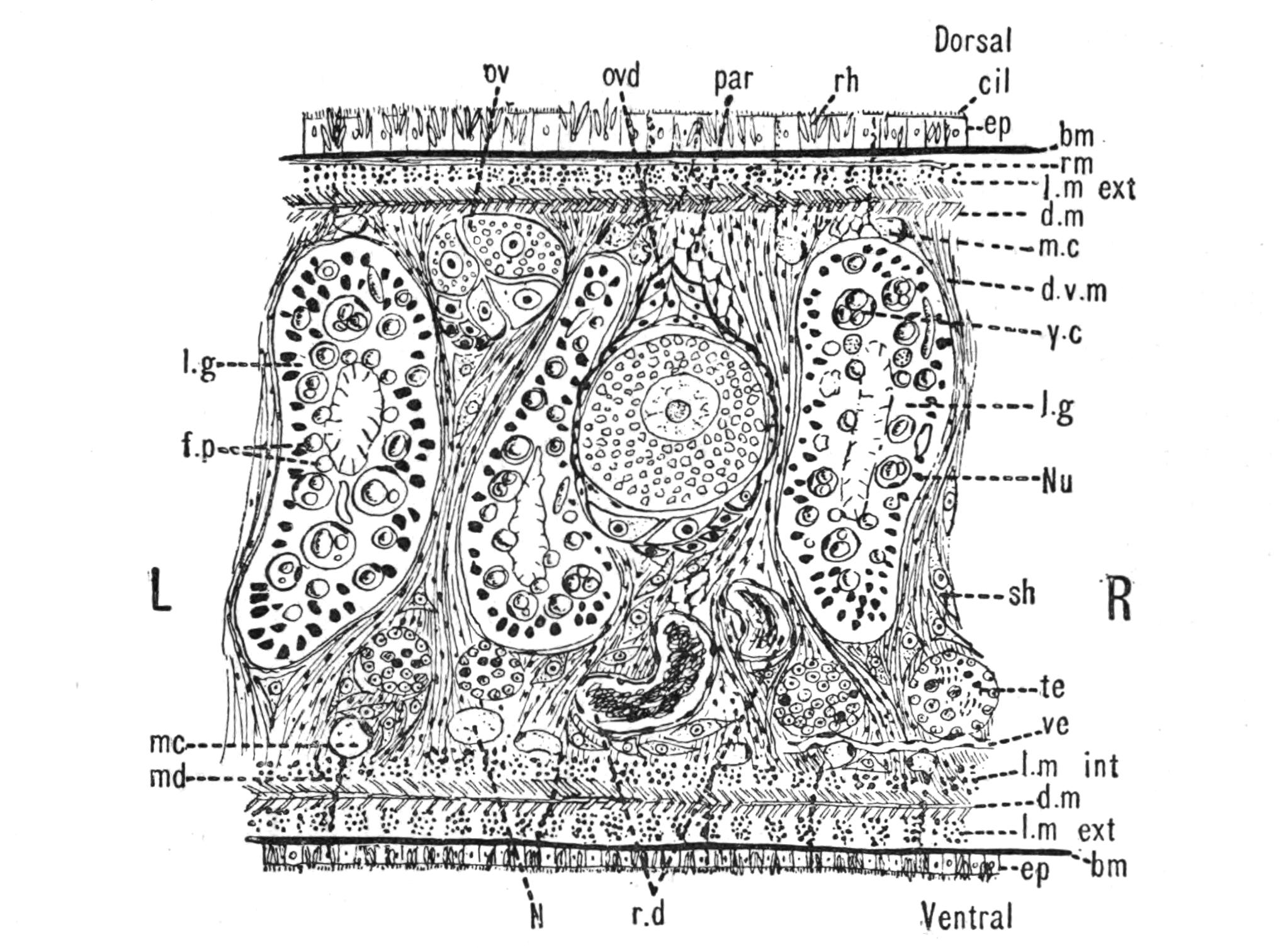
Fig. 4.—Portion of a transverse section of Leptoplana tremellaris in the hinder part of the body. × 100. bm, Basement (skeletal) membrane; cil, cilia; d.m, diagonal muscles; d.v.m, dorso-ventral muscles; ep, epidermis; f.p, food particles; l.g, lateral intestinal branches cut across; l.m ext, external, and l.m int, internal longitudinal muscle layers; m.c, glandular (mucous) cells; md, their ducts; N, longitudinal nerve; Nu, nuclei of the intestinal epithelium; ov, ovary; ovd, oviduct; par, cells of the parenchyma; r.d, vasa deferentia, with spermatozoa; rm, circular musculature; rh, rhabdites; sh, cells of the shell-gland; te, testes; ve, vasa efferentia; y.c, "yellow cells." (After Lang.)
Anatomy of Leptoplana tremellaris.—Leptoplana may be divided into corresponding halves only by a median vertical longitudinal plane. The body and all the systems of organs are strictly bilaterally symmetrical. Excepting the cavities of the organs themselves, the body is solid. A connective "parenchyma" (Fig. 4, par) knits the various internal organs together, while it allows free play of one part on another. These organs are enclosed in a muscular body-wall, clothed externally by the ciliated epidermis, which is separated from the underlying musculature by a strong membrane (Fig. 4, bm), the only skeletal element in the body.
Body-Wall.—The epidermis (Fig. 4, ep) is composed of a single layer of ciliated cells, containing small, highly refractive, pointed rods or "rhabdites" (rh), and gives rise to deeply-placed mucous cells (m.c), which are glandular and pour out on the surface of the body a fluid in which the cilia vibrate. The tenacious hold on a stone which Leptoplana exerts if suddenly disturbed, or when grasping its prey, is probably due to the increased glutinous secretion of these glands, aided perhaps by rhabdites, which on such occasions are shot out in great numbers. The basement membrane is an elastic skeletal membrane composed of stellate cells embedded in a firm matrix. It serves chiefly for the origin and insertion of the dorso-ventral muscles (d.v.m). Under the basement membrane lies a very thin layer of transverse muscular fibres (Fig. 4, rm), which are, however, apparently absent on the ventral surface. Then follows a stout layer of longitudinal fibres (l.m ext), and beneath this a diagonal layer (d.m), the fibres of which intersect along the median line in such a way that the inner fibres of one side become the outer diagonal fibres of the other. Lastly, within this again, on the ventral surface, is a second stout longitudinal layer (l.m int). The sucker (sc, Figs. 2 and 5) is a modification of the body-wall at that point. In addition to the dorso-ventral muscles, there exists a complex visceral musculature regulating the movements of the pharynx, intestine, and copulatory organs.
Parenchyma.—The spaces between the main organs of the body are filled by a tissue containing various kinds of cells, salivary glands, shell-glands, and prostate glands. Besides these, however, we find a vacuolated, nucleated, thick-walled network, and to this the word parenchyma is properly applied. Besides its connective function, the parenchyma confers that elasticity on the body which Leptoplana possesses in such a high degree. Pigment cells are found in the parenchyma in many Polyclads.
Digestive System.—The general arrangement of this system may be seen in Figs. 2, 5, and 7; and may be compared, especially when the pharynx is protruded, as in Fig. 2, with the gastral system of a Medusa. The "mouth" (there is no anus) is placed almost in the centre of the ventral surface. It leads (Fig. 7, B, phs) into a chamber (the peripharyngeal space) divided into an upper and a lower division by the insertion of a muscular collar-fold (the pharynx, ph), which may be protruded, its free lips advancing, through the mouth (Fig. 2), and is then capable of enclosing by its mobile frilled margin, prey as large as Leptoplana itself. The upper division of the chamber communicates by a hole in its roof[20] (the true mouth, Figs. 5 and 7, g.m) with the cavity of the main-gut or stomach (m.g), which runs almost the length of the body in the middle line, forwards over the brain (Fig. 5, up). Seven pairs of lateral gut-branches convey the digested food to the various organs, not directly however, but only after the food mixed with sea-water has been repeatedly driven by peristalsis first towards the blind end of the gut-branches and then back towards the stomach. Respiration is probably largely effected by this means. The epithelium of the intestine (Fig. 4, l.g) of a starving specimen is composed of separate flagellated cells frequently containing "yellow cells."[21] After a meal, however, the cell outlines are invisible. Gregarines, encysted Cercariae, and Orthonectida[22] occur parasitically in the gut-branches.
An excretory system of "flame-cells" and fine vessels has hitherto been seen only by Schultze[23] in this species, which will not, however, resist intact the compression necessary to enable the details to be determined. They are probably similar to those of Thysanozoon described on p. 25.
Nervous System.—The brain, which is enclosed in a tough capsule (Fig. 5, br), is placed in front of the pharynx, but some distance behind the anterior margin of the body. It is of an oval shape, subdivided superficially into right and left halves by a shallow depression, and is provided in front with a pair of granular-looking appendages, composed of ganglion-cells from which numerous sensory nerves arise, supplying the eyes and anterior region. Posteriorly the brain gives rise to a chiefly motor, nervous sheath (Fig. 5, nn), which invests the body just within the musculature. This sheath is thickened along two ventral lines (Fig. 5, ln) and two lateral lines (n.s), but is very slightly developed on the dorsal surface. Ganglion-cells occur on the course of the nerves, and are particularly large at the point of origin of the great motor nerves.
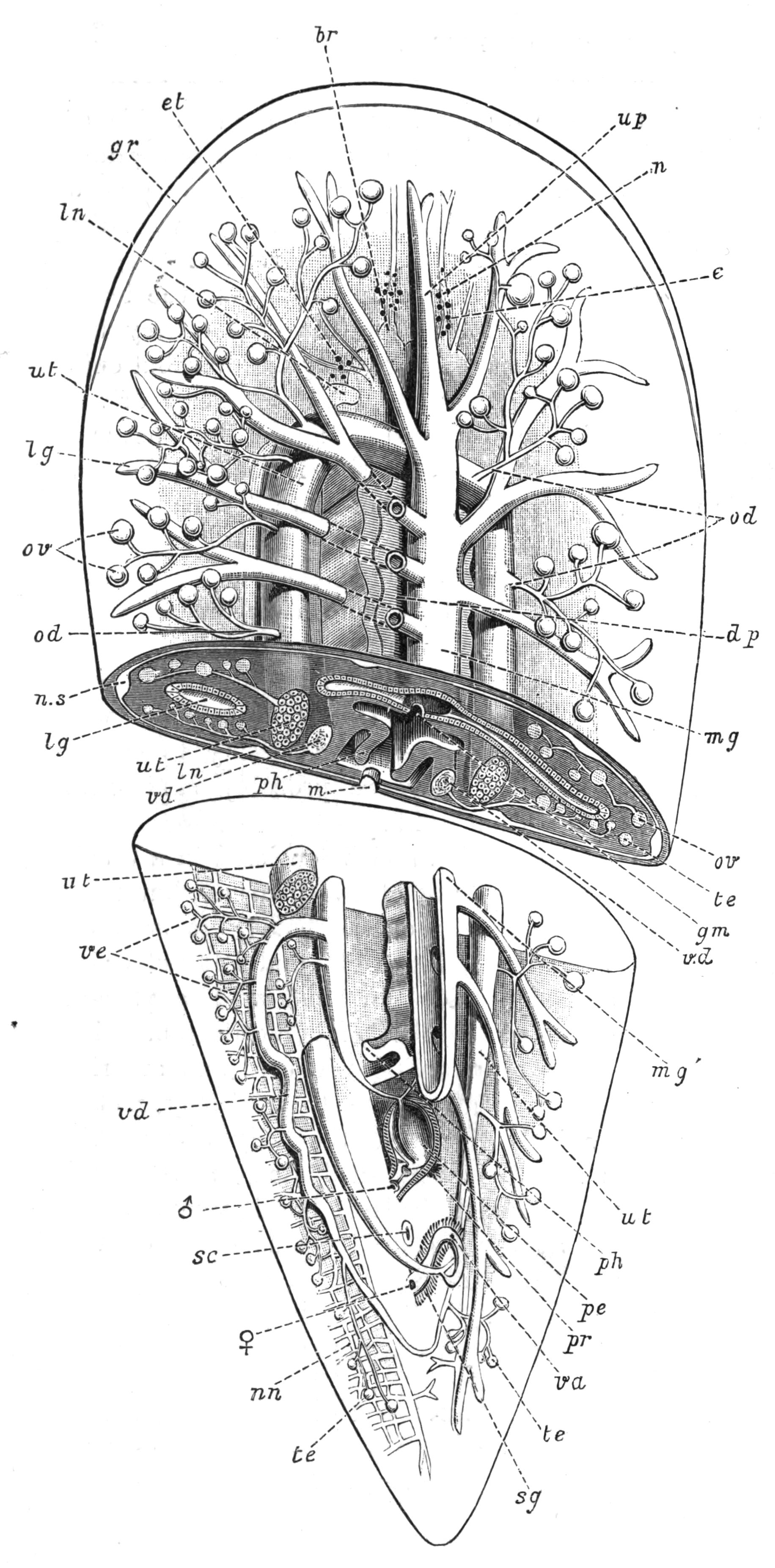
Fig. 5.—Diagrammatic view of the structure of Leptoplana tremellaris as a type of the Polycladida. The body is cut across the middle to show the relative position of organs in transverse section. In the posterior half the alimentary canal has been bisected and removed from the left side, to exhibit the deeply placed nervous sheath (nn) and the male reproductive organs. br, Brain; dp, "diaphragm"; e, cerebral group of eyes; et, tentacular eye-group; gr, marginal groove; gm, true mouth; lg, lateral gut-branch; ln, longitudinal nerve stem; m, external mouth; mg, mg', main-gut, whole, and bisected; n, sensory nerve supplying the eyes; nn, nervous network lying on the ventral musculature; n.s, lateral nerve; od, oviduct; ov, ovary; pe, penis (in section); ph, pharynx; pr, prostate or "granule gland"; sc, sucker; sg, shell-gland; te, testes; up, anterior unpaired gut-branch; ut, uterus; va, vagina (in section); vd, vas deferens; ve, vasa efferentia; ♂, male genital pore; ♀, female pore.
Sense Organs.—Leptoplana possesses eyes, stiff tactile, marginal cilia, and possibly a sense organ in the "marginal groove." The eyes, which are easily seen as collections of black dots lying at the sides of the brain, may be divided into two paired groups: (1) cerebral eyes (Fig. 5, e), and (2) tentacle eyes (et), which indicate the position of a pair of tentacles in allied forms (Fig. 8, A, t and B). Each ocellus consists of a capsule placed at right angles to the surface of the body in the parenchyma, below the dorsal muscles, and with its convex face outwards. It is a single cell in which pigment granules have accumulated. The light, however, can only reach the refractive rods, which lie within it, obliquely at their outer ends. These rods are in connexion with the retinal cells, and thus communicate by the optic nerve with the brain. The cerebral eyes are really paired, and are directed some upwards, some sideways, some downwards.
The "marginal groove" is a shallow depression of the epidermis (Fig. 5, gr) lined by cilia, and containing the ducts of very numerous gland-cells. It runs almost parallel to the anterior margin of the body, a short distance from it, but we have no observations on its functions.
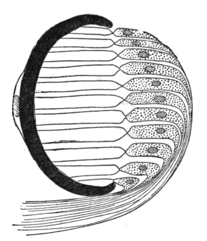
Fig. 6.—Diagram of an eye of Leptoplana from the tentacle group. × 600. (After Lang.)
Reproductive Organs.—Leptoplana is hermaphrodite, and, as in most hermaphrodites, the reproductive organs are complicated. The male organs are the first to ripen, but this does not appear to prevent an overlapping of the periods of maturity of the male and female products, so that when the eggs are being laid, the male organs are, apparently, still in a functional state. The principal parts are seen in Fig. 5. The very numerous testes (te) are placed ventrally, and are connected with fine vasa efferentia (ve), which form a delicate network opening at various points into the two vasa deferentia (vd). These tubes, especially when distended with spermatozoa, may easily be seen (Fig. 2, vd) converging at the base of the penis, and connected posteriorly by a loop that runs behind the female genital pore (Fig. 5). The penis (pe) is pyriform and muscular, and is divided into two chambers, a large upper one for the spermatozoa, and a smaller lower one for the secretion of a special "prostate" gland. The apex of the penis is eversible and not merely protrusible, being turned inside out when evaginated. The ovaries (Fig. 5, ov) are numerous and somewhat spherical. They are dorsally placed, but when fully developed extend deeply wherever they can find room to do so, and they not only furnish the ova, but elaborate food-yolk in the ova, as there are no special yolk-glands. The slender oviducts (od) open at several points into the "uterus" (ut) (a misnomer, as no development takes place within it), which encircles the pharynx, and opens by a single duct into the vagina (va). Here the ova are probably fertilised, and one by one invested by the shell-gland (sg) with a secretion which hardens and forms a resistant shell. They are then laid in plate-like masses which are attached to stones or shells. The development is a direct one, and the young Leptoplana, which hatches in about three weeks, has the outline of a spherical triangle, and possesses most of the organs of the adult. After leading a floating life for a few weeks it probably attains maturity in about nine months.
Classification, Habits, and Structure of the Polycladida.
The Polyclads were so called by Lang on account of the numerous primary branches of their intestine. They are free-living, purely marine Platyhelminthes, possessing multiple ovaries, distinct male and female genital pores (Digonopora), but no yolk-glands. The eggs are small, and in many cases give rise to a distinct larval form, known as "Müller's larva" (Fig. 12). The Polyclads, with one exception,[24] fall into two sub-groups, Acotylea and Cotylea:—
Character. Acotylea. Cotylea.
Sucker A sucker absent.[25] A sucker always present (Figs. 8, D, s; 7, A, sc).
Mouth In the middle, or behind the middle, of the ventral surface. In the middle, or in front of the middle, of the ventral surface.
Pharynx More or less intricately folded. Rarely folded. Usually cylindrical or trumpet-shaped.
Tentacles A pair of dorsal tentacles usually present. A pair of marginal tentacles (except in Anonymus).
Development Usually direct. Larva when present, not a typical Müller's larva. Müller's larva present. Metamorphosis, however, extremely slight.
Fig. 8 shows that, starting with a member (A, D) of each division, in which the mouth is almost in the middle of the ventral surface, and the brain and sense organs somewhat remote from the anterior end, we find in the Acotylea a series leading to an elongated form (Cestoplanidae), in which the mouth, pharynx, and genital pores are far back near the hinder end of the body; while in the Cotylea the series leads similarly to the elongated Prosthiostomatidae, in which, however, the pharynx and external apertures are in the front part of the body. This view of the morphology of the Polyclads is due to Lang, and is based on the assumption that the more radially-constructed forms (Fig. 8, A, D) are the primitive ones.
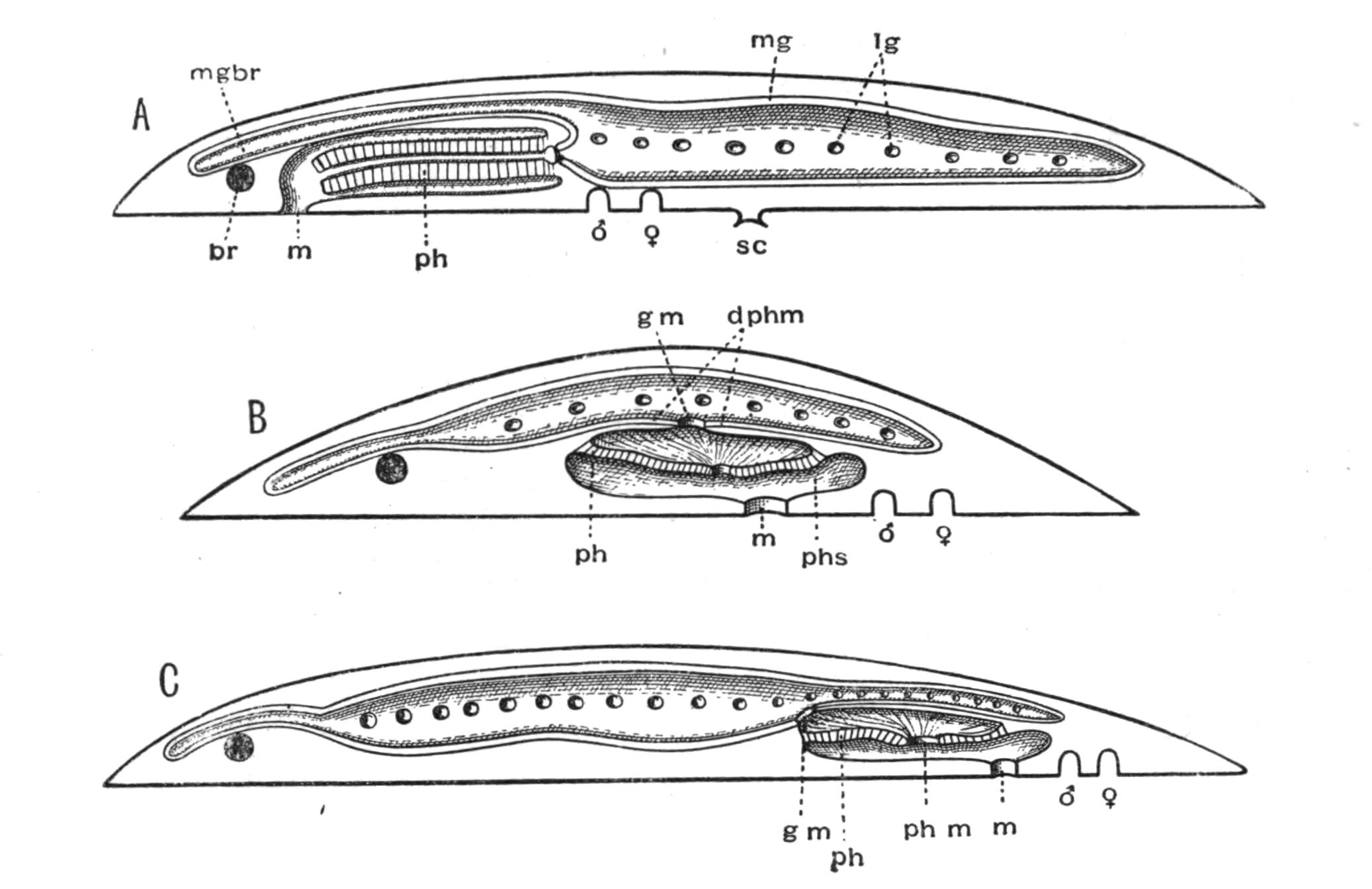
Fig. 7.—Diagrammatic vertical longitudinal sections: A, Of Prosthiostomum (type of Cotylea); B, of Leptoplana; C, of Cestoplana (types of Acotylea). (After Lang.) These figures illustrate the changes which follow the shifting of the mouth from a central position (B) to either end of the body. br, Brain; dphm, "diaphragm"; gm, true mouth; lg, openings of lateral gut-branches; m, mouth; mg, main-gut or stomach; mgbr, median gut-branch; ph, pharynx; ph.m, aperture in pharyngeal fold; phs, peripharyngeal sheath; sc, sucker; ♂, male, and ♀, female, genital aperture.
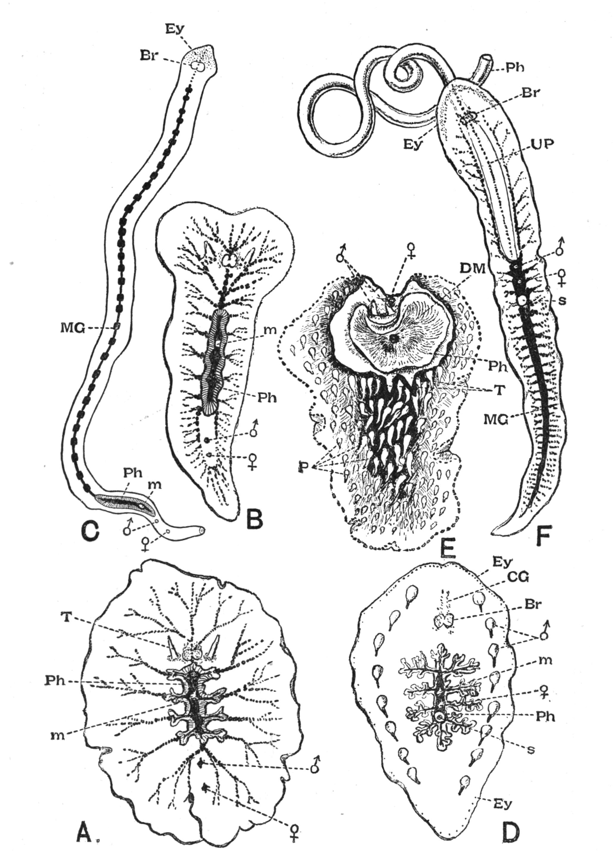
Fig. 8.—Chief forms of Polycladida: A-C, Acotylea; D-F, Cotylea. A, Planocera graffii Lang, nat. size; B, Stylochoplana maculata Stimps, × 7; C, Cestoplana rubrocincta Lang, × 4⁄3; D, Anonymus virilis Lang, × 3, ventral surface; E, Thysanozoon brocchii Grube, nat. size; the head is thrown back and the pharynx (ph) is protruded. F, Prosthiostomum siphunculus Lang, × 3. Br, Brain; CG, cerebral eye group; DM, true mouth; Ey, marginal eyes; m, mouth; MG, main-gut or stomach; P, dorsal papillae; Ph, pharynx; s, sucker (ventral); T, tentacles; UP, dorsal median gut-branch. ♂, male, and ♀, female, genital aperture, except in D, where ♂ refers to the multiple penes. (After Lang and Schmidt.)
Classification of Polycladida.
ACOTYLEA.
Family. Genus. British Representatives.
Planoceridae. With dorsal tentacles. Mouth sub-central. Planocera (Fig. 8, A). Imogine. Conoceros. Stylochus. Stylochoplana (Fig. 8, B). Diplonchus. Planctoplana. Planocera folium Grube. Berwick-on-Tweed. Stylochoplana maculata Quatref. Among brown weeds in Laminarian zone.
Leptoplanidae. Without dorsal tentacles. Penis directed backwards. Discocelis. Cryptocelis. Leptoplana. Trigonoporus. ?Polypostia (see p. 27). Leptoplana tremellaris O. F. Müll. L. fallax Quatref. Plymouth. L. droebachensis Oe. Plymouth Sound. L. atomata O. F. Müll. Doubtful species.
Cestoplanidae. No tentacles. Body elongated. Penis directed forwards. Cestoplana (Fig. 8, C). In Mediterranean and on French side of the Channel.
Enantiidae. No sucker. No tentacles. Main-gut very short. External apertures as in Euryleptidae. Enantia. Adriatic Sea.
COTYLEA.
Anonymidae. Mouth central. No tentacles. With two rows of penes. Anonymus (Fig. 8, D). Naples (two specimens).
Pseudoceridae. Marginal tentacles folded. Mouth in anterior half. Thysanozoon (Fig. 8, E). Pseudoceros. Yungia.
Euryleptidae. Tentacles usually present and pointed, or represented by two groups of eyes. Mouth close to anterior end. Pharynx cylindrical. Prostheceraeus. Cycloporus. Eurylepta. Oligocladus. Stylostomum. Aceros. Prostheceraeus vittatus Mont. On west coast. P. argus Quatref. Guernsey. Cycloporus papillosus Lang. On Ascidians in 2-30 fms. Eurylepta cornuta O.F. Müll. On sponges and shells, 2-10 fms. Oligocladus sanguinolentus Quatref. O. auritus Clap. Doubtful. Stylostomum variabile Lang.
Prosthiostomatidae. Tentacles absent. Body elongated. Pharynx long, cylindrical. Penis with accessory muscular vesicles. Prosthiostomum (Fig. 8, F).
Appearance and Size of Polyclad Turbellaria.—Polyclads are almost unique amongst animals in possessing a broad and thin, delicate body that glides like a living pellicle over stones and weeds, moulding itself on to any inequalities of the surface over which it is travelling, yet so fragile that a touch of the finger will rend its tissues and often cause its speedy dissolution. The dorsal surface in a few forms is raised into fine processes (Planocera villosa), or into hollow papillae (Thysanozoon brocchii), and in very rare cases may be armed with spines (Acanthozoon armatum,[26] Enantia spinifera); in others, again, nettle-cells (nematocysts) are found (Stylochoplana tarda, Anonymus virilis). Some Polyclads, especially the pelagic forms, are almost transparent; in others, the colour may be an intense orange or velvety black, and is then due to peculiar deposits in the epidermal cells. Between these two extremes the colour is dependent upon the blending of two sources, the pigment of the body itself and the tint of the food. Thus a starved Leptoplana is almost or quite white, a specimen fed on vascular tissue reddish. Many forms are coloured in such a way as to make their detection exceedingly difficult, but this is probably not merely due, as Dalyell supposed, to the substratum furnishing them with food and thus colouring them sympathetically, but is probably a result of natural selection.
The largest Polyclad, the bulkiest Turbellarian, is Leptoplana gigas (6 inches long and 4 in breadth), taken by Schmarda, free-swimming, off the coast of Ceylon. The largest European form is Pseudoceros maximus, 3½ inches in length and stoutly built. A British species, Prostheceraeus vittatus, attains a length of from 2 to 3 inches. These large forms, especially the Pseudoceridae (pre-eminently the family of big Polyclads), are brightly coloured, and usually possess good swimming powers, since, being broad and flat, they are certainly not well adapted for creeping rapidly, and this is well shown by the way these Polyclads take to swimming when in pursuit of prey at night. The size of any individual is determined, amongst other factors, by the period at which maturity sets in, after which probably no increase takes place. Polyclads apparently live about twelve months, and mature specimens of the same species vary from ½ inch to 2½ inches in length (Thysanozoon brocchii), showing that growth is, under favourable conditions, very rapid.
Habits of Polyclad Turbellaria.—Polyclads are exclusively marine, and for the most part littoral, animals. Moreover, there is no evidence of their occurrence in those inland seas where certain marine animals (including one or two species of otherwise characteristically marine Rhabdocoelida, p. 46) have persisted under changed conditions. From half-tide mark down to 50 fathoms, some Polyclads probably occur on all coasts, but as to their relative abundance in different seas we have very little accurate information. The southern seas of Europe possess more individuals and species than the northern, and probably the maximum development of the group takes place on the coasts and coral islands of the tropics.[27] No Polyclads have been taken below 60 fathoms; but their delicacy and inconspicuousness render this negative evidence of little value. Six truly pelagic forms, however, are known,[28] and these are interesting on account of their wide distribution (three occurring in the Atlantic, Pacific, and Indian oceans), and also from the distinct modifications they have undergone in relation to their pelagic existence.
Whatever may be the interpretations of the fact, Polyclads are notoriously difficult to detect, and this fact doubtless explains the scanty references to them by the older naturalists who collected even in tropical seas. Lang, who worked seven years at Naples, added to the Mediterranean fauna as many Polyclads as were previously known for all Europe, in spite of the assiduous labours of his predecessors, Delle Chiaje and Quatrefages. Again Hallez, collecting at Wimereux at low-water, obtained some twenty specimens of Leptoplana tremellaris in an hour, while some other collectors working by his side could only find two or three. Yet, even making allowance for the difficulty of finding Polyclads, few of them appear to be abundant.
Leptoplana tremellaris is frequently associated with colonies of Botryllus, and if separated soon perishes, whereas the free-living individuals are distinctly hardy (Hallez). A closely allied but possibly distinct form lives upon the surface of the Polyzoon Schizoporella, on the French side of the Channel, and cannot long endure separation from its natural habitat, to which it is adaptively coloured. A striking case of protective mimicry is exhibited by Cycloporus papillosus, on the British coasts. This species, eminently variable in colour and in the presence or absence of dorsal papillae, is usually a quarter of an inch in length and of a firm consistency. Fixed by its sucker to Polyclinid and other Ascidians, Cycloporus appears part and parcel of the substratum, an interesting parallel to Lamellaria perspicua,[29] though we are not justified in calling the Polyclad parasitic. Indeed, though a few cases of association between Polyclads and large Gasteropods, Holothurians, and Echinids are known,[30] there is only one case, that of Planocera inquilina,[31] in the branchial chamber of the Gasteropod Sycotypus canaliculatus, which would seem to bear the interpretation of parasitism. The jet-black Pseudoceros velutinus and the orange Yungia aurantiaca of the Mediterranean, are large conspicuous forms with no attempt at concealment, but their taste, which is not known, may protect them. Other habits, curiously analogous with devices employed by Nudibranch Mollusca (compare Thysanozoon brocchii with Aeolis papillosa), emphasise the conclusion that the struggle for existence in the littoral zone has adapted almost each Polyclad to its particular habitat.
As regards the vertical distribution of this group on the British coasts, Leptoplana tremellaris has an extensive range, and appears to come from deeper to shallower water to breed.[32] In the upper part of the Laminarian zone, Cycloporus papillosus, and, among brown weeds, Stylochoplana maculata are found. At and below lowest water-mark Prostheceraeus vittatus, P. argus, and Eurylepta cornuta occur. Stylostomum variabile and Oligocladus sanguinolentus, though occasionally found between tide-marks, especially in the Channel Islands, are characteristic, along with Leptoplana droebachensis and L. fallax, of dredge material from 10 to 20 fathoms.
Locomotion.—Locomotion is generally performed by Polyclads at night when in search of food, and two methods, creeping and swimming, are usually employed—creeping by the cilia, aided possibly, as in the case of some Gasteropod Mollusca, by the longitudinal muscles of the ventral surface; and swimming, by undulations of the expanded margins of the body. In the former case the cilia work in a glandular secretion which bathes the body, and enables them to effect their purpose equally well on different substrata. The anterior region is generally lifted up, exploring the surroundings by the aid of the tentacles, which are here usually present. The rest of the body is closely appressed to the ground.
Swimming is particularly well performed by the Pseudoceridae, certain species of Prostheceraeus, the large Planoceridae, some Stylochoplana, Discocelis, and Leptoplana, and in the same manner as in Leptoplana tremellaris (p. 9). In Cryptocelis, Leptoplana alcinoi, and L. pallida, however, the whole body executes serpentine movements like an active leech (e.g. Nephelis); a cross section of the body would thus present the same appearance during the whole movement. Many Polyclads, notably Anonymus (Lang), if irritated, spread out in all directions, becoming exceeding thin and transparent.
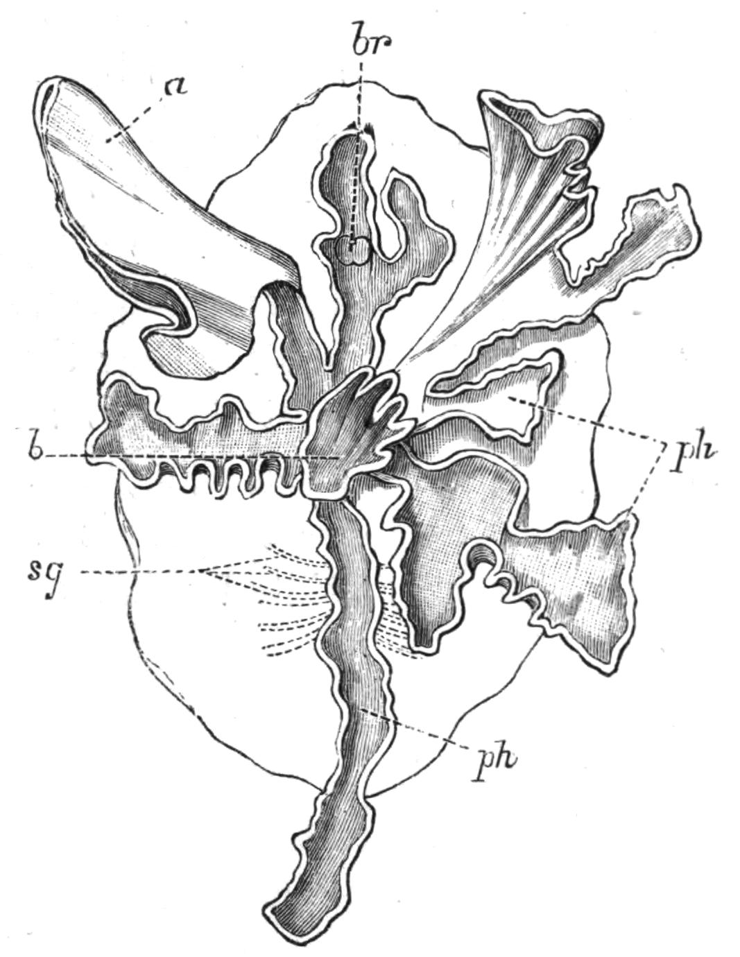
Fig. 9.—Discocelis lichenoides Mert. (after Mertens), creeping on the inner side of a glass vessel by means of the lobes of the extended and exceedingly mobile pharynx (ph). These lobes also serve to enclose Crustacea (a), and one lobe may then be withdrawn independently of the rest, back into the body (b). The brain (br) and shell-gland (sg) are shown by transparency.
Discocelis lichenoides, Planocera graffii, and Anonymus virilis have peculiar modes of progression. The first, according to Mertens, will climb up the sides of a vessel by means of the expanded lobes of the pharynx (Fig. 9, ph), a habit of considerable interest, since we know that certain Ctenophores—Lampetia, for instance—progress when not swimming on the expanded lobes of their "stomach."[33] Planocera and Anonymus creep by extending parts of the anterior margin and dragging the rest of the body behind. In consequence, the brain and dorsal tentacles may come to lie actually behind the middle of the body, and thus no definite anterior end or "head" advances first. Along with this curious habit it may be noticed (Lang) that the radial symmetry of the body is well marked; but even without accepting this author's suggestion of the concurrent development of a "head" with locomotion in a definite direction, the facts, whether these two forms are primitive or not, are highly interesting.
Food.—Though we are probably right in calling Polyclads a carnivorous group, the food of very few forms has been ascertained. Those which possess a large frilled pharynx (most Acotylea) probably enclose and digest large, and, it may be, powerful prey, as appears to be the case in Leptoplana tremellaris. Cryptocelis alba has been seen by Lang with the pharynx so distended, owing to a large Drepanophorus (Nemertine) which it contained, as to resemble a yolk-sac projecting from the under surface of an embryo. The Cotylea such as Thysanozoon, with a bell- or trumpet-shaped pharynx, are fond of fixing this to the side of the aquarium, but whether they thus obtain minute organisms is not clear. Prosthiostomum shoots out its long pharynx with great vehemence (Fig. 8, F) and snaps up small Annelids by its aid (Lang). Those Polyclads which, as Cycloporus and others, are definitely associated with other organisms are not certainly known to feed upon the latter, though "Planaria velellae" has been seen by Lesson[34] devouring the fleshy parts of its host. The salivary glands which open on the lips and the inner surface of the pharynx powerfully disintegrate the flesh of the prey. Digestion takes place in the main-gut, and the circulation of the food is accomplished by the sphinctral musculature of the intestinal branches (conf. Leptoplana, p. 13).
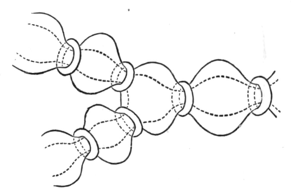
Fig. 10.—Diagram of the musculature, causing peristaltic movements of the intestinal branches of Polyclads. (After Lang.)
A distinct vent or anus is always absent. After a meal the faecal matter collects in the main-gut, and is discharged violently by the pharynx into the water. In a few species, however, the intestinal branches open to the exterior (Lang). Yungia aurantiaca, a large and abundant Neapolitan form, possesses such openings over the greater part of the dorsal surface; Cycloporus papillosus has marginal pores; Oligocladus sanguinolentus apparently possesses an opening at the posterior end of the main-gut; and Thysanozoon brocchii frequently rends at this point, in consequence of the accumulation of food.
Respiration.—The oxygen of the atmosphere dissolved in the sea-water is, in default of a special circulatory fluid, brought to the tissues of Polyclads in two ways. The ciliated epidermis provides a constant change of the surrounding water, by which the superficial organs may obtain their supply; and the peristaltic movements of the digestive system, aided by the cilia of the endoderm cells, ensure a rough circulation of the sea-water, which enters along with the food, to the internal organs. The papillae of Thysanozoon brocchii, containing outgrowths of the intestinal branches, are possibly so much additional respiratory surface, although still larger forms (other Pseudoceridae) are devoid of such outgrowths.
Excretion.—The excretory system of only one Polyclad (Thysanozoon brocchii) is accurately known. Lang, by compressing light-coloured specimens, found the three parts of the system known to occur in many Platyhelminthes: (1) the larger longitudinal canals, and (2) the capillary vessels, which commence with (3) the flame-cells in the parenchyma of the body. The mode of distribution of these parts is not, however, ascertained. The canals are delicate, sinuous, apparently intracellular tubes, coursing close to the margin of the body and sending offsets which suspend the canals to the dorsal surface, where possibly openings may occur. In dilatations of these vessels bunches of cilia, and occasionally flame-cells, are found. Usually, however, flame-cells occur at the commencement or during the course of the capillaries, which are straight, rarely branching, tubes of exceeding tenuity, and appear (Lang) to be outgrowths of the flame-cells, just as the duct is an outgrowth of a gland-cell. In fact there is little doubt that the stellate flame-cells are modified parenchymatous gland-cells, containing a lumen filled with a fluid into which a number of cilia project and vibrate synchronously. The cells excrete nitrogeneous waste substances, which are then discharged into the capillaries, whence the cilia of the main vessels drive them presumably to the exterior, though external openings of the excretory system are not known. Traces of this system have been observed in young Leptoplana (first by Schultze in 1854) and also in Cestoplana.
Sensation.—A nervous sheath, with scattered ganglion cells, everywhere underlies the musculature. It is exceedingly faintly marked on the dorsal surface, but laterally and ventrally forms a dense network with polygonal meshes. Thickenings of this sheath give rise to lateral nerves, and also to a pair of stout longitudinal nerves from which the internal organs are probably innervated. The brain, hardly distinct in pelagic Polyclads, in most forms does not differ greatly from that of Leptoplana (p. 13).
The sense organs of Polyclads have the form of tentacles, eyes, otocysts (in Leptoplana otophora), and stiff tactile cilia. The solid dorsal tentacles of Planoceridae contrast strongly with the folded or pointed hollow processes of the Cotylea. The former (Fig. 8, A, T) are muscular and very contractile, and are placed near the brain some distance from the anterior end. The latter are outgrowths of the front margin of the body, and are sometimes (Yungia) provided superficially with olfactory pits and internally with eyes and intestinal coeca.
The eyes which occur in Polyclads may be divided into (a) a pair of cerebral groups overlying the brain; (b) those embedded in the tentacles (tentacular group); and (c) the marginal eyes, which in Anonymus occur all round the margin. A complex form is sometimes assumed by the cerebral eyes of Pseudoceridae, resulting probably from incomplete fission (Fig. 11). Leptoplana otophora was obtained by Schmarda on the south coast of Ceylon. On each side of the brain is a capsule containing two otoliths. This is the only known case of the occurrence of these organs in Polyclads.
Reproduction.—Although Polyclads are able to repair the result of injuries to a very considerable extent, they are not known to multiply asexually. The two processes are intimately associated, but, though probably all Turbellaria can regenerate certain lost parts, asexual reproduction only occurs sporadically.
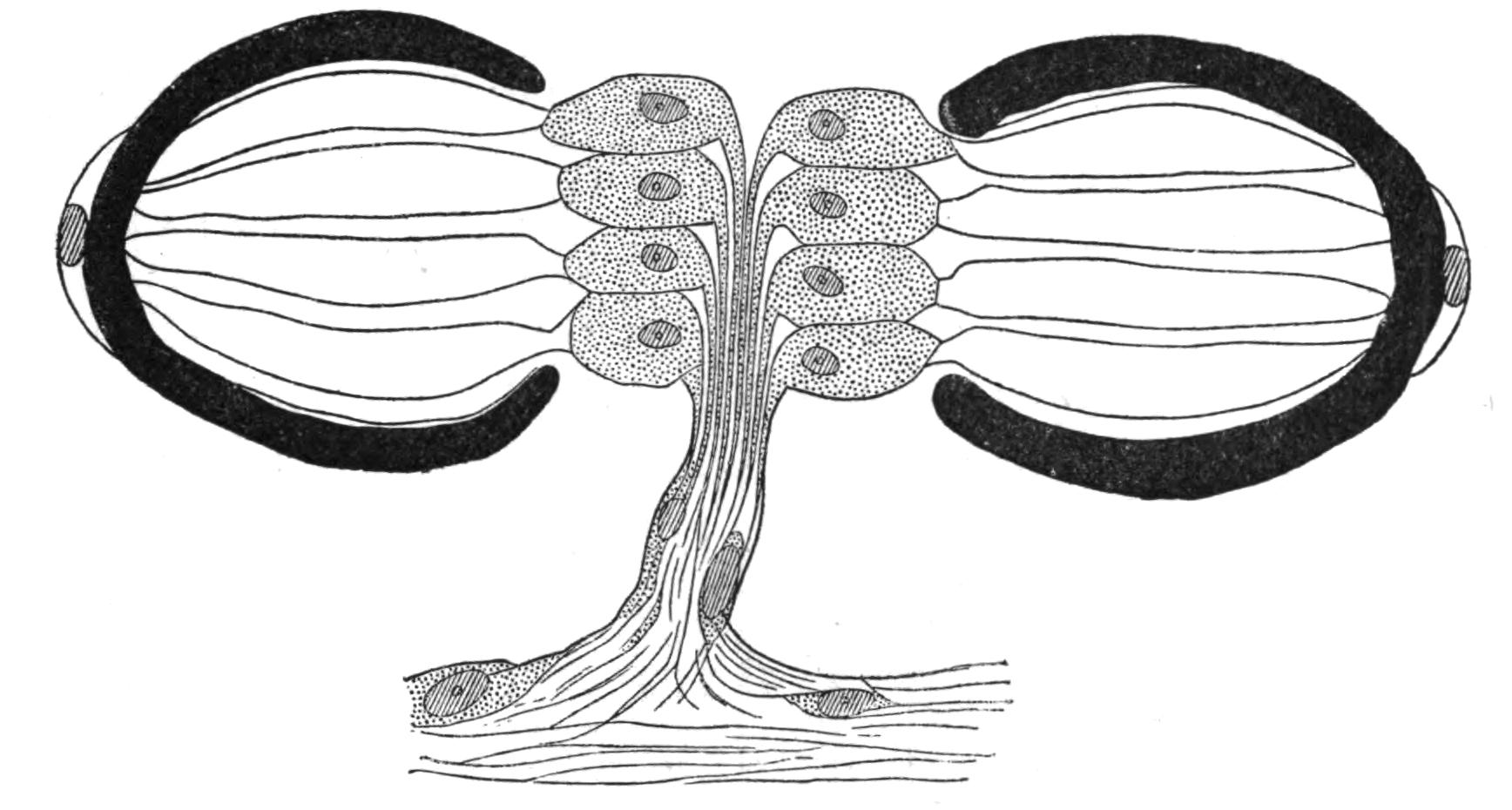
Fig. 11.—Double eye from the cerebral group of Pseudoceros maximus. (After Lang.)
All known Polyclads are hermaphrodite. The male organs, scattered, like the testes of Leptoplana, over the ventral surface, develop earlier than the ovaries, though the periods of maturation overlap; hence the possibility of self-fertilisation, though remote, is still worth consideration. The genital apertures, through which, in the male, spermatozoa, and in the female, ova, are emitted, are usually situated as in Leptoplana (Figs. 2 and 5, ♂ and ♀). In Trigonoporus, a genus once found at Naples, a secondary female aperture has been discovered leading into the female genital canal[35]; and in Anonymus, Polypostia, and Thysanozoon (Fig. 7, E, ♂) two or more male pores and penes have been found. Anonymus has several penes (Fig. 7, D, ♂) arranged radially round the body. Polypostia, a remarkable form described by Bergendal,[36] belonging to the Acotylea, possesses about twenty such structures ranged round the female genital aperture. Lang, whose attention was attracted by these singular facts, made the interesting discovery that Thysanozoon uses its penes as weapons of offence rather than as copulating organs, burying them in the skin of another Polyclad (Yungia) that happened to cross its path, spermatozoa being of course left in the wound. Lang further found that Prostheceraeus albocinctus and Cryptocelis alba in this way implanted a spermatophore in the skin of another individual of the same species, and he suggested that from this point the spermatozoa wandered through the tissues till they met with and fertilised the eggs. It is now known that a similar process of "hypodermic impregnation" occurs sporadically in several groups of animals.[37] Nevertheless, in some Polyclads it is probable, and in Stylochus neapolitanus it is certain, that normal copulation takes place. The sperm-masses are transferred to a coecal diverticulum of the female genital canal, and then by a delicate mechanism, of which we know only the effects, one spermatozoon obtains entrance into one matured ovum, which differs from the ova of most Turbellaria in that it contains in its own protoplasm the yolk necessary for the nutrition of the embryo. In other words, there are no special yolk-glands. After fertilisation, the ovum in all Polyclads is coated with a shell formed by the shell-gland, which also secretes a substance uniting the eggs together. They are deposited on stones and shells, either in plate-like masses or in spirals (like those of Nudibranchs). Cryptocelis alba lays masses of an annular shape, with two ova in each shell, and buries them in sand.
Development.[38]—The first stages in the embryology of Polyclads appear to be very uniform. They result, in all Cotylea and in certain Planoceridae, in the formation of a Müller's larva (Fig. 12) about a couple of weeks after the eggs are laid. This larva (1-1.8 mm. long), which is modified in the Planoceridae, is distinguished by the presence of a ciliated band, running somewhat transversely round the body, and usually produced into a dorsal, a ventral, and three pairs of lateral processes. When swimming the body is placed as in Fig. 12, and twists round rapidly about its longitudinal axis by means of the strong locomotor cilia placed in transverse rows upon the processes. The cilia of each row vibrate synchronously, and recall the action of the swimming plates of a Ctenophore. It is noteworthy that whereas Stylochus pilidium passes through a modified or, according to some authors, a primitive larval stage, its near ally, S. neapolitanus, develops directly. Most Acotylea indeed develop directly, and their free-swimming young differ from Müller's larva merely in the absence of the ciliated band and in the mode of swimming.
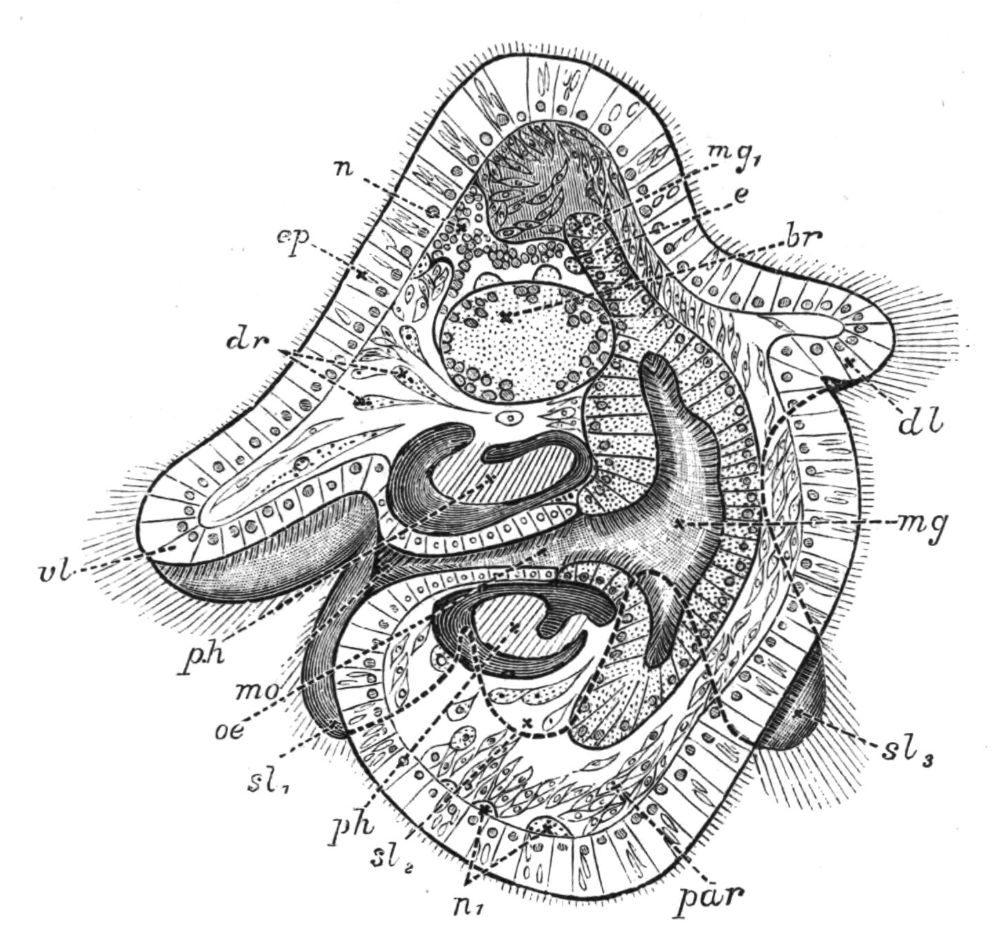
Fig. 12.—Section through Müller's larva of Thysanozoon brocchii (modified from Lang). The right half is seen from inside. × 150. Semi-diagrammatic. br, Brain; dl, dorsal ciliated lobe; dr, salivary gland-cells of pharynx; e, eye; ep, ciliated epidermis containing rhabdites; mg, stomach or main-gut; mg1, unpaired gut branch over the brain; mo, "mouth" of larva; n, n1, section of nerves; oe, ectodermic pit forming oesophagus of larva; par, parenchyma filling the space between the alimentary tract and the body wall; ph, pharynx lying in the cavity of the peripharyngeal sheath, the nuclei of which are visible; sl1, sl2, sl3, lateral ciliated lobes of the right side; vl, ventral ciliated lobe.
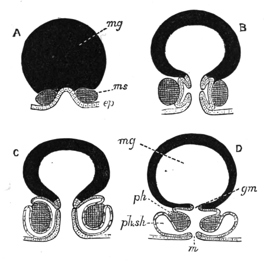
Fig. 13.—Diagrammatic transverse sections of a larval Polyclad at different stages, to illustrate the development of the pharynx. (After Lang.) A, Larva of the eighth day still within the shell. The main-gut (mg) is still solid, the epidermis is slightly invaginated, and a pair of muscular mesodermic thickenings (ms) are present. B, Young pelagic larva. The epidermic invagination has deepened and developed laterally. C, The lateral pouches have formed the wall of the peripharyngeal sheath, enclosing the mesodermic, muscular, thickening or pharyngeal fold (ph). (Compare Fig. 12.) Towards the end of larval life, when the ciliated processes (sl, Fig. 12) have aborted, the stage D is reached. By the opening outwards of the pharyngeal sheath (ph.sh) the two apertures gm, or true mouth, and m, or external mouth, are formed, which together correspond with the oesophageal opening of the younger larva. (Compare the transverse section in Fig. 5.)
Polyclads possess an undoubted mesoderm, which gives rise to the muscles, the pharyngeal fold, and the parenchyma. The ectoderm forms the epidermis, in the cells of which the rhabdites (Fig. 12) arise, apparently as so many condensed secretions. From the ectoderm the brain arises as two pairs of ingrowths, which fuse together, and from these the peripheral nervous system grows out. Three pigmented ectoderm cells give rise, by division, to the eyes—an unpaired cell (Fig. 12, e) to the cerebral group of eyes, and the other two to the marginal and tentacular groups. The copulatory organs apparently arise to a large extent as ingrowths from the ectoderm, from which the accessory glands (prostates, shell-glands) are also formed. The endoderm forms the lining of the main-gut and its branches. The pharynx is developed as in Fig. 13, which shows that the "mouth" of the young larva (C) does not correspond exactly with that of the adult (D). The salivary glands arise from ectoderm cells, which sink deeply into the parenchyma. The reproductive organs (ovaries and testes) possibly arise by proliferation from the gut-cells (Lang, v. Graff). The change from the larva to the adult is gradual, the ciliary band being absorbed and the creeping mode of life adopted.
Turbellaria. II. Tricladida.
The Triclads are most conveniently divided into three groups[39]: (i.) Paludicola, the Planarians of ponds and streams; (ii.) the Maricola, the Triclads of the sea; and (iii.) Terricola or Land Planarians. From the Polyclads they differ in their mode of occurrence; in the elongated form of their body and almost constant, mid-ventral position of the mouth; in possessing a single external genital pore (Monogopora); and in the production of a few, large, hard-shelled eggs provided with food-yolk.
Occurrence of the Paludicola.—The Planarians of our ponds and streams are the most familiar and accessible Turbellaria. Their elongated, flattened bodies, and gliding movements, render them conspicuous objects on the under surface of stones and on the leaves of aquatic plants, where they live gregariously. The variable Polycelis nigra (Fig. 14, H) is very abundant in stagnant water and slowly-moving streams, whereas its ally, P. cornuta (Fig. 14, G), distinguished by a pair of tentacles, is more local. Planaria (Dendrocoelum) lactea (A), P. polychroa (I), P. torva, and P. punctata are not infrequently found together, but the last is at once the largest and rarest.
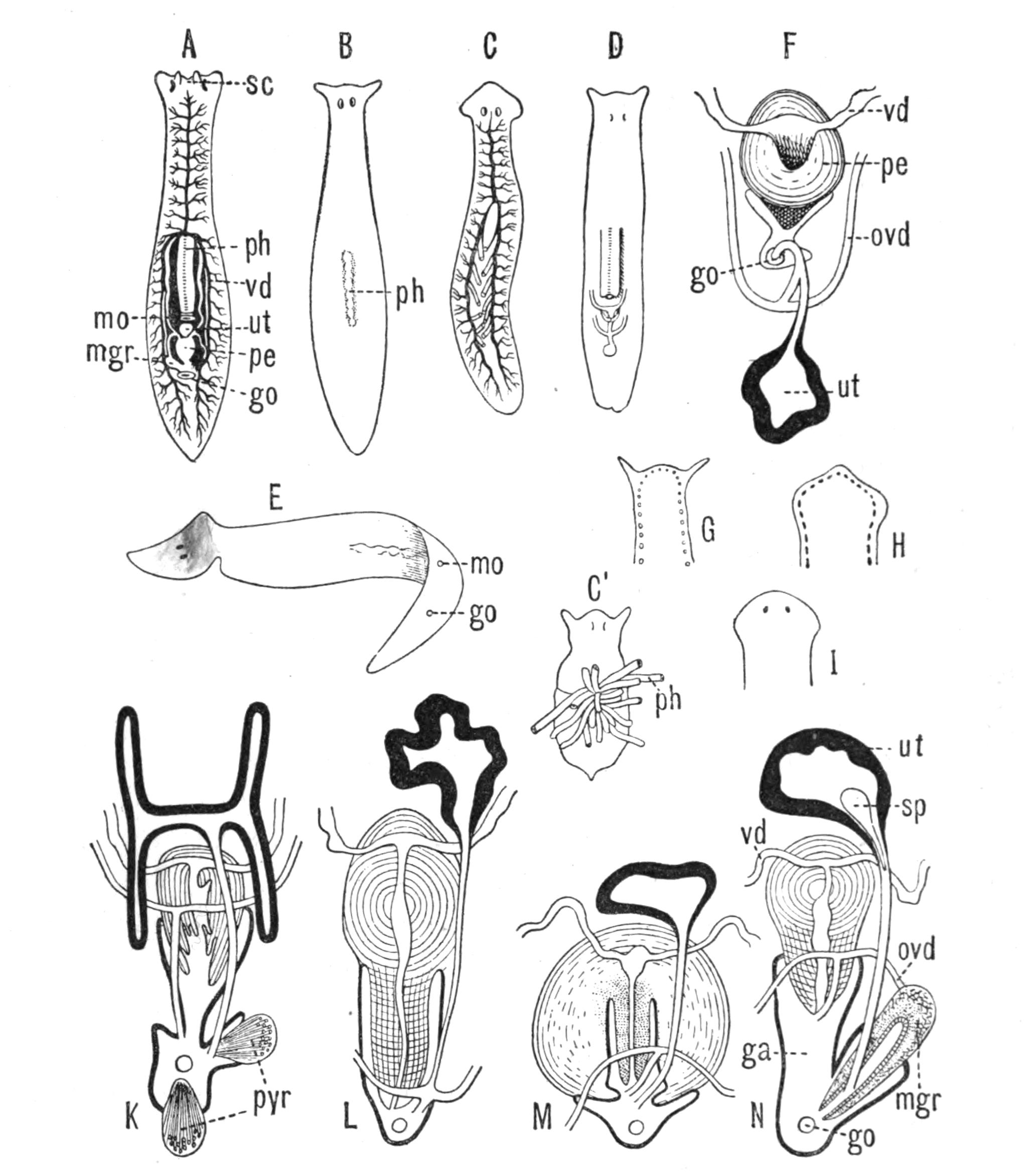
Fig. 14.—Forms of Triclads, with the distinguishing specific characters of certain British forms. A, Planaria lactea O. F. M., × 2; B, Planaria alpina Dana, × 4 (after Kennel); C, Phagocata gracilis Leidy (after Woodworth), × 6; C', the same with the pharynges (ph) extruded; D, Gunda ulvae Oer., × 4; E, Planaria gonocephala Dug. (after Schmidt), × 4; F, genitalia of Gunda ulvae (after Wendt); G, head of Polycelis cornuta Schm.; H, head of Polycelis nigra Ehr.; I, head of Planaria polychroa Schm. K to N show the distinctive characters of the genital ducts in K, Polycelis nigra; L, Planaria polychroa; M, Planaria alpina; N, Planaria torva Schultze (after Iijima and v. Kennel). ga, Genital atrium; go, common genital opening; mgr, "musculo-glandular organ"; mo, "mouth"; ovd, oviduct; pe, penis; ph, pharynx; pyr, pyriform organs of unknown significance; sc, sucker; sp, spermatophore lying in (ut) uterus; vd, vesicula seminalis. (All except C and E are found in England.)
Planaria alpina (Fig. 14, B) is characteristic of cold mountain streams, but occurs down to sea-level in England, the Isle of Man, and Ireland, and from its abundance in spring water, probably enjoys a wide distribution underground. In the Swiss Alps it has been found at altitudes of over 6000 feet, at lower levels in the Rhone, and also in the Lake of Geneva. This wide distribution may perhaps be accounted for, partly, by its faculty for asexual reproduction in summer, and also, by the production, later in the year, of hard-shelled eggs which are laid loosely, not attached to stones or plants.[40] But we have no really direct evidence of the means of dispersal of this or of any of the foregoing species, although they all have a wide distribution in Europe. Of extra-European forms the accounts that exist are very fragmentary. The only indubitable diagnostic character of a Triclad is the structure of its genital ducts, and this is accurately known in only a few cases. Several species such as Dicotylus pulvinar (Fig. 16, B), at present known only from Lake Baikal,[41] and others (Planaria mrazekii, P. albissima) from Bohemia,[42] will doubtless be found elsewhere when they are carefully looked for. Phagocata gracilis is a remarkable North American form, possessing several pharynges (Fig. 14, C and C'), recalling the independent movement of the pharyngeal lobes of Discocelis lichenoides (Fig. 9).[43]
Occurrence of the Maricola.—Little as we accurately know of the distribution of the fresh-water Planariae, our knowledge of the occurrence of the marine forms is still more limited. Gunda (Procerodes) ulvae (Fig. 14, D) is the commonest European form, occurring abundantly in the upper part of the littoral zone, on the shores of the Baltic. G. segmentata from Messina has been carefully described by Lang,[44] but these are almost the only species of Maricola which can be accurately determined. They differ from the Paludicola in the position of the "uterus" behind the genital pore and in the absence of a "musculo-glandular organ" (Fig. 14, F). A special interest attaches to the Bdellouridae, a family containing three species, all parasitic on Limulus from the east coast of America. These remarkable Triclads usually have a sucker at the hinder end of the body, by which they attach themselves firmly to the cephalo-thoracic appendages and to the gill-plates, upon which the eggs may be found in considerable numbers. One species, Syncoelidium pellucidum, possesses a pair of problematical organs in the hinder part of the body, opening to the exterior ventro-laterally by a couple of chitinous mouth-pieces, but having no connexion with the genital ducts.[45]
Occurrence and Distribution of Land Planarians.—The terricolous Triclads or Land Planarians are the most interesting division of the group. Some forms, such as Bipalium kewense, attain large dimensions, being usually 6 to 9 inches in length, and specimens fully extended have measured 18 inches. Their bodies are frequently banded or striped with brilliant colours. Geoplana coerulea Mos. has a blue ventral surface and is olive green or dark Prussian blue above. G. splendens Dendy, is marked dorsally by three stripes of emerald green alternating with four dark brown longitudinal bands. The mode of coloration, though somewhat variable, is an important specific character. Its significance, however, is not clearly understood. The colours may be a warning signal, as some Geoplana at least are disagreeable to the taste of man and some birds[46]; but since Land Planarians are largely nocturnal animals, living by day under logs, banana leaves, and in other moist and dark situations, this explanation is clearly insufficient. Two Geoplana have been noticed by Mr. Dendy which seem to be protectively coloured. G. triangulata var. australis occurs abundantly in the beech forest in the South Island of New Zealand, and its brown back and yellow or orange ventral surface match the leaves around its haunts. G. gelatinosa again looks like a mere slimy patch on the rotten bark where it is found. In arid districts, during the dry season, Land Planarians burrow in the soil and form a cyst, in which they lie coiled up, after the manner of earthworms.[47] The glutinous investment of their delicate bodies forms a moist medium in which the cilia covering the body (and especially the ventral surface) may constantly and evenly vibrate, and by which they adhere firmly to their prey. In some tropical Planarians, in addition to possessing offensive properties, the mucus is so copious in amount and hardens with such rapidity, that these Triclads may creep over bridges of it, and may even be blown from one stem or branch of a plant to another, hanging at the ends of their threads.[48]
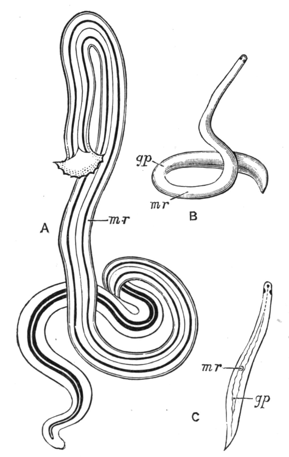
Fig. 15.—Some Land Planarians found in Europe. A, Bipalium kewense Mos. × ⅓ (after Bergendal); B, Rhynchodemus terrestris O. F. M., × 2; C, Geodesmus bilineatus Metsch., × 2½ (after Metschnikoff). mr, Region of mouth; gp, region of genital pore.
In Europe there are only two or three indigenous Land Planarians, of which Rhynchodemus terrestris O. F. M. (Fig. 15, B) is the most widely distributed, and has been found in moist situations for the most part wherever it has been carefully looked for. It measures about ¾ inch in length, and is dark grey above, whitish below, and bears a pair of eyes near the anterior extremity (Fig. 15, B). Bipalium kewense (Fig. 15, A), which has been found in the forests of Upolu, Samoa, by Mr. J. J. Lister, has been accidentally imported, from the (unknown) districts where it is indigenous, with plants and soil to various parts of the world—England, Germany, the Cape, and also to Sydney, where it appears to have established itself. In these Bipalia living in hothouses, the genitalia never appear to attain maturity, and apparently multiple fission and subsequent reparation of the missing parts is the only mode of reproduction. Geodesmus bilineatus (Fig. 15, C), which has occurred at Giessen, Würzburg, and Dresden, has, in all probability, been introduced with ferns from the West or East Indies. Microplana humicola, described by Vejdovsky from dunghills in Bohemia, is doubtfully indigenous.
In marked contrast with the poverty of the temperate zones in Land Planarians, is the abundance and great variety of this group in Southern Asia, South America, and especially in Australasia, where the rich Land Planarian fauna has been carefully investigated by Spencer, Dendy, Fletcher, and others, in certain parts of Victoria, New South Wales, and New Zealand.[49] About forty species of Planarians have been discovered on the Australian continent, thirty-five of which belong to the predominant genus Geoplana, distinguished by the presence of numerous eyes along the border of the simple anterior extremity. Of the remaining five, four belong to the genus Rhynchodemus, with, lastly, the introduced Bipalium kewense. The distribution of any one species, however, is so limited that only three forms are common to the two former colonies; and although some of the twenty known New Zealand Planarians (chiefly species of Geoplana), are identical with Australian species, yet only one, or possibly two, varieties of these species are Australian also. In addition to their prevalence in Australasia, the Geoplanidae also occur in South America, South Africa, Japan, and the East Indies. The Bipaliidae are characteristic of the Oriental region, being found in China, Borneo, Bengal, and Ceylon. The Rhynchodemidae are a cosmopolitan family, occurring in Europe, North and South America, the Cape of Good Hope, Ceylon, the East Indies, Australia (particularly Lord Howe Island), and Samoa.[50]
Habits and Structure of Triclads.—The common Planaria (Dendrocoelum) lactea, which usually progresses by ciliary action, aided, it is said, by muscular contractions of the ventral surface, performs, if alarmed, a series of rapid "looping" movements, by affixing a sucker (Fig. 14, A, sc), placed on the under side of the head, to the substratum, and pulling the posterior end close to this. The sucker, discovered by Leydig, is even better developed in P. punctata (Fig. 16, A), P. mrazekii, and P. cavatica, and is an efficient adhering-organ which has probably been developed from a similar but simpler structure found in a considerable number of both fresh-water and marine Triclads (P. alpina, Fig. 16, E). Probably the sucker of the Land Planarian Cotyloplana (D) is the same structure, but the two suckers of Dicotylus (B) are at present unique. Planaria dioica, found by Claparède on the coast of Normandy,[51] is covered with minute adhesive papillae, similar to those of certain Rhabdocoelida (e.g. Monotus, Fig. 19, D), enabling it to cling tightly to the Zostera, and so to resist the loosening action of the waves.
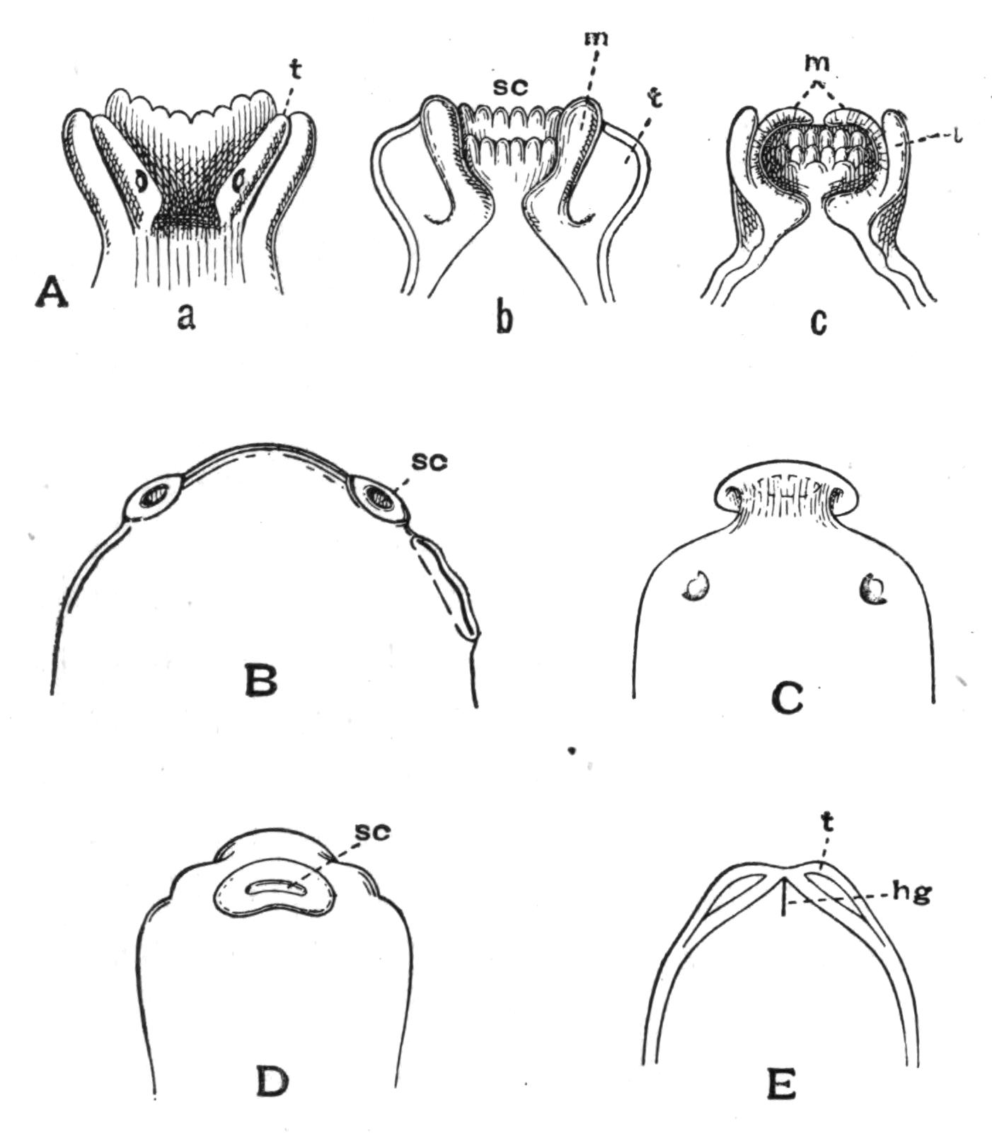
Fig. 16.—Suckers of Triclads. A, Planaria punctata Pall.; a, dorsal surface of head; b, ventral surface (freely moving) showing the sucker; c, sucker contracted (after Hallez): B, ventral surface of head of Dicotylus pulvinar Gr., from Lake Baikal (after Grube): C, dorsal surface of Procotylea fluviatilis Gir. (after Girard): D, sucker of Cotyloplana whiteleggei Sp. (after Spencer): E, ventral view of head of Planaria alpina Dana (preserved specimen); hg, adhering groove; m, thickened musculature forming the margin of the sucker; sc, sucker; t, tentacles.
The movements of Land Planarians are somewhat peculiar. The ventral surface of Bipalium has a median groove, into which the ducts of numerous mucus-glands open. This is bordered by two ridges clothed with long and powerful cilia, which perform the chief part in propelling the animal, aided, according to Lehnert,[52] by muscular waves which pass from the head, backwards, i.e. opposite in direction to those by which a snail slides along. This observation, however, needs confirmation. The whole body executes sinuous movements, during which the crescentic head, lifted slightly above the ground (Fig. 15, A), is constantly altering and regaining its normal shape, somewhat as a Planaria lactea uses the lobes of its head. Further examination shows that the margin of the head of Bipalium is not only provided with eyes, but in addition, with ciliated, (probably) olfactory pits. Such depressions, innervated directly from the cerebral ganglia, have been found in sixteen species of Geoplana, and in one or two species of Rhynchodemus.[53] Some Land Planarians (a species of Rhynchodemus from Ceylon, and a Dolichoplana from the Philippines) wriggle out of a box or the hand with great speed (Moseley).
The skin of Triclads is full of minute rods or rhabdites, which are shot out in great numbers when the animal is irritated, and doubtless serve an offensive purpose. The Terricola possess two kinds of these: (1) needle-like rods; and (2) in Bipalium kewense, flagellated structures, bent into a V-form and with a slender thread attached to one end (Shipley). In Geoplana coerulea these bent rods furnish the blue colour of the ventral surface. The rhabdites arise in all Triclads in cells below the basement-membrane, which they are said to traverse in order to reach the epidermis, thus differing in origin, and also in structure, from the rods of Polyclads.
Food.—Triclads are largely if not wholly carnivorous animals, feeding upon Annelids, Crustacea, Insects, Insect-larvae, and Molluscs. The mouth is usually mid-ventral or behind the middle of the body, but in the anomalous Leimacopsis terricola Schm. from the Andes[54] and in Dolichoplana it is near the anterior end. The pharynx (Figs. 17, 18, ph) is cylindrical or bell-shaped, exceedingly dilatable and abundantly supplied with glands and nervous tissue. It opens into the three main intestinal branches, one of which runs in the median plane forwards, the others backwards right and left, enclosing a space in which the genital ducts lie (Figs. 14, A, 17). The fresh-water Planarians prey upon Oligochaeta, Hydrophilidae (aquatic beetles), and the commoner pond-snails. Bipalium kewense pursues earthworms, seizes the upper surface of the anterior end by the glutinous secretion of its ventral surface, and then proceeds to envelop part or the whole of the worm within its pharynx, which is stretched as a thin skin over the body of its struggling prey (Lehnert). The tissues of the latter pass into the intestine of the Planarian, and distend it greatly. After such a meal, which lasts from one to five hours, a Bipalium may remain for three months without seeking food. Geobia subterranea, a white eyeless form from Brazil, pursues earthworms (Lumbricus corethrurus) in their burrows, and has been seen by Fritz Müller sucking the blood out of a young worm.[55] Geoplana typhlops, a Tasmanian species, is also blind, and pursues worms, as does G. triangulata (Dendy). In Trinidad, von Kennel[56] observed that land-snails (Subulinae) were the food of certain Land Planarians, the name of which, however, he does not state. The pharynx was employed to suck out the soft parts of the snail even from the upper whorls of the shell.
Reproduction.—In Planaria lactea the numerous testes (Fig. 17, te) are placed both above and below the alimentary canal throughout the greater part of its course. The membrane of each gonad is continued into a minute vas efferens, which unites with those of neighbouring testes. Two vasa deferentia (v.d) arise thus on each side, one from the posterior, the other from the anterior testes of the body, and open into the vesiculae seminales (v.s), which may be seen in the living animal as tortuous whitish tubes at the sides of the pharynx (Fig. 14, A). These open into the penis (Figs. 14, A; 17, pe), a large pyriform organ, the apex of which, when retracted, points forwards, projecting into the penial cavity. When this apical portion is evaginated and turned inside out, it is of considerable length, and is able to pass into the long slender duct of the uterus (ut) of another individual. The penial sheath (ps) is part of the genital atrium (gs), which is developed as a pit from the skin, and invests the end of the genital ducts, the mouth of the pit forming the common genital pore (go), through which both male and female genital products are emitted.
There are two ovaries (ov) placed far forwards, between the third and fourth pairs of intestinal coeca. The oviducts (ovd) lie just over the lateral nerves, and have a slightly tortuous course, at each outward bend receiving the duct (yo) of a yolk-gland (yg), so that ova and yolk are already associated when the oviducts open by a short unpaired tube into the genital atrium. The yolk-glands develop rapidly,[57] and when fully formed are massive glands occupying the spaces between the intestinal branches and the testes which are then aborting. The so-called uterus (ut), apparently at first a diverticulum of the genital atrium, expands behind the pharynx into a receptacle lined by long glandular columnar cells, which, however, are not all of the same kind. The uterine duct opens into the atrium just above the aperture of a problematical, eversible, "musculo-glandular organ" (mgr).
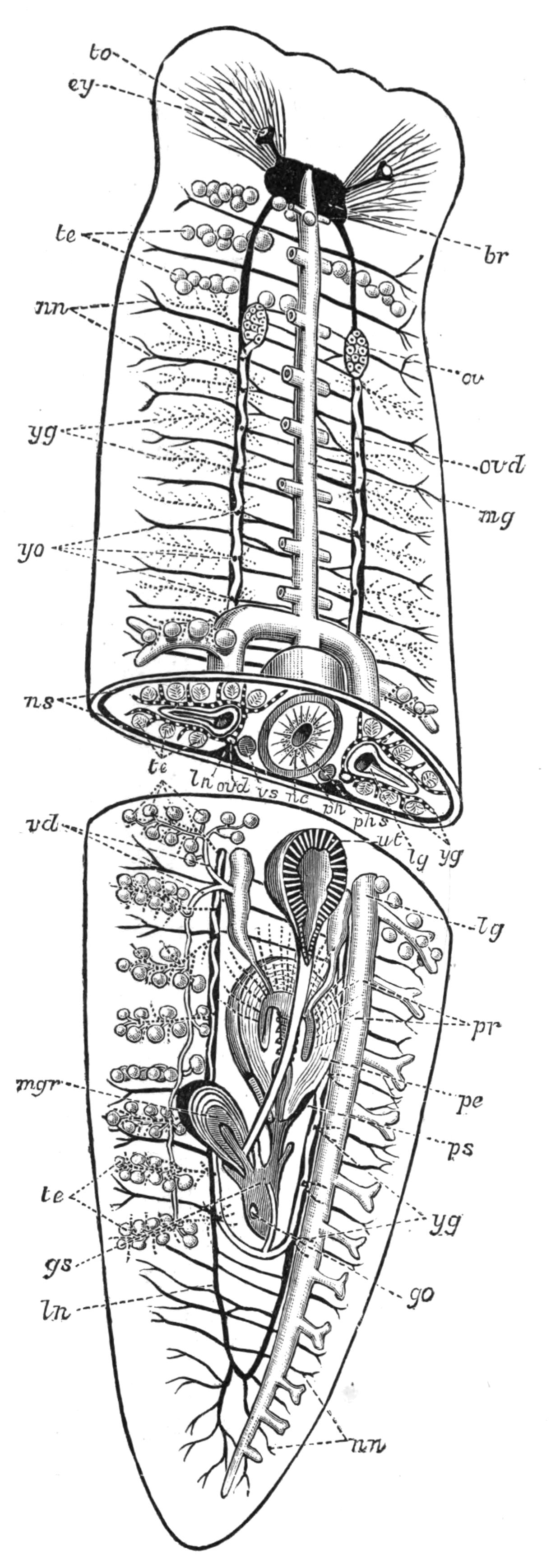
Fig. 17.—Diagrammatic view of the structure of Planaria (Dendrocoelum) lactea. × 7. The body has been cut across and a portion removed. In the posterior half the alimentary tract of the left side is removed and the uterus, penis, and muscular organ sliced open horizontally. The nervous system is represented by black, and the yolk-glands by dotted lines. br, Brain; ey, eye with lens and optic nerve; go, external genital aperture for both male and female products; gs, genital atrium; lg, paired lateral intestinal branch; ln, longitudinal nerve; mg, unpaired anterior intestine, the branches of which are cut off close to the main stem; mgr, eversible "musculo-glandular organ"; nc, nerve-cells in the pharynx; nn, lateral nerve-twigs; ns, nerve-sheath; ov, ovary; ovd, oviduct; pe, the eversible penis, the corrugated inner white portion of which is the apex; ph, pharynx; phs, pharyngeal sheath; pr, "prostate" or granule-gland (represented by dotted lines opening into the penis); ps, penial sheath; te, testes; to, tactile lobe of the head; ut, "uterus" opening into the genital atrium just above mgr; vd, vasa deferentia; vs, vesicula seminalis; yg, yolk-glands; yo, openings of the yolk-ducts into the oviducts.
Fertilisation appears to occur in the uterus, where ova, yolk, and spermatozoa, or (in P. torva) spermatophores (Fig. 14, N, sp), are found. The formation of the cocoon in Planaria lactea is probably begun in the "uterus," but is undoubtedly completed in the genital atrium. In P. polychroa, however, the stalked cocoon is formed wholly in the "uterus." Thus we find two types of cocoons in different species of the genus Planaria associated with two types of reproductive organs (Hallez):—
I. Planariae in which the two oviducts open separately into the posterior part of the duct of the uterus. A musculo-glandular organ is absent. The cocoons are spherical and stalked. Examples—Planaria polychroa (Fig. 14, L), P. albissima, P. gonocephala.
II. Planariae in which the two oviducts open by means of an unpaired duct into the genital atrium. A musculo-glandular organ present (Planaria torva (Fig. 14, N), P. mrazekii, P. lactea, P. cavatica), or absent (P. alpina, Fig. 14, M). The cocoons are sessile.
The genitalia of the Maricola (Fig. 14, F) and Terricola do not differ very much from those of Planaria. The uterus (greatly reduced in the Land Planarians) lies behind the genital pore, and several ova, together with much milky yolk, are enclosed in a capsule which is formed in the genital atrium.
Asexual Reproduction.[58]—It has long been known that fresh-water Planarians have not only great powers of repairing injuries, but that they use this faculty in order to multiply by transverse fission. Planaria alpina and Polycelis cornuta, in summer, separate off the posterior part of the body, and this ultimately becomes an entire individual. P. albissima, and especially P. subtentaculata, anticipate matters so far, that before fission is complete, the new individual has a head nearly fully formed. The new organs are largely regenerated in both parent and young, apparently by the division and specialisation of scattered embryonic cells in the parenchyma. The asexual reproduction of Land Planarians is not fully proved, though it is known that they repair injuries to the body completely, and that Bipalium kewense is often found in hothouses, divided into fragments which regenerate all the organs of the parent, but like the latter, do not mature their sexual organs.
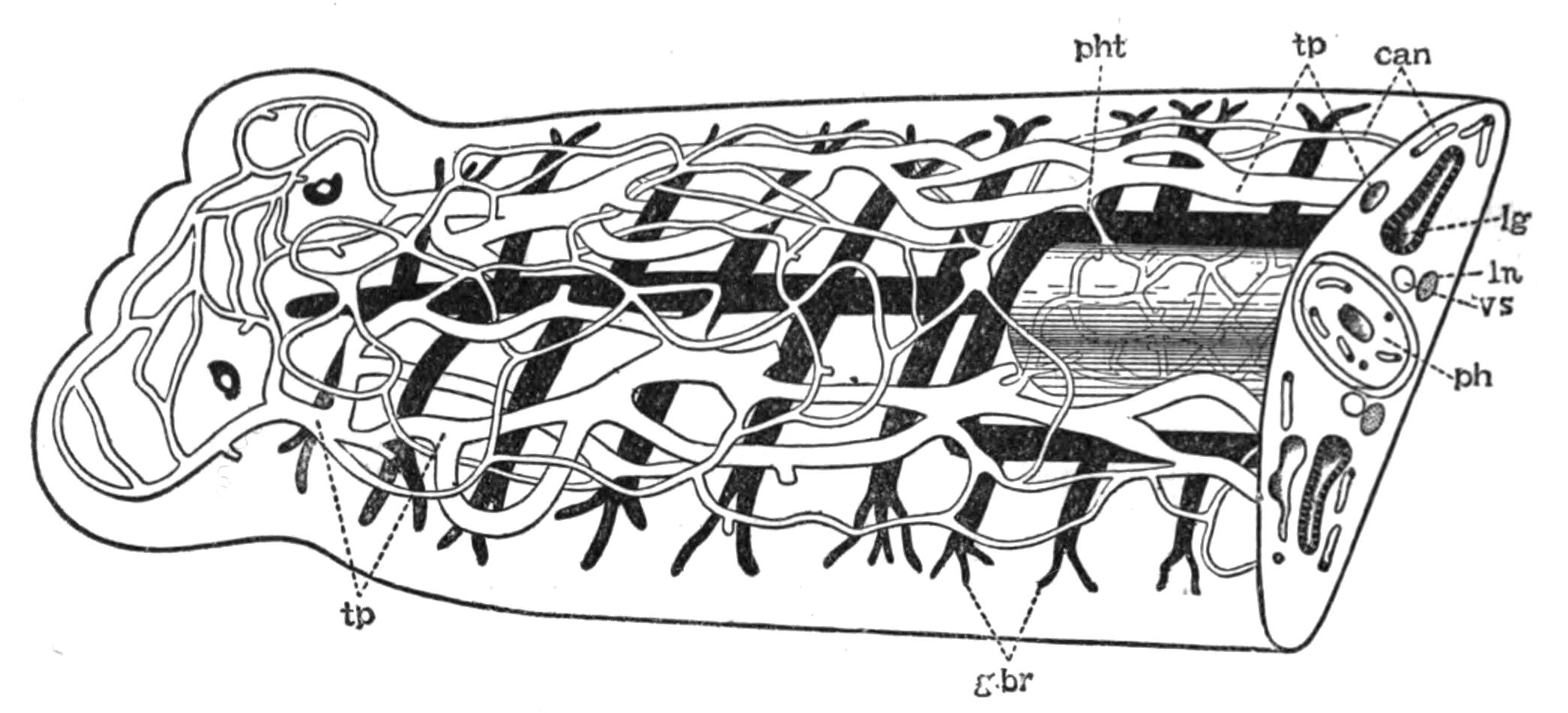
Fig. 18.—Semi-diagrammatic view of the excretory system of Planaria lactea. (Partly after Chickoff.) can, Capillary network on both dorsal and ventral surfaces; g.br, branches of the intestine; lg, lateral branches of the digestive system; ln, longitudinal nerve; ph, pharynx, with intermuscular capillary excretory network arising from the point marked pht; tp, principal vessels of the excretory system, the external opening of which is not certainly known; vs, vesicula seminalis.
Excretion.—The excretory organs of Triclads consist of flame-cells, canaliculi, and a pair of longitudinal canals, the external openings of which, have not been satisfactorily ascertained. The flame-cells are difficult to detect in Planaria lactea, and the latest observer, Chickoff,[59] was unable to see them, although to him we are indebted for figures of this system in P. lactea (Fig. 18) and P. alpina (P. montana). In the latter, the flame-cells are distinct, and may open directly into the two main canals or indirectly through unbranched canaliculi. The pharynx possesses a special supply of excretory tubules communicating with the main canals. A similar system has been described and figured in Gunda segmentata by Lang.[60]
Classification of Tricladida.
PALUDICOLA.
Family. Genus and British Species.
Planariidae Planaria lactea O. F. M., P. punctata Pall., P. polychroa Schm., P. torva M. Sch., P. alpina Dana. Polycelis nigra Ehr., P. cornuta Schm. Anocelis. Oligocelis, Procotyla. (Doubtful genera.) Sorocelis. Dicotylus.
MARICOLA.
Procerodidae (= Gundidae). Procerodes (= Gunda) ulvae Oersted, P. littoralis van Beneden. Cercyra. Uteriporus.
Bdellouridae Bdelloura. Syncoelidium.
TERRICOLA.
Bipaliidae Bipalium kewense Moseley (introduced).
Geoplanidae Geoplana. Geodesmus.
Rhynchodemidae Rhynchodemus terrestris O. F. M.
Belonging to undetermined Families Dolichoplana. Polycladus. Microplana. Leimacopsis.

Turbellaria. III. Rhabdocoelida.
The Rhabdocoelida include a very heterogeneous assemblage of usually minute Turbellaria, distinguished collectively from the Polyclads and Triclads by the form of the digestive tract. This is a simple or slightly lobed sac, except in the Bothrioplanidae, which in this and many other points closely resemble the Triclads. It is to the straight, rod-like nature of the alimentary canal that the name of the group refers. The size and form of the body, and the structure of the pharynx and genitalia, vary within wide limits.
The Rhabdocoelida are subdivided into three tribes:—
(1) Acoela, in which a sub-central mouth and pharynx are present, but lead into the parenchyma of the body, not into an intestine with proper walls. An excretory system has not hitherto been seen. Yolk-glands are absent. An otolith underlies the brain. The Acoela are marine.
(2) Rhabdocoela, which possess a complete alimentary tract separated from the body-wall (except for a few suspensory strands) by a space or body-cavity, filled with fluid. This space is sometimes (Vortex viridis) lined by an endothelium of flattened parenchymatous cells. There are two compact testes, which are enclosed (as are the ovaries and yolk-glands) in a distinct membrane. An otolith is present in some genera and species. Terrestrial, fresh-water, marine.
(3) Alloeocoela, in which the body-cavity is greatly reduced. Except in the Bothrioplanidae, the gonads have no distinct membrane. Testes numerous; yolk-glands present. Marine with a few exceptions.
Occurrence and Habits of the Rhabdocoelida.—The Acoela are usually minute, active Turbellaria abounding amongst weeds throughout the lower half of the Littoral, and the whole of the Laminarian zone, but are most plentiful in the pools exposed during spring-tides on our coasts, especially on the shores of Devonshire. The species of Haplodiscus, however, and Convoluta henseni are modified pelagic forms found in the Atlantic Ocean.[61] Convoluta paradoxa (Fig. 19, B) is the commonest British species. It is from 1 to 9 mm. in length, and of a brown colour, marked above by one or more transverse white bars. The brown colour is due to a symbiotic alga, the nature of which has not been thoroughly investigated. In an allied species, however (C. roscoffensis), from the coast of Brittany, the alga, which is here green, has been carefully examined by Professor Haberlandt,[62] and it appears from his researches that the algae form a special assimilating tissue, enabling the Convoluta to live after the fashion of a green plant. At Roscoff, these elongated green Convoluta live gregariously in the sandy tide-pools, fully exposed to the sun's rays, and have the appearance of a mass of weed floating at the surface of the water. Access to the atmosphere and to sunlight are necessary in order to enable the assimilating tissue to form the carbohydrates, upon which this form lives exclusively. Not only has the alga itself undergone such profound changes (loss of membrane, inability to live independently after the death of the host) as to disguise its true nature (a tissue-cell derived from algal ancestors), but the Convoluta has also undergone concomitant changes, in form, in the loss of a carnivorous habit, and in the development of marked heliotropic movements, thus adapting itself to an holophytic or plant-like mode of nutrition. Nevertheless the Acoela, as a group, are carnivorous, feeding upon Diatoms, Copepoda, and small Rhabdocoela, the absence of a digestive tract indeed being probably more apparent than real.[63]
The Rhabdocoela live under varied conditions. One form, Prorhynchus sphyrocephalus, has been found among plants far from water in the neighbourhood of Leyden, by De Man.[64] With this exception the group is purely aquatic, and though a few genera and even individuals of the same species occur both in salt and fresh water, whole sub-families and genera are either marine or paludicolous. Among the latter, Mesostoma, Castrada, Vortex, and Derostoma are common in brooks and ponds, especially at certain times, often only for one month (May or June) in the year. Species of Macrostoma, Stenostoma, and Microstoma are also abundant in similar places. The two latter occur in chains formed by fission; but the sexual individuals (which are of distinct sexes, contrary to the usual hermaphrodite condition of Flat Worms) only appear at stated times and are not well known. A large number of genera are purely marine, and one family, the Proboscidae (distinguished by having the anterior end invaginated by special muscles and converted into a sensory organ), is entirely so. The most cursory examination of littoral weeds reveals species of Macrorhynchus, Acrorhynchus, Promesostoma, Byrsophlebs, and Proxenetes, the character of which may be gathered from von Graffs great monograph, or from Gamble's paper on the "British Marine Turbellaria."[65] Much, however, still remains to be done before we possess an adequate idea of the occurrence of this group on our coasts.
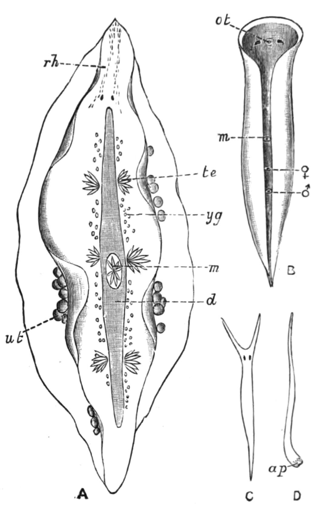
Fig. 19.—Forms of Rhabdocoelida. A, Mesostoma tetragonum O. F. M. (Rhabdocoela), × 10; B, Convoluta paradoxa Oe. (Acoela), × 10; C, Vorticeros auriculatum O. F. M., × 6; D, Monotus fuscus Oe. (Alloeocoela), × 4. ap, Adhesive papillae; d, intestine; m, pharynx; ot, otolith; rh, rhabdites; te, testes; ut, uterus with eggs; yg, yolk-glands; ♂, male, ♀, female genital pores. (A after Braun.)
Some Rhabdocoels are parasitic. Fecampia erythrocephala, which occurs in the lacunar spaces and alimentary canal of young shore crabs (Carcinus maenas), is a white cylindrical animal ¼ inch long, with a red snout. After attaining maturity it works its way out of the crab and encysts under stones, forming a pyriform mass in shape like a "Prince Rupert's drop." Within this case the eggs develop, and the young probably emerge through the open narrow end of the hard white tube, but how they reach the crab is not known. Graffilla muricicola is found in the kidney of Murex brandaris and M. trunculus, at Naples and Trieste; G. tethydicola in the foot of Tethys. Anoplodium parasiticum occurs among the muscles which attach the cloaca of Holothuria tubulosa to the body-wall; and A. schneideri occurs in the sea-cucumber, Stichopus variegatus. These are truly parasitic forms, constituting a special sub-family. They have no rhabdites in the skin; the nervous system and sense-organs are only slightly developed; and the pharynx has undergone a notable reduction in relation to the simpler mode of obtaining nourishment. Other cases of association between certain Rhabdocoels (closely allied to, if not identical with, certain free-living species) and Lamellibranchs or Sea-urchins, are, however, of another kind. Thus on the gills or in the mantle cavity of species of Mytilus, Cyprina, Tellina, and upon the test of Clypeaster, such forms as Enterostoma mytili, Acmostoma cyprinae, and Provortex tellinae have been found. But it is probable that these Turbellaria here obtain merely a temporary shelter and possibly a supply of the food of the mussel or sea-urchin.
The Alloeocoela afford a well-established case of association. Monotus fuscus (Fig. 19, D), an abundant, active, elongated animal, lives on our coasts in the upper part of the littoral zone among Patella, Balanus, and sometimes Chiton. When the tide is low, the Monotus, to obtain moisture and darkness, creeps between the mantle-folds of these animals, where it may readily be found. Upon the return of the tide it leaves its retreat and creeps or swims about freely. Other Alloeocoela collect in great numbers in tufts of red-seaweeds (Florideae). By placing such tufts in vessels, the sea-water, especially as darkness sets in, begins to swarm with Cylindrostoma 4-oculatum, species of Enterostoma and Plagiostoma; P. vittatum, with three violet bands across the white body, being a particularly obvious form. Vorticeros auriculatum (Fig. 19, C), another abundant species, is remarkable for the long tentacles which can be completely withdrawn, and in this condition it completely resembles a Plagiostoma.
The presence of a species (P. lemani) of the characteristically marine genus Plagiostoma, in the Lake of Geneva, and in one or two other Swiss lakes, at depths varying from 1 to 150 fathoms, is very interesting, and is perhaps the only well-established case of the survival of a once marine Rhabdocoelid under changed conditions. Plagiostoma lemani is by far the biggest of the group to which it belongs, being over half an inch in length. It is usually found in fine mud, sometimes among Chara hispida, and has the general appearance of an inactive white slug. We are indebted to Forel and Duplessis for the discovery of this species, and also of Otomesostoma morgiense, a Mesostoma with an otolith, dredged in 10 to 50 fathoms in the Lake of Geneva, the Lake of Zürich, and found recently also by Zacharias in the Riesengebirge. The genus Bothrioplana, first found by Braun in the water-pipes of Dorpat, has been carefully investigated by Vejdovsky,[66] who places it in a special family, Bothrioplanidae, among the Alloeocoela. One species has recently been found near Manchester.
A comprehensive survey of the Rhabdocoelida shows that, with the chief exception of the Proboscidae, the more lowly organised forms, the Acoela and Alloeocoela, are marine, whereas the fresh-water forms are in most cases the most highly organised genera (Mesostoma, Vortex). But Macrorhynchus helgolandicus, though minute (1.5-2 mm. long), has a more complex structure[67] than any other species of the specialised marine genus to which it belongs, and is a remarkable instance of great complexity being associated with small size.
Reproduction.—The Rhabdocoelida present the greatest diversity in the development of the reproductive system. The Acoela and Alloeocoela have the simplest arrangement. Scattered testes, often without a distinct membrane, form the spermatozoa, which in most cases wander into parenchymatous spaces, but in Monoporus rubropunctatus and Bothrioplana, into distinct vasa deferentia. In both groups a protrusible penis opens independently to the exterior, and may be simply muscular or provided with a chitinous armature. Two ovaries are present, and the oviducts, if distinct, are continuations of the ovarian membrane. In most forms a "bursa seminalis," which receives the spermatozoa of another individual, is appended to the female genital canal. In many of the Alloeocoela, however, a portion of the ovary is sterile, and its cells, forming a yolk-gland, feed the fertile portion, the whole structure being then spoken of as a germ-yolk-gland. In many others (Monotidae) this sterile part has become an independent yolk-gland, which communicates by yolk-ducts with the oviducts. The Acoela form no egg-case, the body of the parent becoming a bag for the ova, which elaborate their own food-yolk. The Alloeocoela lay hard-shelled eggs, which are produced in Bothrioplana and Automolos by the activity and interaction of reproductive organs, resembling closely those of certain Triclads.[68]
The Rhabdocoela exhibit every stage in the development of a complex reproductive system, from the simple ovaries and testes of a Microstoma or Macrostoma, to the intricate system of ducts and glands of a Macrorhynchus (Proboscidae), in which there is still much to be made out. The complications of the copulatory organs chiefly arise from the way in which the spermatozoa are brought into contact with a nutritive prostatic fluid, or are formed into spermatophores; and also from the penial armature, which is often very complex, and may consist of a curved chitinoid hook or a coiled loop (Promesostoma), of hooks (Proboscidae), or of an intricate arrangement of plates (Proxenetes); or the penis may take on a complex corkscrew-like form (Pseudorhynchus). The (frequently armed) female genital canal usually possesses a bursa seminalis for the fertilisation of the eggs, but a receptaculum seminis or spermatheca may serve for the reception, the bursa, for the lodgment of the spermatozoa of another individual. The fertilised ovum is provided with a supply of food-yolk and with a shell, which may be formed in a special diverticulum, the "uterus." The development of these organs strains the resources of the animal to the utmost, and in some Proboscidae the alimentary canal is squeezed out and disintegrates, in order to make room for them.
A few Mesostoma (M. ehrenbergii, M. productum, M. lingua) produce two kinds of eggs—thin- and thick-shelled. The latter are laid throughout the summer, and lie dormant through winter. The young which hatch in spring out of these "winter" eggs develop rapidly, and when only 7 to 8 mm. long (i.e. one-third the size of the parent) already possess functional genital organs; the penis, however, is rudimentary, and incapable of being used for copulation. Hence it is probable that this stunted progeny self-fertilise their thin-shelled or "summer" eggs. After the formation of these eggs the same parent is said (Schneider[69]) to produce thick-shelled or winter eggs, but however that may be, the first young which hatch from the thin-shelled ova are produced in great numbers at a time (April to May) when food is abundant. These grow rapidly to the full size, and then having attained maturity, cross-fertilise one another's ova, which become encased in a thick brown shell; and it is these numerous "winter" eggs that lie dormant throughout the autumn and winter. Many Mesostoma, and practically all other Rhabdocoela, however, produce only thick-shelled eggs, and in all cases it is probable that to these many species owe their wide distribution, the exact range of which is, however, unknown, as is also the means of dispersal.
Classification of Rhabdocoelida.
ACOELA.
Family. Genus and British species.
Proporidae Proporus venenosus O. Sch. Plymouth. Monoporus rubropunctatus O. Sch. Plymouth. Haplodiscus.
Aphanostomatidae Aphanostoma diversicolor Oe. Common. A. elegans Jen. Plymouth. Convoluta saliens Grff. Plymouth, Millport. C. paradoxa Oe. (Fig. 19, B). Common. C. flavibacillum Jen. Plymouth, Port Erin, Millport. Amphicoerus. Polychoerus.
RHABDOCOELA.
Macrostomatidae Mecynostoma. Macrostoma hystrix Oe. Stagnant water. Omalostoma.
Microstomatidae Microstoma lineare Oe. Fresh water. M. groelandicum Lev. Plymouth, among Ulva. Stenostoma (Catenula) lemnae Dug. Near Cork. S. leucops O. Sch. Common in fresh water. Alaurina claparedii Grff. Skye.
Prorhynchidae Prorhynchus stagnalis M. Sch. In Devonshire rivers. Promesostoma marmoratum M. Sch. Common. P. ovoideum O. Sch., P. agile Lev. Plymouth. P. solea O. Sch. Plymouth, Port Erin. P. lenticulatum O. Sch. Port Erin.
Mesostomatidae Byrsophlebs graffii Jen. Plymouth, Millport. B. intermedia Grff. Millport, Port Erin. Proxenetes flabellifer Jen. Millport, Plymouth, Port Erin. P. cochlear Grff. Millport. Otomesostoma. Mesostoma productum Leuck., M. lingua O. Sch., M. ehrenbergii O. Sch., M. tetragonum O. F. M. (Fig. 19, A). All at Cambridge. M. rostratum Ehr. Widely distributed. M. viridatum M. Sch. Manchester. M. robertsonii Grff., M. flavidum Grff. Both at Millport. Bothromesostoma personatum O. Sch. Preston. Castrada.
Proboscidae Pseudorhynchus bifidus M‘Int. Millport, St. Andrews, Port Erin. Acrorhynchus caledonicus Clap. Generally distributed. Macrorhynchus naegelii Köll., M. croceus Fabr. Plymouth, Millport. M. helgolandicus Metsch. West coast. Gyrator hermaphroditus Ehrbg. St. Andrews. Also common in fresh water. Hyporhynchus armatus Jen. Plymouth, Port Erin. H. penicillatus O. Sch. Plymouth.
Vorticidae Schultzia. Provortex balticus M. Sch. Generally distributed. P. affinis Jen., P. rubrobacillus Gamb. Plymouth. Vortex truncatus Ehrbg. Abundant in fresh water. V. armiger O. Sch. Millport (fresh water). V. schmidtii Grff., V. millportianus Grff. Millport. V. viridis M. Sch. Generally distributed. Jensenia. Opistoma. Derostoma unipunctatum Oe. Edinburgh. Graffilla. Anoplodium. Fecampia erythrocephala Giard. Plymouth, Port Erin.
Solenopharyngidae Solenopharynx.
ALLOEOCOELA.
Plagiostomatidae Acmostoma. Plagiostoma dioicum Metsch., P. elongatum Gamb., P. pseudomaculatum Gamb., P. sagitta Ulj., P. caudatum Lev., P. siphonophorum O. Sch., P. ochroleucum Grff. All at Plymouth. P. sulphureum Grff. Port Erin. P. vittatum F. and Leuck. Millport, Plymouth, Port Erin. P. koreni Jen. Plymouth, Millport. P. girardi O. Sch. Plymouth, Port Erin, Valencia. Vorticeros auriculatum O. F. M. (Fig. 19, C). Port Erin, Plymouth. V. luteum Grff. Plymouth. Enterostoma austriacum Grff. Plymouth, Port Erin. E. fingalianum Clap. Skye, Plymouth. E. coecum Grff. Millport. Allostoma pallidum van Ben. Millport. Cylindrostoma 4-oculatum Leuck. Skye, Millport, Plymouth. C. inerme Hall, C. elongatum Lev. Plymouth. Monoophorum striatum Grff. Plymouth.
Bothrioplanidae Bothrioplana. Bothrioplana sp.? Manchester. Otoplana.
Monotidae Monotus lineatus O. F. M., M. fuscus Oe. (Fig. 19, D). Both common littoral forms. M. albus Lev. Plymouth. Automolos unipunctatus Oe. Skye, St. Andrews, Plymouth. A. horridus Gamb., A. ophiocephalus O. Sch. Plymouth.