Chapter III
Reading edition
CESTODA
INTRODUCTION—NATURE OF CESTODES—OCCURRENCE OF CESTODES—THE TAPE-WORMS OF MAN AND DOMESTIC ANIMALS—TABLE OF THE LIFE-HISTORIES OF THE PRINCIPAL CESTODES OF MAN AND DOMESTIC ANIMALS—STRUCTURE AND DEVELOPMENT OF CESTODES—TABLE FOR THE DISCRIMINATION OF THE MORE USUAL CESTODES OF MAN AND DOMESTIC ANIMALS—CLASSIFICATION.
The Cestodes or Tape-worms are exclusively endoparasitic Platyhelminthes living, in the adult condition, in the alimentary canal of Vertebrates, with the exception of Archigetes (Fig. 37), which may become mature in the body-cavity of Tubifex. In relation with this wholly parasitic existence, the Cestodes exhibit certain characteristic modifications in structure and mode of development, such as the formation, by the segmentation of the "neck," of a (usually) long chain of "proglottides" or joints, which form the "body" of the Cestode; and the entire absence of an alimentary tract, both in the larva and adult. As an adaptation to the fixed mode of life, the anterior end (head, scolex) is modified to form an adhering organ. Various adaptive forms of larvae are known. These live in the internal organs of one or more intermediate hosts, and are transferred to the final host passively during a meal. Lastly, there is the curious metamorphosis by which the adult is formed from a portion (scolex) of the larva.[94]
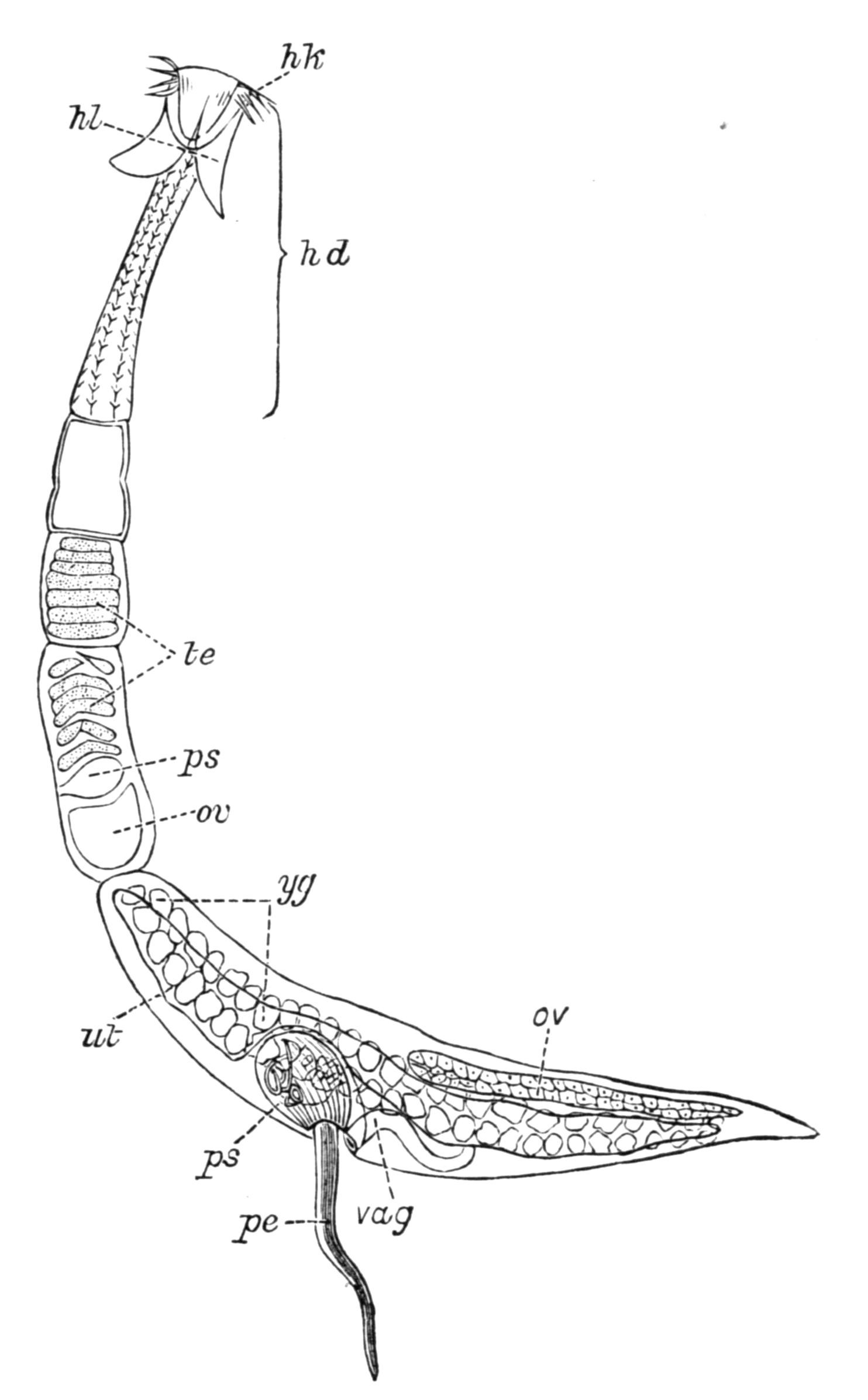
Fig. 36.—Echinobothrium affine Dies., from the intestine of Torpedo, × 43. hd, Head; hk, hooks; hl, lobes of the head; ov, ovary; pe, penis; ps, penis-sheath; te, testes; ut, uterus; vag, vagina; yg, yolk-glands. (After Pintner.[95])
Taenia solium, from man (Fig. 39, B), or Echinobothrium (Fig. 36), from an Elasmobranch fish, is fixed to the mucous lining of the intestine of its host by means of a radially-constructed apparatus of four suckers and a circlet of hooks (Fig. 39), which are borne by the "head" or "scolex," being that part of the worm which is directly derived from part of the larva, and which contains the central, commissural portion of the nervous system. Firm adhesion to the host's intestine is necessary, in order to avoid the loosening action of the peristaltic movements of the intestine as the food passes along. The heads of different Cestodes exhibit a marvellous variety of suckers and hooks, from a mere muscular depression in Schistocephalus, to the compound proboscides of Tetrarhynchus[96] which is found in Elasmobranchs. The jointed body, often of enormous length (up to 20 yards in Bothriocephalus latus), is usually separated from the head by a slender neck, from which the proglottides are segmented off from behind forwards, and become more and more individualised as they recede farther away from the neck by the intercalation of younger joints. Thus in Fig. 36 the mature, distal proglottis has passed through all the stages represented by the other segments.
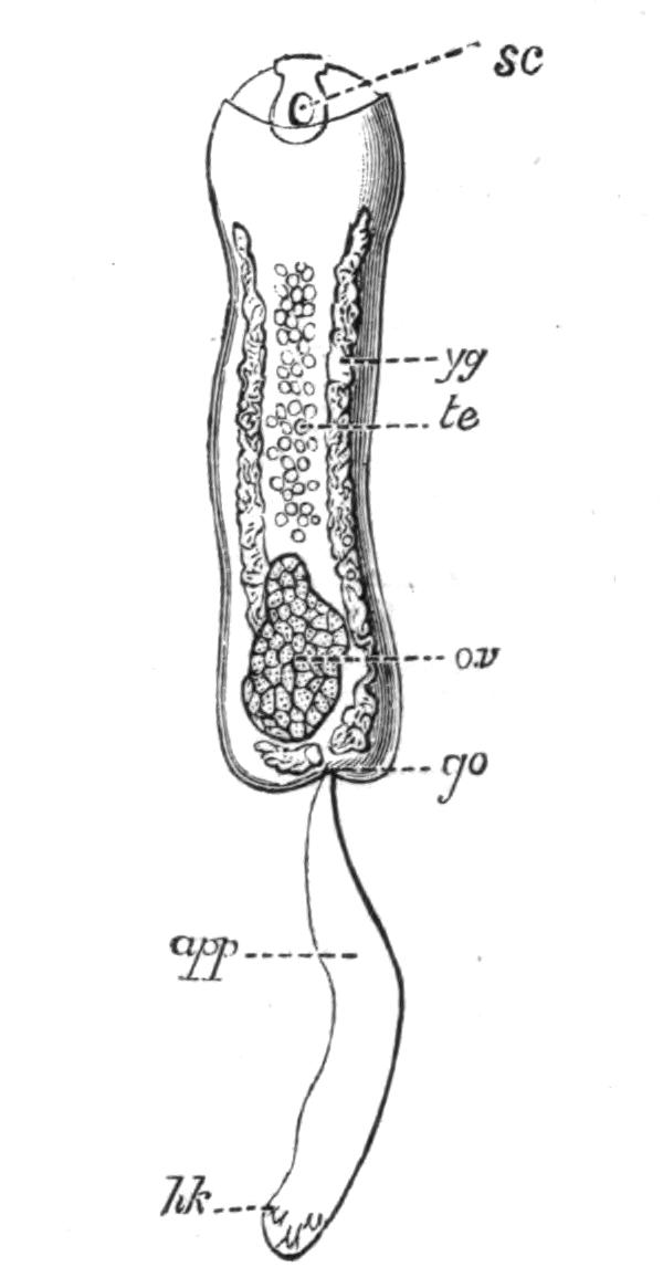
Fig. 37.—Archigetes sieboldii (appendiculatus), from the coelom of Tubifex rivulorum. × 40. app, Persistent larval appendage; go, genital pore; hk, persistent larval hooks; ov, ovary; sc, sucker; te, testes; yg, yolk-glands. (After Leuckart.)
The longitudinal muscles, the nerves, and excretory vessels which supply the proglottides are continuous throughout and with those of the head. Each joint contains at first male genitalia comparable with those of a Trematode; then the female organs develop, and finally self-fertilisation follows. The Cestodes feed through their skin, probably by the aid of fine protoplasmic processes, which penetrate the tough investing membrane and absorb the already digested food which bathes them. When a proglottis of Calliobothrium is approaching maturity it separates from the parent, the broken ends of muscles, nerves, and excretory vessels speedily heal, and it is now capable of continued growth and of fairly active movement if it remains in the intestine of the host. According to van Beneden, it may even attain a size equal to, or exceeding, that of the whole parent or "strobila."[97] These considerations led Leuckart, von Siebold, P. J. van Beneden, and others, to Steenstrup's conclusion that a jointed tape-worm is really a colony composed of two generations—the head and neck derived from the larva, and the proglottides produced by the segmentation of the neck.[98] This view of the colonial nature of jointed Cestodes was generally adopted from 1851 to 1880. During the last fifteen years, however, the varied interpretations of the facts of the ontogeny of this group have led some authors to adopt the monozootic view (that a Cestode is one individual), others are still of the older opinion, and Hatschek (Lehrbuch, p. 349) and Lang take up intermediate positions. Lang considers that the formation of the joints of a tape-worm from a small fixed "scolex," is not only largely comparable with the strobilation of a scyphistoma and the consequent formation of a pile of medusae, as in the life-history of Aurelia, but that both processes have arisen from the power of regenerating the necessary organs in each of the new segments. The result in both cases is the rapid formation of a number of joints, which gradually separate from the parent, to carry the eggs and young to new stations. Just as some Coelenterata (Lucernaria) may be regarded as not having advanced much beyond a scyphistoma stage, so there are unisegmental Cestodes (e.g. Archigetes, Fig. 37) which have remained as a slightly altered but sexual scolex, directly comparable with a Trematode, and, as all authors are agreed, representing one generation only. Such monozootic forms are now classed as a special family, the Cestodaria or Monozoa, of which Caryophylleus mutabilis, from the intestine of various Cyprinoid fish, is the most abundant representative, while Amphiptyches (Gyrocotyle) urna, from Chimaera monstrosa of the northern hemisphere, is paralleled by A. rugosa, found in Callorhynchus antarcticus of the southern seas.
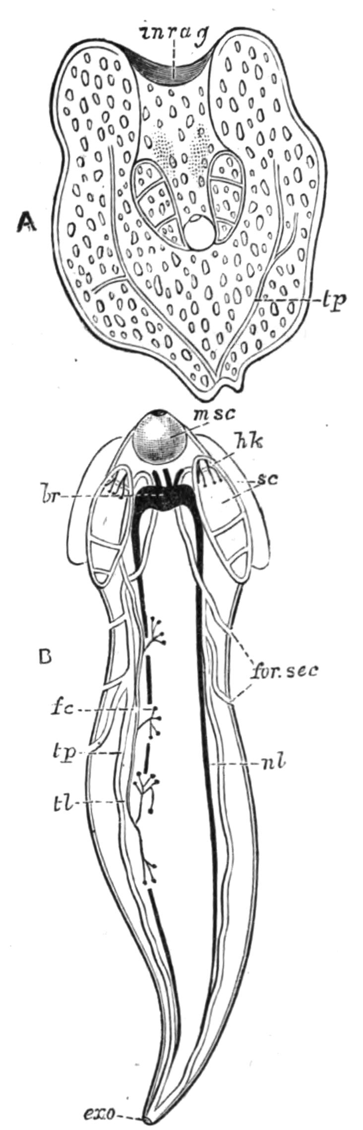
Fig. 38.—Scolex polymorphus Rud. (larva of Calliobothrium filicolle Zschokke), from the muscles of Apogon, a Mediterranean fish; also found in many Invertebrates (e.g. Sepia). A, Inverted scolex, with calcareous bodies; B, everted older larva. br, Brain; exo, terminal excretory aperture; fc, flame-cells; for.sec; secondary excretory pores; hk, hooks of the adult Cestode; inrag, pit at the bottom of which the head is developed; msc, anterior sucker; nl, lateral nerve; sc, suckers; tl, tp, lateral and main excretory vessels. (After Monticelli.)
Occurrence of Cestodes.—The distribution of Cestodes and their larvae is analogous to that of the digenetic Trematodes, although the absence of an alimentary canal limits the habitat of the mature worms to certain sites, such as the blood-vessels, the lymphatic and coelomic spaces, and the digestive system, where their body may be bathed by a nutritive fluid. Almost all groups of Vertebrates are attacked by Cestodes. Those of fishes, and particularly of Elasmobranchs, are distinguished by certain structural and developmental features; those of birds by others; those of mammals, by a third set of characters. The young stages of the Cestodes of Sharks and Rays occur encysted in the body-cavity, or in the pyloric appendages, of Teleosteans, which probably swallow them along with those invertebrate animals upon which they prey. The larvae of the Cestodes of carnivorous mammals or piscivorous birds, live respectively in herbivores and fishes, but how the latter are infected we know in very few instances. Cestode larvae are known to occur in many Invertebrates, and occasionally are taken free swimming in the sea, presumably crossing from one host to the next. Ctenophores, Siphonophores, Copepods, Ostracods, Decapods, various Molluscs especially Cephalopods, Earthworms, and other Annelids, are the intermediate hosts of these larvae (see Fig. 38), the fate of which, however, has been determined in but few cases.
Occurrence of Cestodes in Man.[99]—Tape-worms, either in the adult or larval stages (bladder-worms), have, from ancient times, been known to occur in man, and in the animals that serve him as food. Until comparatively recent times, however, the true nature of these parasites, and particularly of "hydatids" (cystic larvae), was unrecognised. Up to the seventeenth century the larvae were regarded as abscesses or diseased growths of the affected organs, and it was only at the close of that century that their animal nature was even suggested. Even at the beginning of the nineteenth century, three modes of origin of Cestodes—by "generatio aequivoca" from the tissues of the body, or by the union of previously distinct proglottides, or again by metamorphosis of free-living worms drunk with water by cattle or birds (as Linnaeus suggested)—were still variously held, at a time when Malpighi, Pallas, and Goeze had recognised the true connexion between the cystic and segmented states of Taenia crassicollis (the cat tape-worm), and when Goeze had seen the eggs of Taeniae, and Abildgaard[100] had even conducted the first helminthological experiments (conversion of the larval Schistocephalus, Fig. 40, into the adult form).
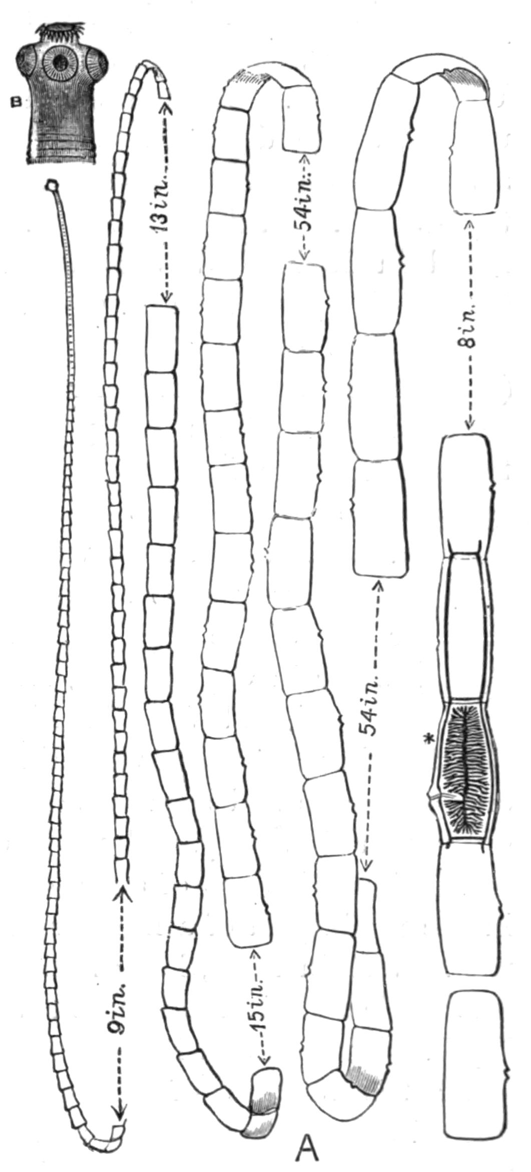
Fig. 39.—A, Taenia saginata Goeze. Nat. size. (From a specimen in the Cambridge Museum.) The approximate lengths of the portions omitted in the drawing are given. At * (after Leuckart) the branched uterus and the longitudinal and transverse excretory vessels are shown. The genital apertures are seen as a lateral opening on each of the larger proglottides. B, Head (scolex) of T. solium Rud. × 12. (After Leuckart.)
Generally speaking, "a tape-worm" in Western Europe will prove to be Taenia saginata Goeze (the beef tape-worm, Fig. 39, A), exceedingly prevalent also in the East, and indeed cosmopolitan, occurring wherever the infected flesh of the ox is eaten in a raw or half-cooked state. Its attacks are fortunately not usually severe. Taenia solium Rud. (the pork tape-worm) is found wherever the pig is kept as a domestic animal, and has consequently a world-wide distribution. Its size (6-9 feet long) and powers of adhesion would alone render T. solium a formidable parasite. But the danger of its presence in the body of man, or in the flesh of pigs, lies in the fact that the larva or bladder-worm (known as Cysticercus cellulosae) can live in the most varied organs. Thus if by accident a mature proglottis be eaten, the embryos escape, bore their way into the wall of the stomach, and entering the portal vein, may reach in time the muscles, the brain, the eye, or even the heart itself, and attain the cystic condition. Even more disastrous may be the result, should some ripe joints of a mature worm work their way from the intestine back towards the stomach. Should this happen (and though it has not been directly proved, the possibility is to be reckoned with), the result would be the release of vast numbers of embryos capable of inflicting fatal injury on the host. An abnormal Cysticercus of this species is probably the Taenia (Cysticercus) acanthotrias Weinl. (see, however, Leuckart, loc. cit. p. 711).
Taenia (Hymenolepis) nana v. Sieb.[101] is found in man in Egypt, Italy, England, Servia, Argentine Republic, and the United States. Though small (¾-1 inch long), its numbers usually excite digestive and nervous disorders of considerable severity, more serious, indeed, than those caused by the commoner tape-worms. H. diminuta Rud. (flavopunctata Weinl.), normally found in Rodents, has been rarely recorded in man. Taenia (Dipylidium) caninum L. (= T. cucumerina Bloch = T. elliptica Batsch), the commonest parasite of pet cats and dogs, and T. (Davainea) madagascariensis Davaine, have occasionally been recorded from infants and young children. But the attacks of these species are insignificant in comparison with those of the cystic stage (Echinococcus polymorphus) of a tape-worm (T. echinococcus v. Sieb.) which lives when mature in the dog.
Echinococcus is most frequent in Iceland, where it affects 2 to 3 per cent of the population, and a still larger proportion of sheep; while in Copenhagen, Northern Germany, some districts of Switzerland, and Victoria it is not uncommon, but is frequently found during post-mortem examinations when no definite symptoms of its presence had been previously noticed. Echinococcus[102] varies greatly in size, form, and mode of growth, but is distinguished in the formation not of one scolex only, as in the Cysticercus, but in the production of a number of vesicles, usually from the inner wall. Within these, large numbers of scolices may be developed. The whole organism continues to swell by the formation of a watery liquid within it, and if its growth be rapid the fluid tension may cause the rupture of the enclosing connective-tissue capsule formed around the parasite, at the expense of the host, and the protrusion of the daughter vesicles. It is the consequent injury to the surrounding organs of the host, at this critical stage, often only reached after the lapse of several years, that occasions serious or even fatal results. Zoologically, Taenia echinococcus and T. coenurus are interesting, since they exhibit an indubitable alternation of asexual generations in the larval state, with a sexual adult stage.
Bothriocephalus latus Brems., the broad tape-worm, which attains a length of 20-30 feet, or even more, occurs in man endemically in the eastern Baltic provinces, certain parts of Switzerland, generally throughout Russia (especially near Kasan), in North America, and commonly in Japan,—that is, in districts where the population partake largely of pike or other fish in a raw or partially-cooked state. Elsewhere it occurs sporadically, and in Munich, where it was unknown before 1880, its presence has been traced to emigrants from infected districts, who settled on the shores of the Starenberger Lake, from which Munich was supplied with fish. How the pike, the usual but not invariable intermediate host, becomes infested (and its musculature is frequently riddled with the larvae) we do not accurately know, but some Invertebrate, the prey of the pike, is probably the first host into which the free-swimming ciliated larva (Fig. 42) finds its way. In Greenland, B. cordatus is very common in the dog, and probably also in man, though few cases have been recorded. B. mansoni Cobb. (= B. liguloides Leuck.) was, till recently, known only in the larval state from China and Japan. Iijima, however, has found older specimens in the latter country. B. cristatus Dav. is a species founded somewhat doubtfully on two fragments found, one in a child, the other in a man, in France.
Occurrence of Cestodes in Domestic Animals.[103]—Among domestic animals, the dog is, undoubtedly, the most frequently attacked by Taeniae. Six species of Taenia (T. serrata, marginata, coenurus, echinococcus, krabbei, and possibly T. serialis), Dipylidium caninum (the commonest form), Mesocestoides lineatus, and three or four species of Bothriocephalus have been found in the dog. The table of life-histories (p. 83) shows that sheep, rabbits and other Rodents serve as the intermediate hosts, in which the cystic stages of the species of Taenia are found. Hence the prevalence of T. serrata in a given locality is connected with the abundance there of the rabbit and hare, in which the larva (Cysticercus pisiformis) occurs. Bothriocephalus cordatus develops from the young stage present in the fish which the Icelanders give to their dogs. In Iceland and certain parts of Australia T. echinococcus infests one-third to one-half the number of dogs examined; a fact connected with the frequency of Echinococcus in man in these countries.
In sheep the most noteworthy and dangerous parasite is Coenurus cerebralis (or the cystic stage of the dog-taenia, T. coenurus), which gives rise to the disease known as "gid" or "staggers." It is found in various parts of the brain or spinal cord, and the symptoms differ according to the position of the parasite. If this presses upon one hemisphere the sheep describes circles and finally falls: if on the optic lobes, the eyes are affected: if the pressure affects the cerebellum the movements of the sheep are uncertain and incoordinated. Four or six weeks after the appearance of the symptoms, death results from cerebral paralysis, or from general debility, and the loss of sheep incurred by this disease (happily less frequent in England than formerly) has been calculated by Youatt at a million for France annually; at 35 per cent of the flocks for England in bad seasons; and about 2 per cent for Germany. Besides sheep, which are most subject to "gid" during their first year, various ruminants—Goat, Ox, Moufflon, Chamois, Roe, Antelope, Reindeer, Dromedary—are attacked in the same way. A similar form, Coenurus serialis Baill., is common in the wild rabbit in this country, and in Australia in the hare and squirrel. It forms large swellings in the connective tissue of various parts of the body, but usually does not affect the health of the host. It is not known in what carnivore Taenia serialis Baill. normally occurs. Experiments have, however, shown that it develops rapidly in dogs.
The preventive measures which are steadily diminishing the prevalence of the Cestode parasites in man in some parts of Western Europe cannot be dealt with here, but it may be noticed that the Jewish observance with regard to swine is the surest preventive measure against taeniasis and trichinosis. Careful inspection of meat and general cleanliness, are the leading measures that in these hygienic matters secure the greatest immunity from disease.
Table of the Life-Histories of the principal Cestodes of Man and the Domestic Animals.
Cestode. Final host. Larva. Intermediate host.
Taenia serrata Goeze Dog Cysticercus pisiformis Zed. Rabbit, Hare, Mice (liver and peritoneum)
T. marginata Batsch Dog, Wolf Cyst. tenuicollis Rud. Monkeys, Ruminants, Ungulates (in peritoneum)
T. saginata Goeze (= T. mediocanellata Küch.) Man Cyst. bovis Cobb. Ox, Giraffe (in muscles)
T. solium Rud. Man Cyst. cellulosae Rud. (? Cyst. acanthotrias Weinl.) Pig, Man, Monkeys, Bear, Dog, Cat, Black Rat (in various organs)
T. crassicollis Rud. Cat and other Felidae, Stoat Cyst. fasciolaris Rud. Rat, Mouse, Bat (liver)
T. coenurus Küch. Dog, Arctic Fox Coenurus cerebralis Rud. Brain of Sheep, Ox, Goat, Dromedary, Camel, Antelope, Horse
T. serialis Baill. ? Dog Coenurus serialis Baill. Rabbit (connective tissue)
T. echinococcus v. Sieb. Dog, Dingo, Jackal, Wolf Echinococcus polymorphus Dies.(incl. E. multilocularis found in Man) Man, Monkeys, many Carnivores, Rodents, Ungulates, Ruminants, and Marsupials; also in Turkey and other birds
Moniezia expansa Rud. Sheep, Ox, Goat, etc. Unknown
Thysanosoma fimbriata Dies. Sheep, Cervidae Unknown
Stilesia globipunctata Riv. Sheep Unknown
Anoplocephala perfoliata Goeze Horse Unknown
Dipylidium caninum L. (= Taenia cucumerina Bloch = T. elliptica Batsch) Man, Dog, Cat Cysticercoid larva (Fig. 43), Cryptocystis trichodectis Vill. Body-cavity of Trichodectes and Pulex of Dog
Hymenolepis murina Duj. Mouse, Rat Cercocystis Vill.[104] (develops in parental host) Usually absent
H. nana v. Sieb. Man Unknown
H. diminuta Rud. (= Taenia flavopunctata Weinl.) Man, Mouse, Rat Cercocystis Vill. Meal-moth, Asopia (Pyralis) farinalis; also certain Orthoptera and Coleoptera
Drepanidotaenia gracilis Zed. Duck, Goose, Wild Duck Cercocystis Vill. The Ostracods Candona rostrata and Cypris compressa, and also Cyclops viridis
D. anatina Krabbe Duck Cerc"cystis V"ll. Cypris incongruens, and also Perch
D. setigera Fröh. Goose Cerc"cystis V"ll. Cyclops brevicaudatus
D. infundibuliformis Goeze Common Fowl Cerc"cystis V"ll. House-fly
Dicranotaenia coronula Duj. Duck Cerc"cystis V"ll. Cypris ovum
Davainea proglottina Dav. Fowl Cerc"cystis V"ll. ? Limax cinereus, L. agrestis
D. madagascariensis Dav. Children Unknown
D. friedbergeri v. Linst. Pheasant Unknown ? Ants
Mesocestoides lineatus Goeze Dog Unknown
Bothriocephalus latus Brems. Man, Dog, ? Cat Plerocercoid, i.e solid, elongate larva, with no bladder Probably first enters an Invertebrate host, which is eaten by Pike, Perch, Trout, etc.
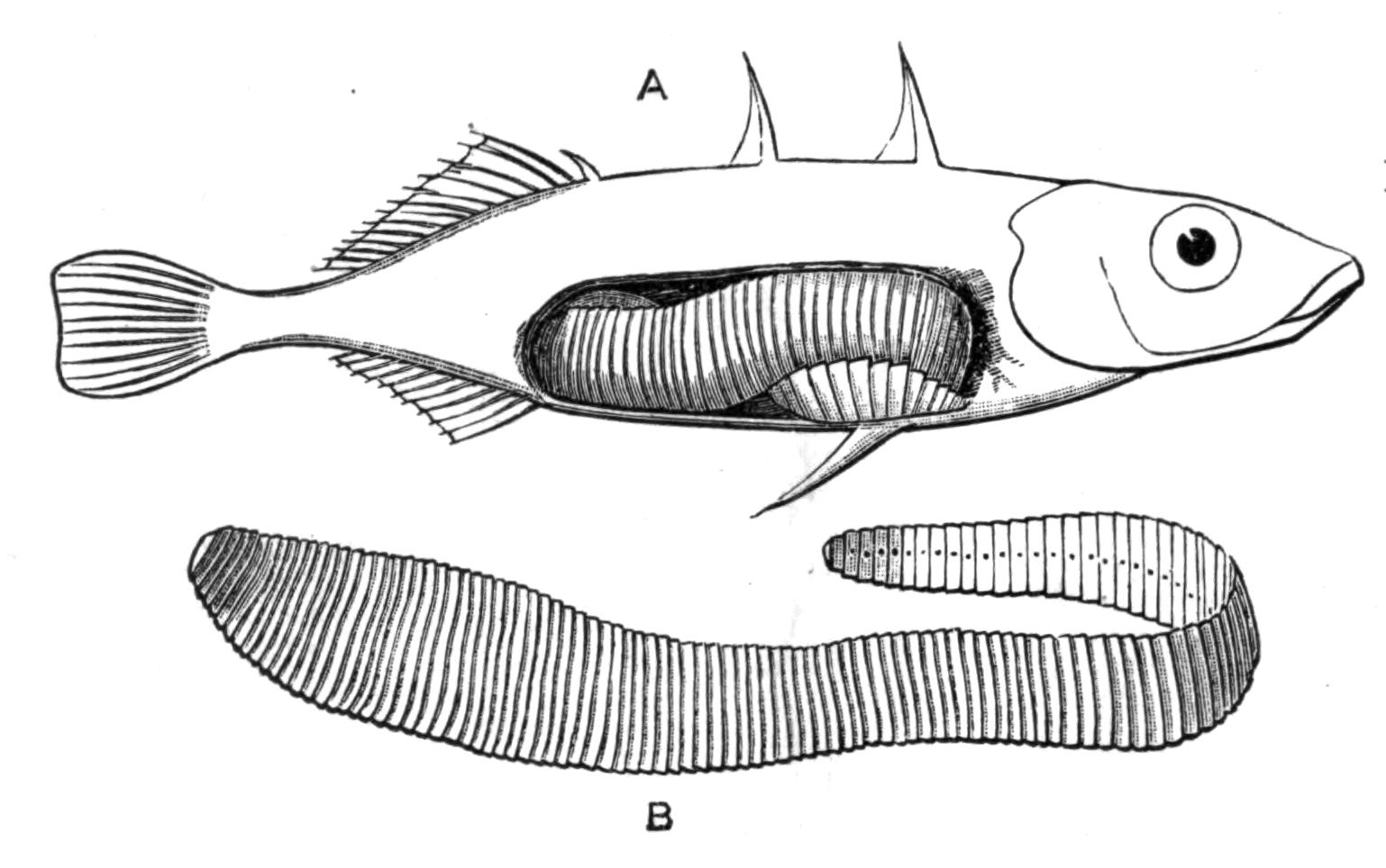
Fig. 40.—A, Stickleback (Gasterosteus aculeatus) infested by an advanced larva of Schistocephalus solidus Crepl. B, The larva. All × 1½. (From specimens in the Cambridge University Museum.)
Structure and Development of Cestoda.[105]—Of the unsegmented Cestodes, Caryophyllaeus mutabilis, from the intestine of carp and other Cyprinoid fishes, is the most easily accessible form. Triaenophorus nodulosus, which is very useful for the study of the excretory system, occurs mature in the pike. In the body-cavity of the Stickleback (Fig. 40) a large, broad, yellow worm may sometimes be found, the larva of Schistocephalus solidus Crepl., which occurs in the intestine of Terns, Storks, Mergansers, and other birds. Species of Ligula are found in the same birds. The intestine of a Lophius or Cyclopterus ("lump-fish") contains, usually, the early and intermediate stages of various Cestodes, while the alimentary canal of Elasmobranchs often contain many peculiar Tetrarhynchidae and other forms. For the study of development, the Taenia anatina from the duck may be used. The ripe proglottides are collected, and the eggs placed with Cypris ovum in an aquarium, with the probability that some of the embryos will enter the Ostracod, and the peculiar Cysticercoid may be bred.[106] Cysticercus pisiformis and Coenurus serialis, which occur commonly in rabbits, are also suitable objects for examination.
A Cestode such as Echinobothrium (Fig. 36) is divisible into head and proglottides. Moniez has suggested that the head is really the morphologically hinder end of the body, in which case the formation of proglottides would closely resemble the mode of segmentation of an Annelid larva. The close similarity, however, between the Cysticercoid larva (Fig. 43, F) and the Cercaria of a liver-fluke, seems to show that the anterior end is the same in both cases, and since it bears the central part of the nervous system, we may reasonably call it the "head." Moreover the hinder end of a Platyhelminth usually possesses the chief excretory pore. Another difficulty is the determination of dorsal and ventral surfaces. Authors are agreed,—on the analogy of Trematodes, in which the testes are usually dorsal and the ovaries ventral,—that the dorsal and ventral aspects of a Cestode are determined by the position of these organs, although the often radially formed "head," the lateral or superficial position of the genital apertures, and the variability of these features, render it a matter of considerable doubt whether "dorsal" and "ventral" are more than useful conventional terms. The suckers and hooks are borne on a muscular cap, the "rostellum," which is only slightly developed in the Ichthyotaeniae. The body is solid, and is divisible into an outer muscular coat—enveloped in a (possibly epidermal) investing membrane—and an inner parenchymatous tissue containing the chief part of the excretory, nervous, and reproductive systems. One or two pairs of longitudinal excretory vessels are present, usually connected by transverse ducts and opening by a single terminal pore. Occasionally a regularly paired arrangement of lateral or secondary pores is present (Figs. 38 and 41, for.sec). Flame-cells occur at the end of the fine tubules (Fig. 38), and the whole system is well developed, but may undergo degenerative changes in the older proglottides. The central nervous system varies according to the degree of differentiation of the rostellum; and, owing to the difficulty of staining the nerves and the contradictory statements of authors, we do not yet possess a fully reliable account of the nervous system even of the commoner Taeniae. Free nerve endings and other sensory terminations have been recently stated to exist in the cuticle of Cestodes and Trematodes. If true, this would tend to show that the parasitic mode of life of these animals demands a complex nervous system comparable with that of the Turbellaria.
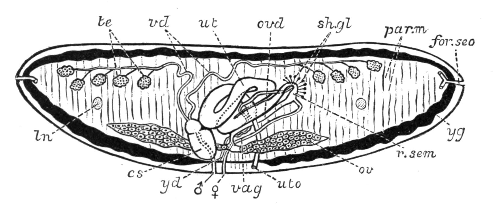
Fig. 41.—Diagrammatic transverse section of Schistocephalus solidus Crepl., from the Wild-duck, illustrative of the Cestodes with uterine aperture (uto). × 12. cs, Cirrus-sac; for.sec, one of the paired lateral openings of the excretory vessels; ln, longitudinal nerve; ov, ovary; ovd, oviduct; par.m, parenchymatous muscles; r.sem, receptaculum seminis; sh.gl, shell-gland; te, testes; ut, uterus; uto, uterine pore; vag, vagina; vd, vasa deferentia; yd, yolk-duct; yg, yolk-glands (black); ♂, male, ♀, female genital aperture. (After Riehm.)
The reproductive organs, unlike the preceding systems, are discontinuous from one proglottis to the next. The male and female organs and their mutual connexions, especially in the unsegmented Cestodes, may be compared in detail with those of Trematodes, but the difference between the arrangement of the generative organs of various Cestodes is very great.[107] The penis (Fig. 41, cs) is evaginated through the male pore (Fig. 41, ♂), and inserted far into the vagina (♀, vag) of the same or another segment of the tape-worm.
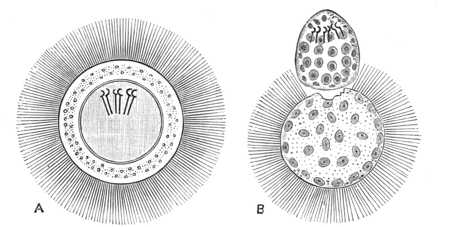
Fig. 42.—A, Free-swimming, six-hooked larva of Bothriocephalus latus Brems. (the broad tape-worm of Man), still enclosed in a ciliated (possibly cellular) double membrane or mantle. In this condition it may continue to live in water for a week or more, but eventually throws off its ciliated coat (as in B) and commences to creep about vigorously by the aid of its hooks, in search of its first host, which is at present unknown. (After Schauinsland.) × 600.
From this fact and the anatomical relations of the vagina, it is becoming increasingly probable that the so-called uterus of Trematodes is an organ corresponding to the vagina of Cestodes, and not to the uterus of Cestodes. The latter opens to the exterior in Schistocephalus, Bothriocephalus, and some other Cestodes of fishes by a special pore (Fig. 41, uto). Through this, some of the eggs (which in these genera give rise to ciliated larvae) are enabled to escape, and need not wait for the detachment of the proglottis, as must happen in the Taeniidae, where the uterus is closed. This uterus, a true physiological one, is probably the homologue of the "canal of Laurer" ("Laurer-Stieda canal," or "vagina") of Trematoda. The fertilised ovum and yolk are brought together into the "ootype," where the shell-gland forms the egg-shell around them (Fig. 41, sh.gl) and the egg is then passed into the uterus. The ovum segments to form a minute six-hooked larva, which may (Bothriidae, Fig. 42) or may not (Taeniidae) be ciliated. Thus in Taenia serrata the proglottides are shed with the faeces of the host (dog), and they protect the young from the desiccating influence of the surroundings. If inadvertently eaten by a rabbit along with herbs, the proglottis and larval envelope are digested, and by its six hooks the tiny larva bores through the gastric wall into the portal vein, and so into the liver. Here the hooks are thrown off, and the solid mass of cells becomes vacuolated.
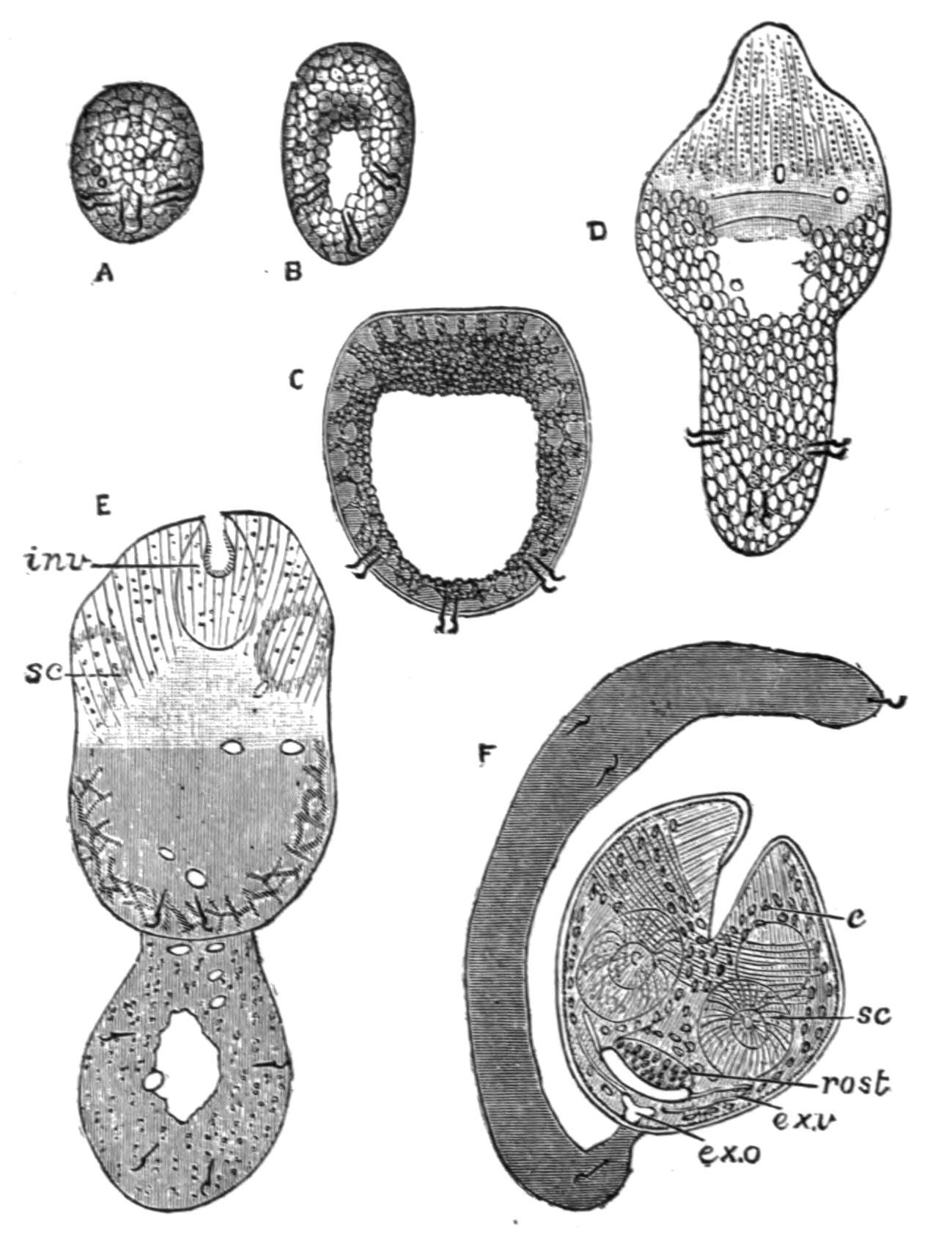
Fig. 43.—Stages in the development of Dipylidium caninum L. (= Taenia elliptica Batsch, T. cucumerina Bloch), the commonest of the Dog-Taeniae; compare Fig. 44. A, Six-hooked larva (now often spoken of as an "Onchosphaera"); B, larva elongating; formation of a central lacuna; C, larva further advanced; D, distinction between body and tail is visible; E, invagination of the rostellum is commencing; F, Cysticercoid larva with four suckers, invaginated rostellum, and excretory vessels. c, Calcareous concretions in cells of the larva; ex.o, excretory aperture; ex.v, excretory vessels; inv, invagination commencing; rost, rostellum; sc, suckers. (After Grassi and Rovelli; highly magnified.)
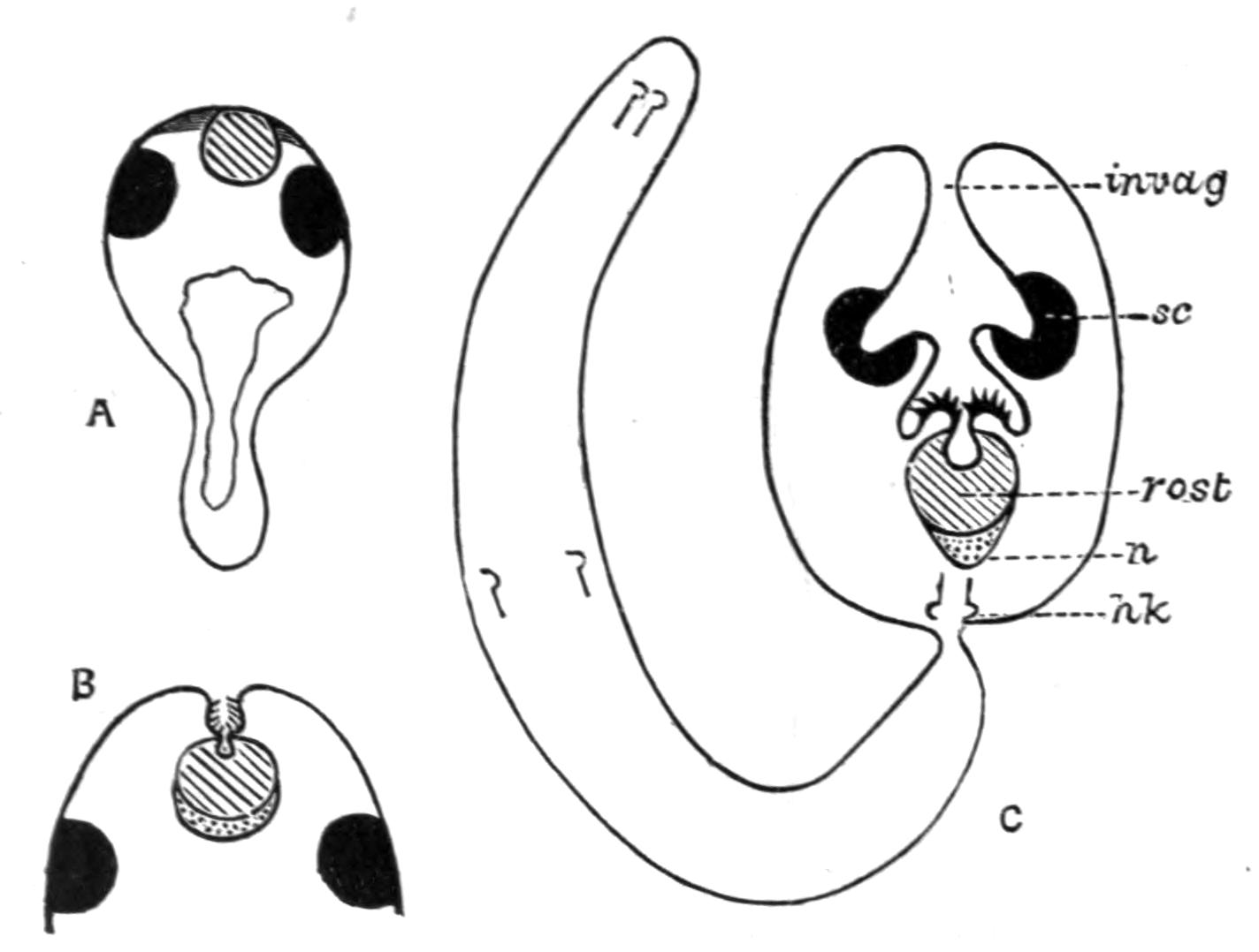
Fig. 44.—Schematic longitudinal sections through the larvae of Dipylidium caninum L. All these stages are passed in the body-cavity of the Dog-flea (Pulex serraticeps). (Compare Fig. 43 for further details.) A, Six-hooked larva with developing rostellum (shaded) and suckers (black). In this species the invagination (C, invag.) occurs after the formation of these organs, and not, as in most Taeniae, before it. B, Invagination commencing; the hooks are developing above the rostellum, while beneath it the nervous system (dotted) is seen. C, The invagination has now carried the suckers inwards. The tail has become distinct, and the whole larva at this stage is known as a Cysticercoid. hk, Larval hooks; invag, mouth of the invagination; n, central nervous system; rost, rostellum and hooks; sc, suckers, of which only two can be seen in a longitudinal section; four are really present. (After Grassi and Rovelli.)
At one pole an invagination occurs, at the bottom of which the rostellum, suckers, and hooks are gradually formed, but inside out as compared with the head of the Taenia serrata. At this stage the larva (Cysticercus pisiformis) has usually issued from the liver and attached itself to the omentum. The invagination projects into the cavity of the bladder, within which a watery fluid accumulates. Thus the "bladder worm" is formed, the head of which is evaginated if the larva be introduced into the digestive system of a dog. The bladder and neck of invagination are digested, while the head, protected by these, remains, and forms the neck, from which the proglottides are afterwards segmented off. In Taenia (Hymenolepis) murina the whole development may take place in the parental host, the larva living in the villi, the adults in the cavity of the same rat's intestine (Grassi). The different forms of Cestode larvae depend largely upon the presence and degree of development of the caudal vesicle or bladder, which in Scolex polymorphus (Fig. 38) (the young stage of Calliobothrium filicolle Zsch.) is practically absent. If the bladder be small, the larva is known as a Cysticercoid. For example, the common Dipylidium caninum, which lives in the dog, has such a larva, the development of which is explained and illustrated by Figs. 43 and 44. The bladder becomes exceeding capacious in Coenurus and Echinococcus.
Table for the Discrimination of the more usual Cestodes of Man and Domestic Animals.[108]
I. Scolex in most cases with hooks; uterus with a median and lateral branches; yolk-glands simple, median; genital pore single; dorsal excretory vessel narrower than the ventral, without a circular commissural trunk; eggs without pyriform apparatus (processes of the ovarian membrane)
Gen. Taenia L. (s. str.)
A. Genital ducts pass on the ventral side of the nerve and of the two longitudinal excretory vessels T. crassicollis Rud.
A. Genital ducts pass on the ventral side of the nerve and of the two longitudinal excretory vessels
T. crassicollis Rud.
B. Genital ducts pass between the dorsal and ventral longitudinal vessels.
a. Nerve present on dorsal side of genital ducts.
α. Head armed T. solium Rud.
β. Head unarmed T. saginata Goeze.
b. Nerve on ventral side of genital ducts.
Dog-Taeniae[109] Head armed; genital pore marginal and — Single
Many proglottides; strobila several centimetres long; small hooks with guard.
Bifid hooks, which are — 230µ-260µ long[110]; genital pore very distinct T. serrata Goeze.
Bifid hooks, which are — 230µ-260µ long[110]; genital pore very distinct
T. serrata Goeze.
— 136µ-157µ long; genital pore not very salient T. serialis Ball.
— 136µ-157µ long; genital pore not very salient
T. serialis Ball.
Entire large hooks, which are — 180µ-220µ long; length of mature segments double their width T. marginata Batsch.
Entire large hooks, which are — 180µ-220µ long; length of mature segments double their width
T. marginata Batsch.
— 150µ-170µ long; length of mature segments treble their width T. coenurus Küch.
— 150µ-170µ long; length of mature segments treble their width
T. coenurus Küch.
3-4 segments; a few mm. long T. echinococcus v. Sieb.
3-4 segments; a few mm. long
T. echinococcus v. Sieb.
— Double and bilateral Dipylidium caninum L.
Head unarmed; two genital pores on ventral surface Mesocestoides lineatus Goeze.
Head unarmed; two genital pores on ventral surface
Mesocestoides lineatus Goeze.
II. Scolex without hooks; one or two transverse uteri present; one or two genital pores and yolk-glands, the latter never median; genital ducts pass on the dorsal side of the nerve; eggs with pyriform apparatus.
A. One transverse uterus present.
a. Uterus with bullate egg-sacs; pyriform apparatus without horns; genital ducts between dorsal and ventral vessels
Thysanosoma Dies.
α. Head large (1.5 mm.); square lobed testes in median field; posterior margin of segments fimbriated; genital pore double
T. fimbriata Dies.
β. Head small; no fimbriae; pore rarely double T. giardii Riv.
β. Head small; no fimbriae; pore rarely double
T. giardii Riv.
b. Uterus without saccular dilatations; segments short, thick, and slightly imbricate Anoplocephala E. Blanch.
b. Uterus without saccular dilatations; segments short, thick, and slightly imbricate
Anoplocephala E. Blanch.
Horse-Taeniae. α. Head very large — No posterior lobes A. plicata Zed.
— Four posterior lobes A. perfoliata Goeze.
β. Head small, without posterior lobes A. mamillana Mehl.
β. Head small, without posterior lobes
A. mamillana Mehl.
B. Two uteri and two genital pores present; horns of pyriform apparatus well developed; genital ducts pass on the dorsal side of the longitudinal vessels
Moniezia R. Bl.
a. Interproglottidal glands[111] arranged in linear series (planissima group)
M. planissima S. and H. M. benedeni Mz. M. neumani Mz.
b. Interproglottidal glands saccular (expansa group)
M. expansa Rud. M. oblongiceps S. and H. M. trigonophora S. and H.
c. Interproglottidal glands absent (denticulata group) M. denticulata Rud. M. alba Perr.
c. Interproglottidal glands absent (denticulata group)
M. denticulata Rud. M. alba Perr.
C. Uterus single or double, without spore-like egg-sacs; eggs with a single shell; genital pores irregularly alternate; strobila narrow; testes absent from median part of the field
Stilesia Raill.
a. A transverse uterus in middle part of median field; head 2 mm. diameter S. centripunctata Riv.
a. A transverse uterus in middle part of median field; head 2 mm. diameter
S. centripunctata Riv.
b. Two lateral uteri in each segment; head less than 1 mm. in diameter S. globipunctata Riv.
b. Two lateral uteri in each segment; head less than 1 mm. in diameter
S. globipunctata Riv.
III. Scolex almost invariably provided with hooks; genital pores on left border of segment; eggs with three shells but no cornua. Segments broader than long; posterior angles salient.
Hymenolepis Weinl.
a. Scolex with a single series of 24-30 hooks, each 14-18µ long
H. nana v. Sieb. H. murina Duj.
b. Scolex very small, unarmed H. diminuta Rud.
IV. Scolex provided with two elongated muscular pits. Body segmented; three genital apertures in middle of ventral surface
Bothriocephalus Rud.
Body 2-20 metres in length
B. latus Brems. B. cristatus Dav. (doubtful species). B. cordatus Leuck. B. mansoni Cobb. (= B. liguloides Leuck.)
Classification of Cestodes.—The following classification, which, so far as the Taeniidae are concerned, follows that employed by Railliet, Blanchard, and most recent writers, includes only a few representative genera:—
1. Fam. Cestodariidae Mont. (Monozoa Lang).
Gen. Caryophyllaeus, Archigetes, Gyrocotyle, Amphilina.
2. Fam. Bothriocephalidae.
Sub-Fam. 1. Bothriocephalinae. Gen. Bothriocephalus, Schistocephalus, Triaenophorus (= Tricuspidaria).
Sub-Fam. 2. Ligulinae. Gen. Ligula.
Sub-Fam. 3. Solenophorinae. Gen. Solenophorus, Duthiersia.
Sub-Fam. 4. Diphyllinae. Gen. Echinobothrium.
3. Fam. Tetrarhynchidae.
Gen. Tetrarhynchus.
4. Fam. Tetraphyllidae.
Sub-Fam. 1. Phyllobothrinae. Gen. Phyllobothrium, Echeneibothrium, etc.
Sub-Fam. 2. Phyllacanthinae. Gen. Calliobothrium, Anthobothrium, etc.
5. Fam. Taeniidae.
Sub-Fam. 1. Cystotaeninae. Gen. Taenia s. str.
Sub-Fam. 2. Anoplocephalinae. Gen. Moniezia, Thysanosoma, Stilesia, Anoplocephala.
Sub-Fam. 3. Cystoidotaeninae. Gen. Dipylidium, Hymenolepis, Drepanidotaenia, Dicranotaenia, Echinocotyle, Davainea.
Sub-Fam. 4. Mesocestoidinae. Gen. Mesocestoides, Dithyridium.
Sub-Fam. 5. Ichthyotaeninae. Gen. Ichthyotaenia, Corallobothrium.