Chapter VI
Reading edition
NEMATHELMINTHES
INTRODUCTION—NEMATODA—ANATOMY—EMBRYOLOGY—CLASSIFICATION—ASCARIDAE—STRONGYLIDAE—TRICHOTRACHELIDAE—FILARIIDAE—MERMITHIDAE—ANGUILLULIDAE—ENOPLIDAE—PARASITISM—NEMATOMORPHA—ANATOMY—CLASSIFICATION—LIFE-HISTORY—ACANTHOCEPHALA—ANATOMY—EMBRYOLOGY—CLASSIFICATION.
The Nemathelminthes include three sub-Orders of very different size and importance. These are—
i. The Nematoda. ii. The Nematomorpha (Gordiidae). iii. The Acanthocephala.
Although the members of these groups differ considerably from one another, on the whole there is a closer resemblance between them than between any one of them and any other group of animals, and there is a certain convenience in arranging them under one head.
The following characteristics are common to all three groups of the Nemathelminthes: they are worm-like in form, and with few exceptions are parasitic in the bodies of other animals, either Vertebrate or Invertebrate. Some of them spend their whole existence within the bodies of their hosts, but more commonly they are only parasitic during a certain period of their life; a few, however, lead a free life in water or in damp earth. None of the Nemathelminthes are segmented—that is, their bodies are not divided into a number of parts which serially repeat each other, and which resemble more or less closely the preceding and succeeding parts. They are not provided with any appendages or limbs, but sometimes bear a few bristles or hooks, and in rarer cases suckers. The body, which is elongated and, as a rule, thread-like and tapering at each end, is enclosed in a thick cuticle or hardened secretion of the underlying cells. In no Nemathelminth is there any closed vascular system, nor are special respiratory organs developed.
In many respects the most remarkable peculiarity of these animals is that, with the possible exception of the excretory organs of the Acanthocephala, there is a complete absence of cilia throughout the whole group. In this respect they resemble the Arthropoda. The universal presence of these small flickering processes of cells from man down to the simplest unicellular organisms makes the absence of these structures most remarkable. In many animals they are the sole organs of locomotion, and in almost all they perform very important functions, both in bringing food and oxygen to the body, and in removing waste matter from it. At present there seems to be no adequate explanation for their absence in the two large groups mentioned above.
Nemathelminthes are, with hardly an exception, dioecious—that is to say, their male and female reproductive organs are in different individuals. Their young do not differ markedly from the adults, except in the absence of sexual organs, but the immature stages are usually termed larvae, and not infrequently either inhabit a different host from the adult, or are free when the adults are parasitic, or vice versâ.
Sub-Order I. Nematoda.
Anatomy.—The Nematode worms, or thread-worms, form by far the largest and most important division of the group Nemathelminthes. The number of species is great, and although the conditions under which they live are of the most varied kind, there is, as a rule, little corresponding difference in structure, and hence the determination of the species is attended with no small difficulty.
With few exceptions the shape of the body is filiform (Figs. 66 and 71), the two ends being more or less pointed, and the posterior end of the male, which is generally a smaller animal than the female, is usually slightly recurved. The worms are, as a rule, white, or of the colour of polished ivory; they may be opaque or semi-transparent, but pigment spots are rarely developed.
Minute Nematodes abound in moist soil, around the roots of plants, etc., and may easily be detected with the aid of a lens wriggling about amongst the particles of sand and earth. Of the animal parasites perhaps the most familiar is the "round worm" (Ascaris lumbricoides, Figs. 66 and 67), which inhabits the alimentary canal of man; others are common in domesticated animals, as A. mystax in the cat and dog, and A. megalocephala in the horse and ox. They are also found living parasitically in plants (Fig. 77), causing the formation of galls and other pathological growths; Anguillula (Tylenchus) tritici causes in this way considerable damage to corn, and others attack root-crops, cabbages, etc. The "vinegar eel" (Anguillula aceti), which occurs so often in weak vinegar, is another familiar example of this group.
The Skin.—The body of the worm is encased in a relatively thick, transparent, smooth cuticle, which is turned in at the various apertures, and lines the tubes connected with them for a greater or less distance. The cuticle is in some cases raised to form spikes or hooks, and in certain species, e.g. Ascaris mystax and A. transfuga, it is produced into two lateral fins, which are supported by a thickened triradiate rod of specialised cuticle (Fig. 62); these fins, however, do not run far down the body. As a rule the cuticle is quite smooth, but it may be ringed, as in Filaria laticaudata and in F. denticulata; and the rings may bear backwardly-projecting teeth.
The skin of Nematodes consists of three layers—(i.) the above-mentioned cuticle, which is presumably secreted by (ii.) the sub-cuticle or epidermis which underlies it; the latter surrounds in its turn (iii.) the muscular layer.
The nature of the sub-cuticle is one of the debateable points in the morphology of the Nematoda. No cell outlines have been detected in it, although nuclei are scattered through it; it is in fact a syncytium, or protoplasmic mass in which cell limits cannot be distinguished. Many of the cells forming it have broken down into fibrils, and these form a close meshwork, which is occasionally specialised, as, for instance, round the nerve-cords. Along the median dorsal and ventral lines, and along the lateral lines, this tissue is heaped up in such a way as to divide the enclosed muscle-cells into four quadrants. These thickenings surround dorsally and ventrally a specialised nerve-cord, and laterally the excretory canals.
According to Jammes[155] this lack of differentiation in the sub-cuticular layer is caused by the early appearance of the cuticle, which he thinks is necessitated, at any rate in many of the parasitic forms, by the action which the digestive juices of the host would have on the otherwise unprotected body-wall.
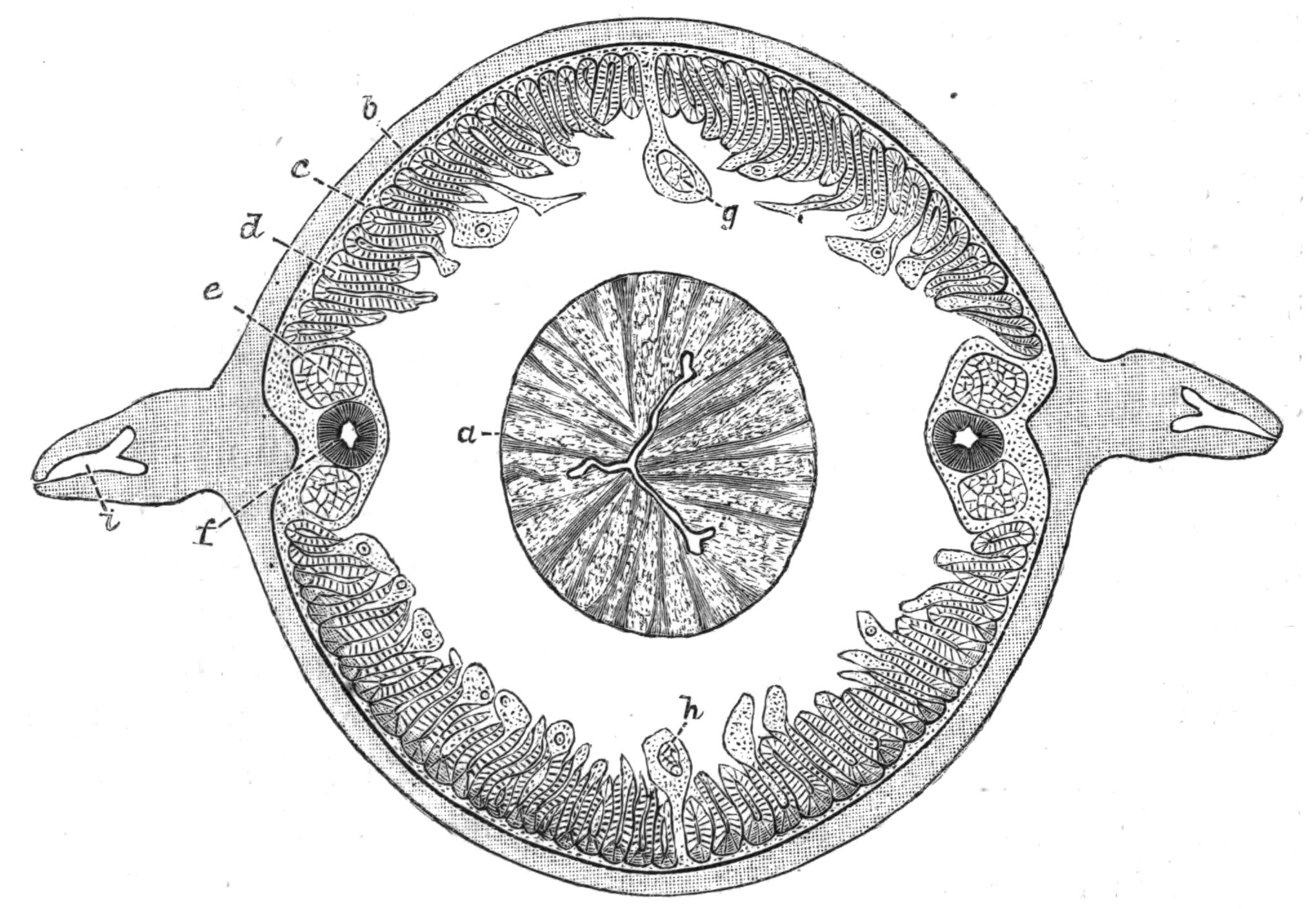
Fig. 62.—A transverse section through the body of Ascaris transfuga Rud., in the region of the oesophagus: a, the muscular oesophagus with its triradiate lumen; b, the cuticle; c, the sub-cuticle; d, the muscular layer; e, the lateral nerves running in the lateral line; f, the excretory canal; g, the dorsal, and h, the ventral nerve; i, the triradiate rod in the fin.
The nervous system, according to the same writer, is of the same nature as this sub-cuticular tissue, only it is more differentiated, or perhaps we should say it has retained more of the primitive cellular character of the embryonic tissue. The fibres of the sub-cuticular tissue are closely connected with the fibrils which compose the spongioplasm (Fig. 64, d) of the muscles,[156] and form also the sheaths of the various nerves; in fact the passage of these fibrils into the nerves is so gradual that it is impossible to make any separation between them.
The Nervous System.—The central organ of the nervous system is the circumoesophageal ring which surrounds the pharynx, close to the anterior end of the body, in A. megalocephala 1½ to 2 mm. behind the mouth.[157] Ganglion cells are found in the ring, but they are not numerous, and are chiefly aggregated round the points of origin of the nerves.
Six short nerves, three on each side of the median line, run forward from the ring, a pair of these ending in each of the three papillae which surround the mouth.
Behind, the nerve-ring gives off six main nerve trunks, of which the dorsal and ventral nerves are usually the largest. These run in the median dorsal and ventral thickenings of the sub-cuticular tissue, and are connected one with another by numerous fine lateral branches running through the sub-cuticle.
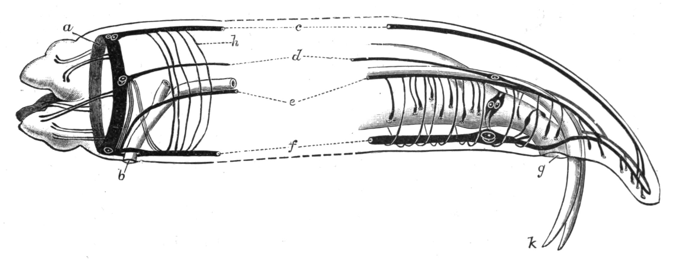
Fig. 63.—Diagram of the nervous system at the two ends of the body in Ascaris megalocephala Cloq., ♂. (After Hesse.) a, Circumoesophageal nerve-ring; b, opening of excretory ducts; c, dorsal nerve; d, dorso-lateral nerve; e, ventro-lateral nerve becoming the bursal nerve posteriorly; f, the ventral nerve; g, cloacal opening; h, sub-cuticular nerves running from c to f; k, spicules.
The lateral nerves, which consist of two or four bundles, one or two lying dorsal and one or two ventral to each excretory canal, have a double origin. The dorsal branches arise directly from the nerve-ring, and at their point of origin there is a considerable accumulation of ganglion cells, from which two commissures on each side run into the ventral nerve (Fig. 63, f). The ventral branches arise from the ventral nerve-cord immediately in front of the excretory pore. At the posterior end the lateral nerves pass into the two branches into which the ventral nerve divides. Just before the point where the ventral nerve splits it swells out into an anal ganglion situated just in front of the anus. In the male[158] this anal ganglion gives off two lateral nerves which pass round the cloaca and form a ring, and in this sex the ventro-lateral nerve, which is much strengthened by fibres from the ventral nerve, and has received, owing to the mistaken impression that it was a special nervus recurrens, the name of the "bursal nerve," gives off numerous branches to the sense papillae which are found in this region of the body and on the tail. The arrangement of these parts is shown in Fig. 63.
Sense organs are but poorly developed in the Nematoda, as is usual in animals which are, as a rule, either parasitic or live underground. Eyes, consisting of masses of dark pigment with or without a lens, occur in the neighbourhood of the circumoesophageal nerve-ring in some free-living forms. Leuckart described as possible auditory organs certain giant-cells lying near the orifice of the excretory ducts. Later research has shown these cells to have some phagocytic action on the contents of the body-cavity. The chief sense organs are the papillae, of which in A. megalocephala there are two kinds, the lip papillae being distinguished from the genital papillae by the fact that the nerve supplying them ends in a fine point and pierces the cuticle in the former case, whilst in the latter it swells out into an "end-organ," which is always covered by a layer of cuticle, though sometimes by a very thin one.
Muscular System.—The muscular system is one of the most characteristic features of the Nematoda, both as regards the histology of the muscle-cells and the way in which the cells are arranged.
Each muscle-cell is of considerable size, and is of the shape of a somewhat flattened spindle produced into a process near the middle. Each end of the spindle cell is said to be continuous with the fibrils of the sub-cuticular layer.[159] The muscle-cell consists of two portions, a contractile part which lies next the sub-cuticle, and which usually, to some extent, wraps round the second or medullary half. The latter consists of a fibrillar spongioplasm, in the meshes of which lies a clear structureless hyaloplasm. The nucleus always lies in the medullary half.
The contractile portion consists of a number of columns, very regularly arranged in two rows and close together, but allowing sufficient space between adjacent columns for fibrils of the spongioplasm to penetrate; and these become continuous with the fibrils of the sub-cuticle, which is thus intimately connected with both nervous and muscular systems.
The medullary portion of the cell varies greatly in size; it may stretch far into the body-cavity, which may be thereby almost occluded, or it may be flattened out, leaving a large space around the alimentary canal. At one point, usually about its middle, it is produced into a process, which bends inwards towards the dorsal or ventral nerve-cord, and by means of this process the muscle receives its nerve supply.
In most Nematodes there are numerous muscle-cells to be seen in any transverse section, forming a layer within the sub-cuticle, and broken up into four quadrants (Fig. 62) by the projection of the dorsal, ventral, and lateral thickenings of the sub-cuticular tissue. In some genera, however, such as Oxyuris, Strongylus, Pelodera, Leptodera, etc., there are but eight muscle-cells in a row, two in each quadrant. Such genera are classed together by Schneider,[160] and termed Meromyarii (vide p. 137).
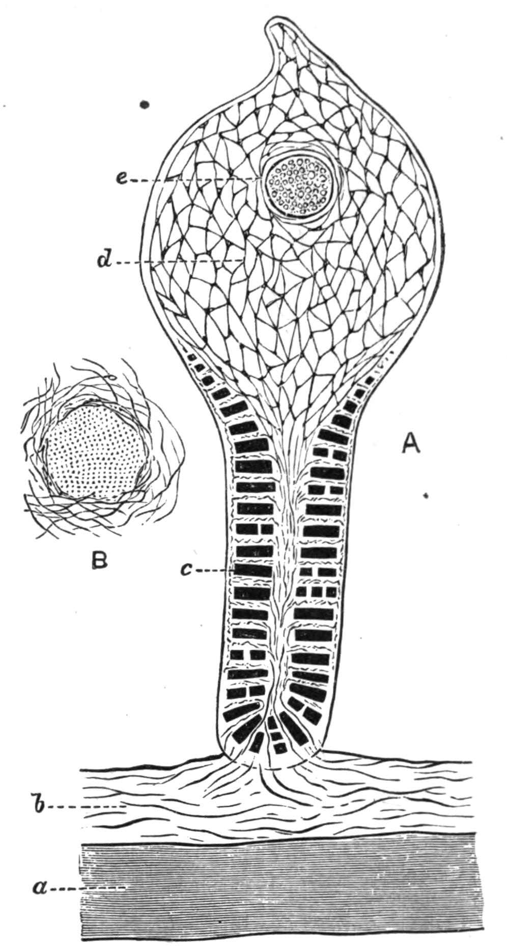
Fig. 64.—A, transverse section through the centre of a muscle-cell; B, the same through a nerve fibre showing the sub-cuticular fibres running into the sheath. (After Rohde.) a, Cuticle; b, sub-cuticular fibres continuous with d; c, contractile columns; d, network of spongioplasm; e, nucleus.
In addition to the characteristic muscles of the body-wall there are others, such as those which move the spicules in the male, which cross the body-cavity obliquely near the anus, and such as sphincter muscles near the latter orifice, which have not the characteristic arrangement of contractile and medullary parts described above.
The Body-Cavity.—The skin of a Nematode, as described above, contains most of the important organs of the body within its thickness. The chief muscular system, the nervous system with its sense organs, and the excretory organs are all embedded in or form part of the skin, which in its turn encloses a cavity—the body-cavity—in which the other two systems of organs which are found in Nematodes lie. These are the digestive system and the reproductive system.
The body-cavity is continuous from one end of the animal to the other, and is in no case divided up into compartments by the presence of septa or mesenteries. It contains a coagulable fluid with numerous corpuscles; this is, as a rule, colourless, but in Syngamus trachealis Sieb. (Fig. 70), which lives on blood, the haemoglobin of its host tinges it red, though the colour is said to disappear if the parasite be isolated and starved.
The morphological nature of this body-cavity affords an interesting problem. It is not a true coelom, such as exists in the earthworm, since it is not surrounded by mesoderm, nor do the excretory organs, with the possible exception of one or two genera, open into it, nor do the generative cells arise from its walls. Essentially it is a space between the mesodermic muscle-cells which line the skin and the endodermic cells of the alimentary canal, and although in many of its functions it resembles the coelom of other animals, its morphological character is quite different.
There are no respiratory or circulatory organs in the Nematoda; possibly the fluid in the body-cavity acts, to some extent, as a carrier of oxygen, but from the inert and almost vegetative life of these animals it seems probable that their respiratory processes are slow, and in fact Bunge[161] has shown that Ascaris mystax, found in the intestine of the cat, will live for four or five days in media quite free from oxygen, and that A. acus from the pike will live and exhibit movements in the same media for from four to six days.
The Digestive System.—The mouth of the Nematoda is usually anterior and terminal, and is surrounded by from two to six projecting lips, the most common number being three. These lips are well provided with sense papillae. The mouth leads into an alimentary canal, which with hardly an exception runs straight through the body to the anus without twists or loops. The anus is usually placed ventrally and is not terminal, but in Trichina and Trichocephalus it is at the end of the body, and in Mermis, where the several parts of the alimentary canal are said not to communicate, it is absent altogether. Ichthyonema, Dracunculus, Allantonema, Atractonema, and other Filariae are also aproctous.
The alimentary canal is divisible into three parts—(i.) the oesophagus, (ii.) the intestine, and (iii.) the rectum. The suctorial oesophagus is a very muscular, thick-walled tube, lined with cuticle continuous with that which covers the body, and like it cast from time to time. Its lumen is usually much reduced, and is almost invariably triangular or triradiate in section (Fig. 62). In many genera the hinder end of the oesophagus is swollen into a muscular bulb, which is armed with teeth in Heterakis, Oxyuris, Pelodera, Leptodera, etc. Other species, such as Tylenchus, Aphelenchus, Dorylaimus, are armed with a spear, which in Onyx,[162] a genus recently described and allied to the last named, is borne on a special bulb. The use of the spear is to pierce the tissue upon the juices of which the animal lives. A gland lies embedded in the thick walls of the oesophagus, and opens into its lumen by a fine tube. This was first described by Schneider[163] in A. megalocephala, and more recently it has been found by Hamann[164] in a number of Ascaridae and Strongylidae from the Adriatic, and also in Lecanocephalus.
With a few exceptions, such as Mermis, where it is blind, the oesophagus opens posteriorly into the intestine. This is a somewhat flattened tube, whose shape and position are often altered by the development of the generative organs. Its wall consists of a single layer of columnar cells, with large nuclei coated internally and externally by a layer of cuticle. The inner layer of cuticle is usually perforated by very numerous minute pores. In some species the intestine is degenerate, in Mermis it is a closed tube opening neither into the oesophagus nor into the rectum; in Trichina spiralis and in the larva of Tylenchus tritici it consists of a single row of cells perforated by a duct, but in the adult of the last named there are many cells in a transverse section.
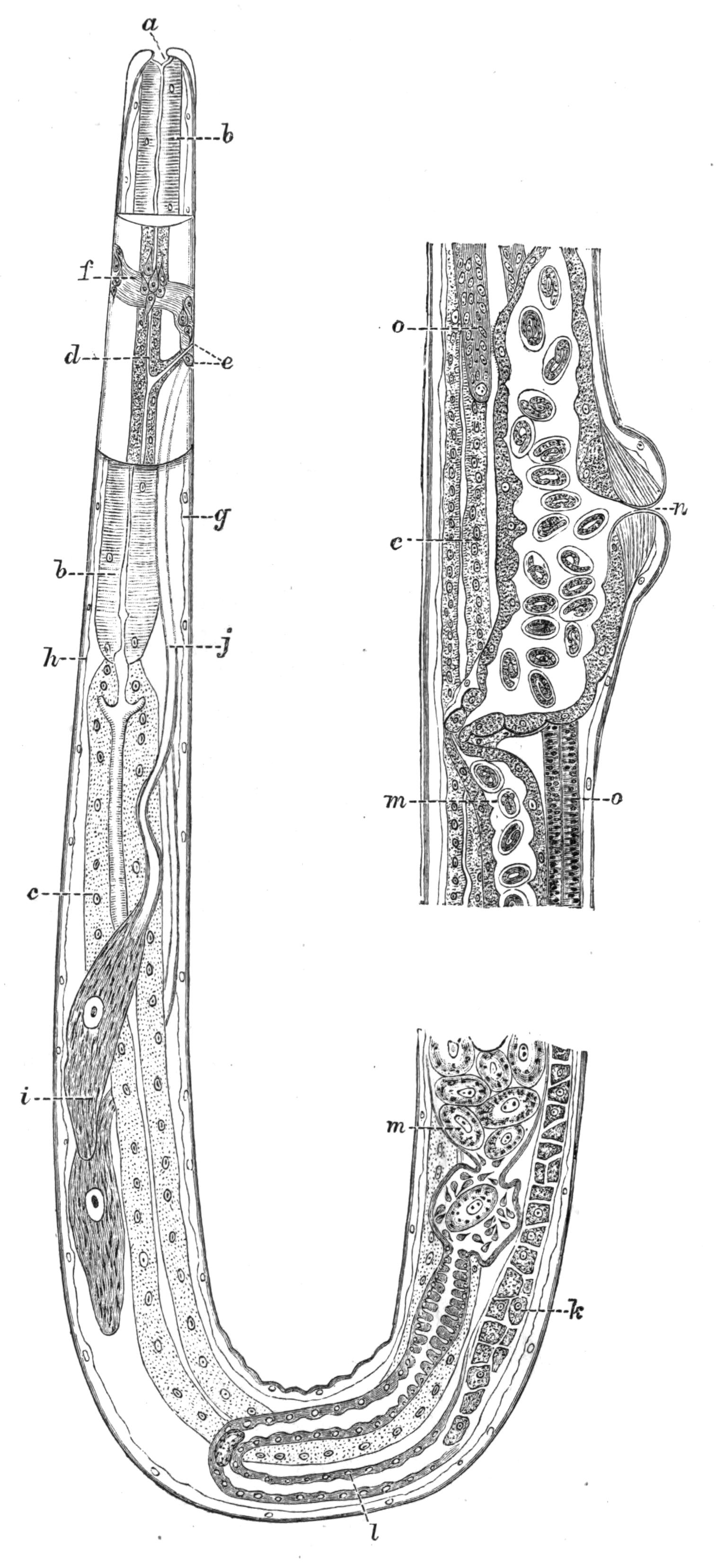
Fig. 65.—A longitudinal section through the body of Strongylus filaria Rud. (From O. Augstein.[165]) A portion of the body, on each side of the excretory pore, is seen in optical section. a, Mouth; b, oesophagus; c, intestine; d, excretory canal; e, excretory pore, and the opening of the poison glands, i; f, circumoesophageal nerve-ring; g, ventral nerve; h, dorsal nerve; i, unicellular poison glands; k, ovary, with the ova separate; l, oviduct; m, uterus, the first egg in the uterus is surrounded by spermatozoa; n, opening of uterus; o, inner end of ovary with the ova undifferentiated.
In some genera, Leptodera and Pelodera, the lumen of the intestine at any one level is bounded by two horseshoe-shaped cells, but by far the commonest arrangement is a tube formed of fairly numerous columnar cells crowded with granules and with large nuclei.
The rectum is usually short; its cuticular lining, like that of the oesophagus, is cast at intervals. At its anterior end there is usually a sphincter muscle, and its walls are divaricated by muscular strands which run from it to the body-wall. The anus is a transverse slit, which in the male Strongylidae is surrounded by a funnel-shaped membrane.
The food of Nematodes seems to be almost entirely fluid, and consists, at any rate in the parasitic forms, of the elaborated juices of their hosts. Little is known about the nutriment of the free-living forms.
The Excretory System.—The excretory organs are peculiar, and, like many other Nematode structures, do not fall readily into line with what is known of similar organs in other animals. They consist of two canals embedded in the lateral thickenings of the sub-cuticular tissue. The canals end blindly behind, but near the anterior end of the body they bend inwards, and after uniting, open by a common pore situated in the middle ventral line, a little way behind the mouth. The lateral canals are in some cases continued in front of the transverse branch, and they then end blindly in the head. The walls of these canals consist of an internal, structureless, refractive layer surrounded by a granular layer with nuclei. They contain a fluid, but nothing is known of its composition.
An interesting divergence from the usual form of excretory organ has been described by Hamann[166] in the genus Lecanocephalus. Here there is only one canal, the right; anteriorly this bends towards the ventral surface and opens by a small median pore close behind the nerve-ring. Posteriorly the canal does not extend much beyond the middle of the body, where it forms a coiled mass, and diminishing in size, opens into the body-cavity. The same author also states that both canals in Dochmius have a similar internal opening; these observations, if confirmed,[167] show a conformity to the ordinary structure of excretory organs which was not supposed to exist in the lateral canals of the Nematoda.
The Reproductive Organs.—With the exception of the genera Angiostomum, Pelodytes, and of Rhabdonema nigrovenosum, which are physiologically hermaphrodite and self-impregnating, the Nematodes have separate sexes. The males are, as a rule, smaller than the females, and may usually be distinguished by the posterior end of the body being curved towards the ventral surface; a genital bursa, and one or more spicules are often found in this sex. Further, the position of the genital opening differs; in the male the vas deferens opens on the ventral surface of the rectum close to the anus, but the oviduct in the female opens in the ventral middle line, usually near the middle of the body, but sometimes close behind the excretory pore, or in some Strongylidae just in front of the anus. The tail of the male bears very numerous papillae, which are of considerable systematic importance.
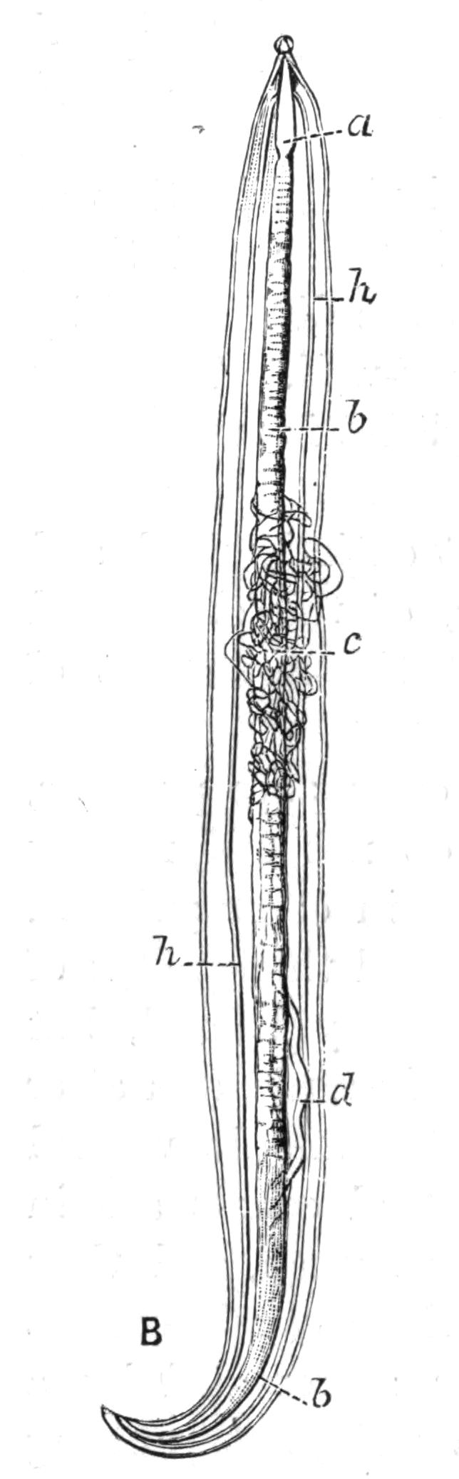
Fig. 66.—Ascaris lumbricoides Cloq. ♂, natural size, cut open along the dorsal middle line. a, Oesophagus; b, intestine; c, testis; d, vas deferens; h, lateral excretory canals.
With rare exceptions, e.g. Filaria attenuata, where it is double, the male reproductive organ consists of a single tube divisible into a testis proper, a vas deferens, a vesicula seminalis, where the spermatozoa are stored up, and a ductus ejaculatorius. The tube stretches through the body in a straight line in the small free-living forms, but is thrown into loops and coils in the larger parasitic Nematodes. Within the testis the mother-cells of the spermatozoa are attached to a rhachis or axial cord; the mother-cells divide, and their products ultimately form spermatozoa. The latter have a very peculiar shape; in accordance with the universal absence of cilia in the Nematoda the spermatozoon has no flagellum, and at first consists of a spherical nucleated cell, on one side of which a cap or covering of some refractive substance appears. The cap elongates and becomes conical, whilst the protoplasmic portion of the spermatozoon throws out pseudopodia and becomes amoeboid, but ultimately rounds itself off again. The spermatozoa do not attain maturity until they reach the uterus of the female.
The internal female reproductive organs are, with few exceptions (Trichina, etc.), double, but the vagina, which is lined with cuticle continuous with that covering the body, is always single. They are usually much coiled, and may be divided into ovary, oviduct, and uterus. The ova arise from a polynucleated mass of protoplasm or syncytium (Fig. 65, o) at the upper end, and acquire distinctness as they approach the oviduct. Fertilisation takes place in the uterus, but the segmentation may not begin until some time after the eggs are laid; in Dochmius, however, it is well advanced at this period, and in many genera, e.g. Pseudalius, Trichina, Dracunculus, etc., the whole development of the larva takes place in the body of the mother.
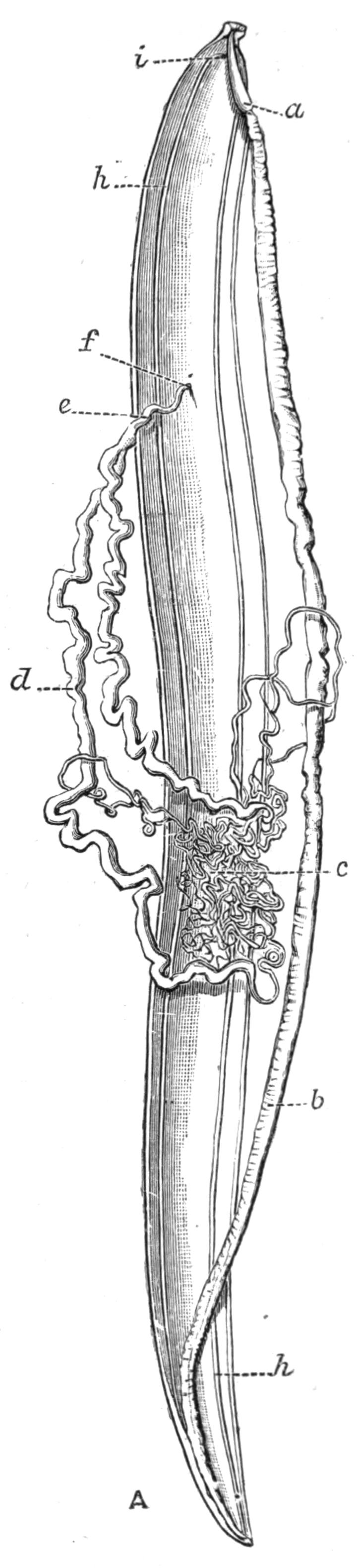
Fig. 67.—Ascaris lumbricoides Cloq. ♀, natural size, cut open along the median dorsal line to show the internal organs. a, The muscular oesophagus; b, the intestine; c, the ovary; d, the uterus; e, the vagina; f, the external opening; h, the excretory canals; i, their opening.
Embryology.—The eggs of many of the parasitic forms require a considerable degree of warmth to develop. Those of Ascaris lumbricoides require a temperature of 20° C., those of Trichocephalus 22.5° C., and those of Oxyuris vermicularis, 40° C. The latter develop in a few hours, the eggs of Dochmius in a few days, whilst those of A. lumbricoides take weeks or even months, and the young of Trichocephalus seldom develop within a year.[168] The ova only develop in a damp atmosphere, and they can be arrested at almost any stage, and for considerable periods, by desiccation.
Our knowledge of the processes by which the fertilised egg-cell develops into the larva is very imperfect. As a rule the segmentation is complete and equal; it results in the formation of a blastula, which may take the form of a hollow sphere of cells—A. megalocephala—or the cavity may be reduced, and the blastula may consist of a double-layered plate, as in Cucullanus.[169] The distinction into cells which will form the three embryonic layers, the ectoderm, mesoderm, and endoderm, is very early evident,—in the eight-cell stage. By the growth of one side of the blastula and the tucking in of the other the blastula becomes converted into a gastrula, which is a two-layered stage with a cavity opening to the exterior by a pore termed the blastopore. In Nematodes the blastopore is elongated and slit-like; it either forms the mouth (Cucullanus) or closes from behind forwards, the mouth ultimately arising at the point where the blastopore finally closed (Rhabdonema nigrovenosum). The mesodermal cells lie between the ectoderm and the endoderm; they ultimately develop into the muscles of the body-wall, the lateral excretory canals, and the reproductive organs; the last-named two systems arise each[170] from a single cell. The nervous system arises from the ectoderm, which also forms the sub-cuticle, and is turned in slightly at the mouth and anus; the remainder of the alimentary canal develops from the endoderm.
The post-embryonic development, which is very variable, and in many cases very extraordinary, will be dealt with under the several families.
Classification.—The classification of the Nematodes is a matter of very considerable difficulty; their structure is unusually monotonous, and, owing perhaps to their largely parasitic mode of life, they show practically none of those external features which are so useful to the systematist in other groups. Schneider in his Monograph divides the group into three subdivisions—(i.) the Polymyarii, in which numerous muscle cells are seen in a transverse section; (ii.) the Meromyarii, in which only eight are seen, two in each quadrant; and (iii.) the Holomyarii, in which the muscles are either not divided, or only divided by longitudinal lines. This grouping has, however, to some extent broken down, since Bütschli[171] and others have shown that the third subdivision is founded on insufficient observation, whilst the first two include, in different subdivisions, Nematodes which are closely allied in all respects except as regards their muscle cells.
The details of the life-history have been used by other writers as a basis of classification. Linstow[172] enumerates fourteen distinct modifications of the post-embryonic development (vide p. 159), and Örley[173] has grouped these under three headings. The animals which fall under each group to some extent resemble one another in structure. Örley's groups are:—
(i.) Nematozoa.—Thread-worms with free larval life, the mature forms being parasitic in animals. Enormous numbers of eggs are produced, and the development is indirect. The genital organs are complicated by many convolutions.
(ii.) Rhabditiformae.—Small, as a rule microscopic, thread-worms, usually living free, but rarely parasitic. They become sexually mature only in decomposing organic substances, or in earth saturated with such substances. They live gregariously and do not produce immense numbers of ova. The metamorphosis is slight, or is complicated by sexual metamorphosis. The oesophagus has two dilatations. The genital tubes are simple and not coiled.
(iii.) Anguillulidae.—Small microscopic thread-worms, with a free existence in mould or water, throughout all stages. They produce large eggs. They are provided with a caudal sucker and bristles, sometimes with eyes and other structures characteristic of a free life. Genital tube simple and not coiled.
The disadvantage of such a system is, that to accurately place a specimen in its proper class we must be acquainted with its life-history, and this is known in but few cases.
The determination of the species to which a Nematode belongs is a matter of considerable difficulty. Amongst the more important features for purposes of classification are the arrangement of the muscles, the character of the tail in the male, especially when papillae are present, the number and the size of the spicules, and the arrangement of the lips and mouth-parts generally.
Cobb[174] has recently devised an ingenious formula in which measurements of different parts of the body appear as percentages of the whole length of the body. The nature of this will be understood by reference to Fig. 68. Such a formula should, however, be used with caution, since it rests on the assumption that the proportions of the various parts of the body are constant in different individuals, and it is by no means certain that this is the case.

Fig. 68.—Diagram to explain the descriptive formula used for Nematodes. (From Cobb.) 6, 7, 8, 10, 6 are the transverse measurements, while 7, 14, 28, 50, 88 are the corresponding longitudinal measurements. The formula in this case is
7 14 28 50 88
6 7 8 10 6
The unit of measurement is the one-hundredth part of the length of the worm. The measurements are therefore percentages of the length.
The measurements are taken with the animal viewed in profile; the first is taken at the base of the oesophagus, the second at the nerve-ring, the third at the cardiac constriction, the fourth at the vulva in females and at the middle in males, the fifth at the anus.
Taking everything into consideration, it has seemed advisable in the following systematic account of the Nematoda to abandon the larger groups, and to deal directly with the families. Claus distinguishes seven of these, and the diagnoses given at the head of each are mainly taken from his Grundzüge der Zoologie.[175]
I. Family Ascaridae.
Body rather stout. A dorsal and two ventro-lateral lips, bearing papillae. Buccal cavity distinct, seldom provided with chitinous armature. The oesophagus often has two dilatations. The tail of the male is ventrally curved, and usually there are two horny spicules. The Ascaridae are found in the intestines of their respective hosts.
Genera: Ascaris, Heterakis, Oxyuris, Nematoxys, Oxysoma, and many others.
Von Linstow[176] enumerates over 250 species of Ascaris, of which it will only be possible to mention here one or two. They are all parasitic in Vertebrata.
A. lumbricoides Linn. is one of the largest known Nematodes ♂ = 4-6 in., ♀ = 10-14 in.; Figs. 66 and 67). It is a common parasite in man, and has been found in the ox. It is now generally recognised as the same parasite which inhabits the pig, and which Dujardin regarded as specifically distinct, and named A. suillae. In the latter host, however, it never attains the dimensions it does in man. It inhabits the upper and middle parts of the small intestine, and has been known to escape into the body-cavity and set up abscesses there, or to make its way into the stomach, and to be voided through the mouth. It is practically cosmopolitan in distribution, and is very common in Japan—Baely found it in twenty-one out of twenty-three post-mortems—and in Tonquin and tropical Africa. Heller[177] states that no one is free from these worms in Finland, and they are common wherever there is a plentiful water supply, as in the marshy districts of Holland and Sweden. In Iceland alone they seem absent. When examined alive they give off an irritating vapour which seriously affects some observers, causing catarrhal symptoms, which in Bastian's case lasted six weeks. The usual number found in one host is small, one to six or eight, but cases are on record where many hundreds occurred in one person.
The details of the life-history of this form are not yet completely worked out. The eggs leave the body of the host with the excreta, and formerly it was thought they re-entered the alimentary canal in drinking-water, etc., and there developed into the adult without change of host. This view has been combated by Leuckart, who failed to rear the Nematodes by direct feeding, and it has been noticed that the youngest parasites found in the intestine are already 2 to 3 mm. long. Von Linstow has recently suggested that the larval stages may be hatched out in the body of the millipede Julus guttulatus, whose habits might easily lead it to eat the eggs of the parasite in manured gardens, etc., and which is itself sometimes unconsciously eaten when hidden in fruit or vegetables. This would account for the frequent presence of the parasite in pigs, and also for the fact that in man it is commonest in children who are apt to eat windfalls, and in maniacs and people with perverted tastes.
A. megalocephala, which is found in the horse, ass, zebra, ox, etc., attains even greater dimensions than the foregoing. The male rarely exceeds 7 inches in length, but the female sometimes reaches 17 inches. They are found in the small intestine of their hosts. Cobbold[178] succeeded in rearing larvae which attained a high degree of organisation when the eggs were placed amongst moist horse-dung, and it seems probable that the larvae pass into the body of their hosts in drinking water; at any rate no intermediate host has yet been found, and Davaine, who fed cows, and Leuckart, who fed horses with the unhatched eggs, both failed to infect the animals they experimented on. A. mystax, which lives in cats, dogs, and other Carnivora, has also been found in man. It is provided with fin-like extensions on the side of its head (cf. Fig. 62), and varies much in size in different hosts. When first found in man it received the name of A. alata. It becomes sexually mature in about three weeks.
One of the most remarkable cycles of development amongst the many curious life-histories met with amongst Nematodes, is that presented by Rhabdonema (Ascaris) nigrovenosum. The free form of this, formerly known as a distinct species, Rhabditis nigrovenosa, lives in the excrement of frogs, and attains sexual maturity in a very short time. The sexes pair, and the fertilised ova give rise to embryos which hatch out within the body of the mother, and then begin to devour her internal organs. After the destruction of the mother, the embryos escape and live in water or slime, and sometimes burrow into water snails, but they undergo no change until swallowed by a frog. Then they make their way into its lungs and grow enormously, attaining a length of almost an inch. This form, parasitic in the frog, is a protandrous hermaphrodite, which first produces spermatozoa and afterwards ova; the latter are fertilised by the spermatozoa, and give rise to rhabditiform embryos, which escape by the alimentary canal and form the free-living sexual stage mentioned above. Thus in the life-history of this form we find an alternation of generation, a sexual free-living form alternating with a hermaphrodite parasitic form.
Of the enormous number of other species of the genus, only a very few can be mentioned. A. transfuga Rud. inhabits bears; A. leptoptera Rud., lions; A. ferox H. and Ehrbg., Hyracoidea; A. depressa Rud., vultures; A. rubicunda Schn., pythons; A. sulcata Rud., turtles; A. mucronata Schn., the cod and pike; A. incurva Rud., the sword-fish.
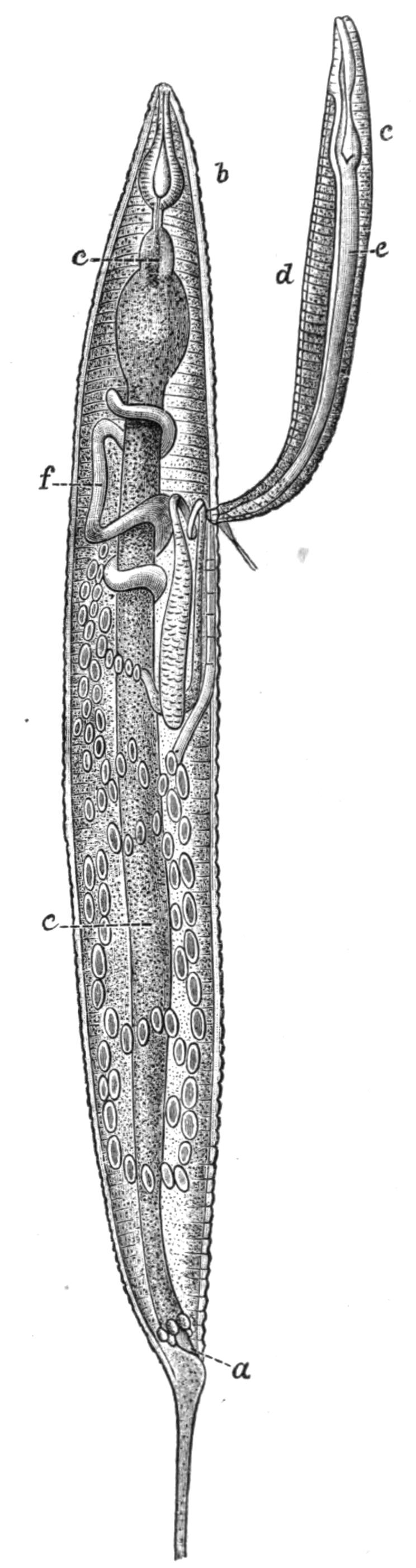
Fig. 69.—A male and female Oxyuris diesingi Ham. in copula, × 60. a, Anus; b, oesophagus; c, bulb; d, testis; e, intestine; f, ovary. (From Galeb.[179])
Oxyuris is Meromyarian (see p. 137), and is characterised by the long capillary tail of the female. It includes another human parasite, O. vermicularis, and it is one which it is difficult to get rid of. The female has the characteristic tail and is about 10 mm. long. The male is smaller. They are found in the caecum and rectum of man, and cause great irritation and sometimes serious functional disturbance. The eggs are laid in immense numbers but perish in water. If whilst still in the egg-shell the larvae are swallowed on fruit or raw vegetables, etc., they are set free in the stomach and small intestine by the action of the digestive secretions. The distribution of this parasite is universal. Besides numerous species that inhabit the alimentary canal of Vertebrates, such as O. ambigua Rud., found in hares and rabbits; O. curvula Rud., in the caecum of horses; O. megatyphlon Rud., in iguanas; several species inhabit the rectum of insects, such as O. blattae, O. diesingi, O. blatticola, found in the cockroach; O. spirotheca, and O. hydrophili in the water beetle Hydrophilus.[180]
The genus Nematoxys has the most complex arrangement of muscles of any Meromyarian, and forms a transition to the Polymyarian type. The whole body of both sexes is covered with numerous irregularly scattered papillae. The members of this genus have hitherto been found in snakes, Amphibia, and eels; there are but few species.
Oxysoma is another small genus with but three species, found in the intestines of opossums, frogs, and turtles respectively.
II. Family Strongylidae.
Mouth surrounded by papillae; an armature of teeth or spines often present. The chitinous lining of the intestine projects into the interior as ridges. No oesophageal bulb. The male orifice at the posterior end of the body is surrounded by a bell-shaped bursa.
Genera: Eustrongylus, Strongylus, Dochmius, Sclerostomum, Cucullanus, Syngamus, Pseudalius, Ollulanus, and others.
The genus Eustrongylus includes two species, E. gigas Rud. and E. tubifex Nitsch. The former attains in the female the gigantic length of 860 mm., with a breadth of 7 mm. and a weight of over 40 grs.[181] The male is a quarter to a third as long as the female. This parasite inhabits the kidney capsules of carnivorous animals, especially of those that eat fish, such as dogs, seals, etc., and has occasionally been found in man, the horse, and the deer. It frequently destroys the substance of the kidney. The worms are red in colour. The eggs die when exposed to desiccation for a few days, but have been kept alive for fifteen months in water; it is believed by Schneider and Leuckart that they are eaten by fish, and that the larvae form the Filaria cystica found in the peritoneal membrane of the fishes Galaxias scriba and Symbranchus laticaudatus, and that they pass into their final host, where they become sexually mature, by the latter eating raw fish. E. tubifex is found in aquatic birds, e.g. ducks, grebes, and divers, etc.
The genus Strongylus is easily recognised by its conspicuous genital bursa, strengthened by variously arranged ridges which are of specific value. There are numerous species, found in man and many other mammals, and also in birds and reptiles. Some species inhabit the intestine, others form aneurisms in the large blood-vessels, and cause considerable mortality amongst horses; others live in the tracheae and lungs of cattle and sheep, their presence often causing great loss to the farmer. No intermediate host has been satisfactorily demonstrated; the larvae live in damp earth, and it seems almost certain that they pass directly into their host with its food.
Dochmius (Ancylostomum) duodenalis, called by Neumann[182] Uncinaria duodenalis, is one of the most dangerous parasites that attack man. It lives in the duodenum and jejunum, and the fertilised eggs leave the body of its host with the excreta, and in damp earth develop into larvae in the course of a few days. These at first eat voraciously, but after undergoing several moults they cease to take food and pass into the resting stage. If now they are swallowed with drinking water, they come to rest in the small intestine of their host, and in a few weeks become sexually mature. They cause great harm by burrowing in the intestinal walls and destroying the capillaries. They are found by hundreds, and even thousands, in the same host, and produce profound anaemia, which is frequently fatal to miners, and was the cause of a great mortality amongst the workers in the St. Gothard Tunnel some fifteen years ago. This species is very widely spread over the face of the globe. Dochmius trigonocephala Rud. and D. stenocephala produce similar diseases in dogs and cats, and D. cernua Crep. is found in sheep and goats.
The genus Cucullanus exists in the adult form in the intestines of fishes, and more rarely of reptiles. C. elegans Zed., which live in fresh-water fish, e.g. the perch, is viviparous; after birth the young pass into the water and make their way into the alimentary canal of the small crustacean Cyclops, and thence into its body-cavity. Here they undergo two moults, accompanied by certain changes in structure. If this second host be swallowed by a fish the parasites are set free, and develop generative organs. Ollulanus tricuspis Leuck., which in the adult state is found in the cat, chiefly in the intestine but also in the bronchi and other parts, gives rise to larvae which are of enormous size compared with the parent; these leave the body, and if eaten by a mouse encyst in its muscles, and if the mouse be devoured by a cat, they complete their life-cycle by becoming sexually mature.
The genus Syngamus infests the trachea and bronchi of birds, more rarely of mammals. The red- or forked-worm, Syngamus trachealis Sieb., is common in poultry and game birds, and causes the disease known as gapes, which is especially common in young birds, and often gives rise to extensive loss. The peculiarity of this genus is that the male is permanently attached to the female, its genital bursa being so closely adherent to the opening of the oviduct that two specimens cannot be separated without tearing the tissues. The ova are not laid, but escape from the body with fully-formed embryos in them, by the decay or rupture of their parent's body. They hatch in damp earth or water in from one to six weeks according to the temperature. When swallowed by a fowl they develop into adults, which reproduce eggs in less than three weeks. No second host is needed, but the embryos remain alive in the alimentary canal of earthworms, and these doubtless to some extent serve to spread the disease.
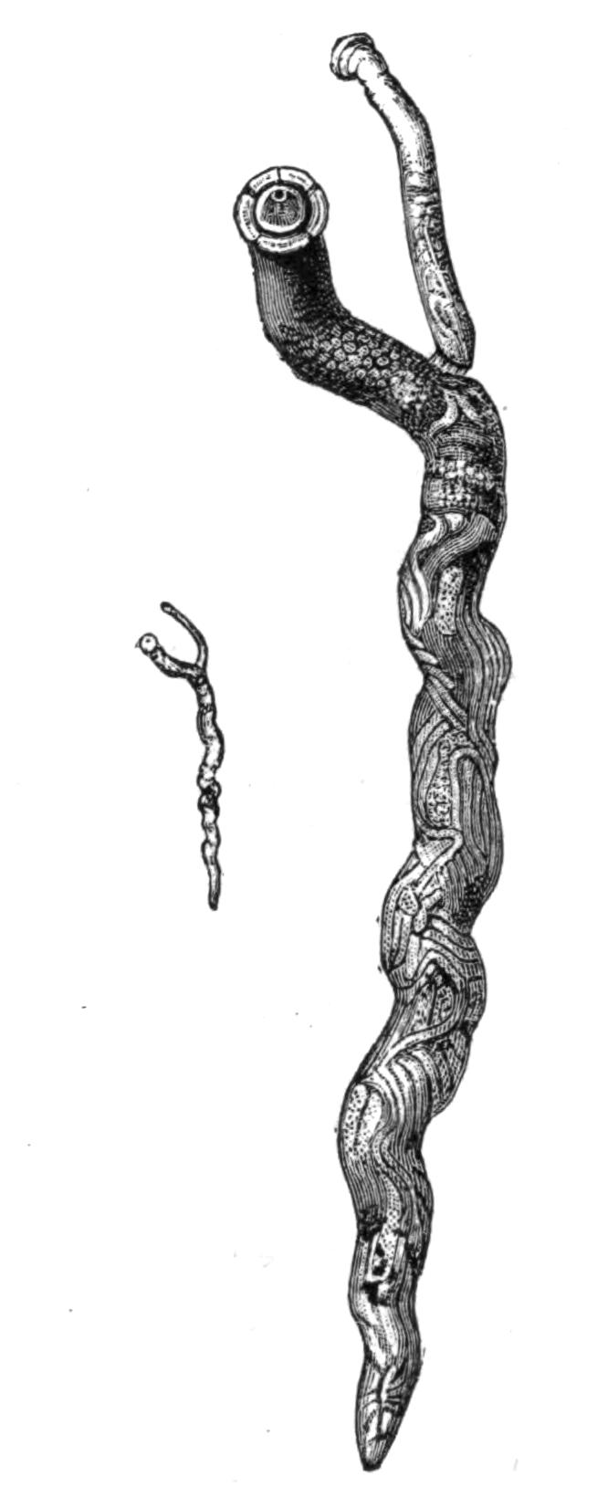
Fig. 70.—Syngamus trachealis Sieb., natural size and magnified four diameters. The small ♂ is permanently attached to the female. (From Warburton.[183])
III. Family Trichotrachelidae.
This family is characterised by the anterior end of the body being produced into a long whip-like neck. The mouth is small and devoid of papillae. The oesophagus is very long, and it traverses a peculiar strand of cells.
Genera: Trichocephalus, Trichina, Trichosoma, and others.
Trichocephalus dispar Rud. (hominis Gmel.) is common in man, and also occurs in some species of monkey. It does not live freely within the intestine, but buries its long whip-like anterior end in the mucous lining of the caecum or colon. The eggs pass out of the body of the host. The development of the embryo is slow, lasting many months; whilst still in the egg-shell the embryos are swallowed, and give rise to the sexually-mature parasite without the intervention of an intermediate host. They are by no means uncommon. Davaine calculated that about 50 per cent of the inhabitants of Paris were infested with them, but they give rise to little disturbance, and only very occasionally cause serious harm. T. affinis Rud. infests sheep; T. crenatus Rud. the pig; T. depressiusculus Rud. the dog; and T. unguiculatus Rud. the hare and rabbit.
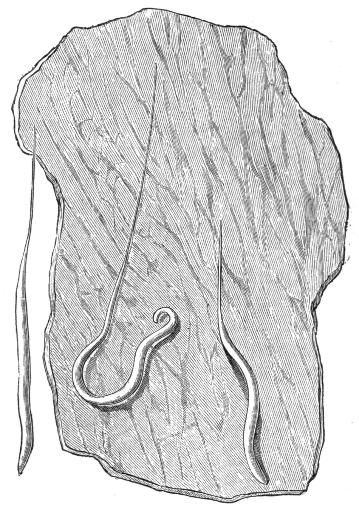
Fig. 71.—Trichocephalus dispar Rud., attached to part of the human colon. × 2.
The genus Trichosoma, with many species, is as a rule found in birds, but it occurs also in mammals, as T. plica Rud. in the bladder of the fox and wolf, T. felis cati in the bladder of the cat, T. aerophilum Duj. in the trachea of the fox and marten. The chief interest of this genus is that, at any rate in T. crassicauda Bel., which infests the rat, the dwarf males live two, three, or four at a time within the uterus of the female, a condition of things which recalls the similar arrangement found in the Gephyrean Bonellia.
Trichina spiralis is the cause of the well-known disease trichinosis, which appears in two forms, intestinal and muscular, according to the habitat of the parasite. The mature forms of both sexes are found in the intestine of man and many other mammals. They have been experimentally developed in birds, though in the latter the larval forms have never been observed. By keeping such cold-blooded animals as the salamander at a constant temperature, Goujon and Legros succeeded in infecting them, but the larvae perished as soon as the artificial heat was withdrawn. Muscular trichinosis is unknown in fishes, but the sexual form develops in their intestine.
The adult parasites of the intestine are scarcely visible to the naked eye; the females are 3 to 4 mm. long and more numerous than the males, which measure 1.4 to 1.6 mm. The eggs are very numerous, a single female containing at one time 1200, and probably producing ten times as many during her life. The embryos are hatched out within the uterus, and the larvae leave the body of the mother through the generative pore. The minute larvae bore through the intestinal walls of their host, and then, either burrowing in the tissues or swept along in the stream of blood or lymph, make their way all over the body, and come to rest most usually in the muscles, but occasionally in other parts. When the larva reaches its resting-place, it either pierces the sarcolemma and establishes itself within the substance of the muscle-fibre, or it comes to rest between and not in the fibres. Here its presence sets up the formation of a spindle-shaped cyst which usually contains but one larva, though any number up to seven have been found in one cyst. Within this the larva may remain dormant for years, the walls of the cyst gradually undergoing a fatty or calcareous degeneration. Almost any muscle may be affected; those most usually infested being the muscles of the diaphragm, of the shoulder-blade, and of the lumbar region; the larvae have also been found in the heart. The ends of the muscles near their points of attachment are always the most thoroughly infested.
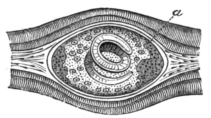
Fig. 72.—Trichina spiralis Owen, encysted in muscle. a, Calcareous deposit. Highly magnified. (From Leuckart.)
The number of the encapsuled larvae in one host is enormous. Leuckart counted between 12,000 and 15,000 in a gramme of muscle, which would give a total of thirty to forty million parasites in one host; other estimates place the total even higher.
When trichinised meat is eaten, unless it has been thoroughly cooked, the cysts are dissolved and the larvae are set free. Within three or four days they become sexually mature and their ova begin to segment. The males after a time leave the body with the excreta and perish, whilst the larvae of the new brood make their way into the tissues of the host.
Man usually acquires trichinosis by eating uncooked or improperly-cooked pork, and the disease is so widely spread and of such a serious nature that most civilised countries have adopted rigorous methods for the detection of trichinised meat. The pigs either acquire the disease by eating uncooked swine's flesh, which is frequently given them in the form of offal, or by devouring rats, which are very susceptible to the disease.
IV. Family Filariidae.
Mouth with two lips, or without lips. Six oral papillae often present, and sometimes a horny oral capsule. Four pre-anal pairs of papillae, and sometimes an unpaired one as well. Two unequal spicula or a single one.
Genera: Filaria, Ichthyonema, Hystrichis, Spiroptera, Dispharagus, and others.
The genus Filaria is a very large one. Like Ascaris, it is confined to Vertebrates, but usually lives in the tissues of the body and not in the intestines. F. (Dracunculus) medinensis Gmel., the guinea-worm, is well known as a human parasite in hot countries; it also occurs in the horse and dog. The female has an average length of 50 to 80 cm., but gigantic forms with a length of 4 metres have been described. The alimentary canal is degenerate. In adult females the body is completely occupied by a uterus crowded with eggs and embryos, which can only escape by the rupture of the mother's body, as the genital ducts have disappeared. Its original home is tropical Asia and Africa, but it has been introduced into South America with the negroes.
The female lives coiled up in the subcutaneous tissues, usually in those of the legs. Its presence gives rise to painful tumours. When these break the female protrudes, and may be withdrawn from the body by very carefully rolling it round a stick or pencil. This must be done very slowly, a few inches a day, as the rupture of the body sets free the contained embryos, and may result in the death of the host. The embryos normally bore their way into the body of the fresh-water Cyclops, and are re-introduced into their Vertebrate hosts with the drinking-water. It is usually stated that the female alone is known, and that it is uncertain whether it is hermaphrodite or whether both sexes are present in the Cyclops. Recently Dr. Charles[184] has described a specimen found in the mesentery of a human subject, from an orifice in the middle of whose body he was able to draw a much smaller specimen, and he thinks this may be the long-sought-for male.
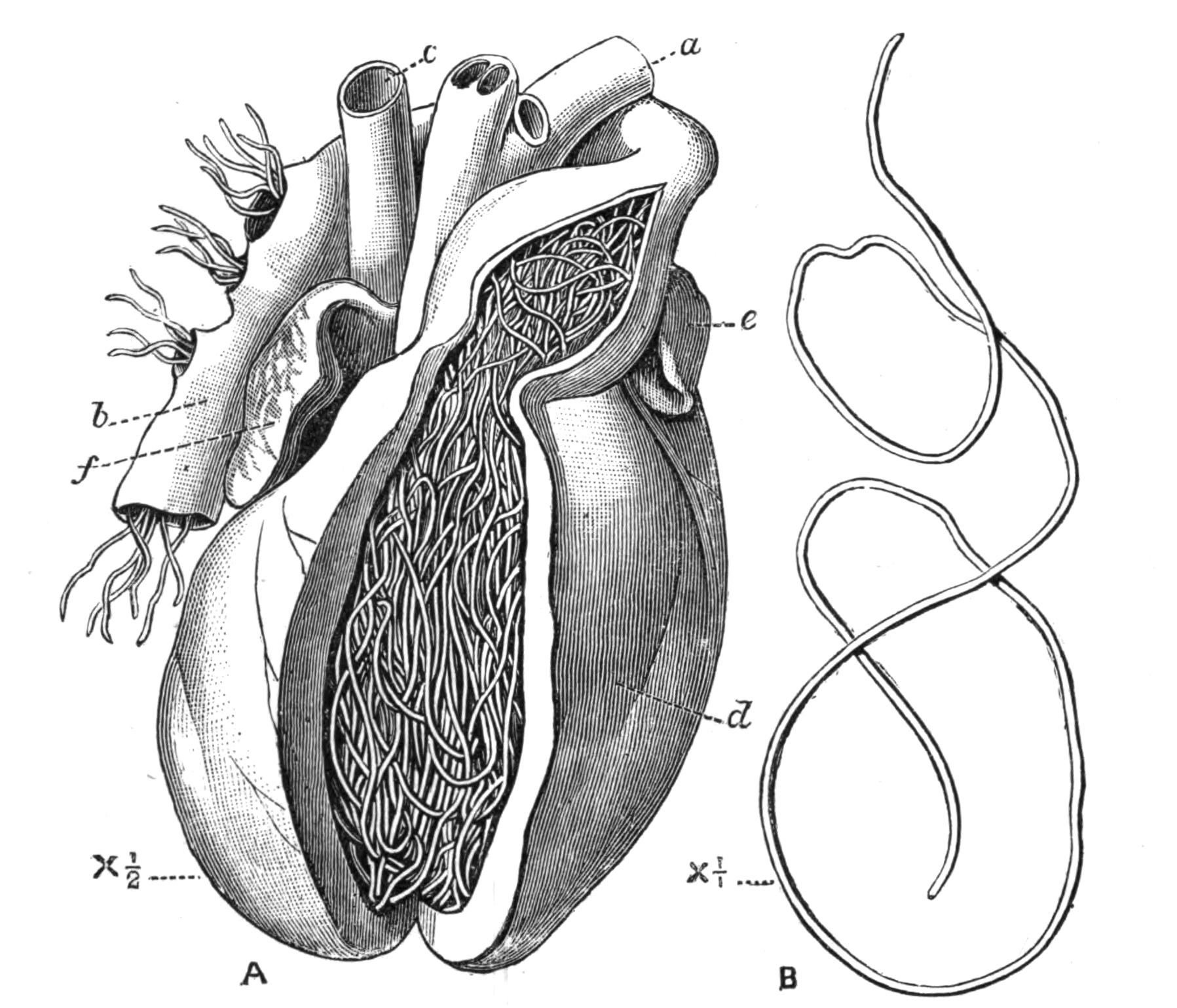
Fig. 73.—A, View of the heart of a dog infested with Filaria immitis[185] Leidy; the right ventricle and base of the pulmonary artery have been opened. a, Aorta; b, pulmonary artery; c, vena cava; d, right ventricle; e, appendix of left auricle; f, appendix of right auricle. B, A female F. immitis removed from the heart to show its length. Natural size.
Filaria immitis Leidy, the cruel worm, is common in dogs in China and the East generally. It is not unknown in America and Europe. It occurs in such large clusters in the right ventricle that it is difficult to see how the circulation can proceed. The intermediate host is unknown, but from the prevalence of the disease in marshy country it is probably some aquatic animal. The larvae are said by Manson to disappear from the peripheral circulation of the dog during the day, but not to such a marked extent as do the F. sanguinis hominis Lew., var. nocturna Man. They were found by Galeb and Pourquier in the foetus of an infested bitch, a fact which establishes the transmission of such parasites through the placenta.
Filaria sanguinis hominis nocturna.—The female of this parasite has been described as living in the lymphatic glands of man. The embryos escape from it into the lymph, and thus reach the blood. According to Manson the intermediate host is the mosquito, in whose stomach the embryos undergo their larval changes. When the mosquito dies the larvae escape into the water, and then make their way into the alimentary canal of man, where they are believed to pair, and whence the female makes its way to the lymphatics. The presence of this Filaria causes great functional disturbance. One of the most remarkable features of it is that the larvae, which are very numerous in the blood during the night, disappear during the day, and are not to be found. Recently Manson[186] has described two new varieties: F. san. hom. diurna, in which the conditions of things are reversed, the larvae being found by day and not by night; and F. san. hom. perstans, in which the larvae occur both by day and by night. The larvae are long-lived, and were found by Manson in the blood of a negro who had not been in Africa, where it is endemic, for six years. The same observer is inclined to associate the presence of F. san. hom. perstans with the fatal disease known as "sleeping sickness." He also suggests that the mature form of the variety diurna is the F. loa, which is not uncommon in the eyes of negroes, and that its intermediate host may be one of the blood-sucking flies so common on the west coast of Africa.
The genus Ichthyonema is confined to fishes. The male is very minute and the female partly degenerate. It has no anus and no external opening to its generative organs. The uterus fills up almost the whole of the body-cavity. I. sanguineum Rud. is found encapsuled in the peritoneum of many fish.
Hystrichis and Dispharagus are confined to birds, where they occur in the oesophagus and stomach. Spiroptera reticulata Crep. occurs in horses, twisted in a spiral round tendons and muscles, forming tumours which require to be opened.
V. Family Mermithidae.
Nematodes without anus and with six mouth papillae. Two spicules in the males and three rows of numerous papillae.
Genera: Mermis, Bradynema, Atractonema, Allantonema, Sphaerularia, and others.
As a rule the Nematoda show but little trace of their parasitic mode of life, but in this family there is considerable degeneration, and in extreme cases the body of the female is reduced to a simple sac crowded with eggs. They are exclusively parasitic in insects. In some respects their structure shows a transition towards Nectonema and the Gordiidae; especially is this the case in the structure of their ventral nerve-cord.
The sexual form of Mermis nigrescens Duj.[187] lives in damp earth, and after storms and in the early morning is sometimes found in such numbers crawling up the stalks of plants, as to give rise to the popular idea that there has been a shower of worms. The male is unknown; the female lays her eggs in the ground, and there they hatch out. It is not known exactly how the larvae make their way into the grasshoppers in whose body-cavity they live, but in an allied species, M. albicans v. Sieb., the larvae have been observed boring their way into small caterpillars through their skin, and it seems probable that the larvae of M. nigrescens burrow in a similar way into young Orthoptera.
Bradynema rigidum Leuck.[188] is found in the adult stage living freely in the body-cavity of a small beetle Aphodius fimetarius, one of the Scarabeidae, from two to three to as many as thirty being found in one host, which does not seem much injured by their presence. The parasite is without mouth, anus, or excretory pore. The eggs hatch out in the uterus of the mother, and the larvae are male and female; they make their way into the body-cavity of the host, and here they pass an unusually long time, five months, soaking in osmotically the nutriment contained in the blood of the insect. Eventually they burrow through the walls of the intestine, and leaving the body of their host through the anus, find their way to the earth. Here, according to zur Strassen, the females die without playing any part in the perpetuation of the species. The males, on the other hand, having developed spermatozoa whilst in the larval stage (paedogenesis), afterwards form ova, and are in fact protandrous hermaphrodites, and become the mature parasites of the beetle, though how they enter the body of the host is unknown.
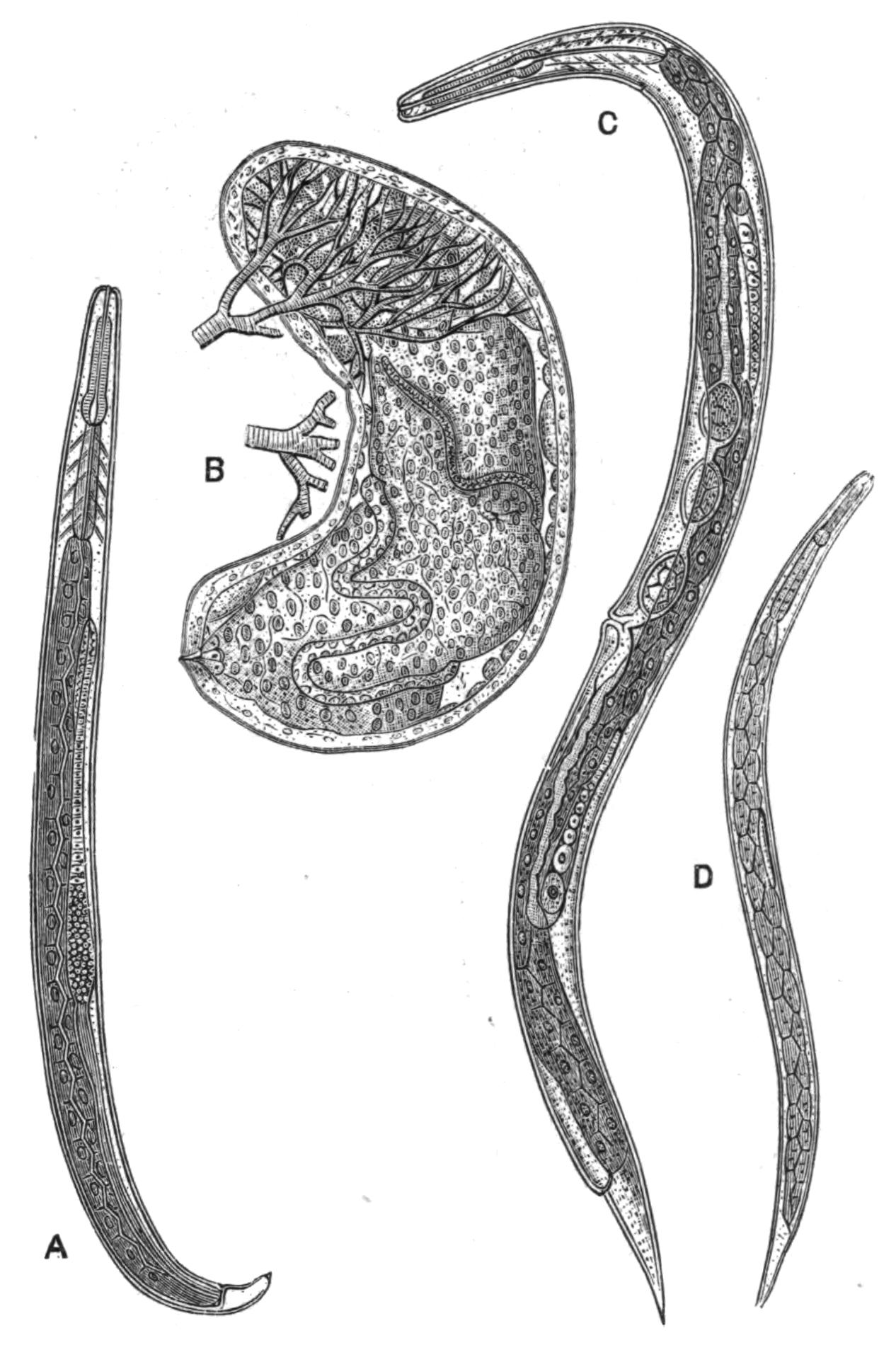
Fig. 74.—Allantonema mirabile Leuck. (From Leuckart.) A, Male Rhabditis stage, sexually mature, × 100; B, the mature female parasitic form, × 17, showing at the upper end part of the capsule richly supplied with the tracheae of the host, a beetle; C, female Rhabditis stage, sexually mature, × 100; D, the larva developed from the Rhabditis form, × 102.
The phenomenon presented by the hermaphroditism of Bradynema is, as far as we know, at present unique, as, though some other Nematodes are hermaphrodite, in their case the hermaphrodite form alternates with a bisexual generation. It is further interesting as showing a means by which hermaphroditism may arise, by the suppression of the females and the assumption of their functions by the male. In the case of Rhabdonema nigrovenosum, no females appear in the alternate generation.
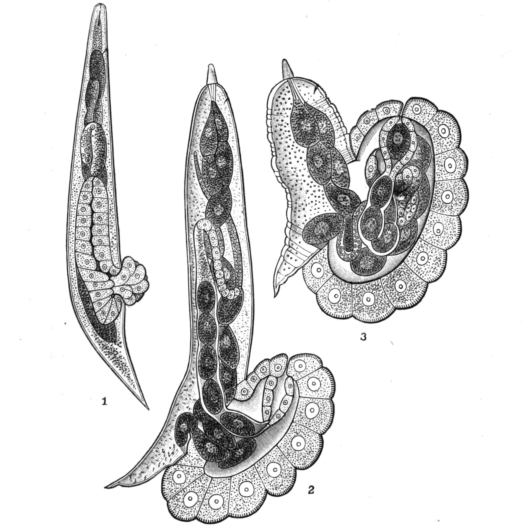
Fig. 75.—Atractonema gibbosum Leuck. (From Leuckart.) 1, Female with commencing prolapsus of the uterus and neighbouring parts, × 130; 2, a further stage, the female being now sexually mature, × 15; 3, a still older stage, with commencing degeneration of the body of the female, × 15.
A similar protandry exists in the parasitic forms of Allantonema,[189] of which there are several species—A. mirabile in Hylobius pini, A. sylvaticum in Geotrupes sylvatica, A. diplogaster in Tomicus typographicus; but in their case the male and female forms which leave their host pair in the damp earth and give rise to larvae which make their way into the body of the beetle-grubs. Here they undergo very extensive retrogressive change. The body of the female, which becomes the shape of a thick sausage, is encapsuled and surrounded by a curious hypertrophied network of tracheae (Fig. 74). As is usually the case with the degenerate parasitic forms, there are practically no organs but the ovary, and this is embedded in a fatty parenchyma which fills all the space within the skin.
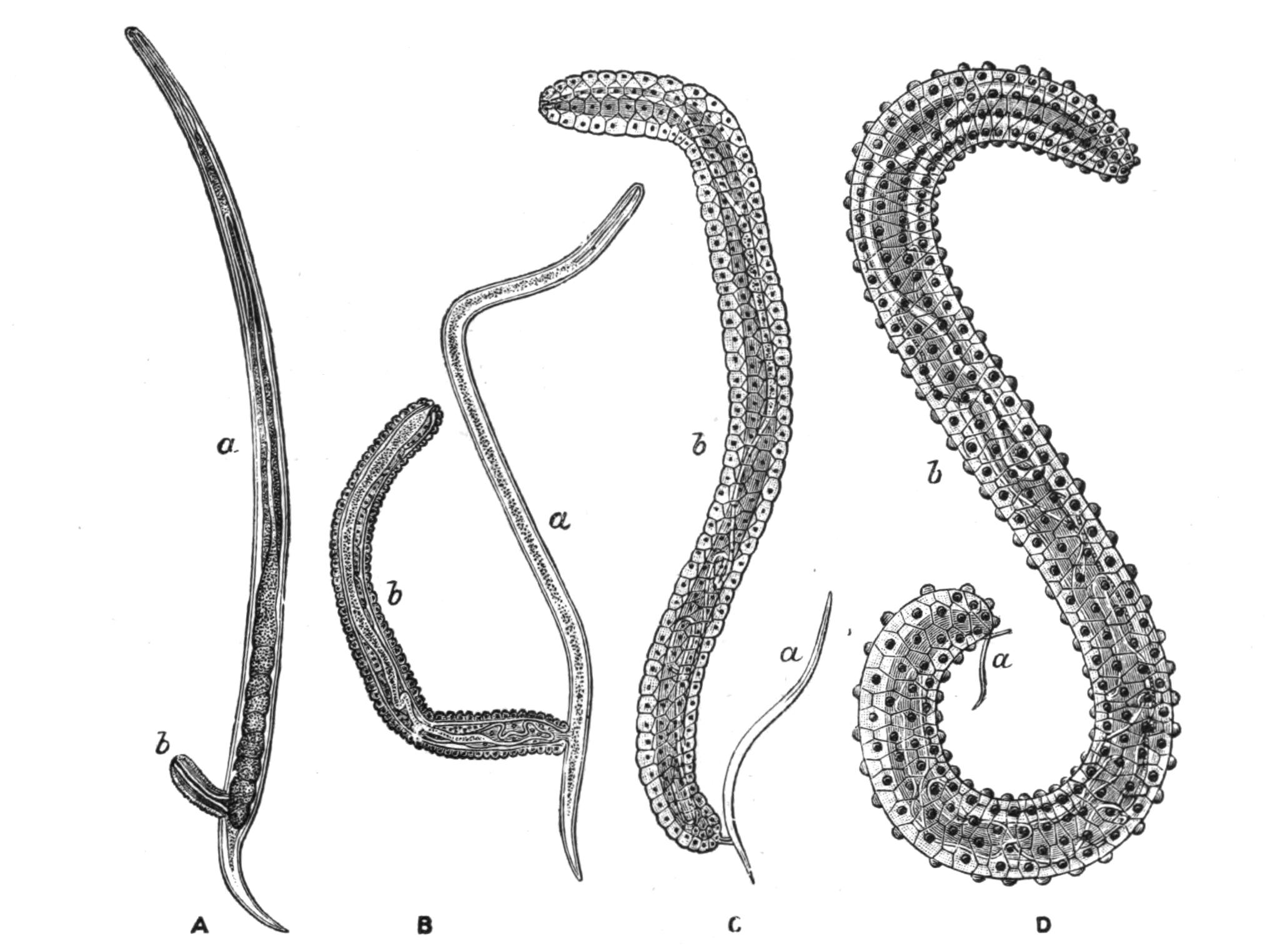
Fig. 76.—Four stages in the life-history of Sphaerularia bombi Dufour, ♀. (From Leuckart.) A, Beginning of the protrusion of the uterus (b), × 66; B, later stage, × 66; C, later stage, × 12; D, the protrusion is complete, × 6. In each case a represents the Nematode, and b its protruded uterus.
Atractonema gibbosum, which lives in the body-cavity of the larva of Cecidomyia pini, has a similar life-history, but the parasitic form has a structural peculiarity which merits attention (Fig. 75). At the time of sexual maturity a swelling, which is caused by the prolapsus of the uterus and vagina, appears at the posterior end of the body; this swelling increases until it equals the rest of the body of the Nematode in size. Even this is far surpassed by a similar protuberance in Sphaerularia bombi, where the evaginated sac grows with such extreme rapidity that in a few weeks its length increases from .25 mm. to 15 mm. and its volume 60,000-fold, the increase being due, according to Leuckart, to the increase in size of the individual cells and not to their multiplication. The Nematode which has produced this enormous growth gets relatively smaller and smaller, and ultimately drops off (Fig. 76). The sexual larvae which arise from the eggs in this sac leave the body of the bee in which this species is parasitic by the anus, and may live in damp earth, moss, etc., for months without taking nourishment, until the autumn, when they become sexually mature and, according to Leuckart, pair. The fertilised female is believed to bore her way into the humble-bee whilst the latter is seeking her underground winter quarters; this accounts for the fact that only queen bees are infected. The parasite is widely distributed both in Europe and North America; it is found in many species of Bombus, but most frequently in B. lapidarius and B. terrestris. The presence of the Sphaerularia affects the reproductive organs of the host, and reduces their fertility, so that an infected queen bee never succeeds in forming a colony.
VI. Family Anguillulidae.
For the most part free living and of small size. The oesophagus has usually a double swelling or two oesophageal bulbs. The male has two equal spicula.
Genera: Diplogaster, Mononchus, Rhabditis, Tylenchus, Anguillula, and many others.
Many species of this family live in humus or decaying matter; others live on, or are parasitic in, plants; some, such as Anguillula aceti, which is found in vinegar and in paste, live in organic fluids.
The part played by the presence of these Nematodes in the soil is not thoroughly understood; sometimes they occur in great numbers, and even when not directly parasitic in plants, probably do them much damage. Cobb[190] has recently described from Australia and Fiji over eighty species, one-half of them new, which occur mostly in the earth, and many of them among plant roots. They frequently crawl up on to plants, especially on to seedlings. An instance of this is given as follows: "The edible part of three bunches of nice-looking celery bought of a Chinaman in Sydney was cut off as far up as it was tender, nearly to the first leaflets. It was washed by hand in a tin dish in tank water, free from Nematodes. The washings gave about 200 to 300 Nematodes, belonging to five different genera."
It is very probable that many of the free-living forms which have received distinct specific names may ultimately turn out to be but stages in the life-history of some of the parasitic species. Von Linstow[191] has pointed out that the free form of A. diplogaster, if found alone, would be placed in the genus Diplogaster; similarly the bisexual form of Ascaris nigrovenosa is known as Rhabditis nigrovenosa.
Those Nematodes which live parasitically in plants, e.g. many of the genera Tylenchus and Aphelenchus and Heterodera, as well as those which only pierce the epidermis of the roots (the remaining species of the above-named genera), are provided with a spine which works to and fro through the mouth and assists the animal to bore into the tissues of the plant. Tylenchus devastatrix lives and reproduces in leaves and stems (never in the roots, except in the case of hops[192]) of many cultivated plants, such as rye, oats, onions, etc. "Clover sickness" is probably caused by this Nematode. The plants become infected by the thread-worms in the soil during the spring; their presence causes swellings and often kills the plant, in which case the worms return to the soil or remain in the straw.
Tylenchus tritici Need. is the cause of "ear-cockles" in corn. These take the form of brown or purple galls, which replace the grains of corn, and which contain hundreds of minute Nematodes. In these galls they are motionless, and are capable of surviving in dryness for at least twenty years; but when moistened,—for instance, by the gall falling on damp earth,—they resume their vitality and make their way to the young wheat plants, and then, wriggling up the leaves and stems, find their way to the ear. Here they pair, and producing a gall-like growth in the flower, lay numerous eggs, from which arise the Nematodes of the ear-cockle.
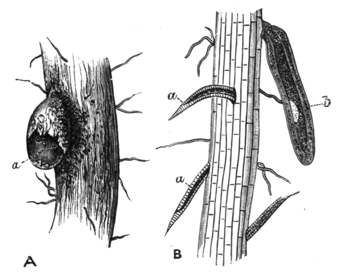
Fig. 77.—A, a, Female Heterodera schachtii Schmidt, breaking through the epidermis of a root; the head is still embedded in the parenchyma of the root: B, a, larvae boring their way into a root; b, larva of the immobile kind surrounded by the old skin, living as an ectoparasite on the outside of the root. (From Strubell.)
Heterodera schachtii[193] Schmidt, is the cause of the "beet sickness," and forms galls or swellings on the roots of many plants, in England especially on the roots of tomatoes and cucumbers. The free larvae live in the earth and make their way into the smaller rootlets; here the female larvae shed their skin, lose their characteristic Nematode form, and become citron-shaped (Fig. 78, D). The male larvae undergo a change, and after a period of rest cast their skin and, leaving the rootlet, seek out the females. The female does not undergo this second ecdysis, but its generative organs grow and mature in what is practically a larval stage. The embryos develop within the body of the mother, and, escaping through the uterus, ultimately cause her death. They then make their way into the earth. The cycle of the development takes but four or five weeks, so that, as in the case of Tylenchus devastatrix, there are several broods in a year; T. tritici, on the other hand, has but one.
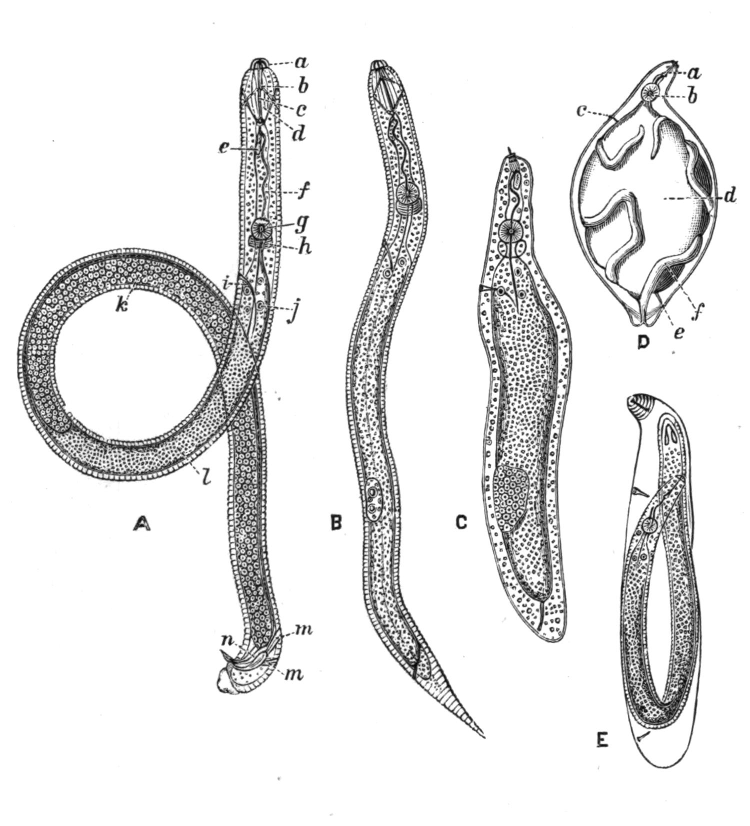
Fig. 78.—A, Male Heterodera schachtii strongly magnified; a, head lappets; b, mouth cavity; c, spine; d, muscle of spine; e, gland; f, oesophagus; g, bulb; h, nerve-ring; i, excretory pore; j, intestine; k, testis; l, intestine; m, muscles moving spicule; n, spicule: B, first motile larva: C, second immovable parasitic larva casting its skin: D, a female with one half of the body-wall taken away to show the coiling generative organs; a, boring apparatus; b, oesophageal bulb; c, excretory pore; d, alimentary canal; e, anus; f, ovary: E, a male shortly before casting its larval skin.
Vuillemin and Legrain[194] point out that while Heterodera is injurious to cultivated plants growing in damp soil, its presence is advantageous to those that grow in deserts. It is very common in the Sahara, and attacks many plants which are immune from it elsewhere. It causes the rootlets to swell out, and the bladder-like extensions thus formed act as reservoirs for water.
Many other species attack plants; Tylenchus millefolii Löw forms galls on Achillea, T. dipsaci Kühn. on the teazle. They all seem to have great powers of resisting desiccation. The former species, when dried and placed in a herbarium in May, gave rise to active worms when moistened the following October; and the corn eel-worm is said to survive twenty-seven years in a state of suspended animation. On the other hand, although these Nematodes like moisture, they cannot withstand submersion in water for any time. They can resist a considerable degree of cold, and a species, Aphelenchus nivalis Auriv.,[195] has been described from Spitzbergen, where it lives in the snow amongst a small red alga, Sphaerella nivalis.
VII. Family Enoplidae.
Small, as a rule free-living, usually marine Nematodes, without a second oesophageal bulb. Eyes and mouth-armature often present. Fine hairs and bristles sometimes surround the mouth.
Genera: Enoplus, Dorylaimus, Enchelidium, and others.
The genus Enoplus is exclusively marine, living amongst Algae and Hydroids in shallow water and moving actively about, but never coiling into spirals. De Man[196] describes Enoplus brevis Bast. as being attacked by a plant parasite, probably a Bacterium, of a greenish colour, which infested the muscles and gave them a peculiar colour.
Numerous other species have been described by De Man from the coast of Holland. It is probable that some of them are the free stages of parasitic forms; a brackish water species found in the East Indies (Dorylaimus palustris) is regarded by Carter as the larva of Filaria medinensis. Oncholaimus echini Leyd. is parasitic in the intestine of the sea-urchin Echinus esculentus. Tricoma cincta[197] has a strongly striated cuticle, which gives it almost the appearance of segmentation. Fimbria tenuis has numerous hairs on the tail, and the mouth is surrounded by bristle-bearing papillae.
Here must be mentioned two families closely allied to the true Nematodes.
(i.) Chaetosomatidae.—This family includes three genera: Chaetosoma, Rhabdogaster, and Tristicochaeta. According to Metschnikoff,[198] although they are not true Nematodes, they have a great likeness to the group. He distinguishes them from the swimming members of the group as "creeping Nematoda." Chaetosoma, of which two species are known, C. ophicephalum and C. claparedii, has a head distinct from the body (Fig. 79). The mouth is at the anterior end, surrounded by a double semicircle of movable spicules; the whole body is covered by fine hairs, and on the ventral surface, just in front of the anus, is a double row of about fifteen cylindrical projections, by whose agency the animal creeps. The female C. claparedii is 1.5 mm. long, the male 1.14 mm. They were found creeping about on sea-weeds in the neighbourhood of Salerno.
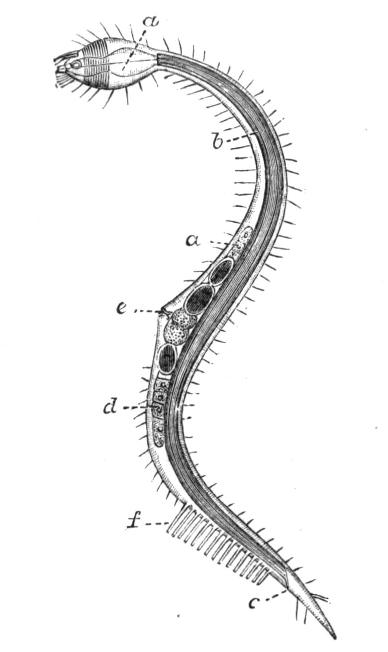
Fig. 79.—Mature female of Chaetosoma claparedii Metschni., × 57. (From Metschnikoff.) a, Oesophagus; b, intestine; c, anus; d, ovary; e, generative pore; f, ventral bristles.
The genus Tristicochaeta[199] differs from the foregoing in having three rows of locomotor projections instead of two.
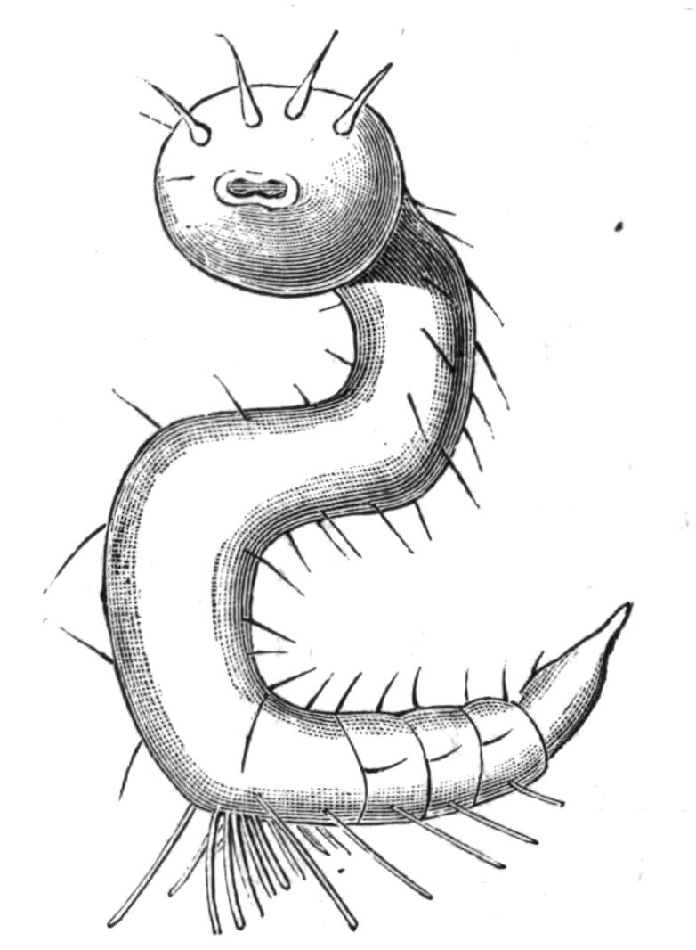
Fig. 80.—Tristicochaeta inarimense Panceri, in one of its most usual positions, showing the triple row of ventral bristles, × 100. (From Panceri.)
Rhabdogaster has no head distinct from the body, though the anterior part of the body is swollen. A second swelling occurs, as is also the case with Chaetosoma, in the region of the opening of the genital ducts. The female in Rh. cygnoides attains a length of 0.36 mm. In this genus the hairs are confined to the dorsal middle line. The locomotor projections are hooked, and are much finer than those of Chaetosoma, and they are situated farther forward than in the last-named genus. Rhabdogaster occurs in the same surroundings as Chaetosoma. Ch. ophicephalum is recorded from the English Channel.
(ii.) Desmoscolecidae.—The members of this family are minute, and are characterised by the presence of well-marked ridges which surround the body and give it an appearance of segmentation. The head, which is somewhat swollen, bears four bristles, and single pairs are borne by a certain number of the ridges, some on the dorsal and some on the ventral surface. These hairs can be moved independently of one another. Two red eye-spots are described between the fourth and fifth rings. The sexes are distinct, and the internal organs generally have a marked resemblance to those of the true Nematoda. The Desmoscolecidae move by looping their bodies after the manner of the Geometrid caterpillars, as well as by creeping with their bristles. The genus contains numerous species[200]: D. minutus Clap. (English Channel), D. nematoides Greef, D. adelphus Greef, D. chaetogaster Greef, D. elongatus Panceri, and D. lanuginosa Panceri. They are exclusively marine.
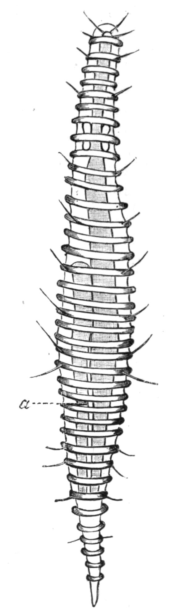
Fig. 81.—Female Desmoscolex elongatus Panceri, ventral view, × 260. a, Ovary. (From Panceri.)
Trichoderma oxycaudatum Greef[201] is a minute animal, 0.3 mm. long, which has no head or ventral spines, but whose body is ringed and covered with long hair-like bristles. The male has two spicules, and the internal organisation recalls that of other Nematodes; still its ringed body has induced some authorities to place it near to Desmoscolex.
The Life-History of Nematodes.
Although, considering the enormous number of species of Nematodes and the remarkable diversity of the conditions under which they live, their bodily structure shows a very striking uniformity, the same is by no means the case with their life-history, which exhibits an astounding variety. Von Linstow[202] has arranged the various modifications, which occur under fourteen heads. He includes in his list the Gordian worms, which we have placed under a different heading. The following account has been taken from his paper, with a few alterations:—
1. The embryos develop, with a larval stage and without any change of medium, directly into the mature sexual forms. They live in fresh, brackish, or salt water, in plants, in the earth or in decaying organic matter: examples, Dorylaimus, Enoplus, Plectus, Monhystera.
2. The larvae live in the earth, the sexual forms in plants: examples, Tylenchus tritici and T. devastatrix, Heterodera schachtii (Figs. 77 and 78).
3. The larvae live in animals, after whose death and decay they are set free and develop into the sexual animals in the earth: example, Rhabditis pellio.
4. The bisexual forms live in the earth, and the fertilised females bore into animals (insects), and here produce embryos: example, Sphaerularia bombi (Fig. 76).
5. The bisexual forms live in the earth; the females do not develop, but the males make their way into Insects (Beetles), and becoming hermaphrodite, develop ova which give rise to the bisexual form: example, Bradynema rigidum.
6. The larvae live in the earth, the sexual form in Vertebrates: examples, Dochmius, Strongylus.
7. The Nematode lives as a hermaphrodite in animals, the offspring of this, by an alternation of generations, become sexual in the earth: example, Rhabdonema in Frog.
8. A bisexual free form gives origin to a bisexual parasitic form living in an animal: example, Leptodera appendiculata in Snails.
9. The eggs develop in the earth, and give rise to embryos which are transferred whilst still in the egg-cell to the body of an animal. The embryos hatch out and form bisexual parasites: examples, Oxyuris, Trichocephalus.
10. The larvae live in insects, the sexual worms in water or in the earth: example, Mermis.
11. The larva lives encapsuled and is passively transferred to a second animal: examples, Ollulanus, from Mouse to Cat; Cucullanus elegans, from Cyclops to Perch; Spiroptera obtusa, from Meal-worm to Mouse.
12. The sexual form lives for a short time in the intestine of a Vertebrate, and produces larvae which bore through the intestinal wall and become encapsuled in the tissues: example, Trichina spiralis.
13. The sexual animal lives in the trachea of birds; the ova containing embryos are coughed up and are taken into other birds with food. They quit the egg-shell and wander into the air-sacs, and finally into the trachea: example, Syngamus.
14. There are two larval forms; the first lives in water, the second in the lungs of Amphibia, whence they wander into the intestine and become sexually mature: example, Nematoxys longicauda in Triton alpestris.
Parasitism.
1. Effect of Parasitism on the Parasite.—The usual effect of parasitism on the parasitic organism is that the various organs necessary for a free life tend to degenerate, whilst there is a multiplication and development of organs of adhesion, by means of which the parasite maintains its hold on its host. There is further an immense increase in the powers of reproduction, which may take the form of an increase in the number of fertilised eggs produced, or the parasite may at some time of its life reproduce asexually, by budding, or fission, or parthenogetically.
Of the various classes of animals which are more or less parasitic, the Nematodes show less difference between the free-living and parasitic members of the group than obtains in any other class. With few exceptions, such as Sphaerularia, Allantonema, and one or two others, the parasitic forms have undergone but little degeneration. It is true that they have no eyes such as the free forms often possess, but in other respects, such as in the nervous, muscular, and digestive systems, they do not show any marked retrogression; further, the mouth-armature is developed in many free forms, and is not confined to the parasites.
The group has developed no methods of asexual reproduction by budding or fission, such as are found in Platyhelminthes; and the cases of an alternation of generations in which a sexual form alternates with a parthenogenetic form, are rare, e.g. Rhabdonema nigrovenosum; and it seems possible that even when parthenogenesis has been described, further observation may show that the parthenogenetic stage is really a protandrous hermaphrodite, in which case the alternation of generations in Nematodes, i.e. the hermaphrodite alternating with the dioecious form, is a case of heterogamy or the alternation of two sexual generations.
On the other hand, parasitic Nematodes produce enormous numbers of eggs. Van Beneden states that 60,000,000 have been computed in a single Nematode, and this multiplication of ova is absolutely necessary, for the chance of the embryo reaching the right host, in which alone it can develop, is always a small one.
It is a common thing to find that parasites are either hermaphrodite or that the male is degenerate, as is the case with many of the parasitic Crustacea, but with one or two exceptions the Nematoda are bisexual, and although, as a rule, the males are smaller than the females, they show no other trace of degeneracy.
In spite of the fact that the class as a whole shows but few special modifications consequent on a parasitic mode of life, it is clear that the Nematoda are peculiarly adapted for such a mode of life. Their elongated thread-like bodies afford little resistance to the passage of the food, which, as it passes through the intestine of the host, might tend to carry the parasites out of the body. At the same time their shape enables them to pierce and wriggle through the various tissues without making any very serious lesions such as might prove fatal to their host. Their extraordinary power of resisting desiccation both in the egg and in the adult state vastly increases their chances of ultimately hitting on the right host. They are capable of living in a state of suspended animation for months, and even years when dried (vide p. 136), and of resuming their activity on being moistened.
The great faculty this group shows for living parasitically is evinced by the extraordinary variety of life-history presented by the different species. There is scarcely a stage which may not be parasitic; the eggs, the larvae, the adults are all in some cases free, in others parasitic, and in many cases first the one and then the other.
2. Occurrence and Effect of the Parasite on the Host.—Von Linstow states that the only law that can be derived inductively from the study of the life-history of Nematodes is that those that live in animals never pass through all their stages of development in the same organ; consequently, in considering the distribution of the parasites within the body of their host we have a double habitat to consider. Many forms, such as Trichina spiralis, wander from the intestine to the muscles; others, such as Filaria medinensis, from the alimentary canal to the lymphatics or blood vessels or subcutaneous tissues. Others pass from the body-cavity to the intestine, as the Mermithidae, which infest Insects, or from the stem and leaves of a plant to its flower, as in the case of Tylenchus tritici.
With regard to their occurrence in the different classes of the animal kingdom, they have been most frequently observed in Vertebrates and in Insects. They are comparatively rare in the other large divisions. Many genera are confined to certain hosts: thus Ascaris, Filaria, Trichosoma occur only in Vertebrates; Spiroptera (with one exception) in Mammals and Birds; Cucullanus in Fishes and Amphibia; Strongylus and Physaloptera in Mammals, Birds, and Reptiles; Dochmius, Pseudalius, Trichocephalus in Mammals; Dispharagus, Hystrichis, Syngamus in Birds; Nematoxys, Hedruris in Amphibia and Reptiles; Ichthyonema in Fishes; and Isacis and Mermis in Insects.
Twenty-two species have been described as parasitic in man, of which perhaps the most dangerous are Filaria medinensis, the three varieties of F. sanguinis hominis; Dochmius (Ancylostomum) duodenalis, and Trichina spiralis. The Ascaridae, as Ascaris lumbricoides and Oxyuris vermicularis, though painful, seldom cause death.
The enormous number of parasites harboured by one host is shown by the fact mentioned in Leuckart's Parasites of Man, that Nathusius[203] took from a single black stork 24 specimens of Filaria labiata from the lungs, 16 Syngamus trachealis from the trachea, more than 100 Spiroptera alata from the coats of the stomach, besides several hundred Trematodes belonging to several different species (see p. 63). Even this has been surpassed in the case of a young horse, in whose body Krause found 500 Ascaris megalocephala, 190 Oxyuris curvula, several millions of Strongylus tetracanthus, 214 Sclerostomum armatum, 287 Filaria papillosa, 69 Taenia perfoliata, and 6 Cysticercus forms.
It is impossible here to enter into a full description of the destruction caused to domesticated animals and crops by the presence of these parasites; full details will be found in books dealing especially with this question, such as Neumann's Parasites and Parasitic Diseases of Domesticated Animals. A couple of cases will show how important this matter is to the farmer. Crisp estimates that Syngamus trachealis causes the death of half a million pullets in England every year, and Mégnin states that in a single pheasantry 1200 victims died daily; again, the loss of one-third the crop of beetroot is by no means uncommon when it is infested with Heterodera schachtii. These show the practical importance of what at first sight seem quite insignificant animals, and the necessity for the minutest observation, for only when we are fully acquainted with all the details of the life-history of a parasite are we in a position to successfully combat it.
Sub-Order II. Nematomorpha.
Until the last few years it has been customary to regard the Gordiidae as a family of Nematodes. Although in external appearance and life-history they closely resemble the members of this group, yet recent research has shown so many important morphological differences between them and the Nematoda, that most zoologists are now agreed in placing them in a different sub-Order, the Nematomorpha, a name first suggested by Vejdovsky.[204]
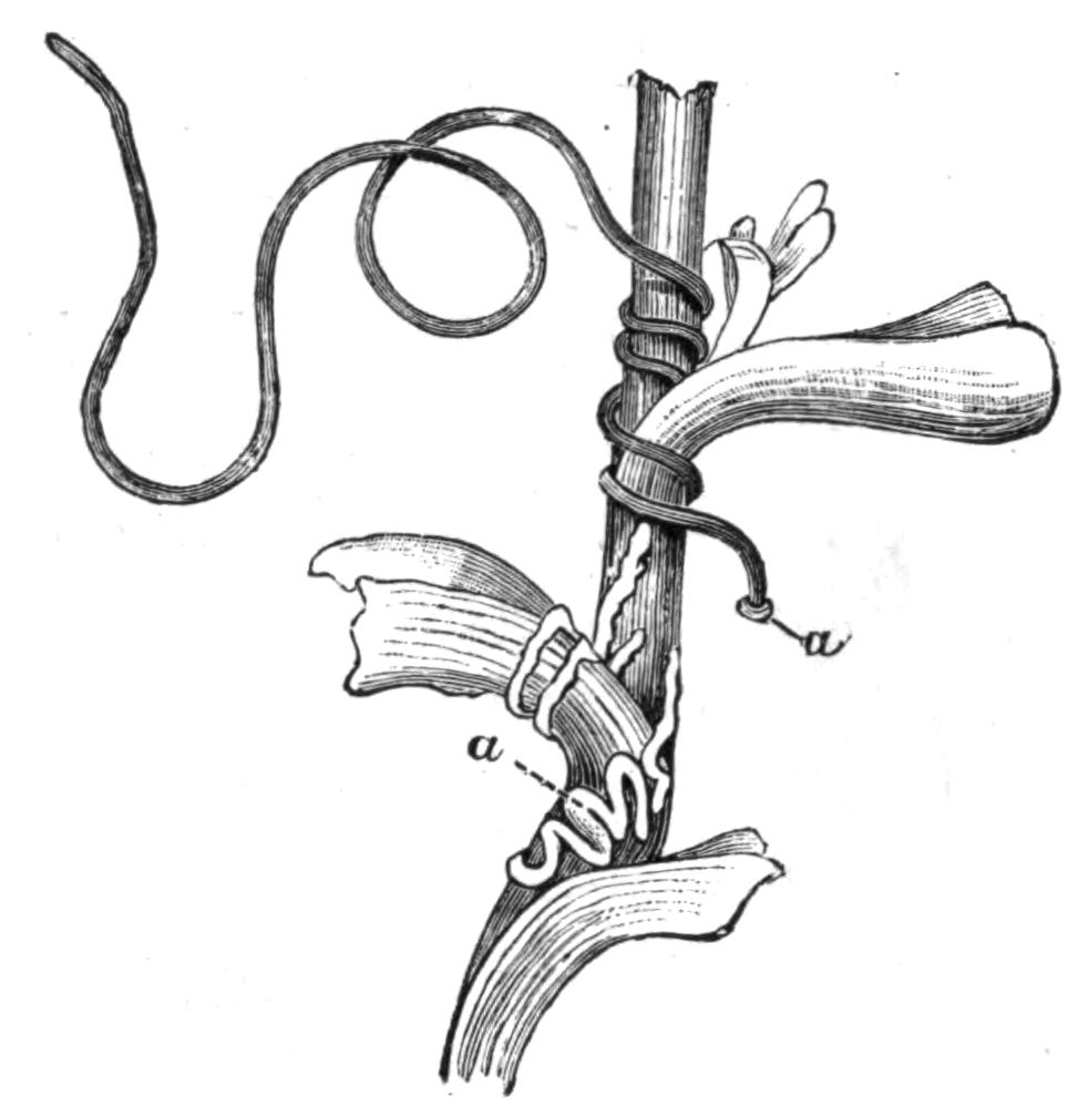
Fig. 82.—A water plant around which a female Gordius is twining and laying eggs. a, a, Clump and string of eggs. (From von Linstow.[205])
The Gordiidae comprise but two genera, Gordius and Nectonema. The latter has but one species, N. agile Verr., and is marine; the former, on the other hand, is exclusively fresh-water, and contains a very large number of species. Gordian worms are frequently to be found in ditches, ponds, or large puddles, moving with an undulating motion through the water, or twining and writhing round water-plants; they are scarcer in running water. In shape they are like a piece of thin whip-cord, slightly tapering at each end; the male, however, is easily distinguished from the female by its forked tail (Fig. 89). Not unfrequently a considerable number are found inextricably tangled together into a knot, and the name of the genus refers to this fact. Where numbers have suddenly appeared in water hitherto free from them, legends have sprung up which attribute their presence to a rain of worms; in reality they have come out of the bodies of Insects in which they are parasitic for the greater part of their life.
The genus Gordius passes through three distinct stages, of which the first two are larval and parasitic; the third is sexually mature and lives in water. The second larval stage closely resembles the adult, but the reproductive organs are not developed. The following account of the structure of this larval form and of the adult is in the main taken from von Linstow.[206]
The whole body is covered with a well-developed two-layered cuticle, which in the adult is marked out into areas, and bears numerous minute sensory bristles, which are especially developed in the neighbourhood of the cloaca of the male. Beneath this is a hypodermis which differs markedly from the sub-cuticle of Nematodes, inasmuch as it consists of a single layer of polygonal nucleated cells. Within this lies a single layer of longitudinal muscle-cells, which differ from the corresponding layer of Nematodes in having that part of their medulla which is not surrounded by the contractile portion directed outwards towards the hypodermis, and not inwards towards the body-cavity.
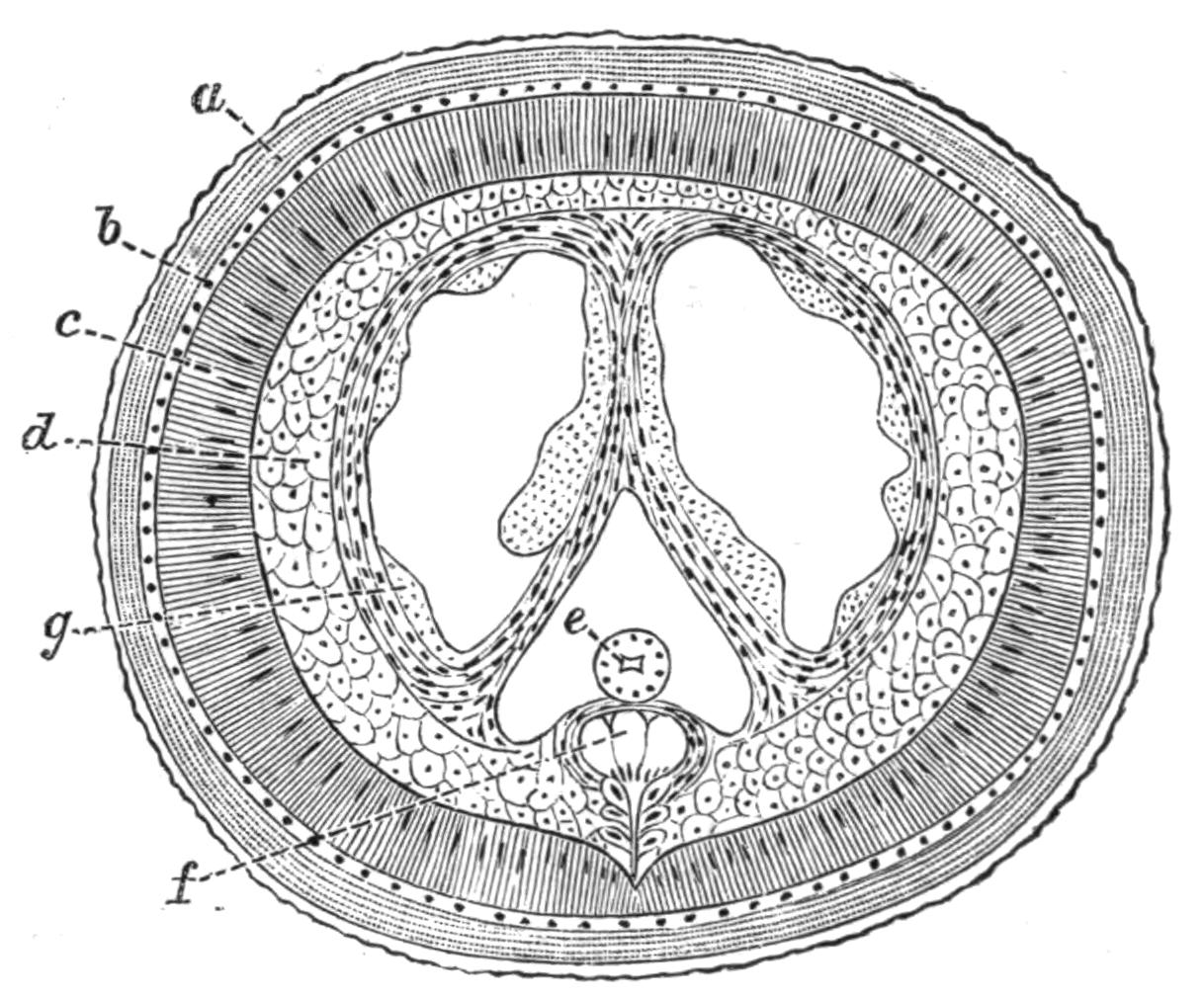
Fig. 83.—Transverse section through a young male Gordius tolosanus Duj. (From von Linstow.) Highly magnified. a, Cuticle; b, hypodermis; c, muscular layer; d, parenchyma; e, alimentary canal; f, nervous system; g, cells of the testis.
The body is in the younger stages practically solid, the interior being filled with clearly defined polygonal cells which are arranged in definite rows; in later life certain splits arise in this tissue which subserve various functions; between these splits strands of tissue are left which form mesenteries, and some of the cells remain lining the muscular layer (Fig. 86). These cells have been described by Vejdovsky as a definite somatic, peritoneal epithelium, but this was not found by von Linstow. Besides forming the mesenteries, and acting as packing between the various organs of the body, these cells also form the ova and the spermatozoa.
The splits which have appeared when the animal has reached the second larval stage, are two dorsal and a ventral; the latter contains the alimentary canal, and may be termed the body-cavity, the former will develop the generative organs. The mouth is occluded in the older larvae, and in the adults there is a distinct but solid oesophagus which passes into a tubular intestine. The intestine consists of a single layer of cells surrounding a lumen; it runs straight to the hinder end of the body, where it opens in both sexes with the ducts of the reproductive organs.
The nervous system consists of a well-defined circumoesophageal ring with two dorsal swellings, and, arising from this, a median ventral cord which runs the whole length of the body. The cord consists of three longitudinal strands with ganglionic cells below them; the latter, though they lie within the muscle layer, maintain a connexion with the hypodermis. Behind, the nerve-cord splits in the male, one half passing into each caudal fork. In the adult a pair of black eyes can be detected on the head; the only other sense organs are the tactile bristles mentioned above. Excretory organs are unknown.
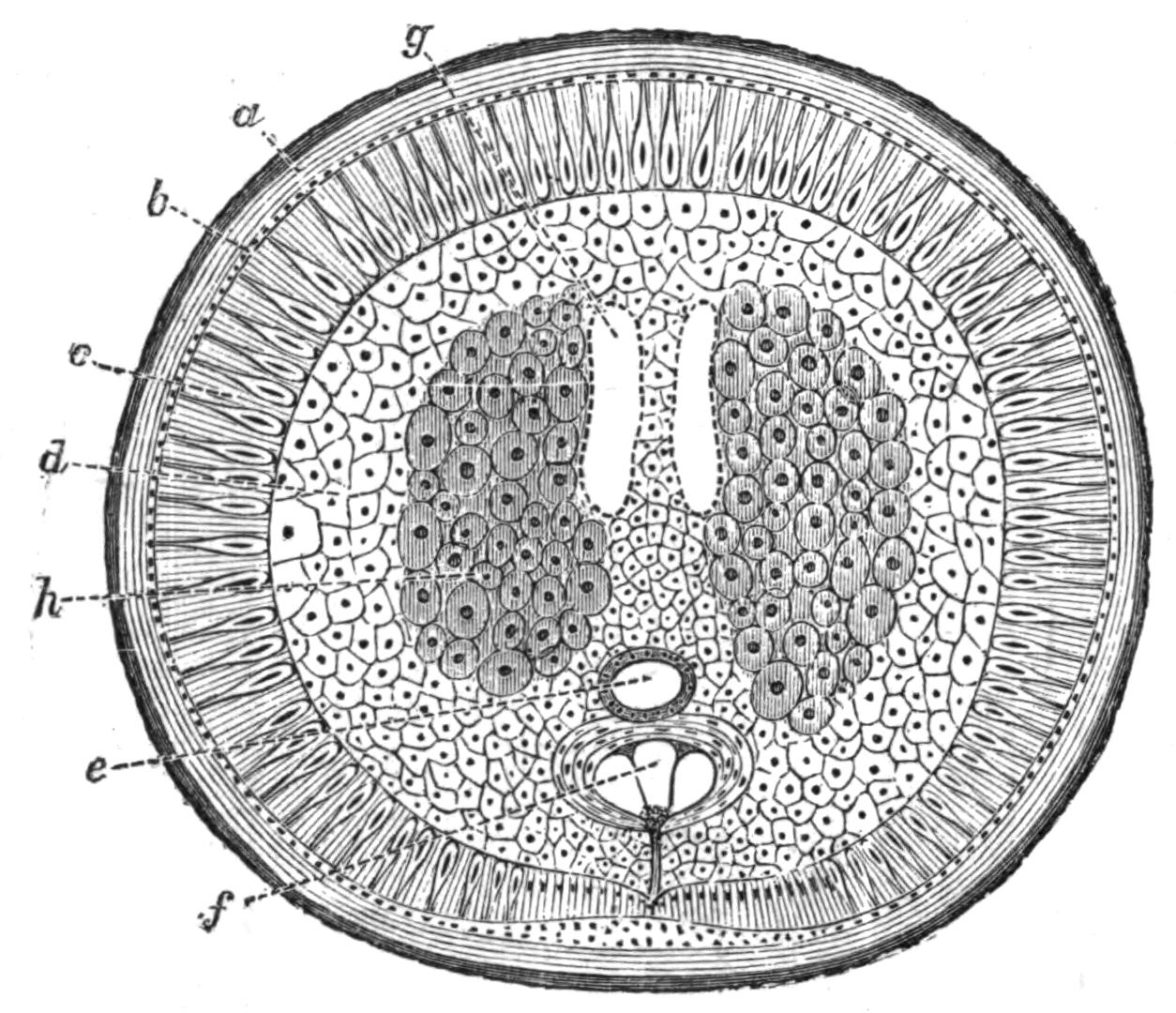
Fig. 84.—Section through a young female Gordius tolosanus. (From von Linstow.) a, Cuticle; b, hypodermis; c, muscular layer; d, parenchyma; e, alimentary canal; f, nervous system; g, egg-sac; h, ovary.
The generative organs only attain maturity in the adult, which is, in fact, exclusively devoted to reproduction. No trace of testes is found in the larva, though the two dorsal splits from the walls of which the spermatozoa will arise are present. They are lined by a definite epithelium (Fig. 83), and this serves at once to distinguish them from the body-cavity. Posteriorly the splits narrow and become the two vasa deferentia which open one on each side into the cloaca. The cells lining the lumen give rise to secondary cells, and these become spermatozoa, the process extending from behind forwards. The external organs—bursa, etc.—described by Vejdovsky were not found by von Linstow.
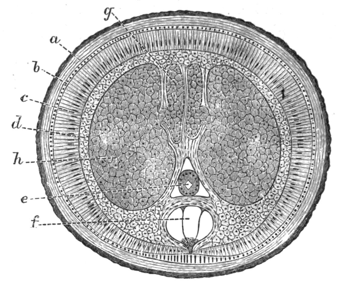
Fig. 85.—Section through a mature female Gordius tolosanus. (From von Linstow.) Lettering as in Fig. 84; g, egg-sac; h, ovary.
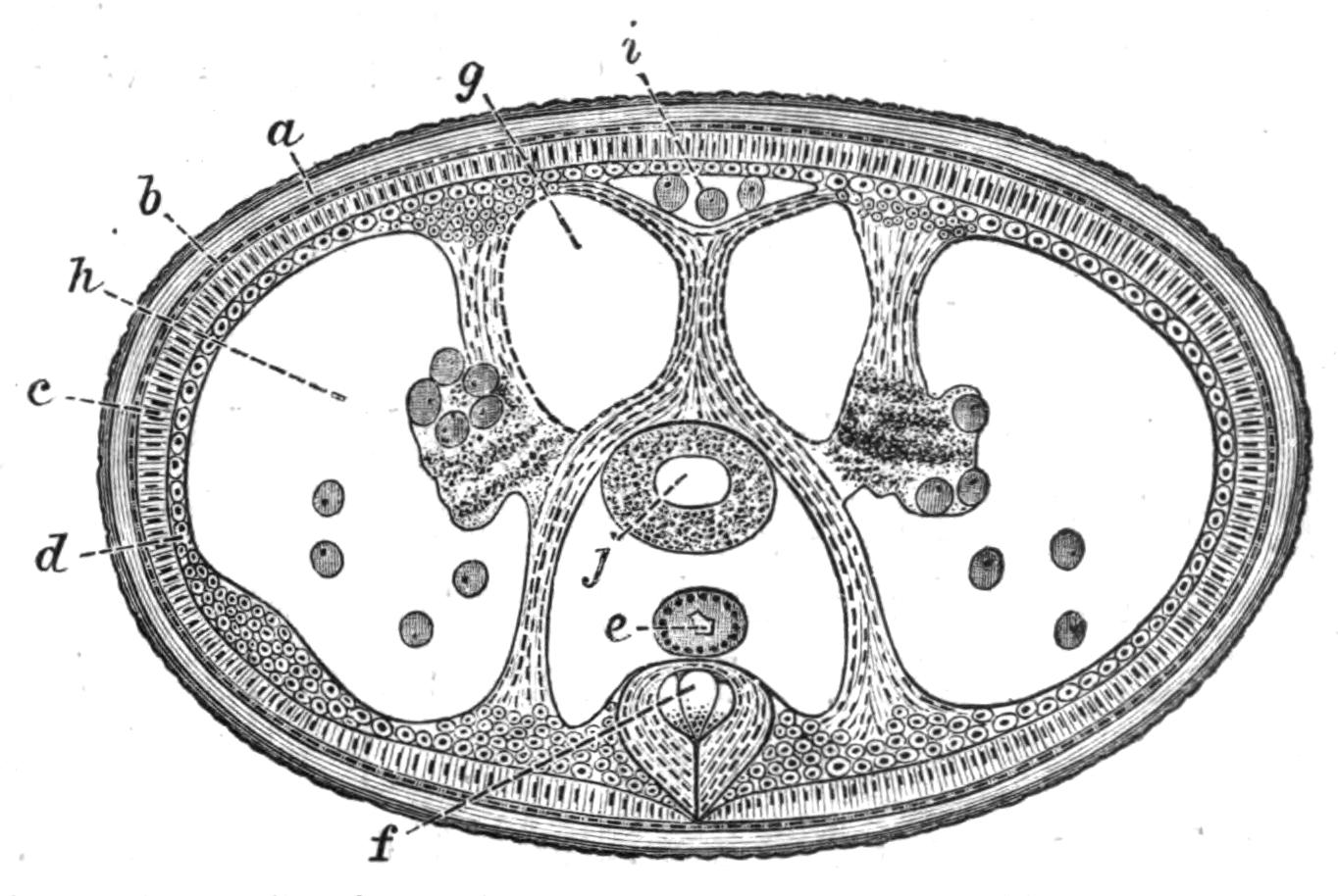
Fig. 86.—Section through a female Gordius tolosanus when the deposition of ova is almost complete. a, b, c, d, e, and f, as in Fig. 84; g, egg-sac; h, ovary almost empty; i, dorsal canal containing eggs; j, receptaculum seminis.
In the female larva two similar splits are present; these form the egg-sacs. Posteriorly they end in two short oviducts which open into a uterus, in which fertilisation takes place, and in which the secretion arises which cements the eggs together. In the adult the ovaries and a receptaculum seminis are found, in addition to the organs present in the larva. The ovaries are formed from modifications of the packing tissue; they begin close behind the head, and soon attain such dimensions as to compress the egg-sacs and body-cavity to small slits. After a time the wall between the ovary and the egg-sacs becomes absorbed, and the eggs grow into the latter. In the old females, where the egg sacs are empty, there is a considerable space round the exhausted ovary, into which eggs continue to fall off; there is also a median dorsal canal which contains a few eggs. By this time the wall between the ovary and the egg-sac has again appeared.
One of the most interesting points about the female is that, according to Vejdovsky, the ovary is segmented, the cells which form the ova being heaped up in segmentally-arranged masses. This observation, if correct, is almost the only instance of segmentation recorded in the group Nemathelminthes.
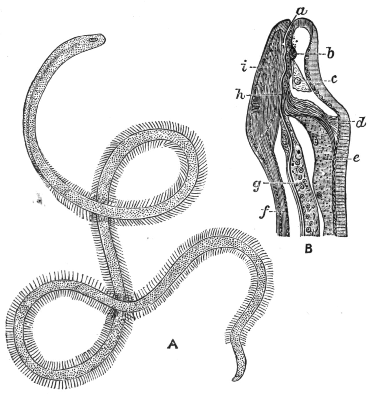
Fig. 87.—Nectonema agile Verrill. A, The adult. Magnified. (After Fewkes.) B, Longitudinal section through the head. × about 20. (From Bürger.) a, Mouth; b, circumoesophageal commissure (dorsal); c, cell of salivary gland; d, septum cutting off head from rest of body; e, testis; f, ventral cord; g, oesophageal cells; h, lumen of oesophagus; i, cerebral ganglion (ventral).
The only other genus which is associated with Gordius in the group Nematomorpha is Nectonema, of which there is as yet but one species known, Nectonema agile Verr.[207] Our knowledge of the anatomy of this worm is due mainly to Bürger[208] and Ward.[209] Nectonema is a marine worm found swimming near the surface of the sea with rapid undulatory motion. The males are from 50 to 200 mm. long, the females from 30 to 60 mm. The body is faintly ringed, and bears two rows of fine bristles on each side. Owing to a curious torsion of the body through a right angle, the lateral bristles of the anterior third seem to be placed in the ventral and dorsal middle line. They are very easily broken off. The body is divided into a small anterior and a large posterior chamber by a transverse septum placed a little way behind the head. The anterior chamber contains the brain and is lined by a definite epithelium, the posterior is not. The layers of the skin correspond with those of Nematodes or of Gordius, but the hypodermal cells show no cell outlines; still they are not so modified as in the former group. The hypodermis is thickened in the median dorsal and ventral line, and the single nerve-cord lies in the latter.
The alimentary canal is degenerate, as in Gordius. A mouth exists, but it is minute, and opens into a very fine tube lined with chitin, which pierces through the substance of a single elongated cell. This minute oesophagus, with its coextensive cell, reaches back to the transverse partition, but behind this a few other cells become associated with it, and ultimately the lumen of the alimentary canal is surrounded by four cells; but the number diminishes behind, and soon only two cells surround the tube at any one level, and the intestine dwindles away some little distance in front of the tail. There is no sign of an anus. A circumoesophageal nerve-ring exists, of which the ventral part is by far the larger (Fig. 87); it gives off a ventral nerve-cord, which swells posteriorly in the male into a large anal ganglion, far bigger than the brain, and larger in the male than in the female.
The testes consist of a dorsally placed sac, continuous behind with a vas deferens; this opens at the posterior end, which is pointed and slightly curved ventrally. The ovary is unknown; but females have been found with their body-cavity crammed with ova; these escape, like the spermatozoa, from a genital pore at the posterior end of the body.
Classification.—The separation of the Nematomorpha from the Nematoda depends mainly on the character of the nervous system, the absence of the lateral lines and of the dorsal line, the character of the contents of the body-cavity, and the character of the reproductive organs. In Gordiidae the latter are always placed dorsal to the intestine, and ovaries and testes open alike at the hinder end of the body. The importance of the differences in the organs just enumerated has been considered sufficient to justify the removal of the Gordiidae from the Nematoda, and the establishment of the special sub-Order Nematomorpha for their reception; and although Nectonema has a dorsal line, and is in some other respects intermediate between the two groups, there can be little doubt that it is more closely allied to Gordius than to any member of the Nematoda, and it must therefore be placed with it in the Nematomorpha.
On the other hand, it ought to be mentioned that Camerano[210] found that the chief details of the fertilisation and development of the egg in Gordius closely conform with what is known of the same processes in Nematodes, and he is of opinion that these resemblances are sufficiently important to justify the retention of the group among the Nematoda.
Life-History.—The life-history of Gordius comprises four stages—the early development of the egg, the first larval form, the second larval form, and the sexually mature form. Both larval forms are parasitic, and during their life they are actively engaged in feeding; the free form, on the other hand, takes in no nourishment, and is exclusively engaged in reproduction.
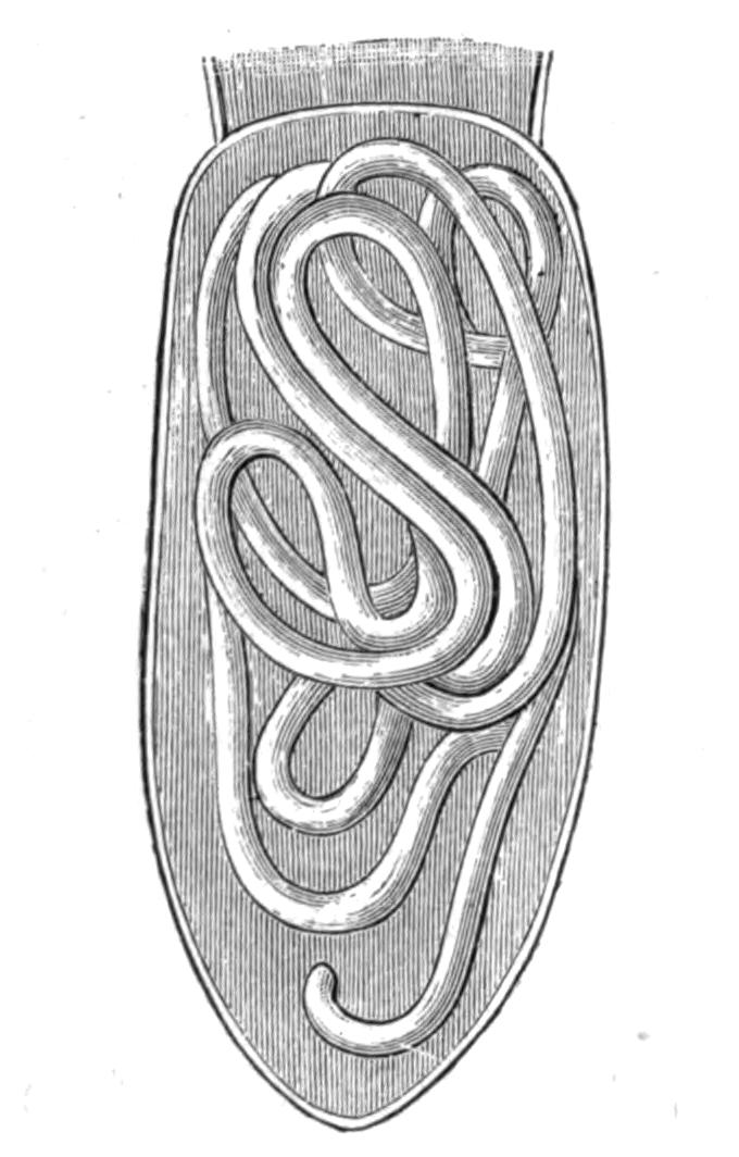
Fig. 88.—Abdomen of Pterostichus niger with the terga removed to expose the Gordius larva within. Slightly magnified. (From von Linstow.)
Von Linstow[211] gives the following account of the life-history of G. tolosanus, a form which has been more fully worked out than any other. In the month of April numerous specimens of the beetle Pterostichus niger were found floating on the surface of the ditches and small ponds in the fields surrounding Göttingen. Some were found dead or dying; others appeared quite healthy, and these were swimming actively, endeavouring to reach land. Within the abdomen of these beetles, in about 20 per cent of those collected, the second larval form of the G. tolosanus was found. The longest larvae were 122 mm. in length, and very soft, partly snow-white and partly brown in colour; traces of the boring apparatus of the first larval form were still to be seen, but in other respects the larva only differed from the free form in the immaturity of its sexual organs. Besides the parasite hardly anything was to be found in the abdomen of the beetle, the larva having eaten up all trace of the fat body and the generative organs of its host. The larvae bored their way out of the body of the beetle and became adult animals.
It is rather difficult to say what brings these essentially terrestrial beetles to the water, but von Linstow suggests that, as they live partly on snails, and at this time of year there are not many land-snails about, they may be in search of water-snails such as Limnaea. They may also be sometimes blown into the water by wind storms, but, whatever the cause is, their presence in water is essential for the continuance of the life of their parasites.
Once free in the water the Gordius is soon sexually mature; the fertilisation takes place in April, and then the female may be seen twisting and writhing round the stems of water-plants and laying the long bead-like strands of eggs (Fig. 82). The first deposition observed by von Linstow took place on 14th April, the last on 2nd August, and the period of egg-laying for each female extended over four weeks. At first the eggs are snow-white, but within twenty-four hours they turn brown in colour.
The development of the first larva within the egg takes about a month. When it emerges from the egg-shell it is minute, .065 mm. long, ringed anteriorly, and provided with a protrusible and retractile boring apparatus consisting of three chitinous rods; round the base of this piercing proboscis is a double crown of papillae, each bearing a spine (Fig. 90).
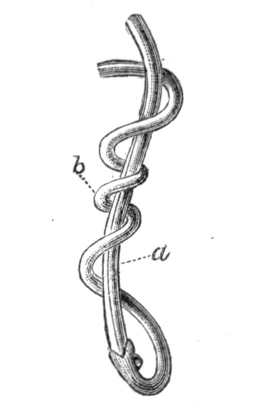
Fig. 89.—The tail ends of a female Gordius (a) and a male (b) in copula. × 1.5. (From G. Meissner.[212])
This first larval form breaks through the egg-shell and sinks to the bottom of the water, where it moves about sluggishly and awaits the arrival of the right host in which to take up its abode. This host is the larva of the Alder-fly, Sialis lutaria Lin. (vide vol. v. p. 444), and into this it bores and comes to rest in the muscles or the fat body. It does not form distinct capsules. It remains in this larva during the following winter, and in the spring passes over into the imago Sialis. The complete insect frequents the small plants growing along the water's edge, and falls an easy prey to the predaceous beetle Pt. niger. The larva is eaten, and undergoing a change becomes the second larval form mentioned above. It remains in the body of the beetle during the second winter, and finally returns to the water as the adult some eighteen or twenty months after it has been hatched from the egg.
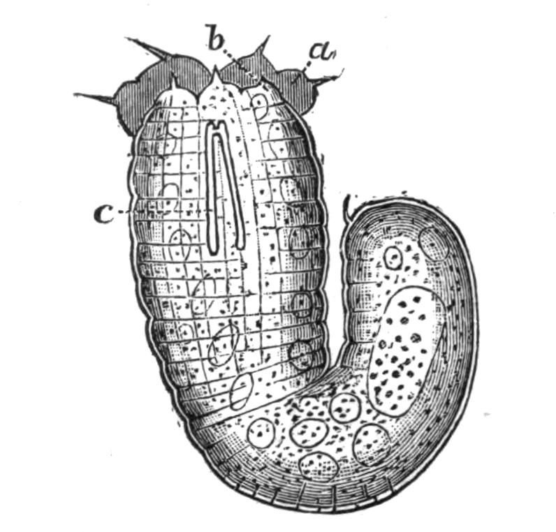
Fig. 90.—Embryo or first larval form of Gordius tolosanus taken from the egg. Highly magnified. a and b, The bristle-bearing papillae on the head; c, the boring apparatus. (From von Linstow.)
From the above account of the life-history of Gordius it will be seen that the chances of an egg reaching maturity are comparatively small, and to compensate for this a very large number of eggs are laid. In addition to the risk of the larvae not finding the right host at the right time, and of the first host not being eaten by the second, and the second not being drowned, there is the danger that the ditches and ponds in which the adults live may dry up, and, in fact, great numbers of worms perish by this taking place.
The sex of the adults may be told from their colour, the males being of a blackish brown, the females of a light clay brown; the former average 120 mm. in length, the latter 170 mm. The males are also more numerous, the proportion being seven to three. Camerano[213] has drawn attention to the fact that there is a certain polymorphism in size, form, and colour which is especially common amongst the males; dwarf forms with mature reproductive organs exist, and he is of opinion that these differences depend both on the size of the second host and on the duration of the parasitic life.