Chapter X
Reading edition
CLASSIFICATION OF THE POLYCHAETA—SHAPE—HEAD—PARAPODIA—CHAETAE—GILLS—INTERNAL ORGANS—JAWS—SENSE ORGANS—REPRODUCTION—LARVAL FORMS—BUDDING—FISSION—BRANCHING—REGENERATION.
The Polychaeta are marine worms whose bodies are usually elongated and cylindrical; they either lead a free life, swimming in the open sea, or crawling along the bottom; or they pass their life in burrows or definite tubes of various kinds.
Each segment is normally provided on each side with a single or a couple of bundles of chaetae, by means of which locomotion is effected. These, in the free-living forms, are carried at the ends of lateral muscular outgrowths of the body, known as "parapodia," which are practically limbs.
The "head" of the worm generally carries eyes, and frequently more or less elongated tactile organs, the "tentacles" dorsally and "palps" ventrally. The foregut is frequently provided with a masticating apparatus in its anterior region, which is capable of protrusion; but this apparatus is absent in many burrowing and tubicolous forms. The sexes are separate, so that there is no such complicated system of generative organs as occurs in the Oligochaeta. The nephridia usually act as genital ducts. In the majority of cases the egg develops into a larva, the "Trochosphere," which leads a free life and undergoes a greater or less metamorphosis into the adult condition.
The classification of Polychaeta adopted in this work is as follows:[312]—
Branch A. Phanerocephala.
Sub-Order 1. Nereidiformia [= Errantia, auctt. + Ariciidae].
Family 1. Syllidae see p. 306 Family 8. Amphinomidae see p. 318
" 2. Hesionidae " 308 " 9. Eunicidae " 318
" 3. Aphroditidae " 309 " 10. Glyceridae " 320
" 4. Phyllodocidae " 313 " 11. Sphaerodoridae " 320
" 5. Tomopteridae " 315 " 12. Ariciidae " 321
" 6. Nereidae " 315 " 13. Typhloscolecidae " 321
" 7. Nephthydidae " 317
Sub-Order 2. Spioniformia.
Family 1. Spionidae see p. 321 Family 4. Magelonidae see p. 325
" 2. Polydoridae " 323 " 5. Ammocharidae " 325
" 3. Chaetopteridae " 323
Sub-Order 3. Terebelliformia.
Family 1. Cirratulidae see p. 325 Family 3. Ampharetidae see p. 330
" 2. Terebellidae " 327 " 4. Amphictenidae " 330
Sub-Order 4. Capitelliformia.
Family. Capitellidae, see p. 331.
Sub-Order 5. Scoleciformia.
Family 1. Opheliidae see p. 331 Family 4. Scalibregmidae see p. 334
" 2. Maldanidae " 332 " 5. Chlorhaemidae " 334
" 3. Arenicolidae " 333 " 6. Sternaspidae " 335
Branch B. Cryptocephala.
Sub-Order 1. Sabelliformia.
Family 1. Sabellidae see p. 336 Family 3. Amphicorinidae see p. 339
" 2. Eriographidae " 338 " 4. Serpulidae " 339
Sub-Order 2. Hermelliformia.
Family. Hermellidae, see p. 341.
Comparative Anatomy of the Polychaeta.
General Shape of the Body.—The majority of the Polychaeta have an elongated and very mobile body, like that of Nereis, consisting of an indefinite and usually of a considerable number of segments; a few, however, have a shorter body, with fewer segments, definite in number, for instance Aphrodite and Polynoë, which have thirty to forty segments; and some Hesionids, with only some seventeen to twenty segments.
In Aphroditidae and certain Amphinomidae the body is more or less oval in shape. In Lipobranchius and Sternaspis it is grub-like, short, and cylindrical, with rounded ends; in the former it is difficult to distinguish head and tail, or dorsal and ventral surfaces.
The segments composing the trunk may be all alike, or may constitute two more or less sharply marked regions, the thorax and abdomen, differing in the character of the chaetae, or in their arrangement, or in some other way, as in the Sabelliformia and the Capitelliformia.
As peculiar cuticular structures, the curious shields of Sternaspis, and of certain of the Maldanidae may be mentioned.
The posterior extremity is generally more or less narrowed, and most of the Nereidiformia are provided with special elongated cirri, borne by the anal segment. In the Maldanidae and others the body terminates in a funnel, at the bottom of which is placed the anus. Only in a few cases is the anus not terminal; in Notopygos and other Amphinomidae, as well as in some species of Polynoë, it is dorsal.[313] In Sabellaria and Pectinaria the hinder end of the body undergoes great degeneration; in the former it is achaetous, but cylindrical and bent forwards alongside the body (Fig. 131). In Pectinaria (Fig. 177), this region, which is called the "scapha," is leaf-like, and serves to close the narrower end of the tube in which the worm lives. Arenicola marina, and some Terebellids have no chaetae in the hinder, narrower part of the body.
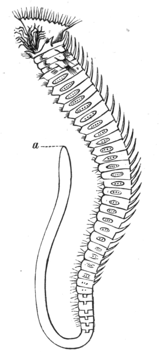
Fig. 131.—Sabellaria alveolata L. × 3. (After Malmgren.) a, Anus.
The Head.—The prostomium is, in the majority of cases, rounded or conical, though it may be square (Nephthys) or elongated and jointed (Glycera), or even hammer-shaped (Tomopteris); or it may be fused with the peristomium, and apparently absent (Arenicola). In the great group Cryptocephala, the peristomium grows forwards so as to hide the prostomium entirely.
In a few of the Nereidiformia the prostomium is compressed, and in the Amphinomidae it is provided with a dorsal ridge or "caruncle," which is a leaf-like process overlapping three or more segments. In many Aphroditidae (as well as in Polydora) there is a peculiar "frontal" ridge passing forwards from the prostomial tentacle, and downwards into the mouth (Figs. 132, c, and 133, A, x).
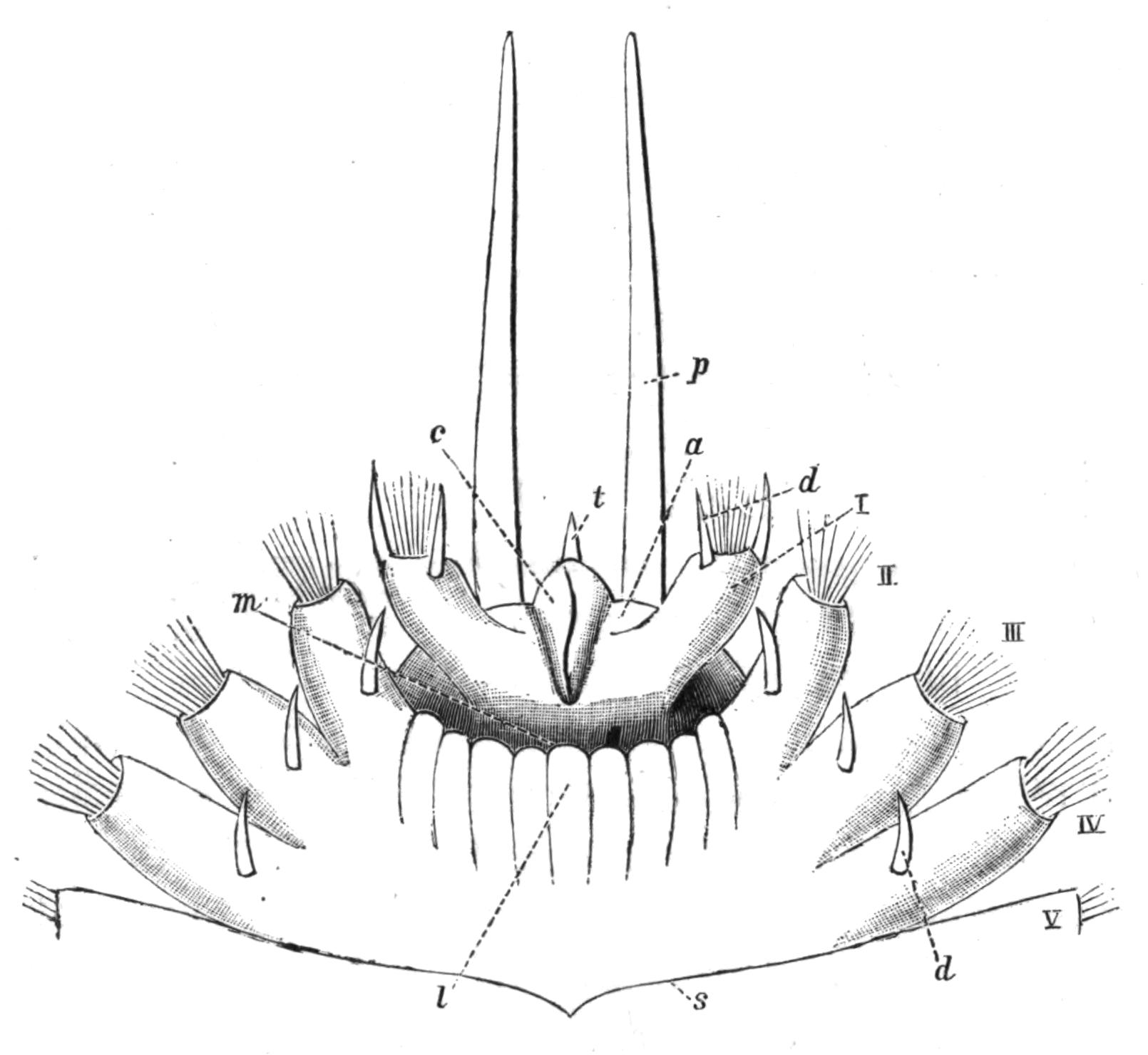
Fig. 132.—Aphrodite aculeata L. Ventral view of anterior region, × 6. a, Prostomium; c, frontal ridge on prostomium; d, neuropodial cirrus; l, lower lip; m, mouth; p, palp; s, intersegmental groove; t, tentacle; I, foot of peristomium, which has shifted forwards so as to lie in front of the mouth; II to V, successive feet.
In all the Nereidiformia, as well as in Sabelliformia and Chlorhaemidae, the prostomium bears sensory processes of two kinds, viz. dorsal tentacles and ventral palps. The latter are invariably two in number, and are particularly well developed in Aphroditidae, Nereidae, Syllidae, some of the Eunicidae, and in Chlorhaemidae. Even when they are apparently absent, as in Nephthys, it is possible that they are represented by certain lobes at the sides of the mouth, for in many Syllidae they are so fused with the prostomium as to be scarcely distinguishable. In the Chlorhaemids the palps[314] are grooved, and in the Sabelliformia they become considerably branched, and extend round the prostomium so as to nearly meet dorsally and ventrally. Each palp is, in this sub-Order, represented by a greater or smaller number of long, mobile filaments, arising from a common base; they are grooved along the inner side, ciliated, and provided with secondary processes. The crown of "gills," in fact, is nothing more than the greatly subdivided and enormously elongated palps, as both Pruvot[315] and Meyer[316] have shown. In such forms as Haplobranchus and Amphicorine the process of subdivision (branching) has only gone a short way. In all the Sabelliformia each filament, in addition to its sensory function, aids in conveying food to the mouth by the action of the cilia, and has a blood-vessel within, thus acting as a respiratory organ. The filament may carry compound eyes (Fig. 143) either at its apex (Branchiomma) or at intervals along its course (Dasychone).
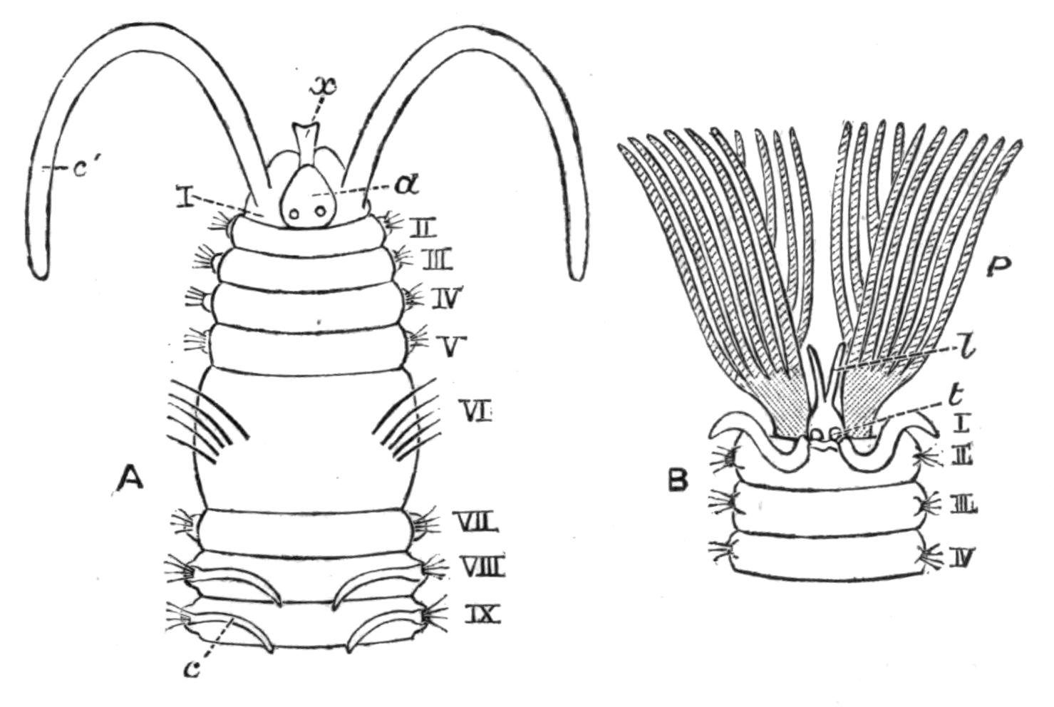
Fig. 133.—A, Anterior end of Polydora enlarged. a, Prostomium; x, frontal ridge; I, peristomium; c', its long cirrus; II, III, etc., the following segments; c, gill; B, head of Sabellid; P, palps (branchial crown); t, position of tentacles; l, processes of upper lip membrane; I, peristomium raised into a collar; II, III, IV, following segments.
In the family Serpulidae one (rarely two) of the most dorsally placed gill filaments is enlarged terminally, and acts as a stopper or "operculum," which closes the mouth of the tube when the animal withdraws into it. Further, in Spirorbis this operculum is grooved on one side, and serves as a brood pouch in which the eggs undergo development (Fig. 184, p. 341). It will be seen, therefore, that the palps may be very important organs for the life of the worm, and they are no less interesting to the comparative anatomist, serving as they do as an excellent illustration of the various uses which Nature finds for one and the same organ.
In the other sub-Orders the prostomium carries neither palps nor tentacles.
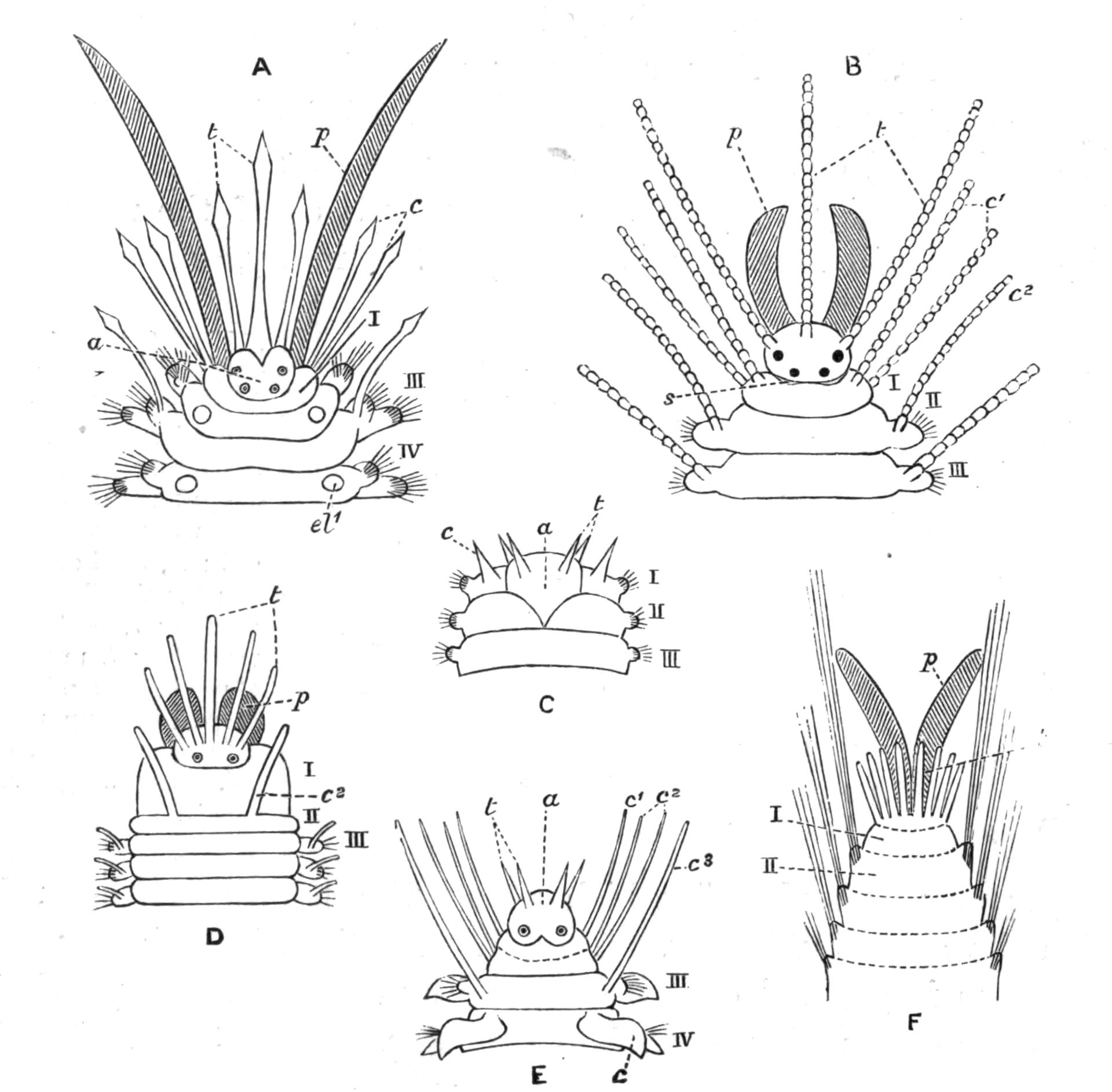
Fig. 134.—Heads of various Polychaeta (diagrammatic). A, Polynoid; B, Syllid; C, Nephthys; D, Eunice; E, Phyllodoce; F, Trophonia: a, prostomium; c, normal cirrus; c1, peristomial cirri; c2, cirrus of second segment; c3, cirrus of third segment; el1 point of attachment of elytron; p, palp: s, nuchal organ (ciliated pit); t, tentacle; I, peristomium; II, III, IV, segments.
The tentacles in the Nereidiformia present a wide variation in number; probably the typical number is three, one of which is median and two lateral—as in Polynoids, Syllidae, and some Eunicidae. Further, there is a certain amount of evidence in the nerve supply of the median tentacle to show that it was originally double. The presence of four tentacles, then, as in Nephthys, Phyllodoce, and Glycera, may be a primitive condition. By the disappearance of the paired lateral tentacles the worm possesses a single median one, as in Aphrodite and Amphinomids;[317] whilst a duplication of these lateral ones leads to the condition of Eunice and Hyalinoecia, which have five tentacles. In the Chlorhaemidae the number is further increased to five or more on each side,[318] and in the Terebellidae these prostomial processes become very numerous.
In the Cryptocephala there is never more than a single pair of tentacles, and these are generally reduced to a group of sensory cells, though in Sabellaria they retain a considerable size.
In a few genera, such as Aphrodite, Nephthys, Capitella, the first postoral segment is distinguished from the succeeding segments only by its position with regard to the mouth (Fig. 132) and by its smaller size. But in the remainder of the Polychaeta, with here and there an exception, the peristomium is achaetous in the adult.[319]
Except in the Nereidiformia, peristomial or tentacular cirri are rare, being represented in the Spioniformia by the very long "tentacles." In the Nereidiformia one or more of the following segments may be added to the peristomium, and share in the "cephalisation," which is so characteristic a feature in this group. In Amphinomids the first three or four chaetigerous segments are incomplete ventrally, owing to the shifting of the mouth backwards; these segments form lateral lips, but they are not otherwise modified. In Phyllodoce, however, there are four cirri on each side of the mouth, and from the arrangement in the Alciopids we are justified in concluding that the segment which carries the four pairs of cirri is really made up of three segments (Fig. 134, E). Among the Hesionids there are four such "cephalised" achaetous segments with long cirri.
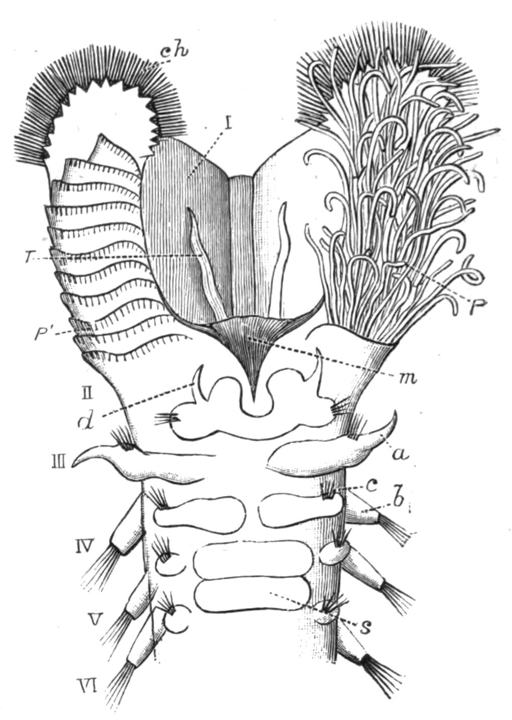
Fig. 135.—Sabellaria alveolata L. Ventral view of anterior region, × 10. a, Notopodial cirrus; b, notopodium; c, neuropodium; ch, peristomial chaetae; d, neuropodial cirrus; m, mouth; P, multifid palp (gill filaments); P', ridges after removal of gill filaments; s, ventral (tubiparous) gland shield; T, tentacle; I, hood formed by peristomium; II to VI, following segments.
In a few cases, such as the Chlorhaemids and Sternaspis, and to a slight degree in Arenicola, the "head" and even the anterior part of the worm is capable of being withdrawn into the body.
The Parapodia and Chaetae.—The typical parts of a parapodium have been described in the preceding chapter; here it is only necessary to refer to the series of diagrams (Figs. 136, 137) representing the parapodia of the more common Polychaetes, and to add a few remarks about them.
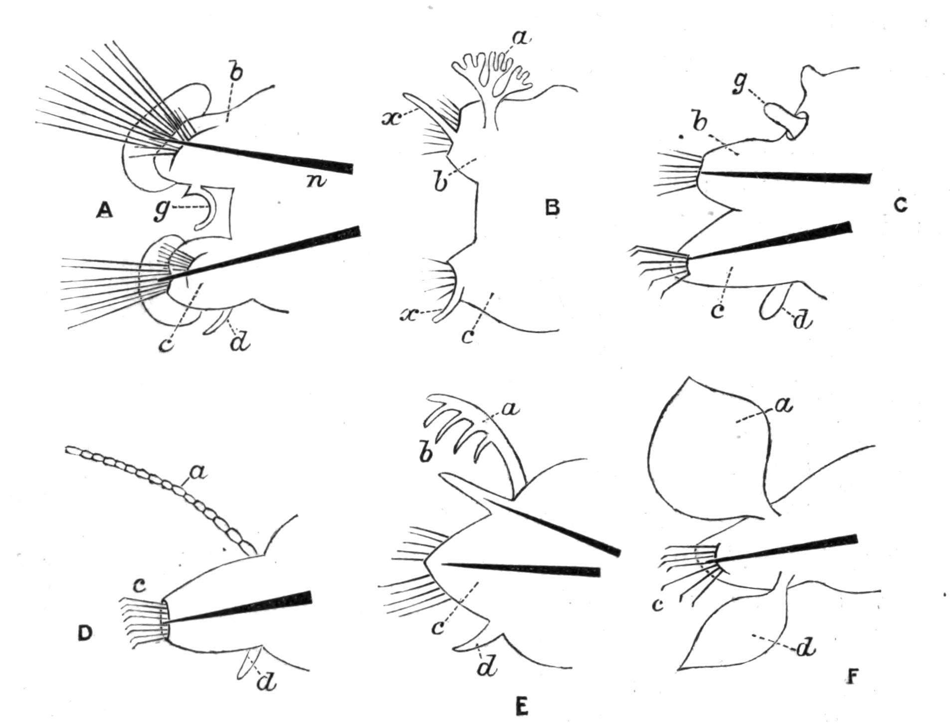
Fig. 136.—Parapodia. A, Nephthys; B, Amphinome; C, Glycera (the unlettered lobe above g is the notopodial cirrus); D, Syllis; E, Eunice; F, Phyllodoce. a, Notopodial cirrus; b, notopodium; c, neuropodium; d, neuropodial cirrus; g, special gill; n, aciculum (omitted in B); x, cirriform lip of chaetigerous sac.
In most Annelids the chaetae are in two bundles on each side, but there are certain families in which the dorsal bundle, and even the notopodium itself, is absent, as in the Eunicidae, Syllidae, and Phyllodocidae; or the dorsal bundle may be absent only in certain regions of the body, as in the hind-body of Terebellids. In some Amphinomidae and Aphroditidae the notopodium is scarcely distinct as a separate lobe, being a slight tubercle on the upper surface of the neuropodium; but the notopodial chaetae are present, and indeed particularly well developed in many cases.
But whilst, in the Nereidiformia, the parapodia, whether consisting of two lobes or only one, are always well developed, and project to a more or less pronounced degree from the sides of the body, it is otherwise in the rest of the group, where the chaetigerous lobes are usually reduced to mere tubercles or ridges, no doubt in relation to their burrowing or tubicolous habits. In Sternaspis the chaetae issue directly from the body-wall.
Amongst the Nereidiformia we find examples in which the parapodia, instead of being more or less conical "legs," are flattened fore and aft so as to serve as efficient "fins," as in the active swimmers, Nereis virens and Nephthys caeca, and in the pelagic Phyllodocids, Alciopids, Typhloscolecids, and Tomopteris.
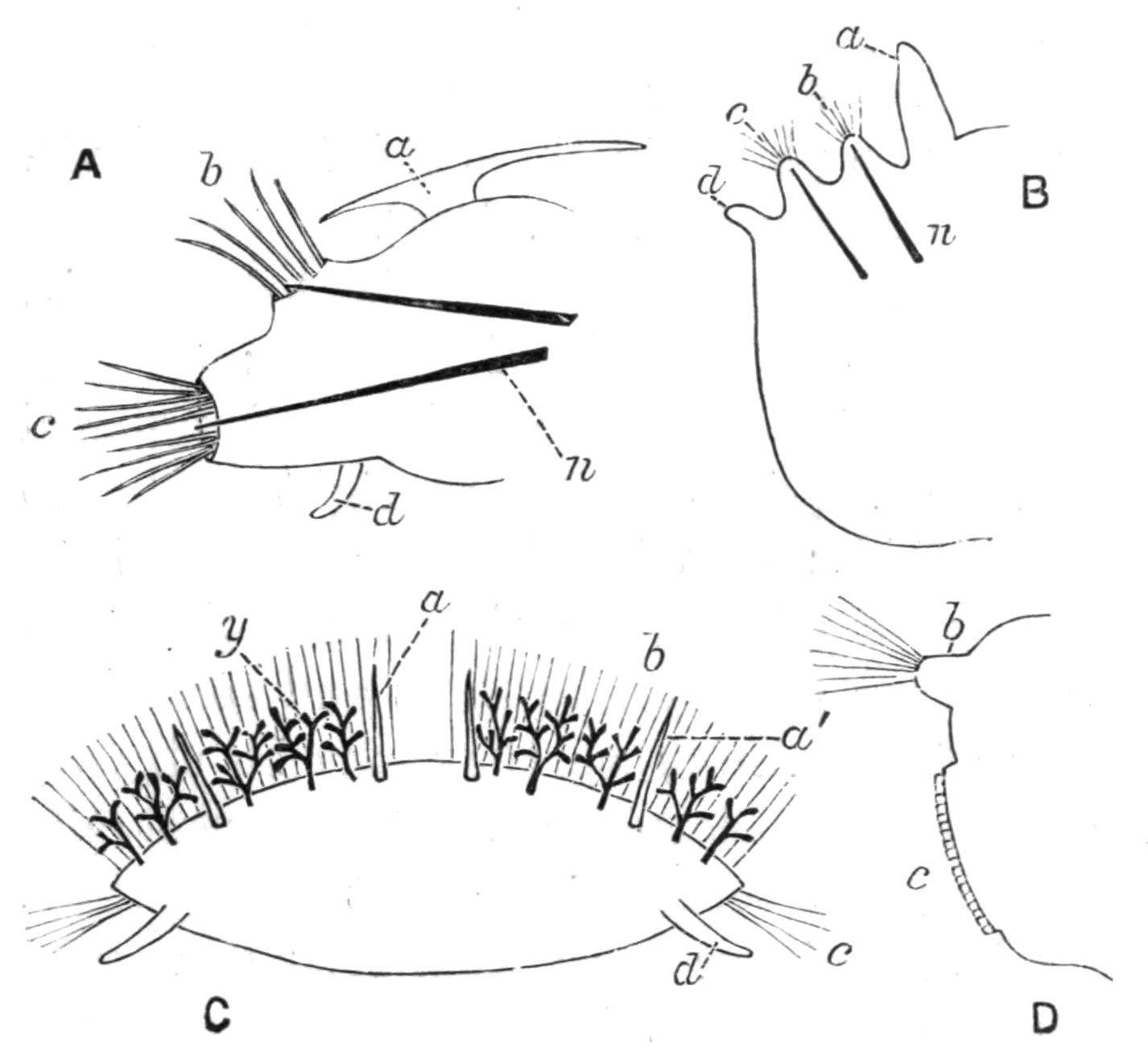
Fig. 137.—Parapodia. A, Polynoë; B, Scoloplos; C, Euphrosyne. (Transverse section of body.) a', Accessory cirrus; y, doubtful branchiae; D, Sabella (thoracic). a, Notopodial cirrus ("elytron" in A, "gill" in B); b, notopodium; c, neuropodium; d, neuropodial cirrus; n, aciculum (accidentally omitted in C).
Of the typical dorsal and ventral cirri, the ventral is only absent in some Amphinomids amongst the Nereidiformia; the dorsal is absent in Nephthys and degenerate in Glycera, whilst in a very large number of families of the other sub-Orders neither cirrus is present. These cirri, though originally filamentous and sensory, may, by virtue of special blood supply, become "gills," and this occurs in several families of different sub-Orders. Thus in Eunice this gill is comb-like; in Amphinome and in Arenicola (on certain segments) it is arborescent, as it is also in one to three segments in Terebellids; whilst in Ariciidae, Spioniformia, Cirratulidae, Opheliidae, and Sabellaria it remains more or less finger-shaped or filamentous. In the family Serpulidae the thoracic cirri, both dorsal and ventral, become flattened and extended antero-posteriorly, and unite with one another to form the "thoracic membrane."[320] In Phyllodocidae the cirri are foliaceous and natatory, and they contain a great quantity of glands of a peculiar character. The Aphroditidae are distinguished from other Annelids by the possession of "elytra" or dorsal scales, which appear to be the dorso-ventrally flattened cirri, retaining their sensory nature, but adding to this function several others.[321]
The chaetae or bristles are mainly used in locomotion, but it is not unreasonable to believe that some of the stronger, serrated kinds may be used as weapons of offence and defence; certainly the Polynoids, bristling as they do with stiff chaetae along each side, must be rather unpleasant to their smaller enemies.
The various bristles may be placed in three chief groups, viz. (1) simple; (2) jointed; (3) uncini (see Fig. 138).
(1) The simple chaetae may be smooth and hair-shaped, i.e. "capillary," such as are present in nearly all families: or they may be forked (Amphinomidae), comb-shaped (Eunice), notched or serrated, or provided with a series of frills at right angles to their length, as in Aphroditidae; or fringed along one or both sides with a membranous expansion, as in Terebellids and Sabellids. The simple chaetae may also be short and spine-like, as in the ventral bundles of Arenicola; or they may be slightly curved at the end and notched, forming what are generally termed "crotchets," such as are common amongst Oligochaeta. These "crotchets" may be simple, or have numerous denticulations at the end (Maldanidae), or be provided with a membranous hood (Spioniformia, Capitelliformia). In Hermione peculiar sheathed, spear-like bristles occur (Fig. 138, N).
(2) Jointed chaetae have already been described (p. 246); they are confined to the sub-Order Nereidiformia, and occur only in certain families.
(3) The uncini are very short chaetae, which are simply embedded in the skin, and do not extend beyond the body-wall into the body-cavity. An uncinus is a sharply curved hook, which may have more or less numerous secondary teeth on it. They are characteristic of the Sabelliformia and the Terebelliformia.
The chaetae appear as solid, usually fibrillated structures, of a yellow or golden tint, transparent and refringent. Chemically they consist of chitin, and each chaeta is the product of a single cell. The chaetae of Euphrosyne are hollow and calcareous, being peculiar in both characters.
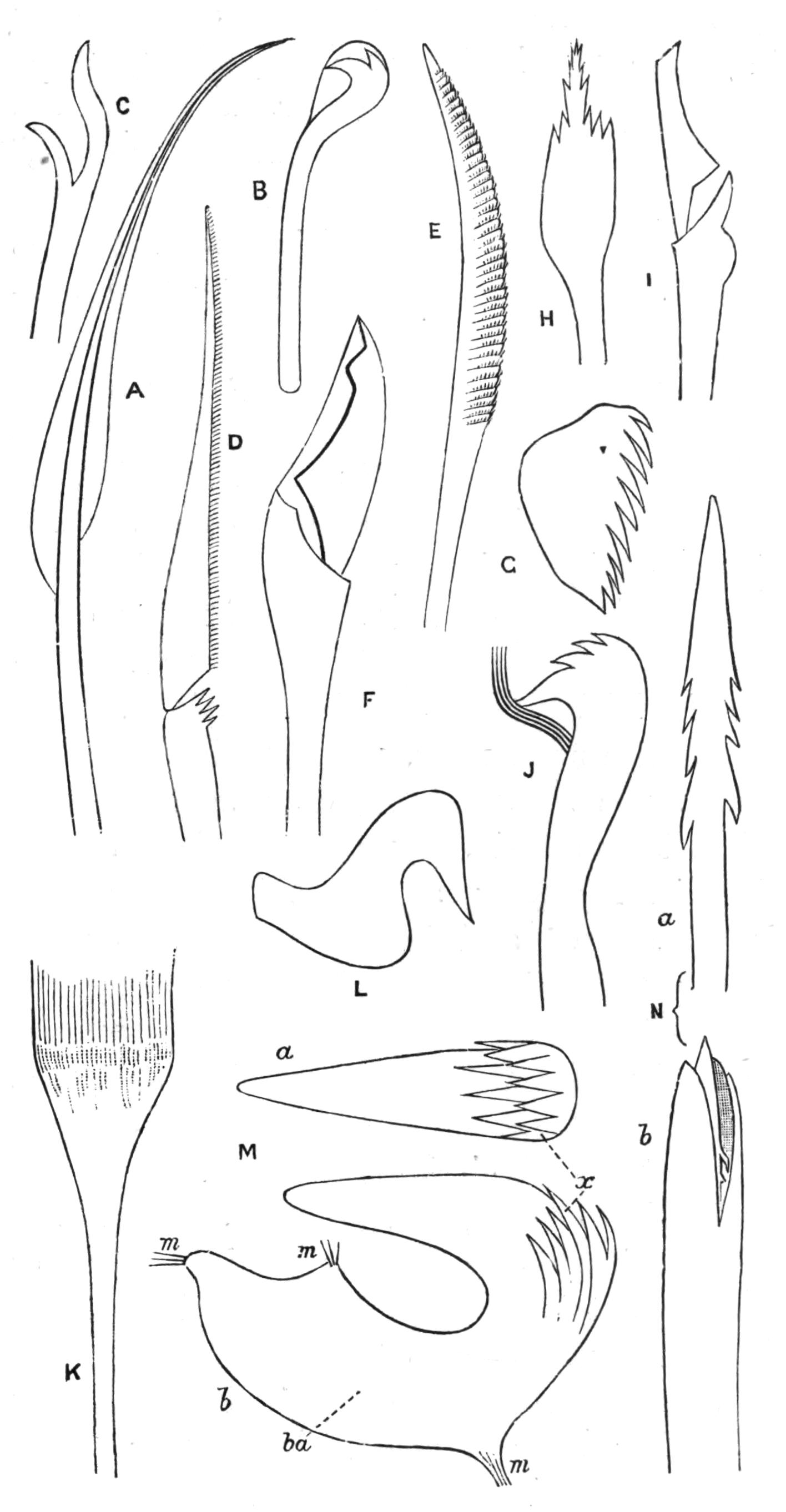
Fig. 138.—Chaetae of various Polychaetes (the magnification is not the same in all cases). A, Doubly-fringed capillary, from Terebellid; B, hooded crotchet, from Polydora; C, a fork, from Euphrosyne; D, jointed chaeta, from Phyllodoce; E, simple chaeta, with serrated ridges or frills, from a Polynoid; F, jointed chaeta, from Eunice; G, uncinus, from Pomatoceros (Serpulid); H, one of the outer series of paleae from the hood of Sabellaria spinulosa; I, jointed chaeta, from a Syllid; J, multidenticulate crotchet, from a Maldanid; K, comb-shaped chaeta, from Eunice; L, uncinus of a Sabellid; M, uncinus of Terebellid (Amphitrite Johnstoni); a, edgewise; b, side view; m, attachments of muscles into ba, basal plate; x, accessory teeth. N, Sheathed spear of Hermione; a, the spear-shaped capillary removed from its sheath; b, the same, with sheath.
Certain modifications of the chaetae presented by various worms deserve mention. In Polydora (Fig. 133, A) and in Chaetopterus (Fig. 173, p. 324) those of one segment are especially strong, but their significance is uncertain. In Capitella those of the notopodium of the eighth and ninth segments are specially modified; they are analogous to the copulatory chaetae of Oligochaeta. In Aphrodite, in addition to the ordinary locomotor chaetae, there are brilliant, iridescent bristles and peculiar felting threads arising from the indistinct notopodium; these latter, however, are not true "chaetae," but are separate chitinous filaments similar to the constituent fibres of an ordinary chaeta.[322]
While the chaetae in the Nereidiformia and others are grouped in bundles, those of many other families are in vertical, transverse rows, as in Maldanidae and in Arenicola. The uncini are always embedded in such rows, usually slightly raised from the general level of the body surface, each being termed a "torus uncinigerus." These tori are usually limited to the sides of the body, but in Myxicola and in Notomastus they encroach upon the dorsal surface, and in Chaetozone, also upon the ventral, so as nearly to encircle the body, recalling the "perichaetous" condition of some earth-worms.
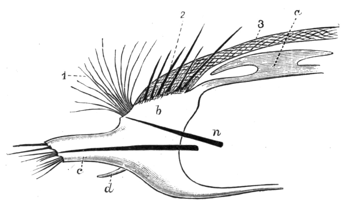
Fig. 139.—Aphrodite. Foot, × 2. a, Elytron; b, notopodium; c, neuropodium; d, neuropodial cirrus; n, aciculum; 1, iridescent bristles; 2, stiff chaetae; 3, felt.
Gills.—We have already seen that several different organs, e.g. the palps in Sabelliformia, the prostomial tentacles of Chlorhaemidae, and the notopodial cirri of sundry other Polychaetes, may take on a respiratory function. There are, however, certain "gills" developed either on the parapodium itself or elsewhere on the body which it is difficult to homologise. Such are the retractile gills on the parapodia of the Glyceridae (Fig. 136, C); those of Dasybranchus, near the abdominal neuropodia; those of Mastobranchus, near the notopodia. Nephthys has a sickle-shaped gill on the under surface of the notopodium. The long gill filaments at the posterior end of Sternaspis, again, are only doubtfully interpreted as the dorsal cirri of some of the posterior segments.
Since primitively the whole skin of the worm is respiratory, any part of the skin may become more or less specialised for this function, and chiefly, of course, on the more actively moving parapodia. The blood-vessels constituting the essential part of the "gill" may make use of any already existing outgrowth (such as a cirrus or a tentacle), or may push the body-wall out on their own account.
Internal Anatomy.
Probably those organs which have the greatest effect in modifying the shape of the body are the septa, for we find in the long, free-swimming worms that these are regularly present throughout the body, and external "segmentation" of the body is well marked. In burrowing and tubicolous forms the septa are frequently incompletely developed, or more or fewer may be absent; and the body becomes less distinctly segmented externally, tends to vary greatly in diameter during movement, or becomes plumper. With the disappearance of the septa there is also a diminution in the number of nephridia, as in Arenicola, with only six pairs. Further, there is frequently a dimorphism of these organs; instead of all of them serving equally as excretory organs and as genital ducts, some of the most anterior in the Sabelliformia and Terebelliformia become greatly enlarged, and take on practically the whole of the former function; whilst more or fewer of the posterior nephridia dwindle in size, and become genital ducts. The absence of septa allows a free communication between the successive segments, and thus a freer flow of coelomic fluid for the distension of the anterior end of the worm during burrowing.
The alimentary system presents certain modifications of a systematic value. In the Nereidiformia the muscular pharynx, which is always protrusible and is preceded by an eversible buccal region, frequently encloses thickened cuticular plates which serve as crushing and grasping organs. The form, number, and arrangement of these "jaws" vary in the different families. They form valuable fossil records of extinct worms.
In the Scoleciformia and Capitelliformia the buccal region exists, but there are no jaws. In the Sabelliformia and Terebelliformia eversion does not take place and jaws are absent.
Amongst the Nereidiformia the jaws are absent in the Phyllodocidae and Hesionidae; when present they are usually set in the direct course of the food. There may be one small tooth used for stabbing, as in some Syllids (Fig. 141, A); or a circle of such denticles (Autolytus, Fig. 140, D). To these are added powerful grasping jaws in Nereis (E); or the latter may alone be present, as in Glycera (F). In Polynoë the four jaws are carried by hard pieces, to which the muscles are attached (C and G). In Nephthys there is a dorsal and a ventral jaw.
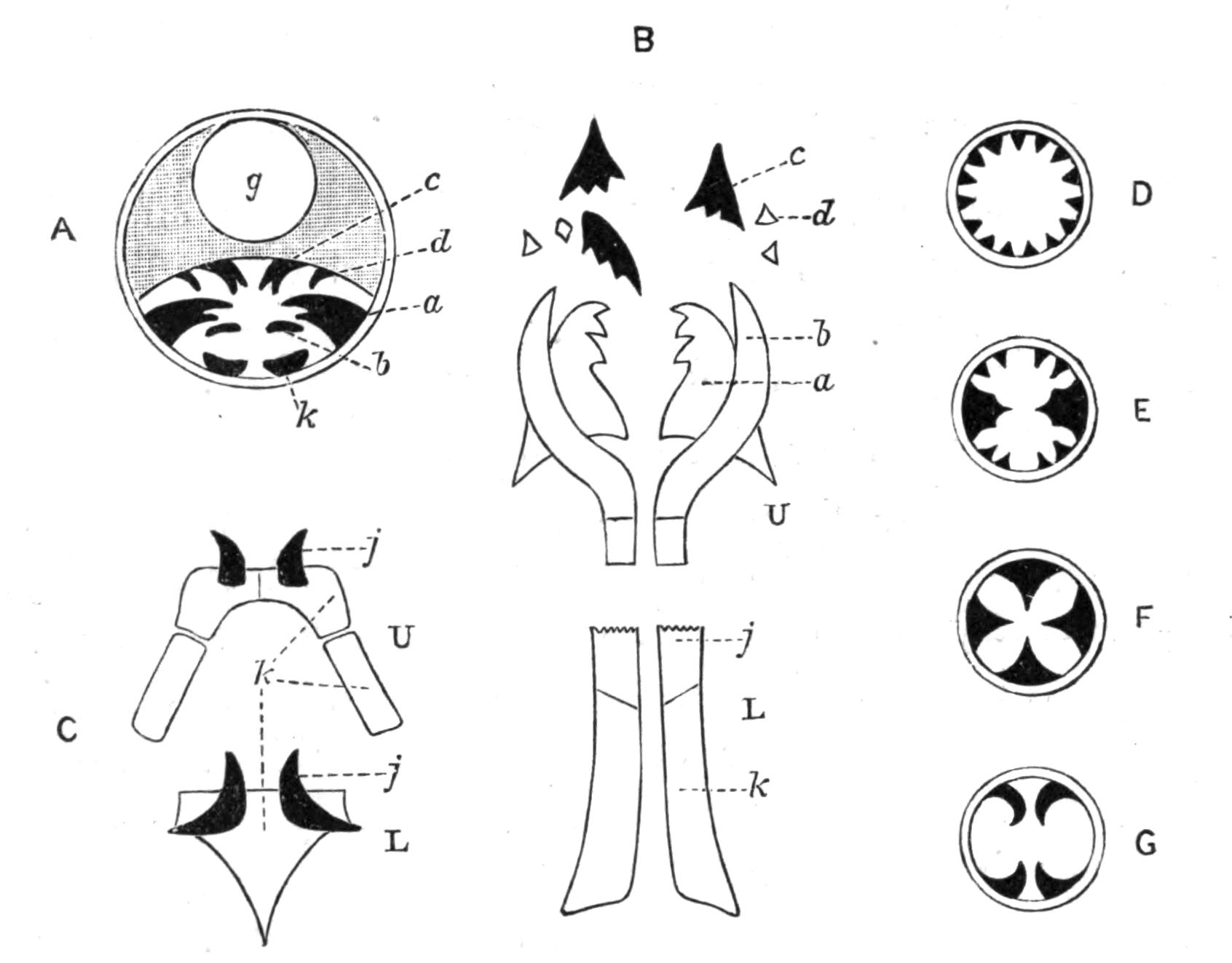
Fig. 140.—Jaws of various Chaetopods. A, Transverse section of the anterior end of Eunice; a, b, c, d, various parts of the upper series of denticles lying in a special chamber; g, oesophagus; k, lower jaw: B, the denticles of Eunice separated; U, upper series; a, grinder; b, forceps; c, rasping plates; d, grater; L, lower series; j, tooth; k, base into which muscles are inserted: C, Polynoid; U, upper, and L, lower jaws; j, tooth; k, base: D, Diagrammatic section across pharynx of Autolytus; E, of Nereis; F, of Glycera; G, of Polynoë.
In the Eunicidae, however, the numerous denticles are carried in a special pouch below the food tract, with which it communicates anteriorly.[323] They are arranged in an upper and lower series. The lower series (L) consists of a pair of flat plates (k) on each side partially embedded in and acted upon by muscles, with a harder enamelled piece—the actual lower "tooth" (j)—at its anterior end. The upper series (U) consists of several pieces, varying in shape and size in the various genera of this family; but developmentally they result from modifications of two rows of small, similar pieces.[324]
The intestine is generally straight and cylindrical, and is usually constricted by the septa, if these are present. In the Polynoids the intervening sacculations become so long as to receive the name of "caeca," which, in Aphrodite, become enormously elongated (Fig. 142); there are eighteen pairs of them (c), each being a slender tube bent upon itself, giving off short branches and dilated distally, where it lies in the base of the parapodium.
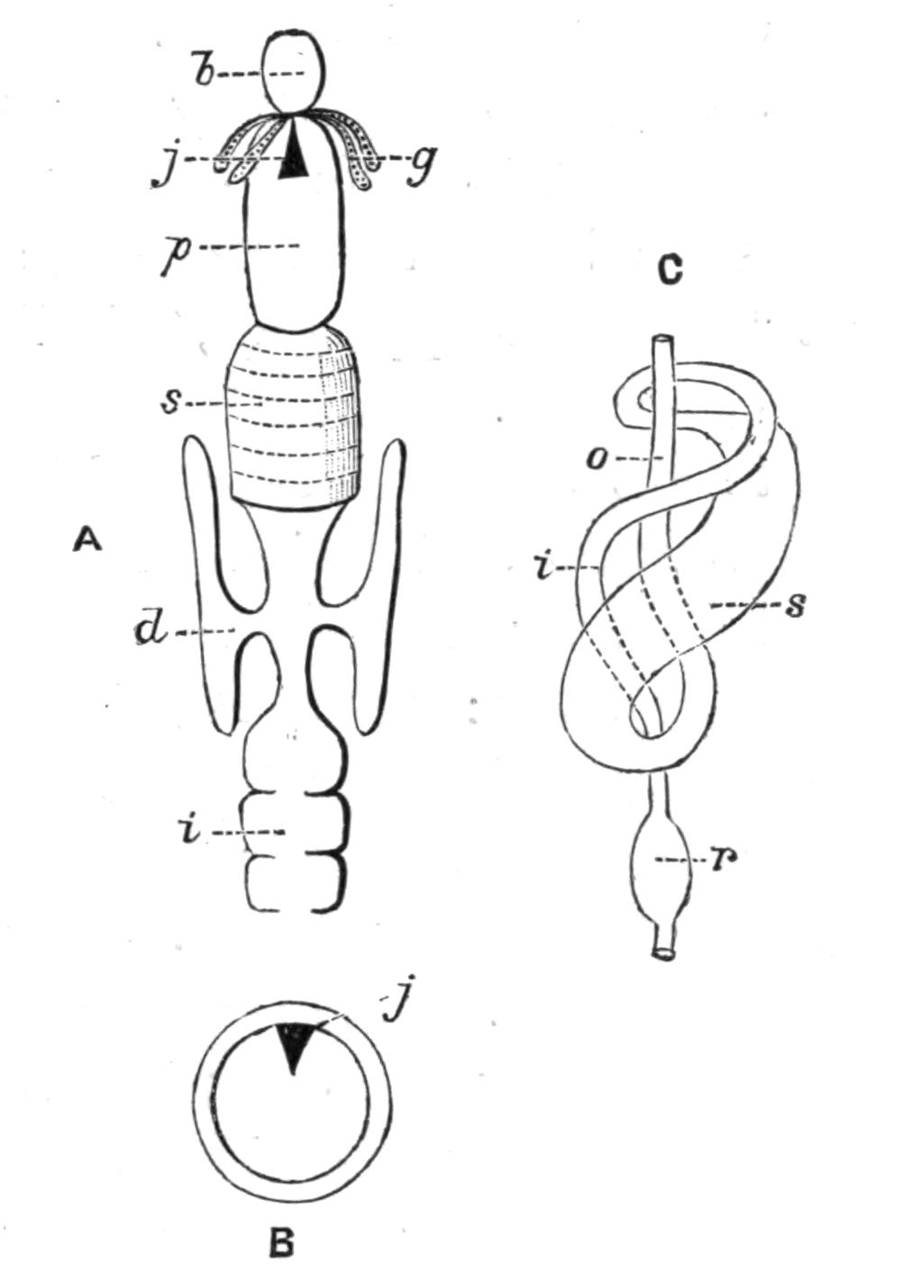
Fig. 141.—A, Alimentary canal of Syllid: B, transverse section of pharynx of the same; b, buccal region; d, oesophageal outgrowth; g, salivary glands; i, intestine; j, tooth; p, pharynx; s, gizzard: C, alimentary canal of Petta (after Wirén); i, intestine; o, oesophagus; r, rectum; s, stomach.
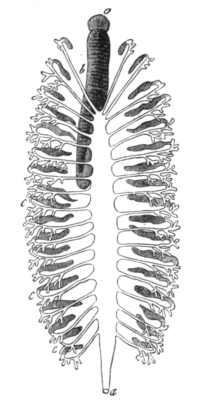
Fig. 142.—Alimentary canal of Aphrodite. × 1. (From Gegenbaur.) a, Anus; b, pharynx; c, caeca; o, mouth.
The intestine is looped in a few genera only, as in Trophonia, or coiled, as in Sternaspis, Petta (Amphictenid, Fig. 141, C), and Ammotrypane. In the course of the tube there may be a thick-walled muscular gizzard, with hard chitinous lining, as in certain Terebellids, where it appears to replace, in function, the pharynx of the Nereidiformia; in the Syllidae the gizzard is present in addition to the pharynx (Fig. 141, A).
Glandular appendages of the oesophagus are present in many worms. Amongst the Nereidiformia, the Syllidae and Hesionidae possess oesophageal diverticula (Fig. 141, A, d), which are used, not for secreting a digestive fluid, but as reservoirs for water and air swallowed by the worms; and are provided with muscular walls, by which their contents can be driven out. They appear, in fact, to be used like the swim-bladder of fishes.[325] Many Chaetopods take in water by the anus—no doubt for respiratory purposes—and pass it forwards along the intestine. In the Capitelliformia a special groove conducts the water for some distance, then the groove becomes closed to form a canal, which, after a course forwards as a free tube below the intestine, again enters the latter, constituting a "siphonal apparatus," similar to that of the Echiuroids and the sea urchins.
Sense Organs.—In addition to the prostomial eyes, which are present in nearly all the Nereidiformia and Spioniformia, eyes may exist elsewhere on the body: thus Myxicola infundibulum and Fabricia possess a pair on the anal segment; in M. aesthetica Clap. there is a pair to every segment; in Branchiomma there is a compound eye near the tip of each gill filament (i.e. palp); whilst in Dasychone a series occurs along each gill filament. All these examples belong to the Cryptocephala, in which, owing to certain peculiar modes of life, these sense organs are required in correspondingly peculiar positions. It is usually stated that Polyophthalmus possesses, in addition to the usual prostomial eyes, twelve pairs on as many successive segments; but the minute structure of these organs points rather to their function as light-producing organs.
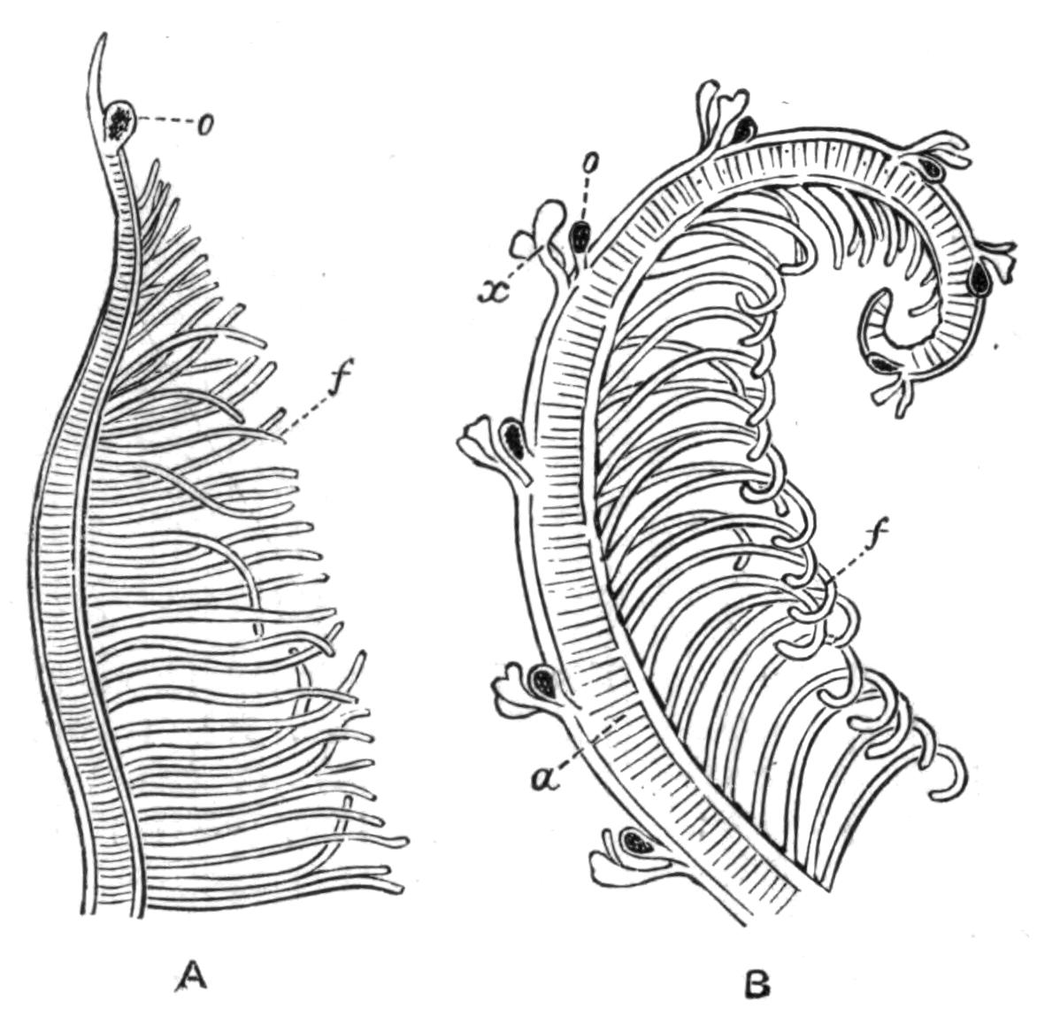
Fig. 143.—A gill filament, A, of Branchiomma, B, of Dasychone. a, Axis; f, secondary filaments; o, compound eye; x, lappets.
The Capitelliformia and Opheliidae possess a pair of peculiar "ciliated pits" or "nuchal organs" at the upper side of the head, between the prostomium and peristomium, and capable of eversion (Fig. 144). They are most characteristically developed in the Capitelliformia, where each organ abuts upon a special lobe of the brain. The function of these "ciliated organs," which bear a great resemblance to those of the Nemertines, is a matter of speculation. Similar organs, in the form of simple pits or grooves, occur in many of the Nereidiformia, Terebelliformia, and others.[326]
Otocysts are rare. Arenicola possesses a pair at the base of the prostomium, each of which in some species retains an opening to the exterior.[327] They probably serve as "organs of direction" rather than of "hearing." Aricia and Polyophthalmus likewise have such organs on the prostomium; whilst Fabricia, Myxicola, Terebella, and a few others possess them in the peristomium, or in some other segment of the body.
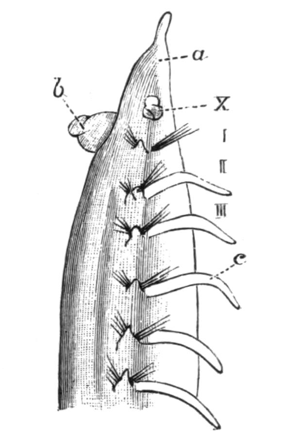
Fig. 144.—Ammotrypane aulogaster Rathke, enlarged. (From Cuningham.) Anterior end. a, Prostomium; b, everted buccal region; c, notopodial cirrus; X, ciliated organ everted; I, II, III, first three segments.
Reproductive Phenomena.—With a few exceptions mentioned below, the Polychaeta are unisexual. The sexual cells are developed in all cases from the lining epithelium of the body-cavity. The exact spot at which this occurs varies in different cases; it may be, though rarely, on the floor of the body-cavity; it is more usually on the wall of some blood-vessel, either the ventral vessel or on branches of it; or on the many blind blood-vessels of Aphrodite. The number of such genital organs is very great in most worms, but in those presenting two regions of the body they are confined to the posterior segments (Sabelliformia, Terebelliformia, Capitelliformia). The number is very limited in Arenicola and other worms presenting but few nephridia: in the former genus there being six pairs, in Trophonia only one pair.
The following genera are hermaphrodite:—Amphiglena, Salmacina, Protula, Spirorbis, belonging to the Sabelliformia, to which must be added some Hesionidae. In this family ova and spermatozoa are developed around the same blood-vessel. But in the former group of worms (as also in Ophryotrocha) the two kinds of cells are produced in different regions of the body. Thus in Protula the anterior abdominal segments are male, the posterior ones female, while in Spirorbis the reverse arrangement holds; and in Syllis corruscans the anterior segments of the body contain eggs, whilst the posterior region contains spermatozoa, and this region separates and becomes a male worm.
The eggs and spermatozoa in the Polychaeta are discharged into the sea either by rupture of the body-wall or through the nephridia; the male and female elements unite, and the resulting fertilised eggs undergo development, either floating separately in the water, or embedded in jelly, or attached to the body or to the tube of the worm.
The result of the segmentation of the egg is a free-swimming larva known as a "Trochosphere," similar to that of Polygordius. The larvae of different species present various more or less marked departures from this type, for instead of the two girdles of cilia there may be only the anterior girdle, or there may be several complete or incomplete girdles between the two typical ones, or there may be (Chaetopterids) only a single girdle of cilia about the middle of the body, the two typical girdles being absent.[328] The postoral region, after elongation, generally becomes marked out into three segments, and these segments develop chaetae, which are usually temporary and specially long.
The little animal is thus equipped for an independent life: the provisional chaetae help in keeping it balanced; and in some cases (Spionidae) serve to protect the little soft creature, for when it is touched it curls up, and its chaetae stick out at the sides, so that it looks like a hairy caterpillar. But the larva is quite at the mercy of the sea, for it is carried hither and thither by currents, and in this way the species is disseminated. The larvae of the Polychaetes, like those of other animals, occur at certain periods of the year in large quantities at the surface of the sea, and serve as food for various larger animals.
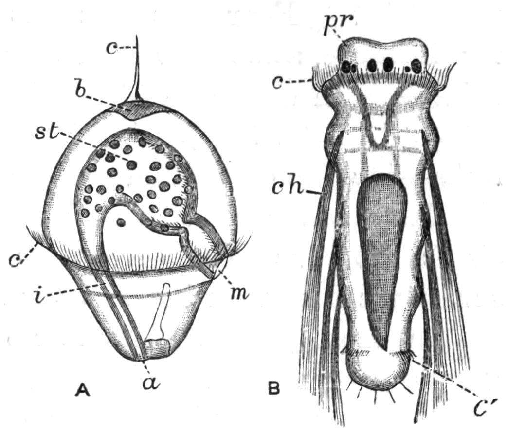
Fig. 145.—A, Trochosphere of Nephthys. × 65. a, Anus; b, apical plate (brain); c, apical tuft of cilia; c', girdle of cilia; i, intestine; m, mouth; st, stomach. B, Larva of Spio, with three segments, eight days old. × 100. c, Preoral girdle of cilia; c', preanal girdle; ch, long provisional chaetae; pr, prostomium with eyes. (From Claparède and Metschnikoff.)
These larvae are at first very different from the adult animal, and the necessary changes to be passed through are more or less great according to the species. It is not our intention to describe these changes in detail.[329] The larva increases in size, the permanent chaetae make their appearance in regular order, and the body exhibits segmentation, the new segments always appearing just in front of the anal segment. The internal organs gradually develop, and the prostomial and parapodial appendages grow out in their turn. In the Sabelliformia the multifilamentous "gills" arise by the continued branching of an at first simple process (the palp) arising from the latero-ventral surface of each side of the preoral lobe.[330] These gradually encroach dorsally and ventrally till the prostomium is more or less encircled; meanwhile the peristomium grows forwards so as to conceal the prostomium, which no longer increases at the same rate as does the rest of the body.
Although most worms appear to discharge their ova directly into the sea and take no further care of them, some make provision for their offspring either by laying the eggs in a jelly, which will serve as food for the young larvae—Aricia, Ophelia, Protula, Phyllodoce—or by attaching them to their body. In certain Polynoids the eggs are attached by means of a secretion to the back, under the elytra, where they undergo development up to a certain stage. In Exogone and some other Syllids they are attached to the ventral cirri, or in Grubea limbata, all over the back. In the female Autolytus (Sacconereis) a ventrally-placed brood sac is formed by the hardening of a secretion; the eggs develop into embryos inside the brood sac, and then become free, with head appendages and three pairs of parapodia. Enormous numbers of such embryos may occur; for instance, some 300 were counted in a brood sac of Autolytus ebiensis. In the case of tubicolous worms, the eggs are frequently attached to the tube, either inside or outside. In Spirorbis and Salmacina the operculum serves as a brood pouch.
Only a very few species are known to be viviparous, viz. Syllis vivipara Kr., Cirratulus chrysoderma Clap., Marphysa sanguinea Mont., and Nereis diversicolor Müll.
In most genera there is no external difference between a mature worm filled with generative products and an immature one, except, it may be, in the colour; for the yolk of the eggs is frequently tinted yellow, or pink, or bluish, while the spermatozoa in mass are white; so that the normal colouring of the worm may be modified when filled with these elements. But in a few instances striking anatomical peculiarities are exhibited by the mature worm.[331] In many species of Nereis, for instance, those segments containing the generative products undergo more or less extensive changes, while the anterior ones remain unaltered. The body of the ripe Nereis is then distinguishable into an anterior non-sexual region and a posterior sexual region; and so great are these changes in certain species that the mature worms were for a long time believed to belong to a different genus, and received the name Heteronereis. But we now know their true relations, thanks to the work of Claparède and others. The males in the Heteronereid phase have fewer unaltered anterior segments than the females, so that there is a sexual dimorphism.
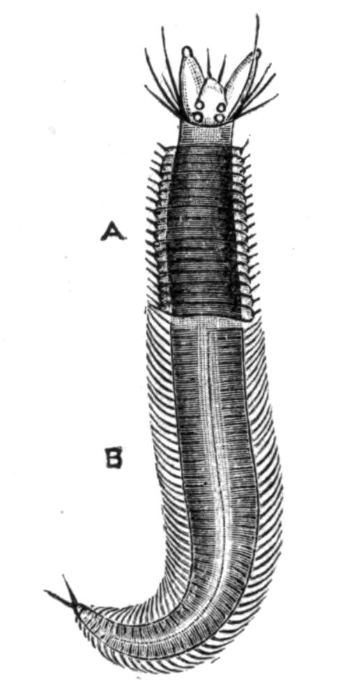
Fig. 146.—Male "Heteronereis" of N. pelagica L. × 1. A, Non-sexual region; B, sexual, modified region. (From Ehlers.)
The changes which Nereis undergoes in its transformation affect chiefly (a) the shape of the parapodia, and (b) the form of the chaetae of these parapodia. Other organs may also be affected, though less noticeably; thus the eyes become enlarged, the intestine may become so compressed by the generative products as to be functionless, and the tail develops special sensory papillae.[332]
In the parapodia an increase in size and a sharper delineation of the various parts take place; then flattened foliaceous outgrowths (Fig. 147, x, y) arise from certain lobes of the feet, in which, too, the blood supply becomes greatly increased. The old chaetae are pushed out by the development of new ones of quite a different shape; these are jointed like the old ones, but the appendix is, in many species at least, flattened and oar-shaped (Fig. 123, C, p. 246); and the chaetae are arranged in a fan-like manner. Both these modifications are in evident relation to the free-swimming habit which the Heteronereid now adopts. The new foot serves as a swimming organ, the old one was a walking appendage.
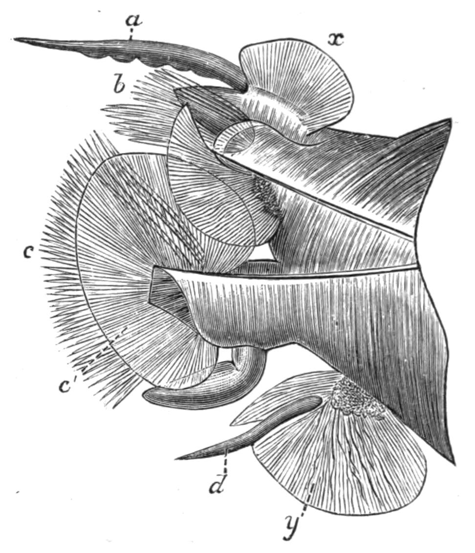
Fig. 147.—Parapodium of male "Heteronereis" of N. pelagica L. × 10. (From Ehlers.) a, Notopodial cirrus; b, notopodium; c, neuropodium with new chaetae; c', foliaceous outgrowth; d, neuropodial cirrus; x, y, foliaceous outgrowths.
Whilst some species, such as the common British N. diversicolor, undergo no change, and others become modified as just described, others, again, are polymorphic. Claparède was the first to show that N. dumerilii may occur in at least five different mature forms; these differ from one another in size, colour, mode of life, character of the eggs, etc. The immature forms may become ripe and lay eggs while still retaining the "Nereid" characteristics, or these immature forms may become "Heteronereids"[333] whilst the sexual elements are ripening. There are then three different kinds of males and of females in this one species, some being found at the bottom of the sea, as the large Heteronereid form, while the small Heteronereid swims on the surface. The relations of these various forms to one another, and the causes leading to the assumption of a Heteronereid condition in some cases and not in others, are unknown.
A somewhat similar phenomenon is exhibited by members of the family Syllidae.[334] In this family sexual reproduction is frequently accompanied by the asexual modes of fission and gemmation. In some genera, such as Eusyllis, Odontosyllis, and Exogone, there occur changes quite similar to those characterising "Heteronereis"—that is, the posterior segments in which the genital organs exist become altered, so that the worm consists of two distinct regions, and is termed a "Heterosyllis." The most marked change is the appearance of a dorsal bundle of long capilliform chaetae in each of the genital segments (Fig. 148, I).
But in other genera the hinder genital region of the body becomes separated, on maturity, from the anterior non-sexual region. Various stages of this "schizogamy," or fission into a sexual and a non-sexual zooid, have been observed in different genera. In the genus Syllis the first segment of the sexual zooid, after its separation from the asexual zooid, proceeds to bud forth a head. The character of the head is alike in both sexes, though different species present heads of different shapes; and as the worms were originally described as distinct genera, the names then given are retained as descriptive terms. Thus the "Chaetosyllis" form has only two tentacles; the "Ioda" form has three tentacles and a pair of palps. One and the same species (e.g. S. hyalina) may successively pass through these stages.
With regard to the asexual portion, there is a regeneration of the tail segments after the sexual zooid has separated; and the number of segments so regenerated is usually equal to those that have become sexual. After a time these newly formed segments will produce generative organs, and take on the characteristic natatory chaetae, and this region will in its turn separate.
But in other genera, such as Autolytus, the regeneration of segments may commence before the separation of the sexual zooid; and the head of the sexual zooid becomes budded out before separation from the asexual portion. So that the animal now consists of two worms, each with its own head, separated by a region or zone of proliferation (Fig. 148, IV). Moreover, in some species not only is the hinder part of the body converted into a sexual zooid, but the zone of proliferation becomes very active, and produces by gemmation a large number of segments, which become marked out, by the appearance of heads at intervals, into a number of zooids, in which genital organs will later make their appearance. A chain of as many as sixteen zooids may be formed in Autolytus (Fig. 148, V)—the hindermost by conversion of the hinder part of the body of the original "stock," the intervening zooids by gemmation.
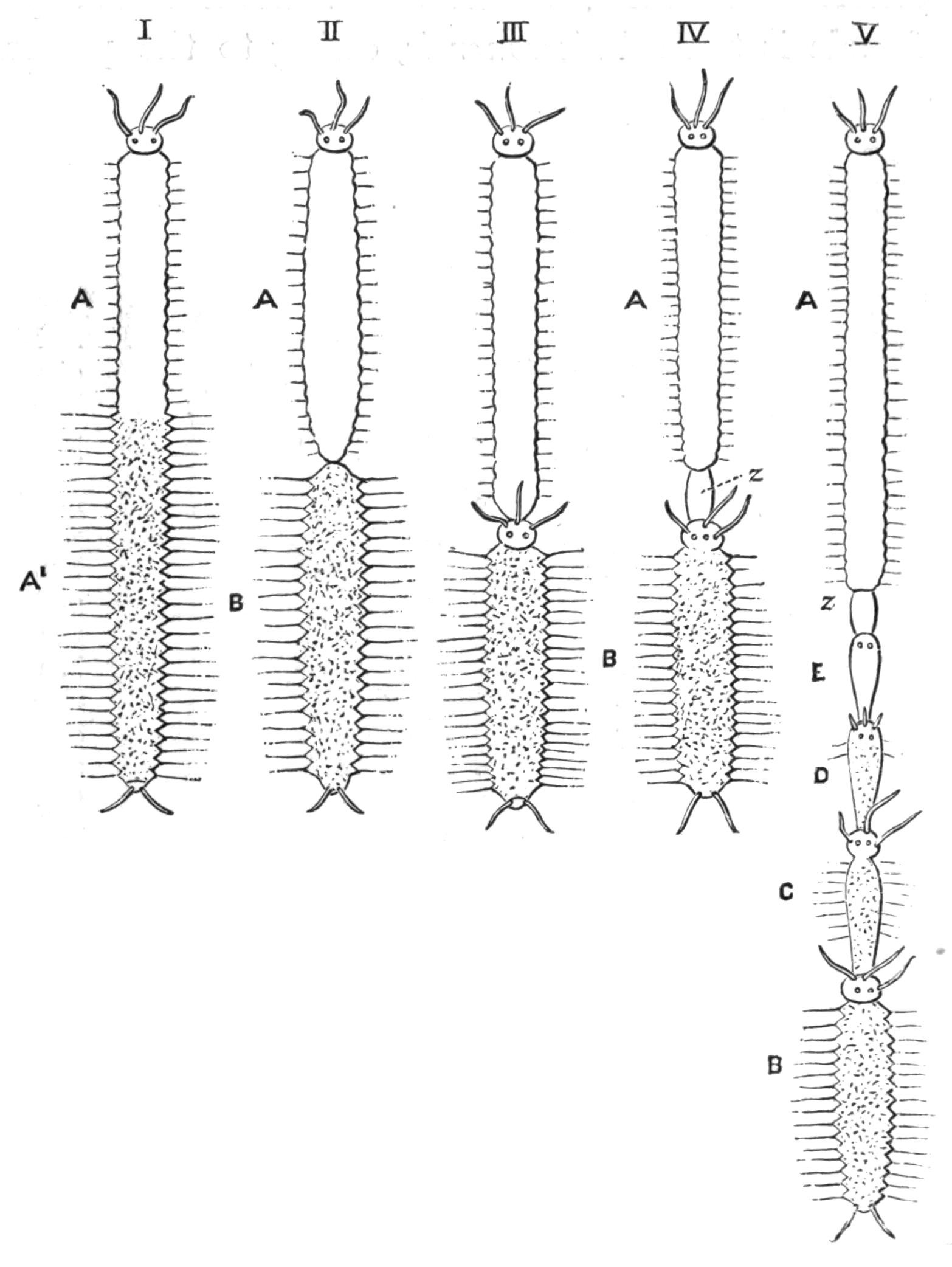
Fig. 148.—Diagrams illustrating the various stages in the asexual formation of a chain of zooids. (Modified from Malaquin.)
I, Heteronereid or Heterosyllid stage. A, Non-sexual; A', sexual region of the body, with modified parapodia.
II, Syllis. The hinder sexual region, B, is similarly modified, and will separate from the parent zooid, A, and become an independent zooid.
III, Autolytus. The hinder zooid, B, develops a head by budding before separation.
IV, Autolytus, etc. A zone of budding (z) makes its appearance in front of the head of B, and by its growth will give rise to a series of new segments in the middle of the body.
V, Myrianida, Autolytus, etc. From this zone of budding a very large number of segments have been formed, which have, further, become grouped so as to form three individuals, C, D, E; B is the hindmost zooid, which is either formed from the hinder segments of the parent zooid or is produced by budding, like C, D, E.
One original "stock," or asexual zooid, thus produces several sexual zooids, but these are only of one sex for a given stock. The males differ in several important characters from the females; so different, indeed, are the two sexes that before their history was worked out by Agassiz[335] they were placed in different genera. The male zooid has thus come to be known as Polybostrichus (Fig. 149, B). It has three tentacles and two bifid palps; there are two pairs of peristomial cirri; the testes are confined to the four anterior segments, which are without natatory chaetae. The female is termed Sacconereis, owing to the possession of a great ventral brood sac; its head possesses no separate palps; the peristomium carries only one cirrus on each side; ova occur in every segment of the body, and may even extend into the hinder segments of the asexual zooid (Fig. 149, C).
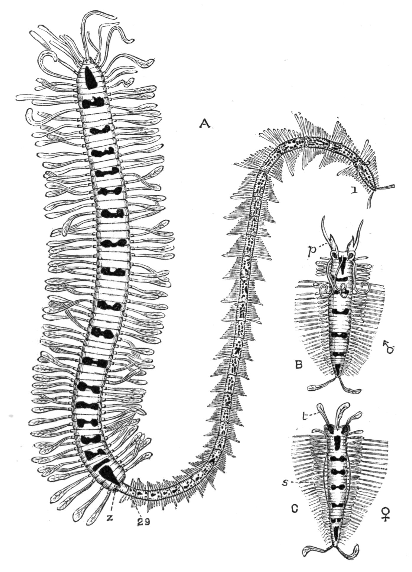
Fig. 149.—Myrianida fasciata. (From Malaquin.) The bright red markings of the living animal are here represented black. A, An asexual individual which has produced by budding from the zone (z) a chain of twenty-nine zooids, the oldest being labelled 1, the youngest 29. B, A ripe male zooid (Polybostrichus), with three tentacles and a pair of forked palps (p). There are five unaltered anterior segments. C, A ripe female zooid (Sacconereis) with the palps fused with the prostomium; s, the ventral brood pouch projecting on each side; t, tentacles.
A further development of this process of gemmiparity is exhibited by Myrianida. Here, there is no conversion of the hinder segments, but the normal preanal zone of proliferation gives rise to a large number of new segments. After a time the most anterior of these becomes a head, and thus a new zooid is marked out. The zone of proliferation immediately in front of the new head now proceeds to form new segments, and a second zooid results. This process goes on till a considerable number of new worms have been formed at the tail of the original one, the oldest of these new ones being the most posterior, the youngest next the original "stock." In each zooid there is a zone of activity which adds to its number of segments, so that as we pass backwards the zooids increase in size. As many as twenty-nine such zooids may be formed in this way entirely by gemmation; and as each zooid becomes completed, genital organs make their appearance, and when these are ripe the zooid separates from the "colony" and leads an independent life. Here, as in Autolytus, the sexes are dimorphic, the male and female resembling those of that genus.
The process of gemmation, as seen in Autolytus, closely resembles that exhibited by certain Oligochaeta (Naididae), where there exists a definite alternation of generations; the production of new individuals by gemmation occurring throughout the greater part of the year, and sexual reproduction recurring only at certain intervals. In the Polychaeta such alternation exists in Myrianida; but it is only the terminal link of a series, which takes its starting-point in the process exhibited by the majority of Annelids, where no sexual character marks maturity. The next stage is presented by "epigamous" forms like Heteronereis and Heterosyllis; then "schizogamy" makes its appearance in certain Syllidae, resulting in the formation of two morphologically and physiologically distinct individuals which lead independent lives. The appearance of a head and of a zone of proliferation leading to the formation of a chain of sexual zooids is accompanied by a delay in the appearance of the genital organs, for in Autolytus these arise during the formation of the new individuals, as part of the general process of new formation; whilst in Myrianida the delay is prolonged, and the generative elements do not make their appearance till after the new individuals have reached some size.
More simple cases of the separation of the body into two parts, sexual and asexual, occur also in some of the Serpulidae. Thus in Filigrana and Salmacina the generative elements make their appearance in the hinder segments, as they do throughout the Sabelliformia; and this hinder part of the body separates from the anterior region after the formation of a new head between the two regions.[336]
Another modification of the process of budding and fission is exhibited by Syllis ramosa, one of the most interesting forms of animal life which was obtained by the "Challenger." This worm consists of a main stem, whence arise a number of lateral branches, which may also branch so as to give rise to an arborescent colony (Fig. 150). The branches of the first and second and higher orders arise by budding from the sides of the original form or branches of lower order; and some of these branches develop generative products, and bud forth a head near the point of attachment. These sexual branches, no doubt, separate from the colony and distribute the ova. The worm lives in a Hexactinellid sponge, Crateromorpha meyeri, living in depths of 95 to 140 fathoms in the Eastern seas.[337]
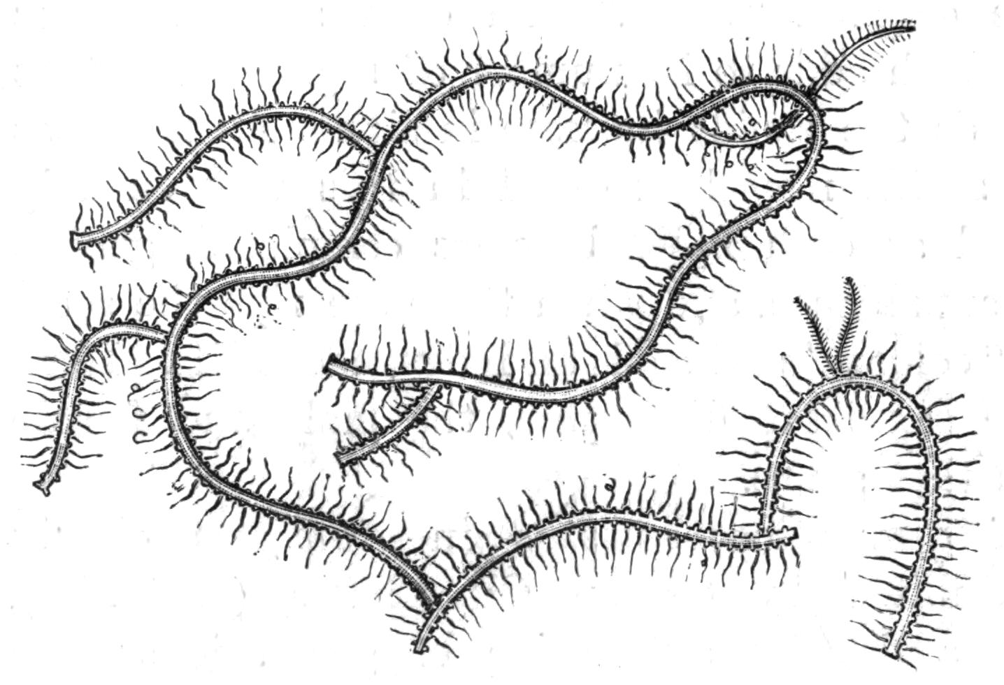
Fig. 150.—Portion of Syllis ramosa. (Reduced from M‘Intosh.)
Regeneration of lost Parts.—The process of budding and fission of the worm into two parts is merely an extension of that resulting in the formation of new segments when the worm is injured. In most of the Nereidiform Polychaetes the number of segments forming the body continues to increase throughout life by the formation of new segments between the anal segment and the one in front of it; that is to say, there is normally a process of budding taking place at this point. Now in many of the longer worms it may be noticed that the segments of the hinder end suddenly become smaller than the rest; these are segments newly formed to replace those lost by the worm. But this "regeneration," though the same in principle as ordinary growth at the penultimate segment, is due to activity in a segment (any segment) further forwards; in other words, in the less modified worms every segment has the power of forming new tissues, just as each of the joints of a crab's leg has the power of forming the remaining joints when injured. It is not therefore surprising that a "zone of budding" arises in an uninjured worm at certain seasons, viz. that of reproduction; it is a property that each worm possesses, though generally it remains latent till injury provides the stimulus.
Moreover, not only can new segments arise at the hinder end, but a new head can be formed at the anterior end, as has been observed in worms belonging to many families—in the less modified Syllidae,[338] in others of the Nereidiformia, and even in Sabellids, where the greatly specialised gill filaments can be reproduced. Thus Sir J. Dalyell[339] noted in Dasychone that the crown of branchiae was regenerated in about a month in springtime, while in winter the process occupied 116 days. He cut a Dasychone into three pieces; the hindermost produced a head, the anterior piece developed an anus, and the middle portion formed both a head and tail!
These regenerated heads are of course at first smaller than the rest of the body, but soon grow to a normal size. Naturally this extensive power of regeneration is of extreme value to the Polychaetes, for if a fish or other enemy bites the head off a worm, a new one can form; and it is not difficult to see in this the origin of the reproduction by fission as a normal process.