Chapter II
Reading edition
TREMATODA
CHARACTERS OF TREMATODES—HABITS AND STRUCTURE OF TREMATODA ECTOPARASITICA (MONOGENEA)—LIFE-HISTORIES OF POLYSTOMUM INTEGERRIMUM, DIPLOZOON PARADOXUM, AND GYRODACTYLUS ELEGANS—TREMATODA ENDOPARASITICA (DIGENEA)—OCCURRENCE AND HABITS OF DIGENEA—LIFE-HISTORY OF DISTOMUM MACROSTOMUM—DISTOMUM HEPATICUM AND ITS EFFECTS—BILHARZIA HAEMATOBIA—BISEXUAL TREMATODES—TABLE OF HOSTS—CLASSIFICATION.
From the Turbellaria we now pass on to a consideration of the second great subdivision of the Platyhelminthes, the Trematodes or "flukes," of which the "liver-fluke" is the best known, since it is one of the most dangerous parasites that infest domestic animals.
It has been pointed out that the Polyclads, Triclads, and Rhabdocoels are carnivorous, and that in each of these groups sporadic cases of parasitism occur. In other words, when the prey is much larger than the Turbellarian, the latter tends to become a parasite, and we can trace the development of the parasitic habit from the gradual association of Turbellaria with Ascidians, Crustacea, Molluscs, and Polyzoa merely for protective purposes, through the adoption, not only of the body of the host for shelter, but of its flesh for food; though it is only in some Rhabdocoels (Graffilla, etc.) that there exists a degeneration corresponding to the easier mode of nutrition and simpler life. The Trematodes,[70] however, are wholly parasitic, either on the outer surface, the gills, or internal organs of their host, which is almost always a Vertebrate. Some Trematodes lodge in the mouth; others wander down the oesophagus into the stomach or intestine, where they fix themselves to the mucous membrane. Again, others work their way into the digestive glands by the ducts, and thus become further and further removed from the external world, and more adapted to live in the particular organs of that host in which they best flourish. The most important result of the adoption of this internal habitat by endoparasitic Trematodes is, however, seen in their life-history. If a liver-fluke were to deposit its million or so of eggs in the bile-ducts of the sheep, and these were to develop in situ, the host could not withstand the increased drain upon its vital resources, and host and parasites would perish together. Hence it is clear that the infection of a second host by Trematodes is highly necessary, whether they be ectoparasitic, in which case the infection is easily effected, when two hosts are in contact, by the adult worms, as well as when they are apart, by free-swimming larvae. In endoparasitic Trematodes it is brought about by the migration of the young to the outer world, their entrance into a, usually, Invertebrate host and their asexual multiplication within it, and the capture and deglutition of this "intermediate host" by the final Vertebrate one. Within the latter the immature parasites find out the organ in which their parents flourished, and here they too grow and attain maturity. The chances of any one egg of an endoparasitic Trematode producing eventually an adult are, therefore, far less favourable than in the case of an ectoparasitic form. In other words, while the former must lay a great number of small eggs, the latter need only deposit a (comparatively) few large ones, and this fact has a corresponding influence on the structure of the genitalia in the two cases. The Digenea, which employ two hosts in a lifetime, have accordingly a different generative mechanism from that of the Monogenea. The great need of the latter is a powerful apparatus for adhering to the surface of the body of its host; while the adaptations which the endoparasite requires are, in addition, (1) protection against the solvent action of the glands of its host, (2) the power of firm adhesion to a smooth internal surface, and (3) the ability not only to produce a large quantity of spermatozoa and ova, but in the absence of a fellow-parasite, to fertilise its own ova; and we find these conditions abundantly satisfied.
Trematoda monogenea (ectoparasitica).
There are four subdivisions of the Monogenea:—
I. Temnocephalidae, with four to twelve tentacles, and one sucker posteriorly (Fig. 20).
II. Tristomatidae, with two lateral, anteriorly-placed suckers. Oral suckers are absent, a large posterior sucker is constant, and is often armed with hooks (Fig. 22, C).
III. Polystomatidae, with, usually, two oral suckers and a posteriorly-placed adhesive disc armed with suckers and hooks (Figs. 23 and 24).
IV. Gyrodactylidae (Fig. 29).
Habits and Structure of Ectoparasitic Trematodes.
I. Temnocephalidae.—These interesting forms, of which a good account has lately been written by Haswell,[71] occur on the surface (rarely in the branchial chamber) of fresh-water crayfish and crabs in Australasia, the Malay Archipelago, Madagascar, and Chili. Others have been found on the carapace of a fresh-water tortoise, and in the branchial chamber of the mollusc Ampullaria from Brazil. Wood-Mason discovered others, again, in bottles containing spirit-specimens of Indian fish. Temnocephala is rarely more than a quarter of an inch long, and looks like a minute Cephalopod or a broad flattened Hydra. By the ventral sucker each species adheres to its own particular host, the tentacles being used as an anterior sucker for "looping" movements. The food, consisting of Entomostraca, Rotifera, and Diatoms, is first swallowed whole by the large pharynx (Fig. 20, ph), which can be protruded through the ventrally-placed mouth, and is then received into a simple lobed intestine (d). The skin, especially on the surface of the tentacles, is provided here and there with patches of cilia borne by the cellular epidermis,—the only undoubted case of external cilia occurring in an adult Trematode. Minute rhabdites formed in special gland-cells, occur plentifully on the tentacles, and are another distinctly Turbellarian feature. The excretory system is peculiar (Fig. 21). Fine ducts proceed from the various organs of the body, and open to the exterior by means of a pair of contractile sacs placed on the dorsal surface. Each sac is a single cell, and within it not one merely, but several "flames," or bunches of rhythmically contractile cilia, are present. These are placed on the course of excessively fine canals, which perforate the protoplasm of this cell. The terminal branches of the excretory canals end in branched cells, apparently devoid of "flames."
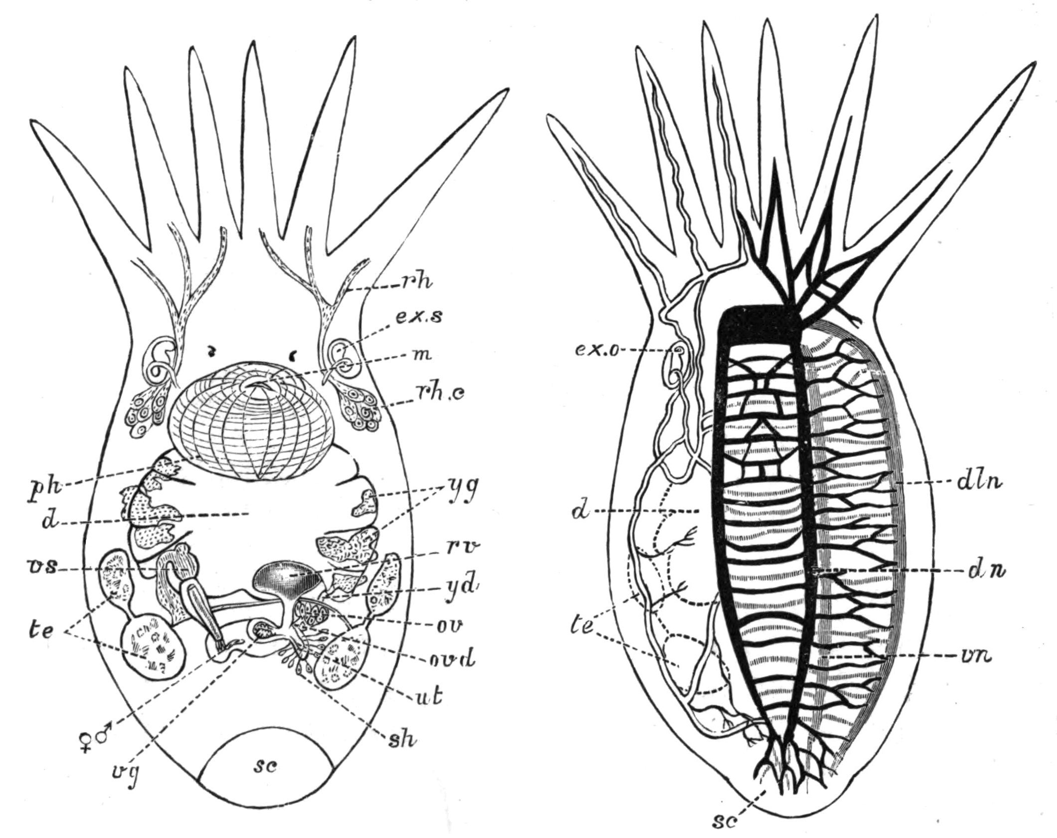
Fig. 20.—Temnocephala novae-zealandiae Has. × 10. Ventral view to show the digestive and reproductive systems. (After Haswell.)
Fig. 21.—The same from the dorsal surface, to show the excretory system (double line), and the nervous system (black and shaded). (After Haswell.)
d, Intestine; dln, dorso-lateral nerve; dn, dorsal nerve; ex.o, excretory aperture on dorsal surface; ex.s, terminal excretory sac; m, mouth; ov, ovary; ovd, oviduct; ph, pharynx; rh, rhabdites; rh.c, cells in which the rhabdites are formed; rv, yolk receptacle; sc, sucker; sh, shell-gland; te, testes; ut, uterus; vg, vagina; vn, ventral nerve; vs, vesicula seminalis; yd, yolk-duct; yg, yolk-gland. ♀, ♂, common genital pore.
The reproductive system is very similar to that of certain Rhabdocoels. An armed penis and the female genital duct open into a genital atrium, and this by a single aperture (♀, ♂, Fig. 20) to the exterior. The fertilised ovum and yolk are enclosed in a stalked shell formed in the uterus.
The interest and importance of the Temnocephalidae lies in the fact that they are almost as much Turbellaria as Trematodes. In habits, in the character of the skin, the muscular, digestive, and reproductive systems, they find their nearest allies in Rhabdocoels (Vorticidae). But in the excretory and nervous systems, the latter composed of two dorsal, two lateral, and two ventral trunks all connected together (Fig. 21), they are Tristomid Trematodes. Thus they may fitly connect an account of the two great groups.
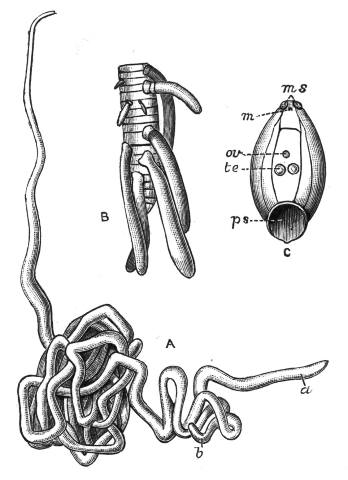
Fig. 22.—A, Nematobothrium filarina van Bened. Nat. size. Two individuals (a and b) are found together, encysted on the branchial chamber of the Tunny. B, Udonella caligorum Johns. A Tristomid, several of which are attached to the ovary of a Copepod (Caligus), itself a parasite on the gills of the Hake. × 8. C, Epibdella hippoglossi O. F. M. A Tristomid found on the body of the Halibut. Nat. size. m, Mouth; ms, lateral suckers; ov, ovary; ps, posterior sucker; te, testes. (All after P. J. van Beneden.)
II. Tristomatidae and III. Polystomatidae.[72]—The members of these families are found on the body, or attached to the gills, of fresh-water and marine fishes. The edible and inedible fish of our coasts have each their particular ectoparasitic Trematodes; while the Minnows, Sticklebacks, and Miller's Thumbs of streams and ponds are attacked by Diplozoon, Gyrodactylus, and other forms. The aquatic Amphibia also harbour a number. Polystomum integerrimum is common in the bladder of Frogs, where it leads a practically aquatic life. Other species of Polystomum inhabit the buccal and nasal cavities of certain Chelonia, but naturally no terrestrial Vertebrates are infested externally by these Trematodes. The blood and epithelia of the host are sucked, and to this end the pharynx has frequently a chitinous armature to aid in the abrasion or inflammation of the tissues upon which the parasite feeds. In the case of a Sturgeon attacked by Nitzschia elongata, a Tristomid, the mouth of the host appeared to be highly inflamed by these attacks (v. Baer).
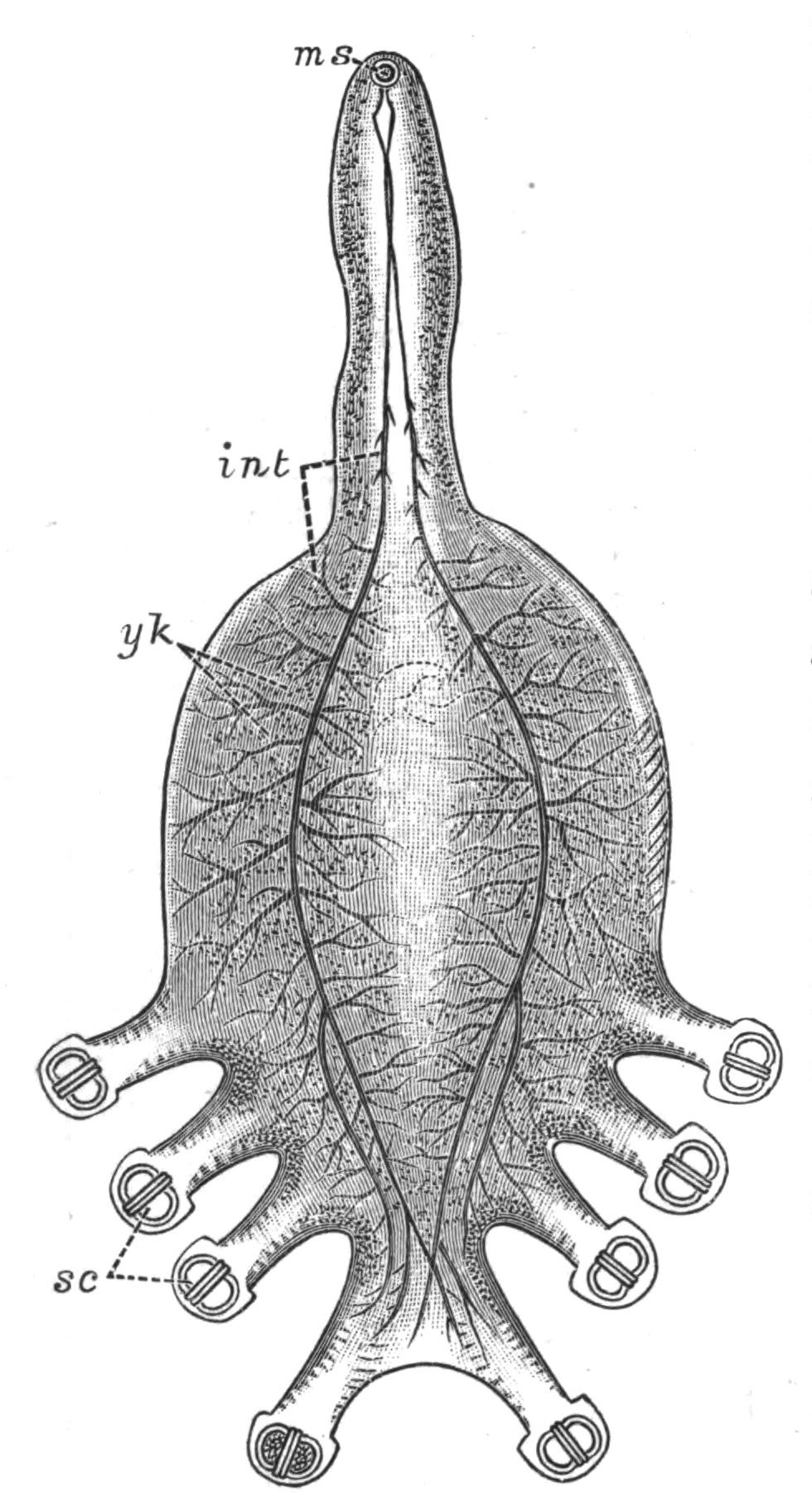
Fig. 23.—Octobothrium merlangi Kuhn, from the gills of the whiting, × 8. int, Intestine; ms, mouth; sc suckers with chitinoid armature; yk yolk-glands. (After v. Nordmann.)
The suckers, in the two families under consideration, vary in number and complexity. There is always a powerful apparatus at the hinder end of the body securing the Trematode firmly to the slimy body or gills of its host, and, usually in the Polystomatidae, a pair of suckers at the sides of the mouth accessory to the pumping action of the pharynx. In Axine, and to a less extent in Octobothrium (Fig. 23), the suckers are strengthened by a complex hingework of chitinoid bars or hooks, which serve as insertions for the muscles of the suckers, and thus increase their efficiency.
The mouth is invariably present just beneath the anterior end of the body. It leads into a muscular, pumping pharynx (Fig. 24, ph), and this into a bifurcated intestine which ends blindly. The two openings of the excretory system lie on the dorsal surface (as in Temnocephala), and the excretory canals branch through the substance of the body, ending usually in "flame-cells." The nervous system is highly developed, and resembles that of Temnocephala (Fig. 21) in detail. Upon the brain one or even two pairs of eye-spots are present in the larvae, and may persist throughout life. Tactile setae occur in Sphyranura, a parasite of the North American Amphibian Necturus, but a cellular epidermis is apparently rendered impossible, perhaps from the nature of the mucus in which the body is bathed, or to the attempts of the host to free itself from these parasites; and hence an investing membrane is present, which morphologically is either a modified epithelium, or a cuticle formed by the glandular secretion of the parenchyma.
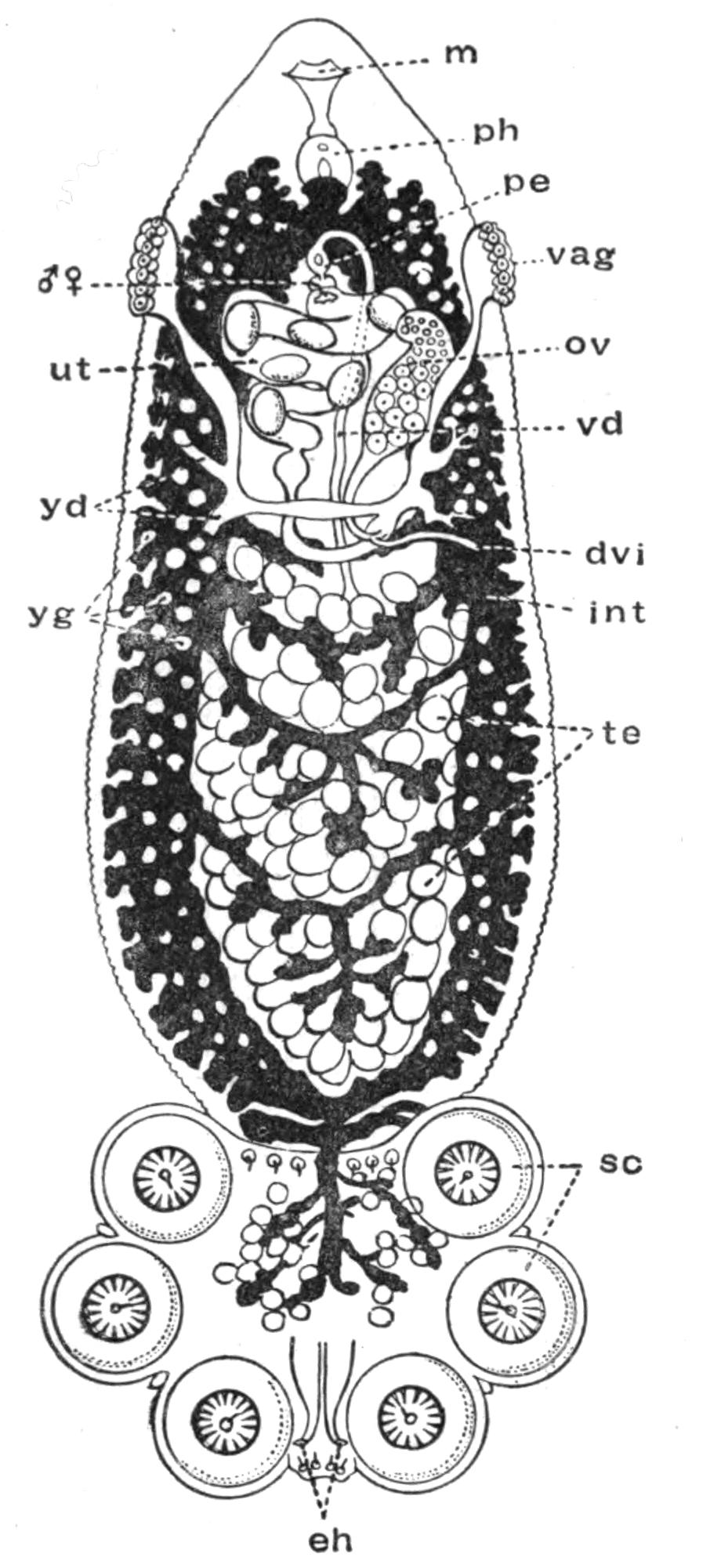
Fig. 24.—Polystomum integerrimum Fröh., from the bladder of the Frog, and seen from the ventral surface. The alimentary canal is black, the white dots upon it being the yolk-glands, dvi, Ductus vitello-intestinalis (probably homologous with the Laurer's canal or "vagina" of Digenea); eh, hooks of sucking disc; int, intestine; m, mouth; ov, ovary; pe, penis; ph, pharynx; sc, suckers with an embryonic hook persisting in each; te, testes; ut, uterus with eggs; vag, left vagina; vd, vas deferens; yd, yolk-duct; yg, yolk-glands; ♂ ♀, common genital aperture. (Modified from Zeller.) × 8.
The reproductive organs of the Polystomatidae may be understood from Figs. 24, 27, and 28. At the point of union of the oviduct (Fig. 28, ovd), the vitelline ducts (yd), and the commencement of the uterus (ut), a slender duct is given off which opens into the intestine, and is known as the "vitello-intestinal canal" (Fig. 24, dvi; Fig. 28, gic). This duct has apparently the same relations as the "canal of Laurer" of Digenea,[73] except only that the latter opens to the exterior directly. In connexion with this vitello-intestinal canal a "vagina" is present, which in Polystomum and most Monogenea is paired (Fig. 24, vag), in Diplozoon and in one or two other forms, however, unpaired. The vagina receives the penis of another individual during copulation (Fig. 26), and does not appear to have an homologue in the liver-fluke or other Digenea.
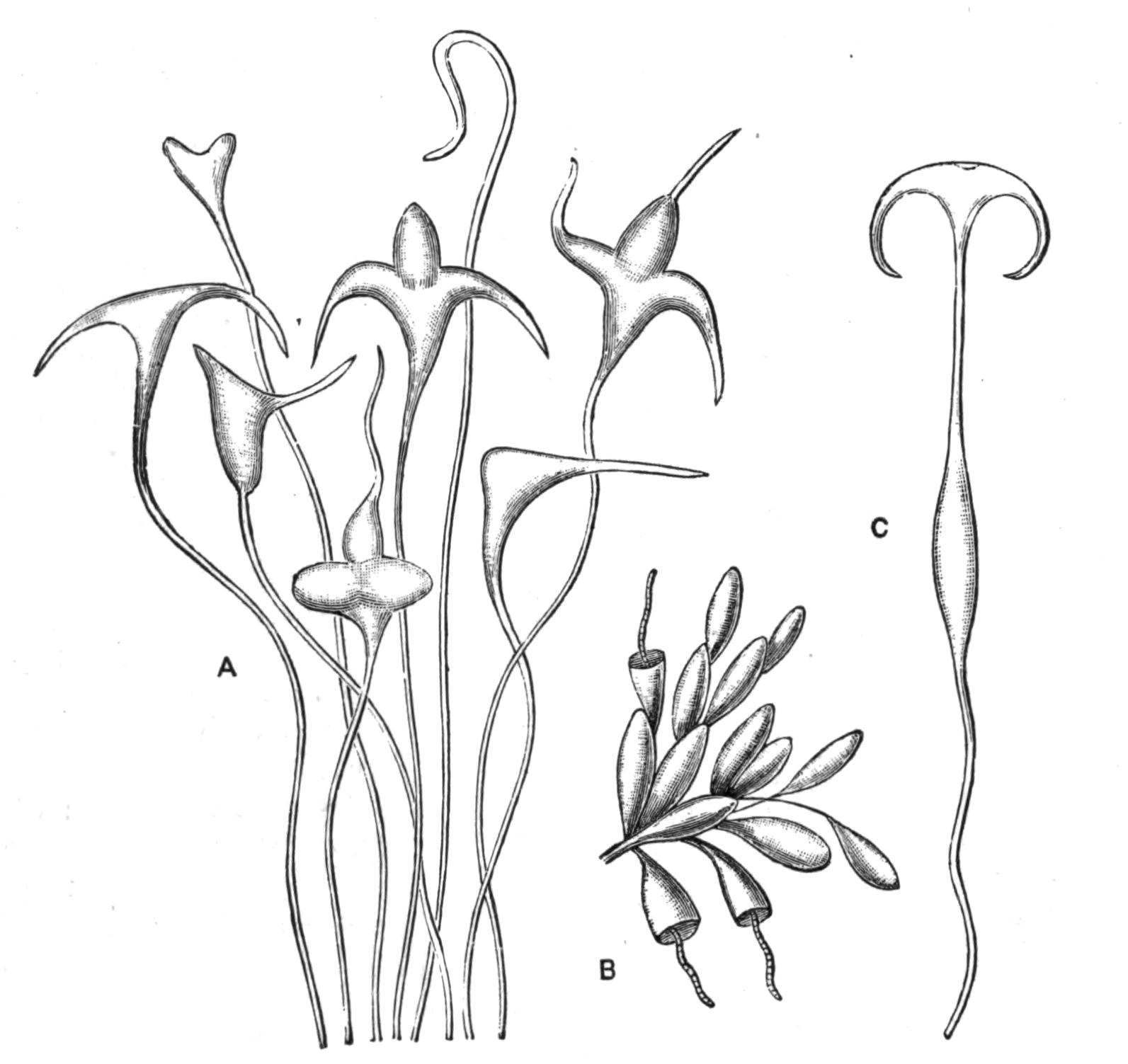
Fig. 25.—Eggs of Monogenea. A, Eggs of Encotylabe pagelli v. Ben.-Hesse; B, eggs of Udonella pollachii v. Ben.-Hesse (with young forms just hatching out); C, egg of Microcotyle labracis v. Ben.-Hesse. (After van Beneden and Hesse.) × 50.
Life-Histories of the Polystomatidae.[74]—Polystomum integerrimum. After the mutual fertilisation of two individuals, the eggs are laid in the water by the protrusion of the body of the parent through the urinary aperture of the Frog. About 1000 eggs are laid in the spring at the rate of 100 a day for ten days. After about six weeks, the larva (.3 mm. long) hatches out, and swims about freely by means of bands of large ciliated cells (Fig. 26, A); but if it does not meet with a tadpole within twenty-four hours, it dies. Should it, however, encounter one, the larva creeps along it in a looping fashion until it approaches the opercular spout, or opening of the branchial chamber, on the left side; into this it darts suddenly, fixes itself, and throws off its cilia. Here it remains eight or ten weeks, feeding, increasing in size, and forming the suckers from behind forwards. At the time of the tadpole's metamorphosis, the young Polystomum works its way down the pharynx into the oesophagus and along the intestine, till it reaches and enters the opening of the bladder. Three years afterwards it becomes mature.
Sometimes, however, Polystomum experiences another fate. The larvae settling down on the external gills of a young, recently-hatched tadpole, and obtaining a richer supply of blood than in the previous case, grow far more rapidly, so that in five weeks they are mature, although still in the branchial chamber of the tadpole. They do not then wander into the alimentary canal, but usually, having discharged their eggs, die at the time of the tadpole's metamorphosis. Still more interesting, however, is the difference between the genitalia in these and in the normal Polystomum. In contrast with the latter, these possess (1) one testis and a rudimentary penis; and their spermatozoa differ in structure and shape from those of the normal Polystomum. (2) The vaginae are absent, a fact connected with the absence of a functional copulatory organ. (3) In compensation for the loss of these, a duct connects the single testis and the point of union of oviduct and yolk-ducts, and by this self-fertilisation occurs. (4) The uterus is absent; the "ootype" or duct into which the shell-gland opens, communicating directly with the exterior. In (1) and (4) these aberrant Polystomum resemble P. ocellatum, from the Tortoise Emys europaea.
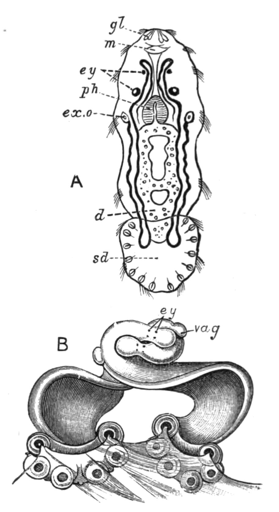
Fig. 26.—Polystomum integerrimum. A, Free-swimming larva, seen from the ventral surface. × 80. B, Two mature individuals in mutual coition attached to the bladder of a Frog. × 5. (After Zeller.) d, Intestine; ex.o, excretory pore, dorsal in position, seen here by transparency; ey, eye-spots; gl, frontal glands; m, mouth; ph, pharynx; sd, adhering disc; vag, vagina.
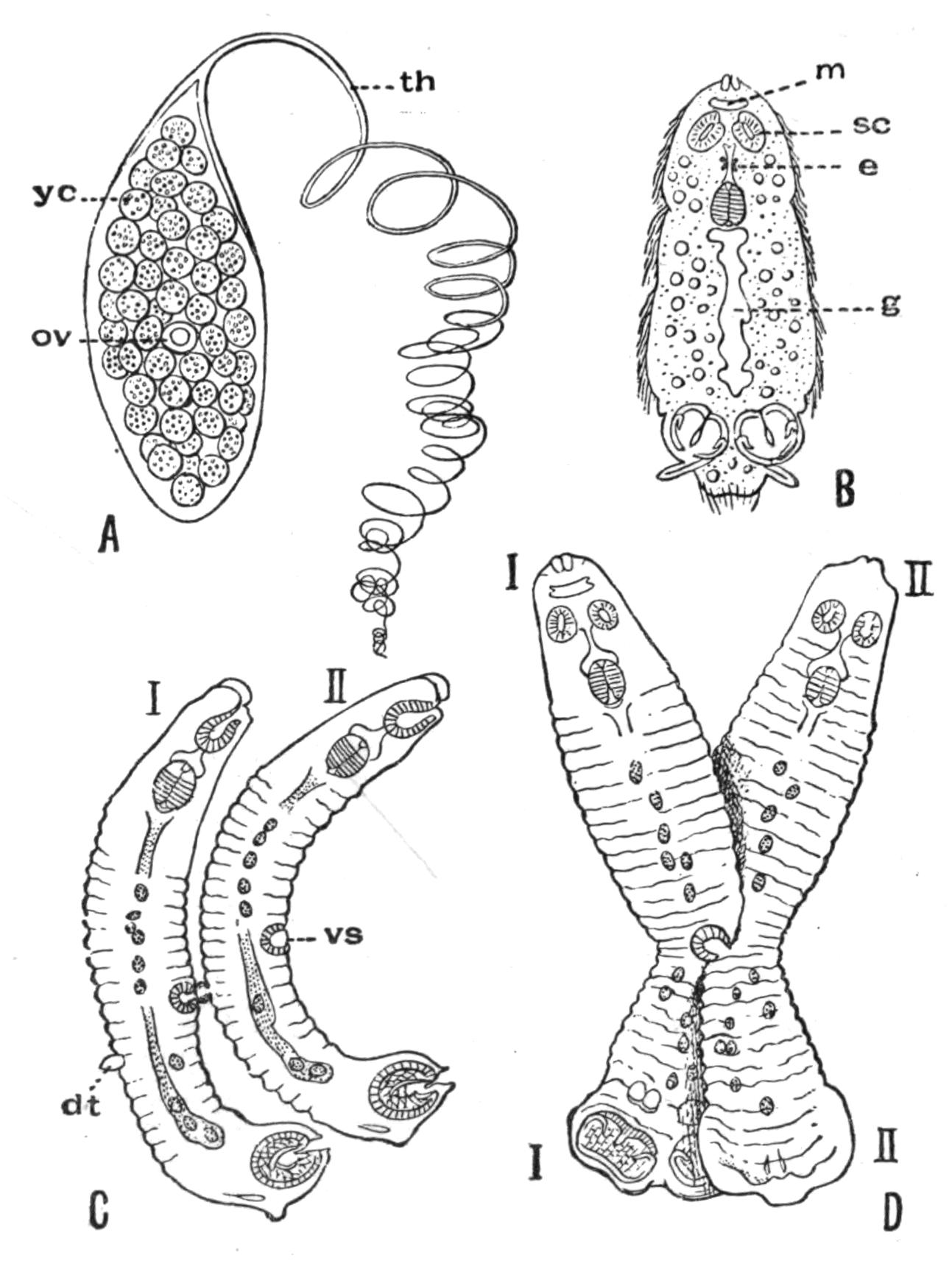
Fig. 27.—A, Egg of Diplozoon paradoxum v. Nord., consisting of a shell enclosing ov, the actual ovum, surrounded by yc, the yolk-cells; B, larva just hatched (× 125); C, two Diporpa (I and II) about to unite; D, conjugation in progress but not yet complete. dt, Dorsal papilla; e, eye; g, intestine; m, mouth; sc, ad-oral sucker; th, spirally-wound thread attaching the egg to the gill of the Minnow; vs, ventral sucker; (in D) I, I, one Diporpa, ventral view; II, II, the other, dorsal view. (After Zeller.)
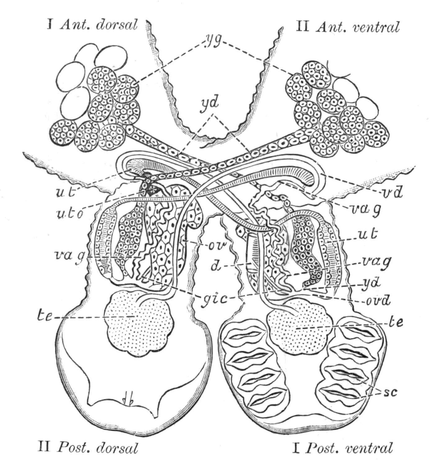
Fig. 28.—Hinder part of the body of Diplozoon paradoxum. The fusion of the two Diporpa, where they come into contact, is now complete. They now cross each other like an X, and are twisted, so that Diporpa I, in front of the point of fusion, is seen from the dorsal surface; behind, from the ventral surface; and the reverse is the case with Diporpa II. The compound animal is seen from the opposite surface to that shown in Fig. 27, D. The digestive and excretory organs are omitted. (After Zeller.) I Ant. dorsal, dorsal surface of Diporpa I, facing the anterior end; I Post. ventral, ventral surface of Diporpa I, posterior end; and similarly for II Ant. ventral and II Post. dorsal. d, Piece of the intestine showing opening of, gic, vitello-intestinal canal; ov, ovary; ovd, point of union of female genital ducts; sc, suckers; te, testis; ut (in Diporpa I), "ootype" or chamber into which shell-glands open. This is continuous with the uterus (ut) of Diporpa I; uto, ventral opening of uterus; vag, vagina, with vd, vas deferens, permanently inserted into it through the genital pore; yd, yolk-ducts; yg, yolk-glands.
Diplozoon paradoxum.—The life-history of Diplozoon is unique. For whereas the larvae of most animals grow up, each into a single adult, in Diplozoon, of the few larvae that survive the dangers of their free-swimming existence, only those become mature which conjugate permanently with another individual. But although there are thus only half as many adult Diplozoon as there were conjugating larvae (or Diporpa, as they were called when they were considered distinct forms), yet the total number of eggs produced is probably as great as if each larva became individually mature.
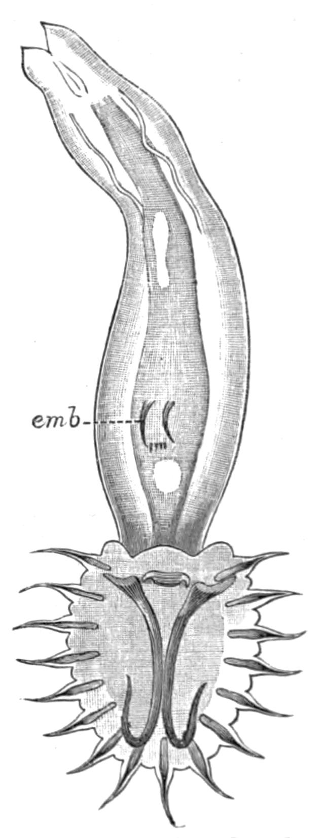
Fig. 29.—Gyrodactylus elegans v. Nord., from the fins of the Stickleback. (After v. Nordmann.) × 125. emb, Embryo.
Diplozoon paradoxum lays its eggs on the gills of the Minnow, which it frequently infests in great numbers. The ovum divides rapidly at the expense of the yolk-cells, and in a fortnight a larva (.2 mm. long) of the shape and complexity shown in Fig. 27, B, hatches out, which, however, succumbs if it does not meet with a Minnow in five or six hours. Should it survive, a dorsal papilla, a median ventral sucker, and a second pair of posterior suckers develop. Thus the Diporpa stage is attained. These Diporpa may acquire a third and even a fourth pair of suckers, and continue to live three months, but they only develop and mature their reproductive organs, if each conjugates with another Diporpa (Fig. 27, C, D), and this only occurs in a small percentage of instances. Each grasps the dorsal papilla of the other by its own ventral sucker, thus undergoing a certain amount of torsion. Where the two bodies touch, complete fusion occurs, and, as shown in Fig. 28, the united Diporpa (or Diplozoon, as the product is now called) decussate, each forming one limb of the X-shaped Diplozoon, within which the two sets of complex genitalia develop (Fig. 28).
IV. Gyrodactylidae.—Gyrodactylus (Fig. 29), the structure of which is in many ways peculiar, produces one large egg at a time. An embryo, in which the large and smaller hooks of the adhesive disc can be seen (emb), develops from this egg while still within the body of the parent, and may give rise to yet another generation within itself. The details of the process have not, however, been well ascertained.
Trematoda digenea (endoparasitica).
Occurrence and Habits of Digenea.—Endoparasitic Trematodes have been found in almost all the organs of Vertebrate hosts excepting in the nervous, skeletal, and reproductive systems. The alimentary canal, however, is the most usual habitat. From the buccal cavity to the large intestine, or even to the cloaca, its different regions are the resorts of various Trematodes. No Digenea have been found in the mouth, pharynx, or oesophagus of Mammals; but in Birds, Reptiles, Amphibia, and especially in Fishes, these parts are largely affected. It is a striking fact that Trematodes should occur in the stomach of (chiefly) large predaceous fishes, such as the Pike, Sharks, the Angler-fish, and others, considering the powerful digestive action of the gastric juice of these carnivores. The peculiar nature of the defence which must be employed by the parasites against this digestive action, becomes still more marked when it is considered that if a Trematode normally living in the stomach of one host be transferred to that of another, it is usually speedily digested, as is shown (p. 65) in the case of Distomum macrostomum. From these considerations the suggestion has been made that the cutaneous secretions of these Trematodes must act, not only as a protection against digestive or other ferments, but that the action in each case must be a specific one (Frenzel, Braun).
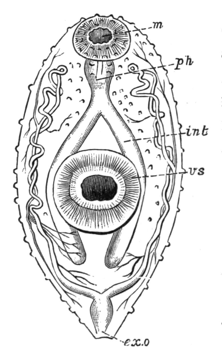
Fig. 30.—Distomum luteum v. Baer (immature), to show the arrangement of the excretory vessels. × 50. ex.o, Excretory aperture by which the terminal contractile duct opens—the finer vessels end in flame-cells; int, intestine; m, mouth-sucker; ph, pharynx; vs, ventral sucker. (After la Valette.)
It is, however, in the small intestine that most Trematodes occur, as the examination of the common Frog[75] will readily demonstrate. Both this and the edible Frog are attacked by a dozen Distomatidae, only a few of which, however, are common to both hosts, and a number of Holostomatidae also pass a stage of their development within these Amphibia. Some idea of the extent to which animals, whose habits lead to infection, may be attacked by Trematodes (to say nothing of Cestodes and Nematodes, which often occur also) may be gathered from the fact that in dissecting a black stork, Nathusius found several hundred Holostomum excavatum and about a hundred Distomum ferox in the small intestine, twenty-two D. hians in the oesophagus, five others in the stomach, and one D. echinatum in the intestine. Snipe, Woodcock, Sandpipers, Dunlin, Gulls, Bittern, Geese, and Wild Ducks are, to mention a few cases, greatly infested by members of this group.
The following Trematodes have occurred in man[76]:—
Distomum hepaticum Abild. Dist"mum lanceolatum Mehlis. Dist"mum conjunctum Cobbold. Dist"mum spathulatum Leuckart (= D. sinense Cobb., D. japonicum R. Blanch.). Dist"mum rathouisi Poir. (probably = D. crassum Busk, D. buskii Lank.). Dist"mum heterophyes v. Sieb. Dist"mum pulmonale Bälz (= D. ringeri Cobb., D. westermanni Kerb.). Dist"mum oculi humani Ammon (= D. ophthalmobium Dies.). Monostomum lentis v. Nord. Amphistomum hominis Lewis and M‘Connell. Bilharzia haematobia Cobb.
Life-histories of the Digenea.—The classification of Trematodes according to their life-histories, expressed in the divisions Monogenea and Digenea, though a very useful one, breaks down entirely in the case of certain forms. Thus the life-history of Gyrodactylus is probably digenetic rather than monogenetic. Aspidogaster conchicola,[77] which lives in the pericardial cavity of the fresh-water mussel (possibly the only case of a Trematode becoming normally mature in an Invertebrate host, since other species of Aspidogaster live in Chelonia), produces larvae which enter another Anodonta and develop directly into the sexual form. In other words, Aspidogaster, though structurally a digenetic form, possesses a life-history which is direct and simple, i.e. monogenetic.
The Holostomatidae, which live in birds of prey and aquatic birds, give rise to eggs from which a minute larva escapes. The fate of this aquatic larva is not directly known, but in all probability after entering a host (Fish, Amphibian, Mollusc), it undergoes a gradual change into what has long been known as a Tetracotyle, from the frequent presence of four (sometimes only three) adhering organs. Fig. 31 exhibits a species which is abundant in the lens and vitreous humour of the eye of the Perch. Its further history is not known, but presumably the Perch is presently devoured by the final host in which the Diplostomum attains maturity. Thus the Holostomatidae are "metastatic" (Leuckart), their (probably) direct development requiring the presence of two hosts.[78]
The other Digenea, the life-histories of which are known, belong to the Distomatidae and Amphistomatidae, and we may distinguish the steps by which the complex life-history of the liver-fluke (Distomum hepaticum) has been brought about, by a consideration of that of Distomum macrostomum.
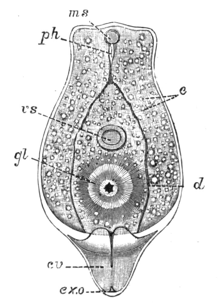
Fig. 31.—Diplostomum (Tetracotyle) volvens. (After v. Nordmann.) × 130. cv, Contractile excretory vesicle; d, intestine; e, calcareous bodies in excretory tubules; ex.o, excretory aperture; gl, glandular adhesive body; ms, oral sucker; ph, pharynx; vs, ventral sucker.
Distomum macrostomum.—This form occurs in the intestine of several common Passerine birds. It is remarkable not only for the large oral sucker, but also on account of the position of the common genital pore at the hinder, and not as usual, at the anterior, end of the body (Fig. 32, A). The eggs pass out through this pore, and are discharged with the bird's excrement. Should a certain snail (Succinea putris) happen to rasp off the epidermis of a leaf upon which the faeces have fallen, the eggs are swallowed and a minute active larva is set free (Fig. 32, B). This penetrates through the thin wall of the digestive tract of the snail, and passing into the connective tissue, throws off its cilia and assumes the shape of Fig. 32, C. This sporocyst, as the larva is now termed, grows rapidly in all directions (Fig. 32, D) at the expense of the snail's tissues, until it becomes impossible to separate parasite and host completely.
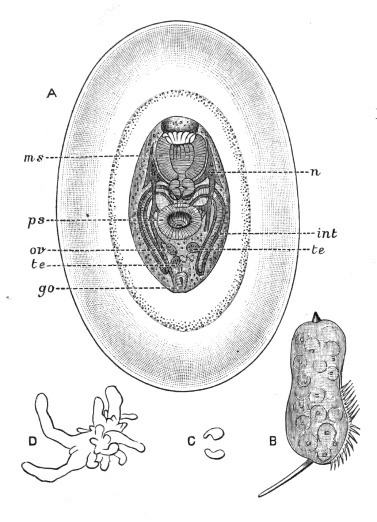
Fig. 32.—Life-history of Distomum macrostomum Rud. A, Immature Distomum (really a tailless Cercaria) found in the swollen terminal parts of Leucochloridium (Fig. 33, B) and enclosed in two protective membranes, × 40; B, larva which hatches out of the egg of D. macrostomum, × 125; C, the metamorphosed larva (sporocyst) fourteen days after having entered Succinea putris, and pierced through its intestinal wall; D, actively growing sporocyst. (After Heckert.) go, Genital aperture; int, intestine; ms, mouth sucker; n, nervous system; ov, ovary; ps, ventral sucker; te, testis.
Those branches which lie superficially in the cephalic region of the snail become greatly swollen, cylindrical, and contractile. They are banded with green and white, ornamented with red terminal spots, and pulsate rapidly. Hence these fertile branches of the sporocyst (which in this condition was known as Leucochloridium paradoxum, Fig. 33, B) naturally attract the attention of insectivorous birds, which peck off the tentacles of the snail, and with it the swollen sporocyst-branch. A sphincter muscle closes the cut end of the fertile sac when the bird's bill nips it off. The sac contains large numbers of young D. macrostomum (Fig. 32, A), produced by the division of embryonic cells of the larva (Fig. 32, B), which are apparently blastomeres of the egg reserved for this future use. It is a remarkable circumstance that the old bird itself is immune from infection, and if it swallows these young Distomes, they are digested. Should, however, the snail's tentacle and its contents be offered as food to the nestlings, their weaker digestive powers merely set the Distomes free from the protective membranes (Fig. 32, A), and thus the Blackcaps, Sparrows, and other birds infested by D. macrostomum have acquired the parasite when they were nestlings by the unintentional agency of their parents.[79] The snail regenerates its lost tentacles only for the sporocyst to again bud off fertile branches into them.
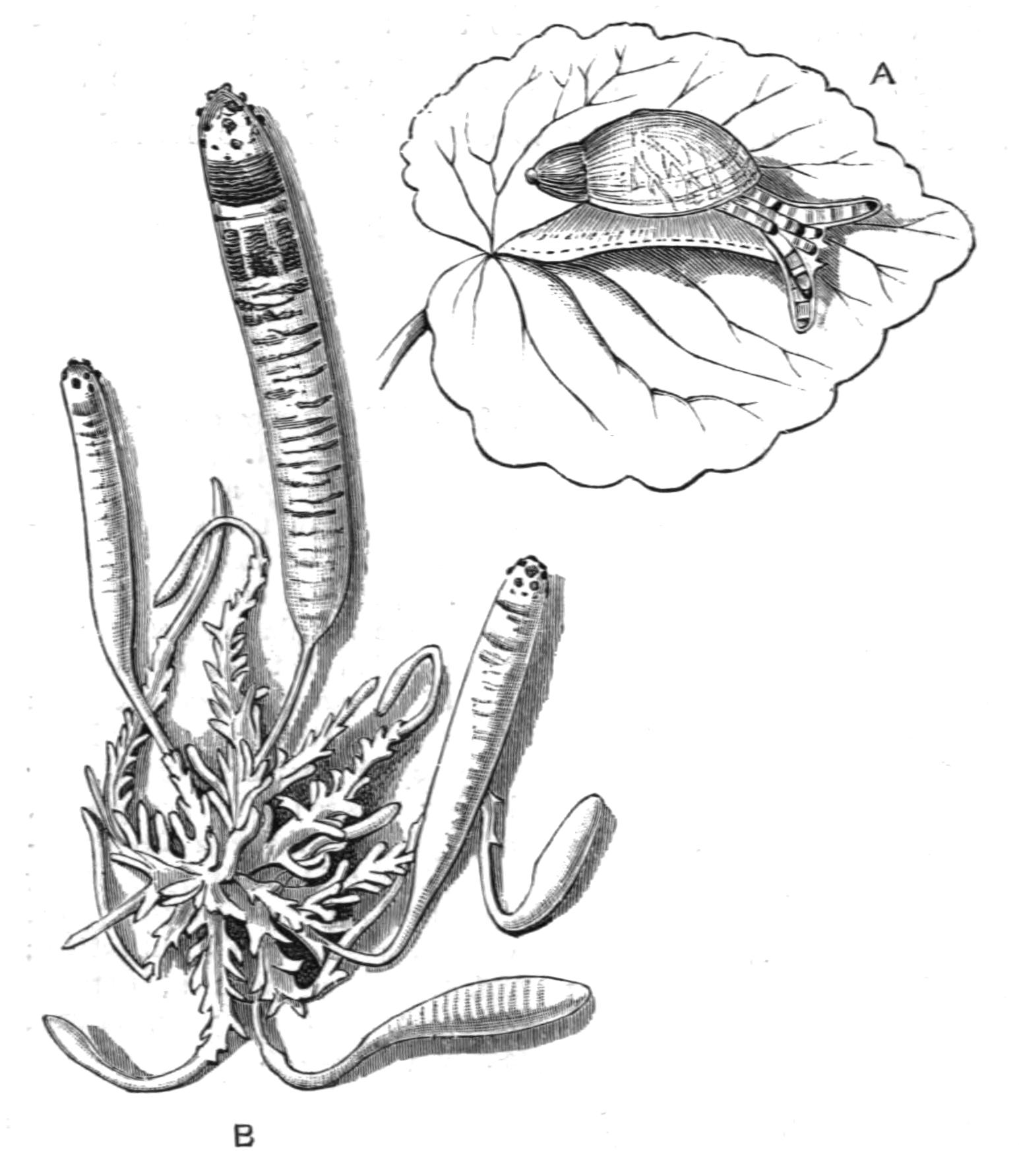
Fig. 33.—A, Succinea putris, infested by B, Leucochloridium paradoxum, or the fully-formed sporocyst of Distomum macrostomum. (After Heckert.) A, Natural size; B, × 7.
The egg of this Distome thus gives rise to a larva which enters the tissues of one particular Mollusc. Here it becomes a branched sporocyst within which the sexual worms are formed, apparently each from a single embryonic blastomere ("Keimzelle"), by a process comparable with the development of a parthenogenetic ovum, and the whole cycle has been termed Alloiogenesis, i.e. alternation of sexual and parthenogenetic generations (Grobben).[80] Leuckart[81] and Looss,[82] however, consider that what was once a metamorphosis of an individual (as in the Holostomatidae) has now become, by maturation of the Cercaria in the comparatively modern warm-blooded bird, a metamorphosis extending over two or more generations.
Distomum (Fasciola) hepaticum.—The liver-fluke of the Sheep, which produces the disastrous disease, liver-rot, has a distribution as wide as that of a small water-snail, Limnaea truncatula, the connexion between the two being, as Thomas[83] and Leuckart discovered, that this snail is the intermediate host in which the earlier larval, sporocyst, and redia stages are passed through, and a vast number of immature flukes (Cercariae) are developed. These leave the snail and encyst upon grass, where they are eaten by the sheep. Over the whole of Europe, Northern Asia, Abyssinia, and North Africa, the Canaries, and the Faroes, the fluke and the snail are known to occur, and recently the former has been found in Australia and the Sandwich Islands, where a snail, apparently a variety of Limnaea truncatula, is also found.[84] Over these vast areas, however, the disease usually only occurs in certain marshy districts and at certain times of the year. Meadows of a clayey soil, liable to be flooded (as in certain parts of Oxfordshire), are the places where this Limnaea occurs most abundantly, and these are consequently the most dangerous feeding-grounds for sheep. The wet years 1816, 1817, 1830, 1853, and 1854—memorable for the occurrence of acute liver-rot in England, Germany, and France—showed that the weather also plays a considerable part in extending the suitable ground for Limnaea over wide areas, which in dry years may be safe pastures. In 1830 England lost from this cause,[85] one and a half million sheep, representing some four millions of money, while in 1879-80 three millions died. In 1862 Ireland lost 60 per cent of the flocks, and in 1882 vast numbers of sheep perished in Buenos Ayres from this cause. In the United Kingdom the annual loss was formerly estimated at a million animals, but is now probably considerably less. After infection during a wet autumn, it is usually in the succeeding winter that the disease reaches its height.
The symptoms of "rot" appear about a month after infection, more acutely in lambs than in sheep, and again, less in oxen than in sheep. At first, death may result from cerebral apoplexy, but if the first few weeks are passed through, a pernicious anaemia sets in, the sheep are less lively and fall at a slight touch, the appetite diminishes, and rumination becomes irregular. The conjunctiva is of a whitish-yellow colour, the dry, brittle wool falls off, and there is sometimes fever and quickened respiration. In January, about three months after infection, the wasting, or fatal, period sets in. Oedemas or swellings, usually visible before, become larger at the dependent parts of the body, a large one in the submaxillary region being especially well marked, and this is considered one of the most characteristic symptoms ("watery poke"). Through this period few of the infected sheep survive, but should they do so, the flukes begin to migrate, though some remain much longer within the liver. Migration is effected through the bile-duct into the duodenum and outwith the faeces, in which the altered remains of the Distomum are sometimes scarcely recognisable. Under these circumstances (or owing to death of the fluke in situ) the sheep recover more or less fully.
The preventive measures seem to be: (1) Destruction of the eggs and of the manure of rotten sheep; (2) slaughter of badly fluked sheep; (3) adequate drainage of pastures; (4) an allowance of salt and a little dry food to the sheep; and (5) dressings of lime or salt on the ground to destroy the embryos.[86]
Distomum hepaticum, contrary to most Trematodes, enjoys a wide range of hosts. Man himself occasionally falls a victim; thus in Dalmatia, in the Narenta Valley, the disease is endemic but slight in its effects. The horse, deer, camel, antelopes, goat, pig, rabbit, kangaroo, beaver, and squirrel have all been known to harbour this fluke occasionally. In the Italian deer-parks at Mandria a large species, D. magnum, decimated the herds some years ago; and this species, probably imported from Italy, is now almost as dangerous a parasite on the western plains of the United States as D. hepaticum.
Bilharzia haematobia.[87]—This formidable parasite was discovered by Bilharz in 1853 in the veins of the bladder of patients at the Cairo Hospital, and is remarkable from its abundance on the east coast and inland countries of Africa from Egypt to the Cape, as well as in the districts bordering Lake Nyassa and the Zambesi river, while westwards it occurs on the Gold Coast. Mecca is a source of infection whence Mohammedans carry the disease to distant places. In Egypt about 30 per cent of the native population is affected by the serious disease known as Haematuria, resulting from the attacks of Bilharzia, so that, of the many scourges from which in Africa man suffers, this one is perhaps the most severe.
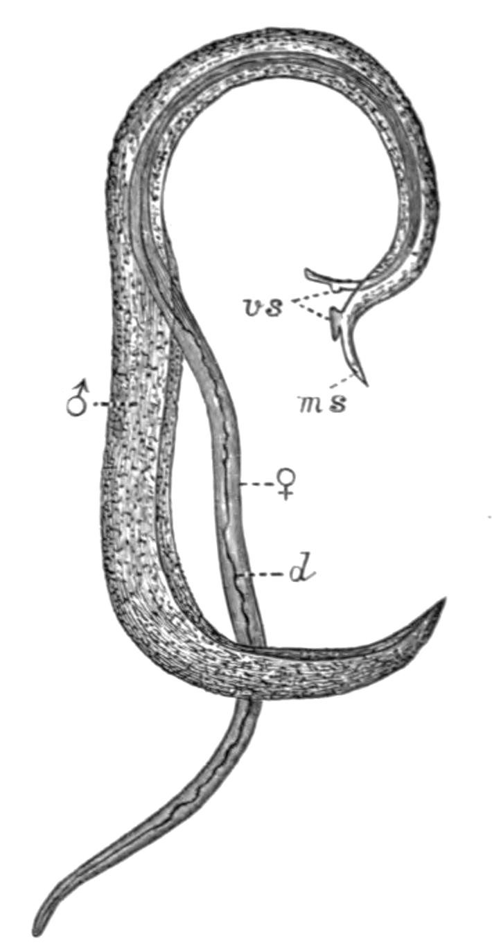
Fig. 34.—Bilharzia haematobia Cobb. × 10. The female (♀) lying in the gynaecophoric canal of the male (♂). d, Alimentary canal; ms, oral sucker of male; vs, ventral suckers. (After Leuckart.)
The worm is found usually in couples, which have been proved to be male and female individuals (Fig. 34), often in considerable numbers in the veins of the pelvic region, chiefly the veins of the bladder and of the large intestine, and it is tolerably certain that Bilharzia enter these vessels from the portal vein. Their long slender bodies enable them to penetrate into the finer vessels, which get partially or entirely choked up, and the circulation accordingly impeded. But the most serious consequences are observed in the urinary bladder. The mucous membrane is swollen and inflamed here and there, chiefly on the dorsal surface, the capillaries appear varicose and covered with mucus, mixed with blood-extravasations in which Bilharzia-eggs are noticeable. The eggs also cause numerous swollen knots in the submucous tissue. Should the disease not pass beyond this stage (and such is usually the case, especially in South Africa), a temporary haematuria ensues. The urine, which is only expelled with great effort, accompanied by intense pain, is mixed with blood, mucous clots, and masses of Bilharzia-eggs, from which some of the embryos have already hatched out. The symptoms, however, may gradually pass away, and a more or less complete recovery accomplished. The disease may indeed be of a far less severe character, and may not interfere with the usual occupations of the patient; but, on the other hand, a far more extensive thickening of the wall of the bladder sometimes occurs; hard masses of eggs, uric acid crystals, and other deposits, may lead to the formation of stones, degeneration of the substance of the ureter, and eventually to that of the kidney itself. The stone, indeed, has long been known to be a prevalent disease in Egypt, and it is now known to arise from concretions formed round masses of Bilharzia eggs. From the portal vein, again, other Bilharzia may gain access to the rectum, or the liver, and it has also been found in the lungs, and may give rise to most serious complications, if indeed the patient lives.
How infection occurs is a question to which at present no satisfactory answer can be made. The attempt to introduce embryos of Bilharzia into the common fresh-water animals of Alexandria has hitherto proved fruitless (Looss[88]), although there seems little doubt that the comparative immunity of Europeans from the disease is in some way owing to their drinking purer water than the natives. Possibly, as Leuckart suggests, the embryo becomes a sporocyst in man himself, somewhat as Taenia murina is known to develop in the rat without an intermediate host.[89] The immense numbers of the parasite in one host would then readily receive an explanation.
A Bilharzia, possibly B. haematobia, was found by Cobbold in the portal vein of Cercopithecus fuliginosus; and B. crassa infests the cattle of Egypt, Sicily, and certain parts of India, but does not produce haematuria.
Of the other Trematodes of man and domestic animals there is not room to speak fully. Distomum pulmonale, which occurs in the lungs of the cat, tiger, and dog, as well as in man, is especially common in Japan, China, Corea, and Formosa. D. sinense and D. rathouisi have been also found in inhabitants of these countries.
Bisexual Trematodes.—Zoologically, Bilharzia is interesting from its bisexual condition. It is not, however, the only bisexual Trematode. In cysts in the branchial chamber of Ray's bream, Brama raii, two worms are found, which are probably the slender male and the swollen female of the same species (Distomum okenii). The only doubt that can arise proceeds from the tendency in all Trematodes for the male organs to ripen before the female organs. Until we certainly know that the swollen egg-bearing form (♀) does not arise from a previously male form (♂), the case is open to suspicion. Since, however, Kölliker[90] never found intermediate hermaphrodite conditions, this Distomum may be almost certainly regarded as of distinct sexes. Didymozoon thynni (Monostomum bipartitum), from cysts on the gills of the Tunny (Thynnus), is another case. Two slender worms flattened posteriorly, come together, and the body of one becomes folded to receive that of the other. They fuse completely except for a small lateral opening through which the anterior parts of both worms may freely protrude. The enclosing individual contains a coiled uterus filled with eggs, and is the female, whereas the smaller individual never possesses eggs, and is probably the male.[91] Nematobothrium (Fig. 22, A), which occurs also in the Tunny, in the form of two immensely long individuals intricately wound about each other in a cyst, is, however, not bisexual.
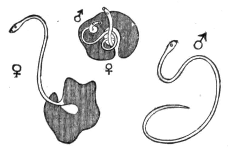
Fig. 35.—Distomum okenii Köll. Showing male and female as they occur together in the branchial cavity of Bramaraii (Ray's bream). (From Bronn, after Kölliker.) Nat. size.
Table of Digenetic Trematodes and their Life-Histories.[92]
Species. Final host. Host into which the larva enters, and in which Cercariae are eventually formed. Host into which the Cercariae migrate and encyst; eaten by final host.
Diplodiscus (Amphistomum) subclavatus Göze Rana, Bufo, Triton Smaller species of Planorbis and Cyclas Insect-larvae, Rana, Bufo, but frequently omitted
Distomum advena Duj. (D. migrans Duj.) Sorex araneus Not known Limax
D. appendiculatum Rud. Clupea alosa Not known Lucullus acuspes, Centropages hamatus (Copepoda)
D. ascidia v. Ben. Species of Bats Limnaea stagnalis Planorbis corneus Ephemera, Perla, Chironomus plumosus
D. atriventre Weinl. Frogs and Toads of N. America Physa heterostropha Not known
D. brachysomum Crepl. The Dunlin (Tringa alpina) Not known Anthura gracilis
D. caudatum v. Linst. Hedgehog (Erinaceus europaeus) Helix hortensis
D. clavigerum Rud. Rana Limnaea ovata Planorbis corneus Not known
D. cygnoides Zed. Rana Pisidium, Cyclas Limnaea sp. (Cercaria macrocerca Fil.)
D. cylindraceum Zed. Rana Limnaea ovata Ilybius fuliginosus
D. dimorphum Dies. Ardea, Ciconia (Brazil) Not known Different species of Fishes
D. echinatum Zed. Cygnus, Anser, Anas Species of Limnaea Species of Limnaea, Paludina vivipara
D. endolobum Duj. Rana Limnaea stagnalis L. stagnalis, Gammarus pulex, larvae of Limnophilus rhombicus
D. globiporum Rud. Perca fluviatilis Not known Limnaea stagnalis, L. ovata, Succinea pfeifferi, S. putris, Physa fontinalis, Planorbis marginatus
D. hepaticum Abild. Sheep, Oxen, Man, etc. Limnaea truncatula Omitted
D. hystrix Duj. Lophius piscatorius Not known Marine Fishes
D. macrostomum Rud. Warblers, Tits, Woodpeckers, etc. Succinea putris Omitted
D. militare v. Ben. Common Snipe Paludina vivipara P. vivipara
D. nodulosum Zed. Perca fluviatilis Bithynia tentaculata Cyprinus, Acerina cernua
D. ovocaudatum Vulp. Rana esculenta Species of Planorbis Probably omitted. (Cercaria known as C. cystophora Wag.)
D. retusum Duj. Rana Limnaea stagnalis L. stagnalis, larvae of Phryganeidae
D. squamula Dies. Polecat Unknown Rana temporaria
D. signatum Duj. Tropidonotus natrix Unknown Rana
D. trigonocephalum Rud. Badger, Polecat Paludina vivipara Unknown
Gasterostomum sp. Dogfish, Rays Ostrea edulis, Cardium rusticum, C. edule Belone vulgaris
G. fimbriatum v. Sieb. Perca, Esox Unio, Anodonta (Cercaria known as Bucephalus polymorphus) Leuciscus erythrophthalmus
G. gracilescens Rud. Lophius piscatorius Unknown Species of Gadus (e.g. G. aeglefinus), Molva, Lophius
Monostomum flavum Mehl. Anas Planorbis corneus Omitted
Classification of Trematodes.—We have seen (p. 63) that it is hardly possible to carry out fully the division of Trematodes into Monogenea and Digenea. Nevertheless, pending further investigation on the doubtful points, this classification may still be used. Monticelli[93] has proposed the main divisions of a new classification, which has been also adopted by Braun, and is based on the nature of the suckers. These divisions are indicated below in brackets.
A. Monogenea v. Ben. (Heterocotylea Mont.).
1. Fam. Temnocephalidae Hasw.
Gen. Temnocephala Hasw.
2. Fam. Tristomatidae Tschbg.
Sub-Fam. 1. Tristomatinae Mont.
Gen. Tristomum, Nitzschia, Epibdella, Trochopus, Acanthocotyle, Phyllonella, Placunella, Encotylabe.
Sub-Fam. 2. Monocotylinae Tschbg.
Gen. Pseudocotyle, Calicotyle, Monocotyle.
Sub-Fam. 3. Udonellinae v. Ben.-Hesse.
Gen. Udonella, Echinella, Pteronella.
3. Fam. Polystomatidae Tschbg.
Sub-Fam. 4. Octocotylinae v. Ben.-Hesse.
Gen. Octobothrium, Pleurocotyle, Diplozoon, Anthocotyle, Vallisnia, Phyllocotyle, Hexacotyle, Platycotyle, Plectanocotyle, Diclidophora.
Sub-Fam. 5. Polystomatinae v. Ben.
Gen. Polystomum, Onchocotyle, Erpocotyle, Diplobothrium, Sphyranura.
Sub-Fam. 6. Microcotylinae Tschbg.
Gen. Microcotyle, Gastrocotyle, Axine, Pseudaxine.
4. Fam. Gyrodactylidae v. Ben.
Sub-Fam. 7. Gyrodactylinae Par. et Per.
Gen. Gyrodactylus, Dactylogyrus, Tetraonchus, Diplectanum.
Sub-Fam. 8. Calceostominae Par. et Per.
Gen. Calceostomum, Anoplodiscus.
5. Fam. Aspidobothridae Burm. (= Aspidocotylea Mont.).
Gen. Aspidogaster, Platyaspis, Cotylogaster, Macraspis.
B. Digenea v. Ben. (Malacocotylea Mont.).
6. Fam. Holostomatidae Brandes (= Metastatica Leuckart).
Gen. Diplostomum, Polycotyle, Hemistomum, Holostomum.
7. Fam. Amphistomatidae Mont.
Gen. Amphistomum, Diplodiscus, Gastrodiscus, Homalogaster, Gastrothylax, Aspidocotyle.
8. Fam. Distomatidae Mont.
Gen. Distomum (and sub-genera), Rhopalophorus, Koellikeria, Bilharzia.
9. Fam. Gasterostomatidae Braun.
Gen. Gasterostomum.
10. Fam. Didymozoontidae Mont.
Gen. Didymozoon, Nematobothrium.
11. Fam. Monostomatidae Mont.
Gen. Monostomum, Notocotyle, Ogmogaster, Opisthotrema.