Chapter XVI
Reading edition
PHORONIS
HISTORY—HABITS—STRUCTURE—REPRODUCTION—LARVA—METAMORPHOSIS—LIST OF SPECIES AND LOCALITIES—SYSTEMATIC POSITION.
This interesting genus was discovered and first described by Dr. Strethill Wright of Edinburgh, who in the year 1856 found specimens of it living on a stone with Caryophyllia sent to him from Ilfracombe. He christened the form Phoronis hippocrepia,[490] the generic name being apparently taken from an epithet applied to Io, the specific name having reference to the beautiful horseshoe shape of its tentacular crown. Two years later a closely allied or identical form was described by Professor P. J. van Beneden under the name of Crepina gracilis.[491]
Phoronis is a sedentary animal living in "colonies," but each member of the colony is distinct, and has no organic connexion with the others, from which it is isolated by the presence of a tube in which it lives, and into which it can be completely withdrawn. The tube is formed from a secretion which probably has its origin from the anterior end of the body-wall. The secretion hardens and forms at first a transparent coating, but it soon becomes opaque, and numerous sand particles, small pieces of shell, sponge spicules, and other marine objects adhere to the outside of the tubes, giving them a very characteristic appearance, and doubtless serving to protect the inhabitants from predatory animals.
What little we know about the habits of Phoronis is in the main due to the observations of Cori,[492] who studied Ph. psammophila at Faro, an inlet of the sea near Messina. The least disturbance causes the animal to withdraw its head with lightning rapidity into the tube, from which after a time it re-emerges very slowly, and does not expand its tentacular crown until its body is completely extended. Cori states that not unfrequently individuals are found either without the crown of tentacles or with the latter in process of regeneration. These may have been bitten off by fish, etc.; but, on the other hand, van Beneden describes in Crepina gracilis (Ph. hippocrepia) the throwing off and regeneration of the crown of tentacles; and Cori confirms his observation, at any rate as far as concerns those individuals kept in captivity, and whose surroundings were presumably somewhat unfavourable. He further observed the interesting fact that the cast-off crown of tentacles continued to live, and suggests that possibly it may develop a new body, in which case the phenomenon would be an interesting case of binary fission producing two new animals.
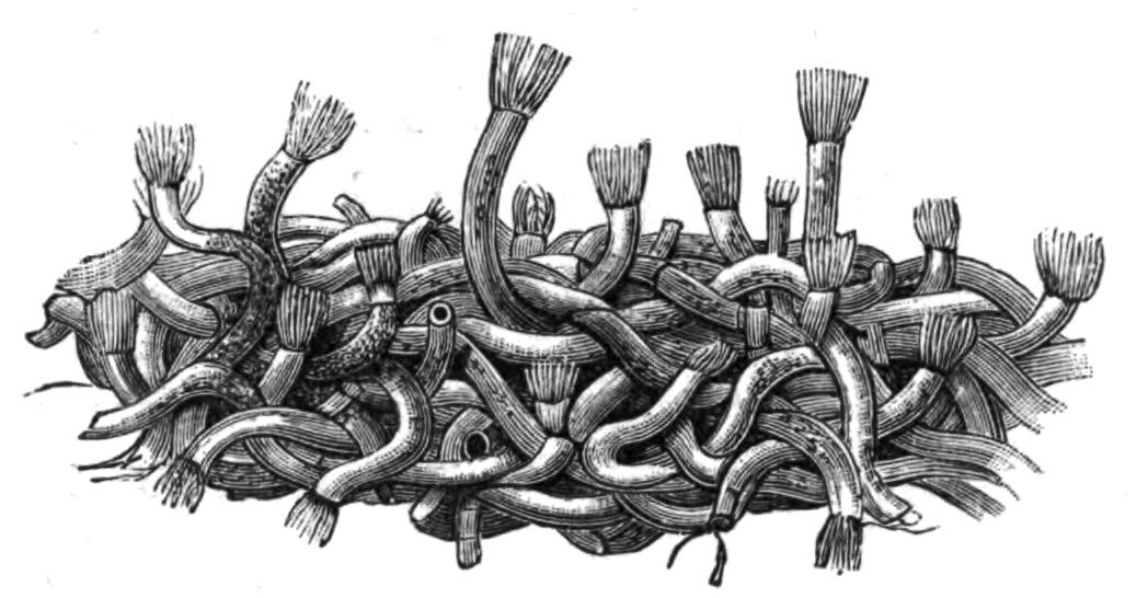
Fig. 226.—A piece of a matted colony of Ph. kowalevskii Cald. Slightly magnified. In most cases the tentacular head is protruding from the tube.
With regard to the habitation of Ph. australis, the largest species known, some discrepancies have crept into the literature of the genus, and to prevent their recurring again it may be worth while to quote the statements of its discoverer, Mr. Haswell.[493] He says: "Phoronis australis occurs in communities of twenty to thirty, in spaces in the substance of the wall of the tube inhabited and formed by a species of Cerianthus. Each worm has a tube of its own, very delicate and transparent, made up of several layers, the mouth opening on the outer surface of the tube of the Cerianthus. The Cerianthus tubes sometimes come up empty, as we should naturally expect, the animal having dropped out; but a sufficient number of occupied tubes are found to show that, under ordinary circumstances, a living Cerianthus occupies the interior of the tube and a community of Phoronis live in its wall. This species of Phoronis is never found anywhere else, and the species of Cerianthus is very rarely found without the Phoronis."
Ph. australis is sluggish in its movements, but other species are capable of very active movement, and withdraw their heads in a moment at the approach of danger. A Neapolitan species, Ph. kowalevskii—known to the fishermen of that place as "Ficchetelli bianchi" or "Vermi di ceppa"—lives chiefly on submarine posts and piles; its tubes, closely interlacing, form a dense feltwork, upon which Ascidians and Sea-anemones often settle, and over which Ophiurids and Polychaets creep. The tubes of this species are rendered opaque by the excreta ejected from the body, and they do not attach foreign substances to the outside to anything like the same degree as Ph. psammophila, which live in sandy places, and are termed by the Sicilian fishermen "Tubi di sabbia." The feltwork of Ph. kowalevskii attains a thickness of 5 to 8 cm. In each case the tube is much longer than the animal it shelters, and is so entangled with its neighbours, to which it frequently adheres, that it is a matter of considerable difficulty to isolate it.
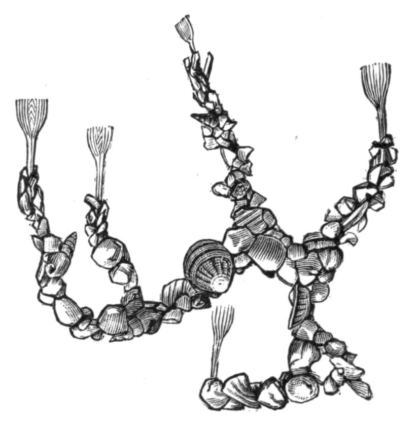
Fig. 227.—A piece of a colony of Ph. psammophila Cori. Slightly magnified. The tubes are covered by particles of sand, small shells, etc.
The various species of Phoronis differ a good deal in size; Cori gives the average length as varying from 1.5 to 7.9 mm. in Ph. hippocrepia and up to 127 mm. (6 inches) in Ph. australis. Probably the very short individuals of the first-named species had not attained their adult stature. Ph. australis has recently formed the subject of a memoir by Dr. W. B. Benham,[494] from whom the following account is mainly taken.
The length of the individuals varied from three to six inches, and their diameter, which is not very uniform, averaged one-eighth of an inch. At one end, which, since it bears the mouth, we may call the oral end, is the very characteristic tentacular crown surrounding the mouth on all sides but one, where there is a slight break in its continuity. The crown of tentacles or lophophore is flattened, and the two ends drawn out, and each is coiled into a spiral (Fig. 229); between the bases of these two spirals three ridges can be seen, each ending in a pore; the median opening is the anus, the two lateral are the openings of the nephridia or kidneys, which also serve as ducts for the reproductive organs. The anus is thus approximated to the mouth, and since the continuity of the tentacular crown is broken at a spot just between the two, there would be nothing to separate these orifices if it were not for the presence of the epistome, a projection or flap of the body-wall which overhangs the mouth between it and part of the crown of tentacles.
The extent to which the ridge bearing the tentacles is incurved at each side varies in different species. In Ph. kowalevskii and Ph. psammophila the ends are only slightly turned in, so that the crown of tentacles is truly horse-shoe shaped; but in Ph. australis they are turned in and form three coils on each side. The number of tentacles also varies, being very numerous in Ph. australis and Ph. buskii—the latter having as many as 300, whilst the other species as a rule have from 60 to 90. The bases of the tentacles are fused for a short distance with one another, forming a thin membrane.
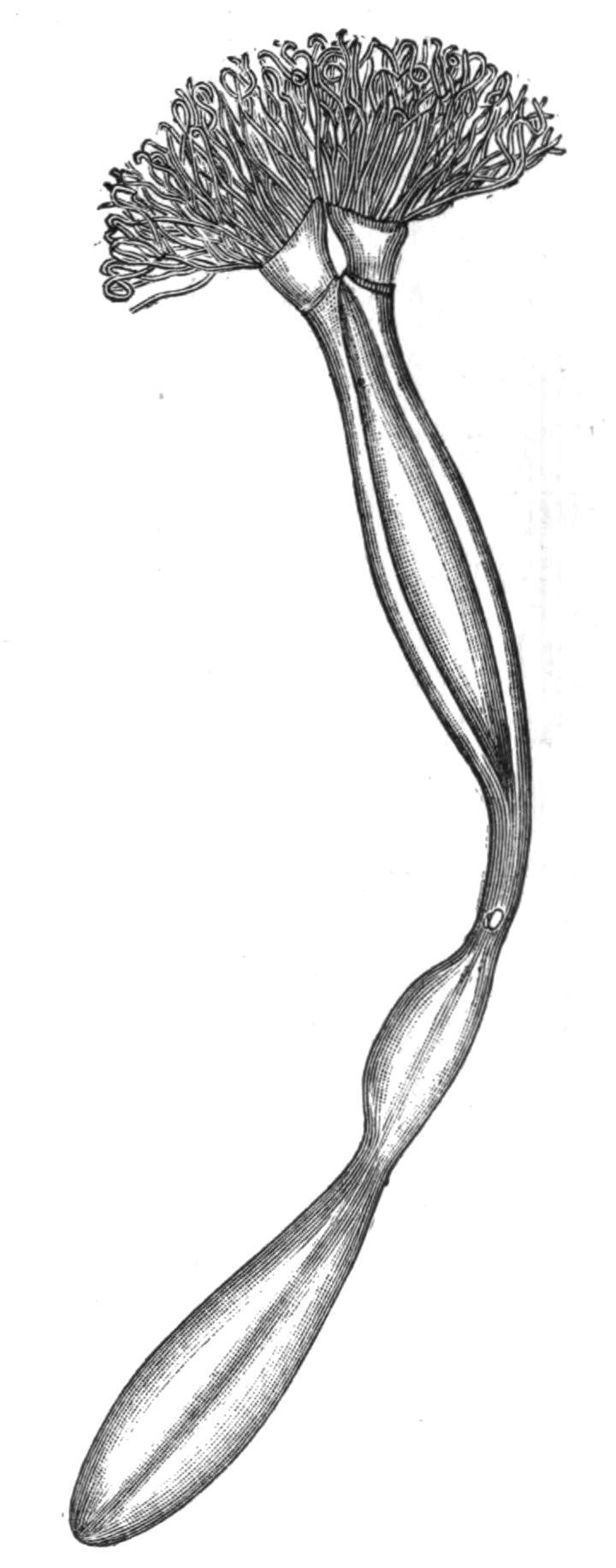
Fig. 228.—A specimen of Ph. buskii M‘Int. removed from its tube and seen from behind, × about 2. (After M‘Intosh.)
The rest of each tentacle is free, and its inner surface, or that turned towards the mouth, is covered with long cilia, which, by the currents they set up, doubtless serve to bring food to the mouth. The tentacles are hollow, and their cavity is kept open by a stiffening of the tissue, which almost resembles an internal skeleton; the cavity communicates with the anterior part of the general body-cavity, and up it runs a single blood-vessel containing red blood. A single nerve is also distributed to each tentacle.
At the base of the two spirals of the tentacular crown lie two ciliated pits, regarded by Caldwell and M‘Intosh[495] as sensory organs, but Benham looks upon them as glandular in structure and function. Perhaps they secrete the substance from which the tubes are formed.
The skin is covered by a delicate cuticle secreted by the underlying epidermis; within the latter is a well-marked basement membrane, and beneath this a layer of circular muscle fibres; these surround a layer of longitudinally-arranged fibres, which do not form a continuous sheet but are arranged in bundles. In both layers the fibres are unstriated. The longitudinal fibres are covered on their inner side by a layer of flat pavement cells, which line the general cavity of the body.
This space, the body-cavity, is divided into two parts by the presence of a diaphragm or septum which runs across from one side of the body to the other about the level of the ridge bearing the tentacular crown. The anterior space is continuous with the cavities of the tentacles and of the epistome. The partition is pierced by the blood-vessels and the oesophagus, but the rest of the alimentary canal, including the anus, the kidneys, and the reproductive organs, all lie in the posterior half of the body-cavity behind the diaphragm. This portion of the body-cavity is further subdivided by the presence of three longitudinal mesenteries supporting the alimentary canal and running between it and the body-wall. One of these mesenteries runs along the outside of the alimentary canal throughout its whole length, attaching both the descending and ascending limbs of the U-shaped tube to the body-wall. The other two are lateral mesenteries, which pass from the body-wall to the sides of the oesophagus. These mesenteries therefore divide the body-cavity into three spaces—one in which the rectum lies, which may be called the rectal, and two lateral; owing to the fact that the lateral mesenteries end before they reach the bend of the alimentary canal, the three chambers are in free communication one with another. The body-cavity is further traversed by irregular strands of tissue which run from the body-wall to the various organs. It contains a corpusculated fluid.
The alimentary canal (Fig. 230) consists of a U-shaped tube which may be divided into four regions. The mouth (m) leads into the oesophagus (oe), which gradually enlarges into the stomach (st) situated just before the bend; a constriction just at the bend separates the stomach from the intestine (int), and this leads into the rectum (r), which terminates in the anus (an). The first three divisions of the alimentary canal are ciliated, but the rectum is not; the walls of the stomach also contain glandular cells, but there are no special glands opening into any part of the tract.
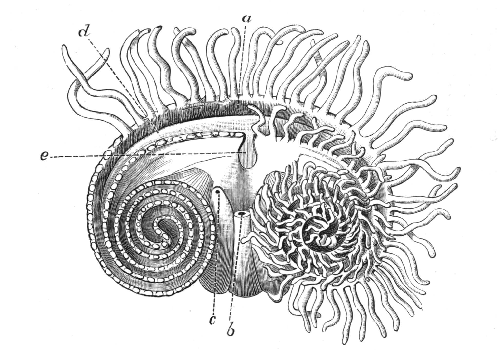
Fig. 229.—The dorsal surface of Ph. australis Has., looking down on the head. The tentacles are cut away on the left side, and the innermost are shortened on the right side to show the arrangement; in reality they are of the same length throughout. a, Mouth; b, anus; c, pore of left nephridium; d, epistome; e, break in the inner series of tentacles. The drawing is to some extent diagrammatic, and is considerably enlarged. (After Benham.)
One of the most interesting features of Phoronis is the presence of a closed system of blood-vessels containing red blood. There are two main blood-vessels; one, lying in the rectal chamber between the two limbs of the U-shaped alimentary canal, has been named the afferent vessel. Just below the diaphragm this splits into two, and each branch, after piercing this partition, runs in a spiral course along the base of the crown of tentacles, giving off a single blood-vessel into each tentacle. At its base each tentacular vessel opens not only into the above-mentioned "distributing" vessel, but also into a "recipient" vessel which takes a course parallel with the former. The two recipient vessels pierce the diaphragm, and after running for some distance apart, fuse to form the efferent vessel, which continues down the body on the left side of the oesophagus. At the aboral end of the body the efferent vessel turns forward and becomes the afferent. Both the main vessels give off numerous blood diverticula, which are developed into plexiform sinuses on the walls of the stomach, and in this region they are covered with the reproductive cells. All the vessels are contractile, and Strethill Wright counted about fifteen pulsations a minute. The blood contains numerous nucleated, disc-shaped corpuscles differing in appearance from those of the fluid in the body-cavity. The corpuscles contain haemoglobin, which gives the red colour to the blood.
The two nephridia or kidneys are essentially tubes which open on the one side into the body-cavity, and on the other to the exterior. The position of the external pores has already been described, one being on each side of the anus. Each pore leads into a tube which passes into that part of the body-cavity situated below the diaphragm, where it divides, and each of the two branches terminates in a ciliated funnel-shaped opening. The smaller of these two funnels pierces the lateral mesentery and opens into the lateral chamber, whilst the larger, whose opening is very much drawn out longitudinally, opens into the rectal chamber. The whole organ is ciliated internally.
The nervous system lies in the skin immediately below the epidermis. This position is very primitive, and forms one of the most interesting anatomical peculiarities of the genus. The nervous tissue is probably diffused all over the body, but there is a special concentration or thickening in the form of a ring which surrounds the mouth, following the base of the tentacular spirals and giving off a nerve to each tentacle. The ring lies at the outside of the base of the tentacles, the anus is not included in it. Caldwell[496] has described in Ph. kowalevskii an asymmetrical nerve-cord given off from the ring and running along the left side of the body; associated with which is a tubular structure of unknown function. In Ph. australis Benham mentions two such tubes, one on each side of the body; their precise value is obscure.
The epithelium covering the nerve-ring is slightly modified in the neighbourhood of the kidney pore, and may have some special sensory function; no other organs of sense are known (but see p. 454).
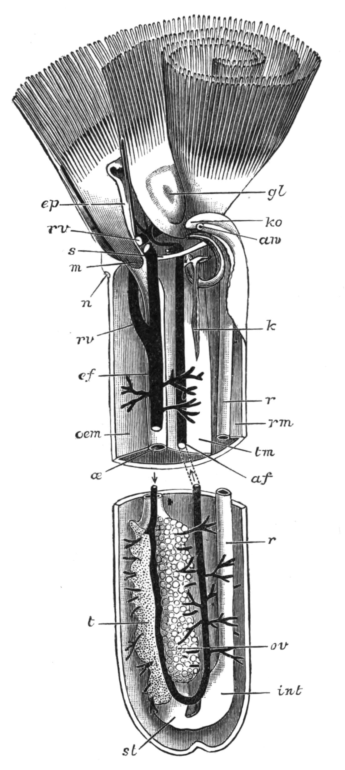
Fig. 230.—A schematic view of the interior of the body of Phoronis. The middle seven-eighths of the body are omitted, af, Afferent blood-vessel; an, anus; ef, efferent blood-vessel; ep, epistome; gl, glandular pit; int, intestine or "second stomach"; k, large funnel of the left nephridium; ko, opening of right nephridium, the opening of the left is seen immediately below in section; m, mouth; n, nerve concentration; oe, oesophagus; oem, oesophageal mesentery; ov, ovary; r, rectum; rm, rectal mesentery; rv, right recipient blood-vessel; s, septum; st, stomach; t, testis; tm, right lateral mesentery. (From Benham.)
Phoronis is hermaphrodite, male and female reproductive cells being formed in the same individual. The testes and ovaries form two white masses lying on the left side of the stomach, one on one side and the other on the other side of the efferent blood-vessel. The glands are traversed in all directions by the diverticula given off from this trunk, and are thus well supplied with blood; in fact both the ovary and the testis are formed by the multiplication and growth of the epithelial cells which cover these diverticula. When ripe the ova and spermatozoa drop off into the body-cavity and make their way to the exterior through the duct of the kidney.
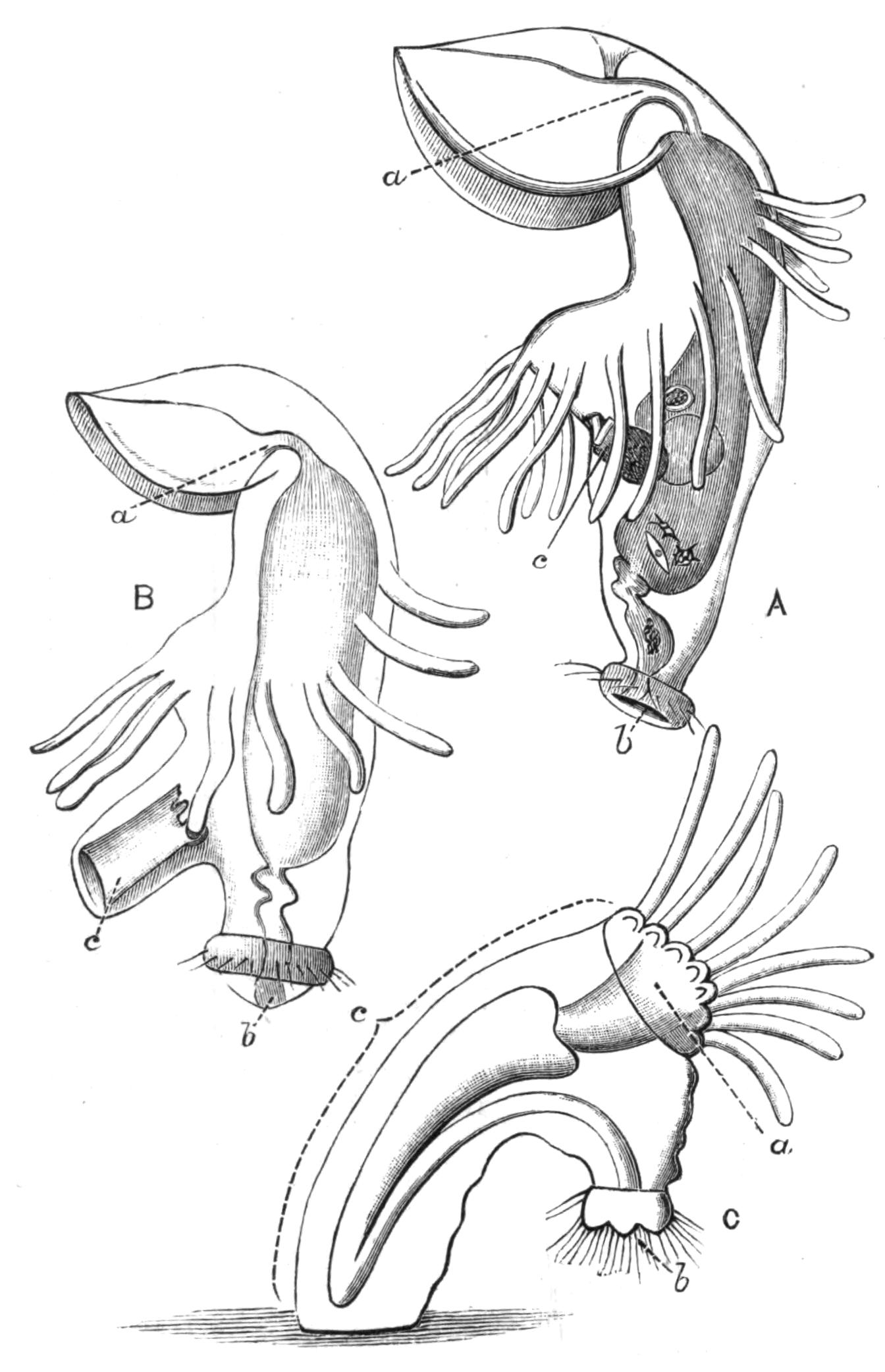
Fig. 231.—Three stages in the metamorphosis of the Actinotrocha into Phoronis. A, Actinotrocha larva with the invagination (c), which will form the trunk of the Phoronis larva beginning to appear. B, Stage with the invagination partly extruded. C, Stage when the extrusion is complete and the alimentary canal has passed into it. C is after Metschnikoff. a, Mouth; b, anus; c, invagination which ultimately forms the greater part of the body of the adult.
The ova are probably fertilised in the sea-water; they undergo the early stages of their development whilst entangled amongst the tentacles of the parent. The larval form to which they give rise was known long before its connexion with the adult was demonstrated by Kowalevsky.[497] It is known as the Actinotrocha (Fig. 231, A), and according to Caldwell has the following structure in Ph. kowalevskii. The mouth is anterior, and the anus terminal and posterior; the mouth is overhung by an immense prae-oral lobe, which bears a special larval nerve ganglion, and in some species four eye-spots; at the base of this, but behind the mouth, is a ring of larval tentacles. The prae-oral lobe and the tentacles are ciliated; the margin of the lobe bears, however, specially long cilia, and there is also a ring of long cilia around the anus.
Before the Actinotrocha stage has been reached the larva has forsaken the shelter of its parent's tentacles, and swims actively about in the open sea. As it grows older a finger-like involution of the skin (c) arises just behind the tentacles on the ventral surface and grows into the body, increasing greatly in length and becoming much folded. The larva now sinks to the bottom of the sea, and after swimming round many times on its axis, undergoes a very astonishing metamorphosis (Fig. 231, B, C). The finger-like involution is suddenly turned inside out, and forms a large projection on the ventral surface, into which the alimentary canal passes, assuming a U-shape, as in the adult. This ventral process in fact forms all the body of the adult behind the line of tentacles, and subsequently contains, not only the alimentary canal, but the kidneys, the reproductive organs, and a large part of the vascular system. At the same time the prae-oral lobe breaks off, and, together with its ganglia and eye-spots, passes into the mouth and is digested in the stomach; the larval tentacles follow the prae-oral lobe, and are similarly digested. Their place is taken by a ring of adult tentacles which commence to appear just behind the larval tentacles before they fall off. The animal is now practically adult.[498]
It is obvious that this astonishing metamorphosis is accompanied by the rotation of the axes of the animal. The adult practically lives at right angles to the larva. In the latter the anus marked the posterior end, and the prae-oral lobe the anterior. The prae-oral lobe has disappeared in the adult, but its position is marked by the mouth. The ventral surface has enormously increased, and corresponds with the whole surface of the trunk. To be consistent we must therefore regard the mouth of the adult as marking the anterior end of the animal, the anus the posterior. The short line between the mouth and anus across the centre of the tentacular crown marks the dorsal surface; and the line running all round the trunk from anus to mouth, the ventral. In fact, in its usual position in its tube Phoronis is lying on its ventral surface, its back faces upwards, and the anterior and posterior ends lie on one side or the other.
Species and Affinities.—In his exhaustive memoir on the anatomy and histology of Phoronis, Cori enumerates seven different species, and quotes the characters of each as enumerated by eight different authors. He, however, reserves his opinion as to the identity or distinctness of some of these species. Benham in his account of Ph. australis enumerates five species, including amongst them Ph. ovalis, which, however, he regards as probably a young form, an opinion in which Cori coincides. The latter regards it as possibly a young form of Ph. hippocrepia.
Without comparing specimens of each of the alleged species, it is difficult to come to any very satisfactory solution of the problem of how many distinct species are at present known, but it seems probable that there are at least six.
(i.) Phoronis hippocrepia Wright.—Under this name is included the first form, described and named by Wright in 1856; also Ph. ovalis, described two years later by the same observer as a distinct form, though it now seems probable that it is but a young form of Ph. hippocrepia. The Crepina gracilis of van Beneden is probably identical with this species.
This species occurs in membranous tubes embedded in limestone, corals, or oyster shells. Its length varies from 1.5 to 15 mm. The number of tentacles varies from 16 to 86. It has been found off the coast of Devonshire and in the Firth of Forth.
(ii.) Phoronis kowalevskii Caldwell.—This name is given by Benham to the species from Naples described by Caldwell, and replaces the name Ph. caespitosa, which was given by Cori. This species is found in the Bay of Naples, living in considerable colonies on submarine piles and posts. It is not firmly attached to its substratum. The tube may be coated with sand or other foreign particles. The length of the individuals varies from 3 to 39 mm. The lophophore is simple, with from 50 to 100 tentacles.
(iii.) Phoronis australis Haswell.—This is the giant of the genus, the length of the individuals being from 3 to 5 (76-127 mm.) or rarely 6 inches. It lives in delicate transparent tubes, interlacing the walls of the tube of a sea-anemone, Cerianthus. The arms of the lophophore coil into two spirals. The colour is reddish or purple. Found in Port Jackson.
(iv.) Phoronis buskii M‘Intosh.—This species was dredged by the Challenger from a sandy bottom at a depth of 10 to 20 fathoms off the Philippines. Its tube is covered with particles of sand, sponge spicules, etc. Its length is 52 mm. or more (more than two inches). The anatomy of this species closely resembles that of Ph. australis, and Benham thinks that, in spite of the difference in their habitat, they may belong to the same species.
(v.) Phoronis architecta Andrews.—A species recently described by Andrews from Beaufort, N.C. Its distinctive features are: "the formation of isolated tubes covered by definite collections of sand grains; the presence of special prostomial organs, possibly of use in the formation of these tubes; the great development of the longitudinal muscles; the presence of a ciliated groove in the digestive tract; the apparent separation of the sexes."
(vi.) Phoronis psammophila Cori.—Found in Faro, near Messina. The tube is hyaline, and is covered by numerous grains of sand, some of considerable size. The length of the individuals is 25 to 50 mm. There are 60 to 90 tentacles. The colour is a fleshy red. A second species discovered by Haswell in Port Jackson had no points of importance to distinguish it from Ph. psammophila, except that no sand adheres to its tube and the number of tentacles is slightly greater.
In addition to the various species of Phoronis, several distinct forms of its larva, Actinotrocha, are known, and have been named without having been traced into their corresponding adult form.
The position of Phoronis in the animal kingdom has formed the matter of considerable divergence of opinion amongst the naturalists who have studied it. The earlier writers regarded Phoronis as allied to the Gephyrea, and it was for a long time classed with these animals, but placed in a separate sub-Order, the Gephyrea tubicola, which was opposed to the Gephyrea nuda, which comprised the true Gephyrea.
Caldwell referred Phoronis, the Brachiopoda, the Polyzoa, and the Gephyrea to the same type of body structure, and Lankester subsequently suggested the provisional name Podaxonia for this miscellaneous collection of animals. Lankester divided his phylum Podaxonia into three classes: (i.) the Sipunculoidea (Gephyrea), (ii.) the Brachiopoda, and (iii.) the Polyzoa. The last-named class he divided into three sections: (a) the Vermiformia, this includes the single genus Phoronis; (b) the Pterobranchia, including the forms Cephalodiscus and Rhabdopleura, whose affinities with Balanoglossus were subsequently demonstrated; and (c) the Eupolyzoa, including the forms treated as Polyzoa in the following pages.
Masterman's recent researches[499] on Phoronis seem to indicate that the Vermiformia, like the Pterobranchia, must in future be grouped with the Hemichordata. He finds three well-defined coelomic spaces corresponding with the epistome, the collar, and the trunk, and also representatives of the collar pores, and is further inclined to believe that structures representing the notochord exist in Actinotrocha.
Should Masterman's researches be confirmed, Phoronis will be removed from its present isolated and enigmatical position, and placed with Cephalodiscus and Rhabdopleura amongst the Hemichordata, which will be described in Vol. VII. of this work.