Chapter VII
Reading edition
CHAETOGNATHA
STRUCTURE—REPRODUCTION—HABITS—FOOD—CLASSIFICATION TABLE OF IDENTIFICATION
At certain seasons and at certain times of the day the naturalist who is investigating the fauna of the surface of the sea is apt to find his tow-net crammed with innumerable transparent spindle-shaped animals, which by their number and the way in which they become entangled with rarer objects, often render useless the result of his labours. These animals belong to the class Chaetognatha, which includes three genera, Sagitta, Spadella, and Krohnia. Amongst them are divided about twenty species, some of which, however, are of doubtful value.
Anatomy.—The body of these animals is as transparent as crystal; it is elongated, and bears a resemblance to certain torpedos, except that the head forms a somewhat blunt termination to the spindle-shaped body. The tail bears a caudal fin, and Spadella and Krohnia have a single pair, and Sagitta two pairs, of lateral fins; all of which are flattened horizontally.
The body is externally divisible into three regions—head, trunk, and tail—and these correspond with the arrangement of the internal organs.
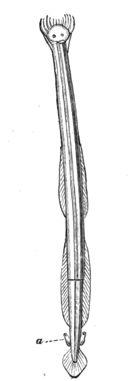
Fig. 101.—Sagitta bipunctata. a, Vesicula seminalis. × 4. (After Hertwig.)
The head is surrounded by a fold of skin, forming a hood, which is most prominent at the sides (Fig. 102, g); within the hood the head bears from two to four rows of short spines, and outside these a right and left row of sickle-shaped hooks, the free ends of which in a state of rest converge round the mouth, but when disturbed these hooks can be widely divaricated.
The cavity of the body, or coelom, is divided into three distinct chambers by the presence of two thin transverse walls or septa, one situated between the head and the trunk, the other between the trunk and the tail (Figs. 104, 105). In the head, this cavity is much reduced by the presence of special muscles which move the spines, hooks, etc.; and in the small species, such as Spadella cephaloptera, the other two cavities are almost entirely occupied by the digestive and reproductive organs[223]; but in the large species, e.g. Sagitta hexaptera, a considerable space is left between the internal organs and the skin, and this is occupied by a coelomic fluid. If the skin of one of these larger species be punctured the fluid escapes and the animal shrivels up. A longitudinal partition or mesentery, with numerous pores in it, runs through these spaces, dividing the body-cavity into a right and left half; in the region of the trunk this mesentery supports the alimentary canal.
In addition to certain muscles in the head, which move the hooks, etc., there is a muscular lining to the body-wall. This is divided into two dorsal and two ventral bands, much in the same way as in Nematodes. The muscle fibres are striated.
The mouth, situated either terminally—Spadella marioni[224]—or below the head, leads into a pharynx; this passes into an intestine lined by a single layer of ciliated cells with a few glandular ones intermingled. The intestine runs straight through the body without loop or coil, and opens by an anus situated at the junction of the trunk and the tail. In most cases the anus is ventral or on the lower surface, but Gourret asserts that in Spadella marioni it is on the upper surface.
There are no special respiratory, excretory, or circulatory organs, unless a glandular structure described by Gourret in the head of Spadella marioni be a real kidney.
The nervous system consists of a supra-oesophageal ganglion or brain situated in the head, and of a ventral ganglion lying in the trunk; both these nerve centres are embedded in the epidermis, and are connected with one another by means of two stout peri-oesophageal nerves (Figs. 102, 104). The brain also gives off a pair of nerves to the eyes, another pair to the olfactory organ, and a pair which ultimately meet one another and so form a ring; on this are certain ganglia giving off nerves which supply the muscles of the head. Both the chief ganglia give off numerous nerves, which divide and split up into a network of fibres which permeate the whole skin.
The sense organs are comparatively simple. A pair of very small eyes lie in the skin of the head; they are of complex structure, and to some extent remind one of the simple eyes of certain Crustacea. Behind the eyes and also on the upper surface of the animal is an unpaired organ which is usually described as olfactory in function (Figs. 103, 105). This is a ring-shaped modification of the epidermis drawn out into different shapes in the various species. The modified epidermal cells bear long cilia. The remaining sensory organs found in the group consist of clumps of modified cells scattered in round groups over the surface of the body and of the fins. The central cells of each group bear long tactile hairs, and are surrounded by supporting cells.
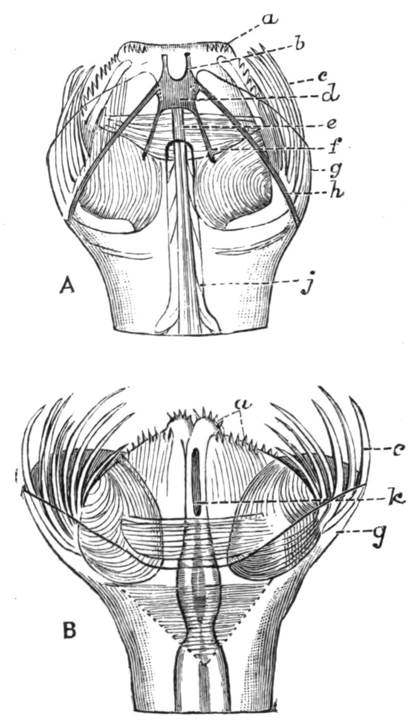
Fig. 102.—Head of Sagitta bipunctata. A, Dorsal view; B, ventral view. × about 33. (From Hertwig.) A, a, spines; b, nerves to lateral cephalic ganglia; c, hooks; d, cephalic ganglion; e, olfactory nerve; f, optic nerve; g, hood; h, commissure to ventral ganglion; j, olfactory organ: B, a, c, and g as in A; k, mouth.
The Chaetognatha are hermaphrodite, and carry the female organs in the trunk, the male in the tail. In a mature specimen the two ovaries occupy almost all the space in the trunk between the alimentary canal and the skin, and each is supported by a narrow lateral mesentery. The ovary is traversed by a oviduct which often contains spermatozoa; it is not clear how the eggs make their way into the oviduct, which seems to have no internal opening and to act largely as a receptaculum seminis. The oviducts open externally on the upper side at the base of the lateral fin, close to the junction of the tail and the trunk.
The cavity of the tail is divided into two lateral chambers by the extension backward of the median vertical mesentery. In each of these a testis and a vas deferens are found. The testes are solid ridges formed by the growth of the lining cells of this part of the body-cavity; the cells mature into spermatozoa, which break off and float freely in the coelomic fluid. At the breeding season the whole tail may be crowded with masses of spermatozoa, which are kept in a more or less regular circulation by the ciliated cells lining the body-wall. The vas deferens opens internally into the space where the spermatozoa lie, and at the other end into a vesicula seminis, which opens to the exterior. The position of the latter structure varies, and is of some systematic value.
The eggs are laid in the water and as a rule float at the surface of the sea. Spadella cephaloptera is, however, an exception to this rule, as it attaches its eggs by means of a gelatinous stalk to sea-weeds. The segmentation of the ovum is regular, and gives rise to a two-layered stage or gastrula, which opens by a pore, the blastopore. This does not, however, become the mouth, but closes up and the mouth arises at the opposite pole. Perhaps the most interesting feature of the development of Sagitta is that the cells destined to form the reproductive organs separate from the other cells of the embryo at a very early date, whilst it is still in the gastrula stage. There is no larval form, but the young hatch out from the egg in a state resembling the adult in all respects but that of size.
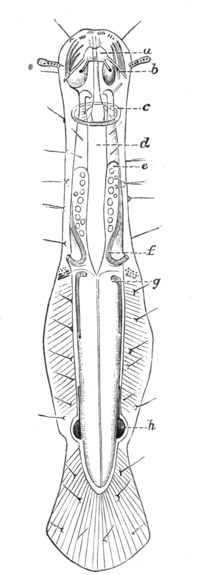
Fig. 103.—Spadella cephaloptera. Dorsal view. x 30. (From Hertwig.) a, Cephalic ganglion; b, commissure to ventral ganglion; c, olfactory organ; d, alimentary canal; e, ovary; f, oviduct; g, testis; h, vesicula seminalis.
Habits.—The Chaetognatha are essentially pelagic, and resemble many other creatures that dwell at the surface of the ocean in being almost completely transparent. Most species have been taken far out at sea, but some are perhaps rather more numerous near the coast, and one species, Spadella cephaloptera, is littoral. They swim by means of muscular movements of the whole body; the fins have no movement of their own, and seem to serve as balancers, and not as locomotory organs. Although usually found at the surface of the water, many species have been taken at considerable depths. Chun[225] states that they are found in countless numbers at depths of from 100 metres to 1300 metres. The commonest species at these depths are Sagitta hexaptera and Sagitta serratodentata. Sagitta bipunctata, according to the same authority, confines itself to the surface. Whether the change of depth is diurnal, or whether it has any relation to sexual maturity, or to any other cause, has not been satisfactorily determined.
The food of the Chaetognatha consists of floating diatoms, Infusoria, small larvae, and such Copepods as Calanus finmarchicus, and small Amphipods as Phoxus plumosus.[226] At times they also devour small larval or post-larval fishes, and owing to their incredible numbers, they doubtless do considerable damage to sea fisheries. It is also recorded that they eat one another, and specimens have been taken which have ingested the whole body of another Sagitta except the head, which hangs out of the mouth of the eater, and gives it the appearance of a double-headed monster.[227] It has been said that they attack hydroid polypes, but here at any rate they do not have it all their own way. Masterman[228] has figured the apical group of five polypes of Obelia, three of which are engaged in ingesting as many young Sagitta.
They exist in incredible numbers; Grassi describes the surface of the sea at Messina on certain days as being literally covered with them, and they must form the food supply of numerous animals which prey upon the pelagic fauna. The immense number of individuals is probably accounted for to some extent by the fact that they lay eggs all the year round, and pass through a very short and rapid development. They are not known to be phosphorescent.
Classification.—The features of the Chaetognatha which have most systematic value are the size of the adult, the relations of the length to the breadth, and of the three divisions to one another; the size, number, and position of the lateral fins, and of the hooks and spines on the head; the thickness of the epidermis, and the structure of the olfactory organ; and, finally, the form of the reproductive organs.
Strodtmann,[229] who gives the latest and most complete account of the species of Chaetognatha, arranges them under three genera, which he characterises as follows:—
(i.) Sagitta Slabber.—Two pairs of lateral fins, two rows of spines on the head. The lateral thickening of the epidermis absent or insignificant.
Under this genus are included nine definite species and five others—S. gracilis Verrill, S. elegans Verrill, S. darwini Grassi, S. diptera d'Orbigny, and S. triptera d'Orbigny—whose position, owing to the inadequacy of their description, is of doubtful validity.
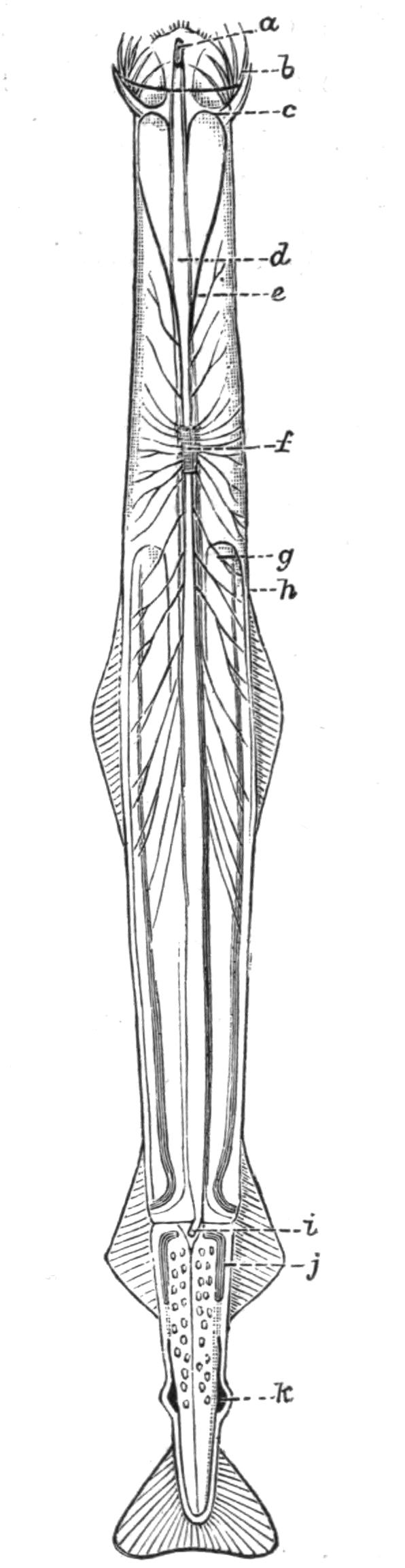
Fig. 104.—Sagitta hexaptera. Ventral view. × 4. (From Hertwig.) a, Mouth; b, hooks; c, anterior septum; d, alimentary canal; e, commissure from the brain to the ventral ganglion; f, ventral ganglion; g, ovary; h, oviduct; i, posterior septum; j, testis; k, vesicula seminalis.
The distribution of the other species may be mentioned. S. hexaptera is the largest Chaetognath known, and reaches in the adult stage a length of 7 cm. It is very widely distributed, being found in practically all the temperate and warm seas, usually at the surface of the water, though at times it is found at a depth of one metre, or even deeper. S. lyra, Mediterranean, very rare. S. tricuspidata, widely distributed. S. magna, Mediterranean and Madeiran, living at the surface. S. bipunctata, the most frequently described form, smaller than the preceding species, 1-2 cm. in length, widely distributed, and as a rule living near the coast line. S. serratodentata, Mediterranean. S. enflata, on the surface of the sea, Mediterranean and Madeiran. S. minima, a very small species, 1 cm. in length, Mediterranean. S. falcidens, Atlantic, off the coast of New Jersey.
(ii.) Krohnia Langerhans.—A single lateral fin extending on to both trunk and tail segment, no lateral epidermal extensions behind the head, only one row of spines on the head. Trunk longer than the tail.
Krohnia has but two species: K. hamata Möbius, with a length of 3-4 cm., found in the North Atlantic and at considerable depths, 200 to 300 fathoms; and K. subtilis Grassi, 1.5 cm. long, with an extraordinary slender body and a relatively large head, found at Messina, but very rare; as a rule only one specimen has been found at a time.
(iii.) Spadella Langerhans.—A single pair of lateral fins; these are situated on the tail segment. Behind the head a thickening of the epidermis extends down each side of the body to the fin, or even farther. Two rows of spines on the head. Small animals, not longer than 1 cm.
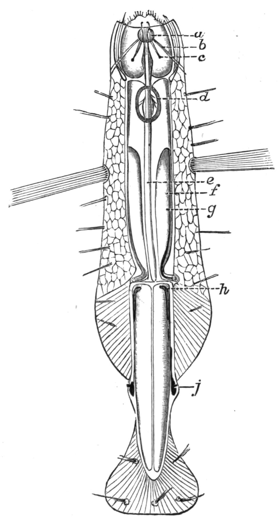
Fig. 105.—Spadella draco. Dorsal view. × 12. (From Hertwig.) a, Cephalic ganglion; b, commissure between the cephalic ganglion and the ventral; c, eye; d, olfactory organ; e, alimentary canal; f, ovary; g, oviduct (the line goes a little beyond the duct); h, testis; j, vesicula seminalis.
S. cephaloptera Busch is the smallest species of Chaetognatha, attaining at most a length of .5 cm. The body is not so transparent as in other species, and is of a yellowish colour. It has been found from the Orkney Islands to the Mediterranean. Strodtmann is of the opinion that the three species S. mariana Lewes, S. batziana Giard, and S. gallica Pagenstecher differ from the above-named only in size, or that their description is too indefinite to permit of accurate characterisation. He recognises three other distinct species: S. pontica Uljanin, from the Black Sea; S. marioni Gourret, from the Gulf of Lyons; and S. draco Krohn, Mediterranean and Madeiran, and from the Canaries.
Much confusion has been introduced into the classification of the Chaetognatha by Grassi,[230] who calls some—but not all—of what other writers term Sagitta, Spadella, and vice versâ. The following table was compiled by Strodtmann,[231] but I have incorporated in it two species recently described from Amboyna by Béraneck,[232] and called by him Sagitta bedoti and Spadella vougai respectively:—
CHAETOGNATHA
I. Two pairs of lateral fins; two rows of spines on the head; slender forms.
(i.) Number of spines in posterior row greater than in anterior.
a. Border of hooks smooth, their point not curved.
α. No interval between the two fins on each side. 3.5 cm. long; 4-7 anterior spines, 8-11 posterior spines; olfactory organ lying entirely on the trunk. The anterior nerves of the ventral ganglion lie close to one another as far as the head.—Sagitta lyra.
β. A distinct interval between the two fins on each side.
aa. Adult animals large; hooks 6-7; anterior spines 3-4; posterior spines 5-7; tail ¼ or ⅕ of the total length; lateral areas relatively larger.—Sagitta hexaptera.
bb. Greatest length 1-2 cm.
αα. Thickening of the epidermis behind the head; prominently projecting vesiculae seminales; olfactory organ very long; hooks 8-10; anterior spines 4-6; posterior spines 10-15.—Sagitta bipunctata.
ββ. No epidermal thickening; two caeca on the anterior end of intestine; length 1 cm.; hooks 6-9; anterior spines 3-4; posterior spines 7-8; point of the hooks somewhat bent round.—Sagitta minima.
γγ. Epidermis thin; no caeca; hooks 8-9, their ends not bent; anterior spines 3-4; posterior spines 7-8; length 2 cm.; small head; trunk proportionately thick.—Sagitta enflata.
δδ. Hooks 11-14, usually 12; length 1.8 cm.; anterior spines 6-7; posterior spines 18.—Sagitta falcidens.
εε. Hooks 7 on each side; length 1.3 cm.; anterior spines 8-10, posterior spines 18-22; no olfactory organ.—Sagitta bedoti.
b. Edge of hooks toothed and their point bent round; hooks 6-8; anterior spines 6-8; posterior spines 10-12; length 1.5 cm.; slender; conspicuously projecting vesiculae seminales.—Sagitta serratodentata.
(ii.) Number of the spines in posterior row smaller than in anterior.
a. Anterior spines 3; posterior spine 1; hooks 8; length 3.5 cm.—Sagitta tricuspidata.
b. Anterior spines 4; posterior spines 3; hooks 10-13; length 4.1 cm.; tail ⅕ of the total length.—Sagitta magna.
II. One pair of lateral fins lying on the trunk and tail; one row of spines; body slender; epidermis not thickened.
(i.) Hooks 8-9, bent like an elbow at the point, serrated in the young; 20-25 spines in a row; ovary reddish; length 3-4 cm.—Krohnia hamata.
(ii.) Hooks 8, broad at their base but very sharply pointed; spines in a curved row, about 18, with a constriction below like the neck of a bottle; body thin; length 1-1.5 cm.—Krohnia subtilis.
III. One pair of lateral fins, these lie on the tail; body relatively very broad in consequence of the thickening of the epidermis lying behind the head; two rows of spines; greatest length 1 cm.; tail and trunk usually the same length.
(i.) A great extension of the epidermis behind the head, consisting of very large cells; amongst these, at the level of the ventral ganglion, lies a bundle of stiff hairs; tactile organ on papillae; hooks 9-10; anterior spines 6-8; posterior spines 12-18.—Spadella draco.
(ii.) Lateral extension of the epidermis not so conspicuous, and the cells composing it smaller. Tactile organs in little depressions. Transverse as well as longitudinal muscles in the trunk. Adhesive cells on the ventral surface of the body. No interval between the lateral fins and the tail fin. Two papillae on the head-hood elongated into club-shaped tentacles. Hooks 8-9, slightly serrated; anterior spines 3-4; posterior spines 3-4.—Spadella cephaloptera.
(iii.) Similar to the last-mentioned species, but the tail segment is larger than the trunk; in the above it is of the same size. No adhesive cells. The fins are covered with papillae, and with a number of serrated spines pointed at both ends.—Spadella pontica.
(iv.) Tactile organs and adhesive cells are unmodified epidermal cells. Anus dorsal. Orifice of oviducts ventral. No olfactory organ. Epidermis colourless. Lateral fins without rays. A pair of ganglia at the postero-lateral angle of the brain.—Spadella marioni.
(v.) Tactile organs well developed on the head, trunk, and fins; tail segment a little shorter than the trunk. Body short, length 3-4 mm. Hooks 9; anterior spines 4-5, posterior spines 6-7.—Spadella vougai.