Chapter VI
Reading edition
LEPIDOPTERA—OR BUTTERFLIES AND MOTHS
Order VI. Lepidoptera.
Wings four; body and wings covered with scales usually variegate in colour, and on the body frequently more or less like hair: nervures moderate in number, at the periphery of one wing not exceeding fifteen, but little irregular; cross-nervules not more than four, there being usually only one or two closed cells on each wing, occasionally none. Imago with mouth incapable of biting, usually forming a long coiled proboscis capable of protrusion. Metamorphosis great and abrupt; the wings developed inside the body; the larva with large or moderate head and strong mandibles. Pupa with the appendages usually adpressed and cemented to the body so that it presents a more or less even, horny exterior, occasionally varied by projections that are not the appendages and that may make the form very irregular: in many of the smaller forms the appendages are only imperfectly cemented to the body.
Lepidoptera, or butterflies and moths, are so far as ornament is concerned the highest of the Insect world. In respect of intelligence the Order is inferior to the Hymenoptera, in the mechanical adaptation of the parts of the body it is inferior to Coleoptera, and in perfection of metamorphosis it is second to Diptera. The mouth of Lepidoptera is quite peculiar; the proboscis—the part of the apparatus for the prehension of food—is anatomically very different from the proboscis of the other Insects that suck, and finds its nearest analogue in the extreme elongation of the maxillae of certain Coleoptera, e.g. Nemognatha. The female has no gonapophyses, though in certain exceptional forms of Tineidae, there are modifications of structure connected with the terminal segments, that have as yet been only imperfectly investigated. As a rule, the egg is simply deposited on some living vegetable and fastened thereto. Lepidoptera are the most exclusively vegetarian of all the Orders of Insects; a certain number of their larvae prey on Insects that are themselves filled with vegetable juices (Coccidae, Aphidae) and a very small number (Tinea, etc.) eat animal matter. In general the nutriment appears to be drawn exclusively from the fluids of the vegetables, the solid matter passing from the alimentary canal in large quantity in the form of little pellets usually dry, and called frass. Hence the quantity of food ingested is large, and when the individuals unduly increase in number, forest trees over large areas are sometimes completely defoliated by the caterpillars.
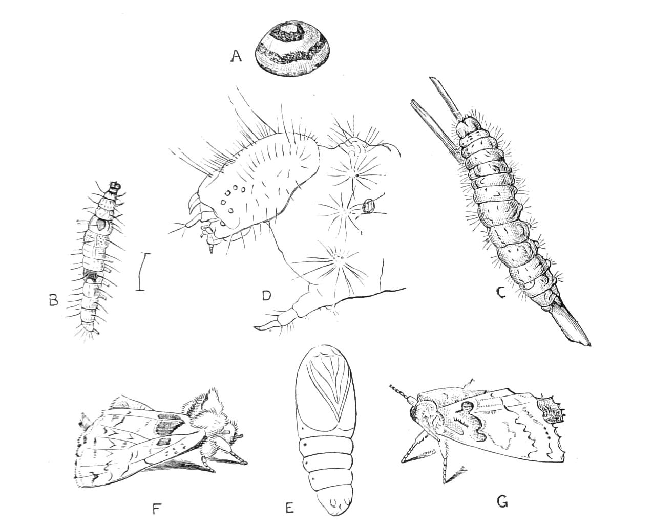
Fig. 157.—Metamorphosis of a Lepidopteron (Rhegmatophila alpina, Notodontidae). (After Poujade, Ann. Soc. ent. France, 1891.) Europe. A, Egg; B, young larva, about to moult; C, adult larva; D, head and first body-segment of adult larva, magnified; E, pupa, × 2⁄1; F, male moth in repose; G, female moth in repose.
Lepidoptera pass a larger portion of their lives in the pupal stage than most other Insects do; frequently during nine months of the year the Lepidopteron may be a pupa. In other Orders of Insects it would appear that the tendency of the higher forms is to shorten the pupal period, and when much time has to be passed between the end of the feeding up of the larva and the appearance of the imago, to pass this time as much as possible in the form of a resting-larva, and as little as may be in the form of a pupa; in Lepidoptera the reverse is the case; the resting-larva period being usually reduced to a day or two. Hence we can understand the importance of a hard skin to the pupa. There are, however, numerous Lepidopterous pupae where the skin does not attain the condition of hardness that is secured for the higher forms by the chitinous exudation we have mentioned; and there are also cases where there is a prolonged resting-larva period: for instance Galleria mellonella spins a cocoon in the autumn and remains in it as a resting larva all the winter, becoming a pupa only in the spring. In many of these cases the resting-larva is protected by a cocoon. It is probable that the chief advantage of the perfect chitinous exudation of the Lepidopterous pupa is to prevent the tiny, complex organisation from the effects of undue transpiration. Bataillon has suggested that the relation of the fluid contents of the pupa to air and moisture are of great importance in the physiology of metamorphosis.
The duration of life is very different in various forms of Lepidoptera. It is known that certain species (Ephestia kuehniella, e.g.) may go through at least five generations a year. On the other hand, certain species that feed on wood or roots may take three years to complete their life-history; and it is probable that some of the forms of Hepialidae are even longer lived than this.
Lepidoptera have always been a favourite Order with entomologists, but no good list of the species has ever been made, and it would be a difficult matter to say how many species are at present known, but it can scarcely be less than 50,000. In Britain we have about 2000 species.
The close affinity of the Order with Trichoptera has long been recognised: Réaumur considered the latter to be practically Lepidoptera with aquatic habits, and Speyer pointed out the existence of very numerous points of similarity between the two. Brauer emphasised the existence of mandibles in the nymph of Trichoptera as an important distinction: the pupa of Micropteryx (Fig. 211) has however been recently shown to be similar to that of Trichoptera, so that unless it should be decided to transfer Micropteryx to Trichoptera, and then define Lepidoptera and Trichoptera as distinguished by the condition of the pupa, it would appear to be very difficult to retain the two groups as distinct.
Structure of Imago.—The head of a Lepidopteron is in large part made up of the compound eyes; in addition to these it frequently bears at the top a pair of small, simple eyes so much concealed by the scales as to cause us to wonder if seeing be carried on by them. The larger part of the front of the head is formed by the clypeus, which is separated by a well-marked line from the epicranium, the antennae being inserted on the latter near its point of junction with the former. There is sometimes (Saturnia, Castnia) on each side of the clypeus a deep pocket projecting into the head-cavity. The other parts of the head are but small. The occipital foramen is very large.[164]
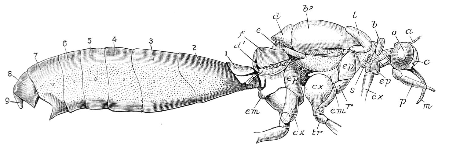
Fig. 158—External structure of a female butterfly, Anosia plexippus. (After Scudder.) a, Base of antenna; b, pronotum; b2, scutum of mesothorax; c, clypeus; cx, coxa; d, scutellum; d1, scutellum of metathorax; e, post-scutellum (= base of phragma); em, epimeron; ep, episternum; f, scutum of metathorax; m, basal part of proboscis (= maxilla); o, eye; p, labial palp; r, mesosternum; s, prothoracic spiracle; t, tegula; tr, trochanter; 1-9, dorsal plates of abdomen.
The antennae are always conspicuous, and are very various in form; they are composed of numerous segments, and in the males of many species attain a very complex structure, especially in Bombyces and Psychidae; they doubtless function in such cases as sense-organs for the discovery of the female.
The largest and most important of the mouth-parts are the maxillae and the labial palpi, the other parts being so small as to render their detection difficult. The labrum is a very short, comparatively broad piece, visible on the front edge of the clypeus; its lateral part usually forms a prominence which has often been mistaken for a mandible; Kellogg has applied the term "pilifer" to this part. In the middle of the labrum a small angular or tongue-like projection is seen just over the middle of the base of the proboscis; this little piece is considered by several authorities to be an epipharynx.
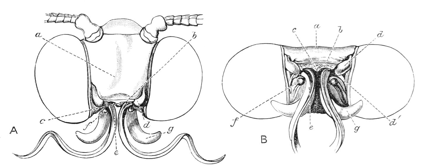
Fig. 159—Mouth of Lepidoptera. Tiger-moth, Arctia caja. A, Seen from front; B, from front and below, a, Clypeus; b, labrum; c, epipharynx; d, mandibular area; d′, prominence beneath mandibular area; e, one side of haustellum or proboscis; f, maxillary palp; g, labial palp.
Mandibles.—Savigny, Westwood, and others considered the parts of the labrum recently designated pilifers by Kellogg to be the rudimentary mandibles, but Walter has shown that this is not the case.[165] The mandibles are usually indistinguishable, though they, or some prominence possibly connected with them,[166] may frequently be detected in the neighbourhood of the pilifers; they are, according to Walter, largest and most perfectly developed in Eriocephala, a genus that was not distinguished by him from Micropteryx and was therefore termed "niedere Micropteryginen," i.e. lower Micropteryges. The opinion entertained by Walter that Micropteryx proper (his "höhere Micropteryginen") also possesses rudimentary mandibles is considered by Dr. Chapman, no doubt with reason, to be erroneous.[167] The mandibles, however, in the vast majority of Lepidoptera can scarcely be said to exist at all in the imago; there being only an obtuse projection—without trace of articulation—on each side of the labrum; and even this projection is usually absent. Meinert recognised these projections as mandibles in Smerinthus populi, and Kellogg in Protoparce carolina, another large Sphinx moth. They appear to be unusually well developed in that group. In Castnia they are even more definite than they are in Sphingidae.
The Maxillae are chiefly devoted to the formation of the proboscis. Their basal portions are anatomically very indefinite, though they exist very intimately connected with the labium. Each usually bears a small tubercle or a segmented process, the representative of the maxillary palpus. The proboscis itself consists of the terminal, or outer, parts of the two maxillae, which parts are closely and beautifully coadapted to form the spirally coiled organ, that is sometimes, though incorrectly, called the tongue. The exact morphology of the Lepidopterous proboscis has not been established. The condition existing in the curious family Prodoxidae (see p. 432), where a proboscis coexists with another structure called a maxillary tentacle, suggests a correspondence between the latter and the galea of a typical maxilla; and between the proboscis and the lacinia or inner lobe of a maxilla: but J. B. Smith is of opinion that the tentacle in question is a prolongation of the stipes. The condition of the parts in this anomalous family (Prodoxidae) has not, however, been thoroughly investigated, and Packard takes a different view of the proboscis; he considers that "it is the two galeae which become elongated, united and highly specialised to form the so-called tongue or glossa of all Lepidoptera above the Eriocephalidae."[168] The proboscis in some cases becomes very remarkable, and in certain Sphingidae is said to attain, when unrolled, a length of ten inches. In some cases the maxillary lobes do not form a proboscis, but exist as delicate structures, pendulous from the mouth, without coadaptation (Zeuzera aesculi, the Wood-leopard moth). In other forms they are absent altogether (Cossus, e.g.), and in Hepialus we have failed to detect any evidence of the existence of the maxillae. On the other hand, in Micropteryx the maxillae are much more like those of a mandibulate Insect; and various other Microlepidoptera approach more or less a similar condition. In the genus last mentioned the maxillary palpi are largely developed, flexible and slender. According to Walter various forms of palpus intermediate between that of Micropteryx and the condition of rudimentary tubercle may be found amongst the Microlepidoptera.[169]
Labium.—The labial palpi are usually largely developed, though but little flexible; they form conspicuous processes densely covered with scales or hairs, and curve forwards or upwards, rarely downwards, from the under side of the head, somewhat in the fashion of tusks. The other parts of the labium are frequently represented merely by a membranous structure, united with the maxillae and obstructing the cavity of the pharynx. Where the proboscis is absent it is difficult to find any orifice leading to the alimentary canal, such opening as may exist being concealed by the overhanging clypeus and labium. In some forms, Saturnia, e.g., there appears to be no buccal orifice whatever. In Hepialus the labium is in a very unusual condition; it projects externally in the position usually occupied by the labial palpi, these organs being themselves extremely short. It is very difficult to form an opinion as to the structure of the labium and other mouth-parts when the maxillae are not developed, as in these cases the parts are of a delicate membranous nature, and shrivel after death. This is the explanation of the fact that in descriptive works we find vague terms in use such as "mouth aborted" or "tongue absent."
The mouth of the Lepidopterous imago is a paradoxical structure; it differs very greatly from that of the larva, the changes during metamorphosis being extreme. We should thus be led to infer that it is of great importance to the creatures; but, on the other hand, the various structures that make up the mouth, as we have remarked, are frequently absent or reduced to insignificant proportions; and even in forms where the apparatus is highly developed the individuals seem to be able to accomplish oviposition without taking food, or after taking only very minute quantities. It is therefore difficult to understand why so great a change should occur during the metamorphosis of the Insects of this Order. It has been ascertained that in some forms where the mouth is atrophied the stomach is in a correlative condition; but we are not aware that any investigations have been made as to whether this correspondence is general or exceptional.
The exact mode in which the proboscis acts is in several respects still obscure, the views of Burmeister and Newport being in some points erroneous. Towards the tip of the proboscis there are some minute but complex structures considered by Fritz Müller to be sense-organs, and by Breitenbach to be mechanical instruments for irritating or lacerating the delicate tissues of blossoms. It is probable that Müller's view will prove to be correct. Nevertheless the proboscis has considerable power of penetration; there being a moth, "Ophideres fullonica" that causes considerable damage to crops of oranges by inserting its trunk through the peel so as to suck the juices.[170] The canal formed by each maxilla opens into a cavity inside the front part of the head. This cavity, according to Burgess,[171] is a sort of sac connected with five muscles, and by the aid of this apparatus the act of suction is performed: the diverticulum of the alimentary canal, usually called a sucking-stomach, not really possessing the function formerly attributed to it.
The Prothorax is very small, being reduced to a collar, between the head and the alitrunk, of just sufficient size to bear the front pair of legs. Its most remarkable feature is a pair of processes, frequently existing on the upper surface, called "patagia." These in many cases (especially in Noctuidae) are lobes capable of considerable movement, being attached only by a narrow base. In Hepialus, on the contrary, they are not free, but are merely indicated by curved marks on the dorsum. The patagia are styled by many writers "tegulae." They are of some interest in connection with the question of wing-like appendages on the prothorax of Palaeozoic insects, and they have been considered by some writers[172] to be the equivalents of true wings. The Mesothorax is very large, especially its upper face, the notum, which is more or less convex, and in the higher forms attains a great extension from before backwards. The notum consists in greater part of a large anterior piece, the meso-scutum, and a smaller part, the meso-scutellum behind. In front of the scutum there is a piece termed prae-scutum by Burgess. It is usually small and concealed by the front part of the scutum; but in Hepialus it is large and horizontal in position. It is of importance as being the chief point of articulation with the prothorax. The scutellum is more or less irregularly rhomboidal in form; its hinder margin usually looks as if it were a lobe or fold placed in front of the base of the abdomen or metathorax, according to whether the latter is concealed or visible. In some of the higher forms this meso-scutellar lobe is prominent, and there may be seen under its projection a piece that has been called the post-scutellum, and is really the base of the great mesophragma, a chitinous piece that descends far down into the interior of the body. In addition to the front pair of wings the mesothorax bears on its upper surface another pair of appendages, the tegulae: in the higher forms they are of large size; they are fastened on the front of the mesothorax, and extend backwards over the joint of the wing with the body, being densely covered with scales so that they are but little conspicuous. These appendages are frequently erroneously called patagia, but have also been called scapulae, pterygodes, paraptera, and shoulder-tufts, or shoulder-lappets. The lower surface of the mesothorax is much concealed by the large and prominent coxae, but the sternum and the two pleural pieces on each side, episternum and epimeron, are easily detected. The area for attachment of the anterior wing on each side is considerable, and appears to be of rather complex structure; its anatomy has been, however, but little studied.
The Metathorax is small in comparison with the preceding segment, to which it is intimately co-adapted, though the two are really connected only by delicate membrane, and can consequently be separated with ease by dissection. The metanotum consists of (1) the scutum, which usually appears externally as an anterior piece on each side; (2) the scutellum, forming a median piece placed behind the scutum, which it tends to separate into two parts by its own extension forwards. In order to understand the structure of the metathorax it is desirable to dissect it off from the larger anterior segment, and it will then be found that its appearance when undissected is deceptive, owing to its being greatly arched, or folded in the antero-posterior direction. A broad, but short phragma descends from the hind margin of the metascutellum into the interior of the body. It should be noted that though the metanotum is forced, as it were, backwards by the great extension of the mesonotum in the middle line of the body, yet at the sides the metanotum creeps forward so as to keep the points of attachment of the hind wings near to those of the front wings. In many forms of Hesperiidae, Sphingidae, Noctuidae, etc. the true structure of the metanotum is further concealed by the back of the mesoscutellum reposing on, and covering it.
Difference of opinion exists as to the thoracic Spiracles; there is one conspicuous enough in the membrane behind the pronotum, and it is thought by some writers that no other exists. Westwood and Scudder, however, speak of a mesothoracic spiracle, and Dr. Chapman considers that one exists. Minot describes[173] a structure behind the anterior wing, and thinks it may be an imperfect spiracle, and we have found a similar stigma in Saturnia pavonia. At the back of the thorax there is on each side in some Lepidoptera (Noctuidae, Arctia, etc.), a curious large cavity formed by a projection backwards from the sides of the metasternum, and a corresponding development of the pleura of the first abdominal segment. Minot and others have suggested that this may be an organ of hearing.
The Abdomen differs according to the sex. In the female seven segments are conspicuous dorsally, but only six ventrally, because the first segment is entirely membranous beneath, and is concealed between the second abdominal ventral plate and the posterior coxae. Besides these segments there are at the hind end two others smaller, more or less completely withdrawn into the body, and in certain cases forming an ovipositor. These nine segments are usually considered to constitute the abdomen; but according to Peytoureau,[174] a tenth dorsal plate is represented on either side of the anal orifice, though there is no trace of a corresponding ventral plate. In the male the segments, externally conspicuous, are one more than in the female. According to the authority quoted,[175] this sex has also truly ten abdominal segments, the ninth segment being withdrawn to a greater or less extent to the inside of the body, and modified to form part of a copulatory apparatus; its dorsal portion bears a process called the "uncus"; the anal orifice opens on the inner face of this process, and below it there is another process—developed to a greater or less extent—called the "scaphium." The ventral portion of the ninth segment bears a lobe, the "saccus" (Peytoureau, l.c.). On each side of the ninth abdominal segment there is a process called the "valve," the internal wall of which bears some hook-like or other processes called "harpes"; it is continued as a membrane surrounding the "oedeagus," or penis, and—bearing more or less distinct prominences—connects with the scaphium. In many forms the parts alluded to, other than the valves, are concealed by the latter, which come together when closed, and may be covered externally with scales like the rest of the abdomen. Peytoureau considers that the uncus is really the dorsal plate of a tenth segment, and that the scaphium is the tenth ventral plate. Thus, according to this view, the ninth segment is extensive and complex, being very highly modified in all its parts: while the tenth segment is greatly reduced. The structure of the male organs is simpler in Lepidoptera, and less varied than it is in the other great Orders of Insects. There are seven pairs of abdominal spiracles on the upper parts of the membranous pleurae.
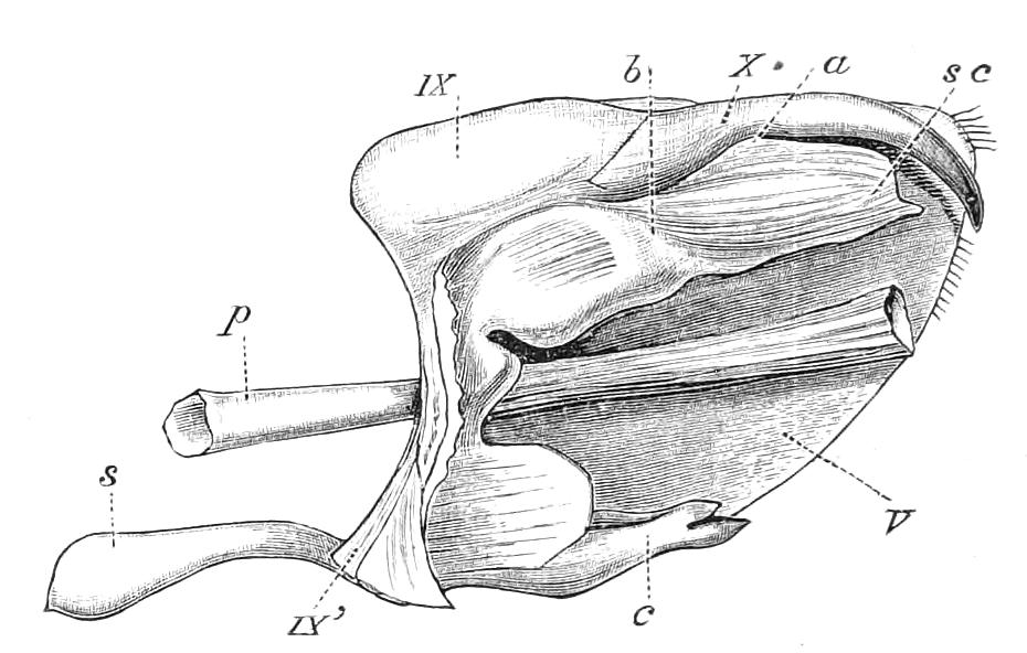
Fig. 160—Acherontia atropos. The termination of ♂ body, one side removed. IX, Ninth dorsal plate; IX’, ninth ventral; s, lobe, saccus, of ninth ventral plate; X, tenth dorsal plate, or uncus; sc, scaphium, or tenth ventral plate; a, position of anus; b, chitinised band of scaphium; V, valve or clasper; c, hooks, or harpes, of clasper; p, penis (or oedeagus). (After Peytoureau.)
Legs.—The legs are long, slender, covered with scales, and chiefly remarkable from the fact that the tibiae sometimes bear articulated spurs on their middle as well as at the tip. The front tibia usually possesses on its inner aspect a peculiar mobile pad; this seems to be in some cases a combing organ; it also often acts as a cover to peculiar scales. The tarsi are five-jointed, with two small claws and a small apparatus, the functional importance of which is unknown, between the claws.
Wings.—The wings are the most remarkable feature of this Order; it is to them that butterflies owe their beauty, the surfaces of the wings being frequently adorned with colours and patterns of the most charming and effective nature. These effects are due to minute scales that are implanted in the wing-membrane in an overlapping manner, somewhat similar to the arrangement of slates on the roof of a house. The scales are very readily displaced, and have the appearance of a silky dust. We shall describe their structure and allude to their development subsequently. The wings are usually of large size in comparison with the Insect's body: in the genus Morpho, the most gorgeous of the butterflies, they are enormous, though the body is small; so that when deprived of these floats the Insect is insignificant. The great expanse of wing is not correlative with great powers of flight, though it is perhaps indicative of flying with little exertion; for the small-winged Lepidoptera, Sphingidae, etc., have much greater powers of aërial evolution than the large-winged forms. The area of the wing is increased somewhat by the fact that the scales on the outer margin, and on a part or on the whole of the inner margin, project beyond the edges of the membrane that bears them: these projecting marginal scales are called fringes. In many of the very small moths the actual size of the wing-membranes is much reduced, but in such cases the fringes may be very long, so as to form the larger part of the surface, especially of that of the hind wings. Frequently the hind wings are of remarkable shape, being prolonged into processes or tails, some of which are almost as remarkable as those of Nemoptera in the Order Neuroptera.
The wings are very rarely absent in Lepidoptera; this occurs only in the female sex, no male Lepidopterous imago destitute of wings having been discovered. Although but little is known of the physiology of flight of Lepidoptera, yet it is clearly important that the two wings of the same side should be perfectly coadapted or correlated. This is effected largely by the front wing overlapping the hind one to a considerable extent, and by the two contiguous surfaces being pressed, as it were, together. This is the system found in butterflies and in some of the large moths, such as Lasiocampidae and Saturniidae; in these cases the hind wing always has a large shoulder, or area, anterior to its point of insertion. In most moths this shoulder is absent, but in its place there are one or more stiff bristles projecting forwards and outwards, and passing under a little membranous flap, or a tuft of thick scales on the under face of the front wing; the bristle is called the "frenulum," the structure that retains it a "retinaculum." In Castnia (Fig. 162) and in some Sphingidae there is the unusual condition of a highly-developed shoulder (s) coexisting with a perfect frenulum (f) and retinaculum (r). The frenulum and retinaculum usually differ in structure, and the retinaculum in position, in the two sexes of the same moth; the male, which in moths has superior powers of flight, having the better retaining organs. Hampson says "the form of the frenulum is of great use in determining sex, as in the males of all the forms that possess it, it consists of hairs firmly soldered together so as to form a single bristle, whilst in nearly all females it consists of three or more bristles which are shorter than that of the male; in one female Cossid I have found as many as nine. Also in the large majority of moths the retinaculum descends from the costal nervure in the male, while in the female it ascends from the median nervure."[176] This sexual difference in a structure for the discharge of a function common to the two sexes is a very remarkable fact. There are a few—very few—moths in which the bases of the hind wings are not well coadapted with the front wings, and do not possess a frenulum, and these species possess a small more or less free lobe at the base of the front wing that droops towards the hind wing, and may thus help to keep up an imperfect connexion between the pair; this lobe has been named a jugum by Professor Comstock. Occasionally there is a jugum on the hind as well as on the front wing. There is usually a very great difference between the front and the hind wings; for whereas in the front wing the anterior portion is doubtless of great importance in the act of flight and is provided with numerous veins, in the hind wing, on the other hand, the corresponding part has not a similar function, being covered by the front wing; hence the hind wing is provided with fewer nervures in the anterior region, the divisions of the subcostal being less numerous than they are in the front wing. In the moths possessing a jugum the two wings differ but little from one another, and it is probable that they function almost as four separate wings instead of as two pairs.
Wing-nervures.—The nervures or ribs of the wings are of great importance in Lepidoptera, as at present they furnish the chief characters for classification and for the discussions of phylogeny that are so numerous in entomological literature. On looking at wings that have been deprived of their scales it will be noticed (Fig. 161) that the ribs are much more numerous at the outer margins than they are near the points of attachment of the wings, and that there is usually but one cell (or area completely enclosed by ribs). This latter point is one of the chief peculiarities of the Lepidopterous wing; in Insect-wings generally the number of cells in proportion to the area of the wings and to the number of nervures is greater than it is in Lepidoptera, for in the latter there are few or no cross-nervures. Hence there is sometimes no closed cell at all on the wing (Fig. 161, II. B). The maximum number of closed cells is six; this is found in some species of Micropteryx, while in Hepialus there may be three or four; but the rule is that there is only one cell in the Lepidopterous wing. When the number of cells is increased this is not necessarily due to an increase in the cross-nervures; and in fact it is generally due to irregular forking or to the sinuous form of the longitudinal nervures themselves (see wing of Castnia, Fig. 162, A.). Some authorities consider that all transverse or cross-veins in Lepidoptera are merely portions of longitudinal veins having diverted courses. When a portion of a nervure beyond the basal or primary portion serves as a common piece to two forked parts external to it, it is called a stalk (Fig. 162, A, e). There are cases in which the furcation takes place in the opposite direction, so that a nervure is double at the base of the wing (Fig. 161, I, A, 1a, and B, 1b). This important condition has not yet been adequately discussed.
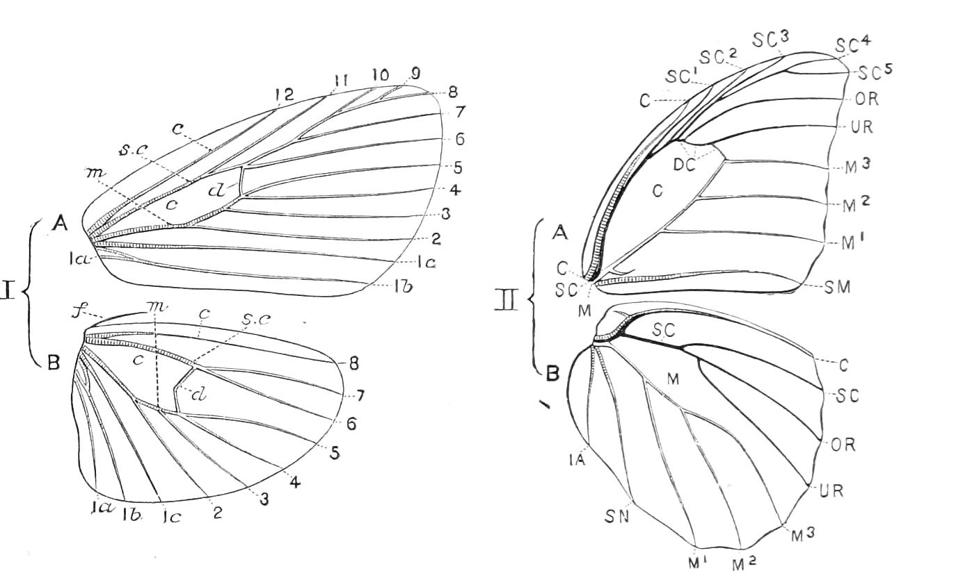
Fig. 161.—Wing-nervuration of Lepidoptera. I, Diagram of moths' wings (after Hampson); II, of a butterfly's wings (Morpho menelaus ♂, after Staudinger and Schatz). A, front, B, hind wing. I.—c, costal; sc, subcostal; m, median; 1a, 1b, 1c, internal nervures; f, frenulum; 2, 3, 4, branches of median nervure; 5, lower radial; 6, upper radial; 7-11, divisions of the subcostal; 12, termination of costal; c, cell; d, discocellular nervure. II.—C, costal; SC, subcostal; M, median; SM and SN, submedian nervures; 1A, inner-margin nervure; UR, lower radial; OR, upper radial; SC1 to SC5, divisions of subcostal; M1 to M3, divisions of median nervure; C, cell; DC, discocellulars.
Turning to the mode of designation of the nervures,[177] we may commence by remarking that no system satisfactory from a practical as well as from a theoretical point of view has yet been devised. The diagrams given in figure 161 will enable us to explain the methods actually in vogue; I. representing the system, dating from the time of Herrich-Schaeffer, chiefly used by British naturalists, and II. that adopted by Staudinger and Schatz in their recent great work on the Butterflies of the world. The three anterior nervures in both front and hind wings correspond fairly well, and are called, looking at them where they commence at the base of the wing, "costal," "subcostal," and "median" nervures. The nervures near the inner margin of the wing (that is the lower part in our figures) differ much in the front and hind wings, consisting either of two or of three separate portions not joined even at the base. British entomologists call these "branches or divisions of the internal nervure": the Germans call the more anterior of them the "submedian," and the more internal the "inner-margin nervure"; they are also frequently called anal nervures. The cross-nervure that closes the cell is called discocellular; when apparently composed of two or three parts joined so as to form angles, the parts are called, according to position, upper, lower, and middle discocellulars. One or more short spurs may exist on the front part of the basal portion of the hind wing; these are called praecostal. The branches or terminal divisions of the nervures should be called nervules; they are usually mentioned by the numbers shewn in the diagram (Fig. 161, I.). In addition to this, it is only necessary to remember that number 2 is always assigned to the posterior division of the median nervure, the nervules below this being all called 1, and distinguished by the addition of a, b, c when requisite. This course is necessary, because if it were not adopted the corresponding nervules on the front and hind wings would bear different numbers.
The use of this system of numbers for the nervules is becoming general, and it answers fairly well for practical purposes. On the other hand, extreme discrepancy exists as to the nomenclature of the nervures and nervules, and there are almost as many systems as there are authorities.
The normal number of nervules is, on the front wing, 11 + 1 or 2 inner marginal, and on the hind wing 7 + 2 or 3 inner marginal. In the aberrant moths of the genus Castnia the nervuration is unusually complex and irregular (Fig. 162), and an analogous condition occurs in our common Goat-moth (Cossus ligniperda). In Hepialus and Micropteryx (the jugate moths of Comstock) the hind wings are less dissimilar in nervuration from the front wings than they are in other Lepidoptera.[178]
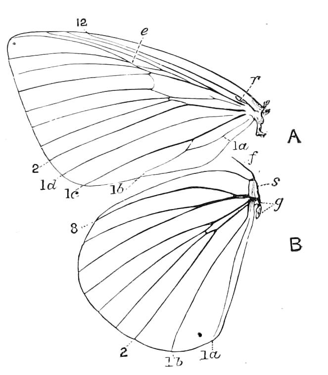
Fig. 162—Wing-nervuration of Castnia. Undersides of, A, front, B, hind wings. 1a, 1b, 1c, 1d, Inner marginal nervures; 2, lower branch of median; 8, subcostal of hind wing; 12, subcostal of front wing; e, "stalk" of 8 and 9; f, frenulum; r, retinaculum; s, shoulder; g, articulation of wing.
Internal Anatomy.[179]—The alimentary canal extends as a long, slender oesophagus through the length of the thorax, dilating when it reaches the abdomen to form a tubular stomach; before this it is somewhat enlarged to form an indistinct crop, and gives off a large diverticulum usually called a sucking stomach. According to Burgess, this structure does not possess the function ascribed to it by this name, and he terms it a food-reservoir. The Malpighian tubes are six in number, three on each side, and each set of three unite to form a common tube opening into the posterior extremity of the stomach; behind them the alimentary canal continues in the form of a slender, tortuous intestine, expanding at the extremity of the body to form a rectum. The dorsal or circulatory vessel commences near the posterior extremity of the body, but in the front part of the abdomen is deflexed to pass under the great phragma into the thorax, where it rises abruptly to the dorsal wall, but is again abruptly deflexed, forming a loop, and is then prolonged above the oesophagus into the head: at the summit of the thoracic loop there may be a dilatation called the aortal chamber. The supra- and infra-oesophageal ganglia are consolidated into a mass pierced by the oesophagus: there is a minute frontal ganglion; the ventral chain consists of three much approximated thoracic ganglia and four abdominal ganglia separated from the thoracic by a long interval.
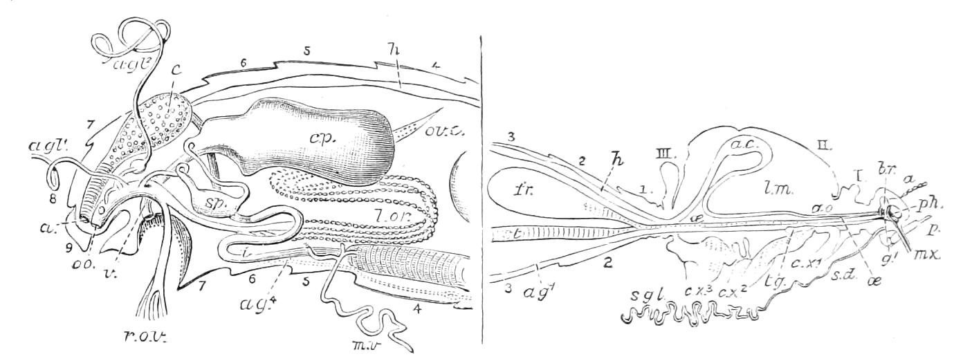
Fig. 163—Internal anatomy of Lepidoptera. Section of the body of a female butterfly, Anosia plexippus. (After Scudder.) The portion to the left of the vertical line more magnified. I. II. III. thoracic segments; 1-9, abdominal segments; a, antenna; a, anus; ac, aortal chamber; ag1, etc., abdominal ganglia; agl1, agl2, accessory glands; ao, aorta; br, brain; c, colon; cp, copulatory pouch; cx1, cx2, cx3, coxae; fr, food-reservoir; g1, suboesophageal ganglion; h, dorsal vessel; i, intestine; lm, area filled by wing muscles; l.or, ovary, or egg-tubes of left side; mv, Malpighian tube (the two others of the right side cut away, except small portions); mx, maxilla; o, oviduct; oo, its orifice; oe, oesophagus; ov.c, end of left ovary; p, labial palp; ph, pharynx; r.ov, terminal parts of right ovarian tubes, turned to one side, after the tubes have been cut away; sd, salivary duct; sgl, salivary gland; sp, spermatheca; st, stomach; tg, thoracic ganglia; v, copulatory orifice.
The male sexual organs consist of the two testes placed in a common capsule, from which proceed a pair of contiguous vasa deferentia (dilated soon after their origin to form the vesiculae seminales); into each vas there opens a long, tubular gland; the two vasa subsequently unite to form a long, coiled, ejaculatory duct. It is in the structure of the female sexual organs that the most remarkable of the anatomical characters of Lepidoptera is found, there being two external sexual orifices. The imago has, in the great majority of cases, four egg-tubes in each ovary; the pair of oviducts proceeding from them unite to form a single unpaired (azygos) oviduct which terminates by an orifice quite at the posterior extremity of the body. There is a sac, the bursa copulatrix or copulatory pouch, which is prolonged in a tubular manner, to open externally on the eighth ventral plate: a tube, the seminal duct, connects the bursa with the oviduct, and on this tube there may be a dilatation—the spermatheca. Besides these structures two sets of accessory glands open into the oviduct, an unpaired gland, and a pair of glands. The development of these structures has been described by Hatchett Jackson,[180] and exhibits some very interesting features. The exact functions of the bursa copulatrix and of the other structures are by no means clear. According to Riley,[181] the spermatheca in Pronuba contains some curious radiate bodies, and Godman and Salvin describe something of the same sort as existing in butterflies. Several variations in the details of the structure of these remarkably complex passages have been described, and the various ducts are sometimes rendered more complex by diverticula attached to them. Some noteworthy diversities in the main anatomical features exist. According to Cholodkovsky, there is but one sexual aperture—the posterior one—in Nematois metallicus; while, according to Brandt, the number of egg-tubes in a few cases exceeds the normal—four—being in Sesia scoliaeformis fourteen. In Nematois metallicus there is individual variation, the number of tubes varying from twelve to twenty.
The egg has been more extensively studied in Lepidoptera than in any other Order of Insects. It displays great variety: we meet with elongate forms (Fig. 164) and flat forms like buttons, while in Limacodes (Fig. 83, Vol. V.) the egg is a transparent scale of somewhat inconstant outline. Some are coloured and mottled somewhat after the fashion of birds'-eggs; this is the case with some eggs of Lasiocampidae and Liparidae; in some the sculpture of the egg-shell is of the most elaborate character (Figs. 77, 78, Vol. V.). The egg-shell or chorion is, according to Korschelt[182] and others, a cuticular product of the epithelium of the egg-chambers of the ovaries. The number of eggs deposited by an individual differs greatly in different species, and has been ascertained to be variable within certain limits in the same species. Speyer thought about 250 to be the average number of eggs deposited by an individual. The number in the case of Aporia crataegi is believed to be from 60 to 100, and in some Hepialus to be several thousands. The mode of deposition also differs greatly; where the eggs are very numerous they seem to be discharged almost at random in suitable spots; but moths such as Clisiocampa neustria fasten their eggs round the stems of the food-plant in a very perfect and artistic manner. Butterflies seem as a rule to prefer to oviposit by placing an egg here and there rather than risk many in one situation; but to this there are many conspicuous exceptions especially in the cases where the larvae live gregariously, as in the Vanessae. Some moths cover the eggs with fur from their own body, which, in the case of certain of the Eggers (Lasiocampidae), seems to have a special supply for the purpose. The period that intervenes between deposition and hatching of the eggs varies from a few days to many months. There seems to be, as a rule, comparatively little power of extending the period of latency beyond a single season; though certain facts have been recorded that would lead us to believe that in Australia eggs may last over the proper time during a drought, and be hatched as soon as rain falls.
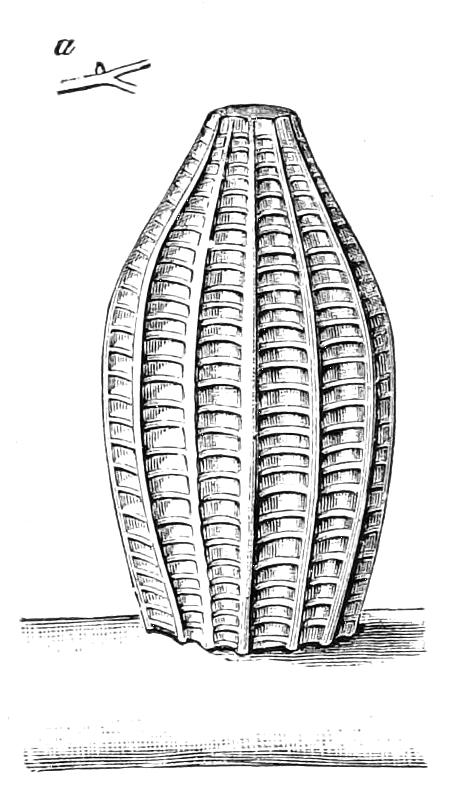
Fig. 164—Egg of Orange-tip butterfly, Euchloe cardamines, magnified. a, The egg of natural size on a stalk.
Larva.—The young condition or larva of the Lepidopterous Insect is commonly called a caterpillar. It is a somewhat worm-like creature—in old English it was sometimes called palmer-worm—and is composed of a head and thirteen divisions or segments of the body; the first three of the latter are called thoracic, the other ten, abdominal segments; in most caterpillars the terminal two or three abdominal segments are more or less run together, and the ninth may be very small, so that the true number is indistinct. The first three segments bear each, on either side, a short limb, ending in a curved spine; the next two (or three or more) segments are destitute of legs, but on some of the following divisions another kind of leg of a more fleshy character appears, while the body is terminated by a pair of these thick legs of somewhat different form. The front legs are usually called the true legs, the others prolegs, but this latter designation is a most unfortunate one, the term "pro" being in entomology used to signify anterior; it is therefore better to call the three anterior pairs thoracic legs, and the others abdominal feet, distinguishing the hind pair of these latter as claspers. There is, too, an unfortunate discrepancy amongst entomologists in their manner of counting the body-segments, some count the head as the first segment, while others apply this term to the first thoracic segment. The latter is the more correct course, for, as the head is not a single segment it should not be called such in a terminology that affects to be morphologically exact, not simply descriptive. The thoracic legs are transversely jointed (Fig. 165, B), but this is not the case with the abdominal feet, which are usually armed beneath with a circle, or with rows, of little hooks. The thoracic legs are, independent of their form, of a different nature from the abdominal, for these latter disappear subsequently, while the former give rise to the legs of the imago. The number of thoracic legs is always six, except in a few cases where there are none at all; the abdominal feet are much more variable, and exhibit so many distinctions that we cannot here attempt to deal with them. M. Goossens has given a concise and interesting account of this subject,[183] and Speyer[184] a summary of the variety in number and position.
The anatomy of the larva is simple in comparison with that of the perfect Insect; its main features will be appreciated from Fig. 165, from which it will be seen that the stomach is enormous, and the silk-vessels are also very extensive.
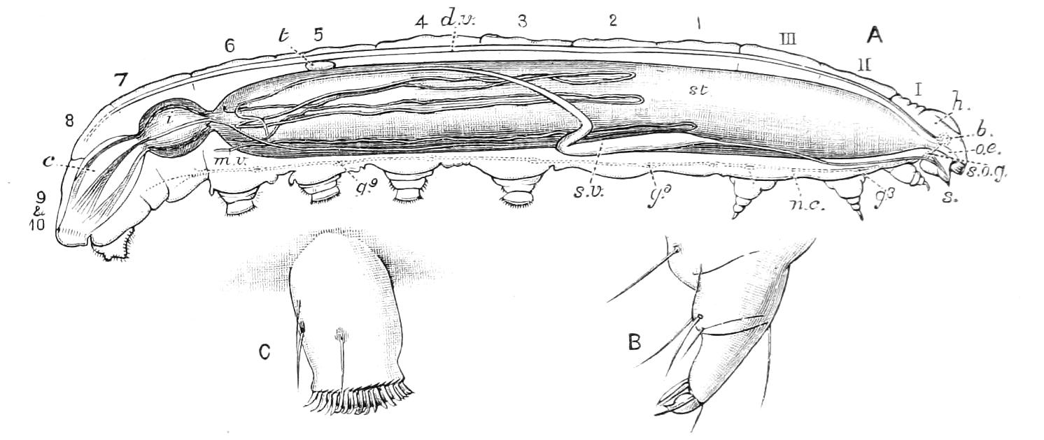
Fig. 165.—A, Section of male caterpillar of Anosia plexippus—muscular and tracheal systems and fat-body not shown: I, pro-, II, meso-, III, meta-thorax; 1-10, abdominal segments; b, supra-oesophageal ganglion; c, rectum; d.v, dorsal vessel; g3-g9, ganglia of ventral chain; h, head; i, intestine; m.v, Malpighian tube; n.c, nerve-cord of ventral chain; oe, oesophagus; s, spinneret; s.o.g, infra-oesophageal ganglion; st, stomach; s.v, silk-vessel; t, testis. B, One of the jointed prothoracic legs. C, An abdominal foot with its hooks. (After Scudder and Burgess, magnification about 2⁄1.)
There are three sets of glands opening by canals on the head, viz. the salivary glands proper, which open into the cavity of the mouth, one close to the base of each mandible; the silk-glands, which terminate by a common canal, continued externally as the spinneret; and the glands of Filippi situate in the head itself, and opening into the ducts of the silk-glands, near their union into a common duct. It should be recollected that Fig. 165 does not indicate all the details of the anatomy; the muscular system, for instance, being entirely omitted, though there are an enormous number of muscles; these however are not very complex, they being mostly repetitions in the successive segments.[185] The mouth-parts are very different from those of the perfect Insect, inasmuch as the maxillae and labial palpi, which are the most remarkable structures of the imago, are small, and are differently constructed in the caterpillar, while the mandibles, which are the largest organs of the caterpillar, disappear in the adult. The little organ by which the caterpillar exudes its silk is called a spinneret; according to Packard it is a "homologue of the hypopharynx." It is a more or less prominent point on the middle of the labium (Fig. 166, g) and sometimes forms a conspicuous spine projecting downwards. The eyes are extremely imperfect organs, consisting merely of six, in some cases fewer, transparent, somewhat prominent, little spaces placed on each side of the lower part of the head; they are called "ocelli," by Landois "ocelli compositi." Under each of these external facets there are placed percipient structures, apparently very imperfect functionally, the caterpillar's sight being of the poorest character.[186] The spiracles of the caterpillar are nine on each side, placed one on the first thoracic segment and one on each of the first eight abdominal segments; there are no true stigmata on the second and third thoracic segments, though traces of their rudiments or vestiges are sometimes visible.
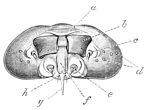
Fig. 166—Front view of head of a caterpillar, with the jaws partially opened, a, Labrum; b, mandible; c, antenna; d, ocelli; e, maxilla; f, lingua; g, spinneret; h, labial palp.
In the caterpillar there are no traces of the external sexual organs, so that the two sexes cannot be distinguished on superficial inspection; it was however long ago demonstrated by Herold[187] that the ovaries and testes exist in the youngest caterpillars, and undergo a certain amount of growth and development in the larval instars; the most important feature of which is that the testes are originally separate but subsequently coalesce in the middle line of the body, and become enclosed in a common capsule. In a few forms—especially of Liparidae—(Lymantriidae of modern authors)—the caterpillars are said to be of different colours in the two sexes. Most of what is known on this point has been referred to by Hatchett Jackson.[188]
The Silk-glands of Lepidoptera are of great interest from the physiological point of view, as well as from the fact that they have furnished for many ages one of the most beautiful of the adornments made use of by our own species. The sericteria, or vessels that secrete silk, are of simple structure, and differ greatly in their size in the various forms of the Order; they sometimes become of great length; in the Silk-worm each of the two vessels is nearly five times as long as the body, while in Bombyx yamamai and others, even this is exceeded. They grow with remarkable rapidity, being in the young silk-worm only 3 mm. long, in the adult 22 mm. The increase in weight is still more remarkable; when the silk-worm is thirty-one days old, the sericteria weigh only 3 mgr., but when the age is fifty days their weight has increased to 541 mgr., being then ⅖ of the whole weight of the body. In the pupa they undergo a gradual atrophy, and in the moth they are, according to Helm, no longer to be found, though earlier authors were of a contrary opinion.[189] According to Joseph,[190] the silk-vessels begin to develop at an extremely early age of the embryo, and are very different in their nature from the salivary glands, the former being derivatives of the external integument (ectoderm), while the salivary glands belong to the alimentary system. This view is to some extent confirmed by the observations of Gilson as to the different manner in which these two sets of glands discharge their functions.
The chief feature in the anatomy of the larva is the great size of the stomach. There is a very short oesophagus and crop; the latter becomes enlarged, spreading out so as to form the stomach, a great sac occupying the larger part of the body-cavity (Fig. 165). On the hinder end of this sac the Malpighian tubes open; they are similar in their disposition to those of the imago; behind the stomach the canal expands into two successive, short dilatations, the first called an intestine, the second a rectum; they are connected by very short isthmuses. The dorsal vessel is a simple, slender tube, extending from the eighth abdominal segment to the head. The main nervous system consists of supra- and infra-oesophageal ganglia, a small frontal ganglion, and a ventral chain of eleven ganglia, three thoracic and eight abdominal, the last of these latter being double. The sexual organs are quite rudimentary, and the passages connected with them very incompletely developed.
Pupa.—The pupa, which is one of the most remarkable of the instars of an Insect's life, attains its highest development in Lepidoptera. The Lepidopterous pupa is frequently called a "chrysalis," a term originally applied to certain metallic butterfly pupae. The Lepidopterous pupa differs from that of other Insects in the fact that its outer skin forms a hard shell, all the appendages of the body being glued together by an exudation so as to form a single continuous outer skin. This form of perfect pupa is called "pupa obtecta." The obtected pupa is exhibited in various stages of perfection in the Lepidoptera; the maximum of perfection is attained by the pupae of such butterflies as are exposed without protection or concealment; on the other hand, we find in various small moths conditions of the pupa that do not differ in any marked manner from the pupae of Insects of other Orders. Moreover, certain Coleoptera and Diptera exhibit obtected pupae of a more or less perfect kind. Hence the pupa obtecta is to be considered as a perfected condition that exists more frequently in the Lepidoptera than in other Orders.
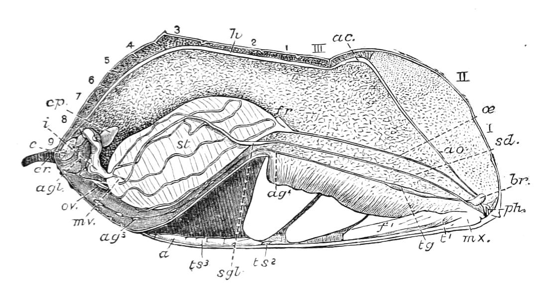
Fig. 167.—Section of female pupa of Anosia plexippus, 3-4 days old. I, pro-, II, meso-, III, meta-thorax; 1-9, abdominal segments; a, antenna(?); ac, aortal chamber; ag1-ag5, abdominal ganglia; agl, accessory glands; ao, aorta; br, brain; c, colon; cp, bursa copulatrix; cr, cremaster; f1, first femur; fr, food-reservoir; h, dorsal vessel; i, part of intestine; mv, Malpighian tube; mx, base of maxilla; oe, oesophagus; ov, ovary; ph, pharynx; sd, salivary duct; sgl, salivary gland; st, stomach; t1 first tarsus; tg, compound thoracic ganglion; ts2, ts3, second and third tarsus. (After Scudder.)
The pupa has no orifices to the alimentary canal or sexual organs, but the respiratory openings are pervious. It has no means of locomotion, but it can move a certain number of the posterior segments (the number variable according to kind). In some cases it is provided with spines, "adminicula," by means of which, aided by the wriggling movements of the abdominal segments, considerable changes of position can be effected. The pupae of the genus Micropteryx apparently use the legs for locomotion, as do the pupae of Trichoptera.
The study of the pupa of Lepidoptera is less advanced than that of the imago and larva, between which it is, in many points of structure, intermediate.[191] The interior of the pupa contains a quantity of cream-like matter, including the results of histolysis—but this, as well as the condition of the internal organs, differs much according to whether the change from the caterpillar to the moth is much or little advanced.
Many pupae are protected by cocoons. These are masses of silk—very various in form—disposed by the caterpillar around itself during the last stage of its existence. Some of these cocoons are so perfect that the moth has considerable difficulty in escaping when the metamorphosis is complete. Various devices are used for the purpose of emergence; the Puss-moth excretes a corrosive fluid, containing potassium hydroxide, and then protects itself from this by retaining on the head while passing through it a shield formed of a portion of the pupa-skin.[192] Lepidopterous pupae usually have the body terminated by a projection of very various and peculiar form called "cremaster." In certain cases these projections are used for the suspension of the pupa, and are then frequently provided with hooks (Fig. 177, C, D). In other cases the cremaster is frequently called the anal armature (Fig. 205, B).
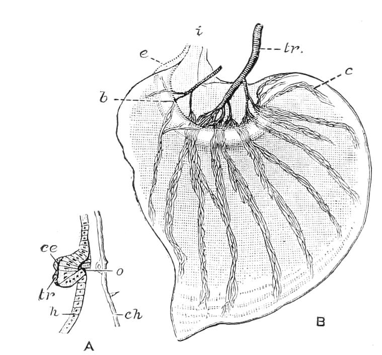
Fig. 168—Wing-rudiments of Pieris brassicae. A, Rudiments of a wing before the first moult of the caterpillar: ce, embryonic cells; ch, external cuticle; h, hypodermis; o, opening of the invagination; tr, trachea. B, posterior wing-rudiment of full-grown caterpillar; b, semicircular pad; c, a bundle of the rolled tracheae; e, envelope; i, pedicel; tr, trachea. (After Gonin.)
The development of the wings of Lepidoptera has recently been much studied. It has been known since the time of Lyonnet, that the rudiments of the wings exist inside the body of the caterpillar when it is nearly adult. Verson considers that he has detected the rudiments in the silk-worm larva even before hatching, and he attributes their origin to a modification of form of those hypodermal cells that occupy the spots where the spiracles of the second and third thoracic segments might be looked for. (It will be recollected that there are no spiracles on these two thoracic segments in Lepidopterous larvae). Gonin has examined the wing-rudiments in the caterpillar, a few days old, of Pieris brassicae,[193] and finds that the future wing is then indicated by a thickening and bagging inwards of the hypodermis, and by some embryonic cells and a trachea in close relation with this mass (Fig. 168, A). The structure grows so as to form a sac projecting to the interior of the body, connected with the body-wall by a pedicel, and penetrated by a trachea forming branches consisting of rolled and contorted small tracheae (Fig. 168, B). If the body-wall be dissected off the caterpillar immediately before pupation the wings appear in crumpled form, as shown in Fig. 169. This fact was known to the older entomologists, and gave rise to the idea that the butterfly could be detected in a caterpillar by merely stripping off the integument.
The exact mode by which the wings become external at the time of appearance of the chrysalis is not ascertained; but it would appear from Gonin's observations that it is not by a process of evagination, but by destruction of the hypodermis lying outside the wing. However this may be, it is well known that, when the caterpillar's skin is finally shed and the chrysalis appears, the wings are free, external appendages, and soon become fastened down to the body by an exudation that hardens so as to form the shell of the chrysalis.
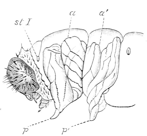
Fig. 169—Anterior parts of a caterpillar of P. brassicae, the body-wall having been dissected off, immediately before pupation. a, a′, Anterior and posterior wings; st I, first spiracle; p, p′, second and third legs. (After Gonin.)
Scales and nervures.—Before tracing the further development it will be well to discuss the structure of the scales and nervures that form such important features in the Lepidopterous wing.
If a section be made of the perfect wing of a Lepidopteron, it is found that the two layers or walls of the wing are firmly held together by material irregularly arranged, in a somewhat columnar manner. The thickness of the wing is much greater where the section cuts through a nervure (Fig. 170, A). The nervures apparently differ as to the structures found in them. Spuler observed in a nervure of Triphaena pronuba, a body having in section a considerable diameter, that he considered to be a trachea, and also a "wing-rib" and blood-cells. He remarks that even in nervures, perfectly formed as to their chitinous parts, either wing-rib or trachea or both may be absent.[194] Schäffer[195] was unable to find any tracheae in the completed wings he examined, and he states that the matrix of the tracheae and even their inner linings disappear. The wing-ribs were, however, found by him to be present (Fig. 170, A and B).
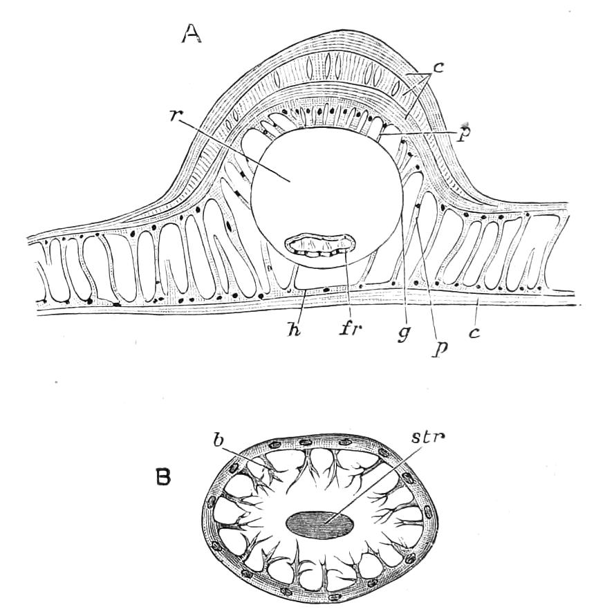
Fig. 170.—Structure of wing of imago. A, Transverse section of basal portion of wing [of Vanessa?] containing a nervure; c, cuticle; fr, wing-rib; g, wall of nervure ("Grundmembran"); h, hypodermis; p, connecting columns: r, lumen of nervure; B, section of a rib; b, one of the chitinous projections; str, central rod. (After Schäffer.)
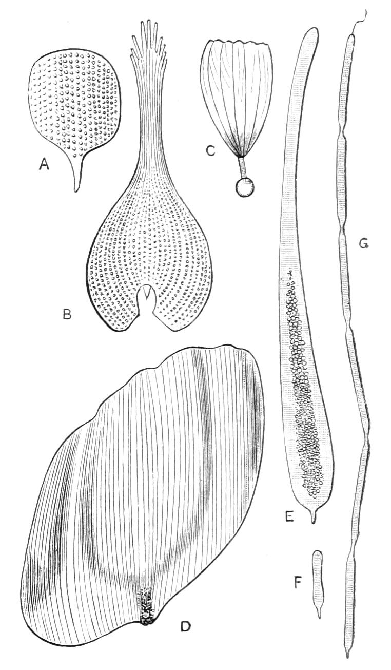
Fig. 171—Scales of male Lepidoptera. A, Scale from upper surface of Everes comyntas; B, from upper surface of Pieris rapae; C, from inner side of fold of inner margin of hind wing of Laertias philenor; D, one of the cover-scales from the costal androconium of Eudamus proteus; E, F, G, scales from androconium of Thorybes pylades. (After Scudder).
The scales that form so conspicuous a feature in Lepidoptera exist in surprising profusion, and are of the most varied forms. They may be briefly described as delicate, chitinous bags; in the completed state these bags are flattened, so as to bring the sides quite, or very nearly, together. Their colour is due to contained pigments, or to striation of the exposed surface of the scale; the latter condition giving rise to metallic "interference-colours." The walls of the scales are themselves, in some cases, tinted with pigment. It is said that some of the scales contain air, and that the glistening whiteness of certain scales is due to this. The exposed surface of the scale usually differs from the surface that is pressed down on the wing in being delicately and regularly striated; the colours of the upper and under surfaces of a scale may also be quite different. Scales are essentially of the nature of hairs, and all the transitions between hairs and true scales may be found on the wings of certain Lepidoptera that bear both hairs and scales, e.g. Ithomia. It has been calculated that there are a million and a half of scales on the wings of an individual of the genus Morpho. The scales are arranged on the wing in an overlapping manner, somewhat like slates on the roof of a house. Each scale has a short stalk, and is maintained in position by the stalk fitting into a cavity in a projection of the wing-membrane (Fig. 172).
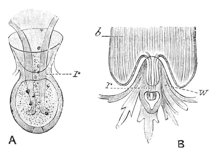
Fig. 172—Insertion of scales. A, Socket holding the stalk in Galleria mellonella; B, insertion of the scale of Polyommatus phloeas. b, Base of scale; r, holding-ring; w, surface of wing. (After Spuler.)
Androconia.—The males of numerous butterflies possess scales peculiar in kind and various in arrangement. They may be either irregularly scattered over the wing, or they may form very complex definite structures (Fig. 173). They were formerly called "plumules," but Scudder has replaced this name by the better one, "androconia." The function of the androconia is still obscure. An odour is believed to be connected with them. Thomas supposes[196] that these scales are hollow tubes in connection with glands at their bases, and that matter secreted by the glands passes through the scales and becomes diffused. In nearly all Lepidoptera it is the male that seeks the female; if therefore odorous scales were present in one sex only we should have supposed that this would have been the female rather than the male. As, however, the reverse is the case, the function of the androconia is supposed to be that of charming the female. Scudder considers that the covering part of the androconial structures is sometimes ornamental. As a rule, however, the "brands" of male Lepidoptera detract from their beauty to our eyes.
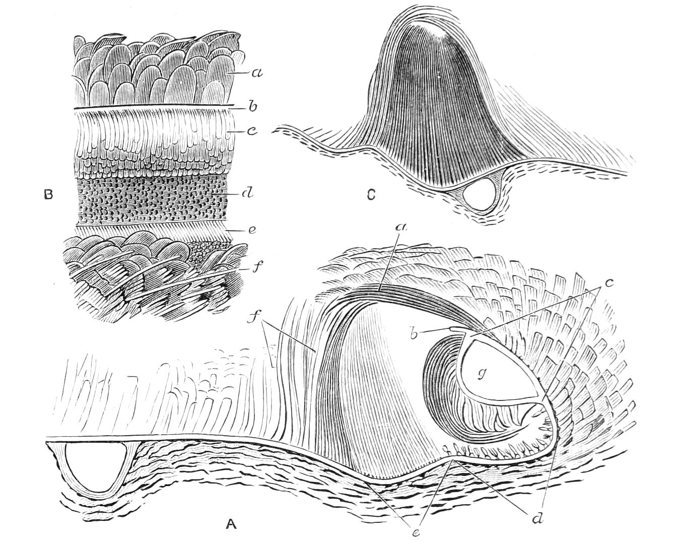
Fig. 173—A, section of part of wing showing the complex androconia of Thanaos tages, a Skipper butterfly. The turning over of the costal margin of the wing is in this case part of the arrangement. a, Upper covering-scales attached to the costal portion of the under surface of the wing; b, edge of costal margin of the wing; c, costal nervure with its scales; d, field of the wing next the costal nervure, bearing stunted scales; e, the androconia proper, or male scales; f, posterior covering scales; g, lumen of the costal nervure: B, a portion of the costal area flattened out and seen from above; lettering as before: C, section of androconium on the second nervure of Argynnis paphia. (After Aurivillius.)
Resuming our consideration of the development of the wings, we may remark that the history of the changes during the pupal state is still imperfect. By the changes of relative size of the thoracic segments the hind wing is brought to lie under the anterior one (i.e. between it and the body), so that in the newly formed pupa the arrangement is that shown in Fig. 174. The wings are two sacs filled with material surrounding peritracheal spaces in which run tracheae. The subsequent history of the tracheae is very obscure, and contrary opinions have been expressed as to their growth and disappearance. We have alluded to the fact that in some nervures tracheae are present, while in others they are absent; so that it is quite possible that the histories of the formation of the nervures and of their relation to tracheae are different in various Lepidoptera. This conclusion is rendered more probable by the statement of Comstock and Needham,[197] that in some Insects the "peritracheal spaces" that mark out the position of the future nervures are destitute of tracheae. Gonin thinks the nervures are derived from the sheaths of the peritracheal spaces, and a review of all the facts suggests that the tracheae have only a secondary relation to the nervures, and that the view that a study of the pupal tracheae may be looked on as a study of the preliminary state of the nervures is not sufficiently exact. It is, however, probable that in Lepidoptera the pupal tracheae play an important though not a primary part in the formation of the nervures; possibly this may be by setting up changes in the cells near them by means of the air they supply. Semper long ago discovered hypodermal cylinders traversed by a string (Fig. 170, B), placed near the tracheae in the pupa.[198] It appears probable that the "wing-ribs" found in the nervures (Fig. 170, A fr and B) are the final state of these cylinders, but the origin and import of the cylinders are still unknown.
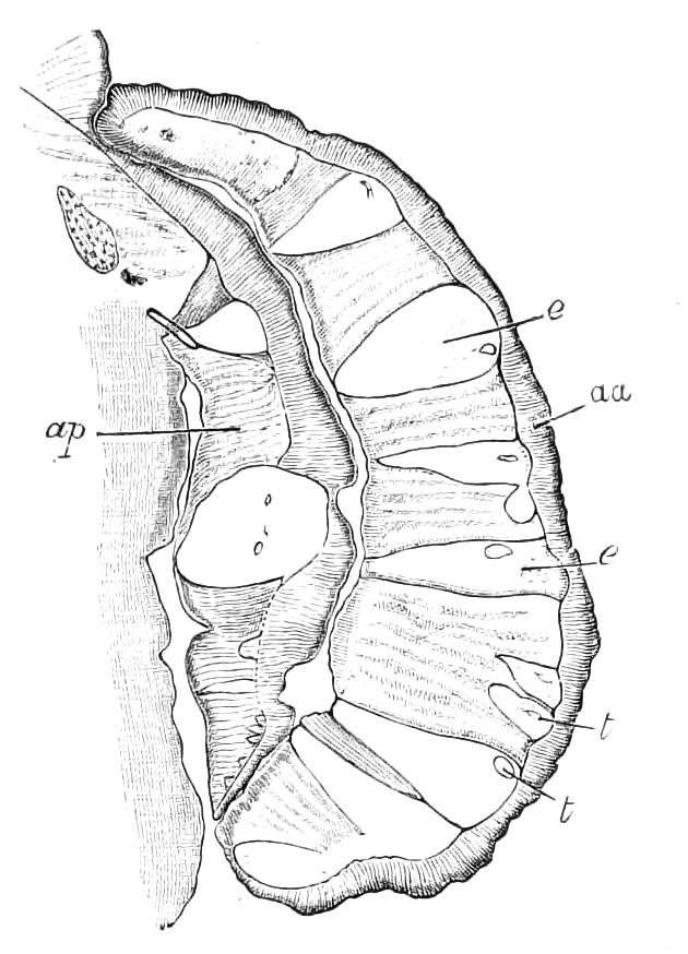
Fig. 174—Transverse section of part of the newly disclosed chrysalis of Pieris brassicae, showing the position and structure of the wings, hanging from one side of the body. aa, Anterior wing; ap, posterior wing; e, e, peritracheal spaces; t, t, tracheae. (After Gonin.)
The formation of the scales of the wing commences very early—apparently soon after the casting of the larval skin—though the completion of the scales and their pigmentation is delayed to a late period of the pupal life. The scales are formed by special cells of the hypodermis that are placed deeper in the interior of the wing than the other hypodermal cells. Each scale is formed by one cell, and protrudes through the overlying hypodermis; the membrane into which the scales are inserted is a subsequently developed structure, and the beautiful articulation of the scale with the wing takes place by a division of the stalk of the scale where it is encompassed by the membrane. Semper was not able to show that the scale-forming cells are certainly hypodermal cells, but this has since been demonstrated by Schäffer, who also shows that each of the cells contains an excretory vesicle.
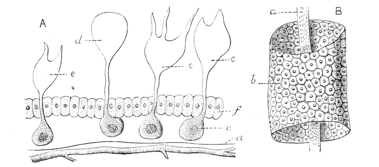
Fig. 175.—Early condition of scales and nervures. (After Semper.) A, Section of portion of wing of pupa of Sphinx pinastri; a, basal membrane with trachea beneath it; c, scale-forming cell; d, early state of a scale; e, e, more advanced stages; f, hypodermal cells. B, part of a cellular cylinder that excretes the nervure [or more probably the rib or "Rippe" of Schäffer; cf. Fig. 170, B]; b, epithelial [hypodermal] cells; a, central string [supposed by Semper to be a nerve].
Very little is positively known as to the development of the colour in the wing-scales. It has been pointed out by Hopkins[199] that in some cases the colours are of the nature of urates; that is, of excretory matter of the kind that usually passes from the body by direct channels, and in the case of Lepidoptera, by the Malpighian tubes. Miss Newbigin suggests that the organic pigments used in scale-coloration will be found to be of two kinds, urates and melanins, the urates being derivatives from nitrogenous, the melanins from carbonaceous, matters.[200] Marchal, who has devoted a great deal of attention to the study of the Malpighian tubes, informs us that the subdermal pigments of caterpillars are frequently in large part deposits of urates, and he is of opinion that, the function of the Malpighian tubes being arrested at certain periods of the metamorphosis, elimination of the matter they separate when functionally active then takes place in a variety of other ways.[201] A similar condition as to the melanin-pigments and the respiratory functions appears also probable. The scales when first formed are pallid, and the physiology of their pigmentation is not fully ascertained; it is, however, known that when the scales are pallid the hypodermis is either pigmented or in close contact with pigmentary matter, and that as the scales become coloured this pigmentation of the hypodermis diminishes; so that it is clear that the colour of the scales is obtained from matter in the interior of the developing wing, and probably by the agency of the hypodermis.
The pattern on the wings of Lepidoptera is formed before the emergence from the pupa. In the Tortoiseshell butterfly, according to Schäffer, it commences to appear about the ninth day of the pupal life, and the pattern is completed about the eleventh or twelfth day. He also states that the process varies in its rapidity, and this, he thinks, may depend on the previous condition of the larva. According to Buckell the pupa of Nemeobius lucina is sufficiently transparent to allow the development of the colour of the imago to be watched. He says that the coloration occurred first in front; that its entire production occupied less than twenty-four hours, and only commenced about forty-eight hours before the imago emerged.[202] When the butterfly leaves the pupal skin the wings are soft, crumpled sacs, of comparatively small size, but, as everyone knows, they rapidly expand and become rigid; the physiology of this process is apparently still unknown.
A great deal of evidence, both direct and indirect, has accumulated showing that the organisation of many Lepidoptera is excessively sensitive, so that slight changes of condition produce remarkable results; and it has also been shown that in the early part of the life this sensitiveness is especially great at the period of ecdysis. Numerous butterflies produce more than one generation a year, and sometimes the generations are so different that they have passed current with entomologists as distinct species. The phenomena of this character are styled "seasonal variations" or "seasonal dimorphism." It has, however, been shown that, by careful management, the eggs of a generation (say form a) may be made to produce form a, whereas in the usual course of nature they would produce form b. A very remarkable condition is exhibited by the North American Papilio ajax. There are three forms of the species, known as P. ajax, P. telamonides, and P. marcellus. It is uncertain how many generations there may be in one year of this species, as the length of the life-cycle varies greatly according to circumstances. But in West Virginia all the butterflies of this species that emerge from the chrysalis before the middle of April are the form marcellus; those produced between the middle of April and the end of May are telamonides; while those that appear after this are ajax. P. telamonides is not, however, the offspring of marcellus, for both forms emerge from pupae that have passed through the winter (and are the offspring of ajax), those that emerge early being marcellus, those that appear later telamonides.
In various parts of Asia and Africa the butterflies produced during the wet season differ more or less markedly from those of the same species produced during the dry season. These are called "wet" and "dry season" forms. Their aetiology has not been investigated, this discovery being comparatively recent.
Turning to the early life we find that some larvae vary in colour, and that this variation is sometimes of a definite character, the larva being one of two different colours—green or brown. In other cases the variation of the species is less definitely dimorphic, a considerable range of variation being exhibited by the species. In tracing the life-histories of Lepidopterous larvae it is not rare to find species in which the larva abruptly changes its form and colour in the middle of its life, and so completely that no one would believe the identity of the individual in the two successive conditions had it not been shown by direct observation; in these cases the change in appearance is usually associated with a change in habits, the larva being, perhaps, a miner in leaves in its first stages, and an external feeder subsequently. In the case of the larval variation we have alluded to above, it is understood that there is no marked change of habits. Poulton has shown[203] that it is not infrequent for some of these latter kinds of variable larvae to change colour during life, and he considers that light or conditions of illumination, that he speaks of as "phytoscopic," are the inducing causes. Great difference is, however, exhibited according to species, some variable species not being so amenable to these influences as others are. In dimorphic forms the change was observed to take place at a moult, the larva changing its skin and appearing of another colour. In some cases the result of the change was to bring the colour of the larva into harmony with its surroundings, but in others it was not so. During the final stage many larvae are susceptible, the result being made evident only when the pupa is disclosed. Variably coloured pupae of certain species of butterflies have long been known, and it has been shown that some of the varieties can be induced by changing the surroundings. The result of the changes is in certain cases correspondence between the colour of the individual and its surroundings. In the case of other species having pupae of variable colour, the colour of the pupa is without relation to, or harmony with, the surroundings.
Experiments have been made on pupae by Merrifield and others, with the result of showing that by changes of temperature applied at certain moments some of the colours or marks of the butterfly that will emerge can be altered.
It is found that in certain localities the colour of various kinds of butterflies more or less agrees, while it differs from that of the same butterflies found in other localities. Thus Weir speaks of a duskiness common to various butterflies in Java, and calls it "phaeism"; and Bates states that in the Amazon valley numerous species of butterflies vary in a similar manner, as regards colour, in a locality. This phenomenon is now called "homoeochromatism," and is supposed to be due to the effect of local conditions on a susceptible organisation, though there is no experimental evidence of this.
Mimicry.—There are many cases in Lepidoptera of species that depart more or less strongly in appearance from those forms to which they are considered to be allied, and at the same time resemble more or less closely species to which they are less allied. This phenomenon is called mimicry.[204] Usually the resembling forms are actually associated during life. Bates, who observed this phenomenon in the Amazon valley, thought that it might be accounted for by the advantage resulting to the exceptionally coloured forms from the resemblance;[205] it being assumed that these were unprotected, while the forms they resembled were believed to be specially protected by nauseous odours or taste. It was, in fact, thought that the destroying enemies were deceived by the resemblance into supposing that the forms that were in reality edible were inedible. This subject has been greatly discussed, and in the course of the discussion numerous cases that could not be accounted for by Bates's hypothesis have been revealed. One of these is the fact that resemblances of the kind alluded to very frequently occur amongst inedible forms. This also has been thought to be accounted for by a supposed advantage to the Insects; it being argued that a certain number of "protected" forms are destroyed by enemies the instincts of which are faulty, and which therefore always require to learn by individual experience that a certain sort of colour is associated with a nasty taste. The next step of the argument is that it will be an advantage to a protected butterfly to form part of a large association of forms having one coloration, because the ignorant enemies will more easily learn the association of a certain form of coloration with nastiness; moreover such destruction as does occur will be distributed over a larger number of species, so that each species of a large, similarly coloured, inedible association will have a less number of its individuals destroyed. It is scarcely a matter for surprise that many naturalists are very sceptical as to these explanations; especially as the phenomena are supposed to have occurred in the past, so that they cannot be directly verified or disproved. It has not, however, been found, as a matter of fact, that even unprotected butterflies are much destroyed in the perfect state by birds. Moreover, in endeavouring to realise the steps of the process of development of the resemblance, we meet with the difficulty that the amount of resemblance to the model that is assumed to be efficient at one step of the development, and to bring safety, is at the next step supposed to be inefficient and to involve destruction. In other words, while analysis of the explanation shows that it postulates a peculiar and well-directed discriminative power, and a persistent selection on the part of the birds, observation leads to the belief that birds have been but little concerned in the matter. If we add to this that there is no sufficient evidence that the species now similar were ever dissimilar (as it is supposed they were by the advocates of the hypothesis), we think it is clear that the explanation from our point of view is of but little importance.[206] The comparatively simple, hypothetical explanation, originally promulgated by Bates, is sometimes called Batesian mimicry; while the "inedible association" hypothesis is termed Müllerian mimicry.
There is one branch of the subject of mimicry that we think of great interest. This is the resemblance between Insects of different Orders; or between Insects of the same Order, but belonging to groups that are essentially different in form and appearance. It is not infrequent for beetles to resemble Hymenoptera, and it is still more frequent for Lepidoptera to resemble Hymenoptera, and that not only in colour and form, but also in movements and attitude. Druce says: "Many of the species of Zygaenidae are the most wonderful of all the moths; in some cases they so closely resemble Hymenoptera that at first sight it is almost impossible to determine to which Order they belong."[207] W. Müller says: "The little Lepidoptera of the family Glaucopides, that are so like certain wasps as to completely deceive us, have when alive exactly the same manner of holding their wings, the same restless movements, the same irregular flight as a wasp."[208] Seitz and others record a case in which a Brazilian Macroglossa exactly resembles a humming-bird, in company with which it flies; and the same naturalist also tells us[209] of a Skipper butterfly that greatly resembles a grasshopper of the genus Tettix, and that moreover makes movements like the jumping of grasshoppers. In most of these cases the probabilities of either original similarity, arrested evolution, or the action of similar conditions are excluded: and the hypothesis of the influence, by some means or other, of one organism on another is strongly suggested.
The classification of Lepidoptera was said by Latreille a century ago to be a reproach to entomologists. Since that time an enormous number of new species and genera have been described, but only recently has much advance been made in the way of improvement of classification. The progress made has been limited to a better comprehension and definition of the families. The nervuration of the wings is the character most in vogue for this purpose. As regards the larger groups, and Phylogeny, there is a general opinion prevalent to the effect that Micropterygidae, Eriocephalidae and Hepialidae are in a comparatively primitive condition, but as to the relations of these families one with the other, or with other Lepidoptera, there is a wide difference of opinion.
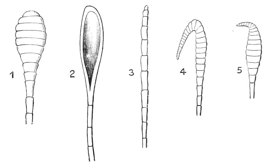
Fig. 176—Clubs of butterflies' antennae. Terminal portions of antenna of, 1, Pieris brassicae; 2, Styx infernalis; 3, Hestia idea (sub-family Danaides); 4, Eudamus proteus, and 5, Limochores taumas (Hesperiidae). (After Schatz and Scudder.)
The primary divisions of the family most often met with in literature are:—either Rhopalocera (= butterflies) and Heterocera (= moths); or Macrolepidoptera and Microlepidoptera; the Macrolepidoptera including the butterflies and large moths, the Microlepidoptera being limited to the families Tineidae (now itself in process of division into numerous families) and Tortricidae; some entomologists including also Pyralidae, Pterophoridae and Orneodidae in Microlepidoptera. The division of all Lepidoptera into two series is merely a temporary device necessitated by imperfect acquaintance with morphology. The division into Macro- and Micro- lepidoptera is entirely unscientific.
Series 1. Rhopalocera or Butterflies.—Antennae knobbed at the tip or thickened a little before the tip, without pectinations, projecting processes, or conspicuous arrangements of cilia. Hind wings without a frenulum, but with the costal nervure strongly curved at the base (Fig. 161, II, B).
Series II. Heterocera or Moths.—Antennae various in form, only rarely knobbed at the tip, and in such cases a frenulum present. In the large majority a frenulum is present, and the costal nervure of the hind-wing is either but little arched at the base (as in Fig. 161, I, B) or it has a large area between it and the front margin; but in certain families the hind wing is formed much as in Rhopalocera.
It may be inferred from these definitions that the distinction between the two sub-Orders is neither sharply defined nor of great importance. The club of the antenna of the Rhopalocera exhibits considerable variety in form (Fig. 176).[210] Butterflies are as a rule diurnal in their activity and moths nocturnal; but in the tropics there are numerous Heterocera that are diurnal, and many of these resemble butterflies not only in colour but even in the shapes of their wings.
Series I. Rhopalocera. Butterflies.
Classification and Families of Butterflies. Although considerable unanimity exists as to the natural groups of butterflies, there is much diversity of opinion as to what divisions are of equivalent value—some treating as sub-families groups that others call families—and as to the way the families should be combined. There is, however, a general agreement that the Hesperiidae are the most distinct of the families, and E. Reuter considers them a distinct sub-Order with the name Grypocera.[211]
Four categories may be readily distinguished, as follows, viz:—
1. The majority of butterflies; having the first pair of legs more or less strikingly different from the other pairs; frequently very much smaller and not used as legs; when not very small, then differing according to sex of the same species, being smaller in the male than in the female; the part most peculiar is the tarsus, which is modified in various manners, but in the males of this great series is always destitute of its natural form of a succession of simple joints five in number. There is no pad on the front tibia.
Fam. Nymphalidae, Erycinidae, Lycaenidae.
[The distinctions between these three families are found in the amount and kind of the abortion of the front legs; for definition refer to the heading of each of the families.]
2. The front legs are in general form like the other pairs; their tibiae have no pads; the claws of all the feet are bifid, and there is an empodium in connection with them.
Fam. Pieridae.
3. The front legs are like the other pairs; their tibiae however possess pads; the claws are large, not bifid, and there is no empodium; the metanotum is completely exposed at the base of the abdomen.
Fam. Papilionidae.
4. The front legs are like the other pairs; their tibiae however possess pads; the claws are small, toothed at the base, and there is an empodium; the metanotum is concealed by the prolonged and overhanging mesonotum.
Fam. Hesperiidae.
The relations between the families Erycinidae, Lycaenidae, and Nymphalidae are very intimate. All these have the front legs more or less modified, and the distinctions between the families depend almost entirely on generalisations as to these modifications. These facts have led Scudder to associate the Lycaenidae and Erycinidae in one group, which he terms "Rurales." It is however difficult to go so far and no farther; for the relations between both divisions of Rurales and the Nymphalidae are considerable. We shall subsequently find that the genus Libythea is by many retained as a separate family, chiefly because it is difficult to decide whether it should be placed in Erycinidae or in Nymphalidae. Hence it is difficult to see in this enormous complex of seven or eight thousand species more than a single great Nymphalo-Lycaenid alliance. The forms really cognate in the three families are however so few, and the number of species in the whole is so very large, that it is a matter of great convenience in practice to keep the three families apart. It is sufficient for larger purposes to bear in mind their intimate connexions.
The Papilionidae and Pieridae are treated by many as two sub-divisions of one group. But we have not been able to find any justification for this in the existence of forms with connecting characters. Indeed it would, from this point of view, appear that the Pieridae are more closely connected with the Lycaenidae and Erycinidae than they are with Papilionidae; in one important character, the absence of the pad of the front tibia, the Nymphalo-Lycaenids and the Pierids agree. It has also been frequently suggested that the Papilionidae (in the larger sense just mentioned) might be associated with the Hesperiidae. But no satisfactory links have been brought to light; and if one of the more lowly Hesperiids, such as Thanaos, be compared with one of the lower Papilionidae, such as Parnassius, very little approximation can be perceived.
It appears, therefore, at present that Hesperiidae, Papilionidae, Pieridae, and the Nymphalo-Lycaenid complex are naturally distinct. But in the following review of the families and sub-families of butterflies, we shall, in accordance with the views of the majority of Lepidopterists, treat the Lycaenidae and Erycinidae as families distinct from both Nymphalidae and Pieridae.[212]
The number of described species of butterflies is probably about 13,000; but the list is at present far from complete; forms of the largest size and most striking appearance being still occasionally discovered. Forty years ago the number known was not more than one-third or one-fourth of what it is at present, and a crowd of novelties of the less conspicuous kinds is brought to light every year. Hence it is not too much to anticipate that 30,000, or even 40,000 forms may be acquired if entomologists continue to seek them with the enthusiasm and industry that have been manifested of late. On the other hand, the species of Rhopalocera seem to be peculiarly liable to dimorphic, to seasonal and to local variation; so that it is possible that ultimately the number of true species—that is, forms that do not breed together actually or by means of intermediates, morphological or chronological—may have to be considerably reduced.
In Britain we have a list of only sixty-eight native butterflies, and some even of these are things of the past, while others are only too certainly disappearing. New Zealand is still poorer, possessing only eighteen; and this number will probably be but little increased by future discoveries. South America is the richest part of the world, and Wallace informs us that 600 species of butterflies could, forty years ago, be found in the environs of the city of Pará.
Fam. 1. Nymphalidae.—The front pair of legs much reduced in size in each sex, their tarsi in the male with but one joint, though in the female there are usually five but without any claws. Pupa suspended by the tail so as to hang down freely. We include in this family several sub-families treated by some taxonomists as families; in this respect we follow Bates, whose arrangement[213] still remains the basis of butterfly classification. With this extension the Nymphalidae is the most important of the families of butterflies, and includes upwards of 250 genera, and between 4000 and 5000 species. There are eight sub-families.
It is in Nymphalidae that the act of pupation reaches its acme of complication and perfection; the pupae hang suspended by the tail, and the cremaster, that is the process at the end of the body, bears highly-developed hooks (Fig. 177, C, D). The variety in form of the chrysalids is extraordinary; humps or processes often project from the body, making the Insect a fantastic object; the strange appearance is frequently increased by patches like gold or silver, placed on various parts of the body. It is believed that the term chrysalid was first suggested by these golden pupæ. The Purple Emperor, Apatura iris, differs strikingly in the pupa as well as in the larva-stage from all our other Nymphalids; it is of green colour, very broad along the sides, but narrow on the dorsal and ventral aspects (Fig. 177). The skin of this pupa is less hard than usual, and the pupa seems to be of a very delicate constitution. The Purple Emperor, like some of the Satyrides as well as some of its more immediate congeners, hibernates in our climate as a partially grown larva and passes consequently only a very brief period of its existence in the form of a pupa.
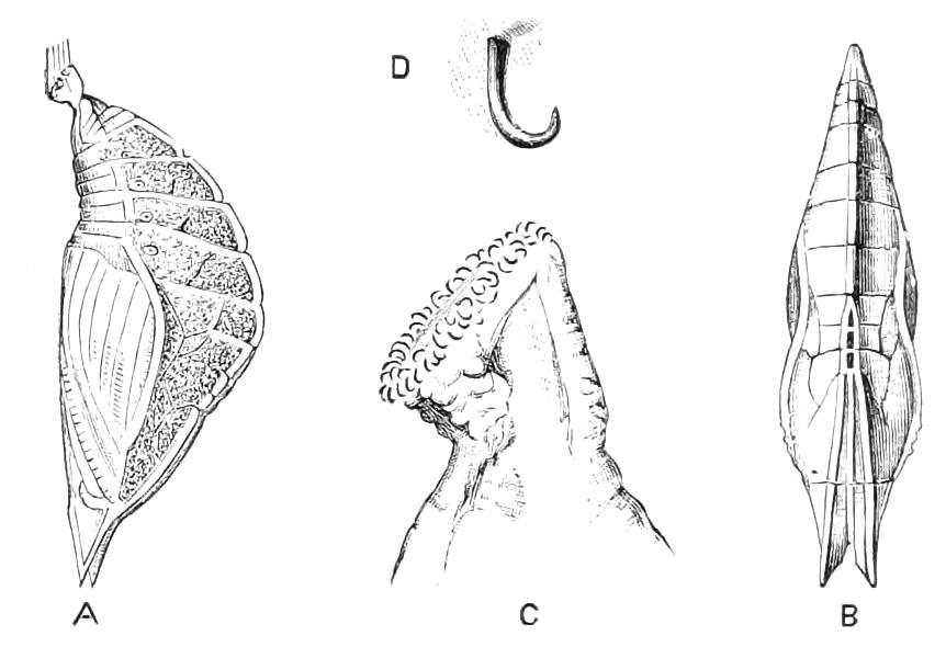
Fig. 177.—Pupa of the Purple Emperor butterfly, Apatura iris. New Forest. A, Lateral, B, dorsal aspect; C, enlarged view of cremaster with the suspensory hook; D, one hook still more enlarged.
Sub-Fam. 1. Danaides.—Front wing with inner-margin (submedian) nervure, with a short fork at the base. Cell of hind wing closed. Front foot of the female ending in a corrugate knob. Caterpillars smooth, provided with a few long fleshy processes. The claws are in a variable state, being sometimes simple, as in Papilionidae, sometimes with an empodium, apparently of an imperfect kind. The Danaides are usually large Insects with an imperfect style of ornament and colour; they have a great deal of black or very dark scaling, and in some Euploea this is agreeably relieved by a violet or purple suffusion, and these are really fine Insects. Usually there are large pale spaces, of some neutral indefinite tint, on which black blotches are distributed in a striking but inartistic manner. In many of the species the markings are almost spot for spot the same on the upper and under sides. About seven genera and 250 species are recognised. Danaides occur in all the warmer parts of the world, but are most numerous in the Eastern tropics. In Europe the family is represented only by an Asiatic and African species, Limnas chrysippus, that has extended its range to Greece. Besides this another species, Anosia erippus, Cr. (unfortunately also called Anosia menippe, Hb., and Danais archippus or even D. plexippus) has in the last two or three decades extended its range to various islands and distant localities, concomitantly, it is believed, with an extension of the distribution of its food-plant, Asclepias. This Insect has several times been taken in this country, and may probably be a natural immigrant. It is a common butterfly in North America, where it is called the Monarch.[214]
Some, at least, of the Danaides are unpleasant to birds in odour or in taste, or both. Among them there occur, according to Moore[215] and others, numerous cases of resemblance between forms that are thus protected. It is possible that the odour and taste are of some value to the Insects;[216] as, however, butterflies of any kind appear to be but rarely attacked in the imago-state by birds, and as their chief enemies are parasitic Insects that attack the larval instar, it is impossible to consider this protection of such prime importance to the species as many theorists assume it to be.
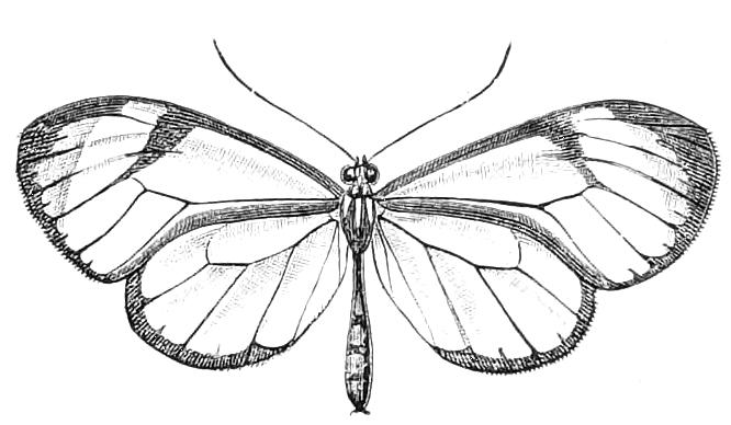
Fig. 178—Ithomia pusio. Brazil.
Sub-Fam. 2. Ithomiides.—Differs from Danaides by the female front foot having a true, though somewhat abbreviate tarsus. The caterpillers have no long processes. There has been considerable difference of opinion as to this division of butterflies. It is the family Neotropidae of Schatz, the Mechanitidae of Berg; also the "Danaioid Heliconiidae" of several previous writers, except that Ituna and Lycorea do not belong here but to Danaides. Godman and Salvin treat it as a group of the Danaid sub-family. The Ithomiides are peculiar to tropical America, where some 20 or 30 genera and about 500 species have been discovered. There is considerable variety amongst them. Ithomia and Hymenitis are remarkable for the small area of their wings, which bear remarkably few scales, these ornaments being in many cases limited to narrow bands along the margins of the wings, and a mark extending along the discocellular nervule. Wallace says they prefer the shades of the forest and flit, almost invisible, among the dark foliage. Many of these species have the hind-wings differently veined in the two sexes on the anterior part, in connection with the existence in the male of peculiar fine hairs, placed near the costal and subcostal veins. Tithorea and other forms are, however, heavily scaled insects of stronger build, their colours usually being black, tawny-red or brown, yellow, and white. In the sub-fam. Danaides, according to Fritz Müller, the male has scent-tufts at the extremity of the abdomen, whereas in Ithomiides analogous structures exist on the upper side of the hind-wing. Ithomiides have various colour-resemblances with members of the Heliconiides and Pieridae; Tithorea has colour analogues in Heliconius, and Ithomia in Dismorphia (formerly called Leptalis). Crowds of individuals of certain species of Ithomia are occasionally met with, and mixed with them there are found a small number of examples of Dismorphia coloured like themselves. They are placed by Haase in his category of secondary models. Belt states that some Ithomiides are distasteful to monkeys and spiders, but are destroyed by Fossorial Hymenoptera, which use the butterflies as food for their young; and he also says that they are very wary when the wasp is near, and rise off their perches into the air, as if aware that the wasp will not then endeavour to seize them. Much information is given about the habits by Bates in the paper in which he first propounded the "theory of mimicry."[217] The larvae are said to live on Solanaceae.
The genus Hamadryas is placed by some writers in Danaides, by others in Ithomiides; and Haase has proposed to make it the group "Palaeotropinae." The species are small, black and white Insects, somewhat like Pierids. They are apparently hardy Insects, and are abundant in certain parts of the Austro-Malay region.
Sub-Fam. 3. Satyrides.—Palpi strongly pressed together, set in front with long, stiff hairs. Front wings frequently with one or more of the nervures swollen or bladder-like at the base of the wing. Cells of both wings closed. Caterpillar thickest at the middle, the hind end of the body bifid. Pupa generally suspended by the cremaster, without girth: but sometimes terrestrial. This is a very extensive group, consisting of upwards of 1000 species. The Insects are usually of small size, of various shades of brown or greyish colours, with circular or ringed marks on the under sides of the wings. It is found all over the world, and is well represented in Europe; our Meadow-browns, Heaths, and Marbled-whites, as well as the great genus Erebia of the highlands and mountains belonging to it. Most of these Insects have but feeble powers of flight, and rise but little from the surface of the ground. The caterpillars live on various grasses. They are usually green or brown, destitute of armature, and a good deal like the caterpillars of Noctuid moths, but the hind end of the body is thinner and divided to form two corners, while the head is more or less free, or outstanding. The pupae are of great interest, inasmuch as in a few cases they do not suspend themselves in any way, but lie on the ground; sometimes in a very feeble cocoon or cell. There are no cremasteral hooks. The pupae of the Grayling butterfly, Hipparchia semele, has been found in loose soil a quarter of an inch below the surface. The chrysalis of the Scotch Argus, Erebia aethiops, was found by Mr. Buckler to be neither suspended nor attached, but placed in a perpendicular position, head upwards, amongst the grass. In the majority of cases the pupa is, however, suspended as is usual in Nymphalidae. Nothing is known as to the nature of the peculiar inflation of the bases of the nervures of the front wings; it is well shown in our common species of Coenonympha; this character is not, however, constant throughout the family. There is in South America a very remarkable group of Satyrides consisting of the genera Cithaerias and Haetera, in which the wings are very delicate and transparent, bearing on the greater part of their area remote fine hairs instead of scales; there are nevertheless some scaled patches about the margins, and one or more of the ringed marks characteristic of the Satyrides; while in some species the distal portions of the hind wings are tinted with carmine. The species of the genus Pierella connect these transparent Satyrids with the more ordinary forms. According to Wallace the habits of these fairy-like forms are those characteristic of the family in general. The genus Elymnias has been separated by some authorities as a sub-family, or even as a family, Elymniidae, chiefly on the ground of a slight peculiarity in the termination of the branches of the veins at the outer angle of the front wings. The Elymnias are said to be of a mimetic nature, having a greater or less resemblance to butterflies of various other divisions; there is also a considerable difference in appearance between their own sexes. The larva of E. undularis is known; it is of the form usual in Satyrides, and lives on the palm Corypha. About 50 species, ranging from India to Australia, with two in Africa, are known of this interesting group.
Sub-Fam. 4. Morphides.—There is no cell on the hind wing, the discocellular nervule being absent (Fig. 161, II. B). Caterpillars smooth or spiny, with the extremity of the body divided; frequently gregarious. These Insects have become notorious from the extraordinary brilliancy of blue colour exhibited by the upper surface of the wings of the typical genus Morpho. The species of Morpho are all Insects of large size, but with wings enormous in proportion to the body; this latter part is carried in a sort of cradle formed by the inner parts of the margins of the hind wings. Although an arrangement of this kind is seen in numerous other butterflies, yet there is perhaps none in which it is carried to quite such a pitch of perfection as it is in Morpho, where, on the under surface no part of the body behind the posterior legs can be seen. There are only about 100 species of Morphides, and 50 of these are included in Morpho, which is peculiar to tropical and sub-tropical America; the other half of the family is divided among ten or twelve genera, found in the Indo-Malay region; there being none in Africa. The eastern Morphides, though fine Insects, are not to be compared, either in size or brilliancy, with their American allies. The species of Morpho are apparently found only in the great forests of South America, where they are far from rare; some have a flapping and undulating flight, straight onwards along the alleys of the forest, and near the ground; others are never seen except steadily gliding with outstretched wings from 20 to 100 feet above the ground, where they move across sunny spaces between the crowns of the taller trees; the low-flyers settle frequently on the ground to suck the juices from fallen fruit, but the members of the other section never descend to the ground. As regards the caterpillars, W. Müller tells us[218] that the spines they are armed with break off, and enter the skin, if the creatures are carelessly handled. Four of the five species known to him are conspicuously coloured with black, red, yellow and white. The individuals are gregarious. The larvae of M. achilles sit in companies, often of more than 100 individuals, on trunks of trees, and so form a conspicuous patch. The caterpillars of M. epistrophis hang together as red clumps on the twigs of their food-plants. Hence it appears that in this genus we have an exception to the rule that night-feeding caterpillars rest in a hidden manner during the day.