Chapter V · (continued 3)
Reading edition
The histories of several other Cantharids have been more or less completely discovered. Fabre has found the larva of Cerocoma schaefferi attacking the stores of provisions laid up by a fossorial wasp of the genus Tachytes, and consisting of Orthoptera of the family Mantidae. The student who wishes for further information may refer to M. Beauregard's work on this family.[144]
Some half-dozen species of the genus Cephaloon found in Siberia, Japan, and North America, have, by some authorities, been separated as the family Cephaloidae. Nothing is known as to the metamorphosis of these rare beetles; and at present it is not necessary to distinguish them from Cantharidæ.
Fam. 76. Trictenotomidae.—Large Heteromera, with powerful free projecting mandibles; the antennae long, but with the terminal three joints short, with angular projections on one side. This family includes only two genera and seven or eight species. They are very remarkable Insects; Autocrates aenea being three inches long. The family is of considerable interest, as it seems to have no affinity with any other Coleoptera. The appearance of the species somewhat reminds one of Lucanidae, or Prionides; but Trictenotomidae have even less relation to those beetles than they have to the members of the Heteromerous series. The Trictenotomidae appear to be found only in the primitive forests of the Indian and Indo-Malayan regions. Nothing is known as to their life-histories.
Series V. Phytophaga.
Tarsi apparently four-jointed, the three basal joints usually densely set with cushion-like pubescence beneath; the third joint different in form, being divided into two lobes, or grooved on its upper surface so as to allow of the fourth joint being inserted near its base instead of at its extremity. Head not forming a definite prolonged beak; its labrum visible, the palpi rarely (and even then not completely) occluded in the mouth.
This great series of beetles includes something like 35,000 species. It approaches, like all the other series, the Polymorpha, especially the family Erotylidae placed therein, but in the great majority of cases there is no difficulty in recognising its members. The tarsi have never the Heteromerous formula, the head is not constructed like that of Rhynchophora, nor the mouth and feet like those of Adephaga; the antennae are different from those of the Lamellicorns. The tarsi are really five-jointed, for careful inspection shows that the long claw-joint has at its extreme base a small nodule, which is undoubtedly the fourth joint (Fig. 142, B). In speaking of the joints it is, however, customary not to refer to this small and functionally useless joint at all, and to call the claw-joint the fourth; when the little joint is referred to it may be called the true fourth joint.
Nearly the whole of the enormous number of species of this series are directly dependent on the vegetable kingdom for their nutriment; they are therefore well styled Phytophaga. This term is, however, restricted by some systematists to the family we have called Chrysomelidae. Although there is enormous variety in this series, three families only can be at all naturally distinguished, and this with difficulty. Of these the Bruchidae are seed-feeders, the Chrysomelidae, as a rule, leaf-feeders, the Cerambycidae wood and stem-feeders. The number of exceptions to this rule is but small, though certain Cerambycidae and certain Chrysomelidae live on roots.
Fam. 77. Bruchidae.—Prosternum extremely short; in front perpendicular; behind the coxae, forming merely a transverse lamina with pointed extremity. Hind femora more or less thickened. This comparatively small family includes about 700 species of small, unattractive beetles. The larvae live in seeds; hence some of the species are liable to be transported by means of commerce; some of them do considerable injury; peas and beans being specially subject to their attacks. They are able to complete their growth with a very small amount of nutriment, some of them consuming only a portion a little larger than themselves of a bean or pea. The larvae are fat maggots without legs, but Riley has discovered that the young larvae of Bruchus pisi and B. fabae have, when first hatched, three pairs of legs which are subsequently lost. They also have peculiar spinous processes on the pronotum. Both of these characteristics may be correlative with the transient differences in the activities of the larva, for the little creature is not at first located in the pea, but mines a gallery in the pod, in which it moves about, subsequently entering the pea and losing its legs. There is a good deal of difference in these respects between the two species—B. pisi and B. fabae—examined by Riley, and as but little is known of the life-histories of other Bruchidae it is probable that still greater variety prevails. Heeger has found that Bruchus lentis sometimes requires two seeds to enable it to complete its growth; it is, notwithstanding its legless state when half-grown, able to migrate by dropping to the earth, and dragging itself along by its mandibles till it comes to another pod into which it bites its way.
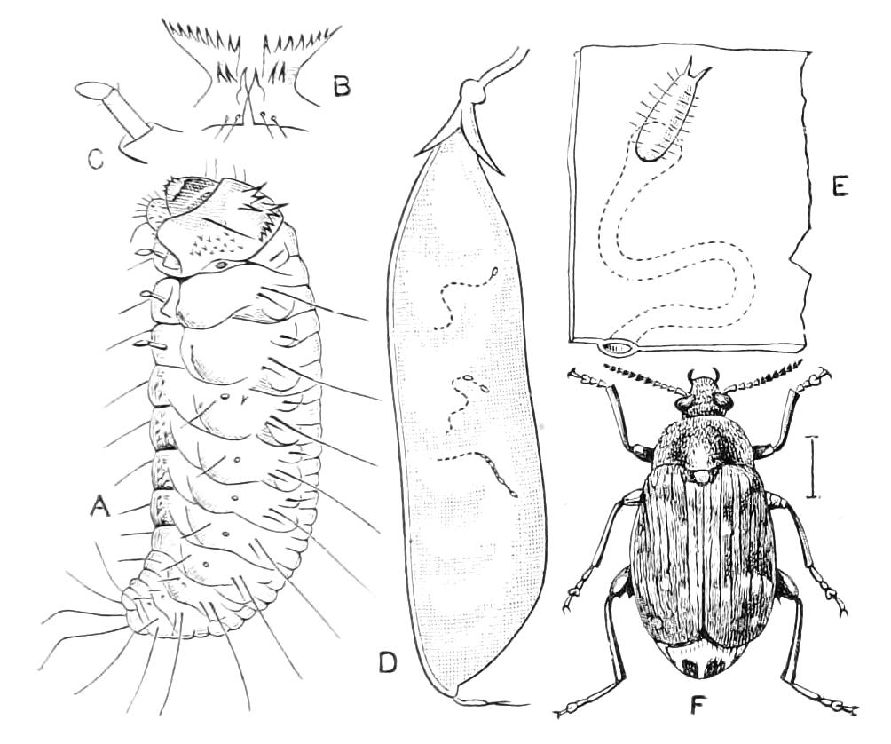
Fig. 141—Bruchus pisi or pea-weevil. A, Young larva; B, prothoracic spinous process; C, post-embryonic leg, greatly magnified; D, pea-pod, with tracks of entry; E, portion of pod, with egg, and the subsequently formed track, magnified; F, imago. (After Riley.)
The family has, until recently, been placed in the Rhynchophorous series, with which it has, however, no direct connection. On the other hand, it is so closely connected with Chrysomelidae that it is not possible to indicate good characters to distinguish the two at present. The Australian genus Carpophagus, and the large South American species of Caryoborus appear to be quite indistinguishable as families, though Lacordaire and Chapuis placed one in Bruchidae, the other in Chrysomelidae. The definition we have given applies, therefore, to the majority of the family, but not to the aberrant forms just mentioned. The European genus Urodon appears to belong to Anthribidae, not to Bruchidae. The family Bruchidae is called Mylabridae by some.
Fam. 78. Chrysomelidae.—Antennae moderately long; eyes moderately large, usually not at all surrounding the insertion of the antennae; upper surface usually bare, frequently brightly coloured and shining. This enormous family comprises about 18,000 species of beetles, in which the form and details of structure are very varied. No satisfactory character for distinguishing Chrysomelidae from Cerambycidae has yet been discovered, although the two families are certainly distinct and natural. Most of the Chrysomelidae live on foliage; few of them are more than half an inch long, whereas the Cerambycidae are wood-feeders and usually of more elongate form and larger size. The potato beetle, or Colorado beetle, that occasioned so much destruction in North America some thirty years ago, and the introduction of which into Europe was anticipated with much dread, is a good example of the Chrysomelidae. The turnip flea, a tiny hopping beetle, is among the smallest forms of the family, and is a member of another very extensive subdivision of Chrysomelidae, viz. Halticides. The term Phytophaga is by many naturalists limited to Chrysomelidae, the Cerambycidae being excluded. The classification of the family is but little advanced, but the enormous number of species of Chrysomelidae are placed in four divisions, viz.:—
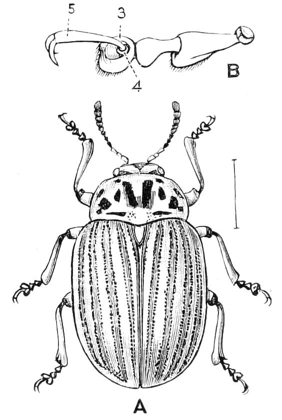
Fig. 142—Doryphora decemlineata, the potato beetle. North America. A, Imago; B, hind-tarsus. 3, third joint; 4, true fourth joint; 5, so-called fourth joint.
Prothorax much narrower at the base than the elytra, and usually without side-margins (raised edges). Sub-fam. 1. Eupoda; with three divisions, Sagrides, Donaciides, Criocerides.
The basal ventral plates of the abdominal segments are somewhat shorter in the middle than at the sides, the fourth one being often invisible in the middle, while the fifth is very large. Sub-fam. 2. Camptosomes; with six divisions, Megascelides, Megalopides, Clythrides, Cryptocephalides, Chlamydes, Sphaerocarides.
In the other two groups there is no great disparity between the fourth and fifth ventral plates.
Prothorax not greatly narrower at the base than the elytra, and usually with distinct edges at the outsides. Sub-fam. 3. Cyclica; with four divisions, Lamprosomides, Eumolpides, Chrysomelides, Galerucides.
Front of the head bent downwards or inflexed, so that the mouth is on the lower aspect. Antennae inserted close together on the most anterior part of the head, so that they are more forward than the mouth. Sub-fam. 4. Cryptostomes; with two divisions Hispides, Cassidides.
In the other three divisions the mouth is placed as usual, but the insertion of the antennae varies a good deal.
The larvae of about 100 species of the family are known; they are arranged in accordance with their habits, by Chapuis,[145] in six groups, viz.:
1. Elongate larvae, living under water, and there undergoing their metamorphosis. (Donaciides.)
2. Larvae mining in leaves, and undergoing their metamorphosis in the leaf. (Hispides and some Halticides.)
3. Short convex larvae, frequently with leathery and pigmented integuments, living exposed on plants. (Most of the Cyclica.)
4. Larvae of short form; covering the body with excrementitious matter. (Some Criocerides.)
5. Peculiar larvae of short form, spiny, and protecting their bodies by excrementitious matter attached by a special apparatus, the excrement itself being modified so as to be suitable for retention. (Cassidides.)
6. Elongate, pallid, larvae with curved abdomen; living in shell-like cases, and undergoing metamorphosis therein. (Most of the Camptosomes, the habits of which are known.)
Though our knowledge of these larvae extends to only about 100 out of 18,000 species, the above category by no means includes all the kinds of larvae; Captain Xambeu having recently discovered that the larva of Chrysochus pretiosus lives in the earth feeding on roots after the manner of a Rhizotrogus larva, which it resembles. The larva of Sagra splendida lives inside the stems of Dioscorea batatas, in swellings; the group Sagrides, to which it belongs, is a very anomalous one.
i. Eupoda. The beetles of the genus Donacia are of special interest. They form, with the genus Haemonia, a peculiar group, well represented in Europe, and also in our own country. They are all connected with aquatic plants, the species of Haemonia living entirely under water, while the Donacia live in the imago-state an aërial life, though many of them enter the water with great readiness, and, it is said, are able to take wing from the surface. The larvae live on the roots of aquatic plants, and derive not only nutriment but air therefrom; they pass several months as pupae (or as resting larvae waiting for pupation), under water in cocoons which they construct, and which, incredible as it may seem, are filled with air, not water. Exact details as to the construction of these cocoons are wanting. It was formerly absurdly supposed that the larva swelled itself out to the size of the cocoon it was about to make, and so served as a mould, subsequently contracting. The observations of Schmidt-Schwedt[146] make it, however, more probable that the plant itself furnishes the air which, under pressure of the water (so he supposes), fills the cocoon; the larva wounds the root, piercing to an air-vessel and then constructs the cocoon on this spot, leaving to the last moment an orifice, according to Schmidt, as an exit for the water. The larva uses a similar artifice for obtaining air; it has no gills, but is provided near the extremity of the body with two sharp chitinous processes which it drives into the root of the plant till it penetrates an air-vessel. Schmidt thinks the processes serve as conduits to conduct the air to the tracheae, but Dewitz thinks the air enters the larva in a more normal manner, by means of a stigma placed at the base of the piercing process. A similar larva exists in Haemonia; which genus is additionally interesting from the fact that the imago lives entirely submerged. It is not known how it breathes. This genus is the only member of the Chrysomelidae that does not possess the structure of the feet that is characteristic of the Phytophaga. The late Professor Babington about sixty years ago found H. curtisi at Cley on the Norfolk coast on submerged Potamogeton pectinatus, but it has not been met with there for a great many years.
The larvae of Criocerides are of two kinds, in one of which the body is peculiarly shaped in conformity with the curious habit of using the excrement as a covering. The larva is less elongate than usual, and has the anus placed on the upper surface, and formed so that the excrement when voided is pushed forward on to the Insect; here it is retained by means of a slimy matter, and a thick coat entirely covering the creature, is ultimately formed. The larva of Lema melanopa is not uncommon about Cambridge, where it feeds on the leaves of growing corn. It is a remarkable fact that even in one genus the species have some of them this habit, but others not. The species of Crioceris living on lilies—C. merdigera, e.g.—are noted for possessing it; while C. asparagi does not protect itself in this way, but emits fluid from its mouth when disturbed. This larva is a serious nuisance in some localities to the cultivators of asparagus. The eggs are deposited on the stems of the plant—as shown in our figure—sometimes in great numbers.
The perfect Insects of many of the Criocerides possess a stridulating organ. Two contiguous areas at the base of the last dorsal segment, where they can be rubbed by the tips of the elytra, are slightly elevated and bear very close and fine straight lines.
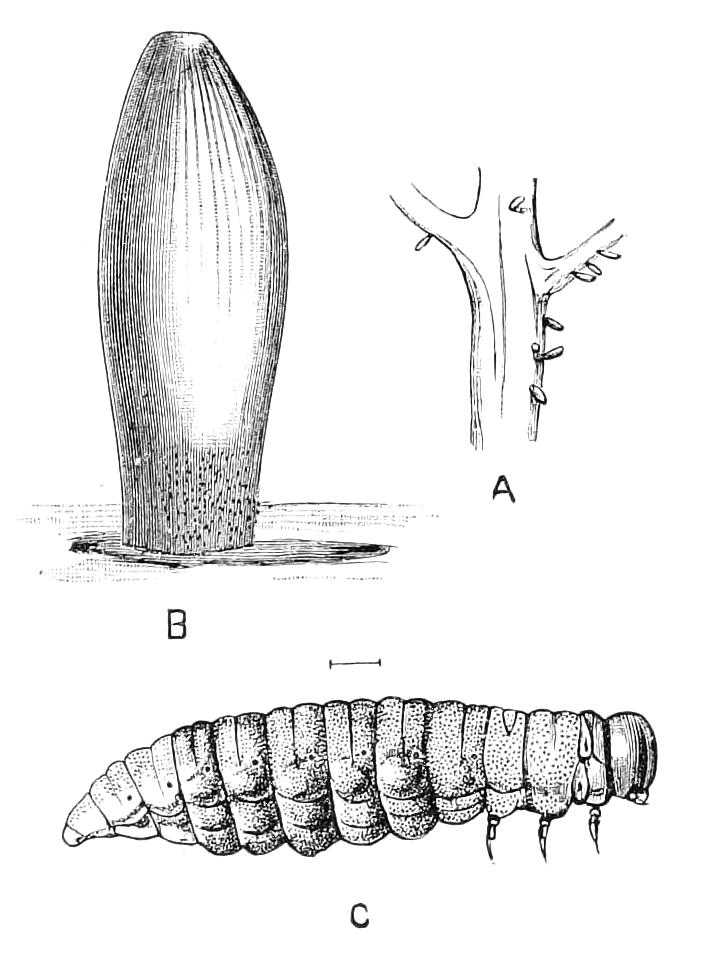
Fig. 143—Crioceris asparagi. A, Eggs in position on stem of asparagus; B, one egg much enlarged; C, young larva. Cambridge.
ii. The Camptosomes, as we have already noticed, are distinguished by a peculiar structure of the abdomen. This character appears to be connected with a very remarkable habit, viz. the formation of a case to envelop the egg. The tip of the abdomen is somewhat curved downwards, and, in the female, bears a hollow near the extremity; when an egg is extruded the female holds it in this hollow by means of the hind legs, and envelops it with a covering said to be excrementitious. When the larva hatches, it remains within this case, and subsequently enlarges it by additions from its own body. The beautiful Insects of the genus Cryptocephalus, which is fairly well represented in Britain, belong to this division. The exotic group Megalopodes is incorrectly placed in Camptosomes; the side pieces of the prothorax meet in it behind the middle coxae, as they do in Rhynchophora. The species of Megalopodes stridulate by means of an area on the base of the meso-scutellum rubbed by a ridge inside the pronotum, as in the Cerambycidæ.
iii. The division Cyclica includes the great majority of Chryomelidae; we have not less than 170 species in Britain. The larvae live, like those of Lepidoptera, at the expense of foliage, and the species frequently multiply to such an extent as to be injurious. Some of them are destroyed in great numbers by Hymenopterous parasites, the Braconid genus Perilitus being one of the best known of these; in some cases the parasite deposits its eggs in either the larva or perfect Insect of the beetle, and the metamorphoses of the parasites in the latter case are sometimes, if not usually, completed, the larvae emerging from the living beetles for pupation.
iv. The Cryptostomes, though comparatively few in number of species, include some very remarkable beetles. There are two groups, Hispides and Cassidides. The former are almost peculiar to the tropics and are not represented by any species in the British fauna. The head in this group is not concealed; but in the Cassidides the margins of the upper surface are more or less expanded, so that the head is usually completely hidden by the expansion of the pronotum. Both the groups are characterised by the antennae being inserted very near together, and by the short claw-joint of the feet. Hispa is one of the most extensive of the numerous genera of Hispides, and is remarkable from the imago being covered on the surface with long, sharp spines. But little is known as to the metamorphosis, beyond the fact already alluded to, that the larvae of several species mine the interior of leaves. The larva of Hispa testacea, according to Perris,[147] makes use of the leaves of Cistus salvifolius in Southern Europe; it is broad and flat, and possessed of six short legs. The eggs are not deposited by the parents inside the leaves, but are probably attached to various parts of the plant. After hatching, the young larva enters a leaf, and feeds on the parenchyma without rupturing the epidermis; but when it has consumed about three-fourths of the soft interior of the leaf it ruptures the epidermis of the upper surface, and seeks another leaf; this found, it places itself on the midrib, tears the upper epidermis, and lodges itself in the leaf. In the case of this second leaf it attacks the parenchyma in the neighbourhood of the petiole, and so forms an irregular tube which has an open mouth, the point of entry. In this tube it undergoes its metamorphosis. Each larva, it is said, always makes use of two leaves, and of two opposed leaves. A knowledge of the habits of some of the larger of the exotic Hispides would be of much interest.
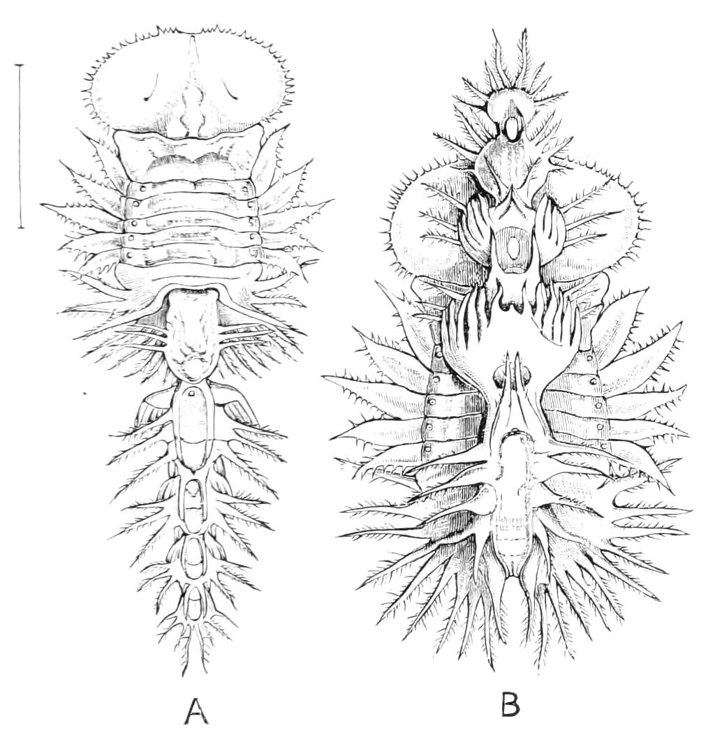
Fig. 144—Pupa of Cassidid beetle (? Aspidomorpha sp.). A, With appendage extended; B, with the appendage reposing on the back. New Britain.
The Cassidides, in addition to the curious marginal expansion of their upper surface, have the power of withdrawing the head into the thorax, and hence they are often called shield or tortoise-beetles. They exhibit considerable variety in form and colour, and some of them display a peculiar metallic reflection of great delicacy and beauty; this disappears entirely after death, but it may be restored by thoroughly moistening the dead Insect. The colour, therefore, probably depends on the presence of water in the integument. The larvae of Cassidides are notorious on account of their habit of covering their bodies with dried excrement, for which purpose they are provided with a forked process at the posterior extremity; this serves to place the protecting matter in a proper position and to retain it there. The excrement assumes in various species forms so peculiar that they cannot be considered merely incidental. In several species this covering-matter is like lichen. This is the case with Dolichotoma palmarum, the larva of which has, in place of the usual fork, a more complex appendage on the back for the purpose of preparing and retaining its peculiar costume. The pupae, too, sometimes retain the larval skin. An extremely remarkable pupa of a Cassidid—possibly of the genus Aspidomorpha—was recently found by Dr. Arthur Willey in New Britain (Fig. 144). The back of the pupa is covered with a complex appendage, so that the creature has no resemblance to an Insect; this appendage is perhaps capable of being moved, or even extended (Fig. 144, A), during life. Whether it may be formed by the retention of portions of the moulted skins of the larva we cannot say with certainty.
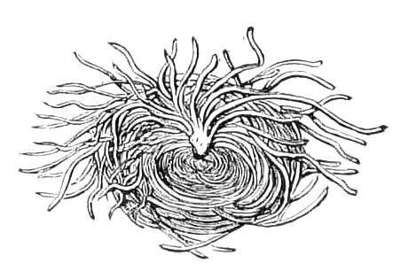
Fig. 145—Nest of intestinally-made filaments under which the larva of Porphyraspis tristis lives.
The most remarkable of the Cassidid coverings yet discovered are those formed by certain small beetles of the tropical American genus Porphyraspis. P. tristis is apparently a common Insect at Bahia, where it lives on a cocoa-palm. The larva is short and broad, and completely covers itself with a very dense coat of fibres, each many times the length of the body, and elaborately curved so as to form a round nest under which the larva lives. On examination it is found that these long threads are all attached to the anal extremity of the Insect, and there seems no alternative to believing that each thread is formed by small pieces of fibre that have passed through the alimentary canal, and are subsequently stuck together, end to end. The process of forming these long fibres, each one from scores of pieces of excrement, and giving them the appropriate curve, is truly remarkable. The fibres nearest to the body of the larva are abruptly curled so as to fit exactly, and make an even surface; but the outside fibres stand out in a somewhat bushy fashion. The construction is much like that of a tiny bird's nest. Señor Lacerda informed the writer that the larva makes a nest as soon as it is hatched. Another Porphyraspis—P. palmarum—has been recorded as forming similar nests on a species of Thrinax in St. Domingo. Candèze says[148] that when it has completed its growth the larva ejects on to the leaf a quantity of semi-liquid matter, and this, on drying, sticks the nest to the leaf, so that the metamorphosis is effected under shelter.
Fam. 79. Cerambycidae (Longicorns).—Form usually oblong, not much curved in outline at the sides; surface very frequently rendered dull by a very minute hairiness, which often forms a pattern; antennae usually long, and their insertion much embraced by the eyes. This great family of beetles includes some 12,000 or 13,000 known species. The elegance and variety of their forms and the charm of their colours have caused them to attract much attention, so that it is probable that a larger proportion of the existing species have been obtained than is the case in any other of the great families of Coleoptera. Still it is not likely that one-half of the living forms are known. It is not possible at present to point out any one character of importance to distinguish Cerambycidae from Chrysomelidae, though the members of the two families have, as a rule, but little resemblance in external appearance. Most of them live on, or in, wood, though many are nourished in the stems of herbaceous plants. The larvae live a life of concealment, and are soft, whitish grubs with powerful mandibles, and usually with a comparatively small head, which is not much exserted from the thorax. Most of them are without legs, but a good many have three pairs of small legs, and there are numerous cases in which the surface of the body is furnished above or below with swellings believed to act as pseudopods (Fig. 84), and help the larvae to move about in their galleries; but this is probably not the sole function of these organs, as their surface is varied in character, and often not of a kind that appears specially adapted to assist in locomotion. There is a slight general resemblance between the larvae of Cerambycidae and those of Buprestidae, and when the thorax of a Longicorn larva is unusually broad, e.g. Astynomus, this similarity is very pronounced.
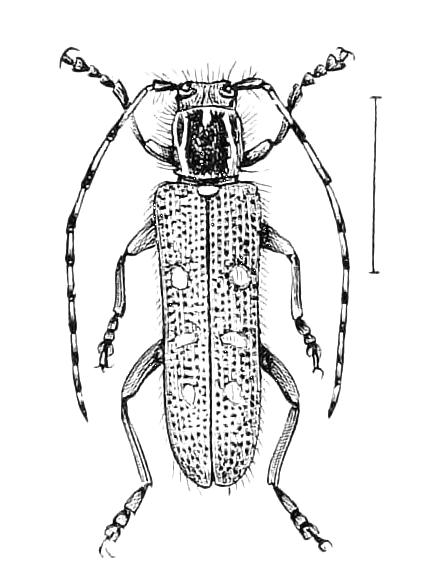
Fig. 146—Saperda populnea. Britain.
The modes of life of Cerambycid larvae exhibit considerable variety, and much perfection of instinct is displayed by the larvae, as well as by the mother beetles. The larvae of Saperda populnea, are common in certain woods in the South of England in the stems of aspen; they consume only a small quantity of the interior of the stem, and are probably nourished by an afflux of sap to the spot where they are situated. Elaphidion villosum is called the oak-pruner in North America. The parent beetle lays an egg near the axilla of a leaf-stalk or small stem, and the young larva enters this and feeds on the tender material; as it grows it enters a larger limb, and makes an incision within this in such a manner that the wood falls to the ground with the larva within it, the dead wood serving subsequently as pabulum and as a shelter, within which the metamorphosis is completed. The species of the American genus Oncideres are called girdlers, because the parent beetle, after laying an egg in a small branch, girdles this round with a deep incision, so that the portion containing the larva sooner or later falls to the ground. The growth of a Longicorn larva frequently takes more than a year, and under certain circumstances it may be enormously prolonged. Monohammus confusus has been known to issue from wooden furniture in a dwelling-house when the furniture was fifteen years old. Individuals of another Longicorn have issued from the wood of a table, twenty and even twenty-eight years after the felling of the tree from which the furniture was made. Sereno Watson has related a case from which it appears probable that the life of a Longicorn beetle extended over at least forty-five years.[149] It is generally assumed that the prolongation of life in these cases is due to the beetle resting quiescent for long after it has completed the metamorphosis. Recent knowledge, however, renders it more probable that it is the larval life that is prolonged; the larva continuing to feed, but gaining little or no nutriment from the dry wood in these unnatural conditions. Mr. C. O. Waterhouse had for some years a Longicorn larva under observation, feeding in this way in the wood of a boot-tree;[150] the burrows in the wood contained a great deal of minute dust indicating that the larva passed much matter through the alimentary canal, probably with little result in the way of nutriment.
There are numerous Longicorns that bear a great resemblance in form and colour to Insects to which they are not related. Haensch[151] has noticed that species of the genus Odontocera resemble various Hymenoptera, one species being called O. braconoides; he also observed that these Hymenoptera-like Longicorns, instead of withdrawing their underwings under the elytra as beetles generally do, vibrate them rapidly like Hymenoptera. A large number of Longicorns stridulate loudly by rubbing a ridge inside the pronotum on a highly specialised, striate surface at the base of the scutellum, and therefore covered up when the Insect is contracted in repose. A few produce noise by rubbing the hind femora against the edges of the elytra, somewhat after the fashion of grasshoppers. In this case there appears to be comparatively little speciality of structure, the femora bearing, however, more or less distinct small granules. The species of the Hawaiian genus Plagithmysus produce sound in both these manners, the thoracic stridulating organ being beautifully developed, while in some species the margin of the elytra and base of the femora are also well adapted for the purpose of sound-production, and in a few species of the genus there are also highly-developed stridulating surfaces on the hind and middle coxae. This is the only case in which a beetle is known to possess more than one set of sound-organs in the imago state.
Three divisions of this family are distinguished, viz.—
1. Front coxae large and transverse; prothorax with distinct side margins. .......... Sub-fam. 1. Prionides.
2. Front coxae not greatly extended transversely, thorax not margined; last joint of maxillary palpus not pointed, usually broader (more or less) than the preceding joint. .......... Sub-fam. 2. Cerambycides.
3. Front coxae usually round and deeply embedded; last joint of maxillary palpus pointed; front tibiae with a more or less distinct, slanting groove on the inner side. .......... Sub-fam. 3. Lamiides.
The Prionides are on the average considerably larger in size than the members of the other divisions, and they include some of the largest of Insects. The Amazonian Titanus giganteus and the Fijian Macrotoma heros are amongst the most gigantic. Some of the Prionides have a great development of the mandibles in the male sex analogous to that we have already noticed in Lucanidae. The larvae of the large Prionides appear in various parts of the world to have been a favourite food with native tribes, and Lumholz states that they are really good eating. In consequence of the destruction of forests that has progressed so largely of late years these gigantic Prionides have become much rarer.
Several aberrant forms are included in Prionides. The genus Parandra has five-jointed tarsi; the third joint being much smaller than usual, so that the fourth joint is not concealed by it. The Brazilian Hypocephalus armatus was for long a subject of dispute as to its natural position, and was placed by different authorities in widely-separated families of Coleoptera. The structure of this aberrant Longicorn seems to be only explicable on the hypothesis of warfare amongst the males.[152] Nothing is, however, known as to the habits and history of the Insect, and only one or two specimens of the female have yet been obtained.
The family Spondylidae has been proposed for some of these aberrant Longicorns, but as it includes but very few, and highly discrepant, species, it is neither natural nor of much use for systematic purposes.
The Lamiides are the most highly specialised division of the Longicorns, and includes the larger number of the species. The front of the head is usually placed at right angles to the vertex, and in some cases (groups Hippopsini, Spalacopsini) it is strongly inflexed, so that the mouth is placed on the under side of the head. The extension of the eyes round the antennae is accompanied by very curious shapes of those organs, and not infrequently each eye is divided into two more or less widely-separated parts, so that the Insect has, on the external surface, four eyes.
Series VI. Rhynchophora.
Head more or less prolonged in front to form a snout or beak, called rostrum. Tarsi four-jointed, usually at least the third joint broad and densely pubescent beneath.
This enormous series includes about 25,000 species, and as may well be imagined shows a great variety of structure amongst its forms. The vast majority may, however, be readily recognised by the two characters mentioned above. There are some cases in which the beak is indistinct, and others in which the tarsi are five-jointed (Dryophthorus), and even slender (Platypides). In these cases a close examination shows that the gular region on the middle of the back of the under surface of the head cannot be detected, and that the back of the prosternum is very strongly consolidated by the side-pieces of the thorax meeting together and being very firmly joined behind the coxae. The beak is in the great majority perfectly distinct, though it varies so extremely in form that it can only be briefly described by saying that it is a prolongation of the head in front of the eyes, or that the antennae are inserted on its sides near to, or far from, the tip. It has been ascertained in many cases that the rostrum is used by the female to assist in placing the eggs in suitable places, a hole being bored with it; in some cases it is also used to push the egg far into the hole in which it has previously been placed by the ovipositor; but there are many forms in which it is fairly certain that it is not so used. What purpose it serves in the male is totally unknown. In many members of the series, the rostrum differs in form in the two sexes, and in most, if not in all, these cases it is clear that the distinctions tend in the direction of making the beak of the female more efficient for the mechanical purpose we have mentioned.
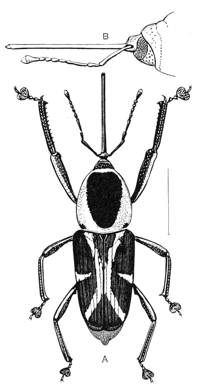
Fig. 147.—Eugnoristus monachus ♀. Madagascar. A, The imago; B, front of pronotum, head, and rostrum.
It was proposed by Leconte and Horn to separate this series from all the other Coleoptera as a primary division, and they looked on it as of lower or more imperfect structure. Packard has very properly protested against this interpretation; and there seems to be no reason whatever for considering the Rhynchophora as "lower" than other beetles; indeed we should be inclined to place such forms as Calandrides amongst the most perfect of Insects; their external structure (as shown by Eugnoristus monachus, Fig. 147) being truly admirable.
Only four families of Rhynchophora can be at present accepted as satisfactory; one of these—Curculionidae—includes an enormous majority of the whole series. Though it is probable that it will ultimately be divided into several families, the attempts to that end that have already been made are not satisfactory.
Fam. 80. Anthribidae.—Palpi usually not covered, but distinct and flexible. Antennae often long, not elbowed, the first joint not very long. Third joint of tarsus small, usually much concealed by being embraced by the second joint. Pygidium exposed; propygidium deeply grooved in the middle. This family includes 800 or more species, which are mostly tropical; it is very sparsely represented in the faunas of Europe and North America. It is quite distinct from Curculionidae with which it was formerly associated. It contains many graceful Insects having a certain resemblance with Longicorns on account of the large development of the antennae. The habits and metamorphoses are but little known. It seems probable that many species find their nutriment in old wood or boleti The larvae of some genera (Cratoparis and Araeocerus) have legs, but in others the legs are wanting, and the larvae are said to completely resemble those of Curculionidae. In the larva of our tiny British species, Choragus sheppardi, the legs are replaced by three pairs of thoracic, sac-like pseudopods. This Insect makes burrows in dead branches of hawthorn. The larvae of the genus Brachytarsus have been ascertained to prey on Coccidae.
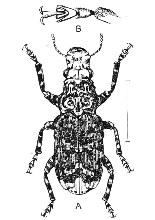
Fig. 148—Platyrhinus latirostris, Anthribidae. Britain. A, the perfect Insect; B, tarsus and tip of tibia.
Fam. 81. Curculionidae (Weevils).—The beak of very variable length and thickness; the palpi small, nearly always concealed within the mouth, short, and rigid. Labrum absent. Antennae of the majority elbowed, i.e. with the basal joint longer, and so formed that when it is laterally extended the other joints can be placed in a forward direction. This enormous family includes about 20,000 known species, and yet a large portion of the species yearly brought from the tropics still prove to be new. The rostrum or beak exhibits excessive variety in form, and is in many cases different in the sexes; in this case it is usually longer and thinner in the female. As the rostrum is one of the chief characters by which a member of the family may be recognised, it is necessary to inform the student that in certain forms (the Australian Amycterides, e.g.) the organ in question may be so short and thick that it is almost absent. In these cases the Insect may be identified as a Curculionid by the gular area being absent on the under side of the head, and by the concealment of the palpi. The tarsi are usually of the same nature as those of Phytophaga, already described, but the true fourth joint is less visible. In the Brachycerides this joint is not present, and the third joint is not lobed. The palpi are flexible and more or less exserted in a very few species (Rhynchitides); in Rhinomacerides there is also present a minute labrum. The front coxae are deeply embedded, and in many forms the prosternum is peculiar in structure; the side-pieces (epimera) meeting at the back of the prosternum in the middle line. This, however, is not universal in the family, and it occurs in some other beetles (e.g., Megalopodides of the Phytophaga). The larvae are without legs. They are vegetarian, the eggs being deposited by the mother-beetle in the midst of the food. These larvae may be distinguished from those of Longicorns by the general form, which is sub-cylindric or rather convex, not flattened, and more particularly by the free, exserted head, the mouth being directed downwards; the attitude is generally a curve, and the anterior part of the body is a little the thicker. No part of plants is exempt from the attacks of the larvae of Curculionidae; buds, twigs, leaves, flowers, fruits, bark, pith, roots and galls may each be the special food of some Curculionid. Certain species of the sub-families Rhynchitides and Attelabides prepare leaves in an elaborate manner to serve as food and dwelling for their young. If young birches, or birch bushes from 5 to 10 feet in height, be looked at in the summer, one may often notice that some of the leaves are rolled so as to form, each one, a little funnel. This is the work of Rhynchites (or Deporaus) betulae, a little Curculionid beetle (Fig. 149). An inspection of one of these funnels will show that it is very skilfully constructed.
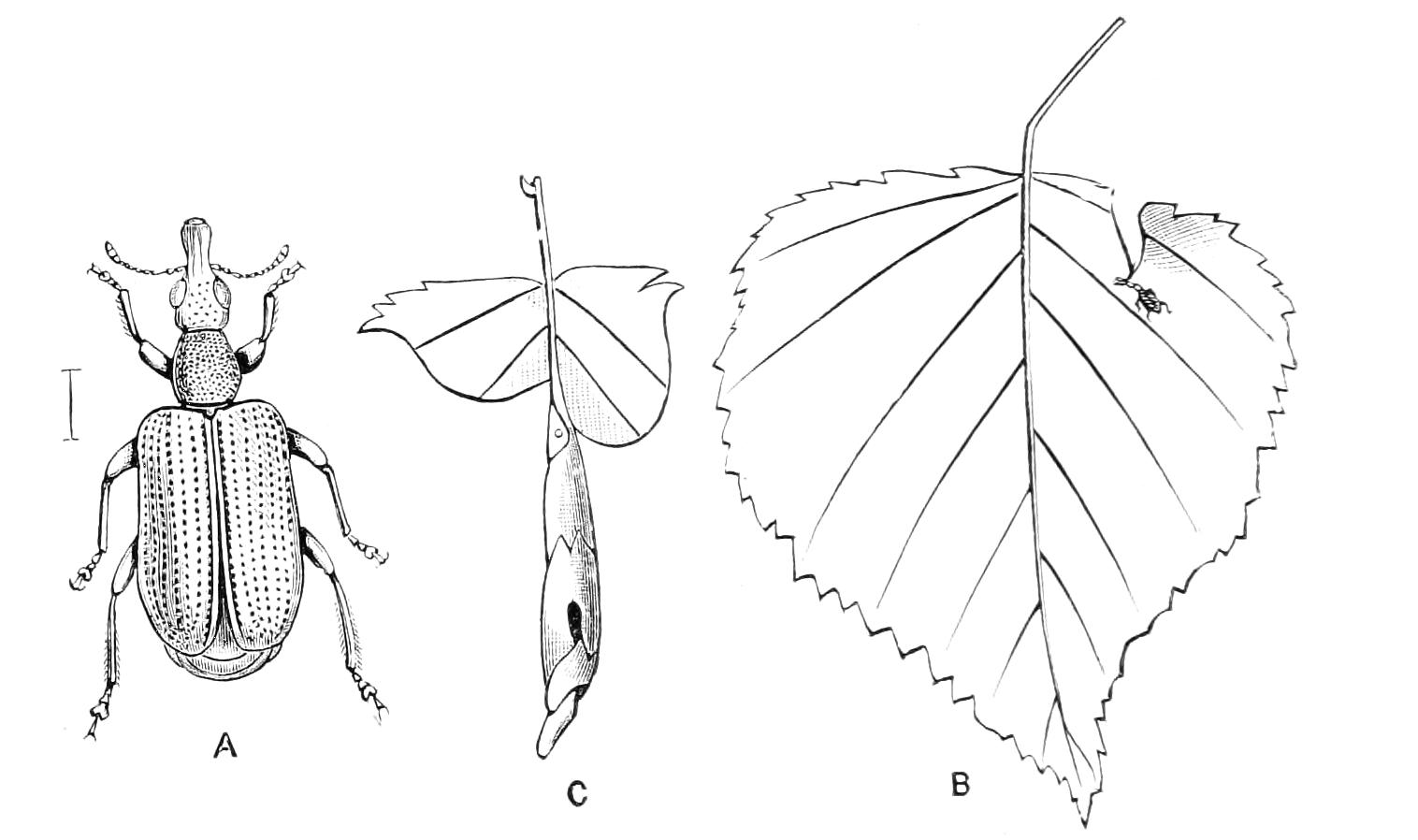
Fig. 149.—The leaf-rolling of Rhynchites betulae. Britain. A, Female beetle, magnified; B, the beetle forming the first incision on a leaf; C, the completed roll. (B and C after Debey.)
The whole of a leaf is not used in the formation of a funnel, cuts being made across the leaf in suitable directions. The beetle standing on a leaf, as shown in the figure, proceeds to cut with its mandibles an incision shaped like an erect S, commencing at a certain part of the circumference, and ending at the midrib of the leaf; the beetle then goes to the other side of the midrib, and continues its incision so as to form another S-like curve considerably different from the first; being prostrate and less abrupt. Thus the blade of the leaf is divided into two halves by certain curved incisions, the midrib remaining intact. The little funnel-twister now commences to roll up the leaf to form the funnel; and this part of the work is greatly facilitated by the shape of the incisions. Going back to the spot where it commenced work, by the aid of its legs it rolls one side of the leaf round an ideal axis, somewhat on the same plan as that adopted by a grocer in forming a paper-funnel for sugar. The incisions are found to be just of the right shape to make the overlaps in the rolling, and to retain them rolled-up with the least tendency to spring back. After some other operations destined to facilitate subsequent parts of its task, the beetle enters the rolled-up part of the leaf and brings it more perfectly together; it again comes out and, pursuing a different system, holds on with the legs of one side of the body to the roll, and with the other legs drags to it the portion of the leaf on the other side of the midrib so as to wrap this part (i.e. the result of its second incision) round the part of the funnel already constructed. This being done the Insect again enters the funnel, bites three or four small cavities on the inside of the leafy wall and deposits an egg in each. Afterwards it emerges and fits the overlaps together in a more perfect manner so as to somewhat contract the funnel and make it firmer; then proceeding to the tip, this is operated on by another series of engineering processes and made to close the orifice; this part of the operation being analogous to the closing by the grocer of his paper-funnel after the sugar has been put in. The operation of the beetle is, however, much more complex, for it actually makes a sort of second small funnel of the tip of the leaf, bends this in, and retains it by tucking in some little projections. The work, which has probably lasted about an hour, being now completed, the creature takes a longer or shorter rest before commencing another funnel. We have given only a sketch of the chief points of the work, omitting reference to smaller artifices of the craft master; but we may remark that the curved incisions made by the beetle have been examined by mathematicians and duly extolled as being conducted on highly satisfactory mathematical principles. It is impossible at present for us to form any conception as to the beetle's conceptions in carrying out this complex set of operations. Our perplexity is increased if we recollect its life-history, for we then see that neither precept or example can have initiated its proceedings, and that imitation is out of the question. The eggs hatch in their dark place, giving rise to an eyeless maggot, which ultimately leaves the funnel for the earth. The parts of this maggot subsequently undergo complete change to produce the motionless pupa of entirely different form, from which emerges the perfect Insect. Hence the beetle cannot be considered to have ever seen a funnel, and certainly has never witnessed the construction of one, though, when disclosed, it almost immediately sets to work to make funnels on the complex and perfect system we have so imperfectly described. More general considerations only add to the perplexity we must feel when reflecting on this subject. Why does the Insect construct the funnel at all? As a matter of protection it appears to be of little use, for the larvae are known to suffer from the attacks of parasites as other Insects do. We have not the least reason for supposing that this mode of life for a larva is, so far as utility is concerned, better than a more simple and usual one. Indeed, extraordinary as this may appear, it is well known that other species of the same genus adopt a simple mode of life, laying their eggs in young fruits or buds. We think it possible, however, that a knowledge of the mode of feeding of this larva may show that a more perfect nutrition is obtained from a well-constructed cylinder, and if so this would to a slight extent satisfy our longing for explanation, though throwing no light whatever on the physiology or psychology of the artificer, and leaving us hopelessly perplexed as to why a beetle in ages long gone by should or could adopt a mode of life that by long processes of evolution should, after enormous difficulties have been overcome, attain the perfection we admire.[153]
Fam. 82. Scolytidae.—Rostrum extremely short, broad; tibiae frequently denticulate externally; antennae short, with a broad club. This family is not at all sharply distinguished from certain groups of Curculionidae (from Cossonides e.g.), but as the species have somewhat different habits, and in the majority of cases can be readily distinguished, it is an advantage to separate the two families. About 1400 species are at present known. Most of them are wood- and bark-feeders; some bore into hard wood; a few mine in twigs or small branches of trees, but the majority live in the inner layers of the bark; and this also serves as the nidus of the larvae. A small number of species have been found to inhabit the stems of herbaceous plants, or to live in dry fruits. Owing to their retiring habits they are rarely seen except by those who seek them in their abodes, when they may often be found in great profusion. The mother-beetle bores into the suitable layer of the bark, forming a sort of tunnel and depositing eggs therein. The young larvae start each one a tunnel of its own, diverging from the parent tunnel; hence each batch of larvae produces a system of tunnels, starting from the parents' burrow, and in many species these burrows are characteristic in form and direction, so that the work of particular Scolytids can be recognised by the initiated.
The Platypides bore into the wood of trees and stumps; they are chiefly exotic, and little is known about them. They are the most aberrant of all Rhynchophora, the head being remarkably short, flat in front, with the mouth placed on the under surface of the head, there being no trace of a rostrum: the tarsi are elongate and slender, the third joint not being at all lobed, while the true fourth joint is visible. Hence they have not the appearance of Rhynchophora. Some authorities treat the Platypides as a distinct family.
Some of the members of the group Tomicides also bore into the wood. Recent observations have shown that there is an important feature in the economy of certain of these wood-borers, inasmuch as they live gregariously in the burrow, and feed on peculiar fungi that develop there, and are called ambrosia. According to Hubbard[154], some species cultivate these fungi, making elaborate preparations to start their growth. The fungi, however, sometimes increase to such an extent as to seal up the burrows, and kill the Insects by suffocation.
Scolytidae sometimes multiply to an enormous extent, attacking and destroying the trees in wooded regions. Much discussion has taken place as to whether or not they are really injurious. It is contended by one set of partisans that they attack only timber that is in an unhealthy, dying, or dead condition. It may be admitted that this is usually the case; yet when they occur in enormous numbers they may attack timber that is in a sort of neutral state of health, and so diminish its vigour, and finally cause its destruction. Hence it is of great importance that they should be watched by competent foresters.
The larvae of Scolytidae are said to completely resemble those of Curculionidae: except in the group Platypides, where the body is straight and almost cylindrical, and terminates in an oblique truncation bearing a short hard spine.[155]
Fam. 83. Brenthidae.—Form elongate; rostrum straight, directly continuing the long axis of the body, often so thick as to form an elongate head; antennae not elbowed. The Brenthidae form a family of about 800 species, remarkable for the excessive length and slenderness of some of its forms, and for the extreme difference in the sexes that frequently exists. It is well represented in the tropics only, and very little is known as to the natural history and development. These beetles are stated to be wood-feeders, and no doubt this is correct in the case of the majority of the species; but Mr. Lewis observed in Japan that Zemioses celtis and Cyphagogus segnipes are predaceous, and enter the burrows of wood-boring Insects to search for larvae as prey: they are very much modified in structure to permit this; and as the other members of the group Taphroderides are similar in structure, it is probable that they are all predaceous. Nothing whatever is known as to the larval history of these carnivorous forms. Indeed an uncertainty, almost complete, prevails as to the early stages of this family. Riley has given a sketch of a larva which he had no doubt was that of Eupsalis minuta, the North American representative of the family; if he is correct the larva differs from those of Curculionidae by its elongate form, and by the possession of thoracic legs: these, though small, are three-jointed. Descriptions, supposed to be those of Brenthid larvae, were formerly published by Harris and Motschoulsky; but it is now clear that both were mistaken.
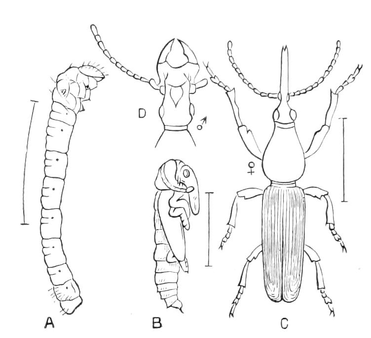
Fig. 150—Eupsalis minuta. North America. A, Larva; B, pupa; C, female imago; D, head of male. (After Riley.)
In the higher forms of Brenthidae the rostrum of the female is perfectly cylindrical and polished, and the mandibles are minute, hard, pointed processes placed at its tip. This organ is admirably adapted to its purpose; it being used for boring a hole in wood or bark, in which an egg is subsequently deposited. The males in these cases are extremely different, so that considerable curiosity is felt as to why this should be so. In some cases their head is thick, and there may be no rostrum, while large powerful mandibles are present.
In other cases the rostrum is slender, but of enormous length, so that it may surpass in this respect the rest of the body, although this itself is so drawn out as to be quite exceptional in the Insect world:[156] the antennae are inserted near the tip of the rostrum instead of near its base, as they are in the female. The size of the males is in these cases usually much larger than that of the female.[157] The males of some species fight; they do not, however, wound their opponent, but merely frighten him away. In Eupsalis it appears that the rostrum of the female is apt to become fixed in the wood during her boring operations; and the male then extricates her by pressing his heavy prosternum against the tip of her abdomen; the stout forelegs of the female serve as a fulcrum and her long body as a lever, so that the effort of the male, exerted at one extremity of the body of the female, produces the required result at the other end of her body. The New Zealand Brenthid, Lasiorhynchus barbicornis, exhibits sexual disparity in an extreme degree: the length of the male is usually nearly twice that of the female, and his rostrum is enormous. It is at present impossible to assign any reason for this; observations made at the request of the writer by Mr. Helms some years ago, elicited the information that the female is indefatigable in her boring efforts, and that the huge male stands near by as a witness, apparently of the most apathetic kind.
Coleoptera of uncertain position.
There are three small groups that it is impossible at present to place in any of the great series of beetles.
Fam. 84. Aglycyderidae.—Tarsi three-jointed, the second joint lobed; head not prolonged to form a beak. The two most important features of Rhynchophora are absent in these Insects, while the other structural characters are very imperfectly known, many parts of the external skeleton being so completely fused that the details of structure are difficult of appreciation. Westwood considered the tarsi to be really four-jointed, but it is not at all clear that the minute knot he considered the third joint is more than the articulation of the elongate terminal joint. The family consists only of two or three species of Aglycyderes, one of which occurs in the Canary Islands, and one or two in New Zealand and New Caledonia. The former is believed to live in the stems of Euphorbia canariensis; a New Zealand species has been found in connection with the tree-ferna. Cyathea dealbata
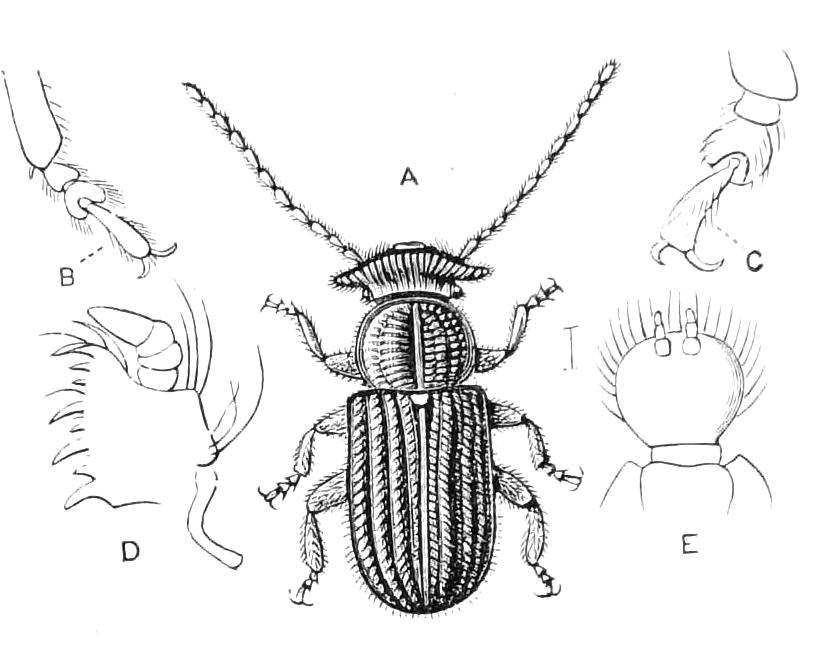
Fig. 151—Aglycyderes setifer. Canary Islands. A, Imago; B, tarsus according to Westwood; C, according to nature; D, maxilla; E, labium.
Fam. 85. Proterhinidae.—Tarsi three-jointed, the second joint lobed; head of the male scarcely prolonged, but that of the female forming a definite rostrum; maxillae and ligula entirely covered by the mentum. As in the preceding family the sutures on the under side of the head and prosternum cannot be detected. The minute palpi are entirely enclosed in the buccal cavity. There is a very minute true third joint of the tarsus, at the base of the terminal joint, concealed between the lobes of the second joint. The family consists of the genus Proterhinus; it is confined to the Hawaiian Islands, where these Insects live on dead wood in the native forests. The genus is numerous in species and individuals.
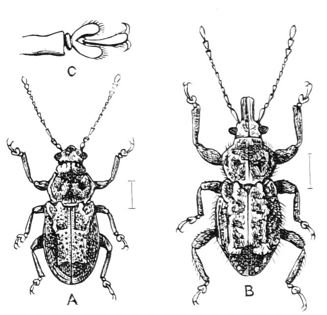
Fig. 152—Proterhinus lecontei. Hawaiian Islands. A, Male; B, female; C, front foot, more magnified.
Strepsiptera (or Rhipiptera, Stylopidae).—Male small or minute; prothorax extremely small; mesothorax moderate, the elytra reduced to small, free slips; metathorax and wings very large; nervuration of the latter radiating, without cross nervules. Female a mere sac, with one extremity smaller and forming a sort of neck or head.
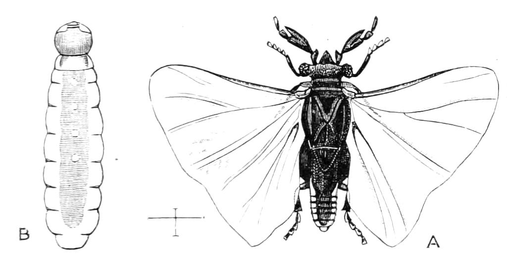
Fig. 153.—Sexes of Strepsiptera. A, Male of Stylops dalii (after Curtis); B, female of Xenos rossii (after von Siebold).
These curious Insects are parasitic in the interior of other Insects, of the Orders Hymenoptera and Hemiptera. Their structure and their life-histories entitle them to be ranked as the most abnormal of all Insects, and entomologists are not agreed as to whether they are aberrant Coleoptera or a distinct Order. The newly-hatched larva is a minute triungulin (Fig. 154), somewhat like that of Meloe; it fixes itself to the skin of the larva of a Hymenopterous Insect, penetrates into the interior, and there undergoes its metamorphoses, the male emerging to enjoy a brief period of an abnormally active, indeed agitated, existence, while the female never moves. It is important to note that these Strepsiptera do not, like most other internal parasites, produce the death of their hosts; these complete their metamorphosis, and the development of the parasite goes on simultaneously with that of the host, so that the imago of the Strepsipteron is found only in the imago of the host.[158] After the young Stylops has entered its host it feeds for a week or so on the fat-body (apparently by a process of suction), then moults and assumes the condition of a footless maggot, in which state it remains till growth is completed. At the latter part of this period the history diverges according to sex; the female undergoes only a slight metamorphic development of certain parts, accompanied apparently by actual degradation of other parts; while the male goes on to pupation, as is normal in Insects. (We may remark that the great features of the development of the sexes are parallel with those of Coccidae in Hemiptera.) When the Hymenopterous larva changes to a pupa, the larva of the Strepsipteron pushes one extremity of its body between two of the abdominal rings of its host, so that this extremity becomes external, and in this position it completes its metamorphosis, the male emerging very soon after the host has become an active winged Insect, while the female undergoes no further change of position, but becomes a sac, in the interior of which young develop in enormous numbers, finally emerging from the mother-sac in the form of the little triungulins we have already mentioned. This is all that can be given at present as a general account; many points of the natural history are still obscure, others have been merely guessed; while some appear to differ greatly in the different forms. A few brief remarks as to these points must suffice.
Bees carrying, or that have carried, Strepsiptera, are said to be stylopised (it being a species of the genus Stylops that chiefly infests bees); the term is also used with a wider application, all Insects that carry a Strepsipterous parasite being termed stylopised, though it may be a Strepsipteron of a genus very different from Stylops that attacks them. The development of one or more Strepsiptera in an Insect usually causes some deformity in the abdomen of its host, and effects considerable changes in the condition of its internal organs, and also in some of the external characters. Great difference of opinion prevails as to what these changes are; it is clear, however, that they vary much according to the species, and also according to the extent of the stylopisation. Usually only one Stylops is developed in a bee; but two, three, and even four have been observed:[159] and in the case of the wasp, Polistes, Hubbard has observed that a single individual may bear eight or ten individuals of its Strepsipteron (Xenos, n. sp.?).
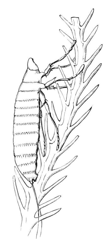
Fig. 154—Young larva of Stylops on a bee's-hair. Greatly magnified. (After Newport.)
There is no exact information as to how the young triungulins find their way to the bee-larvae they live in. Here again the discrepancy of opinion that prevails is probably due to great difference really existing as to the method. When a Stylops carried by an Insect (a Hymenopteron, be it noted, for we have no information whatever as to Hemiptera) produces young, they cover the body of the host as if it were powdered, being excessively minute and their numbers very great; many hundreds, if not thousands, of young being produced by a single Stylops. The species of the wasp genus Polistes are specially subject to the attacks of Stylops; they are social Insects, and a stylopised specimen being sickly does not as a rule leave the nest; in this case the Stylops larva may therefore have but little difficulty in finding its way to a Hymenopterous larva, for even though it may have to live for months before it has the chance of attaching itself to a nest-building female, yet it is clearly in the right neighbourhood. The bee genus Andrena has, however, quite different habits; normally a single female makes her nest underground; but in the case of a stylopised female it is certain that no nest is built, and no larvae produced by a stylopised example, so that the young triungulins must leave the body of the bee in order to come near their prey. They can be active, and have great powers of leaping, so that it is perhaps in this way possible for them to attach themselves to a healthy female bee.
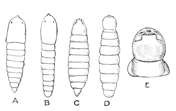
Fig. 155.—Portion of early stages of Xenos rossii. (After von Siebold.) A, Small male larva; B, small female larva; C, full-grown male larva; D, full-grown female larva; E, the so-called "cephalothorax" and adjacent segment of adult female. (The newly-hatched larva is very much like that of Stylops shown in Fig. 154.)
We have still only very imperfect knowledge as to the structure and development of Strepsiptera. Indeed but little information has been obtained since 1843.[160] Before that time the mature female was supposed to be a larva, and the triungulins found in it to be parasites. Although the erroneous character of these views has been made clear, the problems that have been suggested present great difficulties. Apparently the change from the triungulin condition (Fig. 154) to the parasitic larvae (Fig. 155, A, B) is extremely great and abrupt, and it appears also that during the larval growth considerable sexual differentiation occurs (Fig. 155, C, D); details are, however, wanting, and there exists but little information as to the later stages. Hence it is scarcely a matter for surprise that authorities differ as to which is the head and which the anal extremity of the adult female. Von Siebold apparently entertained no doubt as to the part of the female that is extruded being the anterior extremity; indeed he called it a cephalothorax. Supposing this view to be correct, we are met by the extraordinary facts that the female extrudes the head for copulatory purposes, that the genital orifice is placed thereon, and that the young escape by it. Meinert[161] contends that the so-called cephalothorax of the adult is the anal extremity, and that fertilisation and the escape of the young are effected by the natural passages, the anterior parts of the body being affected by a complete degeneration. Nassonoff, in controversion of Meinert, has recently pointed out that the "cephalothorax" of the young is shown by the nervous system to be the anterior extremity. It still remains, however, to be shewn that the "cephalothorax" of the adult female corresponds with that of the young, and we shall not be surprised if Meinert prove to be correct. The internal anatomy and the processes of oogenesis appear to be of a very unusual character, but their details are far from clear. Brandt has given some particulars as to the nervous system; though he does not say whether taken from the male or female, we may presume it to be from the former; there is a supra-oesophageal ganglion, and near it a large mass which consists of two parts, the anterior representing the sub-oesophageal and the first thoracic ganglia, while the posterior represents two of the thoracic and most of the abdominal ganglia of other Insects; at the posterior extremity, connected with the other ganglia by a very long and slender commissure, there is another abdominal ganglion.[162]
It is a matter of great difficulty to procure material for the prosecution of this study; the fact that the instars to be observed exist only in the interior of a few Hymenopterous larvae, which in the case of the bee, Andrena, are concealed under ground; and in the case of the wasps, Polistes, placed in cells in a nest of wasps, adds greatly to the difficulty. It is therefore of interest to know that Strepsiptera occur in Insects with incomplete metamorphosis. They have been observed in several species of Homoptera; and the writer has a large Pentatomid bug of the genus Callidea, which bears a female Strepsipteron apparently of large size. This bug[163] is abundant and widely distributed in Eastern Asia, and it may prove comparatively easy to keep stylopised examples under observation. Both v. Siebold and Nassonoff think parthenogenesis occurs in Strepsiptera, but there appear to be no facts to warrant this supposition. Von Siebold speaks of the phenomena of Strepsipterous reproduction as paedogenesis, or pseudo-paedogenesis, but we must agree with Meinert that they cannot be so classed.
The males of Strepsiptera live for only a very short time, and are very difficult of observation. According to Hubbard the males of Xenos dash about so rapidly that the eye cannot see them, and they create great agitation amongst the wasps in the colonies of which they are bred. Apparently they are produced in great numbers, and their life consists of only fifteen or twenty minutes of fiery energy. The males of Stylops are not exposed to such dangers as those of Xenos, and apparently live somewhat longer—a day or two, and even three days are on record. The individuals of Andrena parasitised by Stylops are apparently greatly affected in their economy and appear earlier in the season than other individuals; this perhaps may be a reason, coupled with their short lives, for their being comparatively rarely met with by entomologists.
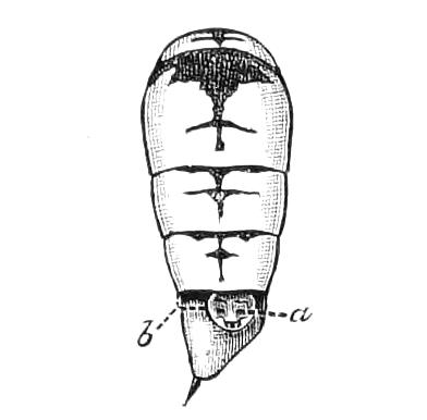
Fig. 156.—Abdomen of a wasp (Polistes hebraeus) with a Strepsipteron (Xenos ♀) in position, one of the dorsal plates of the wasp's abdomen being removed. a, Projection of part of the parasite; b, line indicating the position of the removed dorsal plate.
It is not possible at present to form a valid opinion as to whether Stylopidae are a division of Coleoptera or a separate Order. Von Siebold considered them a distinct Order, and Nassonoff, who has recently discussed the question, is also of that opinion.