ON PARASITES IN GENERAL.
Reading edition
By the term PARASITES is understood living organisms which, for the purpose of procuring food, take up their abode, temporarily or permanently, on or within other living organisms. There are both plants and animals (Phytoparasites and Zoöparasites) which lead a parasitic life in or upon other plants and other animals.
Phytoparasites are not included in the following descriptions of the forms of parasitism, but a very large number of animal parasites (zoöparasites) are described. The number of the latter, as a rule, is very much underrated. How great a number of animal parasites exists may be gathered from the fact that all classes of animals are subject to them. Some of the larger groups, such as Sporozoa, Cestoda, Trematoda and Acanthocephala, consist entirely of parasitic species, and parasitism even occurs among the vertebrates (Myxine). It therefore follows that the characteristics of parasites lie, not in their structure, but in the manner of their existence.
Parasitism itself occurs in various ways and degrees. According to R. Leuckart, we should distinguish between OCCASIONAL (temporary) and PERMANENT (stationary) PARASITISM. Occasional parasites, such as the flea (Pulex irritans), the bed-bug (Cimex lectularius), the leech (Hirudo medicinalis), and others, only seek their “host” to obtain nourishment and find shelter while thus occupied. Without being bound to the host, they usually abandon the latter soon after the attainment of their object (Cimex, Hirudo), or they may remain on the body of their host throughout their entire development from the hatching of the egg (Pediculus). It follows from this mode of living that the occasional parasites become sometimes distinguishable from their free-living relatives, though only to a slight extent. It is, therefore, seldom difficult to determine the systematic position of temporary parasites from their structure.
In consequence of their mode of life, all these temporary parasites live on the external surface of the body of their host, though more rarely they take up their abode in cavities easily accessible from the exterior, such as the mouth, nose and gills. They are therefore frequently called Epizoa or Ectoparasites; but these designations do not cover only the temporary parasites, because numerous epizoa (as for instance the louse) are parasitic during their entire life.
In contradistinction to these temporary parasites, the permanent parasites obtain shelter as well as food from their host for a long period, sometimes during the entire course of their life. They do not seek their host only when requiring nourishment, but always remain with it, thus acquiring substantial protection. The permanent parasites, as a rule, live within the internal organs, preferably in those which are easily accessible from the exterior, such as the intestine, with its appendages. Nevertheless, permanent parasites are also found in separate organs and systems, such as the muscular and vascular systems, hollow bones and brain, while some live on the outer skin. Here again, the terms Entozoa and Endoparasites do not include all stationary parasites; to the latter, for instance, the lice belong, which pass all their life on the surface of the body of their host, where they find shelter and food and go through their entire development. The ectoparasitic trematodes, numerous insects, crustacea, and other animals live in the same manner.
All “Helminthes,” however, belong to the group of permanent parasites. This term is now applied to designate certain lowly worms which lead a parasitic life (intestinal worms); but they are not all so termed. For instance, the few parasitic Turbellaria are never classed with the helminthes, although closely related to them. The turbellarians, in fact, belong to a group of animals of which only a few members are parasitic, whereas the helminthes comprise those groups of worms of which all species (Cestoda, Trematoda, Acanthocephala), or at least the majority of species (Nematoda), are parasitic. Formerly the Linguatulidæ (Pentastoma) were classed with the helminthes because their existence is also endoparasitic, and because the shape of their body exhibits a great similarity to that of the true helminthes. Since the study of the development of the Linguatulidæ (P. J. van Beneden, 1848, and R. Leuckart, 1858) has demonstrated that they are really degenerate arachnoids, they have been separated from the helminthes.
It is hardly necessary to emphasize the fact that the helminthes or intestinal worms do not represent a systematic group of animals, but only a biological one, and that the helminthes can only be discussed in the same sense as land and water animals are mentioned, i.e., without conveying the idea of a classification in such a grouping. It is true that formerly this was universally done, but very soon the error of such a classification was recognized. Still, until the middle of last century, the helminthes were regarded as a systematic group, although C. E. v. Baer (1827) and F. S. Leuckart (1827) strenuously opposed this view. Under the active leadership of J. A. E. Goeze, J. G. H. Zeder, J. G. Bremser, K. A. Rudolphi and F. Dujardin, the knowledge of the helminthes (helminthology) developed into a special study, but unfortunately it lost all connection with zoology. It required the intervention of Carl Vogt to disestablish the helminthes as one class of animals, by uniting the various groups with those of the free-living animals most closely related to them (Platyhelminthes, Nemathelminthes).
Permanent parasitism in the course of time has caused animals adopting this mode of life to undergo considerable, sometimes even striking, bodily changes, permanent ectoparasites having as yet undergone least alteration. The latter sometimes bear so unmistakably the likeness to the group to which they belong, that even a superficial knowledge of their structure and appearance often suffices for the recognition of their systematic position. For instance, though the louse, like many decidedly temporary parasites, has lost its wings—a characteristic of insects—in consequence of parasitism, yet nobody would deny its insect nature; such also occurs in other temporary parasites (Cimex, Pulex). On the other hand, the changes in a number of permanent ectoparasites (such as parasitic Crustacea) are far more considerable, and correspond with those that have occurred in permanent endoparasites.
These alterations depend partly on retrogression and partly on the acquisition of new peculiarities. In the former case, the change consists in the loss of those organs which have become useless in a permanent parasitic condition of existence, such as wings in the louse, and the articulated extremities seen in the larval stage of parasitic Crustacea. The loss of these organs goes hand in hand with the cohesion of segments of the body that were originally separate, and alterations in the muscular and nervous systems. In the same manner another means of locomotion is lost—the ciliated coat—which is possessed by many permanent parasites during their larval period. To all appearances, this character is not secondary and recently acquired, but represents a primary character inherited from free-living progenitors, and still transmitted to the altered descendants, because of its use during the larval stage (e.g., the larvæ of a great many Trematodes, the oncospheres of some Cestodes). Amongst the retrogressions, the loss of the organs of sense may be mentioned, particularly the eyes, which are still present, not only in the nearest free-living forms but also in the free-living larvæ of true parasites. It is only quite exceptionally that the eyes are subsequently retained, as a rule they are lost. Lastly, in a great many cases the digestive system also disappears, as in parasitic Crustacea, in a few nematodes and trematodes, in all cestodes and Acanthocephala. There remain at most the rudiments of the muscles of the fore-gut, but these are adapted to entirely different uses.
The new characters which permanent parasites may acquire are, first of all, the remarkably manifold CLASPING and CLINGING ORGANS, which are seldom (as in parasitic Crustacea) directly joined on to already existing structures. In those instances in which organs for the conveyance of food are retained, these likewise frequently undergo transformation, in consequence of the altered food and manner of feeding. Such alterations consist, for instance, in the transformation of a masticating mouth apparatus into the piercing and sucking organs of parasitic insects.
Hermaphroditism (as in Trematodes, Cestodes, and a few Nematodes) is a further peculiarity of many permanent parasites; moreover, the association in couples that occurs, especially in trematodes, may lead to complete cohesion and, exceptionally, also to re-separation of the sexes. In many cases the females only are parasitic, while the males live a free life, or there may be in addition the so-called complementary males. Occasionally the male alone is parasitic, and in that case lives within the female of the same species, which may live free, like certain Gephyrea (Bonellia); or the female also may be parasitic, as Trichosoma crassicaudum, which lives in the bladder of the sewer rat (Mus decumanus).
We have numerous proofs that demonstrate how considerably the original features of many parasites have become changed. We need only draw attention to the aforementioned Linguatulidæ, also to many of the parasitic Crustacea belonging to various orders. In all of these a knowledge of the larval stages—in which there is no alteration, or at most only a slight degree of change—serves to determine their systematic position, i.e., the nearest conditions of relationship.
The most remarkable changes are observed in those groups that contain only a few parasitic members, the majority leading a free life. A striking instance is afforded by a snail, the well-known Entoconcha mirabilis, Müller. This mollusc consists merely of an elongated sac living in a Holothurian (Synapta digitata). It possesses none of the characteristics of either the Gastropoda or any molluscs, and in its interior there is nothing to be observed but the organs of generation and the embryos. Nevertheless, the Entoconcha is decidedly a parasitic snail, as is clearly proved by its larvæ, but it is a snail which, in consequence of parasitism, has lost all the characteristics of molluscs in its mature condition, but still exhibits them in the early stages of development.
Certain nematodes show very clearly to what devious courses parasitism may lead. The Atractonema gibbosum, the life-history of which has been described by R. Leuckart, and which lives in the larvæ and pupæ of a dipterous insect (Cecidomyia), exhibits, in its early stage, the ordinary characteristics of other threadworms. A few weeks later—the males having died off immediately after copulation—the females are transformed into spindle-shaped bodies, the mouth and anus of which are closed. They carry with them an irregularly shaped appendage, in which the segmenting ova are situated, and in which the further conditions of life of the Atractonema are accomplished. A minute examination has demonstrated that this appendage is the prolapsed and enlarged vagina of the animal which has become merely a supplementary attachment. The conditions present in the Sphærularia, the nematoid nature of which was long undiscovered, are still more remarkable. It was only when Siebold proved that typical nematodes were hatched from their eggs that their nature was recognized. The nematodes thus produced have not the slightest resemblance to the parent.
The researches of Lubbock, A. Schneider, and more particularly of R. Leuckart, have shown that what we call Sphærularia bombi is not an animal but merely an organ—the vagina—of a nematode worm. This vagina at first grows, sac-like, from the body of the tiny nematode; it gradually assumes enormous dimensions (2 cm. in length); it contains the sexual organs and parts of the intestine. The remaining portion of the actual animal then becomes small and shrivelled; it may be easily overlooked, being but an appendage to the vagina with its independent existence, and it finally disappears altogether.
The GREAT FERTILITY of parasites is another of their peculiarities, though this may be also the case to a certain degree with some of the free-living animals, the progeny of which are likewise exposed to enormous destruction.
More remarkable, however, is the fact that the young of the endoparasites only very exceptionally grow to maturity by the side of their parents. Sooner or later they leave the organ inhabited by the parents, frequently reach the open, and after a shorter or longer period of free existence seek new hosts. During their free period, moreover, a considerable growth may be attained, or metamorphosis may take place, or even multiplication. In the exceptional cases in which the young remain within the same host, they nevertheless usually quit the organ inhabited by the parents. They likewise rarely attain maturity within the host inhabited by the parents, but only, as in other cases, after having gained access to fresh hosts.
These transmigrations play a very important rôle in the natural history of the internal parasites, but they frequently conceal the cycle of development, for sometimes there are INTERMEDIATE GENERATIONS, which themselves invade intermediate hosts. Even when there are no intermediate generations, THE SYSTEM OF INTERMEDIATE HOSTS is frequently maintained by the endoparasites.
According to the kind of food ingested by parasites, it has recently become usual to separate the true parasites from those animals that feed on the superfluity of the food of the host, or on products which are no longer necessary to him, and to call the latter MESSMATES or COMMENSALS. As examples, the Ricinidæ are thus designated, because, like actual lice, they dwell among the fur of mammals or the plumage of birds. They do not, however, suck blood, for which their mouth apparatus is unsuited, but subsist on useless epidermic scales. These epizoa, according to J. P. van Beneden, are, to a certain extent, useful to their hosts by removing deciduous materials which under certain circumstances might become harmful to them.1 This investigator, who has contributed so greatly to our knowledge of parasites, assigns the Ricines to the MUTUALISTS, under which term he comprises animals of various species which live in common, and confer certain benefits on one another. The mutualists are usually intimately connected in a mutually advantageous association known as “symbiosis.”2
Incidental and Pseudo Parasites.—In many cases the parasites are confined to certain hosts, and may therefore be designated as specific to such hosts. Thus, hitherto, Tænia solium and Tænia saginata in their adult condition have only been found in man; Tænia crassicollis only in the cat; Brandesia (Distoma) turgida and Halipegus (Distoma) ovocaudatas only in Rana esculenta, and so forth. In many other cases, however, certain species of parasites are common to several, and sometimes many, species of hosts; Dipylidium caninum is found in the domestic cat as well as in the dog; Fasciola hepatica is found in a large number of herbivorous mammals (nineteen species), Diplodiscus (Amphistomum) subclavatus in numerous urodele and ecaudate amphibia, Holostomum variabile in about twenty-four species of birds, and so on. In these cases the hosts are almost invariably closely related, belonging, as a rule, to the same family or order, or at any rate to the same class. Trichinella spiralis, which is found in man, and in the pig, bear, rat, mouse, cat, fox, badger, polecat and marten, and is capable of being artificially cultivated in the dog, rabbit, sheep, horse, in other mammals, and even in birds, is one of the most striking exceptions.
Some parasites are so strictly confined to one species of host that, even when artificially introduced into animals very closely related to their normal host, they do not thrive, but sooner or later, often very quickly, die off, and very rarely establish themselves. For example, repeated attempts have been made to rear the adult Tænia solium in the dog, or to rear Cysticercus cellulosæ in the ox, or the Cysticercus of Tænia saginata in the pig, but they have always proved unsuccessful. Only exceptionally has it been possible to transfer Cœnurus cerebralis, the larval stage of a tapeworm (Tænia cœnurus) of the dog from the brain of the sheep to that of the domestic goat. On the other hand, in the case of the Trichinellæ transference to a different host is easily accomplished.
Under natural conditions, it is not uncommon for certain kinds of specific parasites to occur occasionally in unusual hosts. Their relationship to the latter is that of INCIDENTAL PARASITES. Thus Echinorhynchus gigas, a specific parasite of the pig, is only an incidental parasite of man; Fasciola hepatica and Dicrocœlium lanceatum are specific to numerous kinds of mammals, but may be found incidentally in man. On the other hand, Dibothriocephalus latus, a specific parasite of man, may occasionally take up its abode in the dog, cat and fox. As a rule, all those parasites of man that are only rarely met with, notwithstanding that human beings are constantly being observed and examined by medical men, are termed INCIDENTAL PARASITES OF MAN. In many cases we are acquainted with the normal or specific host of these parasites. Thus we know the specific host of Balantidium coli, Eimeria stiedæ, Fasciola hepatica, Dipylidium caninum, etc.; in others the host is as yet unknown. In the latter case the question partly relates to such forms as have been so deficiently described that their recognition is impossible, partly to parasites of man in various regions of the earth, the Helminthes and parasites of which are totally unknown or only slightly known, or finally to early developmental stages that are difficult to identify. Animals that usually live free, and exceptionally become parasitic, may likewise be called incidental parasites. In this category are included a few Anguillulidæ that have been observed in man; also Leptodera appendiculata, which usually lives free, but may occasionally become parasitic in black slugs (Arion empiricorum): when parasitic it attains a larger size, and produces far more eggs than when living a free life. In order to avoid errors, the term “incidental parasites” should be confined to true parasites which, besides living in their normal host, may also live in other hosts. Leuckart speaks of FACULTATIVE PARASITISM in such forms as Leptodera. L. Oerley3 succeeded in artificially causing Leptodera (Rhabditis) pellio to assume facultative parasitism by introducing these worms into the vagina of mice, where the parasites remained alive and multiplied. Leptodera pellio dies in the intestines of mammals and man; it remains alive in frogs, but always escapes into the open with the fæces.
Recently the incidental parasites of man have also been called “Pseudo-parasites” or “Pseudo-helminthes.” Formerly, however, these terms were applied not only to living organisms that do not and cannot live parasitically, and that only exceptionally and incidentally get into man, but also to any foreign bodies, portions of animals and plants, or even pathological formations that left the human system through the natural channels, and the true nature of which was misunderstood. Frequently these bodies were described as living or dead parasites and labelled with scientific names, as if they were true parasites. A study of these errors, which formerly occurred very frequently, would be as interesting as it would be instructive. It is better not to use the expression pseudo-parasites for incidental parasites, but to keep to the original meaning, for it is not at all certain that pseudo-parasites are not described, even nowadays.
The Influence of Parasites on the Host.—In a great many cases, we are not in a position to state anything regarding any marked influence exercised by the parasite on the organism, and on the conditions of life, of the host. Most animals and many persons exhibit few signs of such influence, an exception being infestation with helminthes and certain other parasites which produce eosinophilia in the blood. As a general rule, the parasite, which is always smaller and weaker than its host, does not attempt to endanger the life of the latter, as simultaneously its own existence would be threatened. The parasite, of course, robs its host, but usually in a scanty and sparing manner, and the injuries it inflicts can hardly be taken into account. There are, however, numerous cases4 in which the situation of the parasites or the nature of their food, added to their number and movements, may cause more or less injury, and even threaten the life of the host. It stands to reason that a Cysticercus cellulosæ situated in the skin is of but slight importance, whereas one that has penetrated the eye or the brain must give rise to serious disorders. A cuticular or intestinal parasite is, as a rule, less harmful than a blood parasite. A helminth, such as an Ascaris lumbricoides or a tapeworm, that feeds on the residues of foodstuffs within the intestine, will hardly affect its host by depriving it of this material. The case is different when the parasites are very numerous, especially when the heavily infested host happens to be a young individual needing all it ingests for its own requirements, and therefore unable to sustain the drain of numerous intruders in the intestine. Disturbances also set in more rapidly when the intestinal helminthes are blood-suckers, the injury to the host resulting from the kind of food taken by the parasite.
Generally, the disorders caused by loss of chyle are insignificant when compared with those induced by the GROWTH and agglomeration of the helminthes. The latter may cause chiefly obstructions of small vessels or symptoms of pressure in affected or contiguous organs, with all those complications which may arise secondarily, or they may even lead to the complete obliteration of the organ invaded. Of course the symptoms will vary according to the nature of the organ attacked.
In consequence also of the MOVEMENTS of the parasites, disorders are set up that may tend to serious pathological changes of the affected organs. The collective migrations, undertaken chiefly by the embryos of certain parasites (as in trichinosis, acute cestode tuberculosis), are still more harmful, as are also the unusual migrations of other parasites, which, incidentally, may lead to the formation of so-called worm abscesses or to abnormal communications (fistulæ) between organs that are contiguous but possess no direct connection.
Recently, several authors have called attention to the fact that the helminthes produce substances that are TOXIC to their host; and the effects of such poisons explain the pathology of helminthiasis far more satisfactorily than the theory of reflex action.
In a number of cases these toxic materials (leucomaines) have been isolated and their effects on living organisms demonstrated by actual experiments. It also appears that the absorption of materials formed by the decomposition of dead helminthes may likewise cause toxic effects. However, our knowledge of these conditions is as yet in its initial stage.5
Nearly all the symptoms caused directly or indirectly by parasites are of such a nature that the presence of the parasites cannot be diagnosed with any certainty, or only very rarely. The most that can be done is to deduce the presence of parasites by the exclusion of other causes. Fortunately, however, there are sufficient means by which we may confirm the diagnosis in a great many cases. Such means consist not only in a minute examination of the patient by palpation, percussion and local inspection, but also in the microscopical examination of the natural secretions and excretions of the body, such as sputum, nasal mucus, urine and fæces. Though such examinations may entail loss of time, they are necessary in the interest of the patient. It appears, moreover, that quackery, which has gained considerable ground even in the treatment of the helminthic diseases of man, can thus be considerably limited.
Origin of Parasites.6—In former times, when the only correct views that existed related to the origin of the higher animals, the mode of multiplication of parasites as well as of other lowly animals was ascribed to SPONTANEOUS GENERATION (generatio æquivoca), and this opinion prevailed throughout the middle ages. The writers on natural science merely devoted their time to the interpretation of the views of the old authors, and perpetuated the opinions of the ancients on questions, which, even in those days, could have been correctly explained merely by observation.
It was only when observations were again recommenced, and the microscope was invented, that the idea of spontaneous generation became limited. Not only did the microscope reveal the organs of generation or their products (eggs) in numerous animals, but Redi succeeded in proving that the so-called Helcophagi (flesh maggots) are only the progeny of flies, and never appear in the flesh of slaughtered animals when fully developed flies are prevented from approaching and depositing their eggs on it. Swammerdam likewise knew that the “worms” living in the caterpillars of butterflies were the larvæ of other insects (ichneumon flies) which had laid their eggs in their bodies; he also discovered the ova of lice. The two authors mentioned were, however, unwilling to see that the experience they had gained regarding insects applied to the helminthes. Leeuwenhoek also vehemently opposed the theory of a spontaneous generation, maintaining that, on a basis of common-sense, eggs, or at all events germs, must exist, even though they could not be seen.
The use of the microscope also revealed a large number of very small organisms in the water and moist soil, some of which undoubtedly resembled helminthes. Considering the wide dissemination of these minute organisms, it was natural to conjecture that after their almost unavoidable introduction into the human system they should grow into helminthes (Boerhave, Hoffmann). Linnæus went even further, for he traced the descent of the liver-fluke of sheep from a free-living planaria (Dendrocœlum lacteum), the Oxyuris vermicularis from free-living nematodes, and the Tænia lata (i.e., Dibothriocephalus latus) from a tapeworm (Schistocephalus solidus) found free in the water. Linnæus’ statements met with general approval. However, we must bear in mind that at that time the number of helminthes known was very small, and many of the forms that we have long ago learned to differentiate as specific were then regarded as belonging to one species. Linnæus’ statements were partly supported by similar discoveries by other investigators, such as Unzer, and partly also by the discovery of eggs in many helminthes. It was believed that the eggs hatched in the outside world gave rise to free-living creatures, and that these, after their introduction into the intestine, were transformed into helminthes. By means of these eggs the old investigators tried to explain the HEREDITARY TRANSMISSION of the intestinal worms, which was universally believed until the commencement of the last century. Some authors went so far as to regard the intestinal worms as congenital or inherited; they maintained the possibility of direct transmission, as in suckling, and denied that the eggs reaching the external world had anything to do with the propagation of the parasites.
The more minute comparison between the supposed free-living stages of the helminthes and their adult forms, and the impossibility of finding corresponding free forms for the ever-increasing number of parasitic species, revealed the improbability of Linnæus’ statements (O. Fr. Müller). It was the latter author also who recognized the origin of the tapeworms (Schistocephalus, Ligula) found free in the water. They originate from fishes which they quit spontaneously.
However, in spite of the fact that van Doeveren and Pallas correctly recognized the significance of the eggs in the transmission of intestinal worms, these statements remained disregarded, as did Abildgaard’s observation, experimentally confirmed, that the (immature) cestodes from the abdominal cavity of sticklebacks became mature in the intestines of aquatic birds. Moreover, at the end of the eighteenth and the commencement of the nineteenth centuries, after helminthology had been raised to a special branch of study by the successful results of the investigations of numerous authors (Goeze, Bloch, Pallas, Müller, Batsch, Rudolphi, Bremser), many of whom experienced a “divine joy” in searching the intestines of animals for helminthes, some authors reverted to generatio æquivoca, without, however, entirely denying the existence of organs of generation and eggs. The fact that a few nematodes bore living progeny—a fact of which Goeze was already aware—had no influence on the erroneous opinion, as in such cases it was considered that the young continued to develop beside the old forms. There were also many helminthes known that never developed sexual organs and never produced eggs, and which therefore were referred to generatio æquivoca. People were convinced that the intestinal mucous membrane or an intestinal villus could transform itself into a worm, either in a general morbid condition of the body, or in pathological changes of a more local character. The appearance of helminthes was even regarded as useful and as a means for the expulsion of injurious matter.
These views, firmly rooted and supported by such eminent authorities as Rudolphi and Bremser, could not easily be overthrown. First, a change took place in the knowledge of the trematodes. In 1773, O. Fr. Müller discovered Cercariæ living free in water. He regarded them as independent creatures and gave them the name that is still used at the present time. Nitzsch, who also minutely studied these organisms and who recognized the resemblance of the anterior part of their bodies to a Fasciola, did not, however, arrive at a correct conclusion. He regarded the combination rather as that of a Fasciola with a Vibrio, for which he mistook the characteristic tail of the cercaria. He also noticed the encystment (transformation into the “pupa”) on foreign bodies of many species of these animals, but was of opinion that this process signified only the termination of life.
Considerable attention was attracted to the matter when Bojanus first published a paper entitled “A Short Note on Cercaria and their Place of Origin.” He pointed out that the cercariæ creep out of the “royal yellow worms,” which occur in freshwater snails (Limnæa, Paludina), and are probably generated in these worms.
Oken, in whose journal, Isis (1818, p. 729), Bojanus published his discovery, remarks in an annotation, “One might lay a wager that these Cercariæ are the embryos of Distomes.” Soon after (1827), C. E. v. Baer was able to confirm Bojanus’ hypothesis that the cercariæ as a “heterogeneous brood” originated from spores in parasitic tubes in snails (germinating tubes). Moreover, Mehlis (Isis, 1831, p. 190) not only discovered the opercula of the ova of Distoma, but likewise saw the infusorian-like embryo emerge from the eggs of Typhlocœlum (Monostomum) flavum and Cathæmasia (Distoma) hians. A few years later (1835) v. Siebold observed the embryos (miracidia) of the Cyclocœlum (Monostomum) mutabile, and discovered in their interior a cylindrical body that behaved like an independent being (“necessary parasite”), and was so similar in appearance to the “royal yellow worms” (Bojanus) that Siebold considered the origin of the latter from the embryos of trematodes as, at all events, possible. Meanwhile, v. Nordmann of Helsingfors had in 1832 seen the miracidia of flukes provided with eyes swimming in water; v. Siebold (1835) had observed the embryos, or oncospheres, of tapeworms furnished with six hooklets in the so-called eggs of the Tænia; while Creplin (1837) had discovered the “infusorial” young of the Diphyllobothrium (Bothriocephalus) ditremum, and conjectured that similar embryos were to be found in other cestodes with operculated eggs. At all events, the fact was established that the progeny of the helminthes appeared in various forms and was partly free living. The researches of Eschricht (1841) were likewise of influence, as they elucidated the structure of the Bothriocephali, and proved that the encysted and sexless helminthes were merely immature stages.
J. I. Steenstrup (1842) was, however, the first to furnish explanations for the numerous isolated and uncomprehended discoveries. Commencing with the remarkable development of the Cœlenterata, he established the fact that the Helminthes, especially the endoparasitic trematodes, multiply by means of alternating and differently formed generations. Just as the polyp originating from the egg of a medusa represents a generation of medusæ, so does the germinal tube (“royal yellow worm”) originating from the ciliated embryo of a Distoma, etc., represent the cercaria. These were consequently regarded as the progeny of trematodes, and Steenstrup, guided by his observations, conjectured that the cercaria, whose entrance into the snails he had observed accompanied by the simultaneous loss of the propelling tail, finally penetrated into other animals, in which they became flukes.
Part of this hypothetical cycle of development was erroneous, and in other particulars positive observation was lacking, but the path pursued was in the right direction. Immediately after the appearance of Steenstrup’s celebrated work, v. Siebold expressed his opinion that the encapsuled flukes certainly had to travel, i.e., to be transmitted with their bearers into other hosts, before becoming mature. This view was experimentally confirmed by de Filippi, La Valette St. George (1855), as well as by Pagenstecher (1857), while the metamorphosis of the ciliated embryo of Distoma into a germinal tube was first seen by G. Wagener (1857) in Gorgodera (Distoma) cygnoides of frogs. All that we have subsequently learned from the works of numerous investigators about the development of endoparasitic trematodes has certainly increased our knowledge in various directions, and, apart from the deviating development of the Holostomidæ has, as a whole, confirmed the briefly sketched cycle of development.
Steenstrup’s work on the cestodes did not attract the same attention as his work on trematodes. Steenstrup always insisted on the “nurse” nature of the cysticerci and other bladder-worms. Abildgaard (1790), as well as Creplin (1829 and 1839), had already furnished the information that certain sexless cestodes (Schistocephalus and Ligula) from the abdomen of fishes only become mature after their transference to the intestine of aquatic birds. These passive migrations were confirmed in an entire series of other cestodes, particularly by v. Siebold (1844, 1848, 1850) and E. J. van Beneden (1849), not by actual experiment, but by undoubted observation.
It was correctly believed that the ova or oncospheres penetrate into certain intermediate hosts, in which they develop into unsegmented larvæ. Here they remain until, with their host, they are swallowed by some predacious animal. They then reach the intestine, being freed from the surrounding membranes through the process of digestion, and settle themselves there to form the adult chain of proglottides. Though some few scientists, such as P. J. van Beneden and Em. Blanchard, deduced from these observations that the bladder-worms (Cysticerci), which had hitherto been regarded as a separate class of helminthes, were only larval Tæniæ, this correct view was not at first universally accepted. The foundation was too slight, and van Beneden was of opinion that the Cysticerci were not necessary, but only appeared incidentally.
v. Siebold was a strenuous opponent to this theory, notwithstanding his experiences on the change of hosts of the Tetrarhynchus. Together with Dujardin (1850) he conjectured that the Tæniæ underwent a deviating cycle of development. He was of opinion that the six-hooked oncospheres left the intestine, in which the older generation lived, and were scattered about with the fæces, and finally re-entered per os (i.e., with water and food) a host similar to the one they had left, in the intestine of which they were directly transformed into tapeworms. A change of host such as occurred in other cestodes was not supposed to take place (the history of the cestodes was at this time not entirely established). As the oncospheres of the Tænia are enveloped in one calcareous or several softer coverings which they cannot leave actively, and as, in consequence of this condition, innumerable oncospheres cannot penetrate into an animal, and others cannot reach the proper animal, v. Siebold conceded, at least for the latter, the possibility of a further development. But this was only supposed to occur because they had either invaded wrong hosts, or, having reached the right hosts, had penetrated organs unsuitable to their development, and had thus gone astray in their travels, and had become hydropically degenerated tæniæ. This was v. Siebold’s explanation of bladder-worms. Naturally, v. Siebold himself conjectured that a recovery of the diseased tapeworm might occur, in a few exceptional cases, after transmission into the correct host, as, for instance, in the Cysticercus fasciolaris of mice, the host of which is the domestic cat, and in which there is a seemingly normally developed piece of tapeworm situated between the caudal vesicle and the cysticercus head.
Guided by correct views, F. Küchenmeister undertook in Zittau the task of confirming the metamorphosis of Cysticercus pisiformis of hares and rabbits, into tapeworms in the intestine of the dog by means of feeding experiments. The first reports on the subject, published in 1851, were not likely to meet with universal approval, because Küchenmeister first diagnosed the actual tapeworm he had been rearing as Tænia crassiceps, afterwards as Tænia serrata, and finally as Tænia pisiformis n. sp. However, in any case, Küchenmeister, by means of the reintroduction of experimental investigation, rendered a great service to helminthology.
The publication of Küchenmeister’s works induced v. Siebold to undertake similar experiments (1852 and 1853), which were partly published by his pupil Lewald in 1852. But the positive results obtained hardly changed Siebold’s opinion, for although he no longer considered the bladder-worms as hydropically degenerated tapeworms, he still regarded them as tæniæ that had strayed. The change of opinion was partly due to an important work of the Prague zoologist, v. Stein (1853). He was able to examine the development of a small bladder-worm in the larvæ of the well-known meal-worm (Tenebrio molitor) and to demonstrate that, as Goeze had already proved in the case of Cysticercus fasciolaris of mice, first the caudal vesicle is formed and then the scolex, whereas Siebold believed that in bladder-worms the posterior end of the scolex was formed first, and that this posterior end underwent a secondary hydropic degeneration.
In opposition to v. Siebold, Küchenmeister successfully proved the necessity of the bladder-worm stage by rearing tapeworms in dogs from the Cysticercus tenuicollis of domestic mammals and from the Cœnurus cerebralis of sheep. He, and simultaneously several other investigators independently, succeeded, with material provided by Küchenmeister, in rearing the Cœnurus cerebralis in sheep from the oncospheres of the Tænia cœnurus of the dog (1854). R. Leuckart obtained similar results in mice by feeding them with the mature proglottides of the Tænia crassicollis of cats (1854).
Küchenmeister also repeatedly reared the Tænia solium of man from the Cysticercus cellulosæ of pigs (1855), and from the embryos of this parasite P. J. van Beneden succeeded in obtaining the same Cysticercus in the pig (1854). As Küchenmeister distinguished the Tænia mediocanellata, known to Goeze as Tænia saginata, amongst the large tæniæ of man (1851), so it was not long before R. Leuckart (1862) succeeded in rearing the cysticercus of the hookless tapeworm in the ox. It is particularly to this last-named investigator that helminthology is indebted more than to any other author. He followed the gradual metamorphosis from oncospheres to cystic worms in all its details.
In view of all the researches that were made, and which are too numerous to mention individually, the idea that bladder-worms are abnormal or only incidental forms had to be abandoned. Everything pointed to the fact that in all cestodes the development is divided between two kinds of animals; in one—the host, the adult tapeworm is found; while in the other, the intermediate host, we find some form or other of an intermediate stage (cysticercus in the broadest sense). The practical application of this knowledge is self-evident. If no infected pork or beef is ingested, no tapeworm can be acquired, and also the rearing of cysticerci in the human body is prevented by avoiding the introduction of the eggs of tapeworms.
Though these results were definitely proved by numerous researches, yet they have been repeatedly challenged, notably by J. Knoch (1862) in Petrograd, who, on the basis of experiments, sought to confirm a direct development without an intermediate host and ciliated stage, at all events as regards Dibothriocephalus latus. However, the repeated communications of this author met with but little favour from competent persons, partly because the experiments were conducted very carelessly, and partly because their repetition on dog and man (R. Leuckart) had no results (1863). It was only in 1883 that Braun was able to prove that the developmental cycle of Dibothriocephalus latus is similar to that of other Cestodes. The results obtained in other places by Parona, Grassi, Ijima and Zschokke render any discussion of Küchenmeister’s conclusions unnecessary.7 Long after Knoch, a French author, P. Mégnin, also pleaded for the direct development of some cestodes, and especially some tæniæ. He (1879) also sought to prove a genetic connection between the hookless and armed tapeworms of mammals, but the arguments he adduced, so far as they rest on observations, can be easily refuted or attributed to misinterpretation. Only one of these arguments is correct, namely, that the number of the species of tæniæ with which we are acquainted is far larger than that of the corresponding cystic forms; but this disparity alone cannot be taken as a proof of direct development. It can only be said that our knowledge in this respect is deficient. As a matter of fact, we have during recent years become acquainted with a large number of cystic forms, hitherto unknown, belonging to tæniæ which have long been familiar. It must also be borne in mind that no man in his lifetime can complete an examination for bladder-worms of the large number of insects, for instance, which may destroy an entire generation of an insectivorous species of bird within a small district.
Naturally it does not follow that direct development in the cestodes is altogether lacking. The researches of Grassi (1889) have furnished an example in Hymenolepis (Tænia) murina, which shows that development may sometimes take place without an intermediate host, notwithstanding the retention of the cystic stage. It was found that the oncospheres of this species, introduced into rats of a certain age, after a time grow into tapeworms without leaving the intestine, but not directly, for they bore into the intestinal wall, where they pass the cystic stage, the cysts afterwards falling into the intestinal lumen, where they develop into tapeworms. The recent experiments of Nicoll (1911) show that the larval stages of Hymenolepis murina also occur in the rat-flea, Ceratophyllus fasciatus.
Important observations were soon made on the remaining groups of helminthes. The discussion on the origin of parasites soon became confined to the helminthes. Amongst the Nematoda, it had long been known that encapsuled forms existed that had at first been regarded as independent species, but very soon they were pronounced to be immature forms, in consequence of their lack of sexual organs. Though Dujardin and also v. Siebold regarded them as “strayed” animals, v. Stein (1853) very promptly demonstrated that the progeny of the nematodes were destined to travel by discovering a perforating organ in the larval nematodes of the mealworm. This was first experimentally confirmed (1860) by R. Leuckart, R. Virchow and Zenker, all of whom succeeded not only in bringing to maturity the muscle Trichinæ (known since 1830) in the intestine of the animals experimented upon, but were likewise able to follow the migrations of the progeny. Of course, the encapsulating brood remained in the same organism, and in this respect deviated from the broods of other helminthes which escape into the outer world and find their way into other animals, but the encapsuled nematodes could no longer be regarded as the result of straying. Subsequently, R. Leuckart worked out, more or less completely, the history of the development of numerous nematodes, or pointed out the way in which further investigations should be made. It has been found that in nematodes far more frequently than in other helminthes, the typical course of development is subject partly to curtailment and partly to complications, which sometimes considerably increase the difficulties of investigation and have hitherto prevented the attainment of a definite conclusion, though the way to it is now clear.
In a similar manner the works of R. Leuckart have cleared up the development of the Acanthocephala and Linguatulida. Of course, much still remains to be done. So far, we do not even know all the helminthes of man and of the domestic animals in all their phases of life, and still less is known of those of other animals. We are indebted to the discoveries of the last fifty years for the knowledge arrived at, though comparatively few names are connected with it. The gross framework is revealed, but the gaps have only been filled up here and there. However, we may trustfully leave the completion of the whole to the future, without fear that any essential alterations will take place.
The deductions to be drawn are as follows: That the helminthes like the ectoparasites multiply by sexual processes, that the entire course of development of the helminthes is rarely or never gone through in the same host as is the case with several ectoparasites, that the progeny at an earlier or later stage of development, as eggs, embryos, or larvæ, quit the host inhabited by the older generation, and almost always attain the outer world: only in Trichinella does the development take place directly in the definite host. Where the eggs have not yet developed they go through the embryonic evolution in the outer world. The young larvæ are transmitted, either still enclosed within the egg or embryonic covering, to the intermediate host or more rarely they are transferred straight to the final host. In other cases they may hatch out from their envelopes, and after a longer or shorter period of free life, during which they may partake of food and grow, they, as before, penetrate, usually in an active way, into an intermediate host, or at once invade the final host. Exceptionally (e.g., Rhabdonema), during the free life there may be a propagation of the parasitic generation, and in this case only the succeeding generation again becomes parasitic, and then at once reaches its final host. The young forms which have invaded the final host become mature in the latter, or after a longer or shorter period of parasitism again wander forth (as the Œstridæ, Ichneumonidæ, etc.), and reach the adult stage in the outer world. The young stages, during which the parasites undergo metamorphoses or are even capable of producing one or several intermediate generations, are passed in the intermediate hosts until, as a rule, they are passively carried into the final host and there complete their cycle of development by the formation of the organs of generation. This mode of development, the spending of life in two different kinds of animals (intermediate and final host), is typical of the helminthes. This is manifested in the Acanthocephala, the Cestoda, the majority of the endoparasitic Trematoda, a number of the Nematoda, and the Linguatulidæ. There are now and then exceptions, however, in which, for instance, the host and intermediate host change order (Trichinella, Hymenolepis murina).
Parasites are hardly ever inherited amongst animals.8 According to a few statements, however, Trichinella and Cœnurus are supposed to be transmissible from the infected mother to the fœtus. Otherwise most animals acquire their parasites, especially the Entozoa, from without, the parasites penetrating either actively, as in animals living in the water, or passively with food and drink. A particular predisposition to worms is not more likely than a spontaneous origin of parasites.
Derivation of Parasites.—Doubt now no longer exists as to the derivation of the temporary and of many of the stationary ectoparasites from free-living forms. This conclusion is founded on the circumstance that not only are there numerous intermediate degrees in the manner of living and feeding between predacious and parasitic animals, but that there is more or less uniformity in their structure. The differences that exist are easily explained as consequences of altered conditions of life. The case is more difficult in regard to groups that are exclusively parasitic (Cestoda, Trematoda, Acanthocephala, Linguatulidæ, and Sporozoa), or groups that are chiefly parasitic (Nematoda), because in these cases the gulf that divides these forms from free-living animals is wider. It is true that we know that the nearest relatives of the Linguatulidæ are found amongst the Arachnoidea, and indeed in the Acarina; that, moreover, the structure and development of the Sporozoa refers them to the Protozoa, and allows some of them to be regarded as the descendants of the lowest Rhizopoda. We know that the Trematoda, and through these the Cestoda, are closely related to the Turbellaria, from which they may be traced. The Nematoda, and still more the Acanthocephala, stand apart. This is less evident, however, in the Nematoda, for there are numerous free-living members of these from which it is possible that the parasitic species may be descended. Indeed, this seems more than probable if such examples as Leptodera, Rhabdonema and Strongyloides are taken into consideration, as well as the conditions of life of free-living nematodes. These mostly, if not exclusively, spend their lives in places where decomposing organic substances are present in quantities; some species attain maturity only in such localities, and there propagate very rapidly. Should the favourable conditions for feeding be changed, the animals seek out other localities, or they remain in the larval form for some time until more favourable conditions set in. It is comprehensible that such forms are very likely to adopt a parasitic manner of life which at first is facultative (Leptodera, Anguillula), but may be regarded as the transition to true parasitism. The great advantages attached to a parasitic life consist not only in protection, but also in the supply of suitable food, and consequently in the easier and greater production of eggs, and thus fully account for the gradual passage of facultative parasitism into true parasitism. In many forms the young stages live free for some time (Strongylidæ), in others, as is the case in Rhabdonema, parasitic and free-living generations alternate; in others, again, the free period is limited to the egg stage or entirely suppressed.
Though it is possible thus to connect the parasitic with the free-living nematodes, by taking their manner of life into account, this matter presents greater difficulties in regard to other helminthes. It is true that the segmented Cestoda may be connected with and traced from the less known and interesting single-jointed Cestoda (Amphilina, Archigetes, Caryophyllæus, Gyrocotyle). Trematodes are all parasites, with the exception of one group, Temnocephalidæ, several genera and species of which live on the surface of the bodies of Crustacea and turtles of tropical and sub-tropical freshwaters. Temnocephalidæ are, nevertheless, predacious. They feed on Infusoria, the larvæ of small insects and Crustacea. So far as is known they do not nourish themselves on part of the host. They belong to the group of commensals, or more correctly, to that of the SPACE PARASITES, which simply dwell with their host and do not even take a portion of the superfluity of its food. However, space parasitism may still be regarded as the first stage of commensalism, which is again to be regarded as a sort of transition to true parasitism.
It is possible that parasitism came about in this way in the trematodes, in which connection we must first consider the turbellaria-like ancestors of the trematodes. Much can be said in favour of such a genetic relationship between turbellaria and trematodes, and hardly anything against it. It should also be remembered that amongst the few parasitic turbellaria there are some that possess clinging discs or suctorial pores, and these are only differentiated from ectoparasitic trematodes by the possession of a ciliated integument, which is found only in the larval stages of the latter.
The Acanthocephala occupy an isolated position. Most authors certainly regard them as related to the nematodes; in any case, the connection is not a close one, and the far-reaching alterations which must have occurred prevent a clear view. Perhaps the free original forms of Acanthocephala are no longer in existence, but that such must have existed is a foregone conclusion.
An explanation of the CHANGE OF HOST so frequent in parasites is more difficult than that of their descent. R. Leuckart is of opinion that the present intermediate hosts, which belong principally to the lower animals, were the original hosts of the parasites, and fostered both their larval and adult stages. It was only in course of time that the original hosts sank to the position of intermediate hosts, the cause for this alteration being that the development of parasites, especially of the helminthes, through further development and differentiation extended over a larger number of stages. The earlier stages remained in their original hosts, but the later stages sought out other hosts (higher animals). To prove this, Leuckart points out that the mature stages of the helminthes, with but few exceptions, occur only in the vertebrates which appeared later in the development of the animal kingdom, while the great majority of intestinal worms of the lower animals only represent young stages, which require transmission into a vertebrate animal before they can become mature. The few helminthes that attain maturity in the lower animals (Aspidogaster, Archigetes) are therefore regarded by Leuckart as primitive forms, and he compares them with the developmental stages of helminthes, Aspidogaster with rediæ, Archigetes with cysticercoids. He classes the nematodes that become mature in the invertebrates with Anguillulidæ, i.e., with saprophagous nematodes from which the parasitic species descend.
Leuckart therefore regards the change of hosts as secondary, so does Sabatier. The latter, however, adduces other reasons for this (lack of clinging organs and the necessity to develop them in an intermediary stage); but in this connection he only considers the Cestoda. In opposition to Leuckart, R. Moniez, however, is convinced that the migrations of the helminthes, as well as the system of intermediate hosts, represent the original order of things. Moniez traces all Entozoa from saprophytes, but only a few of these were able to settle directly in the intestine and there continue their development. These are forms that at the present day still lack an intermediate host, such as Trichocephalus, Ascaris, and Oxyuris. In most other cases the embryos, however, consisted of such saprophytes as were, in other respects, suitable to become parasites, but were incapable of resisting the mechanical and chemical influences of the intestinal contents. They were therefore obliged to leave the intestine at once, and accomplished this by penetrating the intestinal walls and burrowing in the tissues of their carriers. In this position, assisted by the favourable conditions of nutrition, they could attain a relatively high degree of development. Mechanical reasons prevented a return to the intestines, where the eggs could be deposited. Most of them doubtless died off as parasites, as also their young stages do at present when they penetrate wrong hosts. Some of them, nevertheless, passively reached the intestine of beasts of prey. Many were destroyed in the process of mastication; for a small part, however, there was the chance of reaching the intestine of a beast of prey undamaged, and there, having become larger and more capable of resistance, maturity was attained. By means of this incidental coincidence of various favourable circumstances, these processes, according to Moniez, have been established by heredity and have become normal.
This is not the place to express an opinion either for or against the various hypotheses advanced, but the existence of these diametrically opposed views alone will show the great difficulty of the question. Independently, however, it appears more natural to come to the conclusion that parasitism, as well as change of hosts, were gradual transitions.
As a conclusion to this introductory chapter, a list of some of the most important works on the parasitology of man and animals is appended.
LITERATURE.
Goeze, J. A. E. Versuch einer Naturgeschichte der Eingeweidewürmer thierischer Körper. Blankenburg, 1782. 4to, 471 pp., with 44 plates.
Zeder, J. G. H. Erster Nachtrag zur Naturgeschichte der Eingeweidewürmer. von J. A. E. Goeze. Leipzig, 1800. 4to, with 6 tables.
Rudolphi, C. A. Entozoorum sive vermium intestinalium historia naturalis. I, Amstelod., 1808; ii, 1809. 8vo, with 18 plates.
Rudolphi, C. A. Entozoorum synopsis. Berol., 1819. 8vo, with 3 plates.
Bremser, J. G. Ueber lebende Würmer im lebenden Menschen. Wien, 1819. 8vo, with 4 plates.
Bremser, J. G. Icones helminthum, systema Rudolphii entozoologicum illustrantes. Viennae, 1824. Fol. (Paris, 1837).
Dujardin, F. Histoire naturelle des helminthes ou vers intestinaux. Paris, 1845. 8vo, with 12 plates.
Diesing, C. M. Systema helminthum. 2 vols. Vindobonnae, 1850, 1851. 8vo. Supplements by the same author: Revision der Myzhelminthen (Report of the Session of the Imp. Acad. of Science. Wien, xxxii, 1858); with addendum (ibid., xxxv, 1859); Revision der Cephalocotyleen (ibid., xlix, 1864, and xlviii, 1864); Revision der Nematoden (ibid., xlii, 1861); Supplements (ibid., xliii, 1862).
Beneden, P. J. VAN. Mémoire sur les Vers intestinaux. Paris, 1858. 4to, with 12 plates.
Küchenmeister, F. Die in und an dem Körper des lebenden Menschen vorkommenden Parasiten. Leipzig, 1855. 8vo, with 14 plates.
Leuckart, R. Die menschlichen Parasiten und die von ihnen herrührenden Krankheiten. I, Leipzig, 1863; II, Leipzig, 1876. 8vo.
Cobbold, T. Sp. Entozoa; an Introduction to the Study of Helminthology. London, 1864. 8vo. Supplement, London, 1869.
Davaine, C. Traité des entozoaires et des maladies vermineuses de l’homme et des animaux domestiques. 2nd edit. Paris, 1877. 8vo.
Linstow, O. V. Compendium der Helminthologie, ein Verzeichniss der bekannten Helminthen, die frei oder in thierischen Körpern leben, geordnet nach ihren Wohnthieren, unter Angabe der Organe, in denen sie gefunden sind, und mit Beifügung der Litteraturquellen. Hanov., 1878. 8vo. Supplement, including the years 1878–1888, Hanov., 1888.
Cobbold, T. Sp. Parasites; a Treatise on the Entozoa of Man and Animals, including some Account of the Entozoa. London, 1879. 8vo.
Leuckart, R. Die Parasiten des Menschen und die von ihnen herrührenden Krankheiten. 2nd edit. Leipzig, 1879–1886. The Protozoa, Cestodes, Trematodes and Hirudinea have hitherto appeared (continued by Brandes).
BÜtschli, O. Protozoa in Bronn’s Klass. u. Ordn. d. Thierreichs. Vol. i, Leipzig, 1880–1889. 8vo, with 79 plates.
Braun, M. Trematodes in Bronn’s Klass. u. Ordn. d. Thierreichs. Vol. iv, 1, Leipzig, 1879–1893. 8vo, with 33 tables. (The first thirteen sheets, comprising the history of the worms up to 1830, were compiled by H. Pagenstecher.)
Zürn, F. A. Die thierischen Parasiten auf und in dem Körper unserer Haussäugethiere, sowie die durch erstere veranlassten Krankheiten, deren Behandlung und Verhütung. 2nd edit. Weimar, 1882. 8vo, with 4 plates.
Cobbold, T. Sp. Human Parasites; a Manual of Reference to all the Known Species of Entozoa and Ectozoa. London, 1882. 8vo.
Küchenmeister, F., and F. A. Zürn. Die Parasiten des Menschen. 2nd edit. Leipzig, 1888., 8vo, with 15 plates.
Blanchard, R. Traité de zoologie médicale. I, Paris, 1889; II, 1890. 8vo.
Neumann, L. G. Traité des maladies parasitaires non microbiennes des animaux domestiques. 2nd edit. Paris, 1892. 8vo. English edit., translated by G. Fleming. 2nd edit., revised by J. Macqueen. 1905. London: Baillière, Tindall and Cox.
Looss, A. Schmarotzerthum in der Thierwelt. Leipzig, 1892. 8vo.
Railliet, A. Traité de zoologie médicale et agricole. 2nd edit. I, Paris, 1895. 8vo.
Parona, C. L’elmintologia italiana da’ suoi primi tempi all’ anno 1890. Genova, 1894. 8vo.
Braun, M. Cestoda in Bronn’s Klass. u. Ordn. d. Thierreichs. Vol. iv, 2, Leipzig, 1894–1900. 8vo, with 24 plates.
Mosler, F., and E. Peiper. Thier Parasit. (Spec. Path. u. Ther. v. H. Nothnagel. Vol. vi.) Wien, 1894. 8vo, with 124 illustrations.
Laveran, A., et R. Blanchard. Les hématozoaires de l’homme et des anim. Paris, 1895. 12mo, with 30 figs.
Sluiter, C. R. De dierl. paras. v. d. mensch en van onze huisdier. Haag, 1895. 8vo.
Blanchard, R. Malad. parasit., paras. animaux, paras. végét. à l’exclus. des bacter. (Traité de pathol. gén. de Ch. Bouchard, vol. ii.) Paris, 1895. 8vo, with 70 figs.
Huber, J. Ch. Bibliographie der klin. Helminthol. München, 1895. 8vo. With Supplement, 1898, and continued as Bibl. d. klin. Entomol. München, 1899–1900.
Moniez, R. Traité de parasitol. anim. et veget. appl. à la médecine. Paris, 1896. 8vo, with 116 figs.
Weichselbaum. Parasitologie (Weil’s Handb. d. Hyg.). Jena, 1898. 8vo, with 78 illustrations.
Kraemer, A. Die thierischen Schmarotzer des Auges (Gräfe and Sämische’s Handb. d. ges Augenheilk.). Leipzig, 1899. 8vo, with 16 illustrations.
Cholodkowsky, N. A. Icones helm. hominis. St. Petersburg, 1898–99. Fol. (atlas with 15 plates).
Perroncito, E. I parassiti dell’ uomo e degli animali utili e le più comuni malattie da essi prodotti. II_{a} ed. Milano 1902. 8o. con 276 fig. e 25 tav.
Stiles, Ch. W. and A. Hassall. Index Catalogue of Medicine and Veterinary Zoology. Washington, 1902 (U.S. Dept. of Agric., Bur. of Anim. Ind., Bull. No. 39).
Neveu-Lemaire, M. Précis de parasitologie humaine, parasites végétaux et animaux. 4e édit. Paris, 1911.
Hofer, B. Handbuch der Fischkrankheiten. München, 1904. 8o. 18 Taf. 222 Abb.
Guiart, J., and L. Grimbert. Précis de Diagnostic chimique, microscopique et parasitologique. Paris, 1906. With 500 figs.
Ostertag, R. Handbuch der Fleischbeschau. V. Aufl. mit 265 Abb. Stuttgart, 1904.
Stiles, Ch. W. The International Code of Zoological Nomenclature as applied to Medicine (Hygienic Lab., Bull. No. 24, Washington, 1905).
Stiles, C. W., and Hassall, A. Trematoda and Trematode Diseases. (Index Catalogue of Med. and Vet. Zoology.) Hygienic Lab., Bull. No. 37, Washington, 1908.
Stiles, C. W., and Hassall, A. Cestoda and Cestodaria. Hygienic Lab., Bull. No. 85, Washington, 1912.
Laloy, L. Parasitisme et mutualisme dans la nature. Paris, 1906. 8vo, 284 pp., 82 figs.
Theobald, F. V. A Monograph of the Culicidæ of the World. 5 vols. and plates. 1901–1910. London: Brit. Museum, Nat. Hist.
James, S. P., and Liston, W. G. The Anopheline Mosquitoes of India. 2nd edit. 1911. Calcutta: Thacker, Spink and Co.
Howard, L. O., Dyar, H. G., and Knab, F. The Mosquitoes of North and Central America and the West Indies. 2 vols. 1912. Washington: Carnegie Institution.
Austen, E. E. African Blood-sucking Flies. 1909. London: Brit. Museum, Nat. History.
Austen, E. E. A Handbook of Tsetse-flies. 1911. London: Brit. Museum, Nat. History.
Castellani, A., and Chalmers, A. J. Manual of Tropical Medicine. 2nd edit. 1,747 pp. 1913. London: Baillière, Tindall and Cox.
Kolle and Wassermann. Handbuch der pathogenen mikroorganismen. Jena: Gustav Fischer.
Minchin, E. A. An Introduction to the Study of the Protozoa. 1912. London: Arnold.
Laveran, A., et Mesnil, F. Trypanosomes et Trypanosomiases. 2nd edit. 1912. Paris: Masson and Co.
Doflein, F. Lehrbuch der Protozoenkunde. 3rd edit. 1911. Jena: Gustav Fischer.
Nuttall, G. H. F., Warburton, C., Cooper, W. F., and Robinson, L. E. Ticks—a Monograph of the Ixodoidea. Pt. I (1908). Pt. II. (1911). University Press, Cambridge, England.
Brumpt, E. Précis de Parasitologie. 2nd edit. 1913. Paris: Masson and Co.
Patton, W. S., and Cragg, F. W. A Text-book of Medical Entomology. 1913. Christian Literature Society of India: London, Madras, and Calcutta.
JOURNALS.
For current researches the following, among others, should be consulted:—
Annals of Tropical Medicine and Parasitology, Liverpool.
Annales de l’Institut Pasteur, Paris.
Archives de Parasitologie, Paris.
Archives de Zoologie Expérimentale et Générale, Paris.
Archiv für Protistenkunde, Jena.
Archiv für Schiffs- und Tropen-Hygiene, Leipzig.
Bulletin of Entomological Research, London.
Bulletin de l’Institut Pasteur, Paris.
Bulletin de la Société de Pathologie Exotique, Paris.
Bulletins of the Bureau of Animal Industry, Washington.
Centralblatt für Bakteriologie und Parasitenkunde, Jena.
Compt. Rend. Acad. Sci., Paris.
Compt. Rend. Soc. Biol., Paris.
Indian Journal of Medical Research, Calcutta.
Journal of Experimental Medicine, New York.
Journal of Medical Research, Boston.
Memorias do Instituto Oswaldo Cruz, Rio de Janeiro.
Parasitology, Cambridge.
Proceedings of the Royal Society, London.
Quarterly Journal of Microscopical Science, London.
Review of Applied Entomology, London.
Tropical Diseases Bulletin (London: Tropical Diseases Bureau).
Zeitschrift für Infektionskrankheiten, Berlin.
THE ANIMAL PARASITES OF MAN.
Man is one of those organisms in on on which a whole host of parasites find conditions suitable for their existence: Protozoa, Platyhelminthes, Nematoda, Acanthocephala, Hirudinea, and a large number of Arthropoda (Arachnida as well as Insects) all include members which are parasites of man. These animals either live on the external surface of the body or within the intestine and its appendages. Other organs and systems are not quite free from foreign organisms—we are acquainted with parasites in the skeletal system, in the circulatory system, in the brain, in the muscles, in the excretory and genital organs, and even in the organs of sense.
It is possible, and perhaps might be advantageous, to arrange and describe the parasites of man according to the situations in which they are found (parasites of the skin, intestinal parasites, etc.). Their description in the various stages of development would, however, be disturbed when, as is generally the case, the different stages are passed in different organs, and a work which treats more fully of the natural history of the parasites than of the local disorders to which they give rise would suffer thereby. It is, therefore, preferable to describe the parasites of man in their systematic order, and to mention their different situations in man in describing each species.
A. PROTOZOA, BY H. B. FANTHAM, M.A., D.Sc.
All those animal organisms which throughout their entire life never rise above the unicellular stage, or merely form simple, loose colonies of similar unicellular animals, are grouped under the term Protozoa (Goldfuss, 1820), as the simplest types of animal life. All the vital functions of these, the lowest forms of animals, are carried out by their body substance, the protoplasm (sarcode). Often particular parts possess special functions, but the limits of a cell are never over-stepped thereby. These special parts of the cell are called “cell-organs”; recently they have been termed “organellæ.”
The living protoplasm has the appearance of a finely granular, viscid substance which, as a rule, when not surrounded by dense investing membranes or skeletons, exhibits a distinct kind of movement, which has been termed amœboid. According to the species, processes of different forms and varying numbers called pseudopodia are protruded and withdrawn, and with their assistance these tiny organisms glide along—it might almost be said flow along—over the surface. In most Protozoa two layers of cytoplasm may be recognised, and distinguished by their appearance and structure, namely, the superficially situated, viscid, and quite hyaline ectosarc or ectoplasm, and the more fluid and always granular endosarc or endoplasm, which is entirely enveloped by the ectoplasm. The two layers have different functions; the movements originate from the ectoplasm, which also undoubtedly fulfils the functions of breathing, introduction of food and excretion. The endoplasm, which in some forms (Radiolaria) is separated from the ectoplasm by a membrane, undertakes the digestion of the food. To this distribution of functions between the various layers of cytoplasm is due the development of particular cellular organs, such as the appearance of cilia, flagella, suctorial tubules (in the Suctoria) and the myophan striations, which are contractile parts of the ectoplasm in Infusoria and Gregarines. In many cases (Flagellata, Ciliata), an area is differentiated for the ingestion of food (oral part, cytostome) to which there is often added a straight or curved opening (cytopharynx), through which the food reaches the endoplasm. The indigestible residue is either cast off through the oral part or excreted by a special anal part (cytopyge). In rare cases, structures sensitive to light, the so-called pigment or eye spots are developed, e.g., Euglena. In the case of Infusoria the endoplasm circulates slowly, and agglomerations of fluids (food vacuoles) sometimes appear around each bolus of food; in these vacuoles the food is digested under the action of certain materials (ferments). Even in the lowliest Protozoa fluids to be excreted are, as a rule, gathered into one, or, more rarely, several contractile vacuoles, which regularly discharge their contents. This action, however, is to a certain extent governed by the temperature of the surrounding medium. In some Infusoria a tube-like channel in the cytoplasm is joined to the contractile vacuole which usually occupies a certain position; this forms a sort of excretory duct, and there are also supply-canals leading to these organellæ.
Very frequently various substances are deposited in the endoplasm, such as fatty granules, drops of oil, pigment granules, bubbles of gas or crystals. More solid skeletal substances are secreted in or on the ectoplasm. To the latter belong the cuticle of the Sporozoa and Infusoria, the chalky shells containing one or several chambers of the Foraminifera, the siliceous and very ornamental framework of the Radiolaria, and the chitinous coat of many Flagellata, Infusoria, etc. Some forms make use of foreign bodies found in their surroundings, such as grains of sand, to construct their protective coverings.
The food often consists of small animal or vegetable organisms and of organic waste; it is usually introduced in toto into the endoplasm. On the other hand, the Suctoria extract nourishment from their prey by means of their tentacles. Many parasitic species also ingest solid food, others feed by endosmosis.
In all cases one nucleus at least is present. It is true that the existence of non-nucleated Protozoa, the so-called Monera, is still insisted upon, but some of these have already proved to be nucleated, and the presence of nuclei in the others will no doubt be established. Very often the number of nuclei increases considerably, but these multinucleate stages are always preceded by uninucleate stages. In the Infusoria, in addition to the larger or principal nucleus (macronucleus) there is usually a smaller reproductive nucleus (micronucleus). This dualism of the nuclear apparatus is considered by some to be general, and usually to appear first at the onset of reproduction.
The form and structure of the nucleus vary greatly in different species. There are elongate, kidney-shaped, or even branched nuclei as well as spherical or oval ones. In addition to vesicular nuclei with a distinct karyosome and incidentally also with a nuclear membrane, homogeneous and more solid formations are frequently encountered. The nuclei are always differentiated from the protoplasm by their reactions, particularly in regard to certain stains.
In many Protozoa an extra-nuclear mass, sometimes compact, sometimes diffuse, arises from or near the nucleus. This mass, whose staining reactions resemble those of the nucleus, is termed the chromidial apparatus. On the dualistic hypothesis, two varieties of chromidia occur, one originating from the vegetative nucleus (macronucleus), being chromidia in the restricted sense, the other derived from the reproductive or micronucleus being termed sporetia. Chromidia consist of altered (? katabolic) nuclear material.
The nucleus plays the same part in the life of the single celled organisms as it does in the cells of the Metazoa and Metaphyta. It appears to influence in a certain manner all, or at least most, of the processes of life, such as motility, regeneration, growth, and generally also digestion. Its principal influence, however, is exercised in the propagation of the cells, as this is always brought about by the nucleus.
The PROPAGATION of the Protozoa is effected either by division or by means of direct budding. In division, which is preceded by direct or indirect (mitotic) division of the nucleus, the body separates into two, several, or even a great many segments. In this process the entire substance of the body is involved, or a small residual fragment may be left, which does not undergo further division and finally perishes. In the budding method of multiplication a large number of buds are formed, either on the surface or in the interior of the organism. Where divisions or buddings follow one another rapidly, without the segments separating immediately after their production, numerous forms develop, which are often unlike the parental forms, and these are termed swarm spores or spores. Divisions imperfectly accomplished lead to the formation of protozoal colonies.
Sometimes encystment9 takes place previous to division. Frequently, also, sexual processes appear, such as the union of two similar (isogamous) or dissimilar (anisogamous) individuals. In the latter case sexual dimorphism occurs, with the formation of males (microgametes) and of females (macrogametes). The union may be permanent (copulation), the process being comparable with the fertilisation of the ovum by a spermatozoon. On the other hand, attachment may be transient (conjugation) when, after the exchange of portions of the nucleus, the couple separate, to multiply independently of each other. Sometimes there is an ALTERNATION OF GENERATIONS, as there may be several methods of propagation combined in the same species, either direct multiplication, conjugation, or copulation being practised; the different generations may thus, in certain cases, be unlike morphologically.
Protozoa inhabit salt water as well as fresh water; they are also found on land in very damp places, and invade animals as parasites.
Classification of the Protozoa.
Class I.—Sarcodina (Rhizopoda). Protozoa, the body substance of which forms pseudopodia; many of them are capable of developing chitinous, chalky, or siliceous coverings or skeletal structures, which, however, permit the protrusion of the pseudopodia either over the entire periphery or at certain points. They possess one nucleus or several.
Order 1.—Amœbina (Lobosa) naked or with a simple shell, sometimes formed of a foreign substance; the pseudopodia may be lobose or finger-shaped; there may be a contractile vacuole; generally only one nucleus. They live in fresh or salt water, in the soil, and also parasitically.
Order 2.—Foraminifera (Reticularia). Mostly provided with a calcareous shell, usually consisting of several chambers, and allowing the protrusion of the pseudopodia either at the periphery or only at the opening. The pseudopodia are filamentous and frequently anastomosed; there is no contractile vacuole; there are usually several nuclei. Mostly marine.
Order 3.—Heliozoa. Naked, or with a chitinous or simple radial siliceous skeleton; the pseudopodia are filamentous, and are frequently supported by firmer axes, which exhibit no tendency to anastomosis; there is a contractile vacuole; one or several nuclei. Live in fresh water.
Order 4.—Radiolaria. The body has radially-disposed filamentous pseudopodia, and the nucleus is hidden in the central capsule; there is almost always a siliceous framework, consisting of pieces arranged radially, tangentially, or lattice-like; there is no contractile vacuole, but fluid-containing hydrostatic vacuoles are present in the peripheral protoplasm. Marine.
Class II.—Mastigophora (Flagellata). Protozoa with one or several long flagella used for locomotion and for acquiring food; in stationary forms their only function is to take in food. Cytostome and contractile vacuole may be present. May be either naked or provided with protective coverings; one or more nuclei. They live either in fresh or salt water, or may be parasitic.
This class is again divided into several sub-classes and orders, of which only the Euflagellata, with the Protomonadina and Polymastigoda are of interest here.
Class III.—Sporozoa. Protozoa that only live parasitically in the cells, tissues, or organs of other animals. They ingest liquid food by osmosis; the surface of the body is covered with an ectoplasmic layer, or cuticle; they have no cilia in the adult state, but may form pseudopodia. Flagella occur, but only on the male propagating individuals. There may be one or numerous nuclei, but no contractile vacuole. Propagation by means of spores, mostly provided with sporocysts, is characteristic.
Sub-class 1.—Telosporidia. These are usually of constant form, rarely amœboid; they are uninucleate in the mature state; they live within host cells in the first stage. Spore-formation occurs at the end of the life-cycle.
Order 1.—Gregarinida. Body of a constant, usually elongate form, surrounded by a cuticle. In the early stage they lead an intracellular existence; in the mature stage they live within the intestine or body cavity of invertebrate animals, especially the Arthropoda, and, like intestinal parasites, are provided with clinging organs. Copulation usually isogamous; the spores have coats (chlamydospores) and usually contain several minute germs (sporozoites).
Order 2.—Coccidiidea. Body of uniform spherical or oval shape: they lead an intracellular life, but are not freely motile in cavities of the body. Fertilization is anisogamous; the spores have coats or shells (sporocysts), and usually contain several sporozoites. Exhibit alternation of generations.
Order 3.—Hæmosporidia. Parasites of the blood corpuscles of vertebrate animals; they exhibit amœboid movement; fertilization is anisogamous; many present alternation of generations and hosts; spores naked.
Sub-class 2.—Neosporidia. They are multinucleate when adult, and the form of the body varies exceedingly (often amœboid); spore-formation commences before the completion of growth.
Order 1.—Myxosporidia. The spores have valvular coats, with or without caudal appendages, with two, rarely four, polar capsules. They live free in such organs as the gall or urinary bladder, but are chiefly found in connective tissue. They occur especially in fishes.
Order 2.—Microsporidia. Spores with coats or sporocysts; no caudal appendage, with one polar capsule. They usually live in the tissues of Arthropoda.
Order 3.—Sarcosporidia. Elongate parasites of the muscular fibres of amniotic vertebrates, on rare occasions they occur also in the connective tissue; the spores, which are kidney or sickle-shaped, are naked and apparently have no obvious polar capsule.
Order 4.—Haplosporidia. Simple organisms, forming simple spores; they occur in Rotifers, Polychætes, Fish and Man.
Class IV.—Infusoria (Ciliata). The body is generally uniform in shape, with cilia and contractile vacuole, frequently also with cytostome; usually has macro- and micro-nucleus; live free in water and also parasitically.
The orders Holotricha, Heterotricha, Oligotricha, Hypotricha and Peritricha are classified according to the arrangement of the cilia.
Class V.—Suctoria. Bodies with suctorial tubes, contractile vacuoles, macro- and micro-nucleus, no cytostome. They generally invade aquatic animals as cavity parasites, yet also attack plants; early stage ciliated. Live sometimes as parasites on Infusoria. [The Suctoria are frequently regarded as a sub-class of the Infusoria.]
The Protozoa and Protophyta are sometimes united under the term Protista (Haeckel, 1866). The Spirochætes are Protists (see pp. 114–128).
Class I. SARCODINA, Bütschli, 1882. Order. Amœbina, Ehrenberg. A. Human Intestinal Amœbæ.
The first record of the occurrence of amœba-like organisms in the human intestine, that is, in intestinal evacuations, was that of Lambl (1859); nevertheless, the case was not quite conclusive, as the occurrence of testaceous amœbæ of fresh water (Arcella, Difflugia) was also reported. In 1870 Lewis found amœbæ associated with disorders of the large intestine in patients in Calcutta. A year later Cunningham reported from the same locality that he had observed on eighteen occasions, in one hundred examinations of dejecta from cholera patients, colourless bodies with amœboid movements, which became encysted and multiplied by fission. The daughter forms were said to be capable of dividing again, but they might also remain in contact. Contractile vacuoles were not noticed. The same bodies were observed also in simple diarrhœa (twenty-eight cases out of one hundred.)
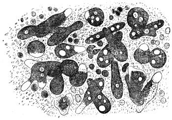
Fig. 1.—Amœba coli, Lösch, in the intestinal mucus. (After Lösch.)
The case reported by Lösch in 1875 attracted more attention. It was that of a peasant, aged 24, who came from the province of Archangel. He was admitted into Eichwald’s clinic at Petrograd with symptoms of dysentery. In the discharges containing blood and pus, Lösch found amœbæ in large numbers. When at rest these amœbæ measured from 20 µ to 35 µ; in a state of movement their length might extend up to 60 µ (fig. 1). The pseudopodia appeared only singly, and, since they were hyaline (ectoplasmic), were thus distinguished from the markedly granular endoplasm that enclosed a spherical nucleus of from 5 µ to 7 µ in diameter. One or more non-contractile vacuoles were present. Quinine enemata had the effect of making the amœbæ disappear from the fæces and thus causing the diarrhœa to abate. Four months after admission the patient died from the results of intercurrent pneumonia. At the autopsy ulceration of the large intestine was found, especially in the lower parts. Lösch connected the amœbæ with the ulcerations by experiments made on four dogs by injecting them with recently passed stools (per os et anum). Eight days after the last injection numerous amœbæ were found in the fæces of one of these dogs; eighteen days after the injection the animal was killed. The mucosa of the rectum was inflamed, covered with blood-stained mucus and ulcerated in three places. Numbers of amœbæ were found both in the pus of the ulcers and in the mucus. The three other dogs remained healthy. From these observations Lösch concluded that the species of amœba described by him as Amœba coli could not be regarded as the primary cause of the disease, but that it was certainly capable of increasing a lesion of the large intestine already present, or at least of preventing its healing.
B. Grassi (1879) found in the stools of healthy as well as in those of diarrhœic patients from various localities in Northern Italy, amœbæ similar to those discovered by Lösch. As this was of frequent occurrence, the pathogenicity could not be definitely established. Normand, formerly naval surgeon at Hong-Kong, observed numerous amœbæ in the dejecta of two patients suffering from colitis.
Many further investigations, which cannot be quoted in detail, showed not only that intestinal amœbæ were widely distributed in man, but indicated with greater certainty their rôle as agents of dysentery. The Commission sent out by the German Government in the year 1883 to investigate cholera in India and Egypt—whose members discovered the cholera bacillus—also collected information with regard to dysentery. In five cases of dysentery examined post mortem at Alexandria, with the exception of one case in which ulceration of the colon had already cicatrized or was approaching cicatrization, R. Koch found amœbæ as well as bacteria in sections from the base of the ulcers, although such had previously escaped notice in examination of the dejecta. Encouraged by these results, Kartulis (1885), who had discovered amœba-like bodies in the stools of patients suffering from intestinal complaints at Alexandria, continued his investigations. The results, obtained from more than 500 cases, gave rise to the theory that typical dysentery was caused by amœbæ as were also the liver-abscesses that often accompany it. Kartulis supported his theory not only by the regular occurrence of amœbæ in the stools of dysenteric patients and their absence in other diseases, and by the occurrence of the parasites in ulcers of the large intestine and in the pus from liver-abscesses, but also by experiments which he performed on cats. These were infected by injection per anum of stool material rich in amœbæ from subjects of dysentery. The infection took place also when amœba-containing, but bacteria-free, pus from liver-abscesses was used. It has been objected that the infection of man with Amœba coli, as the dysenteric amœbæ were then generally designated, does not take place per anum but per os. This difficulty, however, diminished in proportion as the encysted states of amœbæ (fig. 2), long known in the case of other Protozoa, became understood. The infection of man (Calandruccio, 1890) and of cats (Quincke and Roos) succeeded solely when material containing such stages was used. Amœbæ introduced into the intestine multiply there by fission (Harris, 1894). However, this theory, to which various other authors gave support on the grounds of their own observations, encountered opposition. Thus it was established that amœbæ were not found in patients in every place where dysentery was endemic, or else they were much rarer than was expected. Further, amœbæ were present in the most varied kinds of intestinal diseases, both of infective and non-infective characters. Also they were present in quite healthy persons.
Moreover, for various reasons, infection experiments on animals failed to supply proof, and finally a bacterium was discovered (Shiga, 1898) to be the excitant of one form of dysentery. Agglutination attested the specific part played by this organism, as it was produced by the blood serum of a person suffering from or recovered from dysentery, but not by the serum of one who was uninfected. Bacillary dysentery consequently was a distinct entity. The final step to be taken was to decide whether there was a specific amœbic enteritis (amœbic dysentery or amœbiasis, according to Musgrave).

Fig. 2.—Encysted intestinal amœbæ showing nuclear multiplication. (After B. Grassi.)
This question should decidedly be regarded from the positive point of view. It is intimately connected with another, namely, whether there are not several species of intestinal amœbæ. The possibility of this had already been recognized. In addition to the Amœba coli Lösch, R. Blanchard distinguished yet another, Amœba intestinalis, and designated thereby the large amœbæ described in the first communication made by Kartulis; later on he stated the distinction between the species. Councilman and Lafleur10 (1891) considered the amœba of dysentery to be Amœba coli Lösch and so re-named the species Amœba dysenteriæ. Kruse and Pasquale (1893) employed the same nomenclature, but retained the old name Amœba coli Lösch for the non-infectious species. Quincke and Roos (1893) set forth three species: a smaller species (25 µ) finely granular, pathogenic for men and cats (Amœba coli Lösch); a larger species (40 µ) coarsely granular, pathogenic for men but not for cats (A. coli mitis); and a similar species non-pathogenic either for man or cat (A. intestini vulgaris). Celli and Fiocca (1894–6) went still further, they distinguished:
(1) Amœba lobosa variety guttula (= A. guttula Duj), variety oblonga (= A. oblonga Schm.) and variety coli (= A. coli Lösch).
(2) Amœba spinosa n. sp. occurring in the vagina as well as in the intestine of human patients suffering from diarrhœa and dysentery.
(3) Amœba diaphana n. sp. found in the human intestine in cases of dysentery.
(4) Amœba vermicularis Weisse, present in the vagina and in dysentery; and
(5) Amœba reticularis n. sp. in dysentery.
Shiga distinguished two species; a larger pathogenic species with a somewhat active movement, and a small harmless species with a somewhat sluggish movement. Bowman mentions two varieties, Strong and Musgrave (1900) two species—the pathogenic Amœba dysenteriæ and the non-pathogenic Amœba coli; Jäger (1902) and Jürgens (1902) mention at least two species. In the following year (1903) a work by Schaudinn was published which marked a real advance. This, in conjunction with the establishing of a special genus (Endamœba or Entamœba) for human intestinal amœbæ first by Leidy11 and then by Casagrandi and Barbagallo,12 for the time cleared up the confused nomenclature, the old name Amœba coli being retained for the harmless intestinal amœbæ of man, whereas the pathogenic species was designated Entamœba histolytica. The history of more recent work is incorporated in the accounts of the entamœbæ given below.
Entamœba coli, Lösch, 1875, emend. Schaudinn, 1903.
Syn.: Amœba coli, Lösch, 1875. Entamœba hominis, Casagr. et Barbag. 1897.
The amœboid trophozoite, according to Lösch, measures 26 µ to 30 µ and upwards; according to Grassi 8 µ to 22 µ; according to Schuberg 12 µ to 26 µ. A separation of the body substance into ectoplasm and endoplasm is only perceived during movement. The pseudopodia, which are generally only protruded singly, are broad and rounded at the end (lobopodia) and are hyaline, while the remainder of the body is granular. The ectoplasm is less refractile than the rest of the cytoplasm; it also stains less intensely (fig. 1), and is best seen on protrusion of a pseudopodium. Red blood corpuscles are rarely, if ever, found ingested in the cytoplasm.
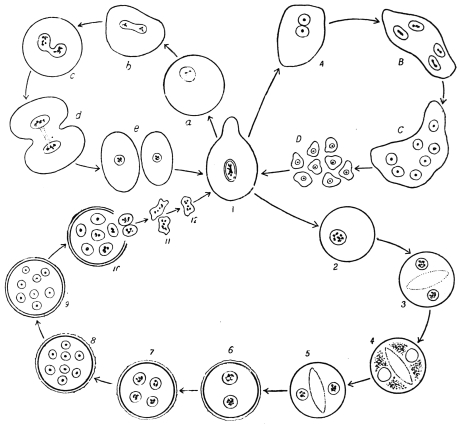
Fig. 3.—Entamœba coli: life-cycle, a-e, stages in binary fission; A-D, schizogony, with formation of eight merozoites; 2–10, cyst formation or sporogony, with formation of eight nucleate cysts. (After Castellani and Chalmers)
The nucleus is vesicular, and is spherical when inactive, measuring 5 µ to 7 µ, with a thick nuclear membrane. In the centre of the nucleus is a chromatinic body or karyosome or sometimes several small nuclear bodies formed of plastin and chromatin; the remaining chromatin is arranged on the achromatic network in the form of fine granules, especially thickly deposited on the nuclear membrane.
Entamœba coli lives as a commensal in the upper portion of the large intestine, where the fæces still possess a pulpy consistency. With their concentration and change in reaction lower in the bowel, the parasites either die or else if they are at a suitable stage of development form resistant cysts. These cysts (fig. 2) can be found in great abundance in normal fæces, as Grassi first observed. Slight laxantia or intestinal diseases of any kind producing increased peristalsis, however, show amœbæ even in the unencysted condition, provided that the person harbours intestinal amœbæ generally. The intensity of infection varies according to the locality; thus Schaudinn found that 50 per cent. of the persons examined were infected with harmless amœbæ in East Prussia, 20 per cent. in Berlin and about 66 per cent. on the Austrian littoral.
The life-history (fig. 3) of the parasite exhibits two phases: (a) asexual multiplication in the intestine, either by binary fission or by schizogony with formation of eight merozoites, and (b) sporogony leading to the production of eight-nucleate cysts. Infection results from ingestion of cysts. Only cysts with eight nuclei are infective. The diameter of such cysts is about 15 µ to 20 µ.
There are varying accounts of the details of the life-cycle of Entamœba coli in its different stages. Thus, regarding schizogony or multiple fission it was formerly stated that the nucleus of the parent amœba divided into eight portions, which after dissolution of the nuclear membrane, passed outwards into the cytoplasm, which segregated around each. Eight merozoites were thus produced. More recently the process of schizogony has been considered to consist in the repeated division of the nucleus into two, four, and finally eight nuclei (fig. 3, A-D), and the formation of eight merozoites or amœbulæ.
The process of encystment is initiated by the extrusion of all liquid and foreign bodies from the protoplasm, which assumes a spherical form (fig. 4, A). The rounded uninucleate amœba then secretes a soft gelatinous coat, which finally differentiates into a double contoured cyst wall in older cysts. According to Casagrandi and Barbagallo, the size of the cyst varies from 8 µ to 30 µ, and averages about 15 µ. According to Schaudinn (1903) the cytological changes during cyst formation are as follows. The nucleus of a rounded uninucleate form divides into two (fig. 4, B). Each of these nuclei fragments into chromidia (fig. 4, C), some of which are absorbed, while others reunite so that the cell becomes binucleate again. Each of these nuclei, by a twice repeated division, produces three nuclei (fig. 4, D), the smaller two of which degenerate and were regarded as reduction nuclei. There is a clear zone or vacuole in the middle of the cyst during these maturation processes, dividing the cyst into two halves. After the nuclear reduction the clear space disappears, and each nucleus (termed by some a gamete nucleus) divides into two pronuclei (fig. 4, E). The pronuclei of the pairs were said by Schaudinn to differ slightly. Copulation occurs between pairs of unlike pronuclei, and is an example of autogamy (fig. 4, F). When complete, each of the fusion nuclei (synkarya) divides twice, giving rise first to four and finally to eight nuclei. Eight amœbulæ are thus formed within the cyst.
According to Hartmann and Whitmore (1911)13, however, autogamy does not occur within the cysts of E. coli. They consider that eight small amœbulæ are formed (fig. 3, 2-10) which escape from the cyst and then conjugate in pairs (fig. 3, 10-12), afterwards growing into a new generation of trophozoites.
Only some 10 to 20 per cent. of the cysts evacuated with the fæces undergo the full course of development, the majority perish previously. In old dry fæces, only cysts with eight nuclei are found, and it is these alone that cause the infection.
Entamœba williamsi, E. bütschlii, E. hartmanni and E. poleki (Prowazek) are probably only varieties of E. coli.
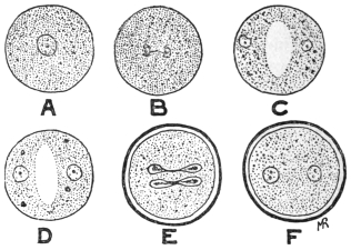
Fig. 4.—So-called autogamy of Entamœba coli. A, rounded amœba; B, nucleus dividing; C, the two daughter-nuclei giving off chromidia; D, each nucleus has formed two reduction nuclei; E, cyst membrane formed, and gamete nuclei are dividing; F, cyst with two synkarya.
The principal feature distinguishing Entamœba coli from E. histolytica is the formation of eight-nucleate cysts by the former as contrasted with the tetra-nucleate cysts of the latter. The cyst-wall of E. coli is thicker than that of E. histolytica (tetragena). Further, E. coli does not usually ingest red blood corpuscles, nor are “chromidial blocks” present inside its cyst (see p. 40).
According to Chatton and Lalung-Bonnaire14 (1912) the entamœbæ of vertebrates should be placed in a separate genus Löschia, as they differ in their life-history from E. blattæ, the type species of Entamœba. Leidy (1879), however, named the genus Endamœba, but further researches are necessary on biological variation among these organisms.
Entamœba histolytica, Schaudinn, 1903.
Syn.: Amœba coli, autt. p. p. Amœba dysenteriæ, autt. p. p.
The average size of the amœboid trophozoite is 25 µ to 30 µ. In fæces diluted with salt solution the amœbæ swell to 40 µ and more. There is sometimes separation of the body substance into a strongly refractile vitreous ectoplasm and a corneous endoplasm, pronounced even in repose, although the former is not equally thick at all parts of the periphery. In the endoplasm generally there are numerous foreign bodies (bacteria, epithelial cells, colourless and red blood corpuscles (fig. 6), and occasionally living flagellates of the intestine). The nucleus is 4 µ to 6 µ in diameter, and may be difficult to recognize because it is sometimes weakly refractile and poor in chromatin. Its shape is slightly variable; it is usually excentric, sometimes wholly peripheral at the limit of the two parts of the body. Vacuoles are not present in quite fresh specimens, but appear later. In the study of E. histolytica, the morphological characters of the trophozoite or vegetative stage of the organism formerly separated as E. tetragena (figs. 5, 6, 8a) must be considered (see p. 38).
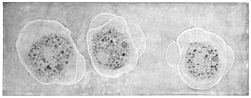
Fig. 5.—Entamœba histolytica (tetragena form), showing three successive changes of form due to movement. × 1100. (After Hartmann.)
The history of the development of these species, which give rise to amœbic enteritis as distinguished from bacillary dysentery, was formerly not so well known as that of E. coli. Upon being introduced into cats (per anum) dysenteric amœbæ provoke symptoms similar to those in man. In the latter, besides metastatic liver abscesses, abscesses of the lungs, and, according to Kartulis, cerebral abscesses are occasionally produced. Marchoux (1899) states that when the disease has lasted for some time liver abscesses are produced in cats also.
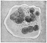
Fig. 6.—Entamœba histolytica which has ingested many red blood corpuscles. × 1100. (After Hartmann.)
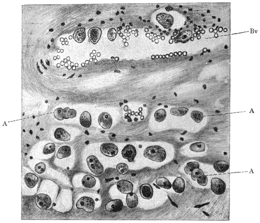
Fig. 7.—Section through wall of large intestine (of a man) close under an ulcer caused by Entamœba histolytica. A, amœbæ that have penetrated partly in blood-vessels (Bv), partly in tissue of submucosa to the muscularis. Magnified. (After Harris.)
In the large intestine of infected cats the amœbæ creep over the epithelium, and here and there they force the epithelial cells apart, as well as removing them or pushing them in front of them; the amœbæ thus insert themselves into the narrowest fissures. They penetrate also into the glands through the epithelium, and thence into the connective tissue of the mucosa. Intestinal and glandular epithelia perish under the influence of these parasites: the cells are pushed aside, fall to pieces or are absorbed by the amœbæ. In the connective tissue of the mucosa the amœbæ migrate further, and often accumulate above the muscles. Finally they rupture this and force their way into the submucosa. In cats, apparently, the penetration is not so great as in men, according to Kruse and Pasquale. During their migration the parasites also gain access to the lymph-follicles of the wall of the intestine, which become swollen and commence to suppurate; follicular abscesses arise and after their rupture follicular ulcers. The diseased patches in the mucosa are markedly hyperæmic and numerous hæmorrhages are set up. Roos and Harris state that the amœbæ also penetrate into the blood-vessels (fig. 7) and this explains the occurrence of metastatic abscesses.15 The whole submucosa is severely swollen at the diseased spot and undergoes small-celled infiltration in the neighbourhood of the colonies of amœbæ. From these findings Jürgens (1902) draws the conclusion16 which is followed here, that the amœbæ are causative agents of the enteritis of cats, which disease is well defined, both pathologically and anatomically. Subsequent researches confirm the experience of earlier authors; great precautions were taken to exclude errors, hence, as with Gross and Harris, no exception can be taken to their results. The inoculation material was derived from soldiers who suffered from amœbic enteritis in China and who were admitted into the garrison hospital at Berlin. In order to be independent of the patients themselves, transmission experiments from cat to cat were performed, after the first experiments on cats yielded positive results. This was also effected by rectal feeding as employed by earlier workers. Such appeared necessary in order to prevent the evacuation of the inoculation material per anum, as well as to avoid the employment of morphia and ether narcosis. Forty-six cats were used for the experiments. Ten cats received tested stools containing motile amœbæ from soldiers suffering from amœbic enteritis contracted in China. Sixteen other cats received stools from cats infected by inoculation. All the animals sickened and suffered from the disease. Five cats received dejecta from human amœbic enteritis in which, however, no motile amœbæ were present. Thirteen cats received stools from soldiers who suffered from bacillary dysentery. None of the latter cats took the complaint and none showed changes in the large intestine upon sectioning. The injection of various bacteria, obtained from a stool of amœbic enteritis pathogenic to cats, remained without result in both the cats employed for this experiment. Lastly, two cats, which had been kept with those artificially infected, were taken ill spontaneously and suffered from the disease. In the opinion of Harris, who ascertained the harmless nature of bacteria derived from the intestinal flora containing dysenteric amœbæ, young dogs are capable of being infected.