ON PARASITES IN GENERAL. (continued 9)
Reading edition
In man, and only rarely in cattle, the mother cyst first forms “daughter cysts” (E. hominis, Rud. [fig. 255]), which, though smaller than the “mother cyst,” resemble it in the structure of their walls; thus they are covered externally by a laminated cuticle and internally by the parenchymatous layer. They originate:
(1) Between the laminæ of the cuticle of the mother cyst from small, detached portions of the parenchymatous layer; during their growth they bulge inwardly or outwardly and may separate themselves entirely from their parent cyst. In the latter case they lie between the mother cyst and the capsule of connective tissue formed by the host (E. granulosus or E. hydatidosus exogenus); when growing inwardly they reach the interior of the mother cyst (E. hydatidosus endogenus). Their number is very variable and does not depend on the size of the mother cyst. They are as big as, or bigger than, gooseberries.
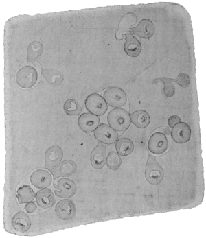
Fig. 254.—A piece of the wall of an Echinococcus veterinorum stretched out and seen from the internal surface. A few brood capsules (the outline of which is only faintly shown), with scolices directed towards their interior and exterior. 50/1.
(2) According to some authors, endogenous daughter cysts arise also from a metamorphosis of scolices that have separated off from the brood capsule. This takes place in the following way: Fluid accumulates in the interior of the scolex, so that eventually nothing remains except a sac consisting of cuticle lined by parenchyma. The cuticle gradually thickens and several layers form (fig. 257).
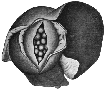
Fig. 255.—Echinococcus hominis in the liver. The fibrous capsule and the wall of the echinococcus have been incised, so that the endogenous daughter cysts may be seen. Reduced. (After Ostertag, from Thoma.)
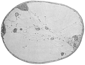
Fig. 256.—Section through an echinococcus scolex in process of vesicular metamorphosis, twenty-six days after insertion in the pleural cavity. × 250. (After Dévé.)
(3) Transformation of Brood Capsules into Daughter Cysts.—This is also held to be possible by various observers. New epithelial layers are deposited between the cuticle which lines the brood capsule and the outer parenchymatous layer. This parenchymatous layer gradually disappears and a new parenchymatous layer forms in the interior from the parenchyma of the scolex or scolices. Although it appears strange that a completely formed scolex with specifically differentiated tissues and organs should retrogress to more primitively organized matter, and again become a proliferating bladder, yet we can hardly doubt that the older observations, regarding such a vesicular metamorphosis, of Bremser (1819), v. Siebold (1837), Naunyn (1862), Rasmusser (1866), Leuckart (1881), Alexinsky (1898), Riemann (1899), Dévé (1901), and Perroncito (1902) are correct.
(4) Further, a fourth method of formation of daughter cysts is described by Naunyn as occurring in sterile hydatids, i.e., those containing no brood capsules. In this case a portion of the mother wall of the hydatid gets constricted off.
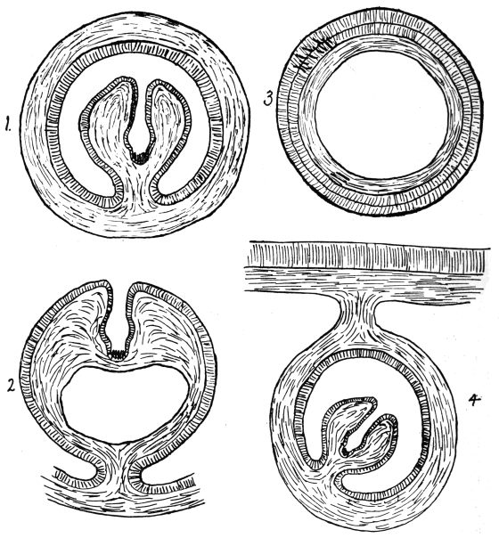
Fig. 257.
Figs. 257 and 257a.—Diagram of transformation of a scolex into a daughter cyst (1 to 3): 1, scolex in brood capsule; 2, liquefaction of scolex; 3, daughter cyst; and (4 to 6) of a brood capsule into a daughter cyst; 4, brood capsule with scolex; 5, deposition of new epithelial layers on the inner layer of the parenchyma; 6, disappearance of outer parenchyma and formation of inner parenchyma from the parenchyma of scolex, which has now disappeared. (Note.—The scolices are out of proportion to the brood capsules and to the daughter cysts. Stephens.)
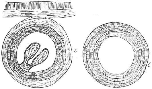
Fig. 257A.
It has also been established that not only daughter cysts transplanted into animals develop further (Lebedeff, Andrejew, Stadnitzky, Alexinsky, Riemann), but that this also holds good if only hydatid scolices from man or animals are transplanted into animals (rabbits). They develop into echinococci and can then give rise to brood capsules and scolices. As Dévé further established, hydatid scolices are not capable of developing in guinea-pigs, while corresponding experiments with rabbits are in the large majority of cases successful where the scolices are introduced subcutaneously or into the pleural or peritoneal cavities. It is only in the case of daughter cysts that further growth is obtained in the case of guinea-pigs. Finally it appears, as has been already stated, that brood capsules can transform themselves into daughter cysts, but according to Dévé only within the mother cyst, not after transplantation. Daughter cysts that have been formed in the mother cyst of man and animals behave themselves just as the mother cyst does, i.e., they can remain sterile, or give rise to brood capsules and scolices, or even again to fresh cysts—granddaughter cysts. The mother cyst can also die, so that the daughter cysts then lie in the cavity of the connective tissue capsule. The number of the daughter cysts in either case may attain several thousands.
The echinococcus fluid, which originally is formed from the blood of the host, is light yellow, with a neutral or slightly acid reaction; its specific gravity averages 1009 to 1015. It contains about 1·5 per cent. of inorganic salts, half of which is common salt; in addition (besides water) it contains sugar, inosite, leucine, tyrosin, succinic acid (associated with lime or soda) and albumens which are not coagulated by heat; occasionally also the fluid has been found to contain hæmatoidin and uric acid salts (in echinococcus of the kidneys), which doubtless demonstrates that the echinococcus liquid originates from the host. It has been generally assumed that echinococcus fluid contains a toxic substance the escape of which into the body cavity (at operation or by bursting of a hydatid cyst) produces more or less severe symptoms (fever, peritonitis, urticaria), so much so that one speaks of hydatid intoxication. The investigations of Kobert, Joest, etc., have, however, shown the harmlessness of fresh undecomposed hydatid and cysticercus fluid for rabbits, mice and guinea-pigs, whether inoculated intraperitoneally, subcutaneously or intravenously. Contrary data or clinical experience must accordingly depend on other factors.
According to the researches of Leuckart, the growth of the echinococcus is very slow; four weeks after infection the average size is only 0·25 to 0·35 mm., at the age of eight weeks it is 1 to 2·5 mm., and at this period the formation of the central cavity commences; at the age of five months, and with a size of 15 to 20 mm., the first brood capsules with scolices are formed. The consequence of this gradual increase of size is that the organ attacked can maintain its functions by vicarious hypertrophy, and that many echinococci induce no special symptoms and cannot even be diagnosed, the latter circumstance being due to their hidden position.
The echinococcus cannot be said to be scarce in man, as is shown by the following table for Central Europe:—
PlacePeriodNo. of post-mortemsNo. of cases of echino.Percentage
Rostock1861–831,026252·43
Greifswald1862–933,429511·48
Jena1866–874,998420·84
Breslau1866–765,128390·761
Berlin1859–684,770330·69
Würzburg—2,280110·48
Göttingen—63930·469
Dresden1852–621,93970·36
Münich1854–8714,183350·25
Vienna18601,22930·24
Prague—1,28730·23
Kiel1872–873,58170·19
Zürich, Basle, Berne—7,982110·13
Erlangen1862–731,75520·11
These, however, are only cases that have become known by post-mortem; in addition, there are cases that have been treated medically, of which there are a few statements, at all events relating to the principal districts of Germany. According to Madelung, one case of echinococcus occurs in every 1,056 inhabitants in the town of Rostock, in the district of Rostock one to every 1,283, in Schwerin one to every 5,887, and in Ludwigsort one to every 23,685; according to Peiper, in Upper Pomerania one case occurs to every 3,336, in the district of Greifswald one to every 1,535 inhabitants. The northern districts of Pomerania are more affected than the southern ones.
Accordingly, echinococcus is also considerably more frequent in cattle in Pomerania. On an average in Germany 10·39 per cent. oxen, 9·83 per cent. sheep, and 6·47 per cent. pigs are infected, whereas in Upper Pomerania 37·73 per cent. oxen, 27·1 per cent. sheep, and 12·8 per cent. pigs are infected; in Greifswald, indeed, 64·58 per cent. oxen, 51·02 per cent. sheep, but only 4·93 per cent. pigs are infected. In accordance with these figures Tænia echinococcus must be frequent in dogs in Pomerania, especially in Upper Pomerania; on the other hand, the conjecture that the frequency of echinococcus in Mecklenburg is explained by the occurrence of Tænia echinococcus in foxes has not been confirmed, as the fox does not harbour this worm in Mecklenburg.
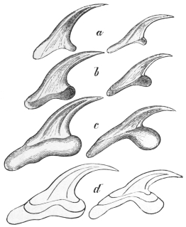
Fig. 258.—Hooklets of echinococcus. a, of Echinococcus veterinorum; b, of Tænia echinococcus, three weeks after infection; c, of the adult Tænia echinococcus; d, the three forms of hooklets outlined one within the other. 600/1. (After Leuckart.)
Beyond the European continent, echinococcus is frequent in the inhabitants of Iceland, Argentine, Paraguay and Australia. In Iceland, according to Finsen, 1 in every 43 inhabitants is affected with echinococcus; according to Jonassen the proportion is 1 to 63; this is due to the habits of the people of Iceland or, in fact, to the frequency of Tænia echinococcus in dogs, and the prevalence of the hydatid in cattle. In certain districts of Australia it is just as frequent. In Cape Colony, Egypt and Algeria echinococcus is not rare, but it is scarce in America and in Asia, with the exception of the nomadic tribes of Lake Baikal.
Echinococcus attacks persons of every age, though it is rare in children up to 10 years of age and in old people. It occurs most frequently between the ages of 21 and 40 years. According to all statistics it preponderates in women (about two-thirds of the cases). The liver is its favourite seat (57·1 per cent. of the cases); next in order come the lungs (8 per cent.), kidneys (6 per cent.), cranial cavity, genitalia, organs of circulation, spleen (3·8 per cent.), etc. As a rule one organ only is invaded; multiple occurrence may originate from one infection, or eventually from a later infection (?), or it may come to pass that from some cause (through the spontaneous rupture of an echinococcus, or the rupture of one caused by an injury or surgical operation) daughter cysts, brood capsules or scolices escape into the abdominal cavity,294 where they settle or become transformed and go on growing. In the distribution of this secondary echinococcus the great powers of motility of the free scolices must be taken into account (Sabrazès, Muratet, and Husnot).
Human echinococci may also die at various stages of development, become caseous or calcified, or may be absorbed, the cause for this being either disease of the hydatid itself or inflammation of its connective tissue capsule; the discovery of the laminated cuticle, which has great powers of resistance, or the finding of the hooklets of the scolices is sufficient to form a conclusion as to the nature of such formations.
Siebold (1853) was the first to rear Tænia echinococcus in the dog by feeding it with the echinococcus of cattle and especially of sheep. Küchenmeister, van Beneden, Leuckart, Railliet and others obtained similar results, and Thomas, Naunyn, Krabbe and Finsen succeeded in rearing T. echinococcus in dogs from the bladder worms of human beings; these grow comparatively slowly (one to three months295) and only during the process of growth develop their hooklets in their definite form (fig. 258). It lies in the nature of things that dogs, whether experimentally or naturally infected, almost always harbour T. echinococcus in large quantities. That cats exceptionally harbour these worms has been already mentioned (Dévé). Finally, Leuckart infected young pigs by feeding them with mature segments.
Echinococcus multilocularis (alveolar colloid).
In addition to the form of echinococcus already described, and which is also frequently termed Echinococcus unilocularis, there is a second form which occurs in man as well as in animals, and which is termed E. multilocularis, s. alveolaris (alveolar colloid).
It was originally regarded as a tumour; its animal nature was first established by Zeller and R. Virchow. The parasite, which varies in size from that of a fist to a child’s head, presents a collection of numerous cysts, measuring between 0·1 and 3 to 4 mm. to 5 mm. in diameter, which are embedded at first in a soft, connective tissue stroma; the cut surface has therefore a honeycomb appearance. The cysts are surrounded by a pellucid and laminated cuticle, and each according to its size encloses either a small-celled tissue or a cavity lined by a parenchymatous layer; the fluid contained in such a cavity may be transparent, or is rendered opaque by globules of fat, bile-pigment, hæmatoidin and fat crystals. According to some authors all or most of these cysts intercommunicate; others state that this is the case at least as regards the cuticle. The scolices are by no means found in all the cysts, and when present only a few, rarely half, of the cysts contain scolices (one or more); it is supposed that at least some of these scolices are formed in brood capsules, and that the former are capable of undergoing a cystic metamorphosis.
One circumstance is peculiar to the multilocular echinococcus of man, namely, the disintegration that sets in at certain stages; in the centre of the parasite a cavity forms that frequently becomes very large and is filled with a purulent or brownish or brownish-green viscid fluid; in this fluid one finds shreds of the wall of the cavity, calcareous bodies, echinococcus cysts, also scolices and hooklets, as well as fat globules and crystals of hæmatoidin, margarine and cholesterin and concretions of lime. Such ulcerative processes, according to Ostertag, are never present in the multilocular echinococcus of oxen,296 in which the separate cysts are larger and the connective tissue integument less powerfully developed.
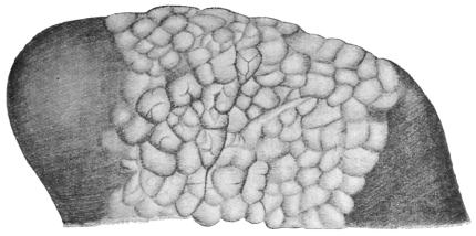
Fig. 259.—Echinococcus multilocularis in the liver of the ox. Natural size. (After Ostertag.)
Hardly anything positive is known with regard to the development of the alveolar echinococcus; its peculiar conformation is attributed by some to enormous infection of oncospheres, by others to the abnormal situation of one oncosphere; a few authors ascribe it to infection of lymphatic vessels, others to infection of the biliary ducts or to peculiarities of the surrounding hepatic tissue; Leuckart ascribes it to a grape-like variety of form which continues budding; a few more recent authors consider multilocular echinococcus to be specifically different from unilocular echinococcus, and therefore also different the species of Tænia arising from them. Melnikow-Raswedenkow is also of this opinion. According to this author the oncospheres infect the lumen of a branch of the portal vein in Glisson’s capsule of the liver and grow into an irregularly shaped formation (chitinous coil), which breaks through the vascular walls and thus forms the alveoli. So far the data coincide well with Leuckart’s opinion of the original grape-like form of the Echinococcus multilocularis; according to Melnikow-Raswedenkow the “granular protoplasmic substance” (parenchymatous layer) is not only present in the interior of the loculi but also outside, and, moreover, “ovoid embryos” are supposed to develop in the chitinous coils, which, “thanks to their amœboid movements, reach the lumen of a vessel, where, under favourable circumstances, they begin to develop further,” that is to say, they become “chitinous cysts with fantastic outlines,” or also “single-chambered chitinous cysts”; scolices may develop in both. Dévé, however, considers that these embryos are only prolongations of the protoplasmic layer which secondarily cuticularize.
The multilocular echinococcus, which in man produces a severe disease and almost always leads to premature death, infects most frequently the liver, but may also be found primarily in the brain, the spleen and the suprarenal capsule; from the liver by means of metastasis it may reach the most various organs, especially those of the abdomen, but also the lungs, the heart, etc. Up to 1902, 235 cases have been described and up to 1906, 265, being 70 from Russia, 56 from Bavaria, 32 from Switzerland, 30 from the Austrian Alps, 25 from Würtemberg; the remaining cases are distributed over Central Germany, Baden, Alsace, France, Upper Italy, North America. In some the origin is doubtful; in any case after Russia, the mountainous South of Europe is the principal region of distribution. As to the domesticated animals, the same parasite is found principally in the ox (according to Meyer, in Leipzig, in 7 per cent. of the oxen affected with echinococcus); it is rarer in the sheep and very scarce in the pig.
It has already been mentioned above that recently the multilocular echinococcus has been stated to be specifically different from hydatid or unilocular echinococcus. To this may be added the fact that Mangold, who fed a young pig with oncospheres of a Tænia reared from the multilocular echinococcus, found two growths in the liver four months later, which he took to be E. multilocularis, and consequently one has to assume the existence of two different worms. The chief defender of this view, already put forward by Vogler, Mangold, and Müller, is Possett. He bases his opinions on (1) the more restricted distribution of the multilocular hydatid, the former occurring in districts where only cattle are raised, the latter where sheep-breeding is established; (2) that those engaged in looking after sheep are attacked by multilocular, whereas those looking after cattle are attacked by unilocular hydatid; (3) that among the cases of unilocular hydatid occurring in the distribution areas of multilocular hydatid no transitions between the two forms are observed; (4) on the difference in the hooks both in the hydatid as well as in the Tænia stage; the hooks of Tænia echinococcus are plump, sharply curved, and have a short posterior root process the length of which is to that of the total length as 1 to 4·7, whereas on the contrary the hooks of the alveolar echinococcus are more slender, slightly bent, and have a long posterior root process (1 to 2·5); and (5) on the form of the uterus, which in the alveolar Tænia has the form of a spherically distended sac anteriorly.
Serum Diagnosis of Echinococcus.
(1) Precipitin Reaction.—Mix equal parts of hydatid fluid (of the sheep) and serum of patient. Keep at 37° C. The reaction is not decisive as it may be given by normal sera.
(2) Complement Deviation.—Required: (1) Hydatid fluid of sheep (antigen), (2) guinea-pig complement, (3) patient’s serum, (4) red cells of sheep, (5) hæmolytic serum (of rabbit) against sheep’s red cells, (6) 0·8 per cent. salt solution. Mix the antigen + patient’s serum (heated) + complement + salt solution at 37° C. for one hour. Add red cells of sheep + hæmolytic serum. Allow to stand for half an hour at 37° C. It is imperative to make adequate control observations. An example will indicate the method. Salt solution 1·3 c.c. + patient’s serum (heated) 0·2 c.c. + hydatid fluid 0·4 c.c. + complement 0·1 c.c. of serum diluted to a quarter strength + hæmolytic serum and red cell emulsion 1 c.c. Result: no hæmolysis, i.e., the patient’s serum contains specific (echinococcus) antibodies.
C. NEMATHELMINTHES. BY J. W. W. STEPHENS, M.D., B.C., D.P.H.
Bilaterally symmetrical animals, without limbs and with a body cavity in which the gut or other organs float. They are generally cylindrical.
Class. NEMATODA.
Nemathelminthes with an alimentary canal.
Nematodes are as a rule elongated round worms of a filiform or fusiform shape; their length varies according to the species from about 1 mm. to 40 to 80 cm. The outer surface of the body is smooth or annulated, and at certain points provided with papillæ, occasionally also with bristles and alar appendages. The anterior end carrying the oral aperture is usually rather slender, occasionally quite thin; the posterior end is pointed or rounded; the anus, as a rule, lies somewhat in front of the posterior extremity. The sexes are almost always separate, and the male can as a rule be easily distinguished from the female because the former is smaller and more slender, its posterior extremity is often spiral or incurved, or carries an alar appendage, whereas the female is larger and thicker, and its posterior extremity is straight. In the male the genitalia open into the anus; the sexual orifice of the female opens ventrally along the median line in the anterior half of the body, in the middle, or a little further back. Both sexes, moreover, have an orifice, the excretory pore, which is situated ventrally in the median line and about the level of the œsophageal nerve ring.
In large species, even with the naked eye, two lighter transparent bands—the lateral lines—may be distinguished; they run along the sides of the body from the anterior to the posterior end, while two other bands, the median lines, running along the ventral and dorsal mid-lines, are less evident; in exceptional cases there are also four sub-median lines. These bands or lines are inward projections of the ectoderm, and in them lie the nerves and excretory vessels (fig. 260).
Some Nematodes live free in fresh or salt water, in soil, mud or decaying vegetable matter, others parasitically in the most various organs of animals, frequently also in plants.
Anatomy of the Nematodes.
All the Nematodes are covered by (1) a CUTICLE, which in the small species is thin and delicate, while in the larger species it is thickened, and may consist of several layers of complicated structure. Canalicular pores do not occur. According to general opinion, which is confirmed by the history of development, the cuticle is a product of (2) the EPITHELIUM or ectoderm that had formerly existed or is still found beneath it; in young specimens and small species it is perceptible, but in older worms it frequently alters so considerably that not only do the borders of the cells disappear,297 but a fine fibrous differentiation appears in their cytoplasm. The matrix or ectoderm then has the appearance of an ectodermal syncytium permeated by fibres and strewn with nuclei, so that it is hardly distinguishable from the tissue of (3) the CUTIS, which is always present, though developed to a varying degree. Both layers, matrix and cutis, project internally as ridges and form the lateral lines, while the less marked median lines are produced apparently only by the ectoderm (fig. 260).
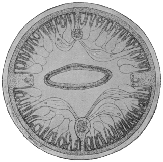
Fig. 260.—Diagram of a transverse section of Ascaris lumbricoides, showing thick cuticle, and beneath it the matrix or syncytial ectoderm. The flat intestine is in the middle, and to the right and left near it in the body wall the lateral lines with excretory vessel and lateral nerves; above and below in the centre the dorsal or ventral median lines with the nerves radiating to the muscles, also the muscle cells with their striated outer contractile portion and inner nucleated vesicular protoplasmic portion. About 50/1. (After Brandes.)
Unicellular cutaneous glands are known in parasitic as well as in free-living species; they vary in number and arrangement, and are found discharging some at the anterior extremity and others in the vicinity of the genital orifices. In other cases large numbers of them are present along the lateral lines; they are strongly developed in most of the Trichotrachelidæ, where they discharge either along a part of the ventral surface or along the lateral and median lines; they are placed so closely together that the ridges of the cuticle perforated by the orifices have long been known, and have been described, as “rodlet borders,” or “fields of rods.”
As the cutis is immediately adjacent to (4) the DERMO-MUSCULAR TUBE the simple layer of the muscular cells is divided into four quadrants by the longitudinal lines—two dorsal and two ventral (fig. 260). The MUSCLES are in the simplest cases large rhomboid cells that lie two by two in each quadrant, so that on transverse section of the entire worm only eight cells are perceptible. The outer border of the cells is converted into contractile fibrils, while the contiguous inner portion has remained protoplasmic, and contains the nucleus. In large species the muscular cells do not only increase in length (up to 3 mm.) and in number in every quadrant, but their contractile portion curves up to form a groove (like that of a dead leaf) thereby even becoming thicker; simultaneously space is gained for more cells, the protoplasmic parts of these cells (on transverse section) project out of the grooves like vesicles. In all cases there is only one layer of longitudinal muscular cells, which, by contracting, can shorten the body or, by contracting one side, can bend it. In the latter case the muscles of the opposite side have an antagonistic effect, or when all the muscles are contracted, the elasticity of the cuticle acts in the same way. Special muscles exist at the beginning of the gut and at sections of the genital apparatus.
The existence of a cavity between the body and the gut wall has hitherto been generally assumed, and has been referred to the cleavage cavity, and consequently designated as a primary body cavity. More recent investigators, however, state that such a cavity does not exist, but that the space between the longitudinal muscles or their protoplasmic portions and the gut epithelium is filled by a complicated “isolation tissue.” This in the main proceeds from a large cell (Is., fig. 262) which lies directly behind the nerve ring dorsal to the œsophagus, and consists of a system of lamellæ which sheathe the muscles and penetrate through them to the cutis and also cover the gut in a thin layer.
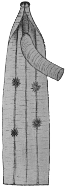
Fig. 261.—Anterior end of an Ascaris megalocephala cut open and showing the four tuft-like organs lying on the lateral lines. Natural size. (After Nassonow.)
We may now consider the “tuft-like” or “phagocytic” organs, which attain 1 cm. in size, and consist of four, six, or even more ramified cells, which lie close to the walls of the body (fig. 261). They are found either only in the anterior part of the body (Ascaris), or throughout the whole length of the body (Strongylus, syn., Sclerostomum), and their position usually corresponds to the lateral lines. In some species there are small protoplasmic cells on the processes of these organs. In consequence of their size they can be recognized with the naked eye, especially when they are loaded with granules of stain (carmine, Indian ink) injected into the body cavity.
Intestinal canal.—The oral aperture, which is situated at the tip of the anterior extremity, is frequently surrounded by thick lips, or small bristles, or papillæ; it leads to a more or less strongly developed buccal cavity, which is lined by a continuation of the body cuticle, and which in some species is provided with “teeth,” representing differentiated portions of the cuticle.
The œsophagus (fig. 262), which arises from the base of the oral cavity, is as a rule a short, bottle-shaped tube with triradiate lumen; its wall is chiefly composed of radiating muscular fibres, which give it the appearance of being transversely striped when viewed from the surface. There exist also in its wall three large gland cells (œsophageal glands) and nerves arising from the lateral lines and running forward. The radial fibres cause a dilatation of the lumen, and exercise an effect antagonistic to the elasticity of the cuticle lining the inner surface. The latter has its own particular layer, which is not in direct connection with that of the oral cavity. Special dilator muscles, arising from the dermo-muscular tube and situated at the commencement of the œsophagus, are only known in a few species. The posterior end of the œsophagus presents a bulb-like dilatation, and is frequently provided with small chitinous movable valves. In a few forms, which belong to the Trichotrachelidæ (Trichocephalus, Trichinella), the œsophagus is a very long cuticular tube, beset on its dorsal surface with a series of large nucleated cells. In others (Cucullanus, Ascaris, etc.), a tube, the so-called glandular stomach, lined only by epithelial cells, follows behind the muscular œsophagus. This glandular stomach is, from its structure, easily distinguished from the mid-gut, or chyle intestine, which is likewise cellular. The so-called mid-gut is a tube lined by flat, cubical, or cylindrical cells (fig. 260) surrounded by “isolation tissue”; its transverse section is circular or flattened dorso-ventrally; the lumen may run in a straight line, or it runs a sinuous course through the alternating prominences of the then flat epithelial cells.
The ectodermal hind gut is, as a rule, very short. At the anal opening the cuticle and the subcuticular layers are reflected inwards, forming the lining of the hind gut. In large species the subcuticular tissue forms large cells on which anteriorly lie in addition large “glandular cells.”298 In the male the ejaculatory duct opens at this point. Around the end part of the gut, either on the chyle intestine or at the beginning of the end gut, there exists a sphincter muscle arising from a muscle cell which acts antagonistically to the two diaphragm-like dilator muscle cells which stretch from the gut to the body wall. In many species large stretches of the gut are provided with dilator muscles. There is sometimes a retrogressive absorption of the gut in the adult stage of a few parasitic species.
Intestinal cæca and œsophageal glands sometimes exist as intestinal appendages; the former are tubular appendages of various size, running backwards or forwards, and arising from the posterior extremity of the œsophagus. They are lacking in many species. The œsophageal glands are unicellular; a dorsal and two subventral glands may be distinguished according to their position; as a rule they open into the œsophagus at a distance from one another. The body of the gland lies in the bulb of the œsophagus, or in the dorsal cul-de-sac arising from it.
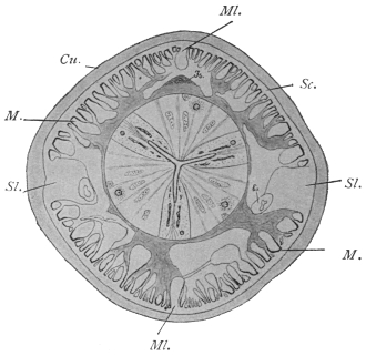
Fig. 262.—Transverse section through Ascaris lumbricoides at the level of the œsophagus behind the nerve ring. Cu., cuticle; Sc., subcuticular layer; Ex., excretory vessel; Is., isolation cell and the system of lamellæ proceeding from it; M., muscles; Ml., median line; Sl., lateral line. Magnified. (After Goldschmidt.)
The nervous system is sufficiently known in a few species only; it consists of a ring containing fifty to sixty fibres closely surrounding the œsophagus, various groups of ganglion cells, and a certain number of nerves extending anteriorly as well as posteriorly. The remarkably small number of fibres, as well as ganglion cells, is characteristic of the nervous system of all Nematodes. Immediately behind the œsophageal ring (fig. 263, Lg.) an agglomeration of ganglion cells lies at either side (lateral ganglia); part of their off-shoots form the œsophageal ring, and part are directed posteriorly and ventrally, and unite partly in front of and partly at the back of the excretory pore, with fibres originating direct from the œsophageal ring, and passing along the ventral median line to the back; these fibres then together form the ventral median nerve (fig. 263, V.m.n.). This nerve, originally consisting of thirty to fifty fibres, becomes in the female attenuated quite evenly in its further course. There is also an agglomeration of ganglion cells close in front of the anus (anal ganglia), and then the median nerve divides in order to combine with the lateral nerves on either side. In the male the median nerve enlarges to nearly the original number of fibres in front of the anal ganglion, which contains seven cells; there is also an anal ring embracing the terminal gut, and there are two ganglion cells in it on each side. In the dorsal median line the dorsal median nerve is alike in both sexes; arising in front with a single root from the œsophageal ring, it gathers its fibres from the lateral ganglia; in the anterior part of the body it consists of thirteen to twenty fibres; in the posterior part of the body the fibres are reduced to four or six; behind the anus it divides and combines with the lateral nerves; the latter consists of two fascicles at either side right up to their most posterior extent—one dorsal and one ventral—which in the greater part of the body do not run in, but beside the lateral lines, and exhibit a different origin anteriorly. The ventral fascicle at each side branches off from the ventral median nerve in front of the excretory pore, whereas the dorsal fascicles originate from the œsophageal ring close to the lateral ganglia. Each of the four fascicles contains only two or three fibres, which run backwards parallel to the lateral lines; a few centimetres in front of the caudal extremity they enter the lateral lines and remain separate from one another up to the level of the anal ganglion; here they amalgamate on either side, after each interpolating one ganglion cell, with the single short lateral nerve which first takes up the forked ends of the ventral and then of the dorsal median nerve; finally, both lateral nerves unite with each other at the back in an arch-like manner.
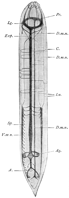
Fig. 263.—Schematic representation of the nervous system of a male Ascaris megalocephala. A., anus; Ag., anal ganglion; C., commissures; D.m.n., dorsal median nerve; Exp., excretory pore; Pr., œsophageal sensory ring; Lg., lateral ganglia; Ln., lateral nerve; Sp., papilla; V.m.n., ventral median nerve. (After Brandes.)
In the male each ventral part of the lateral nerves becomes thickened by taking up fibres from the ventral nerves, which become thickened posteriorly to the nervus bursalis, which towards the middle gives off a mass of fibres to the “genital papillæ” situated in front of and behind the anus; the number of these fibres averages eighty to 100; in its further course the bursal nerve resembles the corresponding ventral part of the lateral nerves of the female.
The ventral and dorsal nerves are connected by a number of semicircular commissures, which originate from the ventral nerves and serve to supply the dorsal nerve, which is always being decreased by fibres departing from it. It is remarkable that these commissures are not placed symmetrically, and their position also is different in the two sexes; in the female there are thirty-one on the right side and only thirteen on the left side. In the male there are thirty-three commissures on the right side and fourteen on the left, which run into the subcuticular layer, generally in pairs, and usually cross at the level of the lateral lines.
The fibres of the two median nerves are chiefly motor; fascicular processes run from each protoplasmic part of the muscular cells to the median nerves; from these they take up bundles of primitive fibrils, which separate, pass through the protoplasmic part and enter the contractile part (fig. 260). One part of the fibrils, however, penetrates beyond the muscles into the subcuticular layer, where they form a network, probably of a sensory nature, with contiguous fibrils. Nerves directed anteriorly finally originate from the œsophageal ring; they consist each of three fibres, carry three ganglion cells at their point of origin, and enter the sensory organs of the three papillæ surrounding the oral aperture. Two of these little trunks lie in the lateral lines, the remaining four are situated in the middle of the four quadrants (Nn. sub-mediani anteriores).
Parasitic species lack higher ORGANS OF SENSE; free-living worms occasionally have two rust-red eyes, sometimes with lenses, at the anterior part of the body. In addition to the above-mentioned sensory papillæ surrounding the oral aperture and the genital papillæ of the male at the end of the body, another pair exist in the vicinity of the lateral ganglia, the “cervical papillæ,” and two dorsal papillæ in the central region of the body and two lateral ones near the tip of the tail (Ascaridæ). The differences in the distribution and number of the sensory papillæ serve for characterizing the larger and smaller groups of Nematodes.
The excretory organs of the Nematodes are variable. In a great many cases the apparatus is symmetrical, and consists of a vessel commencing in the posterior extremity in each lateral line (fig. 260), and passing anteriorly. In the vicinity of the anterior extremity both tubes pass out of the lateral lines, bend ventrally, and, in the median ventral line, unite into a short vesicle formed by an ectodermal cell—the cavity of which is lined by a continuation of the cuticle of the body—which opens into the excretory pore (fig. 263, Exp.). Asymmetry is occasioned through the excretory duct proceeding from the ventral pore to the lateral line, and it here proceeds as (or takes up) the left excretory canal, which anteriorly is a broader tube and runs along the left lateral line; shortly before its union with the excretory duct it throws out a branch to the right towards the lateral line, which, however, always remains weak, and runs posteriorly in the right lateral line; a few smaller branches in addition spring from the left main stem. In other species the right branch is completely suppressed; the entire organ thus lies in the left lateral line, and consists of the excretory duct, which occasionally opens quite in front near the lips, as well as the excretory canal, which throws out a number of lateral branches.
This excretory vesicle is a single elongated or horse-shoe-shaped cell, with a large nucleus and an intracellular tubular system, which is connected with the excretory duct arising from the excretory pore on the outer surface (fig. 326). The so-called ventral gland is the only excretory organ of marine Nematodes, and probably represents a primitive form. Goldschmidt, who has investigated the excretory apparatus of Ascaris lumbricoides, considers that the vessels running in the lateral lines are only ducts to which belong a glandular system hitherto overlooked or otherwise interpreted. This system also lies in the lateral lines, and takes the form of two glandular tracts, forming a syncytial tissue in which lie the ducts, one dorsal, one ventral. In parts these tracts are connected by commissures, although their junction with the excretory vessels cannot be clearly made out. These statements, however, require confirmation. The author has further found that the anterior ends of the lateral canals, directly before they bend ventrally, anastomose with one another and give off anteriorly a small blind process, which can be interpreted as a rudiment of a canal coming from the head end, and as a matter of fact, according to Golowin, such anterior excretory canals exist in a number of genera.
In a number of Nematodes (Cheiracanthus, Capillaria, Trichocephalus, Trichinella, etc.), however, special excretory organs are lacking; possibly the cutaneous glands, which are in these species generally powerfully developed, replace these organs.
Sexual organs.—With the exception of a few species, the Nematodes are sexually differentiated.
(a) Female Sexual Organs.—The sexual orifice (vulva), surrounded by thick labia, is, as a rule, ventral and varies in position from near the head to near the anus. It leads into a short or long vagina (ectodermic), bifurcating into the two uteri, which may be long or short; the long filiform ovaries are continuations of them (fig. 264). Further there is often, e.g., in Ankylostoma, a differentiation into the following parts: (1) Ovejector: the specialized portion of the uterus before it joins the vagina; there may be a separate one for each uterus, or a common one for both uteri. (2) Seminal receptacle: at the other extremity of the uterus. (3) Oviduct: a narrow tube connecting the ovary with the uterus proper. (For the explanation of the terms convergent and divergent uteri vide footnote p. 432.) Uterus and ovaries, which arise in the first place from a single cell, lie between the body wall and the gut and are surrounded by connective tissue. In some species (for instance, Trichinella) the ovary is single.
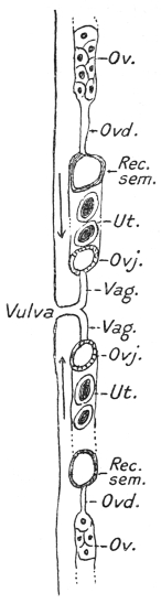
Fig. 264.—Diagram of female genitalia. Ov., ovary (in part); Ovd., oviduct; Rec. sem., seminal receptacle; Ut., uterus (in part); Ovj., ovejector; Vag., vagina.
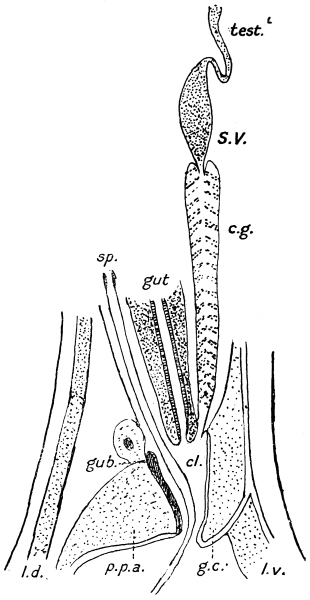
Fig. 264a.—Diagram of male genitalia of a strongylid. Test., testis (in part); S.V., seminal vesicle; c.g., cement gland surrounding ejaculatory duct; sp., spicules; cl., cloaca; gub., gubernaculum; p.p.a., pulvillus post-analis; g.c., genital cone; l.d., dorsal lateral line; l.v., ventral lateral line (the bursa is not shown).
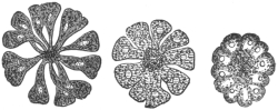
Fig. 265.—Transverse section through the ovarian tube of Belascaris cati of the cat at various levels. To demonstrate the development (right to left) of the ova and of the rhachis. Magnified.
At the blind end of the ovary there is a mass of protoplasm with numerous nuclei that multiply continuously. Gradually the nuclei arrange themselves in longitudinal rows (fig. 265) and the protoplasm commences to leave the periphery and surround each nucleus. The nearer to the uterus the more progressive is this loosening process, until club-shaped cells each containing a nucleus are developed. The most pointed end of each, however, is still attached to an axial fibre of protoplasm, the rhachis; probably this has some connection with the nutrition of the ova. Finally the ova fall off and reach the uterus, where they are fertilized and enclosed in shells.
(b) Male Sexual Organs.—There is never more than one testis (fig. 266), which is a straight or sinuous tube of the same construction as an ovary, and in which the mother cells originate in the same manner as the ova. In the same way as the ovary passes into the uterus, so does the testis pass into the spermatic duct; the latter is often divided into the somewhat dilated seminal vesicle and into the muscular ductus ejaculatorius, which, running ventral to the intestine backward (fig. 267), finally opens into the cloaca. In many species, e.g., A. duodenale, the ejaculatory duct is surrounded for a greater or less portion of its extent by the cement gland, the secretion of which (brownish or blackish in colour) serves for copulation. The ejaculatory duct of the large Ascaridæ is for the most of its course surrounded by a muscular network which takes its origin from the two dilator cells of the gut (fig. 268 F.). The spermatozoa of the Nematodes, it may be noted, only attain their full development after the sperm mother cells have been conveyed by copulation into the uteri of the female genitalia. In their form (sheathless, capable of amœboid motion) they differ from those of most other animals.
Spicules.—The male genital apparatus is also provided with one or two sacs, situated on the dorsal side of the intestine, and opening into the cloaca. In each sac there is a chitinous rod-like body, the spicule. Further, in many cases there exists, more or less fixed in the dorsal wall of the cloaca, a chitinous structure, the accessory piece or gubernaculum, the latter name implying its function of guiding the spicules during copulation (fig. 264A). A special muscular apparatus, consisting of protractors and retractors, moves the spicules. The protractors or exsertors in the large Ascaridæ consist of four flat band-like muscles which surround the spicule sac. Two long muscle cells which arise proportionally far forward on the dorsal side of the lateral line and are inserted into the base of the spicules serve as retractors. The spicules can be projected from the cloacal orifice (anus) during copulation, and when they are introduced into the vagina they serve as prehensile organs, perhaps also as stimulatory organs.
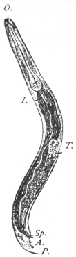
Fig. 266.—Male of the rhabditic form of Angiostomum nigrovenosum. A., anus; I., mid-gut; T., testicular tube; O., oral orifice; P., papillæ; Sp., spicule. Magnified.
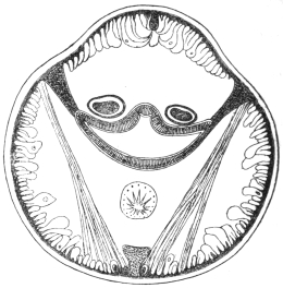
Fig. 267.—Transverse section through the posterior extremity of the body of Ascaris lumbricoides (male). The intestine is in the middle, and the lateral lines are subjoined thereto; above the intestine the two spicule sacs are seen; below is the ductus ejaculatorius. The muscular fibres are between the lateral and median lines. Magnified.
Bursa copulatrix.—The males in many genera possess epidermal wing-like appendages at their posterior extremity. These are supported by elongated tactile papillæ called ribs. In the most highly developed bursæ, e.g., in the Strongylidæ, the ribs are called rays, as they consist not only of nerve fibres but mainly of “pulp,” i.e., prolongations of the subcuticular layer. Bursæ are either open, i.e., bilaterally symmetrical, or closed, when the posterior border is continuous all round. A pseudo-bursa is one unsupported by ribs or rays, e.g., in Trichuris. The bursa serves as an organ of prehension during copulation. Some forms, moreover, carry a sucker at the posterior extremity (e.g., Heterakis); in others the spicules and other prehensile organs are absent; they are then replaced by an evertible cloaca, e.g., Trichinella.
Development of the Nematodes.
After impregnation, the ovum develops around itself a delicate membrane (vitelline membrane), and subsequently an egg-shell is formed. This is derived either as a secretion from the uterine wall or it is a further differentiation of the vitelline membrane, the original single membrane splitting into two, the outer becoming the egg-shell. Further the uterus often secretes a special albuminous covering around the egg-shell. The “yolk” granules of the ovum are secretions of the protoplasm of the ovum itself and first appear when the rhachis is formed. In certain cases ova lie in follicles or capsules formed of epithelium cells derived from the ovarian tubes. These cells subsequently fuse and form a membrane—the CHORION.
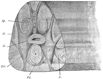
Fig. 268.—Hind end of a male Ascaris lumbricoides cut across at the level of the dilator cells of the gut. D., gut; Dil., dilator cells of the gut; F., a process of the dilator cells forming a network over the vas deferens; Sl., lateral line; Sp., spicule; Vd., vas deferens. The anterior end of the worm lies to the right. Magnified. (After Goldschmidt.)
The shape of the completed eggs is characteristic of the different species, and therefore a single egg often suffices to diagnose the species. According to the species, the eggs may be deposited sooner or later, either before or during segmentation, or with the embryo perfectly developed. Only a few species are viviparous, e.g., Dracunculus mediensis, Trichinella spiralis; in the other Nematodes the further development of the extruded eggs takes place after various lengths of time in the open, in moist earth, or in water. Thick-shelled eggs can maintain their developmental capacity for a long time, even after prolonged desiccation.
Finally, a nematode-like embryo develops, which usually lies somewhat coiled up within the shell, and varies in its further development according to the species to which it belongs.
In the simplest forms, as in the free-living Nematodes, the embryos, apart from their size, resemble their parents, and grow up into these after leaving the egg-shell. In many parasitical Nematodes, however, the young must be called larvæ, as they present characters which are subsequently lost.
The manner of conveyance of the eggs or the embryos contained in them after they have left the body into the definite host is very different in the various species.
(1) Without Intermediate Host.—(a) In many the conveyance into the definite host is effected directly after the larvæ have developed within the eggs; thus, for instance, the feeding of suitable animals with the embryo-containing eggs of species of Trichocephalus and Ascaris leads to an infection of the gut, for the young Trichocephali or Ascarides only leave the egg-shell when they have attained the intestine of the final host, in which they become adult.
In other cases (b) Ancylostoma, Necator, the larvæ hatch in the open, and live for a time free, changing their form; they grow, cast their skin, and finally gain the intestine of the host by means of water or through the skin, when they lose their larval characters and assume the structure of the adult worm.
(c) In a number of Nematodes, however, HETEROGONY occurs. This terms signifies a mode of development in which two structurally different sexual generations of the same species alternate with each other. To these appertains, for instance, Angiostomum (syn.: Rhabdonema) nigrovenosum, which lives in the lungs of frogs and toads; this Nematode measures about 1 cm. in length and is hermaphrodite (protandric). The eggs are deposited in the pulmonary cavity, and through the cilia of the same reach the oral cavity, where they are swallowed and thus conveyed into the intestine. They pass through the entire gut, and are finally evacuated with the fæces; often, indeed, the young themselves emerge from the egg-shell within the hind-gut of the frogs. These young forms become sexually differentiated, remain much smaller than the parent, their œsophagus is differently constructed (rhabditis form), and they are non-parasitic (fig. 266). After having grown in the open they copulate; the males die soon after copulation, and the females in their own bodies develop a few young, which, given the opportunity to get into frogs, infect them, and are transformed into the hermaphroditic Angiostomum. The same manner of development occurs in other species of the same genus, and also in the case of Strongyloides stercoralis.
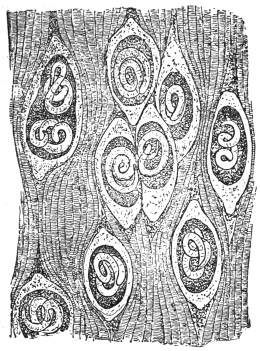
Fig. 269.—A piece of the trunk muscle of the pig with encapsuled embryonic Trichinæ. Magnified.
(2) With Intermediate Host.—(a) Frequently, however, the larvæ of Nematodes make use of one or even two intermediate hosts; their condition then resembles that of Cestodes or Trematodes, excepting that there is never a multiplication within the intermediate hosts. The larvæ become encapsuled amongst the tissues of the intermediate host, and wait till they are introduced with the latter into the final host. For instance, Ollulanus tricuspis, the adult form of which is found in cats, previously lives encysted in the muscular system of mice. Cucullanus elegans, which attains the adult stage in fishes (perch, etc.), is found encysted in species of Cyclops. Other examples of species that require an intermediate host are Filaria bancrofti and Dracunculus medinensis.
Peculiar conditions prevail in the case of (b) Trichinella spiralis. This species, which in its adult state lives in the intestine of man and of various mammals, is viviparous; the young Trichinæ, however, do not leave the intestine, but reach the intestinal wall (Cerfontaine, Askanazy) in the following way: the female intestinal Trichinæ bore into the intestinal wall, where they are found in the submucosa, or in the lumen of the dilated lacteal vessels. Here the young are born, in the intestinal wall, and leave this position with the lymph stream. Some of them, no doubt, actively bore through the intestinal wall, reaching the lymph or blood-stream, or even pass into the body cavity. What occurs during their further migrations is difficult to say at present. It has hitherto been maintained that the wandering is entirely active; for instance, the ligaturing of an artery would be no protection against the part of the body supplied by such artery being invaded by Trichinella. This observation cannot be otherwise explained than by the active progress of the young Trichinella. The question, however, may be mooted as to where and when the worms quit the blood-vessels, which they for the most part reach through the thoracic duct, the natural connection between the vascular system and the lymphatic system, to wander further independently, and ultimately reach the muscular system, in which they become encysted (fig. 269). Thus the progeny does not leave the body of the host inhabited by the parents, as is generally the case amongst helminthes, but uses it as an intermediate carrier to reach another host, which is then the final host. The latter may belong to another species, or may be another individual of the same species. This second migration is, of course, purely passive.
Classification of the Nematoda.
The Nematodes are usually divided into a number of families, some of which it is at present impossible to define accurately; moreover, the definition of many genera is also in an unsatisfactory state.
Family. Anguillulidæ, Gervais and van Beneden, 1859.
A “family” name not definable. They comprise a vast number of small forms, most of which live free in fresh water, in soil, or in macerating substances; amongst them there are some which live parasitically on plants, more rarely on animals. They do not exceed 8 mm. in length. The large majority are only 1 to 2 mm., or even 0·5 mm. The uterus is straight. Eggs in the uterus at one time, one to four. Genera very numerous, but many of them insufficiently defined (Anguillula, Anguillulina, Rhabditis, Heterodera, etc.).
Family. Angiostomidæ, Braun, 1895.
Small Nematodes undefined morphologically, but characterized by heterogony, i.e., there is a free-living “rhabditic” generation and a parasitic “filariform” generation which succeed one another (e.g., Angiostomum, Strongyloides, Probstmayria).
Family. Gnathostomidæ.
Cuticle covered totally or partly with cuticular laminæ fringed posteriorly with multiple points. Head subglobular, covered with simple spines. Two spicules. Vulva behind middle of body, parasitic in vertebrates, especially mammals (e.g., Gnathostoma, Tanqua, Rictularia).
Family. Dracunculidæ, Leiper, 1912.
Males very small in proportion to females. Anus absent. Vulva absent (?). Genera: Dracunculus, Icthyonema (in body cavity of eel and other fish).
Family. Filariidæ, Claus, 1885.
Long thread-like Nematodes. Anus present. Œsophagus without bulb. Vulva usually in anterior half of body. Two ovaries. Generally ovoviviparous. Development often requires an intermediate host. This family is at present ill-defined, but has been already subdivided into several sub-families, Filariinæ, Onchocercinæ, Arduenninæ.
Family. Trichinellidæ, Stiles and Crane, 1910.
Œsophagus consisting of a chain of single cells, the lumen of the œsophagus passing through their centre. Ovary single. Vulva at junction of anterior and posterior portions. Sub-families: (1) Trichurinæ, (2) Trichinellinæ.
Family. Dioctophymidæ.
Body anteriorly armed with spines or unarmed; mouth without lips, with six, twelve, or eighteen papillæ in one or two circles; œsophagus very long without a bulb; anus terminal in female; one ovary; vagina very long; spicule in male very long; bursa cup-shaped without rays (Dioctophyme, Hystrichis, Eustrongylides).
Family. Strongylidæ, Cobbold, 1864.
Bursa, supported by rays, always present. Oviparous.
Family. Physalopteridæ.
Mouth with two large lips. Bursa with supporting papillæ in form of a lanceolate cuticular expansion, with genus Physaloptera.
Family. Ascaridæ, Cobbold, 1864.
Rather thick Nematodes. Mouth with three lips—one dorsal, two latero-ventral. Sub-families: (1) Ascarinæ, (2) Heterakinæ, etc.
Family. Oxyuridæ.
Smallish forms, 4 to 45 mm., with cuticle thickened on each side for the whole length of body in the form of a lateral flange or wing. Œsophagus long with a well-marked bulb containing a valvular apparatus. Tail end of female drawn out into a long point. Eggs asymmetrical. Males very small (about 2 mm.). One spicule. Genera: Oxyuris, Passalurus, Ozolaimus, Atractis, etc.
Mermithidæ, greatly elongated “Nematodes,” which, in the larval stage, are parasitic in insects, but in their adult condition are free living. Cuticle with diagonal striation. Without an open mouth or anus. Oral papillæ present. Characteristic eggs with two processes, ending in a tuft of filaments. Larvæ with a movable boring spine at the head end.
Gordiidæ.—Long, thread-like “Nematodes.” Mouth and anterior portion of gut atrophied in adult. Oral papillæ absent.
The Nematodes Observed in Man.
FamilySub-familyGenusSpecies
Anguillulidæ—RhabditisR. pellio.
R. niellyi.
Rhabditis sp.
AnguillulaA. aceti.
AnguillulinaA. putrefaciens.
Angiostomidæ—StrongyloidesSt. stercoralis.
Gnathostomidæ—GnathostomaGn. siamense.
Gn. spinigerum.
Dracunculidæ—DracunculusD. medinensis.
FilariidæFilariinæFilariaF. bancrofti.
F. demarquayi.
F. taniguchi.
F. (?) conjunctivæ.
Group. Agamofilaria—Ag. georgiana.
Ag. palpebralis.
Ag. oculi humani.
Ag. labialis.
F. (?) romanorum- orientalis.
F. (?) kilimaræ.
F. (?) sp. ?
(Mikrofilaria)Mf. powelli.
Mf. philippinensis.
SetariaS. equina.
LoaL. loa.
AcanthocheilonemaAc. perstans.
DirofilariaDi. magalhãesi.
OnchocercinæOnchocercaO. volvulus.
TrichinellidæTrichurinæTrichurisT. trichiura.
TrichinellinæTrichinellaT. spiralis.
Dioctophymidæ—DioctophymeD. gigas.
StrongylidæMetastrongylinæMetastrongylusM. apri.
TrichostrongylinæTrichostrongylusT. instabilis.
T. probolurus.
T. vitrinus.
HæmonchusH. contortus.
Mecistocirrus (Nematodirus)M. fordi.
Ancylostominæ
Group. ŒsophagostomeæTernidensT. deminutus.
ŒsophagostomumŒ. brumpti.
Œ. stephanostomum var. thomasi.
Œ. apiostomum.
Group. AncylostomeæAncylostomaA. duodenale.
A. ceylanicum.
A. braziliense.
Group. BunostomeæNecatorN. americanus.
N. exilidens.
Group. SyngameæSyngamusS. kingi.
Physalopteridæ—PhysalopteraP. caucasica.
P. mordens.
AscaridæAscarinæAscarisA. lumbricoides.
A. sp.
A. texana.
A. maritima.
ToxascarisT. limbata.
BelascarisB. cati.
B. marginata.
LagocheilascarisL. minor.
Oxyuridæ—OxyurisO. vermicularis.
Mermithidæ—MermisM. hominis oris.
(Agamomermis)Ag. restiformis.
Family. Anguillulidæ.
Genus. Rhabditis, Dujardin, 1845.
Buccal cavity elongated, with lips. Its chitinous wall uniformly thick. Lateral lines absent. Males with bursa.
Rhabditis pellio, Schneider, 1866.
Syn.: Pelodera pellio, Schn., 1866; Rhabditis genitalis, Scheiber, 1880; Rhabditis pellio, Schn., 1866.
Males 0·8 to 1·05 mm. in length; females, 0·9 to 1·3 mm. in length. The posterior extremity of the body of the male has a heart-shaped bursa, and seven to ten ribs on each side; the bursa may, however, be lacking. The spicules measure 0·027 to 0·033 mm. in length, but are never quite alike. The posterior extremity of the female is long and pointed; the vulva lies somewhat behind the middle of the body, the ovary is single, the eggs are oval, 60 µ by 35 µ.
This species was found in Stuhlweissenburg by Scheiber in the acid urine (containing albumin, pus and blood) of a woman suffering from pyelonephritis, pneumonia and acute intestinal catarrh; the observer was able to convince himself that the Nematodes which were found during the whole period of the illness lived in the vagina, and were evacuated with the urine.
Oerley proved that this species had long been known; during its larval stage (Anguillula mucronata, Grube, 1849) it lives in earthworms; in its adult stage it lives in decomposing matter in the soil. By introducing individuals of this species into the vagina of mice, Oerley succeeded in obtaining infection and multiplication (facultative parasitism). These Nematodes must in some such way have got into the vagina of Scheiber’s patient.
Two other cases described by Baginsky and Peiper probably belonged to the same or a nearly related species.
Rhabditis niellyi, Blanchard, 1885.
Syn.: Leptodera niellyi, Blanchard, 1885.
In 1882 Nielly had a cabin-boy, aged 14, under observation in Brest. The lad had never left the neighbourhood of Brest, and had suffered from itching papules on the skin for five or six weeks; in the papules the observer found one or several rhabdites, measuring 0·33 mm. in length by 0·30 mm. in breadth. Their cuticle presented a delicate transverse striation; the intestine was the only internal organ recognizable, and it opened somewhat in front of the posterior extremity. Therefore, it must have belonged to the rhabditis-like larva of a Nematode, the adult stage of which is unknown.
The manner of infection was established almost certainly by a further observation of Nielly’s: at the commencement of the illness small Nematodes were found in the blood of the patient; later on, however, they disappeared, neither were Nematodes found in the fæces, urine or sputum. Therefore it must be concluded that the cabin-boy, who was in the habit of drinking water from brooks, had thus ingested embryo-containing eggs of a Nematode; the young hatched out in the intestine, perforated it, reached the blood and then settled in the skin; but, on the other hand, the entry may have been direct through the skin.
In connection with the foregoing, reference should be made to a communication by Whittles, insufficient from a zoological point of view. In a case of hypertrophic gingivitis occurring in a female patient, aged 19, who had never left Birmingham, he found Nematode larvæ in the periosteum of the upper jaw, which was excised after extraction of the right premolar; the genital rudiment could be recognized in them. Similar larvæ were found in the same patient in abscesses in various regions of the skin, and in the case of her mother in the blood. The author considers that the infection took place through a dog, and refers to the case of O’Neil (1875), who found Filariæ in the skin (in the condition known as “craw-craw”), referred by Manson to Filaria perstans. O’Neil’s case was quoted, and attributed to Filaria sanguinis hominis. In conclusion, the author states that he has repeatedly found Nematode larvæ in the blood of persons who suffered from pruritus; in his opinion the parasite had been imported through the agency of troops returned from South Africa. Glatzel found true Filaria larvæ in a pustule of a cutaneous eruption of the trunk and extremities in a patient at Dar-es-Salam.
Skin diseases which are caused by young Nematodes are also observed in dogs (Siedamgrotzky, Möller, J. G. Schneider, Künnemann), foxes (Leuckart), and horses (Semmer). Zürn found young Nematodes (Anguillulidæ) also in pig’s flesh. In Künnemann’s case it was shown that the adult Rhabdites lived in the straw upon which the dog lay.
Rhabditis, sp.
In the fluid obtained by lavage from the stomach of a female patient, aged 16, suffering from ozæna, O. Frese found during two consecutive months Rhabdites of various ages, 0·275 to 0·64 mm. in length, the adults all with eggs; males were not found; transmission into rabbit’s stomach failed, but they could be kept alive in much diluted hydrochloric acid (2 : 1,000) for several weeks. Neither eggs nor larvæ appeared in the fæces of the patient. The nature of the infection, which was perhaps of unique occurrence, remained doubtful.
Genus. Anguillula, Ehrenberg, 1826.
Buccal cavity very small, without lips. Males without bursa, but with a series of papillæ. Lateral lines absent.
Anguillula aceti, Müller, 1783.
Cuticle unstriped, body cylindrical, anterior end tapering but little, posterior end long, pointed. Male up to 1·45 mm. long, 0·024 to 0·028 mm. wide; two pre-anal papillæ, one post-anal; spicules equal, curved, 0·038 mm. long; gubernaculum present; testis extending in front of mid-line of body. Female up to 2·4 mm. long, 0·040 to 0·072 mm. wide; anterior uterus reaching to near the œsophagus, posterior to hind gut. Viviparous; embryos in both or only in one uterine horn, 0·22 mm. long, 0·012 mm. broad.
The species is a frequent inhabitant of vinegar (prepared by older methods), and was once observed for some time by Stiles and Frankland in the urine of a woman; the urine had an acid reaction, and once had a distinct odour of vinegar. It was assumed that the patient, who was hysterical and suffered from chronic nephritis, employed vaginal douches with diluted vinegar, perhaps to deceive her physician or to protect herself against conception. According to Ward, Billings and Miller are said to have reported on two other cases. Ill-effects which might be connected with the vinegar eel (Anguillula aceti) were not present.
Genus. Anguillulina, Gervais and Beneden, 1859.
Syn.: Tylenchus, Bastian, 1864.
Characterized by the possession in the buccal cavity of a spine knobbed posteriorly; bursa present; uterus asymmetrical. Numerous species parasitic in plants.
Anguillulina putrefaciens, Kühn, 1879.
Syn.: Tylenchus putrefaciens, Kühn; Trichina contorta, Botkin, 1883.
In 1883 Botkin (Pet. klin. Wochenschr., 1883) found a small Nematode, which was, however, entirely mistaken, in the material vomited by a Russian; this was not a species of Trichinella, but an Anguillulina living in onions which had already, in 1879, been described by Kühn as Tylenchus putrefaciens; the Nematodes got into the stomach with the onions, causing nausea and vomiting.
Family. Angiostomidæ, Braun, 1895.
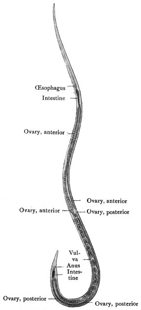
Fig. 270.—Strongyloides stercoralis, female: parasitic generation from gut of man. × 70. (After Looss.)
Genus. Strongyloides, Grassi, 1879.
Syn.: Pseudorhabditis, Perroncito, 1881; Rhabdonema, Leuckart, 1882, p.p.
The genus is insufficiently defined. The parasitic form possesses a simple mouth opening directly into the long cylindrical œsophagus which occupies the anterior third of the body. The free-living forms possess a small buccal cavity; the œsophagus is short, with a double bulb, in the hinder one there is a Y-shaped chitinous valve; two spicules of equal size.
Strongyloides stercoralis, Bavay, 1877.
Syn.: Anguillula intestinalis et stercoralis, Bavay, 1877; Leptodera intestinalis et stercoralis, Cobb.; Pseudorhabditis stercoralis, Perroncito, 1881; Rhabdonema strongyloides, Leuckart, 1883; Strongyloides intestinalis, Grassi, 1883; Rhabdonema intestinale, Blanchard, 1886.
In 1876, a number of French soldiers returned to Toulon from Cochin China suffering from severe diarrhœa. Dr. Normand, under whose treatment they were, discovered a large number of Nematodes in the evacuated fæces, and Bavay described them as Anguillula stercoralis. Soon after Normand, at the post-mortem of five patients who had died of Cochin China diarrhœa, found numerous other Nematodes in the intestine, from the stomach to the rectum, in the bile-ducts and in the pancreas, and these he handed over to Bavay. The latter diagnosed another species, and described them as A. intestinalis. Both forms were then regarded as the cause of Cochin China diarrhœa until, in 1882, Leuckart was able to demonstrate that the two forms are only two succeeding generations of the same species, of which the one (A. intestinalis) lives parasitically in the intestine, whereas its young (A. stercoralis) attain the open, where they come to maturity and propagate. The young of these again live parasitically. There thus exists the same heterogony as was discovered by Leuckart in Angiostomum nigrovenosum of frogs, which heterogony, indeed, according to v. Linstow, appertains to the entire family of the Angiostomidæ.
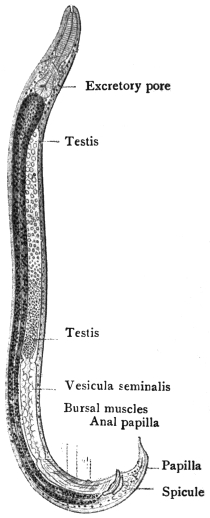
Fig. 271.—Strongyloides stercoralis, male: free-living generation. × 170. (After Looss.)
(1) The parasitic generation (strongyloid or filariform ♀) is quite colourless and cannot be seen in situ even with a lens. To detect them it is necessary to scrape the mucosa of the jejunum and examine the scrapings microscopically. It measures 2·2 mm. in length, and 34 µ to 70 µ in breadth; the cuticle is finely transversely striated; the mouth is surrounded by four lips; the œsophagus is almost cylindrical and a third the length of the entire body. The anus opens shortly in front of the pointed posterior extremity; the vulva is situated at junction of middle and posterior thirds of the body; the uterus has no special ovejector; the eggs measure 50 µ to 58 µ in length, and 30 µ to 34 µ in breadth, and lie in a chain one behind the other (fig. 270). As in the case of Angiostomum nigrovenosum, Leuckart considers this stage to be hermaphroditic, the testes degenerating after having functioned; other authors (Rovelli) regard it as a female reproducing by parthenogenesis.
(2) The free-living generation (♂ and ♀) has a smooth body, cylindrical, somewhat more slender at the anterior extremity and pointed at the tail end. The mouth has four indistinct lips; the œsophagus is short with a double (rhabditis-like) bulb; there is a Y-shaped valve in the posterior bulb; the anus opens in front of the tail end. The males measure 0·7 mm. in length, 0·035 mm. in breadth. Their posterior end is rolled up; the two brown spicules are small (38 µ) and much curved. There is also a gubernaculum. The females measure 1 mm. in length or a little over; 0·05 mm. in breadth. The tail end is straight and pointed; the vulva lies somewhat behind the middle of the body. The yellowish, thin-shelled ova measure 70 µ in length and 45 µ in breadth.
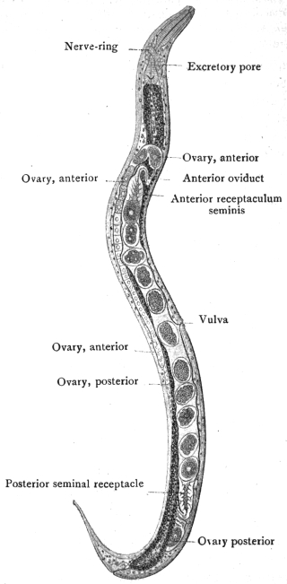
Fig. 272.—Strongyloides stercoralis, female; free-living generation, × 170. (After Looss.)
As Askanazy has shown, the parasitic form bores deeply into the mucous membrane of the intestine, and frequently into the epithelium of Lieberkühn’s glands, both for nourishment and oviposition. The eggs then develop in the intestinal wall. The eggs which are found in scrapings from the mucosa occur, at least in the case of Strongyloides of the sheep, in chains enclosed in a thin tube or sheath, the origin of which is doubtful; possibly it is the uterus. The eggs themselves are only rarely found in stools, e.g., after a strong purge. The larvæ, which are hatched out, and measure 0·2 to 0·25 mm. long by 0·016 mm. broad, again reach the lumen of the intestine,299 and grow to double or three times that size, until they are passed out with the fæces. They already differ from the parent (♀) in the shape (rhabditiform) of the œsophagus. When the external temperature is sufficiently high (26° to 35° C.), they become sexually mature after moulting. In about thirty hours they are completely developed and copulate, now forming the free-living rhabditiform generation. At lower temperatures the larvæ only moult, but do not escape from the old cuticle and do not develop further. At a temperature of about 25° C. only some of the larvæ attain maturity.
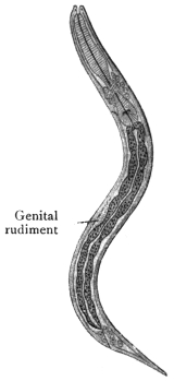
Fig. 273.—Strongyloides stercoralis: larva from fresh human fæces. × 310. (After Looss.)
The females of the free-living generation (rhabditiform) deposit from thirty to forty eggs, which develop rapidly, sometimes even within the uterus in the case of old females. After the larvæ have emerged from the egg-shell, they measure 0·22 mm. in length, and possess the characteristics of the parents (rhabditiform larvæ). When they have grown to 0·55 mm. they moult, and while losing their own characteristics they acquire the characteristics of their parasitic grandparents (strongyloid or filariform). After about eight days the free-living adult generation in the cultures have disappeared, and all the rhabditiform larvæ have been transformed into strongyloid or filariform larvæ; they then die off unless they reach the intestine.
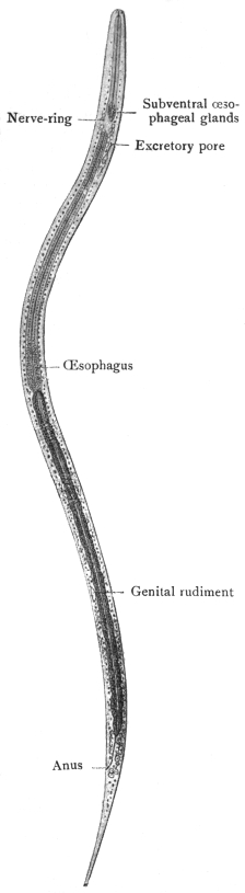
Fig. 274.—Strongyloides stercoralis: mature filariform larva showing long transparent œsophagus, slender granular intestine and characteristic tip to the tail ending in two small points. × 620. (After Looss).
This cycle of development holds good for Strongyloides stercoralis of tropical origin (Bavay, Leuckart, Leichtenstern, Zinn). In the European Strongyloides the free-living generation, as a rule, is absent (Grassi, Sonsino, Leichtenstern, Braun); the rhabditis-like larvæ evacuated with the fæces are transformed into the strongyloid or filariform type of larva (in cultures which are easily made) which will only become adult if introduced into man.
So that we have these two cycles: (A) (1) ♀ parasitic, (2) eggs, the rhabditiform larvæ in fæces, (3) free-living strongyloid or filariform larva, (4) ♀ parasitic. (B) (1) (2) (3) as before, then (4) adult ♀ and ♂, free living, (5) eggs, (6) rhabditiform larva, (7) strongyloid or filariform larva, (8) ♀ parasitic.
Infection of man results not only from direct entry into the stomach but also, according to van Durme and Looss, through the skin.
Occurrence in Man.—As already mentioned, Strongyloides stercoralis was first observed in persons suffering from so-called Cochin China diarrhœa. From the enormous numbers of parasites evacuated with the fæces, the cause of the disease was apparently evident. It appeared, however, that only some of the soldiers returning from Cochin China and Martinique, and suffering from diarrhœa, harboured Strongyloides (Chauvin). Breton made the same observations in Cochin China and found that only 10·4 per cent. of cases of chronic dysentery, and 8·8 per cent. of chronic diarrhœa, show Strongyloides. Normand, moreover, found that only a few of the Europeans residing in Cochin China are exempt from S. intestinalis, yet the people exhibit no intestinal symptoms; if, however, from any cause a catarrhal condition of the intestine supervenes the condition is changed, the parasites appear in larger numbers, and the disorder is considerably intensified.
S. intestinalis, besides being present in the Indo-China region, also occurs in the Antilles, in Brazil, Africa, and Europe; in 1878 it was discovered in Italy by Grassi and C. and E. Parona; in 1880 it was also found in the labourers working at the St. Gothard tunnel. It was imported into Germany, Belgium, and the Netherlands by Italian labourers. One sporadic case has been observed in East Prussia, and the worm has also been reported from Siberia.
In mammals the following species are found: Probstmayria (Strongyloides) vivipara, Ransom, 1907, in Equus caballus; Strongyloides fülleborni, v. Linst., in Anthropopithecus troglodytes and Cynocephalus babuin.
Their development is, so far as is known, the same as that of Strongyloides stercoralis (v. Linstow, Centralbl. f. Bakt., Path. u. Infektionsk., 1905, Orig. xxxviii, p. 532).
Family. Gnathostomidæ.
Genus. Gnathostoma, Owen, 1836.
Syn.: Cheiracanthus, Diesing, 1839.
Easily recognizable by the numerous spines which cover the entire body or only the anterior extremity, and terminate in several points; head globular and beset with bristles; mouth with two lips; two spicules; vulva situated behind the middle of the body.
Gnathostoma siamense, Levinsen, 1889.
Syn.: Cheiracanthus siamense, Lev., 1889.
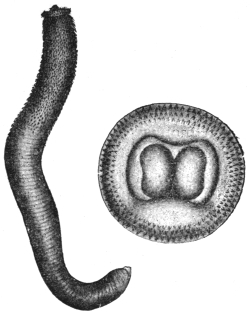
Fig. 275.—Gnathostoma siamense: to the left, the entire worm (8/1); to the right the head seen from above, with two fleshy lips (about 40/1). (After Levinsen.)
Female measures 9 mm. in length, 1 mm. in breadth. There are eight rows of simple spines on the head; the armature of spines extends over the anterior third of the body only; each spine on the anterior region of the body spreads into three points, of which the middle one is the longest; the posterior spines are simple; they gradually become smaller and then disappear entirely. The vulva is situated behind the middle of the body.
Male.—10·5 mm. long by 0·6 mm. broad. Head terminates in a globular swelling with two large lips. Neck 3 mm. broad. In front of neck eight rows of simple spines directed backwards. Anterior half of body with cuticular laminæ, posterior unarmed. Two pre-anal and two post-anal papillæ. Bursa wanting.
Spicules 1·1 and 0·4 mm. respectively.
Leiper considers Gnathostoma siamense to be identical with Gnathostoma spinigerum.
The single specimen described by Levinsen was found by Deuntzer in Bangkok (Siam), and was obtained from a young Siamese woman who suffered from a small tumour of the breast which had developed in the course of a few days. After the disappearance of the tumour, nodules the size of beans were found in the skin; out of one of these the worm was obtained. The same observer saw this affection in two other persons.
A closely related species, Gnathostoma spinigerum, Ow., lives in the stomach of wild cat (Felis catus), puma (Felis concolor), tiger (Felis tigris), and domestic cat (India); another species, Gnathostoma hispidum, Fedsch., 1839, in the stomach of pigs in Turkestan, Annam, Hungary, Congo, and by Collin in the stomach of an ox (Berlin).
Gnathostoma sp. in pariah dogs, Calcutta. Gnathostoma sp. in monkeys, French Guiana. They produce large fibrous thickenings in the stomach wall.
Gnathostoma spinigerum, Owen, 1836.
Cuticle of bulb with eight rows of chitinous laminæ with their posterior edges notched into spines. The laminæ on the anterior portion of the body are similar trident laminæ. In the middle of the body, the laminæ are simple and conical, cuticle posteriorly is unarmed. Mouth with two fleshy lips.
Male 5 mm. long by 0·5 mm. broad; tail spiral, four pairs of papillæ.
Female about twice as long; tail straight, trilobed.
Family. Dracunculidæ, Leiper, 1912.
Genus. Dracunculus, Kniphoff, 1759.
Anterior end rounded with a cuticular thickening or shield. Mouth triangular with two lips. Alimentary canal atrophied.
Dracunculus medinensis, Velsch, 1674.
Syn.: Vena medinensis, Velsch, 1674; Dracunculus persarum, Kämpfer, 1694; Gordius medinensis, Linné, 1758; Filaria dracunculus, Bremser, 1819; Filaria æthiopica, Valenciennes, 1856; Dracunculus medinensis, Cobbold, 1864; Guinea worm, Medina worm.

Fig. 276.—Guinea worm (Dracunculus medinensis). (After Leuckart.)
The females attain a length of 50 to 80 cm., or even more, and average 1·5 to 1·7 mm. in diameter. They are whitish or yellowish in colour. The anterior extremity is roundish and bears a cuticular thickening or shield. The triangular mouth opening is surrounded by two projections or lips, behind which on the shield there are two lateral and four sub-median papillæ; the posterior end terminates in a spine, ventrally directed, and about 1 mm. in length; the alimentary canal below the œsophagus is atrophied, but not entirely obliterated; anus absent; the lateral lines are very flat. The greater part of the body is occupied by the long uterus, in which a great number of young larvæ are always found. The ovaries probably lie at the ends of the uterus; the vulva lies just behind the cephalic shield. During parturition the uterus is prolapsed through this opening.
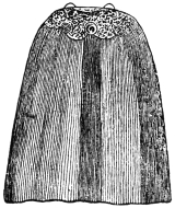
Fig. 277.—Anterior extremity of Guinea worm, showing dorsal and ventral lips, one lateral and two submedian papillæ and the lateral line. (After Leuckart.)
The male is almost unknown. Leiper in an experimentally infected monkey found two males 22 mm. long, one from the psoas muscle, the other from the connective tissue behind the œsophagus.
Occurrence.—Filaria medinensis has been known since the most remote period. The “fiery serpents” that molested the Israelites by the Red Sea, and which Moses mentioned, were probably filariæ. The term Δρακὁντιον occurs in Agatharchides (140 B.C.). Galen called the disorder dracontiasis; the Arabian authors were well acquainted with the worm. It is found not only in Medina or Arabia, but also in Persia, Turkestan, Hindustan. The Guinea worm is also widely distributed in Africa, on the coasts as well as in the interior. It occurs in the Fiji Islands. It was carried to South America by negro slaves, but is said at the present time to exist in only quite a few places (British Guiana, Brazil [Bahia]); it is also observed in mammals (ox, horse, dog, leopard, jackal [Canis lapuster], etc.).
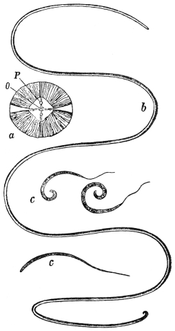
Fig. 278.—Dracunculus medinensis. a, anterior extremity seen end on; O, mouth; P, papillæ; b, female reduced more than half; c, larvæ enlarged. (After Claus.)
Dracunculus medinensis in its adult stage lives in superficial ulcers on the body surface; it is seen most frequently on the lower extremities, more especially in the region of the ankle, but it also occurs in other parts of the body—on the trunk, scrotum, perineum, on the upper extremities, and in the eyelids and tongue. Sometimes there is only one ulcer and one worm, but more commonly several. It attacks man without distinction of race, age or sex. It is observed most frequently during the months of June to August.
Life history.300—When about a year old the worm seeks the surface of the body and produces there a thickening as big as a florin. Over this a vesicle forms which eventually ruptures, and at the bottom of the ulcer can be seen a hole from which a part of the worm may project. On bathing the sides of the ulcer with water, a drop of fluid, at first clear then milky, exudes. This contains numerous larvæ. In other cases a thin tube an inch long is prolapsed (through the vulva). This is probably the uterus, but the mechanism of parturition is not clearly known. It lasts for about a fortnight. An abundant supply of larvæ can be got by placing wet compresses on a fresh ulcer. In a few hours a mass of larvæ is obtained.
The larvæ are 500 µ to 750 µ by 15 µ to 25 µ, with a long slender tail about one-third of the total length. The cuticle is transversely striated. The body is flattened. They possess an œsophagus and gut. At the anus there are apparently glandular structures.
The larvæ live and move actively in water for about two days, the majority dying on the third (Leiper). If a number of Cyclops sp. have been collected and isolated in clean water, and the larvæ are now added, the further development can be traced.
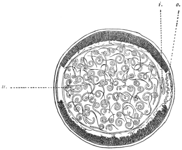
Fig. 279.—Transverse section of female Guinea worm; u., uterus containing embryos; i., intestinal canal; o., ovary. (After Leuckart.)
The larvæ enter the Cyclops, according to most authorities, by penetrating the exoskeleton, but according to Leiper this is impossible; they must enter by the mouth and penetrate the gut in order to reach the body cavity. In eight days moult 1 takes place, the striated cuticle being cast off. In ten days moult 2 takes place. In five weeks the larva is mature. If now the infected Cyclops is placed in 0·2 per cent. HCl solution the Cyclops is killed immediately, but the larvæ are stirred into activity, escape from the body, and swim about in the acid. This suggests that infection in nature probably takes place by the swallowing of infected Cyclops; Leiper, by feeding Cyclops containing mature larvæ to a monkey, found in it, post mortem six months later, two immature females 30 cm. long and two males 22 mm. long.
In certain areas the new cases occur principally in June. Five weeks later the larvæ will become mature in Cyclops, so that infection of Cyclops is taking place in July or August, and from then to June about ten months elapse, giving the period of development in man.
Pathology.—The initial induration is accompanied by itching. Urticarial eruptions are described in Dahomey and Mauretania accompanied by fever, rigors, blood-shot conjunctiva, and prostration resembling fungus poisoning. Symptoms last for one to two days, later the worms appear on the surface.
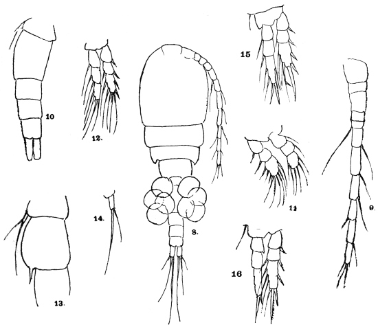
Fig. 280.—Cyclops virescens, ♀. 8, Female, ventral view, × 120; 9, anterior antennæ × 240; 10, urosome and last thoracic segment, × 240; 11, foot of first pair, × 320; 12, 15, 16, foot of second, third and fourth pairs, × 240; 14, foot of fifth pair, × 440; 13, last thoracic segment and first segment of urosome of male, × 240.
If the worm is ruptured in an attempt to extract it, disastrous results may occur through the escape of the larvæ into the tissues: fever, inflammation, abscess, sloughing, ankylosis, even death from sepsis. Eosinophilia is often marked, 11 to 13 or even 50 per cent.
Extraction.—(1) The native method consists in rolling the worm round a stick; 1 in. to 2 in. are extracted each day, the process taking about a fortnight; (2) Emily used injections of 1 in 1,000 sublimate into the swelling or into the worm itself fixed by a ligature. (3) Béclère chloroforms the worm; (4) the worm can be more easily removed when all the embryos have been deposited (two to three weeks).
Cyclopidæ.—Cephalothorax ovate, clearly separated from abdomen. Anterior antennæ of female when bent back scarcely ever stretch beyond the cephalothorax. The second antennæ are unbranched. First four pairs of feet two-branched, outer branches three-jointed. The fifth pair of limbs are rudimentary alike in both sexes, usually one-jointed. There is no heart. The female has two egg sacs containing about fifty eggs.
Genus. Cyclops, Müller, 1776.
Mandible palp rudimentary, reduced to a tubercle bearing two branchial filaments. Maxillary palp rudimentary (obsolete). Lower foot-jaw non-prehensile. Head ankylosed to first thoracic segment.
Family. Filariidæ.
Sub-family. Filariinæ.
The residue after exclusion of the Arduenninæ and Onchocercinæ.
Genus. Filaria, O. Fr. Müller, 1787.
Very long, slender Nematodes, without excretory vessels or excretory pore, the males of which are usually considerably smaller than the females. Mouth round, without lips, unarmed. The lateral lines occupy one-sixth of the circumference of body. The tails of the males are bent or spirally rolled, and bear little wing-like appendages. The two spicules are unequal; almost always there are four pre-anal papillæ, but the number of post-anal papillæ varies. The vulva is always situated at the anterior extremity. Parasitic chiefly in the serous cavities and in the subcutaneous connective tissue. Insufficiently defined.
Filaria bancrofti, Cobbold, 1877.
Syn.: Trichina cystica, Salisbury,301 1868 (nec Filaria cystica, Rud., 1819); Filaria sanguinis hominis, Lewis, 1872; Filaria sanguinis hominis ægyptiaca, Sonsino, 1875; Filaria wüchereri, da Silva Lima; Filaria sanguinis hominum, Hall, 1885; Filaria sanguinis hominis nocturna, Manson, 1891; Filaria nocturna, Manson, 1891.
These parasites of man were for a long time only known in their larval stage. They were discovered in 1863 in Paris by Demarquay, in the hydrocele fluid of a Havanese emptied by puncture; they were next observed by Wücherer, in Bahia, in the urine of twenty-eight cases of tropical chyluria; they were likewise observed in North America by Salisbury, who gave them the name of Trichina cystica. The next discoveries (in Calcutta, Guadeloupe, and Port Natal) related to chyluria patients, until Lewis discovered the larvæ in the blood of man (India), and found they were almost always present in persons suffering from chyluria, elephantiasis, and lymphatic enlargements; he also, in exceptional cases, found them in apparently healthy persons (Filaria sanguinis hominis). Lewis and Manson studied the disease and the filariæ of the blood very minutely, and became aware that the filariæ were sucked up by mosquitoes with the blood. Manson described the metamorphoses that take place within the body of the mosquito. The adult female was discovered in Queensland by Bancroft, and soon after Lewis found it in Calcutta; it was described by Cobbold as F. bancrofti. The male was first seen by Bourne in 1888.
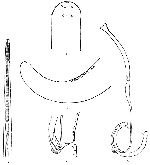
Fig. 281.—Filaria bancrofti. 1, Anterior portion of male; 2, two rows of papillæ on head; 3, papillæ of tail of male; 4, cloaca of male showing tips of spicules and gubernaculum; 5, the spicules and gubernaculum of male. (After Leiper.)
Head bougie-like, i.e., separated by a narrowing from the neck, having two rows of minute papillæ. Cuticle has extremely fine striations.
Female.—50 to 65 mm. long by 1·5 to 2 mm. broad. Vulva 0·4 to 0·7 mm. behind the head. Anus about 1/4 mm. from the tip of the tail (vulva 1 to 1·3 mm. from head, and anus 0·17 to 28 mm. from tail according to other authors). The vagina is a muscular tube forming three bold loops, and has terminally a pyriform enlargement. Uterus double (or single). Ovoviviparous.
Male.—25 to 30 mm. long by 0·1 mm. thick (40 by 0·1 mm. according to various authors). Probably two pairs of pre-anal papillæ, eight pairs of peri-anal, two pairs of post-anal papillæ, and one pair terminal. Tail curved. Two spicules, 0·2 and 0·6 mm. respectively, and a cup-like gubernaculum. The long spicule is cylindrical, expanded proximally and tapering distally to a filament with wings. At the tip it is spoon-like. The short spicule is of the same diameter throughout. It is gutter-like, coarsely marked. Testis uncoiled, terminating in a snowdrop-like process (Leiper).
Eggs.—40 µ by 25 µ. They do not appear to possess a true shell, but only an embryonal or vitelline membrane secreted by the ovum.
Embryos.—In the posterior part of the uterus eggs occur, in the anterior part embryos; the larvæ at birth measure 127 µ to 200 µ by 8µ to 10 µ. In the blood they measure in the fresh 260 µ by 7·5 µ to 8 µ. In stained films, owing to shrinkage, there is great variation in size, from 154 µ to 311 µ. Probably 260 µ to 285 µ is the average in stained films.
Geographical Distribution.—Europe: Two cases recorded, one from near Barcelona. The patient suffered from hæmato-chyluria and enlarged scrotum with mikrofilariæ in the blood. A second case from Siena. Africa: The filarial index has not been estimated for various parts. In Nigeria it is about 10 per cent.
Habitat.—Lymphatic glands: e.g., inguinal, femoral, iliac, lumbar, mesenteric, bronchial, superficial cervical, epitrochlear.
Lymphatic vessels: e.g., those draining into the receptaculum chyli of the spermatic cord, in the thoracic duct and in various different parts.
Organs, etc.: Testis, epididymis, spermatic cord, tunica vaginalis, mammary cyst, and in abscesses.
They may occur in masses, but usually only a few (one to eight). Females are commoner than males. Dead and calcified worms are common in the various sites.
Distribution of Larvæ in Body.—These are by no means uniformly distributed, but occur in greater number in the capillaries of the lungs. Besides the lungs they occur in the capillaries of other organs, as the following data of Rodenwaldt show:—
MikrofilariæMikrofilariæ
Lungs134,821†Spleen1,666
Liver4,884 Brain3,833
Kidneys15,253 Glands0
Glomeruli8,008 Marrow0

Parenchyma7,245 Blood3,000
† These figures refer to 1 c.c. of each organ, and were estimated by cutting sections of definite thickness (30 µ to 40 µ) and counting the filariæ in a definite area of section, e.g., 1/4 cm.2 The organs before removal from the body have their vessels tied, and are then fixed in hot alcohol.
The following data of Rodenwaldt refer to the larvæ of Filaria immitis in the dog. They are commoner in organs than in vessels, and especially in the capillaries of the organs, but in the lungs they appear to be equally distributed in capillaries, arteries and veins.
The length of life of larvæ is unknown, but they appear to be destroyed in the kidneys, as dead calcified specimens are fairly numerous in the capillaries of the vasa recta of the medullary substance.
Kidneys: mainly in the glomerular capillaries and those of the vasa recta.
Liver: in the capillaries of the portal system, especially in those between the interlobular and the central intralobular veins.
Periodicity of Larvæ.302—Roughly speaking, the larvæ of Filaria bancrofti are found in the peripheral blood only during the night, disappearing (but not entirely) during the daytime. Their periodicity and that of Loa loa larvæ is shown by the table on p. 394, based on that of Smith and Rivas (Amer. Journ. Trop. Dis. and Prev. Med., 1914, vol. iii, p. 361).
It was discovered by Mackenzie that this periodicity could be reversed by making the patient sleep during the daytime, showing that the phenomenon was in some way dependent on sleep or its attendant phenomena. Rodenwaldt gives the following explanation of the phenomenon of periodicity:—
Mikrofilariæ come to rest in capillaries. After passing up the thoracic duct they would reach the capillaries of the lungs by the superior vena cava. Here they occur in immense numbers. In the case of Loa loa larvæ (which have a diurnal periodicity) some of these are forced out by the increased force and rapidity of the pulmonary circulation during the day, but are able to rest (owing to their sticky sheath?) in the peripheral capillaries on their way to the capillaries of the organs. During the night the force of the current through the lungs is relaxed and consequently they are able to remain in the pulmonary capillaries and do not appear in the capillaries of the systemic circulation. If it is true that the periodicity of Loa loa cannot be reversed by changing the hours of sleep, then the explanation is incomplete. In the case of the larvæ of Filaria bancrofti (which have a nocturnal periodicity), in order to apply the same explanation we must further assume that the mikrofilariæ have less power of resisting the force of the capillary current (i.e., are less sticky). They are washed out of the pulmonary capillaries by day and by night, but it is only at night, when the blood stream in systemic capillaries is less rapid, that they are able to rest there. In the daytime they are washed on until they reach the capillaries of the organs (possibly again the lungs). The reversal of the periodicity by sleeping during the daytime admits of a similar explanation. If this explanation be true, then a prolongation of the day conditions, e.g., by continued exercise, should result in still keeping the larvæ out of the circulation, but this does not appear to be the case.
Larvæ of L. loa in equal quantities of bloodAverage 132. Deviations from averageCase 1. F. bancrofti larvæ in 1 c.c. of bloodAverage 1,000 (about). Deviations from averageCase 2. F. bancrofti larvæ in 1 c.c. of bloodAverage 1,570 (about). Deviations from average
2a.m.9– 1233,500+ 2,5006,500+ 3,930
4a.m.11– 1213,200+ 2,2005,200+ 3,630
6a.m.41– 912,800+ 1,8002,000+ 430
8a.m.168+ 36900– 1001,100– 470
10a.m.298+ 166210– 790350– 1,220
12noon531+ 38930– 97050– 1,520
2p.m.252+ 12020– 98040– 1,530
4p.m.146+ 1410– 99030– 1,540
6p.m.91– 4140– 96040– 1,530
8p.m.23– 9960– 940100– 1,470
10p.m.5– 127600– 400800– 770
12midnight5– 127750– 2502,600+ 1,030
Total1,580—12,120—18,810—
In certain countries, e.g., Fiji, Samoa, Philippines, West Africa, larvæ, apparently those of Filaria bancrofti, show no periodicity. In Fiji the usual intermediate host is Stegomyia pseudoscutellaris, a day-biting mosquito, so that possibly, as Bahr suggests, the mikrofilariæ have partly adapted themselves to the habits of their intermediate host, as the nocturnal mikrofilariæ are adapted for transmission by a nocturnal feeding mosquito, e.g., Culex fatigans, but how this could come about is a mystery. It is not certain in all cases whether the non-periodic mikrofilariæ really belong to Filaria bancrofti; some may be L. loa larvæ, or possibly unknown larvæ. An exact morphological description of these larvæ is therefore always necessary.
Preservation of Living Larvæ.—Blood from the vein (or finger puncture) is shaken up with twenty times its volume of sterile 0·9 per cent. salt solution, and kept in an ice cupboard (Fülleborn).
Concentration of Larvæ.—(a) The above mixture is hæmolysed with water and then sufficient salt solution added to make up to 0·9 per cent. The solution is allowed to stand or can be centrifugalized. (b) The blood is mixed with sodium citrate and centrifugalized; the larvæ are found in the leucocytic layer (Bahr). (c) Allow blood to clot in a small tube; the larvæ appear on the surface of the clot and are so got in pure serum. A drop of blood may also be allowed to clot on the slide; the larvæ are found in the clear areas of serum. (d) Hæmolyse blood with water or acetic acid. Centrifugalize, make smears from, or examine the sediment.
Removal of Red Corpuscles.—The blood film is allowed to stand for some minutes in a moist atmosphere. The staining solution is sucked through with blotting paper: the larvæ stick to the slide, while the corpuscles are washed out.
Morphology of Larvæ.—Wet staining: Azur II one part, 0·9 per cent., salt solution 3,000, or very dilute Giemsa or ripened methylene blue or neutral red solutions. Place a drop on the slide and add a drop of blood to this. The larvæ remain alive for one or more days; it sometimes takes twenty-four hours to stain some particular structure. Differentiation by drawing through weak eosin solution is often useful. This method is the best for finest details. The excretory pore, anal pore, excretory cell, and chief “genital” cell stain first, then the matrix cells and finally the column of nuclei.
Wet fixation and staining: The blood is spread on a large cover-glass—floated on the surface of 70 per cent. alcohol heated to about 70° C. Wash in water, (1) overstain with 1 in 1,000 azur II solution, warming slightly; (2) differentiate with (a) absolute alcohol (containing, if necessary, a trace of HCl), or (b) with absolute alcohol 96 per cent. ninety parts, anilin oil ten parts; (3) clear in origanum, bergamot or cajeput oil; (4) mount in balsam. Or stain with hæmatoxylin, e.g., Mayer’s glycerine alumhæmatein, heating till slightly steaming. Differentiate with acid (2 per cent. HCl) alcohol if overstained. Clear and mount as above.
Dry fixation and staining: (1) With azur II as above, or (2) with hæmatein (warm). Examine the dried films in the usual way without a cover-glass. The azur stains the excretory and genital cells clearly.
Thick films: (1) The blood is smeared out fairly thickly over an area as big as a sixpence.
(2) Dry quickly to prevent shrinking, using carefully a spirit lamp in a moist climate.
(3) Place films downwards in water for a few minutes.
(4) Fix in alcohol.
(5) Stain with azur II, 1 in 1,000. Differentiate as above. Examine as a dry film. This method suffices for showing the excretory cell and the G1 cell; or
(6) Stain with hæmatein (slightly steaming), especially for the column of nuclei and the sheath. The fixation in alcohol in this case may be omitted.
(7) The removal of the hæmoglobin and the fixation may be combined by using Ruge’s mixture (formalin 2 per cent., containing 1 per cent. acetic acid) or acetic alcohol (glacial acetic 1, alcohol 3).303
Structure of Larvæ.—(1) Subcuticular cells: By vital staining, at intervals underneath the cuticle are seen a series of spindle-shaped cells—the subcuticular matrix cells of Rodenwaldt, the muscle cells of Fülleborn. There are thirty or forty or more of these.
(2) Nerve ring: Appears as a break in the nuclear column about 20 per cent. of total length from the head.
(3) Excretory system: Consists of a lateral spherical hollow excretory pore which shows a radial striation. Connected with the pore is an excretory cell which appears to be canalized. Excretory pore, 29·6 per cent. of length from head. Excretory cell, 30·6 per cent. of length from head.
(4) “Genital” cells and anal pore: Consists of a pore opening ventrally on a very fine papilla with which are connected four other cells in series, the chief “genital” cell (G1) being some distance from the three others, which lie close to the pore. G1, 70·6 per cent., anal pore, 82·4 per cent. of length from head.
(5) Internal body, viscus, or reserve material: Best shown by vital staining with neutral red. This is a granular strand-like body extending from 52·7 per cent. to 65 per cent. of length from head.
(6) Tail end: (i) Rod-like structures resembling those in the head, 90 per cent. of length. (ii) The column of nuclei extends to 95 per cent. of length, so that the terminal portion is free from nuclei.
(7) Mouth: Terminal according to some authors, lateral according to others. Some describe a fang on the head, others not. By vital staining and eosin differentiation two rod-like structures with mushroom-like caps can be seen behind the head.
(8) Cuticle: Transversely striated. There is a longitudinal break in the striation on each side corresponding to the lateral lines. The striation is best shown by vital staining with azur II and eosin differentiation.
(9) Column of nuclei: These nuclei of the gut cells form the main feature in ordinary dry films stained with hæmatoxylin. They are separated by a space from the subcuticular cells.
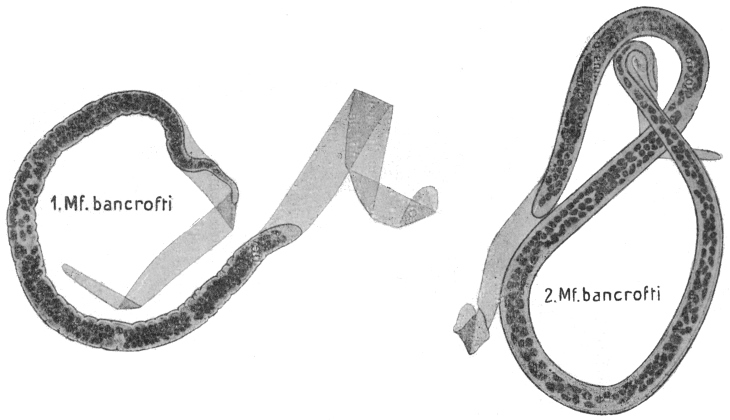
Fig. 282.—Mf. bancrofti in thick film, dried and stained with hæmatoxylin: 1, shrunken; 2, unshrunken. × 1,000. (After Fülleborn)
Distinction between Mikrofilaria bancrofti AND Mikroloa loa.
Dry Films, Hæmatoxylin Staining:—
Mf. bancrofti.Ml. loa.
(1) In graceful curves (but only if quickly dried).(1) Kinked.
(2) Tip of tail free from nuclei.(2) Nuclei extend to tip.
(3) Column of nuclei separated by a space from the cuticle.(3) Not so distinctly.