ON PARASITES IN GENERAL. (continued 13)
Reading edition
Conorhinus renggeri, Herr-Schäff (Great Black Bug of Pampas).
This large black bug is mentioned by Darwin,367 who states as follows: “At night I experienced an attack (for it deserves no less a name) of the benchuca, a species of Reduvius, the great black bug of the Pampas. It is most disgusting to feel soft wingless insects, about an inch long, crawling over one’s body. Before sucking they are quite thin, but afterwards they become round and bloated with blood, and in this state are easily crushed. One which I caught at Iquique (for they are found in Chili and Peru) was very empty. When placed on a table, and though surrounded by people, if a finger was presented the bold insect would immediately protrude its sucker, make a charge and, if allowed, draw blood. No pain was caused by the wound. It was curious to watch its body during the action of sucking, as in less than ten minutes it changed from being flat as a wafer to a globular form. This one feast, for which the benchuca was indebted to one of the officers, kept it fat during four whole months, but after the first fortnight it was quite ready to have another suck.” Mr. Kirby368 also refers to this species.
Conorhinus variegatus (Variegated Cone-nose).
Occurs in Florida in houses, and chases bugs (Cimex) and flies; not definitely known to bite man.
Conorhinus nigrovarius.
This species occurs in South America. It is one of the forms known as bichuque. Its bite makes a troublesome swelling.
Conorhinus protractus.
also attacks man in Utah.369 It has been called the “big bed bug.”
Genus. Reduvius, etc.
Reduvius personatus, Linné.
Syn.: Reduvius personatus, Leconte, 1855.
European, but also found in the United States. The bite causes intense pain. It bites when caught or handled, but does not seem to do so voluntarily. Swelling and irritation result which may last a week, and may even cause death.370 In 1899 it was very abundant at Washington and elsewhere; other species occurred, and so no definite opinion existed as to the actual biter, but some people took R. personatus actually biting. It was first described as a parasite of man in America by Walsh and Riley.371
A popular name for this bug is the wheel or masked bug—a black insect, three-fourths of an inch long. The larva of this bug is carnivorous and covers its body with dust so as to conceal itself from its prey. The adult is active on the wing.
Coriscus subcoleoptratus, Kirby, 1837.
Syn.: Nabicula subcoleoptrata, Kirby, 1837; Nabis subcoleoptratus, Reuter, 1872; Coriscus subcoleoptratus, Stål, 1873.
Northern United States. Howard was bitten by one between the fingers—the pain was intense, like a needle prick, but the swelling was small.372 No other case known.
Rasahus biguttatus, Say, 1831.
Syn.: Pirates biguttatus, Stål, 1862; Callisphodrus biguttatus, Stål, 1866; Rasahus biguttatus, Stål, 1872.
Common in southern United States, and found in Cuba, Panama and Pará, etc. Known as the two-spotted corsair on account of the great spot on the hemielytra. Frequently found in houses, where it chases the bed bug. It also bites man frequently. From 1869 Walsh and Riley placed it amongst the parasites of man. In the United States Davidson373 is of opinion that all cases attributed to spider bites are due to this insect.
Melanolestes morio, Erichson, 1848 (Non-walker).
Syn.: Pirates morio, Erichson, 1848; Melanolestes morio, Stål, 1866; Pirates picipes, Herrich-Schäffer, 1848; Melanolestes picipes, Howard, 1900.
Guiana and Mexico and eastern and southern United States. Length 20 mm., hides under stones and logs during daylight, and flies at night. Attracted by lights into houses. Very abundant in 1899 at Washington. Howard cites cases where it was proved to bite man.
Melanolestes abdominalis, Herrich-Schäffer, 1848.
Syn.: Pirates abdominalis, Herrich-Schäffer; Melanolestes abdominalis, Uhler, 1875.
Allied to the former; some say similar, but can be told by the shorter wings on the female. It occurs in the same localities as M. morio.
Phonergates bicoloripes.
This reduvid attacks man in Africa.
Family. Aradidæ.
Broad and very flat bugs, with antennæ of four segments and the beak of three; scutellum short, no cuneus to elytra and the tarsi of two segments. They normally live under the bark of trees, etc., and are found in most parts of the world.
Dysodius lunatus, Fabr. (Pito Bug).
A large species which is found in South America, frequenting houses, and bites very severely.
The Ochindundu.
The bug is described by Wellman (Journ. Trop. Med., April 2, 1906, p. 97) as not only feeding on ticks, such as Ornithodorus moubata, but as also attacking man. It is called by the Angola Bantus the ochindundu. It is black in colour; the first two pairs of legs are of a bright red hue. It has curious paddle-like structures on the front four legs, which seem to be designed for securely holding the ticks. It infects native kraals for the sake of preying on ticks. The natives also state that it inflicts a bite which far exceeds in painfulness that of the tick. They compare the bite with that of a poisonous snake.
Family. Lygæidæ.
Scutellum short; antennæ four-jointed; ocelli present; membranous part of hemielytra with never more than five nervures. Nearly all vegetable feeders. A few are recorded here as biting man.
Lyctocoris campestris, Fabricius.
Syn.: Acanthia campestris, Fabr. (Lyctocoris domesticus).
Rare in habitations, lives on human blood. Found by Blanchard in a bed at an hotel at Liverpool. The bite is undoubtedly worse than that of Cimex; cosmopolitan. In colour it is ferruginous, shining, legs testaceous; hemielytra slightly shorter and narrower than the abdomen; membranous portion transparent, the apex broadly fuscous. Length 3·8 to 4·8 mm.
Rhodinus prolixus, Stål, 1859.
Sometimes attacks man, and the bite is very painful. It is 25 mm. long and 8 mm. broad, and occurs in Colombia. It is found also in Cayenne and Venezuela. This like other species is known in South America as bichuque or benchuca.
[A few other unimportant species are also recorded as biting man, such as Harpactor cruentas, in the South of France; Eulyes amœna, from Borneo and Java; Arilus carinatus, Forster, from Brazil. The latter appears to be the same as the Acanthia serratus, Fabricius.—F. V. T.]
Order. Orthoptera.
[The only Orthoptera recorded as doing actual harm to man are certain wingless locusts found in Africa. The cysticercus stage of a small tapeworm found in rats and man has been found in an earwig (Alcock).
[The strange Hemimeridæ found in West Africa, resembling wingless cockroaches, are parasitic on rats (Cricetomys). Phasmidæ, or stick insects, are said to be able to eject a fluid which may cause blindness if it comes in contact with the eyes.
Locusts Injurious to Man.
[A wingless locust—Enyaliopsis durandi, Luc—is recorded by Wiggins374 as injurious to man in Uganda. “The bite of this insect,” it is said, “gives rise to a very nasty eruption, which may extend over the whole body, with high temperature and general malaise. The skin at the site of the bite sloughs away, and generally leaves a large deep cavity, which heals very slowly.”
[An allied species—E. petersi, Schaum—emits a clear yellow fluid, but according to Marshall this does no harm.375 Stannus writes that “for some years I have been cognizant of the fact that among the natives of Nyasaland an allied if not the same species is held to cause skin lesions by the emission of a fluid on the bare skin surface of the body. I have seen cases of ulcers on various parts of the body, for which the ‘nantundua’ was assigned as the cause.” He then describes the destruction of the superficial layers of the skin which he observed after the yellow fluid had been on the skin twelve hours.—F. V. T.]
Order. Coleoptera.
The larvæ of beetles, similarly to those of some other Arthropoda (myriapods and the larvæ of gnats), have sometimes been observed in man as purely accidental guests. In one case or another, such accounts may have originated through a mistake of the observer. Thus English doctors report the presence of the larvæ of Blaps mortisaga in the stools of human beings, Sandberg of the larva of Agrypnus murinus in his ten year old son, and Blanchard mentions the larva of a beetle that was vomited by a child. All these cases, however, do not represent actual parasitism, although there are beetles living parasitically.376
Silvanus surinamensis, Linnæus (Saw-toothed Grain Beetle).
[Taschenberg records this beetle as having invaded some sleeping apartments adjoining a brewery where stores were kept, and annoying the sleepers at night by nipping them when in their beds.
[This beetle is common in many parts of the world amongst groceries, corn, meal, seeds, dried fruits, etc. It is about 1/10 in. long, much flattened and chocolate-brown in colour. The thorax has two shallow grooves and bears six minute teeth on each side. The jaws are strong, but the bite cannot be very serious.—F. V. T.]
Order. Diptera or Siphonaptera.
Aphaniptera (Fleas).
Wingless, the thoracic rings distinct and free; antennæ of three segments; legs very powerful; abdomen with nine segments. [Ten segments are present, but only nine are visible.—F. V. T.] The mandibles transformed into serrated puncturing organs, which are situated in the split sheath of the rostrum; the maxillæ are laminated and have palpi, and more or less conceal the other parts.
The importance of fleas lies mainly in the fact that they act as plague carriers. About 150 species have already been described. The only ones of importance for this work are those found on man and those on rats and mice. The two families in which these are found are known as Pulicidæ and Sarcopsyllidæ.
The eggs of fleas are laid on the ground, on rugs, etc., and in birds’ and rodents’ nests. They hatch rapidly in warm weather and in warm climates, varying from two to five days; in cold countries they may take two or three weeks to incubate.
The larva is a footless creature, pearly white in colour, the head sometimes being darkened, composed of fourteen segments including the head, and although apodal can move with considerable agility. It lives amongst dust and dirt, and feeds upon any organic matter it can find. In about two weeks it is said to become mature, and then spins a cocoon in which pupation takes place.
The cocoons of the common human flea and the fowl flea become covered with dust and dirt. The period of pupal life seems varied, for I have had the fowl flea hatch out in ten days, and others in three weeks at the same time of year.
The adults are blood suckers and cause considerable irritation as well as acting as disease carriers, and in the Sarcopsyllidæ the females attach themselves permanently to their hosts, embedding themselves under the skin, where they become pregnant. Some kinds harbour the cystic stage of tapeworms, and the rat trypanosome passes certain stages in the rat flea. Most fleas have definite hosts, but some, like the rat and fowl fleas, attack man.
The fleas which can carry the bacillus of plague are Xenopsylla cheopis, Pulex irritans, Ceratophyllus fasciatus and Hoplopsyllus anomalus.
The two families, Pulicidæ and Sarcopsyllidæ, can be distinguished as follows:
Thoracic segments much foreshortened, coxæ and femora of hind legs very slightly enlargedSarcopsyllidæ.
Thoracic segments normal, coxæ and femora of all the legs much enlargedPulicidæ.
Family. Sarcopsyllidæ (Jiggers).
The members of this family are not confined to one host.
Three genera are known and tabulate as follows:—
α. Hind coxa without a patch of spines on the inside.
α1. Hind femur simple1. Dermatophilus.
α2. Hind femur with a large tooth-like projection near the base2. Hectopsylla.
β. Hind coxa with a patch of short spines on the inside3. Echidnophaga.
Genus. Dermatophilus, Guérin.
Dermatophilus cæcata, Enderl.
The eyes of the female vestigial. Taken on and behind the ears of Mus rattus in Brazil.
Dermatophilus penetrans, L., 1758 (Jigger, Chigoe).
Syn.: Sarcopsylla penetrans.
About 1 to 1·2 mm. in length; brown in colour. Eyes distinct. The males only occasionally visit man to bite; the fertilized female, on the other hand, bores into the skin with her head, particularly about the toes of the host, and then attains considerable dimensions. The eggs develop on the soil with a metamorphosis similar to that of the common flea.
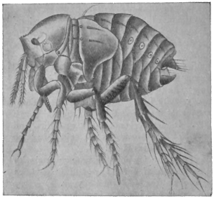
Fig. 380.—Dermatophilus penetrans: young female. Highly magnified. (After Moniez.)
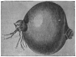
Fig. 381.—Dermatophilus penetrans: older female. Enlarged. (After Moniez.)
The sand flea (nigua) particularly infests Central and South America, and, in 1873, was carried by ships from Brazil to the West Coast of Africa. In a comparatively short time it has become disseminated throughout Africa and has also appeared in Madagascar; recently also it has been reported from China.
Besides attacking man, it also settles on mammals, for instance, on dogs, pigs, etc. According to Jullien the wound or little swelling caused by the female has no particular significance, as children infested with ten or eleven sand fleas quietly proceeded with their games. It will be understood, however, that the wound easily affords the opportunity for the setting up of inflammation or even septic processes, as is the case in any kind of wound.
[The jigger is also well known in the West Indies.—F. V. T.]
Genus. Echidnophaga, Olliff.
Four species found on rats, etc.
Echidnophaga gallinacea, Westwood (Chigoe of Fowls).
[This flea is a native of tropical Asia and Africa. It lives on the fowl chiefly, attacking the neck and around the eyes. Specimens were sent me from Texas, where they not only attack poultry but also children, the latter somewhat severely. It also occurs on cats, and is found on rats in Africa. It has been introduced into North America.
[Three other species are found on rats, viz.: E. myrmecobii, Rothsch., from Australia; E. murina, Tirah, from Southern Europe; and E. liopus, Rothsch., India and Western Australia.—F. V. T.]
Family. Pulicidæ (True Fleas).
Rothschild’s classification is as follows:—
Section I.—Club of antennæ distinctly segmented only on the hind side.
Key to Genera.
a. No comb on head and thorax.
a1. The internal incrassation, which extends from the insertion of the mid coxa into the thorax, joins the anterior edge of the mesosternitePulex.
b1. This incrassation joins the upper edge of the mesosterniteXenopsylla.
b. With a comb on the pronotum onlyHoplopsyllus.
c. With a comb on the pronotum and at the lower edge of the headCtenocephalus.
Section II.—Club of antennæ distinctly segmented all round.
Key to Genera.
a. Eye developed.
a1. No comb on head.
a2. Pygidium not projecting backwards; frons with tubercleCeratophyllus.
b2. Pygidium strongly convex, projecting backwards; frons without tuberclePygiopsylla.
b1. Two spines at angle of genæChiastopsylla.
b. Eye vestigial or absent.
a1. Abdomen without comb.
a2. Hind edge of tibiæ with about eight short and several long bristles, which do not form a comb.
a3. Fifth segment in fore and mid tarsi with five, and in hind tarsus with four bristlesNeopsylla.
b3. Fifth segment in fore and mid tarsi with four, and in hind tarsus with three lateral bristles, there being an additional pair of bristles in all the tarsi on the ventral surface in between the first pairCtenophthalmus.
b2. Hind edge of tibiæ with about twelve short and three long bristles, the short ones forming a kind of combCtenopsylla.
b1. Abdomen with at least one combHystricopsylla.
Genus. Pulex, Linn.
Pulex irritans, L., 1758.
Male 2 to 2·5 mm. in length, females about 4 mm.; reddish or dark brown; head without bristles; thoracic and abdominal rings of bristles on the dorsal aspect, and small hairs directed backwards at the posterior margin. The barrel-shaped white eggs are deposited in cracks in the boards, sweepings, spittoons, etc.; they produce legless larvæ consisting of fourteen segments, which, after about eleven days, are transformed into pupæ; after another eleven days the flea emerges.
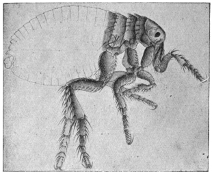
Fig. 382.—Pulex irritans. 14/1.
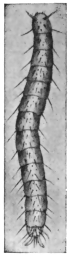
Fig. 383.—Larva of flea. Enlarged. (After Railliet.)
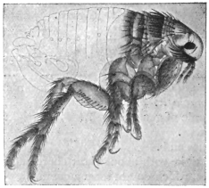
Fig. 384.—Pulex serraticeps. 22/1.
Fleas live in human dwellings all over the world, and periodically pass on to persons to suck their blood. They may deposit their eggs on very uncleanly individuals, and even undergo development, therefore it is possible to find larvæ and pupæ on such persons.
The dog flea, Pulex serraticeps, is easily distinguished from the flea of man by the large thick bristles on the posterior margin of the first thoracic ring (fig. 384).
Genus. Xenopsylla, Glink.
Xenopsylla cheopis, Rothschild.
This is the common rat flea of tropical countries. Rothschild377 says: “Although practically cosmopolitan, it cannot apparently flourish in temperate and cold climates.”
In the male the bristles of the flap-like process of the clasper all slender; in the female the narrow portion of the receptaculum seminis long. Originally discovered in Egypt.
This is apparently the chief plague flea. The Indian Plague Committee have proved that this flea is easily infected when fed on plague rats, and that the bacillus multiplies rapidly in the flea’s stomach and that the fleas may remain infective for fifteen days. How the flea infects man does not apparently seem to have been proved, as it does not do so through its bite, but the excrement is highly infective. It is probable that this poisoned fæcal matter gets to the wound caused by the piercing mouth.
Xenopsylla brasiliensis, Baker,
occurs on rats in West Africa and has been introduced into Brazil.
Genus. Ctenocephalus, Kolen.
Includes the cat and dog fleas. The dog flea, C. canis, Dugès, is found on the dog all over the world, but especially in temperate climates. It also occurs on rats. Man is often badly bitten by this insect and it overruns houses. The eggs are laid on rugs, carpets and dust and dirt and amongst dogs’ hair, but are not fastened to it and fall anywhere. The ova may hatch in about fifty hours and the larvæ live for seven days and then spin their cocoons amongst dust and dirt. The pupal stage lasts about eight days.
The cat flea (C. felis) is widely distributed over the world, and occurs on many mammals beyond the cat, and is also found on rats.
Genus. Hoplopsyllus, Baker.
A genus found in North America related to Pulex, but at once recognized by the prothorax bearing a comb.
Hoplopsyllus anomalus, Baker,
which is found on the ground squirrel (Citellus beecheyi) in California, and according to Rothschild once found on the rat, has been proved to carry the plague bacillus and to play an important part in plague infection in California.378
Genus. Ceratophyllus, Centis.
Ceratophyllus fasciatus, Bosc.
This flea is also found on the rat in Europe and will attack man. It is a plague carrier. It has eighteen to twenty teeth on the prothoracic comb and no black spines on the head. The genus Ceratophyllus of Centis has a pronotal comb and three ante-pygidial chætæ on each side. Two other specimens recorded: C. londiniensis, Rothsch., and C. anisus, Rothsch. The former on rats and mice in London, the latter on Felis sp. from Japan and Mus norvegicus in California.
Genus. Ctenopsylla, Kolen.
This genus contains a very abundant rat and mouse species, C. musculi, Dugès, which is widely distributed over the globe.
Genus. Hystrichopsylla, Tasch.
Large hairy fleas, with no eyes and one or more combs on the abdomen. In the Mediterranean area one species, H. tripectinata, Tirah, is common on rats and mice, and also in the Azores. Several others occur on rats and mice. For information concerning these the reader is referred to Rothschild’s papers.
Pulex pallipes is another species found on the rat and man.
Systematic, Anatomical, and Biological Remarks on Mosquitoes.
Mosquitoes come in the Nematocera, one of the sub-orders of the Diptera, and are divided into numerous families, of which, however, the Culicidæ are of most interest to us here. Other families as the Psychodidæ and Chironomidæ are also of considerable importance, vide following pages. The head is small, the facetted eyes are placed laterally, but there are no accessory eyes (ocelli). In front of the eyes are situated the comparatively long antennæ, the differences of which strongly mark the distinction of sex.379
The antennæ are composed of fifteen or sixteen segments. In the male they are covered with long whorl-like hairs, while in the female the antennal hairs are short—differences that are perceptible even with the naked eye.380 The proboscis, which is longer than the antennæ, protrudes from the inferior aspect of the head and is composed of the following parts (figs. 387 and 388): Two grooved half tubes, facing one another, of which the upper one is the upper lip (labrum) and the lower one the lower lip (labium), which represents a pair of coalesced maxillæ. Within the tube formed by the labrum and labium are the mandibles and maxillæ, transformed into instruments for piercing, and a single puncturing organ, the hypopharynx. On the right and left, next to the proboscis, are placed the straight five-jointed palpi, the final joint of which is thickened in the male.381 In biting, the labrum, which is swollen at its free end, is not introduced into the wound like the other mouth parts, but is bent backwards. The labium and hypopharynx push direct into the skin; the maxillæ and mandibles, however, which are needle-like and serrated at the tips, penetrate with a saw-like movement. [The swollen free end of the labrum really means the labellæ, two articulated pieces, supposed by some to be the labial palpi. In most species the mandibles are not serrated at their ends.—F. V. T.] The saliva is introduced into the wound through the lumen of the hypopharynx, while the blood is sucked up by the mosquito in the groove of the labium.
The three thoracic segments are soldered together. The central one carries the membranous wings on the sides of the dorsal surface; the posterior somite carries the small halteres (rudimentary posterior wings). There are three pairs of long slender legs on the lower side.
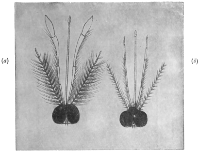
Fig. 385.—Head of a male (a) and of a female (b) Anopheles. Slightly enlarged. (After Giles.)
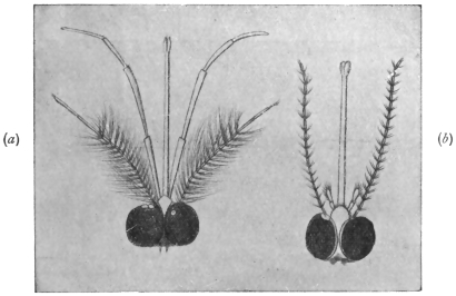
Fig. 386.—Head of a male (a) and of a female (b) Culex. (After Giles.)
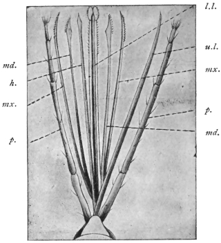
Fig. 387.—Mouth parts of Anopheles claviger.† h., hypopharynx; md., mandible; mx., maxilla; u.l., upper lip; l.l., lower lip; p., palpi. (After Grassi.)
† [This should read Anopheles maculipennis, Meig.; there was no type of A. claviger.—F. V. T.]
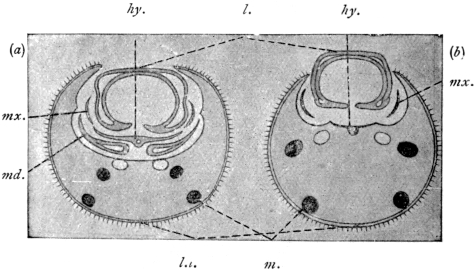
Fig. 388.—Anopheles maculipennis: transverse section through the proboscis of a female (a) and a male (b). hy., hypopharynx, with duct of the salivary gland; m., muscles; md., mandibles; mx., maxillæ; l., labium; l.l., labrum. (After Nuttall and Shipley.)
The abdomen has no limbs, is composed of eight (rarely nine) distinct segments; the sexual and anal orifices are at the posterior end, the stigmata on the sides. The intestinal canal (fig. 389) is composed of three principal divisions; the anterior part reaches as far as the front pair of legs, and consists of the œsophagus, which is provided with two small lateral diverticula. [At the commencement of the œsophagus are one or more diverticula, which vary in size; they contain air, food and bacteria.—F. V. T.] The mid gut reaches as far as the fifth and sixth abdominal ring; in front it is thin, and has numerous small supra-œsophageal ganglia; the posterior part is, however, more dilated. Four or five Malpighian tubes, the excretory organs, discharge at the place where the mid gut passes into the terminal gut.
The pair of salivary glands have one common excretory duct leading into the hypopharynx.
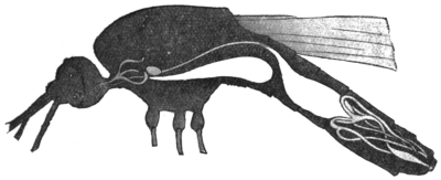
Fig. 389.—Longitudinal section of an Anopheles, showing alimentary canal. In the forepart of the thorax is the salivary gland consisting of three tubules; ventrally, the suctorial stomach extending into the abdominal cavity; the stomach, and at the posterior end of the abdomen the Malpighian vessels. (After Grassi.)
These glandular bodies are situated in the thorax; each consists of three slightly serpentine tubules, the dorsal and ventral tubes being long, the central one shorter. The above-named characteristics apply to both genera Culex and Anopheles, but in the genus Culex is smaller, Anopheles larger. [In Anopheles the ends of the ducts in the lobules are dilated, whilst in most of the genera the ducts are the same size all along. The lobules may bifurcate, and in Psorophora there are five lobules.—F. V. T.] The legs of the genus Culex are about the same length as the whole body; in Anopheles they are double that length.382 In Anopheles the palpi and proboscis are of equal length; in Culex the condition is different, according to sex. In the male the palpi are longer than the proboscis; in the female considerably shorter and the number of segments diminished. The venation of the wings exhibits further points of differentiation, as also their adornment, though this last sign is not by any means always conclusive; most species of the genus Culex have unspotted wings, whilst those of Anopheles are usually spotted. More important is the fact that in Culex the abdomen is decorated with small scales, similar to those on butterflies, whereas there are small bristles on the abdomen of Anopheles. [This cannot be said to be a character by which an Anopheline may be told from a Culicine, for in such common Anopheline genera as Cellia and Neocellia we get plenty of scales on the abdomen.—F. V. T.] An experienced observer can, however, separate the two genera by the difference in size and their manner of resting. When settled they either touch the resting place with all the legs or only with the four anterior legs. In consequence of the different length of the legs, the body of Culex approaches the resting place more closely; moreover, Culex holds the abdomen parallel or at an acute angle to the resting surface, whereas Anopheles carries the abdomen directed upwards (at an angle of about 145°) and holds the head down. Both genera, however, usually only rest on the four anterior legs, and then, as has long been known, Culex carries the third pair directed towards the dorsum, while those of Anopheles hang down.
In regard to the differentiation of the species, I must refer you to the special literature, and content myself by observing that about 150 species of Culex and about fifty species of Anopheles have been described, of which fifty about four are found in Europe. [The number of known Anophelines now is more—100 species—of other Culicidæ over 700.—F. V. T.] According to our present knowledge it appears that the entire genus Anopheles can transmit malaria to man; this observation has been confirmed in Anopheles claviger, Fabr.; A. maculipennis, Meig.; A. bifurcatus, L.; A. superpictus, Grassi; A. pseudopictus, Gr., all of which are found in Italy,383 Germany, etc., as well as in the tropics. Moreover, in A. costalis, Loew; A. funestus, Giles (Africa); A. quadrimaculatus, Say (North America), and A. rossii, Giles; the latter is perhaps identical with A. superpictus, Gr., as well as with A. culicifacies (India). [Anopheles maculipennis and A. claviger are the same. Certainly neither maculipennis nor bifurcatus has been found in the tropics. Anopheles quadrimaculatus, Say, is the same as A. maculipennis. There is no evidence that all Anophelines carry malaria, but there is much to show that certain species only are capable of so doing. A list of known carriers is given later.—F. V. T.]
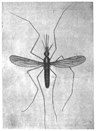
Fig. 390.—Anopheles maculipennis, Meigen. Enlarged. (After Grassi.)
Everyone is aware that mosquitoes swarm at sunset in fine weather, and then seek out human beings and other warm-blooded animals to take food. In this regard, however, the sexes differ, for it is almost without exception that the females only suck blood, while the males subsist on the juices of plants (blossoms or fruits).384 After sucking, and when night has fallen, the mosquitoes find a place of refuge, for which purpose they utilize the grasses or foliage of trees and bushes, or inhabited or uninhabited rooms of houses, also cellars, stables, verandahs, etc., where they also pass the day.
[Some mosquitoes bite in the daytime—Stegoymia and some Anophelines; some bite right into the night, as Culex fatigans and C. pipiens.—F. V. T.]
The period required for digestion varies according to the temperature. It takes two days in summer, and may take up to ten days or more in cool weather. After digestion is complete more food is taken up, this being necessary [in some species only—F. V. T.] for the maturing of the sexual products in the female.
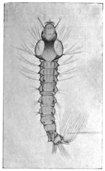
Fig. 391.—Larva of Anopheles maculipennis, Fabr. Enlarged. (After Grassi.)
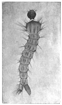
Fig. 392.—Larva of Culex. Enlarged. (After Grassi.)
It is still unknown under what circumstances copulation takes place;385 in any case, sooner or later the females are fecundated, and when the ova have become mature, and the season is not too far advanced, they seek a suitable place in which to deposit them.386 These are larger or smaller, permanent or temporary, collections of standing water, pools, puddles, lakes, pits, water in rain-water barrels, basins, etc. Nevertheless, certain kinds prefer certain waters; thus Anopheles (claviger) maculipennis and several of the Culices seek stagnant water overgrown with swamp vegetation and decomposing vegetable matter; A. bifurcatus and certain Culices, clear water with some vegetation (such as fountains and the lakes in gardens and parks); Culex pipiens has a preference for rain-water barrels, even though the water be dirty and evil-smelling. [I have found the larvæ of Anopheles bifurcatus living in great numbers in ponds and lakes completely overgrown with floating water-weeds, and those of Culex pipiens in liquid manure.
Sexual Organs of the Mosquito.—The female has a pair of ovaries, opening into a single tube by the ovarian tubes; into the single tube opens a duct coming from the spermathecæ, and also a mucous gland. The spermathecæ store up the male cells. The male organs consist of two testes joined by ducts (vasa deferentia) to the ejaculatory duct formed by their union. Each vas deferens is joined by a short tube with the sac-like vesicula seminalis.—F. V. T.]
There is also a difference in the manner in which Culex and Anopheles deposit their ova. Culex deposits two to three hundred eggs in compact heaps that float on the water, and in which the eggs stand perpendicularly one next the other; whereas Anopheles maculipennis deposits only three or four up to twenty eggs, united in groups that float horizontally on the water; the eggs of A. bifurcatus, again, are arranged in star-like groups. The eggs are about 0·75 mm. in length, and assume a dark hue soon after being laid. The development only occupies a few days. The young larvæ grow rapidly, changing their integument several times; the larvæ also differ in the various genera, though they have a general resemblance (figs. 391 and 392).
The long legless larva has a flattened head, a fairly broad, rectangular, or trapeziform thorax, on which there are bristles, and an abdomen distinctly segmented, and on the segments of which there are also lateral bristles. The situation of the stigmata marks the difference between the two genera. Though in both genera the stigmata are at the posterior end and on the dorsal surface, they are in Anopheles close to the surface of the body; in Culex, however, they are on the free end of a long tube (siphon).
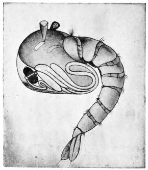
Fig. 393.—Pupa of Anopheles maculipennis, Meig. Enlarged. (After Grassi.)
The position of the larva in the water also differs. The larva of Anopheles lies almost horizontally beneath the surface of the water, the posterior border of the penultimate abdominal segment, upon which the stigmata are situated, being on the surface; whereas the larva of Culex hangs head downwards perpendicularly in the water, the point of the siphon only touching the surface.
In about a fortnight the larva is fully grown and becomes a pupa. The pupa (fig. 393), which moves in jerky movements, remains in the water, but partakes of no food. In shape it somewhat resembles a tadpole, that is to say, it consists of a bulky anterior portion, on the surface of which the head, with its appendages, is recognizable, and a more slender segmented abdomen. Above, on the thorax, there are two small trumpet-shaped breathing tubes for the conveyance of air to the tracheal system. After three or four days the perfect mosquito hatches out, remains a short time on the surface of the water until its chitinous integument is hardened, and then flies away.
The females that are fertilized in the autumn hibernate in sheltered spots in the open air, or in houses, cellars, under stairs, in stables, barns, etc., and are the progenitors of the first generation of the following year.
In accordance with the climate of a country, or the kind of weather of a year, the conditions in regard to the manner of life and the duration of the development of the mosquito vary. At all events, the life-history of the mosquito elucidates many points relating to malaria which were hitherto not understood.
[The length of the egg, larval and pupal life varies so much that it is not possible to give an account of any value here. Frequently the eggs may incubate in two days, whilst I have had Stegomyia fasciata eggs from Cuba that have hatched out under abnormal circumstances more than two months after they were laid (“Mono. Culicid.,” iii, p. 6). Some larvæ, as Anopheles bifurcatus, live for months during the winter. Some mosquitoes therefore hibernate as larvæ. The larvæ and pupæ of the different genera present very marked characters, mainly in regard to the structure of the siphons. Specific differences may be found in the frontal hairs of Anopheline larvæ and in the number and arrangement of a group of spines at the base of the siphon in Culicines.—F. V. T.]
Culicidæ or Mosquitoes.
The importance of these insects to man is very great. They not only produce painful bites, which may become inflamed and give rise to a considerable amount of œdema, but they are more important on account of the part they play in the distribution of various diseases. Culicidæ may not only carry disease germs, but act as intermediate hosts for certain parasites, such as some of the Anophelina for malarial parasites, Culex for Filariæ, and Stegomyia for yellow fever, etc.; the last-named is in any case the distributor of that fatal disease. It is therefore very necessary to know the life-history, habits and characters of these pests.
Mosquitoes exist in almost all parts of the world from the Arctic circle to the tropics; temperate regions suffer from them less than the two extremes, but even there they form not only a source of great annoyance but of danger as malaria and possibly now and again yellow fever carriers. A few years ago comparatively few species were known, now some 800 odd have been described. Their number will probably not stop far short of 1,000, in spite of the fact that many have been described under different names, yet really the same species. Some are purely domestic, others entirely sylvan; the former, as we might expect, often have a very wide distribution, having been taken from place to place in boats and trains. The more rapid transport becomes, the greater becomes the possibility of this wide distribution of many species increasing, and the spread of other species from their natural home to foreign parts by sea and then by trains further inland.
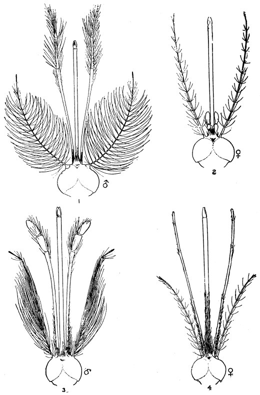
Fig. 394.—Heads of Culex and Anopheles: (1) Culex male; (2) Culex female; (3) Anopheles male; (4) Anopheles female. (After Daniels.)
All Culicidæ are aquatic in their larval and pupal stages. Almost all small collections of water, both natural and artificial, may form breeding grounds for these pests. Some even breed in pitcher plants and many in bromelias. The favourite resorts for the larvæ of Anophelina are small natural collections of water, such as puddles, ditches and small pools around swamps; certain species (A. maculipennis, etc.) live in rain barrels as well. They may also occur in the sluggish water at the edges of rivers or even in mid river, where the flow is checked by masses of water weeds (Myzomyia funesta, etc.). The Stegomyias prefer artificial collections of water, but also occur in natural pools. The yellow fever species (S. fasciata) prefers small collections, such as in barrels, pots, jars, etc. Culex occur in all manner of places—rain barrels, tanks, cisterns, ponds and ditches. Some of the South American species of Culex, Wyeomyia, Joblotia, etc., breed in the collections of water at the base of bromelia leaves.387 Very few Culicid larvæ live in salt water except in Australia, where Dr. Bancroft has found them in salt water of specific gravity 1·040 (Mucidus alternans and Culex annulirostris). Other salt water mosquitoes are known in America. The food of the larvæ is very varied; the majority appear to feed upon confervæ, small crustacea and insects; some are cannibals, readily devouring others of their own kind. The larger larvæ of Megarhinus, Psorophora, Toxorhynchites and Mucidus are extremely ravenous and devour one another.
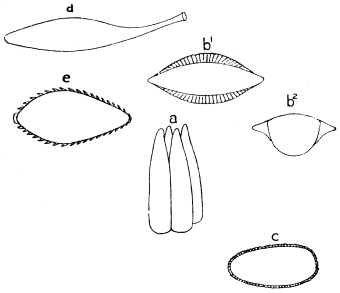
Fig. 395.—a, eggs of Culex; b1 b2, eggs of Anopheles; c, egg of Stegomyia; d, egg of Tæniorhynchus; e, egg of Psorophora.
There are two main types of larvæ, the Anopheline and Culicine; in the former there is no respiratory siphon, in the latter the siphon is long or moderately long. The head offers certain marked peculiarities which are of specific value; this especially applies to the Anophelina, in which the frontal hairs are of great service in distinguishing the larvæ,388 whilst in Culex the number and position of the spines at the base of and on the siphon are characteristic. The position assumed by the larvæ in the water also varies in the different groups; most of the Anophelines lie horizontally, most of the Culicina and Ædeomyina hang head downwards. The pupæ also vary, but not to the same extent; the chief differences to be noticed are in the form of the two respiratory trumpets.
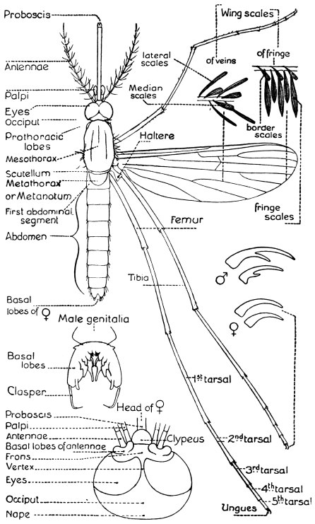
Fig. 396.—Diagram showing the structure of a typical mosquito. (Theobald.)
The eggs, which may be laid separately (Anopheles maculipennis, Stegomyia fasciata, Joblotia nivipes, etc.), or in rafts (Culex pipiens, C. fatigans) or in chains (Pseudotæniorhynchus fasciolatus), present a great variety of forms. The most peculiar are shown in fig. 395 (Tæniorhynchus, Culex, Stegomyia, Anopheles, Psorophora).
As in all insects, they differ very materially in each species of one genus. Those best known are the Anopheline eggs.
The eggs always float on the surface of the water; immersion soon destroys them, but many may occur in mud and can resist desiccation.
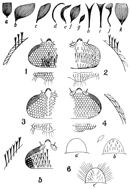
Fig. 397.—Types of scales, a to k; head and scutellar ornamentation, 1 to 5; forms of clypeus, 6. (Theobald.) 1, head and scutellum of Stegomyia, etc.; 2, of Culex and Mansonia; 3, of Howardina, Ædes, etc.; 4, of Megarhinus and Toxorhynchites, etc.; 5, of Cellia and some other Anophelines; 6, a′, clypeus of Culex; b′ of Stegomyia; c′, of Joblotia.
Characters of Adult Culicidæ.—The chief characters by which true mosquitoes, or Culicidæ, are known are the following:—
(1) Wings always with the veins covered with scales; the longitudinal veins, usually six in number (in one genus seven); the costal vein carried round the border of the wing.
(2) Head, thorax and abdomen usually, but not always (Anopheles, etc.), covered with scales.
(3) Mouth parts formed into a long piercing proboscis.
As a rule the males may be told from the females by their antennæ being plumose, whilst in the females they are pilose (vide fig. 394), but this does not invariably hold good, for in Deinocerites, Theobald, and Sabethes, Desvoidy, and others, they are pilose in both sexes. The labial palpi are very variable in regard to their form and the number of segments; in the Anophelina they are long in both sexes, as long or nearly so as the proboscis, more or less clubbed in the males; in Culicina, Joblotina and Heptaphlebomyia, they are long in the males, short in the females; in Ædeomyina, short in both sexes.
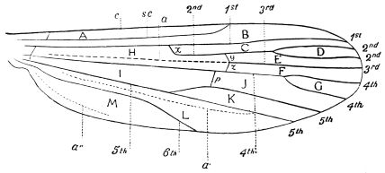
Fig. 398.—Neuration of Wing. Explanation of Wing, Veins and Cells.—A, costal cell; B, sub-costal cell; C, marginal cell; D, first sub-marginal cell (= first fork cell); E, second sub-marginal cell; F, first posterior cell; G, second posterior cell (= second fork cell); H, first basal cell; I, second basal cell; J, third posterior cell; K, anal cell; L, auxiliary cell; M, spurious cell; c, costal vein; 1st—6th, first to sixth longitudinal veins; a, a′ and a′′, incrassations (a′ called by Austen the sixth vein, a′′ the eighth vein); y, supernumerary cross vein; z, mid cross vein; p, posterior cross vein; s.c., sub-costal. (Theobald.)
Scales.—The most important structural peculiarities in Culicidæ are the scales, which form the chief and most readily observed characters for separating genera and species. The importance of scale structure has been recently ignored by some workers, who are probably right academically, but as a means of separating groups, and so more easily running down a species, the practical man is strongly advised to follow this method. As to what a genus is, is purely a matter of personal opinion. If one examines any recent standard work on entomology one will find a species being placed in varied genera by the varied authorities.
The head, thorax, abdomen and wings are in nearly all cases clothed with squamæ of varied form, of which the following are the main types (fig. 397):—
(1) Flat, spade-shaped scales (a). (2) Narrow curved scales (e). (3) Hair-like curved scales (d). (4) Spindle-shaped scales (f). (5) Small spindle-shaped scales (g). (6) Upright forked scales (h) and (i). (7) Twisted upright scales (j). (8) Inflated or pyriform scales (k). (9) Mansonia scales (b). (10) Small broad asymmetrical scales (c).
Various other varieties are found on the wings, such as:—
(1) Narrow linear lateral scales. (2) Narrow lanceolate scales. (3) Broad lanceolate scales. (4) Elongated, broad, truncated scales (= Pseudotæniorhynchus-like scales). (5) Pyriform scales. (6) Asymmetrical broad or Tæniorhynchus scales. (7) Flat spade-like scales.389
The wings have a series of scales along the middle line of the veins, and also lateral scales to all or nearly all the veins. The wing is also fringed by a series of scales (fig. 396), which, however, are of little systematic importance; the so-called “border scales” (b.s.) vary, however, to some extent, and are useful characters in separating some of the Tæniorhynchus.
The Classification of Culicidæ.
Section A.—Proboscis formed for piercing; metanotum nude. Scutellum simple.
I.Wings with six-scaled longitudinal veins.
A.Palpi long in the male.
α.Palpi long in both sexes, clavate in ♂ Anophelina.
I.First submarginal cell as long or longer than the second posterior cell.
Antennal segments without dense lateral scale tufts.
Thorax and abdomen with hair-like scalesProthoracic lobes simple; no flat head scalesWing scales lanceolateAnopheles, Meigen.


Wing scales mostly long and narrowMyzomyia, Blanchard.
Wing scales as above, but fourth long vein near base of third and outstanding scales on prothoracic lobeNeomyzomyia, Theobald.
Wing scales partly large and inflatedCycloleppteron, Theobald.
Prothoracic lobes mammillated; some flat head scales. Basal lobe of ♂ genitalia of two segmentsStethomyia, Theobald.
Prothoracic lobes with dense outstanding scalesFeltinella, Theobald.
Thorax with some narrow curved scales; abdomen hairyPyretophorus, Blanchard.
Wing scales small and lanceolate. Wing scales broad and lanceolateMyzorhynchella, Theobald.
Thorax with hair-like curved scales, some narrow curved ones in front; abdomen with apical lateral scale tufts, scaly venter; no ventral tuftArribalzagia, Theobald.
Thorax with hair-like curved scales; abdominal scales on venter only, with a distinct ventral apical tuftMyzorhynchus, Blanchard.
Much as above, but abdomen with long spine-like dense lateral tuftsChrystia, Theobald.
Thorax with very long hair-like curved scales; abdomen pilose, except last two segments which are scaly; dense scale tufts on third femora; wings with broadish, blunt, lanceolate scalesLophoscelomyia, Theobald.
Thorax and abdomen with scalesAbdominal scales as lateral dorsal patches of small flat scales; thoracic scales narrow and curved, or spindle-shapedNyssorhynchus, Blanchard.

Abdomen nearly completely covered with irregular scales and with lateral tuftsCellia, Theobald.
No lateral scale tuftsNeocellia, Theobald.
Thoracic scales hair-like except a few narrow curved ones in front; abdominal scales long, broad and irregularKerteszia, Theobald.
Thorax with hair-like curved scales and some broad straight scales, others spatulate on sides. Abdomen covered with fine hairs except last three segments, which are scaly. Tufts of scales on hind femora. Wing scales lanceolateManguinhosia, Cruz.
Antennal segments with many dense scaly tuftsChagasia, Cruz.
Antennæ with outstanding scales on second segment, more appressed ones on the first. At least one segment of abdomen with long flat more or less spatulate scalesCalvertina, Ludlow.
II.First submarginal cell very smallBironella, Theobald.
With a distinct cylindrical tubercle projecting obliquely from the prothoracic regionDactylomyia, Newstead and Carter.390
Scutellum trilobed.
First submarginal cell much smaller than the second posterior cell; proboscis long and bentMegarhininæ.
Palpi long in both sexesMegarhinus, Rob. Desvoidy.
Last segment of ♂ palpi blunt. Last segment of ♂ palpi long and pointedAnkylorhynchus, Lutz.
β.Palpi short in the femaleToxorhynchites, Theobald.
First submarginal cell longer than the second posterior cellCulicinæ.
Legs more or less densely scaly; head not entirely clothed with flat scales; all the legs densely scaly.
Wings with large pyriform scalesMucidus, Theobald.
Wings with narrow scalesPsorophora, Rob. Desvoidy.
Hind legs only densely scaledJanthinosoma, Arribalzaga.
Head entirely clothed with flat scales. Legs uniformly scaled with flat scales. Head and scutellar scales all flat and broad. Palpi of ♀ short, of ♂ thickened apically and tuftedStegomyia, Theobald.
Palpi of ♀ longer than in Stegomyia and in ♂ long and thin, acuminate, simpleDesvoidea, Blanchard.
Head scales mostly flat, but a median line of narrow curved ones; scutellar scales flat on mid lobe, narrow curved on lateral lobes and palpi longer than proboscisMacleayia, Theobald.
Head scales mostly flat, irregular, narrow curved ones behind; mid lobe scutellum with flat scales, lateral with narrow curved; ♂ palpi shorter than proboscisCatageiomyia, Theobald.
Head scales mostly flat, but a few narrow curved ones in middle in front; scutellar scales all flatScutomyia, Theobald.
Head scales all flat; scutellar scales all narrow curvedSkusea, Theobald.
Head with flat scales, except a small median area of narrow curved ones; scutellar scales all narrow curvedHowardina, Theobald.
Head with all flat scales except a thin line of narrow curved ones behind; scutellar scales all narrow curvedDanielsia, Theobald.
Head with small flat scales over most of surface, with median line and line around eyes of narrow curved ones; scutellar scales bluntly spindle or club-shapedHulecoetomyia, Theobald.
Head and scutellar scales narrow curved. Wing scales long, narrowly lanceolate, collected in spots; palpi clubbed in ♂; five-jointed and rather long in ♀Theobaldia, Neveu-Lemaire.
Wing scales (lateral) long and narrow, and ♀ palpi three-jointed, ♂ not clubbed and hairyCulex, Linnæus.
Wing scales at apex of veins dense and rather broad, femora swollen; small dark speciesMelanoconion, Theobald.
Wings with short, thick, median scales and short, broadish lateral ones on some of the veins; scales mottled; fork-cells rather shortGrabhamia, Theobald.
Wings with dense, broadish, elongated, truncated scalesPseudotæniorhynchus, Theobald.
Wings with broad, short, asymmetrical scalesTæniorhynchus, Arribalzaga.
Head covered with rather broad, flat, spindle-shaped scales; scutellum with small flat scales to mid lobeGilesia, Theobald.
Head clothed with flat, irregularly disposed scales all over, with patches of narrow curved ones; ♂ palpi clubbedAcartomyia, Theobald.
Abdomen with projecting flat lateral scales with deeply dentate apices; wings not ornamentedLasioconops, Theobald.
Wings ornamented; scutellum with flat and narrow curved scalesFinlaya, Theobald.
γ.Palpi short in ♂ and ♀Ædeomyina.
Wings unornamented.
Antennæ pilose in ♂ and ♀; second joint very longDeinocerites, Theobald.
Antennæ plumose in the ♂.
Head clothed with narrow curved and flat scales.
Mid-lobe of scutellum with six border-bristles.
Scutellum with narrow curved scales.
Palpi in ♀ four-jointed, in ♂ two-jointedÆdes, Meigen.
Mid-lobe of scutellum with four border bristles.
Scutellum with flat scales.
Head clothed with flat scales only.
Fork-cells normal length.
Mid-lobe of scutellum with four border-bristles.
Palpi of ♀ two-jointedVerallina, Theobald.
Palpi of ♀ five-jointed, metallicHæmagogus, Williston.
Fork cells very small or small.
Scutellar scales flat.
First submarginal cell longer than the second posterior cell; no flat scales on mesothoraxFicalbia, Theobald.
First submarginal cell smaller than the second posterior cell; flat scales on mesothoraxUranotænia, Arribalzaga.
Scutellar scales narrow curved.
First submarginal cell as in UranotæniaMimomyia, Theobald.
Wings ornamented with Mansonia-like scalesÆdeomyia, Theobald.
Section B.—Metanotum ornamented with chætæ, squamæ or both.
α.With chætæ only.
Proboscis longer than whole body; lateral wing scales Tæniorhynchus-likePhoniomyia, Theobald.
Proboscis as long as whole body in ♀ frons drawn out into a prominence; wing scales rather broad and longBinotia, Blanchard = Runchiomyia, Theobald.
Proboscis not as long as the whole body; lateral vein scales narrowWyeomyia, Theobald.
Proboscis not as long as whole body, swollen apically; wing scales long and broadDendriomyia, Theobald.
β.Metanotum with squamæ and chætæ.
Palpi short in ♂ and ♀.
Proboscis straight in ♀ and ♂; legs with scaly paddlesSabethes, Rob. Desvoidy.
Venation like Sabethes.
Legs simpleSabethoides, Theobald.
Venation like CulexGoeldia, Theobald.
Proboscis in ♂ elbowed, with two scaly tuftsLimatus, Theobald.
Palpi long in ♂, short in ♀Joblotina, Blanchard.
II.Wings with seven-scaled longitudinal veins: Culex typeHeptaphlebomyia, Theobald.
Section C.—Proboscis short, not formed for
piercingCorethrina.
Metatarsus longer than first tarsal jointCorethra, Linnæus.
Metatarsus shorter than first tarsal jointMochlonyx, Ruthe.391
Notes on the Different Genera.
Sub-family. Anophelina.
The following Anophelines have been recorded as malaria carriers:—
*Anopheles maculipennis, Meigen. Anopheles bifurcatus, Linnæus. *Myzomyia funesta, Giles. Myzomyia lutzii, Theobald. *Myzomyia rossii, Giles. Myzomyia listonii, Liston. Myzomyia culicifacies, Giles. Pyretophorus superpictus, Grassi. *Pyretophorus costalis, Loew. Pyretophorus chaudoyei, Theobald. *Cellia argyrotarsis, Robineau Desvoidy. Myzorhynchus pseudopictus, Grassi. Myzorhynchus barbirostris, Van der Wulp. Myzorhynchus sinensis, Wiedemann. Myzorhynchus paludis, Theobald. Myzorhynchus mauritianus, Grandpré. Neocellia stephensii, Liston. Neocellia willmori, James. Nyssorhynchus theobaldii, Giles. Nyssorhynchus fuliginosus, Giles. Nyssorhynchus annulipes, Walker.
Those marked with an asterisk (*) also carry the larvæ of Filaria bancrofti, as also do Myzorhynchus minutus, Theobald, and Myzorhynchus nigerrimus, Giles.
Genus. Anopheles, Meigen.
“Syst. Beschr. Europ. zwei. Ins. I,” 1818, ii, p. 2, Meigen; “Mono. Culicid.,” 1903, i, p. 191; iii, p. 17; and 1910, v, p. 3, Theobald.
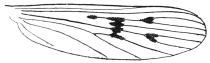
Fig. 399.—Wing of Anopheles maculipennis, Meigen.
This genus contains a few large species found either in temperate climates or in hills and mountains of warm climates. The type is the European and North American A. maculipennis.
A. maculipennis, Meigen. This species and A. bifurcatus are malaria carriers. True Anopheles only occur in Europe, North America, the North of Africa and in the mountains of India, and one has been found by Bancroft similar to A. bifurcatus in Queensland. They are easily told by the absence of scales on thorax and abdomen, and by the rather densely scaled wings with lanceolate scales.
Genus. Myzomyia, Blanchard; Grassia, Theobald.
Comp. rend. heb. Soc. Biol., No. 23, p. 795, Blanchard; “Mono. Culicid.,” 1910, iii, p. 24; v, p. 16, Theobald.
This genus occurs in Asia, Africa and South America, Europe and East Indies. The type is M. funesta, Giles, found in Central and West Africa. Although structurally there is not much difference between this genus and Anopheles, they differ greatly in appearance, and there are usually a few narrow curved thoracic scales projecting over the head, whilst the wing scales are much smaller in proportion, and the wings more uniformly spotted, always so along the costa. Funesta and lutzii are undoubtedly malaria bearers and also rossii.
Genus. Neomyzomyia, Theobald.
“Mono. Culicid.,” 1910, v, p. 29.
A single species only occurs in this genus, N. elegans, James, from India. In this genus, which is near to Myzomyia, the fourth long vein is very near the base of the third, and there are outstanding scales on the prothoracic lobes, and there is a marked tuft of dense scales at the posterior angles of the head.
Genus. Cycloleppteron, Theobald.
“Mono. Culicid.,” 1903, ii, p. 312; 1903, iii, p. 58; 1910, v, p. 33.
Two common species only occur in this genus, C. grabhamii, Theob., from Jamaica, and C. mediopunctatus, Theob. (Lutz., ms.), from South America. The chief character is the presence of large black inflated pyriform scales on the wings. The palpi are densely scaled. Neither have been shown to be malaria bearers.
Genus. Feltinella, Theobald.
“Mono. Culicid.,” 1907, iv, p. 56.
A single species, so far only found in this genus. The basal lobes of the male genitalia of two segments, the prothoracic lobes with dense outstanding scales.
The species, F. pallidopalpi, Theob., occurs in Sierra Leone.
Genus. Stethomyia, Theobald.
“Mono. Culicid.,” 1903, iii, p. 13; 1907, iv, p. 59; 1910, v, p. 35.
Four species occur in this marked genus—one S. nimba, Theob., from British Guiana and Para, another S. fragalis, Theob., from the Malay States, S. culiciformis, James and Liston, from India, and S. pallida, Ludlow, from India.
The former may be a malaria carrier, for Dr. Low says: “Malarial fever is got amongst the Indians and often of a severe type. In that connection it is interesting that in the interior, at a place called Corato, I got an entirely new Anopheles in large numbers.” The genus is easily told by its unornamented wings, flat head scales, mammillated prothoracic lobes and long thin legs.
Genus. Pyretophorus, Blanchard; Howardia, Theobald.
Compt. rend. heb. Soc. Biol., No. 23, p. 705, Blanchard; Journ. Trop. Med., v, p. 181; and “Mono. Culicid.,” 1903, iii, p. 13; 1910, v, p. 36, Theobald.
Forty-four species come in this genus, of which Anopheles costalis, Loew, is the type.
This genus is found in Africa, India, Europe and in Australia. Three species are proved malaria bearers, namely, P. costalis, Loew, P. chaudoyei, Theob., and P. superpictus, Grassi. Members of this genus can be told by having narrow curved thoracic scales, hairy abdomen, and much-spotted wings.
Genus. Myzorhynchella, Theobald.
“Mono. Culicid.,” 1907, iv, p. 78.
In this genus the thorax has distinct, narrow curved scales, and the abdomen is hairy, the wing scales broad and lanceolate, and the head with broad scales not closely appressed, but not forked or fimbriated.
Five species are known: lutzi, Cruz; parva, Chagas; nigritarsis, Chagas; tibiomaculata, Neiva; gilesi, Neiva; and nigra, Theobald. They are all recorded from Brazil, and nigra also from Mexico.
Genus. Manguinhosia, Cruz, in Peryassu.
“Os Culicideos do Brazil,” 1908, p. 112.
A single marked species from the Brazils. The thorax has piliform curved scales, and some narrow curved and flattened ones on the sides. Abdomen pilose, except the last three segments which are scaled. No tufts of scales on posterior femora.
Allied to Lophoscelomyia, but at once told by the absence of scale tufts on the hind femora. M. lutzi, Cruz, Brazil.
Genus. Chrystya, Theobald.
“Rep. Sleeping Sickness, Roy. Soc. Eng.,” 1903, vii, p. 34.
A very marked genus in which the hairy abdomen has very long, dense, hair-like, apical, scaly tufts to the segments. A single species only so far known, C. implexa, Theobald, from Africa (Uganda, Sudan, etc.).
Genus. Lophoscelomyia, Theobald.
Entomologist, 1904, xxxvi, p. 12.
A single species only, from the Federated Malay States. The hind femora have dense, apical scale tufts; the thorax long, hair-like curved scales; abdomen pilose, except the last two segments which are scaly; wings with broad, blunt, lanceolate scales.
Genus. Arribalzagia, Theobald.
“Mono. Culicid.,” 1903, iii, pp. 13 and 81; and 1910, v, p. 48.
Two species only occur, found in South America. The thorax and abdomen have scales and hairs respectively, as in Pyretophorus, but the abdomen has in addition prominent lateral apical scale tufts to the segments and a scaly venter. Wings with membrane tinged in patches and wing scales bluntly lanceolate and very dense. The type is A. maculipes, Theob. found in Trinidad and Brazil; A. pseudomaculipes, Cruz, also in Brazil.
Genus. Myzorhynchus, Blanchard; Rossia, Theobald.
Compt. rend. heb. Soc. Biol., 1902, No. 23, p. 795, Blanchard; Journ. Trop. Med., 1902, p. 181, Theobald; “Mono. Culicid.,” 1903, iii, p. 84; 1907, iv, p. 80; 1910, v, p. 49.
A very marked genus of large, dark, densely scaled species, found in Europe, Asia, Africa and Australia. The thorax with hair-like curved scales; the abdomen with ventral and apical scales, and a median ventral apical tuft, and with very densely scaled palpi in the female, and densely scaled proboscis. It seems to be mainly an Asiatic and East Indian genus, but three species occur in Africa and one in Australia. They are mostly sylvan species and bite severely.
Fourteen species are known. Five are malaria carriers (vide list, p. 566).
Genus. Nyssorhynchus, Blanchard; Laverania, Theobald.
“Mono. Culicid.,” 1910, iii, p. 14; v, p. 55, Theobald; Compt. rend. heb. Soc. Biol., No. 23, p. 795, Blanchard.
A group of small, closely allied species found in Asia, Africa and Australia, twelve out of the twenty species coming from India.
The thorax is covered with narrow curved and spindle-shaped scales, abdomen with small, flat or narrow curved dorsal scales, especially on the apical segments or in patches; the legs are always banded or spotted with white, and the tarsi have as a rule one or more pure white segments. (This banding and spotting is of no generic value, however.)
The species show considerable seasonal variation. The type of the genus is N. maculatus, Theobald.
Three are malaria carriers (vide list, p. 566).
Genus. Cellia, Theobald.
“Mono. Culicid.,” 1903, iii, p. 107; 1910, v, p. 67.
Very marked Anophelines, with densely scaly abdomens, the scales irregularly disposed on the dorsum and forming dense lateral tufts; thorax with flat spindle-shaped scales; palpi densely scaled and also the wings.
The type of the genus is the African C. pharoensis, Theob. It is represented in Asia by C. kochii, Dönitz; in West Indies and South America by C. argyrotarsis, Desvoidy, and C. bigotii, Theob.; in Africa by C. squamosa, Theob., etc.
C. argyrotarsis, Desvoidy, and C. albimana, Wiedemann, are undoubtedly malaria bearers.
Genus. Neocellia, Theobald.
“Mono. Culicid.,” 1907, iv, p. 111.
Allied to Cellia, but has no lateral scale tufts. Three species recorded from India.
Genus. Kertészia, Theobald.
“Ann. Mus. Nat., Hung.,” 1905, iii, p. 66.
This genus has the thoracic scales hair-like, except a few narrow curved ones in front; abdominal scales long, broad and irregular.
A single species, K. boliviensis, Theob. from Bolivia.
Genus. Manguinhosia, Cruz.
The thorax has narrow hair-like curved scales and some broad straight scales; others spatulate on the sides. Abdomen with fine hairs, except the last three segments which are scaly. Tufts of scales on the hind femora. Wing scales lanceolate.
The type is M. lutzi, Cruz, from Brazil.
Genus. Chagasia, Cruz.
“Brazil-Medico,” 1906, xx, pp. 20, 199.
This genus can at once be told by the antennal segments having many dense scaly tufts. Type, C. fajardoi, Lutz, from Brazil.
Genus. Calvertina, Ludlow.
Canadian Entomologist, 1909, xli, pp. 22, 234.
The antennæ in this genus have outstanding scales on the second segment, more appressed ones on the first. At least one abdominal segment with long, flat, more or less spatulate scales. Type, C. lineata, Ludlow, from Philippine Islands.
Genus. Birónella, Theobald.
“Ann. Mus. Nat. Hung.,” 1905, iii, p. 69.
At once told by the first submarginal cell being very small. Type, B. gracilis, Theob. from New Guinea.
Sub-family. Megarhininæ.
Three genera occur in this marked sub-family; they are the largest of all mosquitoes, and are very brilliantly coloured, and many have tail fans. They occur in North and South America, Asia, Africa, and Australia. The long curved proboscis is very marked. They are usually spoken of as elephant mosquitoes; some are vicious blood-suckers at times.
The three genera tabulate as follows:—
α. Palpi long in both sexes.
β. Last segment of ♀ palp round or blunt as if brokenGenus Megarhinus, R. Desvoidy.
ββ.Last segment of ♀ palp long and pointedGenus Ankylorhynchus, Lutz.
αα.Palpi of female short of male long. Palpi of female not more than one-third length of proboscisGenus Toxorhynchites, Theobald.
Genus. Megarhinus, Robineau Desvoidy.
“Mém. Soc. d’Hist. nat. de Paris,” 1827, iii, p. 412; “Mono. Culicid.,” 1901, i, p. 215; 1903, iv, p. 163; 1907, iv, p. 128; 1910, v, p. 89.
All large brilliant mosquitoes with long palpi in both sexes and, as a rule, with a caudal fan of scales; the proboscis is long and bent. They are all sylvan species, and are not so far recorded as biting man.
Genus. Toxorhynchites, Theobald.
“Mono. Culicid.,” 1901, i, p. 244; 1903, iii, p. 119; 1907, iv, p. 140; 1910, v, p. 95.
Differs from the former genus in that the female palpi are short. The palpi may have one, two or three minute terminal segments. Banks’s genus Worcesteria has three.
The elephant mosquito of India (T. immisericors), Walker, bites very severely. They are sylvan species.
Sub-family. Culicinæ.
Genus. Mucidus, Theobald.
“Mono. Culicid.,” 1901, i, p. 268; 1910, v, p. 125.
This genus is so far confined to Australia, West and Central Africa, India, East Indies and Malay Peninsula. They are all large mosquitoes, easily told by the whole body being more or less covered with long twisted scales, giving them a mouldy appearance, and the legs densely scaled with outstanding scales; the wings with large parti-coloured scales. The Australian M. alternans, Walker, occurs in larval form both in fresh and salt water. The adults bite man.
Genus. Psorophora, Robineau Desvoidy.
“Mém. de la Soc. d’Hist. nat. de Paris,” 1827, iii, p. 412, R. Desvoidy; “Mono. Culicid.,” 1901, i, p. 259; 1903, iii, p. 130; 1907, iv, p. 158; 1910, v, p. 123, Theobald.
This genus is confined to the Americas and the West Indies. Several species exist which can easily be told from Mucidus by the absence of long twisted scales and the narrower wing scales. The legs are densely scaled and the thorax ornamented with flat spindle-shaped scales.
P. ciliata, Robineau Desvoidy, occurs in both North and South America, and bites man.
Genus. Janthinosoma, Arribalzaga.
“Dipt. Arg.,” 1891, p. 52, Arribalzaga; “Mono. Culicid.,” 1901, i, p. 253; 1903, iii, p. 124; 1907, iv, p. 152; and 1910, v, p. 118, Theobald.
Hind legs only densely scaled; some of the hind tarsi are always white. The venation is as in Culex. The abdomen is metallic and iridescent. They all bite man and occur only in the Americas and West Indies.
Genus. Stegomyia, Theobald.
“Mono. Culicid.,” 1901, i, p. 283; 1903, iii, p. 130; 1907, iv, p. 170; 1910, v, p. 151.
This, the most important genus in the Culicinæ, can be told by the head and scutellum being clothed with flat scales and the thorax with narrow curved ones.
About forty species are known in this genus, occurring in Southern Europe, Asia, Africa, Australia, the Americas, East and West Indies, and on most oceanic islands. Many of them seem to be vicious blood-suckers. They are mostly black and white mosquitoes, and several seem to go by the name of tiger mosquitoes. The genus contains the yellow fever mosquito (S. fasciata, Fabricius), the only one that need be dealt with in detail here. The chief known species tabulate as follows:—
A.Proboscis banded.
α.Legs basally banded.
Thorax brown, with scattered creamy-white scalesannulirostris, Theobald.
Thorax black, with narrow, curved golden scalesperiskelta, Giles.
αα.Legs with basal and apical banding. Fore legs with no bands; mid with apical and basal bands on first and second tarsals, hind with basal bands.
Thorax white in front, with a brown eye-like spot on each sidethomsoni, Theobald.
AA.Proboscis unbanded.
β.Legs basally banded.
γ.Abdomen basally banded.
Thorax with one median silvery-white linescutellaris, Walker.
Thorax as above, but pleuræ with white linespseudoscutellaris, Theobald.
Thorax similar, but two white spots near where line endsgelebinensis, Theobald.
Thorax with two median yellow lines and lateral curved silvery linesfasciata, Fabr.
Thorax with two short median lines and a white patch on each sidenigeria, Theobald.
Thorax with large lateral white spots in front, smaller ones by wings, two narrow median lines and two posterior sub-median white lineslilii, Theobald.
Thorax with a white W-shaped area in front, a prolongation curved on each side enclosing a brown eye-like spotW-alba, Theobald.
Thorax with white frontal median spot, two large lateral spots, a small one in front of the wings, a narrow median white line and narrow sub-median ones on posterior half. Last two hind tarsi whitewellmannii, Theobald.
Thorax brown, with broad white line in front extending laterally towards wings, where they swell into a large patch, a white line on each side just past wing roots. Last two hind tarsi whitealbipes, Theobald.
Thorax with silvery white spot on each side in front, small one over roots of wings and white over their base. Last two hind tarsi whitepseudonigeria, Theobald.
Thorax with two lateral white spots, front ones the largest, a small median one near head, two yellow median lines, a short silvery one on each side before the scutellumsimpsoni, Theobald.
Thorax with silvery-white scaled area in front and another on each side in front of wingsargenteomaculata, Theobald.
Thorax with median yellowish-white line, a silvery patch on each side in front of wings extending as a fine yellow line to the scutellum, and another silvery spot before base of each wingpoweri, Theobald.
Thorax with small grey-scaled area in front of wing roots and three short creamy lines behindminutissima, Theobald.
Thorax (?) denuded; abdomen black; fifth segment with yellow basal band; sixth unbanded; seventh, two median lateral white spots; eighth, two basal lateral white spots; second hind tarsal nearly all whitedubia, Theobald.
γγ.Abdomen unbanded.
First hind tarsal all white, second basally white, last two dark. Thorax chestnut brown, with a broad patch of white scales on each side in front and a median pale lineterreus, Walker.
ββ.Legs with white lines as well as basal bands.
Thorax brown, with white lines; abdomen with basal bandsgrantii, Theobald.
βββ.Fore and mid legs with apical bands, hind basal.
Fourth tarsal of hind legs nearly all whitemediopunctata, Theobald.
Mid metatarsi with basal pale banding, base and apex of hind, also base of first tarsal paleassamensis, Theobald.
ββββ.Legs unbanded.
δ.Abdomen basally banded.
Thorax with front half white, rest bronzy-brownpseudonivea, Ludlow.
Thorax deep brown, with scattered golden scales, showing two dark eye-like spots; head white, dark on each side and behindalbocephala, Theobald.
Thorax brown with golden stripes; abdomen with narrow basal bands on fifth and sixth segments onlyauriostriata, Banks.
δδ.Abdominal banding indistinct.
Thorax with broad silvery white patch on each side in frontalbolateralis, Theobald.
δδδ.Abdomen unbanded.
Thorax with six silvery spotsargenteopunctata, Theobald.
δδδδ.Abdomen with apical white lateral spots.
Thorax unadorned, except for pale scaled lines laterallypunctolateralis, Theobald.
δδδδδ.Abdomen with basal white lateral spots.
Thorax with two pale median parallel lines and two silvery lateral spotsininuta, Theobald.
Thorax unadorned.
A white spot middle of headtripunctata, Theobald.
No white spotamesii, Ludlow.
AAA.Proboscis yellow basally, dark apically.
Abdomen with apical pale bandscrassipes, Van der Wulp.
AAAA.Proboscis with median interrupted white line on basal half.
Head black, anterior margin greyalbomarginata, Newstead.
Stegomyia fasciata, Fabricius (Yellow Fever Mosquito).
This insect, which is the proven carrier of yellow fever, is commonly called the tiger, brindled, spotted day or striped mosquito. It is also referred to by some writers as S. calopus, Meigen. It is subject to considerable variation in colour, but the thoracic markings are generally very constant. The general colour is almost black to deep brown, the head with a median white area, white at the sides and in front around the eyes; the thorax has two median parallel yellow lines, a broad curved silvery one on each side and white spots at the sides; the scales on the intervening spaces of the thorax are brown. The dark abdomen has basal white bands and basal white lateral spots. The dark legs have basal white bands, the last segment of the hind legs being all white except in a variety from South America and the West Indies (luciensis), which has the tip of the last hind tarsal dark. The abdomen may also vary in colour, some having pale scales over most of the surface (queenslandensis).
The food of the adult female consists mainly of man’s blood, but she will also feed on dogs and other animals. The male has been said to bite, but such is very unusual. This mosquito bites mainly in the daytime up till about 5 p.m.
The adults breed the first day after emergence. They may live a considerable time, Bancroft having kept females for two months in confinement. The ova are laid separately, often in chains; they are black, oval, with a reticulated membrane outside, some of the reticulated cells containing air. They may hatch in from six to twenty hours, the larval stage nine days, the pupal stage three; thus the whole cycle may be completed in from twelve to thirteen days. The ova when dry can remain undeveloped for a considerable time. The larvæ are greyish-white, with short, thick siphon, and feed at the bottom of the water, only coming to the surface now and again to breathe. This is almost entirely a domesticated gnat, seldom being found far from man’s habitations. Its larvæ occur in such small collections of water as old sardine tins, jam-pots, calabashes, puddles, barrels, wells—in fact, wherever water is held up, even to the gutters of houses. Not only are they found breeding on land, but also on board ship, although they prefer artificial collections of water. They may also breed in larger natural collections.
This insect is easily transported by steam and sailing ships and by train, and this doubtless explains its very wide distribution. The adults may live for fifty days, and it is on this account and their frequent occurrence on ships that danger lies in regard to the Panama Canal. An infected insect may leave that endemic centre of yellow fever and live until the vessel arrived at the Philippine Islands and fly ashore, and so introduce the disease for the native fasciata possibly to spread.
Roughly the distribution of this pest is as follows: Africa from South to North, but especially along the coast and up the Nile. In Asia, in India, Ceylon, Burma, Siam, along the ports of the Malay Peninsula, in French Cochin China, Philippine Islands, the Andaman and Nicobar Islands, Japan, Malay Archipelago, and East Indies, Turkey in Asia, Arabia and Palestine.
In Australia it occurs in Queensland, New South Wales, Victoria and South Australia.
In Europe in Italy, Spain, Portugal, Greece, in the Mediterranean Islands.
In South America, Central America, Mexico, North America, and the West Indies it is very abundant, and it also is found in the Bahama Islands, Fiji, Sandwich Islands, Samoa, the Azores, Teneriffe and Santa Cruz, Pitcairn Islands and Bermuda.
For a full account of its distribution the reader is referred to the following: “The Distribution of the Yellow Fever Mosquito (Stegomyia fasciata, Fabricius) and General Notes on its Bionomics;” “Mém. 1er Congrès international d’Entomologie, 1911, ii, pp. 145–170, F. V. Theobald.” In addition to being the yellow fever carrier, it is supposed by Wenyon to be the intermediate host of the parasite of Bagdad sore.
Stegomyia scutellaris, Walker.
A vicious biter, found in India, China, Malay, East Indies, and Ceylon. The thorax has one median silvery stripe, and so can easily be told from S. fasciata.
A very similar species occurs in Fiji, but can be told by the pleuræ having white lines, not spots (S. pseudoscutellaris, Theobald). It is the intermediate host of filaria in Fiji (Bahr).
A number of nearly allied genera occur here (vide synoptic table).
Genus. Theobaldia, Neveu-Lemaire.
Theobaldinella, Blanchard.
Includes several large Culicines, of which T. annulata, Meigen, is the type. The wings are usually spotted (annulata, incidens, etc.), but may be nearly plain (spathipalpis). The males have the palpi swollen apically, and the females have long five-jointed palps.
Several of these are vicious biters.
Theobaldia annulata, Meigen.
This large gnat (6 mm. long) can be told by its wings having five large spots of dark scales and by its legs having broad basal white bands to the tarsi. The larvæ occur in rain barrels and small pools. It is essentially a domestic form, occurring in houses and privies. Its distribution is Europe generally and North America. The bite is very severe, and in some districts gives rise to painful œdema.392
Theobaldia spathipalpis, Rondani, occurs in Italy, Mediterranean Islands, Palestine, the Himalayas, Khartoum, and in South Africa. It is about the same size as T. annulata, but is yellowish-brown in colour, with striped thorax and mottled and banded legs. It occurs in privies and bites very severely.
Genus. Culex, Linnæus.
“Syst. Nat. Ed.,” 1758, x, Linnæus; “Mono. Culicid.,” 1901, i, p. 326; 1910, v, p. 322, Theobald.
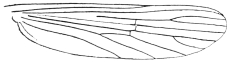
Fig. 400.—Wing of a Culex.
This large genus still contains many forms which should be excluded. The species normally have narrow curved median head-scales, and similar ones on the scutellum; the female palpi are shorter than in the former genus and the male palpi are pointed; the lateral vein-scales are narrow and linear.
The type is Culex pipiens, Linn., the common gnat of Europe. The thorax is covered with narrow curved golden-brown scales, the abdomen has basal pale bands to the segments and the legs and proboscis are unbanded. The stem of the first submarginal cell is always less than one-fifth the length of the cell. It lays its eggs in rafts in water-butts, etc., and even in the foulest water. They are first deposited in England in June and July, and again soon after hatching in August. In some districts this gnat bites man viciously, in others not at all.
The common tropical gnat (Culex fatigans, Wied). This resembles the European Culex pipiens, but can always be told by the stem of the first submarginal cell always being much longer than it is in C. pipiens. This is one of the species that has been proved to transmit filariæ to man, etc. Varieties of it occur in almost every country between 40° N. and S., having a very similar range to S. fasciata. In all countries it appears to be connected with the transmission of Filaria bancrofti, and it is also said to carry the micrococcus of dengue fever.
Genus. Melanoconion, Theobald.
“Mono. Culicid.,” 1903, iii, p. 238; 1907, iv, p. 507; 1910, v, p. 455.
This genus is composed of eight species, most of which are small black gnats which bite viciously and which occur in swamps and jungles. They can at once be told from Culex by the veins of the wings having dense broadened scales on their apical areas and along the upper costal border. The femora and apices of the tibiæ are swollen.
The black mosquito, Melanoconion atratus, Theob. This small gnat is a very troublesome pest in swamps in the West Indies. The female bites both by day and by night, and the bite causes severe irritation. The larvæ live in permanent ponds. It is almost black in colour, but sometimes presents a dull coppery sheen; each segment has small lateral basal white spots. Length 2·5 to 3 mm.
It occurs in Para and British Guiana as well as in the West Indies.
Ordinary mosquito netting is no use for keeping off this pest.
Genus. Grabhamia, Theobald.
“Mono. Culicid.,” 1903, iii, p. 243; 1907, iv, p. 284; and 1910, v, p. 277.
Allied to Culex, but separated by the wings having short fork-cells, mottled scales, the median ones thick and also some of the lateral ones short and broad; the last two joints of the male palps are very slightly swollen. The eggs are laid singly, not in rafts, and the larvæ have short, thick siphons. Ten species occur and are found in Europe, North America, West Indies and Natal. G. dorsalis, Meigen, bites severely in Europe. G. sollicitans, Walker, is a great scourge along the New Jersey Coast and at Virginia summer resorts and in Florida. It breeds in brackish water and is the most common mosquito of the Atlantic seaboard.
Genus. Pseudotæniorhynchus, Theobald; Tæniorhynchus, Theobald, non-Arribalzaga.
Differs from the former in having the whole wing veins clothed with dense, broadish elongated scales. They occur in South America (T. fasciolatus, Arri.), in Africa (T. tenax, Theob.), in Europe (T. richardii, Ficalbi). The latter bites very severely.
Genus. Tæniorhynchus, Arribalzaga; Mansonia, Blanchard; Panoplites, Theobald.
Compt. rend. heb. Soc. Biol., 1901, iii, 37, p. 1046; “Mono. Culicid.,” 1901, ii, p. 173; and 1910, v, p. 446, Theobald.
A very marked genus, easily told by the broad asymmetrical wing scales. It occurs in Africa (T. africana and T. major, Theob.); in Asia (T. uniformis, Theob.; T. annulipes, Walker, etc.) and in Australia (T. australiensis); in the Americas and West Indies (T. titillans, Walker). The eggs (fig. 395, d) are peculiar in form and are laid separately; the larva has not been described; the pupa has long curved siphons. They mostly occur along rivers, in swamps and forests, and bite very severely. They also enter houses (T. titillans). T. uniformis is most troublesome during the rains. The saliva is strongly acid. Both these species carry the larvæ of Filaria bancrofti.
Genus. Chrysoconops, Goeldi.
“Os Mosq. no Para,” 1905, p. 114, Goeldi; “Mono. Culicid.,” 1910, v, p. 433, Theobald.
Bright yellow or yellow and purple mosquitoes, with rather dense wing scales. Numerous species occur in Africa (aurites, annettii, fuscopennatus, etc.), others in India, Australia and South America.
Low found filariæ in the thoracic muscles of fuscopennatus in Uganda.
Several of the Ædeomyina bite, especially the small Uranotænias. They are all sylvan species, seldom entering houses. They need not, therefore, be referred to here.
For full details of the Culicid genera and species the reader is referred to my monograph393 and other works mentioned below.
Other Nematocera.
Other nematocerous flies are midges, daddy-long-legs and sand-flies. The ones which cause annoyance to man besides Culicidæ are the following:—
Sand-flies (Simulidæ), certain midges (Chironomidæ), and a few owl midges (Psychodidæ).
The Nematocera have long thread-like jointed antennæ and their pupæ are, as a rule, naked; the larvæ have a distinct head and can thus be told from the next section (Brachycera).
Family. Simulidæ.
This family consists of a single genus, Simulium, Latreille, which Roubaud has recently divided into two sub-genera called Pro-Simulium and Eu-Simulium. These insects, which are frequently spoken of as sand-flies, are found in all parts of the world; they are all small insects varying from 1·5 to 3 mm. The females are very bloodthirsty, but the males appear to be incapable of sucking blood.
The head sunk under the humped thorax; antennæ short, straight; palpi short and broad, of four segments, bent; wings broad and in some iridescent, legs stout. The male has holoptic eyes, whilst in the female they are small and widely separate. The sucking proboscis is short. The thorax and abdomen are clothed with short hairs which may form spots and markings; these are golden, silvery, grey, or brownish. In the sub-genus Pro-Simulium the second segment of the hind tarsi in both sexes is elongate, linear, and without a basal notch; in Eu-Simulium it is short, curved, and dorsally notched at the base.
Simulidæ often occur in swarms, and attack not only man but cattle, horses, and poultry. In some districts they are more annoying than mosquitoes.
Their life-cycle has been most completely worked out by King, in Africa.
The larvæ and pupæ occur in swiftly flowing water, by waterfalls, in rapids, etc. The ova are laid in gelatinous masses on plants or rocks close to or overhanging the water. The larva is cylindrical, enlarged posteriorly, where it is provided with a sucker, by means of which it attaches itself to a rock, water weeds, debris, etc.; anteriorly it has a proleg close behind the head on the lower surface. The head is dark and chitinous. The respiration takes place by means of branched tracheal gills which protrude from the dorsal surface of the last body segment; they are retractile. The colour varies from deep green to yellow or almost black. Their food consists of algæ and other organisms in the water brought to their mouth by two fan-like organs placed on the head. The larvæ can crawl from place to place by means of the thoracic proleg; they occur in masses, usually in a more or less erect attitude. A network of threads is spun on their support, by means of which King tells us “they are enabled to maintain their position against the strongest current; frequently they will leave their support and let themselves out into the stream anchored by threads of silk and enabled by them to return.”
When full fed the larva spins a pocket-shaped cocoon on the support, within which it pupates. The pupa is motionless and has a pair of branched spiracles projecting from behind the head. When the adult emerges, a bubble of air collects around it, and in this it floats to the surface and at once takes wing. The European species take a month to complete larval life, a week being spent in the pupal stage. The flies are most restless, and even when stationary continually move their legs about like feelers. Sometimes the swarms consist entirely of females, sometimes early in the season mostly of males.
The females pierce the skin of humans on tender spots, such as ears, the forehead, around the eyes and nose, and crawl into the cavities. They are quite harmless at night, mainly attacking about sunrise and sunset. Some crawl up the arms and legs and down the neck, and leave behind little red weals which itch intensely (S. damnosum, Theob.), and blood may flow freely from the wounds.
The following are some of the worst species:—
Simulium columbaschensis, the “Kolumbatz fly,” which abounds in the damp marshy lands along the Danube, and is a great plague to man and beasts in Hungary, and is also abundant in Austria and Moravia, and is most numerous after inundations from the Danube. They sometimes appear in such swarms that it is impossible to breathe without getting them into one’s mouth. There are instances of children being killed by these flies when left on the ground by their mothers when working in the fields.
S. damnosum, Theob. This occurs throughout Equatorial Africa and is known as the “jinja fly” in Uganda, the “fouron” in the French Congo, the “kilteb” in the Sudan. It is a most vicious biter, and in some parts occurs in “belts”; Dr. Christy found one such extending from the shores of the Victoria Nyanza northwards along the right bank of the Nile for twelve or fifteen miles or more, and perhaps three or four miles wide. In this area the flies swarm in millions at certain seasons, so much so that the natives have to leave their plantations. The bite causes a weal, marked by a drop of blood.
S. griseicollis, Becker. The so-called “nimitti” occurs in Upper Egypt and the Anglo-Egyptian Sudan. It lives near the river and is not found more than half a mile from it. Human beings are bitten on the face and hands, animals in the region of the pudenda.
S. latipes, Meigen. This is a European species, also found in Natal.
S. wellmanni, Roubaud. The “ohomono” of Angola, where it bites viciously and is dreaded by the naked porters.
S. buissoni, Roubaud. Occurs in abundance in the Marquesas Islands. It has been suggested that this species may help to propagate leprosy.394
A large number of these insects have been described by Lutz in Brazil.395
A Simulium sp. (?) is very harmful to poultry in Cape Colony.396
In America, Simulidæ are most annoying. One, S. meridionale, Riley, also known as the turkey gnat in the Mississippi Valley, has been supposed to be the carrier of chicken cholera; anyhow, it has caused the death of thousands of chickens and turkeys in Virginia annually.397
In Mexico Townsend found a Simulium which was named S. occidentalis, which caused great annoyance to man, many people being so susceptible to them as to preserve through the gnat season a chronic inflammation of the exposed parts of face and neck, resulting from the repeated bites giving rise to sores.398
Men and horses have been partially incapacitated by the bites of sand-flies or Simulium in a Hampshire wood (Cantlie, Brit. Med. Journ., April 28, 1900, v, No. 2,052, p. 1023).
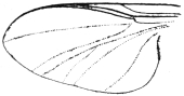
Fig. 401.—Wing of Simulium.
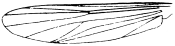
Fig. 402.—Wing of Chironomus.
Family. Chironomidæ (Midges).
The Chironomidæ or midges are not only frequently mistaken for mosquitoes, but some are very annoying to man by biting him as mosquitoes do. They are easily distinguished from true mosquitoes (Culicidæ) by the following characters: (1) head small, often retracted under the cowl-like thorax; (2) no scales to the wings or body; and (3) the different arrangement of veins on the wings (fig. 402).
Two genera are important as annoying man, namely, Culicoides, Latreille, and Johannseniella, Williston. The larvæ of Chironomidæ are either aquatic, both fresh water and marine, and help to make the former foul,399 according to Slater, or may, as in Ceratopogoninæ, live beneath the bark of trees, etc. The pupæ are very varied and also the life-histories of the different genera.400 The blood-sucking habit is confined to the sub-family Ceratopogoninæ.
Sub-family. Ceratopogoninæ.
This sub-family of midges consists of very small species varying from 1 to 2 mm. in length; the wings have darkened areas, and the second longitudinal vein is wanting, and the first and third veins are stouter than the others and placed close to the anterior margin, the fourth and fifth are forked; the antennæ in both male and female are composed of fourteen segments, six or eight in the males bearing long hairs.
The chief blood-sucking species belong to the genera Culicoides, Latreille, and Johannseniella, Williston. The latter genus differs from the former in the absence of an empodium or median appendage on the last segment of the tarsi. The genus Ceratopogon, as restricted by Kieffer, is not supposed to take vertebrate blood, but Austen has recently noticed that the type specimen of C. castaneus, Walker, and a new species described by him, apparently have their bodies distended with blood. The wings in the Ceratopogoninæ are carried flat when at rest.
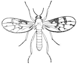
Fig. 403.—A Ceratopogon, or midge. Greatly enlarged.
In spite of their small size the females are the most bloodthirsty and annoying of all insects. The Culicoides, which are often called “sand-flies,” bite during the day and rarely at night. Usually they are most troublesome between 3 and 6 p.m. They frequently attack in swarms, especially in the open, and owing to their minute size can get through fine mosquito netting. Some of them produce a distinct “buzz” when on the wing. These insects are found in all parts of the world. No species has been definitely connected with any disease, but Culicoides has been suspected of carrying the germs of Delhi boil. The larvæ of Culicoides are elongate in form and have smooth bodies composed of thirteen segments including the head, which is horny; there is no proleg on the first segment as seen in Chironomus, and on the anal segment are retractile gills. They are very active and live in the sap of various trees which saturates diseased bark.
The pupæ are smooth, but the abdominal segments bear a transverse row of small spines. Austen describes a number of Culicoides and one Johannseniella and three Ceratopogons from Africa,401 and Lutz402 a number of this sub-family from Brazil, including a new genus, Centrorhyncus. Another genus, Tersesthes, Townsend (Centrotypus, Grassi; Mycterotypus, Noe), also occurs in Brazil.
Culicoides ornatus, Taylor, is described from Townsville, Australia, found in mangrove swamps. It is a very vicious biter and causes considerable irritation, settling on hands and wrists (Taylor, Rep. Ent. Aust. Inst. Trop. Med. (1912), 1913, p. 24).
Family. Psychodidæ (Owl Midges).
This family of diptera is of considerable importance, not only on account of the blood-sucking habits of some species, but especially on account of one at least having been proved to be the carrying agent of “papataci” fever, a three-day fever very prevalent in Malta and several parts of Southern Europe in the autumn.
It is also possible that these small flies are connected with the formation of “Delhi boil,” caused by a protozoan parasite.
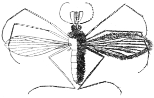
Fig. 404.—An owl midge, Phlebotomus sp. Greatly enlarged. (From Giles’s “Gnats or Mosquitoes.”)
Psychodidæ are all very small flies, many of which have a moth-like appearance, and owing to their fluffy nature are spoken of in Britain as “owl flies,” sometimes also as “window flies.” Their bodies and wings are covered with hairs, densely in some (sub-family Psychodinæ), and in a few with patches of flat squamæ. In the non-blood-sucking Psychodinæ the wings are carried in a peculiar manner downwards over the body, to a slight extent resembling the Hepialidæ, or swift moths. The wings may be ovoid or lanceolate, and have a marked venation as seen in the figure. The proboscis is short and non-suctorial in the majority of genera, but in the sub-family Phlebotominæ it is elongated and hard. The antennæ are long and of sixteen segments, and bear whorls of fine hair.
There are two sub-families, Psychodinæ and Phlebotominæ; in the former the mouth is not suctorial; the female has a horny ovipositor and the second longitudinal vein is branched at the root of the wing; in the second sub-family the proboscis may be formed for sucking, the female has no horny ovipositor, and the second long vein has its first fork near the middle of the wing.
The sub-family Phlebotominæ contains the genus Phlebotomus, which occurs in South Europe, South Asia, Africa, North and South tropical America. They are all small grey, brown, or dull yellow-coloured flies, and carry their wings when at rest upwards like a butterfly. The proboscis is moderately long and the legs long and thin.
The females are most vicious blood-suckers, but in some species anyhow the males also bite (P. duboscii). They are mainly nocturnal feeders and hide away during the day in any dark corners or crevices.
The life-cycle has been worked out by Newstead403 and Grassi404 in Europe, and by Howlett405 in India.
The larvæ have been found in crevices in rocks and caves, in dirty cellars, and dark damp places containing rubbish, and are also said to live in crevices in the walls of privies and cesspits.
The minute larva is very marked; as figured by Newstead it has two long chætæ projecting upwards, in some stages branched, in others simple, and on the segments a few blunt spine-like processes. The pupæ are found in similar situations. The ova are very minute, elongate, translucent white, and covered with a thin coating of viscous matter when first laid; soon after they become dark brown, shiny, with long black wavy lines. Newstead found the incubation period in Malta to last for about nine days in P. papatacii. Five species are known in Europe, five in Africa,406 two in North America, and eight are described by Annandale407 in the Oriental region. Lutz and Neiva have described three species from Brazil408 (P. longipalpis, intermedius and squamiventris).
Brachycera (Flies).
The antennæ as a rule have three segments, and are usually shorter than the head. The first segment of the antennæ is frequently very small, and the third one is generally the largest, and sometimes possesses a terminal annulated bristle. The palpi have from one to three segments; the mandibles are covered by the labium. The three thoracic rings are coalesced; wings are almost always present, the posterior ones being rudimentary and covered with a little scale. From the ova legless maggots are hatched, which as a rule have not a distinct head, but occasionally possess two claw-like hooklets. These maggots live in decomposing organic matter; they rarely live in water and some of them are parasitic. They either become barrel-shaped pupæ within the last larval integument or, after casting it, are transformed into naked pupæ. The larvæ of numerous Brachycera have been observed in man, some in ulcers or on mucous membranes, others in the skin or in the intestine, etc. In many cases the report only mentions the presence of the larvæ of flies; in other cases the species has been determined; whilst in still other cases the corresponding adult creature is unknown. We must therefore confine ourselves to describing the most common varieties.
Family. Phoridæ.
These flies belong to the same division of Diptera, the Aschiza, as the family Syrphidæ or “hover flies.” They are all small insects with marked antennæ and wings; the former have the third segment globular and enlarged, and thus hiding the first two; the wings are short and broad, the venation shows two short, thick, long veins with four thin ones running out from them. The larvæ normally live in decaying animal and vegetable matter, but one species, Aphiochæta ferruginea, Brun., has been found as an intestinal parasite of man.
Aphiochæta ferruginea, Brun.
This small fly belonging to this family is of an orange-ochreous colour, the upper part of the thorax tawny, and with dark bands on the abdomen, legs pale yellow, the hind femora tipped with dark brown. It measures only 2 to 3 mm. in length. This insect is shown by Austen to be widely distributed in the tropics, being found in India, Burma, West Africa, and Central America. The larvæ breed in decaying animal matter, such as putrid meat, decomposing shell-fish, etc.
Heusner bred out sixty-three flies from larvæ taken from an Indian’s foot.
Baker (Proc. Burma Branch Brit. Med. Assoc., 1891, p. 11–16) found that the maggots of this fly were passed per anum by a European at intervals during a period of ten months. Baker found that the larvæ fed on human fæces; from the egg stage to the deposition of eggs from the resultant brood of flies occupied twenty-two days. He concludes that they are capable of propagating, and do so while living within the human intestines. He also records the larvæ in two girls.
The larva does not seem to have been described, but Austen describes the pupa (Trans. Soc. Trop. Med. and Hyg., iii, No. 5, p. 229).
Phora rufipes, Meig.
The larvæ of the “hump-backed fly” live in rotting potatoes, mushrooms, radishes, etc., and when accidentally introduced into the intestine of man can, like other larvæ, live there twenty-four hours and even more, and may set up serious gastric disturbances.
P. rufipes is the same as P. pallipes, Latr.
Family. Sepsidæ.
Small blackish flies, elongate, with abdomen narrowed at the base, thickened and curved downwards towards the extremity. Larvæ often found in decaying vegetables, ham, cheese, etc. The larvae have the power of skipping; conical in form, pointed in front, truncated behind, about 5 mm. long, shiny and smooth, the anal segment with fleshy protuberances. The genus Piophila has a short proboscis and the cross-veins of the wings approximate.
Piophila casei, L.
Cheese flies. The larvæ live in ripe cheese, with which they are sometimes introduced into human beings (Meschede).
The larvæ of the cheese flies (Piophila casei) may pass through the alimentary canal of human beings alive, and have been occasionally referred to in cases of internal myiasis. It also breeds in dead bodies in adipose tissue. Howard records it on human excrement. It is thus possible that some of the recorded cases of this pest being passed alive may be due to eggs deposited on human fæces.
Family. Syrphidæ (Hover and Drone Flies).
Amongst the large family of Syrphidæ is found a section known as the Eristalinæ or drone flies, whose curious long-tailed larvæ are popularly called “rat-tail larvæ,” on account of the end of the body being drawn out into a long telescopic tail of two segments, at the end of which are placed the breathing pores. These larvæ live in water, no matter how foul, and in liquid manure. They have occasionally been obtained in foul drinking water by human beings and from eating watercress improperly washed or from badly kept beds. Austen (Trans. Soc. Trop. Med. and Hyg., iii, No. 6, p. 221) records that in the autumn of 1907 a number of the larvæ of the common drone fly (Eristalis tenax) were passed per rectum by a woman in Hampshire who had recently arrived from France. The patient had eaten a considerable quantity of watercress before leaving France. I have twice found small Eristalis larvæ clinging by their long tails on watercress served at table.
Family. Drosophilidæ.
Small, rather plump flies, with short, broad abdomen, with bristles on the head and legs. Often abundant in decomposing fruit, and may occur in dense masses.
Drosophila melanogaster, Br.
The larvæ of this fly occur in over-ripe fruit and in fungi, often also in human habitations, and live in substances undergoing acid fermentation (vinegar, decaying fungi, rotting fruit, in damaged spots in diseased trees), much more rarely in animal substances, and they occasionally gain access to the human intestine (for example, by the medium of sour milk). When introduced in any quantity, they cause vomiting or attacks resembling colic; when taken in the pupal stage no unpleasant results are produced.