ON PARASITES IN GENERAL. (continued 3)
Reading edition
In sheep and goats T. rhodesiense causes an acute disease, marked by high fever, œdema of the face, and keratitis, as shown by Bevan and others, death resulting after a relatively short period. T. gambiense gives rise, in these animals, to no symptoms except fever, which may be overlooked. T. rhodesiense produces keratitis in dogs.
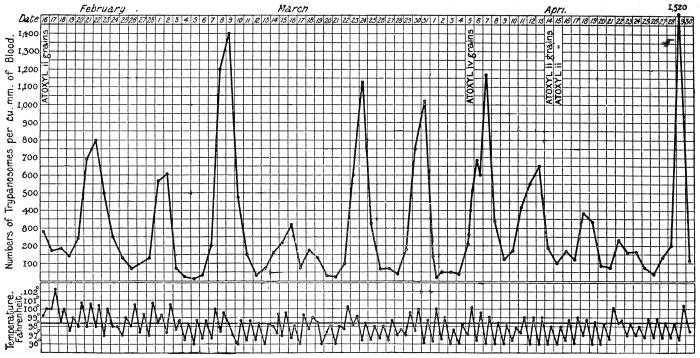
Fig. 32.—Chart showing daily counts of number of trypanosomes per cubic millimetre of peripheral blood from a case of Rhodesian sleeping sickness. (After R. Ross and D. Thomson.)
Stannus and Yorke (1911) observed T. rhodesiense in animals inoculated from a case of sleeping sickness in Nyasaland. Sir D. Bruce and his colleagues73 have shown (1912) that T. rhodesiense is the parasite usually found in man and in animals sub-inoculated from cases of sleeping sickness in Nyasaland. It has since been found in German East Africa and Portuguese East Africa, while Ellacombe has described a case from North-western Rhodesia.
(3) Serum Reactions.—Interesting experiments on this subject were performed during 1911 and 1912 by various French investigators.
(a) Action of Immune Serum (Mesnil and Ringenbach)74: (1) A goat was infected with T. rhodesiense. Twenty-two days later its serum mixed with T. rhodesiense was injected into a mouse. Result: Protection. (2) The serum mixed with T. gambiense was injected into a mouse. Result: Infection.
(b) Action of Baboon Serum.—Contrary to T. gambiense, T. rhodesiense is very susceptible to human and baboon sera. Mesnil and Ringenbach75 showed that a dose of 1 c.c. of baboon (Papio anubis) serum cured mice infected with T. rhodesiense. In the same dose it acted very feebly on T. gambiense.
(c) Action of Human Serum.—1 c.c. of human serum cured T. rhodesiense mice in three out of four cases; on T. gambiense mice there was no appreciable effect.
Laveran and Nattan-Larrier76 have shown the same, namely, that human sera act on T. rhodesiense, but are quite without action on T. gambiense.
(d) Trypanolytic Reactions.—Mesnil and Ringenbach77 have also shown that the sera of animals (man, monkey and guinea-pig) infected with T. gambiense are trypanolytic for the homologous trypanosome, that is, T. gambiense, but have no action on the heterologous trypanosome, that is, T. rhodesiense.
(4) Cross Immunity Experiments.—(a) Mesnil and Ringenbach78 immunized a monkey (Macacus rhesus) against T. gambiense. It was inoculated with T. rhodesiense on June 7, 1911; on June 27 trypanosomes appeared, the infection being slight; on July 4 it died. A control died in ten and a half days.
(b) Laveran79 immunized a goat and mice against T. gambiense. When they had acquired a solid immunity, they were inoculated with T. rhodesiense. They became infected like the controls.
(c) Laveran and Nattan-Larrier80 immunized a ram against T. brucei, it subsequently became infected with T. rhodesiense.
(d) Laveran81 immunized a ram and a sheep against different strains of T. brucei. Inoculated with T. rhodesiense they both acquired acute infections and died. Conclusion: T. rhodesiense is not T. brucei.
When the converse set of experiments is tried, namely, immunizing an animal against T. rhodesiense, and then inoculating with T. gambiense, the difficulty immediately arises that it is impossible to immunize an animal against T. rhodesiense, owing to its virulence. But a partial and transitory immunity to T. rhodesiense can be obtained by treating the infected animal with drugs, such as arsenophenylglycin. The results, so far as they go, seem to show that an animal immunized against T. rhodesiense is immune not only to T. rhodesiense, but also to T. gambiense, a fact which, according to Mesnil and Léger, does not invalidate the specificity of T. rhodesiense, but tends to show that the two trypanosomes are closely related.
(5) Mode of Transmission and Reservoir.—Kinghorn has shown that T. rhodesiense is transmitted by Glossina morsitans in which it undergoes development. Kinghorn and Yorke82 found that about 16 per cent. of the wild game examined in Northern Rhodesia was naturally infected with T. rhodesiense. The wild game examined included waterbuck, hartebeest, mpala, bushbuck and warthogs. One native dog near the Nyasaland border was found infected, but not domestic stock. Taute doubts whether T. rhodesiense really occurs in wild game. Approximately 3·5 per cent. of the tsetse flies fed on infected animals may become permanently infected with T. rhodesiense, and capable of infecting clean animals. Furthermore, a tsetse fly when once infective probably remains infective for the rest of its life.
Kinghorn and Yorke, however, have shown that climatic conditions, namely, those of temperature, also affect the infectivity of the tsetse fly, as the ratio of flies capable of transmitting T. rhodesiense to those incapable of transmitting the virus is 1 : 534 in hot valley districts (e.g., Nawalia, Luangwa Valley, temperature 75° to 85° F.), while on elevated plateaux (e.g., Ngoa, on the Congo-Zambesi watershed, temperature 60° to 70° F.) the ratio falls to 1 : 1312.
Mechanical transmission by the tsetse fly does not occur, if a period of twenty-four hours has elapsed since the infecting meal.
Developmental Cycle in the Fly.—The period which elapses between the infecting feed of the flies and the date on which they become infective varies from eleven to twenty-five days in the Luangwa Valley, according to Kinghorn and Yorke. Attempts carried out at laboratory temperature on the Congo-Zambesi plateau, during the cold season, to transmit T. rhodesiense by means of G. morsitans were always unsuccessful. The developmental cycle of the trypanosome in the fly is influenced by the temperature to which the flies are subjected (as stated above). The first portion of the developmental cycle proceeds at the lower temperatures (60° to 70° F.), but higher temperatures are necessary for the completion of the development of the trypanosome. Kinghorn and Yorke found that the trypanosomes may persist in the fly, at an incomplete stage of their development, for at least sixty days when the climatic conditions were unfavourable.
The first portion of the developmental cycle of the trypanosome takes place in the gut of the fly. Invasion of the salivary glands of the tsetse is secondary to that of the intestine, but is necessary for the infectivity of the fly. A relatively high mean temperature, 75° to 85° F., is essential for the passage of the trypanosomes into the salivary glands and the completion of their development therein.
Kinghorn and Yorke83 state that the predominant type of trypanosome in the intestine of infected G. morsitans was a large broad form, quite different from that which is most common in the salivary glands. The trypanosome in the glands resembles the short form seen in the blood of the vertebrate host. The authors quoted state that both the intestinal and salivary gland forms of infective G. morsitans are virulent when inoculated into healthy animals.
Bruce and colleagues84 have quite recently (June, 1914) published an account of their investigations of T. rhodesiense in G. morsitans in Nyasaland. (Incidentally it may be remarked that Bruce considers T. rhodesiense to be identical with a polymorphic strain of T. brucei—see pp. 83, 94). The development of T. rhodesiense takes place in the alimentary canal and salivary glands, not in the proboscis, of the tsetse fly. In feeding experiments with laboratory bred flies, as well as with a few wild flies, fed on infected dogs or monkeys, only 8 per cent. of the flies were found to be infected on dissection. Of such infected flies, however, only some allow of the complete development of the trypanosomes within them, in other words only about 1 per cent of the flies become infective. The length of time which elapses before a fly becomes infective varies from fourteen to thirty-one days, averaging twenty-three days, when kept at 84° F. (29° C.). The dominant intestinal type of flagellate in the fly is that seen in the proventriculus, which contains many long, slender trypanosomes. These proventricular forms find their way to the salivary glands, wherein crithidial and encysted forms are seen. They change into “blood forms,” which are short, stumpy trypanosomes and are infective. “The infective type of trypanosome in the salivary glands—corresponding to the final stage of the cycle of development—is similar to the short and stumpy form found in the blood of the vertebrate host.” The cycle is thus very similar to that of T. gambiense in G. palpalis (fig. 30).
Culture.—J. G. Thomson (1912),85 and subsequently Thomson and Sinton, succeeded in cultivating T. rhodesiense in a modified Novy-MacNeal medium. The development obtained resembled that of the trypanosome in the intestine of Glossina.
General Note on Trypanosomes with Posterior Nuclei.
Posteriorly placed nuclei have been found to occur not only in T. rhodesiense by Stephens and Fantham (1910), but also in T. pecaudi by Wenyon (1912), in T. brucei by Blacklock (1912), and in T. equiperdum by Yorke and Blacklock (1912).
Recently Stephens and Blacklock (1913)86 have shown that two trypanosomes, different morphologically, have been confused under the name T. brucei. One of these is polymorphic (i.e., it exhibits long and slender as well as short and stumpy forms) and came from Uganda, while the other is monomorphic and is the original Zululand strain described by Bruce from cattle suffering from “nagana.” Bruce (1914) considers that morphological change has occurred in T. brucei in its passage through laboratory animals, and thus explains the diversity of views. The posterior nuclear forms described by Blacklock occurred in the Uganda strain of T. brucei. (See p. 95.) Similarly, a posterior nuclear form, T. equi, has been separated from T. equiperdum. (See p. 98.)
Again, Bruce and his colleagues on the Royal Society Commission investigating sleeping sickness in Nyasaland, have stated (April, 1913) that “evidence is accumulating that T. rhodesiense and T. brucei (Plimmer and Bradford) are identical.” The exact identity of trypanosomes showing posterior nuclei is, then, far from settled, although Laveran by cross immunity tests has declared that T. brucei is distinct from T. rhodesiense. No one has yet seen posterior nuclei in T. gambiense.
Trypanosoma cruzi, Chagas, 1909.
Syn.: Schizotrypanum cruzi, Chagas, 1909.
The trypanosome was discovered by Chagas87 in the intestine of the bug, Triatoma (Conorhinus) megista, in Brazil, and then in the blood of a small monkey bitten by the bug. A little later it was found in the blood of a child, aged two years, suffering from irregular fever, extreme anæmia and enlarged glands in the State of Minas Geraes, Brazil. Chagas found that he was able to infect many of the usual laboratory animals with the trypanosome, by allowing the bug to bite them. He was also able to culture the parasite on blood agar.
Chagas found the Reduviid bug, Triatoma megista, in the houses of the poorer inhabitants of the Brazilian mining State, and that it attacked the people, more especially the children, at night, biting the face. On this account the insect is called “barbeiro” by the inhabitants. The bite is somewhat painful. The disease has since been found in other parts of Brazil, e.g., Matta de São João in Bahia province, Goyaz, Matto Grosso and São Paulo provinces, as well as in Minas Geraes.
Morphology.—The trypanosome has a large blepharoplast or kinetic nucleus. It is stated to occur both free and in the red blood corpuscles in the peripheral blood. It is about 20 µ long, on an average.
Two forms of the parasite (fig. 33, 6, 7) are described in the human blood. In one free form there is a large egg-shaped blepharoplast and the posterior (aflagellar) end of the parasite is drawn out. The blepharoplast (kinetic nucleus) may have a chromatin appendage. The nucleus is oval or band-like, containing a karyosome. The flagellum, starting close to the blepharoplast or its appendage, has a free portion of variable length. The other free form in the blood has a more or less round, terminal blepharoplast, smaller than in the first form, without a chromatin appendage as a rule. The body of this second form is decidedly broader than that of the first mentioned.
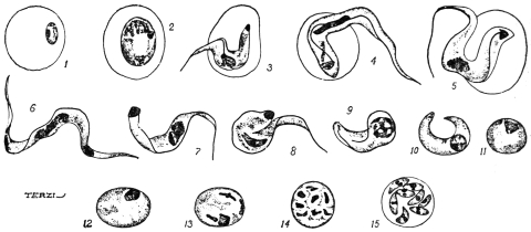
Fig. 33.—Trypanosoma cruzi. Schizogony. 1, merozoite in red blood corpuscle; 2, parasite totally enclosed in red cell, no flagellum or undulating membrane; 3-5, parasites partially enclosed in red cell; 6, 7, parasites in human blood; 8-11, parasites in lungs of the monkey, Callithrix; 12, 13, initial forms of schizogony; 14, 15, schizogony in the lungs of Callithrix. (After Chagas.)
The dimorphism has been interpreted sexually, the first mentioned forms being termed males, the second ones females. The correctness of this interpretation is very doubtful.
No sign of longitudinal division was ever seen in the peripheral blood or in the internal organs. The “endocorpuscular” forms may be completely or partially enclosed in the red cell or only attached thereto (fig. 33, 1-5). At the beginning of infection the endocorpuscular forms are the more numerous. Some authorities, however, doubt these stages.
Life-history in the Vertebrate Host.—Chagas found fluctuations in the number of the parasites in the peripheral blood. He believes the increase of the parasites to be periodic.
The investigations of Chagas and of Hartmann have revealed two types of multiplication which take place in the internal organs of the vertebrate host.
(a) The first type—which possibly belongs to another organism, Pneumocystis carinii, see p. 90—occurs in the capillaries of the lungs. The flagellate parasite entering the lung capillaries loses its flagellum and undulating membrane. Its body becomes curved, and the two ends fuse, and so an oval mass is formed (fig. 33, 8-11). In some cases the blepharoplast disappears, in other cases it blends or fuses with the nucleus. The nucleus of the rounded parasite then divides into eight by successive divisions (fig. 33, 12-15). Next the body, which is surrounded by its own periplast, also divides, giving rise to eight tiny daughter individuals or merozoites (fig. 33, 15). The merozoites lie inside the periplast, which acts as a sort of “cyst wall.” The merozoites are said to exhibit dimorphism, and Chagas has interpreted the dimorphism in terms of sex. The daughter forms, produced by the parent trypanosomes which kept their blepharoplasts, themselves have blepharoplasts as well as nuclei, and have been termed “males” or “microgametes.” The merozoites, arising from parent trypanosomes which lost their blepharoplasts, have themselves only nuclei, and have been called “females” or “macrogametes.” In the case of the so-called “female” forms the single nucleus divides into two unequal parts, of which the smaller becomes the blepharoplast, and a flagellum is formed later. The so-called “males” possess early a rudiment of a flagellum. Both kinds of merozoites escape from the parent periplast wall, and enter red blood corpuscles. They grow into flagellates within the corpuscles, and then become free as adult trypanosomes in the blood-stream.
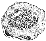
Fig. 34.—Trypanosoma cruzi. Transverse section of a striated muscle containing rounded forms of the parasite in the central portion. × 1,000 approx. (After Vianna.)
(b) The second mode of multiplication is one of asexual reproduction (schizogony or agamogony). It was first described by Hartmann from hypertrophied endothelial cells of the lungs. It has since been found in the cardiac muscle, in the neuroglia of the central nervous system, and in striped muscle (fig. 34). In laboratory animals it has also been found in the testicle and suprarenal capsules. In these tissues the parasite is intracellular, appearing as a small rounded body with nucleus and blepharoplast, without flagellum or undulating membrane. In other words the parasite is Leishmania-like in the body tissues, and recalls the organism of kala-azar.
Chagas considers this second mode of multiplication to be strictly asexual. By this means the number of parasites in the vertebrate host is increased, and symptoms are produced. On the other hand the first mode of multiplication, seen in the lung capillaries, is considered by Chagas to be a process of gametogony, in which sexual forms are differentiated. He finds that (1) the adult trypanosomes exhibit a dimorphism in human blood rarely seen in artificially infected guinea-pigs. In these guinea-pigs (infected from guinea-pigs) the so-called gametogony in the lungs is seldom seen. (2) The intermediate host, Triatoma (Conorhinus), becomes infective if fed directly on infected human blood, but very rarely so if fed on guinea-pigs. Chagas is led to believe that the occurrence of sexual forms constantly in the blood of man implies a greater resistance to infection on the part of man than on the part of guinea-pigs or other animals, assuming the general hypothesis that the formation of gametes represents a reaction of the Protozoön to unfavourable conditions. In human infection the number of parasites is always less than in laboratory animals, and their presence in the blood is transitory, lasting from fifteen to thirty days in acute cases. In many cases examination of the tissues at death has shown the presence of parasites in patients who did not exhibit them in the general circulation.
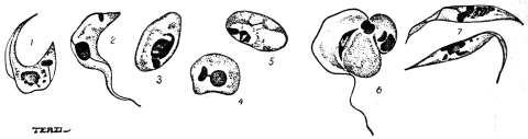
Fig. 35.—Trypanosoma cruzi. Development in Triatoma megista. 1-6, forms found in the mid gut of Triatoma; 7 flagellate forms found in the posterior part of the gut of Triatoma. (After Chagas.)
Life History in the Invertebrate Host.—About six hours after the ingestion of infected blood by the bug (Triatoma megista), the kinetic nucleus of the trypanosome moves towards the nucleus, and the flagellum is usually lost (fig. 35, 1-5). The parasite becomes rounded and Leishmania-like (fig. 35, 3-5), and multiplies rapidly by division. After a time, multiplication having ceased, the rounded forms become pear-shaped and develop a flagellum at the more pointed end. Crithidial forms (fig. 35, 7) are thus produced and pass into the intestine, where they multiply and may be seen in about twenty-five hours after the ingestion of blood. The crithidial forms may also be found in the rectum and fæces. The last stage in the invertebrate is a small, trypanosome-like type, long and thin with a band-like nucleus and conspicuous kinetic nucleus. These parasites are found in the hind gut and in the body cavity. They find their way into the salivary glands, and are the forms (fig. 36) which are transmissible to a new vertebrate host. The development in the bug takes about eight days altogether, after which time the bugs are infective.
There are thus three principal phases in the development of T. cruzi in Triatoma megista: (1) A multiplicative phase (Leishmania-like) in the stomach of the bug, (2) a crithidial phase, which is also multiplicative, in the hind-gut, and (3) a trypanosome phase, which is “propagative,” and apparently passes through the wall of the alimentary canal into the body cavity and so into the salivary glands.
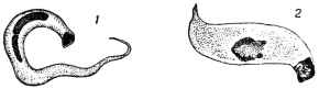
Fig. 36.—Trypanosoma cruzi. Forms found in the salivary glands of Triatoma megista. (After Chagas.)
Brumpt found that T. cruzi could live in Cimex lectularius, C. boueti, and Ornithodorus moubata. The Cimex fæces may be infective. Blacklock found multiplication of the parasite in C. lectularius.
Culture.—The trypanosome can be cultivated on Novy-MacNeal’s blood agar, and the cultural forms resemble those described in the bug.
Possible Reservoir.—Chagas thinks that probably the armadillo or “tatu” (Dasypus novemcinctus) may be the reservoir of T. cruzi. He also thinks that Triatoma geniculata is a transmitter; it lives in the burrows of the armadillo. Other carriers may be Triatoma infestans and T. sordida.
Clinical Features.—The trypanosomiasis of Brazil, produced by T. cruzi and spread by Triatoma spp. has received various names, such as oppilação, canguary, parasitic thyroiditis, and coreotrypanosis. It is also known as the human trypanosomiasis of Brazil, South American trypanosomiasis, and Chagas’ disease.
Chagas88 reports two principal forms—acute and chronic. The acute infection is rare, and is characterized by increase in the volume of the thyroid gland, pyrexia, a sensation of crackling in the skin, enlarged lymphatic glands in the neck, axilla, etc., while the liver and spleen are increased in volume. Sclerosis of the thyroid gland is found at autopsy and fatty degeneration of the liver. During an attack of fever, trypanosomes are found in the blood. The acute form was only observed in children.
In the chronic form Chagas reports several varieties: (a) A pseudo-myxœdematous form, occurring in most cases, especially up to the age of 15. There is hypertrophy of the thyroid gland or at least signs of hypothyroidism, general hypertrophy of glands, disturbance of heart rhythm, and nervous symptoms. (b) The myxœdematous form is characterized by similar symptoms, especially by considerable swelling of the thyroid body, and myxœdema of the subcutaneous cellular tissue; sometimes there is a true pachydermic cachexia. (c) In the nervous form there are motor disturbances, aphasia, disturbances of intelligence or signs of infantilism, athetosis of the extremities and idiocy. There are also paralytic symptoms of bulbar origin, disturbances of mastication, phonation and deglutition, and in some cases convulsive attacks. (d) The cardiac form, characterized by disturbance of the heart rhythm. In all these forms the parasite is found at autopsy in the nervous substance, brain, bulb and heart.
Vianna (1911)89 has studied the histopathology of the disease. Some of the chief points are: in the heart muscle destruction of the sarcoplasm, followed by interstitial myocarditis; in the central nervous system invasion of the neuroglia cells and inflammatory reaction; in the suprarenal capsule invasion of medulla or cortex; inflammatory reaction can also be seen in the kidneys, the hypophysis and thyroid gland.
Recently Chagas states90 that “schizotrypanosomiasis” has been found in a child 15 to 20 days old, and that Trypanosoma cruzi has also been found in a fœtus—the mother being infected with the trypanosome. The trypanosomiasis can, then, be transmitted hereditarily.
Trypanosoma lewisi, Kent, 1881.
The trypanosome has a nucleus somewhat displaced anteriorly, about one-third of the way from the anterior (flagellar) end of the body, a relatively straight edge to the undulating membrane, and a rod-shaped blepharoplast (fig. 37, A). It averages about 25 µ long and 1·5 µ broad.
Much attention has been devoted in recent years to the elucidation of the life history of the rat parasite, Trypanosoma lewisi. It is usually non-pathogenic to its host. It has been shown that the trypanosome can be transmitted from rat to rat by the rat-flea, Ceratophyllus fasciatus, and by Ctenocephalus canis (the so-called dog-flea). (See also p. 92). The flagellate may also persist, but doubtfully develop, in the rat-louse, Hæmatopinus spinulosus. These researches may now be summarized.
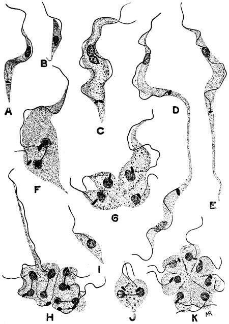
Fig. 37.—Trypanosoma lewisi, from rat’s blood. a, ordinary form; b, small form; c, d, stages in equal binary fission; e, elongate form (longocaudense type), resulting from division as seen in d; f, unequal binary fission; g, h, multiple fission into four and eight; i, small form; j, binary fission of small form; K, division rosette. × 2,000. (After Minchin and Thomson.)
Life Cycle in the Vertebrate Host.—After infection of a rat, the trypanosomes usually appear in the animal’s blood in five to seven days. This incubation period applies either to a natural or an artificial infection. The trypanosomes first observed in the rat’s blood are diverse in form (fig. 37), being small, medium and large in size. This diversity is explained by the rapid multiplication taking place. A trypanosome may divide by equal longitudinal fission (fig. 37, C, D), but more commonly multiple fission occurs (fig. 37, G, H), and is unequal. Rosette forms are produced, in which the parent form can be recognized by its long flagellum (fig. 37, H) and attached to it are daughter individuals, smaller in size, from which flagella are growing. Minchin and J. D. Thomson (1912) find that the daughter forms may be set free sometimes with a crithidia-like facies (fig. 37, I), the blepharoplast being anterior but near to the nucleus. The daughter forms, when set free, may themselves divide by binary or multiple fission, in the latter case forming rosettes (fig. 37, K). Rosette forms were described by Moore, Breinl and Hindle in 1908.
Lingard, some years ago, described as a distinct species, T. longocaudense, certain forms with markedly elongate posterior ends (fig. 37, E). According to Minchin, “these forms appear to arise by binary fission” (fig. 37, D). These long drawn-out forms “are of constant occurrence and very numerous at a certain stage of the multiplication period.” It is about the eighth or tenth day after infection that the multiplication of T. lewisi is at its maximum in the rat’s blood. About the twelfth or thirteenth day the trypanosomes seen in the blood appear uniform. According to Minchin (1912)91 the rat “gets rid of its infection entirely sooner or later, without having suffered, apparently, any marked inconvenience from it, and is then immune against a fresh infection with this species of trypanosome.” There is, then, a cycle of development in the vertebrate host. Minchin notes that the records of the pathogenicity of T. lewisi in rats, causing their death, need further investigation.
T. lewisi inoculated into dormice (Myoxus nitela) and jerboas may become pathogenic thereto.
Carini found cysts in the lungs of rats infected with T. lewisi. He thought the cysts were schizogonic stages of the trypanosome, comparable with those found in the lungs of animals sub-inoculated with T. cruzi. Delanoë (1912)92 has found, however, that such cysts, containing eight vermicules, occurred in rats uninfected with T. lewisi. Delanoë concludes that the pneumocysts are independent of T. lewisi, and represent a new parasite, Pneumocystis carinii. The pneumocysts may be allied to the Coccidia, and must be considered when investigating the life-cycle of a trypanosome in a vertebrate host. Some of the stages of T. cruzi may possibly be of this nature.
Life-cycle in the Invertebrate Host.—This occurs in fleas, and has been investigated in considerable detail by Minchin and Thomson in Ceratophyllus fasciatus, and by Nöller in Ctenocephalus canis and Ctenopsylla musculi.
When infected rat’s blood is taken up by the flea, the parasites pass with the ingested blood direct to the mid-gut of the Siphonapteran. In the flea’s stomach they multiply in a somewhat remarkable manner, namely, by penetration of the cells of the lining epithelium, and division inside the epithelial cells. Inside these lining cells the trypanosomes first grow to a large size and then form large spherical bodies, within which nuclear multiplication occurs (fig. 38, A-F). Any one of these large spherical bodies contains at first a number of nuclei, blepharoplasts and developing flagella, the original flagellum still remaining attached for a time. The cytoplasm then divides into daughter trypanosomes which are contained within an envelope, formed by the periplast of the parent parasite. Inside the periplast envelope are a number of daughter trypanosomes “wriggling very actively; the envelope becomes more and more tense, and finally bursts with explosive suddenness, setting free the flagellates, usually about eight in number, within the host-cell” (fig. 38, F). The daughter forms escaping from the host cell into the stomach lumen of the flea are fully formed, long trypanosomes.
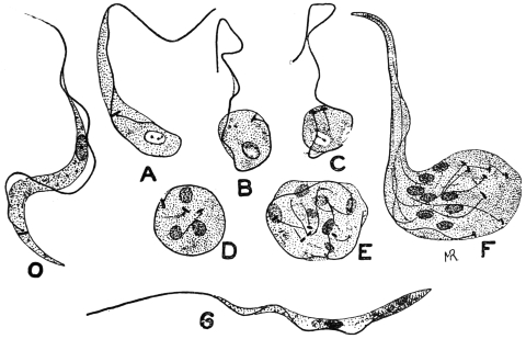
Fig. 38.—Trypanosoma lewisi. Developmental stages from stomach of rat flea. O, ordinary blood type; A-F, stages occurring in gut-epithelium of flea, when the trypanosome becomes rounded and undergoes multiplication, forming in F eight daughter trypanosomes; G, type of trypanosome resulting from such division which passes back to the rectum. × 2,000. (After Minchin.)
The trypanosomes (fig. 38, G) pass into the flea’s rectum. The next phase is a crithidial one. The parasites become pear-shaped, in which the blepharoplast (kinetic nucleus) has travelled anteriorly past the nucleus towards the flagellum (fig. 39). The crithidial forms attach themselves to the wall of the rectum, and multiply by binary fission (fig. 39, D). A stock of parasites is thus formed which, according to Minchin and Thomson, “persist for a long time in the flea—probably under favourable conditions, for the whole life of the insect” (fig. 39, A–I).
From the crithidial forms of the rectum, according to Minchin, small infective trypanosomes arise by modification morphologically (fig. 39, J–M). The flagellum grows longer and draws out more the anterior part of the body, the blepharoplast migrates posteriorly, behind the nucleus, and carries with it the flagellar origin. These trypanosomes are small, but broad and stumpy (fig. 39, N), and can infect a rat. Minchin and Thomson formerly considered that the small, stumpy, infective trypanosomes pass forwards from the rectum into the stomach, and “appear to be regurgitated into the rat’s blood when the flea feeds.” However, the small infective trypanosomes were previously described by Swellengrebel and Strickland.93 They may be found in the flea’s fæces. Nöller (1912)94 has found that the development of T. lewisi proceeds quite well in the dog flea (Ctenocephalus canis) in Germany. Wenyon confirms this, and states that the human flea, Pulex irritans, and the Indian rat-flea, Xenopsylla cheopis, are also able to serve as true hosts for T. lewisi.
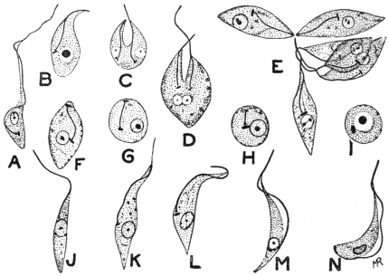
Fig. 39.—Trypanosoma lewisi. Developmental stages from rectum of rat-flea. A, early rectal form; C, D, division of crithidial form; E, group of crithidial forms; F–I, crithidial forms without free flagella, some becoming rounded; J–M, transitional forms to trypanosome type seen in N, which represents the final form in the flea. × 2,000. (After Minchin.)
Nöller stated that rats were not infected with T. lewisi by infective fleas biting them, but by the rats licking up the fæces passed by the fleas while feeding. This is not in agreement with Minchin and Thomson’s earlier views of regurgitation, which, apparently, they have now abandoned.95 Wenyon (1912) confirms Nöller’s experiments. He took a dog flea, containing infective trypanosomes in its fæces, and allowed it to feed on a clean rat. The fæces of the flea, passed while feeding, were carefully “collected on a cover glass and taken up in culture fluid with a fine glass pipette.” The contents of the pipette were discharged into the mouth of a second clean rat. Injury to the rat’s mouth was carefully avoided. The first rat, on which the infective flea was fed, did not become infected, while the second rat, in whose mouth infective flea fæces were placed, became infected in six days.
When infective forms of T. lewisi have been developed within the gut of a rat flea, they may enter and infect the vertebrate host by96 (a) being crushed and eaten by the rodent; (b) the rat may lick its fur on which an infected flea has just passed infective excrement; or (c) the rat may lick, and infect with flea excrement, the wound produced by the bite of the flea.
The time taken for the full development of T. lewisi in the flea is about six days. The intracellular phase is at its height about the end of the first day; the crithidial phase, in the flea’s rectum, begins during the second day; the stumpy, infective trypanosomes are developed in the rectum about the end of the fifth day.
Wenyon97 writes that, “the fleas, when once infected with T. lewisi, remain infected for long periods, for though many small infective trypanosomes are washed out of the gut at each feed, those that remain behind multiply to re-establish the infection of the hind gut. Further, the infection is still maintained even if the flea is nourished on a human being, so that fresh human blood does not appear to be destructive to the infective forms in the flea.”
The best method of controlling fleas during experiments is that due to Nöller. He adopted the method of showmen who exhibit performing fleas, and secure them on very fine silver wire.
Of fleas fed on an infected rat only about 20 per cent. become infective. About 80 per cent. are immune. If fleas are examined twenty-four hours after feeding, trypanosomes will be found in all, so that many of the parasites are destined to degenerate.
It may be of interest to note that Gonder98 (1911) has shown that a strain of T. lewisi resistant to arsenophenylglycin loses its resistance after passage through the rat-louse, Hæmatopinus spinulosus. These experiments suggest that physiological “acquired characters” may be lost by passage through an invertebrate host.
Trypanosoma brucei, Plimmer and Bradford, 1899.
Trypanosoma brucei was discovered by Sir D. Bruce in 1894 in cattle in Zululand and was named T. brucei by Plimmer and Bradford in 1899 in honour of its discoverer. This trypanosome is of considerable economic importance, as it is responsible for the fatal tsetse fly disease, or “nagana,” in cattle, horses and dogs. The disease is widely distributed in Africa and is transmitted from host to host by the tsetse, Glossina morsitans, and other species of Glossina. The virus is maintained in nature in certain big game, such as wildebeest, bushbuck and koodoo, which thus act as living reservoirs of disease from which the tsetse may become infected. These reservoir hosts are not injured, apparently, by the presence of the parasites.
T. brucei is rapidly fatal to the small laboratory animals, such as rats and mice. Horses, asses and dogs practically always succumb to its attacks, while a very small number of cattle recover from “nagana.” The disease is characterized by fever, destruction of red blood corpuscles, severe emaciation and by an infiltration of coagulated lymph in the subcutaneous tissue of the neck, abdomen and extremities giving a swollen appearance thereto. The natural reservoirs in which T. brucei has been long acclimatized are unaffected by the trypanosomes, while the newer hosts, such as imported cattle in Africa, are rapidly destroyed by their action.
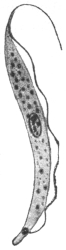
Fig. 40.—Trypanosoma brucei. × 2,000. (After Laveran and Mesnil.)
The general morphology and life history in the vertebrate host is that of a typical trypanosome (fig. 40). Its length is from 12 µ to 35 µ, its breadth from 1·5 µ to 4 µ. Multiplication by longitudinal division proceeds in the peripheral blood (fig. 26), while latent, leishmaniform bodies are produced in the internal organs.
Bruce and colleagues99 have quite recently (June, 1914) described the development of a Zululand strain of T. brucei in G. morsitans. The tsetse flies were bred out in Nyasaland. In vertebrate blood the brucei strain was polymorphic. The development was like that found for T. gambiense in G. palpalis (fig. 30), and by Bruce and colleagues for T. rhodesiense in G. morsitans in Nyasaland. Long trypanosomes were found in the proventriculus of the tsetse. Crithidial, rounded or encysted, and immature “blood forms” occurred in the salivary glands; and finally infective, stumpy, “blood forms” were differentiated in the salivary glands. The period of development of T. brucei in G. morsitans takes about three weeks, and then the fly becomes infective. Bruce believes that T. rhodesiense of Nyasaland and T. brucei of Zululand are the same, their cycles of development in G. morsitans being “marvellously alike.” (But see Laveran, p. 80.)
T. brucei has been cultivated with difficulty by Novy and MacNeal, using blood agar. The best treatment for nagana is arsenic in some form.
It is probable that more than one trypanosome has been confused under the name T. brucei, more especially as the occurrence of many species of trypanosomes in various animals in Africa was not suspected until comparatively recent times. It has been shown by Stephens and Blacklock (1913) that the original Zululand strain of T. brucei was monomorphic, while the organism sent from Uganda, and at the time believed by Bruce to be the same as the Zululand trypanosome, has been found to be polymorphic, with morphological resemblances to T. rhodesiense. Stephens and Blacklock100 have suggested the name T. ugandæ for the polymorphic trypanosome, which, however, has marked resemblances with Trypanosoma pecaudi, and they are, perhaps, identical. T. pecaudi was the name given by Laveran101 in 1907 to the causal agent of “baleri” in equines and sheep in the French Sudan. T. pecaudi, which is dimorphic, is widely distributed in Africa. An extremely small number of both T. pecaudi and T. ugandæ have been shown to possess posterior nuclei. T. pecaudi is transmitted by various species of Glossina, and is said to develop in the gut and proboscis of the fly.
On the other hand, Bruce and colleagues (1914), examining a strain sent from Zululand in 1913, state that T. brucei is polymorphic. Bruce (1914) suggests that passage through laboratory hosts has influenced and altered the morphology of the parasite.
Trypanosoma evansi, Steel, 1885.
Syn.: Spirochæta evansi, Steel, 1885; Hæmatomonas evansi, Crookshank, 1886; Trichomonas evansi, Crookshank, 1886.
Trypanosoma evansi, first found by Evans in 1880, in India, is the causal agent of the disease known as “surra.” The malady affects more particularly horses, mules, camels and cattle in India and neighbouring countries, such as Burma and Indo-China. It occurs also in Java, the Philippines, Mauritius and North Africa. Elephants may be affected. A serious outbreak among cattle in Mauritius occurred in 1902, the disease being imported into the island. The symptoms are fever, emaciation, œdema, great muscular weakness and paralysis culminating in death.
T. evansi varies from 18 µ to 34 µ in length and 1·5 µ to 2 µ in breadth. It has a pointed posterior extremity, and, anteriorly, there is a free portion to the flagellum (fig. 41). It is possibly monomorphic, but a few broad forms occur. The trypanosome multiplies by longitudinal fission in the blood. Rounded leishmaniform stages occur in the spleen of the vertebrate host, which stages Walker102 (1912) considers to be phases of schizogony.
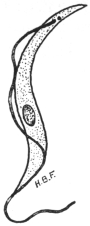
Fig. 41.—Trypanosoma evansi. × 2,000. (Original. From preparation by Fantham.)
The parasite is transmitted in nature by various species of Tabanus and Stomoxys, though at present little is known of the life-history within these invertebrate hosts.
Dogs are said to contract the disease by feeding on animals dead of surra.
A variety of T. evansi is the cause of “mbori” in dromedaries in Africa (Sahara and Sudan). Another possible variety, or closely allied form, is T. soudanense, the causal agent of “el debab” in camels and horses in North Africa, especially Algeria and Egypt.
An extraordinary example of the possible infection of a human being with an animal trypanosome is recorded in the case of Professor Lanfranchi, of the Veterinary School, Parma. The Professor became infected with trypanosomes, although only nagana and surra were maintained in his laboratory, and he himself had never visited the tropics. He suffered from irregular attacks of fever and was œdematous, but his mind remained clear. The identification of the trypanosome from Lanfranchi’s blood has been a matter of great difficulty. Apparently Mesnil and Blanchard (1914)103 consider the strain found in the patient is almost indistinguishable in its reactions from T. gambiense, though the parasite is monomorphic. Lanfranchi considers that he was infected with T. evansi.
Trypanosoma equinum, Voges, 1901.
Syn.: Trypanosoma elmassiani, Lignières.
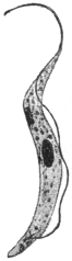
Fig. 42.—Trypanosoma equinum. × 2,000. (After Laveran and Mesnil.)
Trypanosoma equinum was found by Elmassian to be the cause of the fatal disease, “mal de caderas,” of horses and dogs, in South America (Paraguay, Argentine, Bolivia). The name refers to the fact that in the disease, as in other trypanosomiases, the hind quarters become paralysed. Cattle are refractory to inoculation.
T. equinum is about 22 µ to 24 µ long and about 1·5 µ broad (fig. 42). Although this trypanosome is very active, yet it is characterized by the blepharoplast (kinetic nucleus) being very minute or even absent, as the granule sometimes seen may be the basal granule of the flagellum.
The mode of transmission of T. equinum is not known with absolute certainty. Migone has shown that the parasite causes a fatal disease in the large South American rodent, the capybara (Hydrochœrus capybara). This animal appears to be a reservoir of the parasite. Dogs may become infected by eating diseased capybaras, and it is suggested that the infection is spread from the dogs to horses by the agency of fleas. Some authorities consider that T. equinum may be spread by various Tabanidæ and by Stomoxys. Neiva (1913)104 doubts all these modes of transmission in Brazil, and suggests Chrysops or Triatoma as vectors.
Trypanosoma equiperdum, Doflein, 1901.
Syn.: Trypanosoma rougeti, Laveran and Mesnil.
The malady of horses known as “dourine” or “mal du coït” is due to a trypanosome, T. equiperdum, discovered by Rouget in 1894. “Dourine”—also known as “stallion disease” or “covering disease”—is found among horses and asses in Europe, India, North Africa and North America. The trypanosome is transmitted by coitus, and so far as is known not by insect agency.
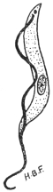
Fig. 43.—Trypanosoma equiperdum. × 2000 approximately. (Original. From preparation by Fantham.)
The progress of the disease may be considered under three periods. The period of œdema, when signs of œdema of the genitalia are seen. The œdema is generally painless and non-inflammatory. This period lasts about a month. It is succeeded by the period of eruption, which sets in about two months after infection. Circular œdematous areas (“plaques”), often about the size of a two-shilling piece, appear under the skin of the sides and hind quarters, and also, at times, under the skin of the neck, thighs and shoulders. The eruption is variable, but usually lasts about a week and leaves the animal in an enfeebled condition. Gland enlargement and swelling of the joints and synovia also may occur. The third period of the disease is described as that of anæmia and paralysis. The animal becomes very anæmic, emaciation is marked, superficial non-healing abscesses often form, and conjunctivitis and ulcerative keratitis can occur. Paralysis ensues, and in from two to eighteen months the animal dies. In the acute form of the disease the animal may die after the first period from acute paralysis.
It is difficult to find the trypanosomes in naturally infected animals, and they are best obtained from the plaques of the eruption. Apparently the parasite occurs more in the lymph than in the blood.
Ruminants are said to be refractory to this trypanosome.
T. equiperdum is about 25 µ to 28 µ in length on an average, but varies from 16 µ to 35 µ. Its cytoplasm is relatively clear, and does not show chromatic granules (fig. 43). It is stated to be monomorphic.
It has been shown recently by Blacklock and Yorke (1913)105 that there is another trypanosome giving rise to dourine in horses. This trypanosome is dimorphic (resembling T. pecaudi and T. ugandæ), and is named T. equi. Previously T. equiperdum and T. equi had been confused.
Uhlenhuth, Hübner and Worthe have demonstrated the presence of endotoxins in T. equiperdum. These endotoxins may be set free by trypanolysis.
Trypanosoma theileri, Bruce, 1902.
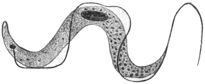
Fig. 44.—Trypanosoma theileri. × 2,000. (After Laveran and Mesnil.)
This parasite, 60 µ to 70 µ long, and 4 µ to 5 µ broad, is distinguished for its large size, though it is not so large as T. ingens from Uganda oxen, whose length may be 72 µ to 122 µ, and breadth 7 µ to 10 µ. The posterior end of T. theileri is drawn out. Small forms of the flagellate are known, 25 µ to 53 µ in length. Probably other forms of the parasite have the nucleus posterior, and these flagellates were formerly separated as T. transvaaliense (Laveran, 1902). Myoneme fibrils may be seen on its body. The pathogenicity of this organism is doubtful, it was formerly thought to be the causal agent of “gall-sickness” in cattle in South Africa. T. theileri also occurs in Togoland, German East Africa, and Transcaucasia. Allied or identical parasites occur in cattle in India.
Trypanosoma theileri, specific to cattle, is perhaps transmitted by the fly Hippobosca rufipes in South Africa.
Trypanosoma hippicum, Darling, 1910.
Trypanosoma hippicum causes the disease of mules known as “murrina.”106 It was found in mules imported to Panama from the United States. It can live in other equines. The parasite varies from 18 µ to 28 µ in length, and is from 1·5 µ to 3 µ broad. Its undulating membrane is little folded. The trypanosome has a noticeable blepharoplast. It can penetrate mucous membranes, and it is thought that the trypanosome may be transmitted during coitus. It may also be spread mechanically by species of Musca, Sarcophaga and Compsomyia, sucking the wounds of infected animals and carrying over the trypanosomes to wounds on healthy ones.
Endotrypanum schaudinni, Mesnil and Brimont, 1908.
This organism was discovered in the blood of a sloth (Cholœpus didactylus), in South America (French Guiana).107 It possesses special interest, in that the best known form of the organism is endoglobular, inhabiting the erythrocytes of the sloth. A free trypanosome in the same animal was considered to be different from the endoglobular form, which was somewhat like a peg-top, and possessed a short flagellum. Darling108 (November, 1914) has seen the organism in Panama. He describes free crithidial forms in shed blood, but not in the blood-stream of the sloth.
Trypanosoma boylei, Lafont, 1912.
This is a parasite of the Reduviid bug, Conorhinus rubrofasciatus. The insect attacks man in Mauritius, Réunion and other places. Lafont infected rats and mice by intraperitoneal injection with the gut-contents of infected bugs. Trypanosomes appeared in the mice. Other flagellate types were assumed by the parasites in the bug.
Monomorphic Trypanosomes.
A number of trypanosomes, characterized by relative uniformity in size and structure, may be considered under this heading. They occur in cattle, sheep, goats and horses in Africa, especially West Africa. Morphologically, they are characterized by the posterior (aflagellar) part of the body being swollen, while the anterior part narrows. The nucleus is central and situated at the commencement of the narrowing of the body. The blepharoplast is almost terminal, the undulating membrane is narrow and not markedly folded, so that the flagellar border lies close to or along the body. The flagellum may or may not possess a free portion.
Some recent workers have considered that T. brucei (Zululand strain) and T. evansi are also monomorphic, but they do not exhibit the general characteristics outlined above. T. brucei and T. evansi have already been considered separately.
The monomorphic trypanosomes, as defined above, include:—
Trypanosoma vivax, Ziemann, 1905.
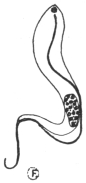
Fig. 45.—Trypanosoma vivax. × 2,000. (Original. From preparation by Fantham.)
This trypanosome109 occurs in cattle, sheep and goats, and was first found in the Cameroons. It is fatal to cattle. Equines are also affected. Antelopes are the possible reservoirs of the trypanosome. It is probably transmitted by Glossina palpalis and other tsetse flies. Its movement is very active. It possesses a free flagellum (fig. 45) and it averages 23 µ to 24 µ in length. T. cazalboui (Laveran, 1906)—the causal agent of “souma” in bovines and equines in the French Sudan—is probably synonymous with T. vivax.
Trypanosoma capræ (Kleine, 1910) is allied, but is somewhat broader and more massive. It was found in goats in Tanganyika.
Trypanosoma congolense, Broden, 1904.
Probable synonyms.—Trypanosoma dimorphon, Laveran and Mesnil, 1904; Trypanosoma nanum, Laveran, 1905; Trypanosoma pecorum, Bruce, 1910; Trypanosoma confusum, Montgomery, 1909.
This trypanosome causes disease among horses (e.g., Gambia horse sickness), cattle, sheep, goats, pigs, and dogs. It is widely distributed in Central Africa (e.g., Gambia, Congo, Uganda, Nyasaland), the strain probably being maintained naturally in big game. It is transmitted by various Glossinæ, and perhaps by Tabanus and Stomoxys. It is said to develop in the gut and proboscis of Glossina palpalis and G. morsitans. The trypanosome averages 13 µ to 14 µ in length and has no free flagellum (fig. 46). It is about 2 µ broad. Formerly T. nanum and T. pecorum were said to differ in their pathogenicity, the former being said not to infect the smaller laboratory animals. Yorke and Blacklock (1913), however, consider that the virulence varies and that these trypanosomes are probably the same.
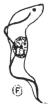
Fig. 46.—Trypanosoma congolense. × 2,000. (Original. From preparation by Fantham.)
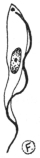
Fig. 47.—Trypanosoma uniforme. × 2,000. (Original. From preparation by Fantham.)
The T. dimorphon originally obtained by Dutton and Todd (1903) in Gambian horse sickness has been shown to be a mixture of T. vivax and T. congolense.
Trypanosoma simiae (T. ignotum) is like T. congolense. It averages 17·5 µ long. It is virulent to monkeys and pigs.
Trypanosoma uniforme, Bruce, 1910.
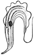
Fig. 48.—Trypanosoma rotatorium, from blood of a frog. × 1,400. (After Laveran and Mesnil.)
This trypanosome was found in oxen in Uganda.110 It can be inoculated to oxen, goats and sheep, but is refractory to dogs, rats and guinea-pigs. It has been found in antelopes. It resembles T. vivax, but is smaller (fig. 47), averaging 16 µ in length. A free flagellum is present. It is transmitted by Glossinæ.
Many other trypanosomes occur in mammals, while birds, reptiles, amphibia (fig. 48) and fish also harbour them. The discussion of these forms does not come within the scope of the present work. They are dealt with in Laveran and Mesnil’s “Trypanosomes et Trypanosomiases,” 2nd edit., 1912.
General Note on Development of Trypanosomes in Glossina.
Before concluding the account of trypanosomes, it may be of interest to remark that several African trypanosomes develop in various species of Glossina, and are found in different parts of the alimentary tract and in the proboscis. Thus (a) T. vivax, T. uniforme and T. capræ develop in the fly’s proboscis (labial cavity and hypopharynx) only; (b) T. congolense, T. simiæ and T. pecaudi develop first in the gut of the fly and then pass forward to its proboscis; and (c) T. gambiense and T. rhodesiense develop first in the gut and later invade the salivary glands of the tsetse. The proboscis or the salivary glands in such cases are termed by Duke111 the anterior station of the trypanosome, wherein it completes its development.
Adaptation of Trypanosomes.
These flagellates may exhibit power of adaptation to changes of environment, such as those due to the administration of drugs, change of host, etc. A few examples of such mutations may be briefly considered:—
(1) Blepharoplastless Trypanosomes.—T. brucei may become resistant to pyronin and oxazine. Accompanying this drug resistance is a change in morphology, namely, the loss of the blepharoplast (Werbitzki).112 A race or strain of blepharoplastless trypanosomes may be thus produced which retains its characteristic feature after as many as 130 passages (Laveran).113 Oxazine is the more powerful drug, and it acts directly on the blepharoplast. (Compare the natural blepharoplastless character of T. equinum.)
(2) Reference has been made on p. 93 to the experiments of Gonder, who showed that a strain of T. lewisi rendered resistant to arsenophenylglycin lost its resistance after passage through the rat louse. This is in marked contrast with the retention of drug resistance during passage by inoculation from rat to rat.
(3) T. lewisi from the blood of a rat when transferred to a snake seems largely to disappear, as very few flagellates are seen. When blood from the snake is inoculated into a clean rat, then trypanosomes reappear in the rat, but they are not all like those originally inoculated. It seems certain that, in such a case, changes in form and virulence of the trypanosome have occurred. Similar experiments were made with T. brucei from rats to adders and other animals and back to rats. Changes in the form and virulence of T. brucei occurred.
These interesting experiments were performed by Wendelstadt and Fellmer.114
Genus. Herpetomonas, Saville Kent, 1881.
Herpetomonas is a generic name for certain flagellates possessing a vermiform or snake-like body, a nucleus placed approximately centrally, and a blepharoplast (kinetic nucleus) near the flagellar end. There is no undulating membrane (fig. 49, a). The organisms included in this genus certainly possess one flagellum, while according to Prowazek (1904) Herpetomonas muscæ-domesticæ, the type species, possesses two flagella united by a membrane. Patton,115 Porter116 and others affirm, however, that the biflagellate character of H. muscæ-domesticæ (from the gut of the house-fly) is merely due to precocious division. The matter is further complicated by the generic name Leptomonas, given by Kent in 1881, to an uniflagellate organism found by Bütschli in the intestine of the Nematode worm, Trilobus gracilis. This parasite, Leptomonas bütschlii, has not yet been completely studied. Until these controversial points relating to the identity or separation of Herpetomonas and Leptomonas have been satisfactorily settled, we may retain the better known name Herpetomonas for such uniflagellate, vermiform organisms. However, the name Leptomonas, having been used by Kent two pages earlier in his book (“Manual of the Infusoria”) than Herpetomonas, would have priority if the two generic names were ultimately shown to be synonymous.
A full discussion of these interesting and important flagellates hardly comes within the purview of the present work; brief mention can only be given here to certain species.
The Herpetomonads occur principally in the digestive tracts of insects, such as Diptera and Hemiptera. They are also known in the guts of fleas and lice, but are not confined to blood-sucking insects. One example, H. ctenocephali (Fantham, 1912)117 occurs in the digestive tracts of dog fleas, Ctenocephalus canis, in England, France, Germany, Italy, India, Tunis, etc. It is a natural flagellate of the flea, and might easily be confused with stages of blood parasites in the gut of the dog flea. Dog fleas are stated by Basile to transmit canine kala-azar, which is believed to be the same as human infantile kala-azar. Confusion is further likely to arise since herpetomonads pass through pre-flagellate, flagellate and post-flagellate or encysted stages; pre- and post-flagellate stages being oval or rounded and Leishmania-like. The post-flagellate stages are shed in the fæces, and are the cross-infective stages by means of which new hosts are infected by the mouth. The possible presence of such natural flagellates must always be considered when experimenting with fleas, lice, mosquitoes, etc., as possible vectors of pathogenic flagellates like Leishmania and Trypanosoma. H. pediculi (Fantham, 1912) occurs in human body lice.118 See further remarks on pp. 107, 112.
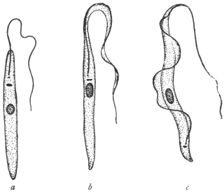
Fig. 49.—a, Herpetomonas; b, Crithidia; c, Trypanosoma. (After Porter.)
Laveran and Franchini (1913–14)119 have recently succeeded in inoculating Herpetomonas ctenocephali, from the gut of the dog flea, intraperitoneally into white mice, and producing an experimental leishmaniasis in the mice. A dog was also infected. They have also succeeded in infecting mice with H. pattoni—a natural flagellate of the rat flea—by mixing infected rat fleas with the food of the mice, and by causing them to ingest infected fæces of rat fleas. Further, they have shown that infection with the herpetomonas occurs naturally by this method, that is, by the rodents eating the fleas and not by the insects inoculating the flagellates into the vertebrates when sucking blood. These experiments shed an interesting light on the probable origin of Leishmania and its cultural herpetomonad stage, which were very probably once parasitic flagellates in the gut of an insect.
Fantham and Porter120 (1914–15) have shown that young mice may be inoculated or fed with Herpetomonas jaculum, from the gut of the Hemipteran, Nepa cinerea (the so-called “water-scorpion”), with fatal results. The pathogenic effects are like those of kala-azar. They also showed that the post-flagellate stages of the herpetomonads seemed most capable of developing in the vertebrate.
A herpetomonad, H. davidi, has been found in the latex of species of the plant-genus Euphorbia in Mauritius, India, Portugal, etc. It is apparently transmitted to the plants by Hemiptera. The plants sometimes suffer from “flagellosis.”
Franchini (1913)121 has described a new parasite, Hæmocystozoon brasiliense, from the blood of a man who had lived in Brazil for many years. It possesses flagellate and rounded stages, and is closely allied to the herpetomonads.
Genus. Crithidia, Léger, 1902, emend. Patton, 1908.
Crithidia is the generic name of vermiform flagellates with a central nucleus, a blepharoplast or kinetic nucleus in the neighbourhood of the principal nucleus, and a rudimentary undulating membrane bordered by a flagellum arising from a basal granule, which is the centrosome of the kinetic nucleus (fig. 49b). The anterior or flagellar end of the body is attenuated and fades off as the undulating membrane.
Crithidia fasciculata, the type species, was found by Léger in the alimentary canal of Anopheles maculipennis. Crithidia occur in bugs, flies, fleas,122 and ticks. Some of them are found in the body-fluid of the invertebrate host as well as in the gut. Others may be restricted to the body cavity or intestine respectively. C. melophagia from the sheep-ked, Melophagus ovinus, and C. hyalommæ from the hæmocœlic fluid of the tick, Hyalomma ægyptium, pass into the ovaries and eggs of their hosts, and the young keds or ticks are born infected.
C. fasciculata has been shown by Laveran and Franchini to be inoculable into white mice, producing a sort of experimental leishmaniasis therein. In one case cutaneous lesions were produced like those of Oriental sore.
Crithidia are natural flagellates of Arthropoda, with their own pre-flagellate, flagellate and post-flagellate stages, and must not be confused with transitory crithidial stages of trypanosomes.
Genus. Leishmania, Ross, 1903.
With an oval body containing nucleus and blepharoplast (kinetic nucleus) but no flagellum. An intracellular parasite in the vertebrate host.
Included in the genus Leishmania are three species, namely:—
(1) Leishmania donovani, Laveran and Mesnil, 1903, the parasite of Indian kala-azar, a generalized systemic disease, usually fatal, occurring in subjects of all ages.
(2) Leishmania tropica, Wright, 1903, the parasite of Delhi boil, Oriental sore, Aleppo button—a localized, cutaneous disease, usually benign.
(3) Leishmania infantum, Nicolle, 1908, the parasite of infantile kala-azar, occurring in children (and a few adults) around the shores of the Mediterranean. The disease is perhaps a form of Indian kala-azar, and the parasite is probably identical with L. donovani.
These diseases may be termed collectively leishmaniases. The morphology of the various species is practically identical.
Leishmania donovani, Laveran and Mesnil, 1903.
Syn.: Piroplasma donovani, Laveran and Mesnil.
The parasite of Indian kala-azar was demonstrated in 1900 by Leishman from a post-mortem examination of a case of “Dum-Dum fever,” but details were not published till May, 1903. In July, 1903, Donovan found similar bodies from cases in Madras. Rogers succeeded in cultivating the parasite in July, 1904.123 The original centre of the disease was probably Assam; it occurs also in Madras, Ceylon, Burma, Indo-China, China and Syria. A variety of this leishmaniasis is found in the Sudan. The patient becomes emaciated, with a greatly enlarged spleen. There is anæmia and leucopenia.
The parasite, commonly known as the Leishman-Donovan body, is intracellular (fig. 50, 2, 3). It is found in the endothelial cells of the capillaries of the liver, spleen, bone-marrow, lymphatic glands and intestinal mucosa, and in the macrophages of the spleen and bone-marrow. Some host cells may contain many parasites. It is rather rare in the circulating blood, but may be found in the blood from the femoral, portal and hepatic veins. It does not occur in the red blood corpuscles as was formerly thought. The parasites liberated from the endothelial cells are taken up by the mononuclear and polymorphonuclear leucocytes. The Leishman-Donovan body is the resting stage of a flagellate. As found in man it is a small, oval organism, about 2·5 µ to 3·5 µ in length by 2 µ in breadth, and containing two chromatinic bodies, corresponding to the nucleus and kinetic nucleus (blepharoplast) of a flagellate. The latter element is the smaller and more deeply staining, and is usually placed at the periphery, transversely to the longer axis of the oval organism. There is sometimes a very short, slightly curved filament to be seen, which may be a rhizoplast. Multiplication takes place by binary or multiple fission. The presence of the parasite used to be demonstrated by splenic or hepatic puncture; nowadays it can be demonstrated in peripheral blood, e.g., of the finger, or by culture of infected blood.
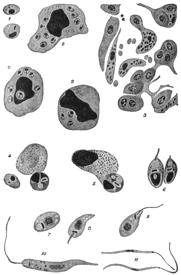
Fig. 50.—Leishmania donovani. 1, Free forms, each with nucleus and rod-shaped blepharoplast (after Christophers); 2, endothelial cell and leucocytes containing parasites (after Christophers); 3, capillary in the liver showing endothelial cells containing parasites (after Christophers); 4, two parasites escaping from a leucocyte in the alimentary canal of the bug (after Patton); 5, further development in bug (after Patton); 6, young flagellate forms in bug (after Patton); 7-11, culture forms (after Leishman); 7, 8, 9, show development of flagellum.
L. donovani can be cultivated in citrated splenic blood, under aerobic conditions, at 22° to 25° C. This was first accomplished by Rogers (1904). It is not so easily culturable as L. infantum on the Novy-MacNeal-Nicolle medium.124 L. donovani is inoculable with some difficulty into experimental animals—in India, white rats, white mice, dogs and monkeys (Macacus spp.), have been inoculated. The Sudan variety, somewhat less virulent, is inoculable to monkeys. Row also produced a local lesion in Macacus sinicus by subcutaneous inoculation of L. donovani. Parasites taken from such a local lesion were found to be capable of producing a generalised infection in Macacus sinicus and white mice.
In cultures the various species of Leishmania all grow into herpetomonad, uniflagellate organisms (fig. 50, 10), about 12 µ to 20 µ in body length. On this account Rogers125 and Patton place the Leishman-Donovan body within the genus Herpetomonas. The method of culture may be used in diagnosing leishmaniases.
Kala-azar is very probably an insect-borne disease. Patton126 suspects the bed-bug to be the transmitter and finds (fig. 50, 4-6) that the Leishman-Donovan body can develop into the flagellate stage in the digestive tract of the bed-bug. Feeding experiments are unsatisfactory, since there are very few cases in which the parasites occur in sufficient numbers in the peripheral blood to make the infection of the insect possible, or at any rate easy. In examining the alimentary tracts of insects for possible flagellate stages of Leishmania, it must be remembered that in many insects natural flagellate parasites, belonging to the genus Herpetomonas, may occur therein; such natural insect flagellates may be harmless, and have no connection with the life-cycle of L. donovani. Natural herpetomonads are known to occur in the alimentary tracts of flies, mosquitoes, sand-flies, fleas and lice, but not in bed-bugs. Further, if such flagellates are able to be inoculated into and live within vertebrate hosts, producing symptoms like those of leishmaniasis, the origin of kala-azar is indicated (see pp. 104, 112).
Leishmania tropica, Wright, 1903.
Syn.: Helcosoma tropicum, Wright, 1903; L. wrighti, Nicolle, 1908; Ovoplasma orientale, Marzinowsky and Bogrow.
It is believed by some that the parasite was first described by Cunningham in 1885, and studied by Firth in 1891, being called by him Sporozoon furunculosum. If these earlier studies were of the parasite, then its correct name is L. furunculosa, Firth, 1891.
The benign disease produced by this parasite has received many names, among the best known being Oriental sore, Tropical sore, Delhi boil and Aleppo button. These names, however, are not happy ones, as cutaneous leishmaniasis (e.g., on the ear) is now known to occur in the New World, for example in Mexico, Venezuela, Brazil and neighbouring States. However, it may be necessary to subdivide cutaneous leishmaniases later.
In the Old World the disease occurs in India, Persia, Arabia and Transcaucasia. It is also known in Algeria, Northern Nigeria, Egypt, Sudan, Crete, Calabria, Sicily and Greece.
The boils often occur on the face, and before ulceration the parasites may be found in the cells at the margin and floor of the “button.” In searching for parasites the scab should be removed and scrapings made from the floor and edges. Where lesions occur atrophy of the epidermis takes place, and infiltration of mononuclear cells (e.g., plasma cells, lymphoid and endothelial cells) follows. The parasites are intracellular, being found inside mononuclear cells. In non-ulcerating sores, Cardamitis found some free parasites. Non-ulcerating forms are said to occur in the Sudan. In the Old World the sores are often limited to exposed surfaces of the body. Infection of mucous membranes (such as the lip, palate, buccal and nasal membranes) may occur, especially in South America, and are often known there as “Espundia.” Christopherson (1914) has recorded a case in Khartoum.
Leishmania tropica is equally well cultivated on Novy-MacNeal-Nicolle medium or on citrated blood. The usual temperature for cultivation is 22° to 28° C., though Marzinowski claims to have cultivated the parasite at 37° C. L. tropica can be inoculated into monkeys and dogs, with the production of local lesions. Material from a human sore or flagellates from a culture may be thus successfully inoculated. Also infected material may be rubbed directly into a scarified surface. The incubation period is long, extending over several months. The duration of the disease may be from twelve to eighteen months. Recovery from one attack of tropical sore confers immunity, and the Jews in Bagdad inoculate their children with the disease on a part of the body which will be covered, and so secure immunity in adult life.
The mode of transmission of L. tropica is unknown. Wenyon (1911)127 has found that the parasite develops into the flagellate stage in the digestive tract of Stegomyia fasciata in Bagdad. Patton (1912)128 has found similar development in the bed-bug in Cambay. The house-fly, Phlebotomus and Simulium have been suspected as transmitters in different parts of the world.
An interesting announcement has been made recently (May, 1913), that Neligan has found that L. tropica occurs in dogs in Teheran, Persia, producing ulcers on the dogs’ faces (cf. natural occurrence of L. infantum in dogs—see p. 110). Yakimoff and Schokhor (1914),129 have found the disease in dogs in Tashkent.
Gonder130 (1913) has performed some interesting experiments showing the relation of infantile kala-azar to Oriental sore. Gonder infected mice with L. infantum and with L. tropica. He used culture material and injected intraperitoneally or intravenously. In each a general infection resulted, with enlargement of the liver and spleen. Later, however, mice injected with Oriental sore (North African variety) developed peripheral lesions on the feet, tail and head, and the lesions contained Leishmania. No such peripheral lesions developed in the case of the mice infected with the kala-azar virus. Gonder suggested that Oriental sore, like kala-azar, is really a general infection overlooked in its earlier stages, and that it is in the later stages that peripheral lesions on the skin are developed. Row (1914)131 also obtained a general infection in a mouse by the injection of cultures of L. tropica from Oriental sore of Cambay.
Leishmania infantum, Nicolle, 1908.132
Infantile splenic anæmia has been long known in Italy. It also occurs in Algeria, Tunis, Tripoli, Syria, Greece, Turkey, Crete, Sicily, Malta,133 Spain and Portugal. This leishmaniasis is, then, distributed along the Mediterranean littoral; also in Russia. Cathoire (1904) in Tunis and Pianese (1905) in Italy were among the first to see the parasite. Nicolle then found the parasite in patients in Tunis, and further found spontaneous infection in dogs. The patients are usually children between the ages of 2 and 5 years. There are a few cases known in which the infantile type of leishmaniasis occurred in youths and adults of the ages of 17 to 19, while one patient in Calabria was 38 years old. The symptoms are like those of Indian kala-azar. Several Italian investigators and others consider that L. infantum is the same as L. donovani, and that the latter name should be used for the parasite of Mediterranean leishmaniasis. This view, as to the identity of L. donovani and L. infantum, seems coming into general favour.
There are, however, differences between the Indian and infantile kala-azars, in addition to the ages of the patients affected, thus: (a) As regards cultures, it is found that L. infantum is readily grown on the Novy-MacNeal-Nicolle (“N.N.N.”) medium (saline blood-agar), and that sub-cultures are easily obtained; in citrated blood L. infantum grows with difficulty. The reverse is the case with regard to culture media for L. donovani, which grows with difficulty on the N.N.N. medium, but relatively easily in citrated splenic blood. (b) Considering inoculability into experimental animals, it is found that L. donovani is inoculated generally with some difficulty into white rats, white mice and monkeys, and with greater difficulty into dogs, while L. infantum can be inoculated into several experimental animals, especially into dogs and monkeys, with ease. (c) At present L. donovani is not known to occur spontaneously in animals, but L. infantum is found naturally in dogs in the Mediterranean region, and the disease in dogs is often referred to as canine kala-azar. Kittens have occasionally been found infected. However, these differences must not be emphasized too much.
The material for cultivation is obtained from punctures of spleen, liver or bone-marrow of cases infected with L. infantum. It is not always easy, however, to infect from cultures, as the cultural flagellates inoculated into the body are often phagocytosed.
Similarly, the material for animal inoculation is obtained from emulsions of infected spleen, liver or bone-marrow. Dogs and monkeys are easily inoculated with such material; Nicolle inoculates into the liver or the peritoneal cavity. Mice, white rats, guinea-pigs and rabbits only show slight infections after such inoculations.
Dogs infected experimentally with infantile leishmaniasis may show either acute or chronic symptoms. The acute course occurs more often in young dogs, and is usually fatal in three to five months. The chronic course is found more commonly in older dogs, and may last seventeen to eighteen months. In acute forms there is irregular fever, progressive wasting, diarrhœa occasionally, motor disturbances involving the hind quarters, and the animal dies in a comatose condition. In the chronic form the animal may appear well, except for loss of weight. The parasites may be found in the internal organs of these experimental dogs, but are not numerous in the peripheral blood except at times of high fever. Experimental monkeys live about three months.
It may be interesting to record the number of dogs found to be infected naturally with leishmaniasis in various countries. In Tunis, Nicolle and Yakimoff found about 2 per cent. infected out of about 500 dogs examined. Sergent in Algiers found 9 infected out of 125 dogs examined. In Italy and Sicily, Basile found about 40 per cent. of the dogs to be infected out of 93 examined at Rome and Bordonaro. Cardamitis found 15 infected out of 184 examined in Athens. In Malta, Critien found 3 infected out of 30 dogs examined. Alvares found 1 infected dog out of 19 examined in Lisbon. Pringault has recently (December, 1913) found an infected dog in Marseilles.134 Yakimoff and Schokhor found 24 per cent. infected out of 647 dogs examined in Turkestan.
The distribution of the parasites in the body of the human patient is much the same as in the case of Indian kala-azar. Critien records the finding of parasites in the mucous flakes of the stools of a three-year-old Maltese child.135 Intestinal lesions rarely occur in infantile leishmaniasis.
Ætiology.—Infantile leishmaniasis is stated to be transmitted by fleas, especially dog fleas, Ctenocephalus canis (= Pulex serraticeps), and by Pulex irritans. Children living in contact with infected dogs may be bitten by infected dog fleas, and so contract the disease. Basile (1910–11) and Sangiorgi (1910) state that they found L. infantum parasites in the digestive tract of the dog flea. After searching they found infected dog fleas on the beds, mattresses, and pillows used by children suffering from the disease. Franchini (1912) thinks that Anopheles maculipennis may be concerned in the transmission.
Basile136 tried a number of experiments to show that infantile leishmaniasis is transmitted by fleas, thus:—
(1) Fleas were taken from a healthy dog. They were placed in vessels containing infected spleen-pulp and allowed to feed thereon. The fleas were then killed and dissected, and portions of the gut-contents examined for parasites. The remainder of the gut was emulsified and injected into a young puppy, whose bone-marrow had been shown previously to be uninfected. Basile states that the puppy became infected. The parasites are said to increase in number in the flea’s gut.
(2) Two healthy pups, each a month old, and born in the laboratory, were placed in a disinfected, flea-proof cage. A few days after, an infected dog was placed in the cage, so that fleas from the infected dog could pass on to the puppies. A month later the two pups became infected, parasites being found in them after liver puncture. A number of control puppies from the same litter remained uninfected and in good health.
(3) Basile next used other laboratory-born puppies, a month old. Four of the litter were placed in a disinfected, flea-proof gauze cage in Rome. The cage was isolated from other dogs. Fleas obtained from an infected area in Sicily were placed in the cage. The puppies were examined by hepatic puncture, but were found to be negative for two months. Then two of the puppies showed infection, and six days later the remaining two puppies were found to be infected, and all four died. They showed irregular temperatures, and were getting thin. Control puppies remained healthy.
From these experiments Basile concludes that fleas transmit leishmaniasis. However, Basile did not exclude the possible occurrence of natural herpetomonads in the gut of the fleas.137 Herpetomonas ctenocephali is known to occur in the gut of Ctenocephalus canis. A natural Herpetomonas is also known in the gut of Pulex irritans, as well as a Crithidia (C. pulicis, Porter). These natural flagellates of the fleas pass through non-flagellate stages, like the Leishman-Donovan body. In consequence Wenyon and Patton, among others, have criticized Basile’s results. Further, other investigators, such as Wenyon and Da Silva (1913), have repeated Basile’s flea experiments and been unable to confirm them.
In feeding and inoculation experiments the incubation period of the parasite may be long, and so it is necessary to wait a long time to see whether the parasite will develop.
Immunity.—Nicolle has tried some experiments with L. infantum and L. tropica. He finds that in animals recovery from an attack of the former confers immunity against infection by the latter and vice-versâ.
Laveran138 records that a monkey having an immunity against L. infantum was also immune to L. donovani.
As mentioned on p. 103, Laveran and Franchini (1913), working in Paris, have succeeded in inoculating Herpetomonas ctenocephali, a natural flagellate in the gut of the flea, Ctenocephalus canis, into white mice. Leishmaniform stages of the flea flagellate were recovered from the peritoneal exudate, blood and organs of the mice some weeks after inoculation. The parasites may also be conveyed by way of the digestive tract of the vertebrate. Similar experiments have succeeded with H. pattoni. These experiments go to show, together with those of Fantham and Porter with H. jaculum (see p. 104), that, in the words of the latter authors, “it may be expected that the various leishmaniases, occurring in different parts of the world, will prove to be insect-borne herpetomoniases.”
Genus. Histoplasma, Darling, 1906.
Under the name Histoplasma capsulatum,139 Darling described small round or oval parasites, enclosed in a refractile capsule, and each containing a single nucleus. The bodies were found in cases of splenomegaly in Panama. They occurred in the endothelial cells of the small blood-vessels of the liver, spleen, lungs, intestine and lymphatic glands, and also within the leucocytes. A few flagellates were stated to occur in the lungs. The parasite has usually been placed near Leishmania, but recently Rocha-Lima has stated that Histoplasma is a yeast.
Genus. Toxoplasma, Nicolle and Manceaux, 1908.
The genus was created for crescentic, oval or reniform parasites, 2·5 µ to 6 µ by 2 µ to 3 µ, possessing a single nucleus and multiplying by binary fission. They occur in mononuclear and polymorphonuclear cells in the blood, spleen, liver, peritoneum etc. (fig. 51). The parasites have been found in the gondi, dog, rabbit, mole, mouse, pigeon and other birds. Although various species names have been given to the parasites in these hosts, it seems probable, from cross infection experiments, that there is but one species with several physiological races. Splendore140 (1913) has described a flagellate stage.
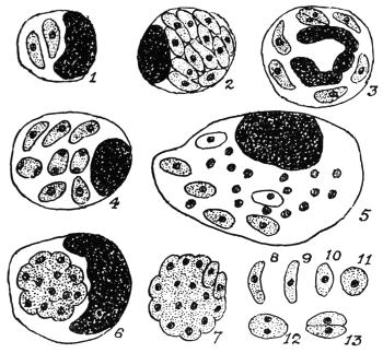
Fig. 51.—Toxoplasma gondii, endocellular or free in the peritoneal exudate of infected mice. 1, 2, mononuclear leucocytes containing toxoplasms. 3, polynuclear, containing parasites. 4, 5, 6, endothelial cells containing toxoplasms, agglomerated in 6. 7, agglomeration forms. 8–11, free forms. 12–13, division stages. × 1,600. (After Laveran and Marullaz.)
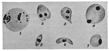
Fig. 52.—Toxoplasma pyrogenes. 1, body found in blood. 2–7, bodies found
in spleen. [1 is about the size of a red blood corpuscle, as drawn in the figures]. Magnification not stated. (After Castellani.)]
Castellani (1913–14)141 has described similar parasites from a case of splenomegaly, with fever of long standing, in a Sinhalese boy. The bodies were found in the spleen and more rarely in the blood (fig. 52). Castellani has named them Toxoplasma pyrogenes. Further researches are needed.
THE SPIROCHÆTES.
The Spirochætes are long, narrow, wavy, thread-like organisms, with a firm yet flexible outer covering or periplast. There is a diffuse nucleus internally in the form of bars or rodlets of chromatin distributed along the body. In some forms there is a membrane or crista present (fig. 53), which in the past was compared with the undulating membrane of a trypanosome, but the membrane of a spirochæte does not undulate. Progression is very rapid, corkscrew-like and undulatory movements occurring simultaneously.
The genus Spirochæta was founded by Ehrenberg in 1833 for an organism which he discovered in stagnant water in Berlin. Ehrenberg named the organism Spirochæta plicatilis. According to Zuelzer (1912) S. plicatilis does not possess a membrane or crista, but an axial filament. S. gigantea has been described by Warming from sea-water.
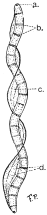
Fig. 53.—Spirochæta balbianii. a, basal granule or polar cap. b, chromatin rodlets. c, membrane (“crista”). d, myonemes in membrane. (After Fantham and Porter.)
Spirochætes occur in the crystalline style and digestive tract of many bivalve molluscs. The first molluscan spirochæte to be studied was that of the oyster, named by Certes (1882) “Trypanosoma” balbianii (fig. 53). Similar spirochætes, probably belonging to the same species, occur in various species of Tapes and in Pecten (the scallop). S. balbianii has rounded ends (fig. 53). Other spirochætes occur in freshwater mussels (Anodonta spp). S. anodontæ, studied by Keysselitz (1906) and by Fantham (1907), has pointed ends. Gross (1911) suggested the generic name Cristispira for molluscan spirochætes, because they possess a well-marked membrane or “crista,” which appears to be absent from S. plicatilis, according to Zuelzer’s researches.
Schaudinn in 1905 founded the genus Treponema for the parasite of syphilis (T. pallidum), discovered by him and by Hoffmann. According to Schaudinn the Treponemata have no membrane or crista. The pathogenic agent of yaws or frambœsia, discovered by Castellani, is also placed in the genus Treponema, as T. pertenue.
There remain the blood spirochætes. It is somewhat disputed as to whether these organisms possess a membrane. The present writer considers that they have a slight membrane or crista. The name of the genus in which to place the blood-inhabiting forms is somewhat uncertain and disputed. Various generic names given to them are Spirochæta, Treponema, Spiroschaudinnia (Sambon) and Borrelia (Swellengrebel). Included in this division are the causal agents of relapsing or recurrent fever. These Protists will be named, for description, Spirochætes without prejudice as to the ultimate correct generic name.
It is sometimes made a matter of argument as to whether the spirochætes are Protozoa or Bacteria. Such arguments are somewhat unprofitable. Morphologically the spirochætes are like the Bacteria in possessing a diffuse nucleus. They differ from Spirillum, an undoubted bacterial genus, in being flexible and not possessing flagella. Molluscan spirochætes, however, may appear to have flagella if their membrane becomes frayed or ruptured, when the myonemes therein (fig. 53), becoming separated, form apparent threads or flagella (Fantham, 1907–08).142
Again, the mode of division of spirochætes has been used as a criterion of their bacterial or protozoal affinity. They have been stated to divide transversely, longitudinally, and by “incurvation,” or bending on themselves in the form of a U, “a form of transverse fission.” The present writer believes that they divide both transversely and longitudinally, and that there is a periodicity in their mode of division at first longitudinal (when there are few spirochætes in, say, the blood) and then transversely (when spirochætes are numerous in the blood).143 Some authors consider that longitudinal division is explained by “incurvation.”
The spirochætes of relapsing fever show a remarkable periodic increase and decrease in numbers in the blood. They are transmitted by ticks or by lice. They react to drugs (e.g., salvarsan or “606”) rather like trypanosomes, and—like Protozoa, but unlike Bacteria—they are cultivated with difficulty. These and other criteria have been used to endeavour to determine whether they are Protozoa or Bacteria. The present writer believes that they are intermediate in character, showing morphological affinities with the Bacteria and physiological and therapeutical affinities with the Protozoa. The group Spirochætacea, as an appendix to the Protozoa, has been created for them by the present writer (Jan., 1908). Others have placed them in the Spirochætoidea of the Bacteria or with the Spirillacea. Doflein (1909) called them Proflagellata. Further discussion is unnecessary, as they are undoubtedly Protista (see p. 29).
There is no true conjugation, sex or encystment in spirochætes, but morphological variation may occur.144 They may agglomerate.
The Spirochætes form an interesting chapter in the evolution of parasites. There are free living forms, parasitic forms in the guts of both vertebrates and invertebrates, and blood-inhabiting forms. These probably represent the order of evolution of parasitism. The blood-inhabiting forms are pathogenic to warm-blooded hosts.
We must now consider the blood Spirochætes and the Treponemata (organisms of syphilis and of yaws).
THE SPIROCHÆTES OF THE BLOOD.
There are at least two important human parasites included hereunder:—
(a) Spirochæta recurrentis (=S. obermeieri), (b) Spirochæta duttoni.
More is known of the life-cycle of Spirochæta duttoni, and it will be convenient to consider that first.
Spirochæta duttoni, Novy and Knapp, 1906.
The specific name duttoni was also given, independently, to this parasite in 1906 by Breinl and Kinghorn.
S. duttoni is the pathogenic agent of African tick fever in man, prevalent in the Congo State and other parts of Africa. The full-grown organism is about 16 µ to 24 µ long, and has pointed ends. It is 0·25 µ to 0·5 µ broad. P. H. Ross and Nabarro were among the earliest to see a spirochæte in the blood of patients in Uganda. It is transmitted by the tick, Ornithodorus moubata.
In the blood of the patient some of the spirochætes may show, after staining, lighter and darker portions (chromatin dots) and evidence of the possession of a very narrow membrane (fig. 54). The mode of division has already been discussed. Periodicity in the direction of division was first described by Fantham and Porter,145 (1909). Just before the crisis in African tick fever, Breinl has stated that S. duttoni becomes thinner in the spleen and bone-marrow and rolls up into skein-like forms, which are surrounded by a thin “cyst” wall (probably the periplast). Such occur in apyrexial periods. Inside the cyst the spirochæte breaks up into granules. Balfour and Sambon have described somewhat similar rolled up forms, breaking into granules, inside the red blood cells of Sudanese fowls in the case of S. granulosa (possibly only a variety of S. gallinarum). The intracorpuscular stage is not definitely established.
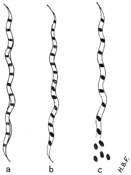
Fig. 54.—Spirochæta duttoni. a, blood form showing slight membrane; b, granules or coccoid bodies clearly formed within the organism; c, beginning of extrusion of coccoid bodies in the tick. (After Fantham.)
The granule phase, however, is an essential one in the invertebrate transmitter (fig. 54c). In 1905,146 Dutton and Todd proved experimentally that O. moubata transmitted S. duttoni. They fed ticks, obtained from Congo native huts in which infected persons lived, on monkeys and the latter became infected. Dutton and Todd also found the offspring of infected ticks to be capable of transmitting the infection to experimental animals. They concluded that O. moubata was a true intermediate host.
A little later in 1905, Koch stated that spirochætes from the gut of the tick penetrated the gut wall and tissues and found their way into the eggs in the ovary. Koch figured tangled masses of spirochætes as occurring in the tick eggs. He found ticks infective to the third generation. He thought that the infection was spread by the salivary fluid of the tick, in the act of biting. (This is now known to be incorrect.) Markham Carter (1907) corroborated Koch’s work on the spirochætes in the tick eggs, and they have been seen since by Kleine and Eckard (1913).
Sir William Leishman,147 in 1909–10, found that at ordinary temperatures the salivary glands of infected ticks (O. moubata) were not themselves infective, and that the infection occurred by way of the ticks’ excretion. The spirochætes (contained in the ticks’ excrement) found their way into the vertebrate host through the wound made by biting. While feeding, ticks pass large quantities of clear fluid from the coxal glands; in this fluid an anticoagulin occurs. Some of the ticks also pass thick, white Malpighian secretion, that is, excrement, towards the end of the feed. Leishman, using experimental monkeys, showed that if infected ticks were interrupted while feeding, then no infection resulted in the monkeys. If, however, the ticks were allowed to finish their feed, and the Malpighian secretions were passed, then the experimental monkeys became infected. Fantham148 and Hindle149 (1911), independently, have repeated the experiments with mice.
Leishman’s methods and results may be summarized thus: Saline emulsions of the organs of infected ticks were made, after the organs had been most carefully dissected out. The ticks were first kept for several days at certain constant temperatures, such as 24° to 25° C. or blood heat, 37° C. The saline emulsions of the organs were inoculated, separately, into experimental animals, and the results recorded:—
At 24° C.At 37° C.
Salivary glandsNegativePositive
Malpighian tubulesPositivePositive
Gut and contentsPositivePositive
ExcrementPositivePositive
Genital organsPositivePositive
Coxal fluid is usually negative; thick, white excrement from Malpighian tubes is positive.
When the ticks were incubated at 21° to 24° C. no spirochætes, as such, were seen in the organs, except perhaps in the gut, where they often disappeared in a few days. When the ticks were previously incubated at 35° to 37° C. for two to three days, spirochætes, as such, reappear in the gut, organs and hæmocœlic fluid. The infection proceeds, not from the salivary gland, but from the infective excrement, that is, from the thick, white material voided by the tick while feeding, usually towards the end of the meal. This Malpighian excrement passes into the wound caused by the bite, being greatly aided by the clear and more limpid coxa fluid, which bathes the under surface of the tick’s body, and mixes with and carries the infective excrement into the wound. Ticks remain infective for a long time.
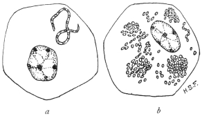
Fig. 55.—Spirochæta duttoni and its coccoid bodies in the tick (O. moubata).—Mononuclear cells of the tick (O. moubata) containing (a) Spirochæte breaking up into coccoid bodies; (b) Similar tick-cell containing coccoid bodies or granules. Such mononuclear cells occur in various organs of ticks and in developing Malpighian tubules. (Original. From preparations by Fantham.)
The spirochætes in the gut of infected ticks divide by a process of multiple transverse fission into granules, which are composed of chromatin (fig. 54). These granules—sometimes known as coccoid bodies—are capable of multiplication. Leishman first found them in clumps inside the cells of the Malpighian tubules (cf. fig. 55).
To summarize, when spirochætes are ingested by a tick, some of them pass through the gut-wall into the hæmocœlic (body) fluid. They then bore their way into the cells of various organs (fig. 55a) and break up into coccoid bodies. In this manner the granules find their way into the ovaries and ova, thus explaining how the young ticks are born infected. Inoculation of these chromatinic granules usually produces infection. Infective granules are also seen in the rudiments of the Malpighian tubules of embryo ticks. Bosanquet and Fantham (1911), independently, have shown that molluscan spirochætes also break up into similar granules or coccoid bodies. Gross has also demonstrated multiple transverse fission in molluscan forms. Marchoux and Couvy (1913) and Wolbach (1914) consider the granules or coccoid bodies to be degeneration products. This is unlikely (see below).
Schuberg and Manteufel have found that certain O. moubata, perhaps 30 per cent. of the specimens of a given neighbourhood, may acquire a natural active immunity against infection with S. duttoni.
S. duttoni, or a closely allied form (by some termed S. novyi), occurs in Colombia, and is spread by the tick Ornithodorus turicata. In Panama a similar spirochæte is probably spread by O. talaje.
Spirochæta gallinarum, Stephens and Christophers, 1905 (= Spirochæta marchouxi, Nuttall, 1905).
This Spirochæte, which occurs in fowls and is pathogenic, is transmitted by the tick Argas persicus. It is about 10 µ to 20 µ long. There is a pathogenic spirochæte known to occur in geese, named by Sakharoff (1891) S. anserina, and found in Caucasia. This may be the same as S. gallinarum, in which case the name S. anserina will have priority. These organisms cause fever, diarrhœa, anæmia and death. The life history of the avian pathogenic spirochætes has been studied by Balfour, by Hindle150 and by Fantham.151 It is essentially similar to that of S. duttoni.
Marchoux and Couvy152 (1913) consider that the “fragmentation of the chromatin” in spirochætes is a process of degeneration. Working with A. persicus and S. gallinarum, they state that a large number of the spirochætes ingested by the Argas almost immediately pass through the wall of the alimentary canal and appear in the hæmocœlic fluid. Marchoux and Couvy consider that Leishman’s granules may be found in the Malpighian tubules of various Arachnids. They found spirochætes in the cephalic glands of infected Argas. They consider that spirochætes remain as wavy spirochætes within the tick, if they are to be infective, though the spirochætes may become so thin as to be invisible! The latter argument is obviously weak, and it was never asserted that all granules in the Malpighian tubules of infected ticks were derived from spirochætes. With dark-ground illumination small, refractile spirochætal granules may be seen to grow into spirochætes. The granule phase of spirochætes has recently been discussed by Fantham153 (1914).
Spirochæta recurrentis, Lebert, 1874.
Syn.: Spirochæta obermeieri, Cohn, 1875.
This organism was discovered by Obermeier (1873) in cases of relapsing fever in Berlin. Short forms 7 µ to 9 µ long, and longer (probably adult) forms, 16 µ to 19 µ, are found in the blood. The width is 0·25 µ. Parasites 12 µ or 13 µ long are often observed.
The spirochæte is found in the blood during febrile attacks and relapses, but not during intervening periods. It can be inoculated into monkeys, rats and mice. It can live in the bed-bug, Cimex lectularius, and Nuttall has succeeded in transmitting S. recurrentis from mouse to mouse by the bites of the same bug. The French investigators Sergent and Foley (1908–9) in Algeria, and Nicolle, Blaizot and Conseil (1912) in Tunis, have shown experimentally that S. recurrentis (var. berbera) is transmitted by lice. The latter workers also demonstrated the method of infection that commonly occurs, namely, by the scratching of the skin and crushing of lice containing spirochætes on the excoriated surface of the body.
Lice as transmitting agents for relapsing fever were indicated by Mackie154 in 1907. An epidemic among Indian school children furnished the materials.155 It was noted that out of 170 boys, 137 were infected, and the boys were very verminous. Among the girls, 35 out of 114 suffered, and few lice were found on them. Twenty-four per cent. of the lice taken from the boys contained spirochætes as compared with 3 per cent. of those from the girls. As the epidemic died out among the boys, the lice also became fewer, and an increase in the number of cases among the girls coincided with an increase in the number of lice. Spirochætes were found in the gut, Malpighian tubules and genital organs of the lice. Mackie thought that infection of the patients was brought about by the regurgitation of the spirochætes when the lice fed, but proof of this was lacking.
In 1912, Nicolle, Blaizot and Conseil,156 working in Tunis and using chiefly an Algerian strain of relapsing fever spirochætes (sometimes called S. berbera), showed by direct experiments that infection by means of the bites of Pediculus vestimenti and P. capitis was untenable. As many as 4,707 infected lice were fed on one man, and 6,515 on another occasion were allowed to bite a man after they had fed on a monkey heavily infected with spirochætes, yet no infection of the man followed. Examination of the lice showed that the spirochætes left the gut soon after they were ingested, and passed into the body cavity, which swarmed with spirochætes. The contents of the alimentary tract and the fæces of the lice alike were uninfective. The spirochætes did not reappear in the gut till eight days after an infective feed, but some persisted as late as the nineteenth day when kept at 28° C.
It was noted that the irritation due to the lice caused scratching, and that thereby lice became crushed on to the skin. An emulsion was made of two infected lice and rubbed on to the slightly excoriated skin of one of the above workers. Infection followed five days later. A drop of emulsion placed on the conjunctiva of the human eye produced spirochætosis after an incubation of seven days. The body contents of such lice, then, produce infection when they reach the blood by any excoriated or penetrable surface. The stages leading up to infection in nature briefly are: The irritation due to the louse bites causes scratching, and the lice are crushed on to the skin. The slight abrasion is quite sufficient to permit the entry of the parasite. The louse bite alone is harmless. Infection by way of the eye is quite probable in Africa, remembering the constant trouble due to sand, dust, insects, etc., resulting in frequent touching of the eyes.
The spirochætes occur in the body fluid of the lice and can pass in it to the adjacent organs. Thus they probably find their way into the genital organs, and into the eggs of the lice. Eggs laid twenty to thirty days after the parent became infected have retained the infection, and the larvæ issuing from such eggs must have contained some form of spirochætes, for an emulsion of either the eggs or the larvæ produced spirochætosis when inoculated into monkeys. Further details regarding the spirochætosis in the eggs of the lice and in the larvæ are needed. Hereditary infection, however, has been demonstrated, but is not very common. Sergent and Foley (1914) state that the spirochæte possesses a very small and virulent form which it assumes during apyrexial periods in man and during a period following an infecting meal in the louse. Nicolle and Blanc (1914) find that the organisms are infective in the louse just before they reappear as spirochætes. Nicolle and Blaizot found that female lice were more susceptible to spirochætes than males, four times as many females as males being infected.
Tictin (1897) found S. recurrentis in bugs recently fed on patients, and infected a monkey with the fluids of crushed bugs. Karlinski (1902) found the spirochætes in bed-bugs in infected houses. There is some other evidence to show that bugs may transmit the spirochæte in Nature. Further researches are needed regarding the relationship of bed-bugs and human spirochætosis.
Multiplication of S. recurrentis is by longitudinal and transverse division (including so-called “incurvation”), and the organism forms small, ovoid bodies (“coccoid” bodies) in the same way as S. duttoni.
S. recurrentis is the cause of European relapsing fever, and a number of possible varieties of it are associated with relapsing fevers in other parts of the world. Such spirochætes only differ by biological reactions, such as acquired immunity tests. They include:—
S. rossii, the agent of East African relapsing fever; S. novyi, the agent of North American relapsing fever; S. carteri, the agent of Indian relapsing fever; S. berbera, the agent of North African and Egyptian relapsing fever.
Other Human Spirochætes are:—
S. schaudinni. This organism, according to Prowazek, is the agent of ulcus tropicum. It varies in length from 10 µ to 20 µ.
S. aboriginalis has been found in cases of granuloma inguinale in British New Guinea and Western Australia. It also occurs in dogs, and may not be truly parasitic.
S. vincenti. This spirochæte is 12 µ to 25 µ in length, tapers at both ends and has few coils. It has been associated with angina vincenti. It often occurs in company with fusiform bacilli.
S. bronchialis, found by Castellani in 1907 in cases of bronchitis in Ceylon. The parasites are delicate, but show morphological variation. This organism is important and has since been found in the West Indies, India, Philippine Islands and various parts of Africa, such as the Anglo-Egyptian Sudan, Uganda and West Africa. It has recently been the subject of research by Chalmers and O’Farrell, Taylor, and Fantham.