ON PARASITES IN GENERAL. (continued 14)
Reading edition
Family. Muscidæ.
Teichomyza fusca, Macq.
Syn.: Scatella urinaria, Rob. Desv.; Ephydra longipennis, Meigen.
The larvæ live in the urine in privies. Several authors state they have found them in fresh fæces or in vomited matter. Pruvot states that they continue for three days in the stomach of rats into which they have been intentionally introduced. (Pruvot, G., “Contrib. à l’étude des larves de dipt, trouv. dans le corps humain,” Thèse de Par., 1882; Chatin, J., in Comp. rend. Soc. de Biol., Paris, 1888 (8), v, p. 396; Roger, H., ibid., 1851 (1), iii, pp. 88, etc.)
Homalomyia canicularis, L., etc.
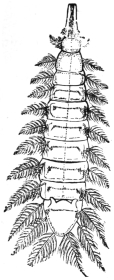
Fig. 405.—Larva of Homalomyia canicularis. Enlarged.
Homalomyia manicata, Meig., live as larvæ in decomposing vegetable matter or in cultivated vegetables (cabbage); they are easily recognizable by their plumed bristles, which are situated laterally on the body segments. They obtain access fairly often to the human intestine and give rise to very uncomfortable symptoms. Cases have been recorded from Germany, Austria, France, England, North America (Wacker, in Artzl. Intelligenzbl., 1883, xxx, p. 109; Florentin, in Compt. rend. Soc. de Biol., Paris, 1904, lvi, p. 525; and other authors).
The larvæ of an allied genus (Anthomyia), which, however, does not possess plumed bristles, has been found in the external auditory meatus of a man (A. pluvialis, according to Danthon).
[H. canicularis is common to Europe and North America, and is an abundant house-fly. It is the small house-fly so often seen on windows. Besides living on vegetable matter, they have also been found in the nests of the humble bee. Larvæ of this species (fig. 405) were sent to the British Museum, taken from the fæces of a woman suffering from cancer.409 They were found at Shrewsbury. Hagen410 reports the larvæ of this fly as occurring alive in the urethra of a patient.—F. V. T.]
Homalomyia scalaris, Fabr.
[This is not a synonym of the above, but a distinct species.
[H. manicata, Meigen, is also distinct.—F. V. T.]
Anthomyia desjardensii, Macq.
This fly, allied to Homalomyia, is the cause of both intestinal and cutaneous myiasis at Bihé, Angola (Wellman, Journ. Trop. Med. and Hyg., June, 1907, x, p. 186).
Hydrotæa meteorica, L.
The larvæ live in decaying vegetable substances, also in dung, and have been evacuated in some cases by man (Zetterstedt, Joseph).
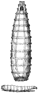
Fig. 406.—Larvæ of Calliphora vomitoria. Enlarged.
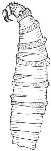
Fig. 407.—Larva of Chrysomyia macellaria. 4/1. (After Conil.)
Cyrtoneura stabulans.
Larvæ in fungi, but occasionally also on larvæ of butterflies and Hymenoptera; occasionally introduced into the human intestine (Joseph).
Musca domestica, L.,
and M. (Calliphora) vomitoria, L., and allied species; larvæ of these have been repeatedly found in the intestine and nose of man (Mankiewicz, etc.).411
Musca domestica, Linn. (Common House-fly).
It is not only on account of a few larvæ of the common house-fly (Musca domestica) being found in the intestines of man that it is of importance medically. It is far more important on account of the part it plays in the spread of diseases of the intestines, such as typhoid fever and cholera, infantile diarrhœa and dysentery.
Howard and Clark (Journ. Exp. Med., 1912, xvi, No. 6, pp. 850–859) have shown that the house-fly is capable of carrying the virus of poliomyelitis for several days on the surface of the body and for several hours in the gastro-intestinal tract. The house-fly may also distribute the ova of Tænia solium and the white worms (Oxyuris and Ascaris). It has also been proved that they may carry the germs of tuberculosis, and it is said that they play an important part in the spread of infectious ophthalmia in Egypt.
This insect is found in all parts of the world. In warm countries it breeds all the year round, and it may do so even in temperate climates in warm places, such as stove houses. Most, however, die off in the autumn; but some survive the winter as adults, in such places as kitchens, restaurants, and warm houses. I have never failed to find a few Musca domestica in houses during the winter. The majority, however, hibernate as puparia.
The females deposit from 120 to 150 eggs in a batch in stable manure, rotting vegetation, house refuse, spent hops, old soiled bedding, etc. A single female may lay as many as six batches of ova during her life. The eggs are shiny white, and hatch in from eight to twenty-four hours in warm weather to three or four days in cool weather. The white footless maggots are cylindrical, tapering to a point at the head end, truncated posteriorly. The head consists of two dark mandibular hooks and two short antennæ. On the tail end are two plates, the stigmata, in which the main tracheal trunks open; in the second segment are a small pair of projecting stigmata. The larval stage lasts from seven to five days in hot weather; but in cold weather in temperate climes it may last six or eight weeks.
The larva on reaching maturity becomes a barrel-shaped puparium of a dark brown to black colour, and in this case changes to the pupa. This stage lasts from three days in the tropics to four or five weeks in cold weather, the life cycle thus varying from ten days in the tropics to fourteen in warm weather in Europe up to three or four months under unfavourable conditions.
All breeding grounds should be burnt or otherwise done away with, such as stable manure, house and kitchen refuse, human excrement and soiled substances, also decaying vegetation as soon as possible, certainly by every sixth day. Stable manure should be kept in closed receptacles and should be removed by every sixth day to at least one mile from habitations and sprinkled with chloride of lime. All kitchen and household refuse should be burnt at once or buried in pits and covered with soil. Latrines should be as far as possible from hospitals, mess rooms and tents. Food—especially milk, sugar and fruit—should be kept screened with muslin when house-flies are about. Mess rooms and tents and hospitals should have doors and windows screened with fine wire gauze during the fly season. All possible steps should be taken to prevent them contaminating man’s food and from breeding in human excrement and from entering hospitals. When present in dwelling-houses in numbers they may be killed by fumigation with pyrethrum or sulphur.
Genus. Chrysomyia, Rob. Desv.
Chrysomyia (Compsomyia) macellaria, Fabr.; Lucilia macellaria, Fabr.
Syn.: Lucilia hominivorax, Coq.; Calliphora infesta, Phil.; Calliphora anthropophaga, Conil.
A species distributed from the Argentine to the south of the United States which deposits its ova on ulcers, in the aural meatus or in the nasal cavities of persons who sleep in the open air. The larvæ are yellowish white, 16 mm. long, are armed with two strong mouth hooks, and provided with spinous rings (screw-worm); they lie hid in the nasal and frontal sinuses, in the pharynx, larynx, etc.; they perforate the mucous membranes, even cartilage, migrate into the eyes, the cranial cavity, middle ear, and cause severe disturbances; after the mature stage, in which the larvæ leave the host to enter the pupal state, these symptoms often spontaneously abate after a lapse of eight days, leaving behind greater or less cicatrices, and consequently also defects in function of the organs attacked. Very often, however, sepsis sets in, usually with a fatal termination.
(Coquerel in: Arch. gén. de méd., 1858 (5), p. 513; 1859, xiii, p. 685; Ann. Soc. ent. France, 1858 (3), vi, p. 171; 1859, vii, p. 234. Weber in: Rec. de mém. de méd. milit., 1867 (3), xviii, p. 159. Francius, A., in: Arch. f. path. Anat., 1868, xliii, p. 98. Conil in: Bol. Acad. nac. cienc. Cordoba, 1881, iii, p. 296. Humbert, Fr., in: Proc. U.S. Nat. Mus., 1883, vi, p. 103; Amer. Nat., 1884, xviii, p. 540. Lindsay in: Journ. Trop. Med., 1902; v, p. 220, and other authors.)
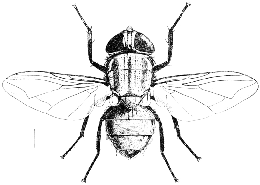
Fig. 408.—The screw-worm fly (Chrysomyia macellaria).
[This species is known as the screw-worm fly. It attacks animals as well as man, especially laying its eggs on wounds formed by barbed wire. It may also be found on dead flesh. Dr. St. George Gray sent me specimens from St. Lucia, from the nose and mouth of a patient in Victoria Hospital. Others were found in the vagina of another patient. Out of the four patients attacked, two occupied the same bed, one after the other, and a third the next bed to it. The other case was in a more remote part of the hospital. There are numerous records of this fly attacking man. It occurs from the Argentine to Texas.—F. V. T.]
Chrysomyia viridula, Rob. Desv.
[This species is somewhat larger than the former; the body is metallic bluish-green, the dorsum of the thorax with three blackish, longitudinal stripes, and the face ochraceous; about 10 mm. long. Austen records this species from man, Dr. Daniels having bred it from larvæ from a sore on a human being in New Amsterdam, British Guiana. Dr. Laurence also bred it in Trinidad. In the latter case between 100 and 150 maggots were discharged from the nose of a woman suffering from facial myiasis (Brit. Med. Journ., January 9, 1909, p. 88 + fig.).—F. V. T.]
Genus. Lucilia, Rob. Desv.
Lucilia nobilis, Meig.
The larvæ were observed by Meinert in Copenhagen in the auditory meatus of a person who, after taking a bath, fell asleep in the open air, and on waking felt singing in the ears, and had a sensation as if there were water in the auditory canal. During the next days severe pains set in, and there was a discharge of blood and pus from both ears, as well as from the nose. On washing out the meatus the maggots made their appearance.
Lucilia cæsar and L. sericata have also been observed in the larval state in man (Thompson, Hope, Henneberg and Calendoli, Napoli, 1907).
[This golden-green fly usually lays its eggs on decomposing organic matter; now and again it lays its eggs in wounds on man.—F. V. T.]
Genus. Pycnosoma, Brauer and v. Bergenstamm.
The species of this genus have a general resemblance to the Lucilias and Chrysomyias, but the body is stouter and the abdomen banded. The genus can be distinguished from Chrysomyia by the absence of the three thoracic stripes and by the eyes of the male, in which the facets forming the upper portion are much enlarged, whereas in Chrysomyia they are not noticeably larger. Austen also points out that the sterno-pleural bristles in Pycnosoma are 1 : 1, in Chrysomyia 2 : 1. The genus is found in tropical Asia and Africa only. All records of Chrysomyia (Compsomyia) in India must be referred to this genus. Bezzi and Stein (“Katalog der Palăarktischen Dipteren,” 1907, iii, p. 543), however, regard the two as synonymous.
The larvæ are frequently found in the nostrils of man and burrow into the sinus, but normally they live on decaying animal matter.
Pycnosoma forms the so-called Indian screw-worm. Patterson (Ind. Med. Gaz., October, 1909, xliv, No. 10) records the case of a woman at Tezpin, Assam, from whom as many as 100 larvæ were removed at one time, and later the left orbital cavity was found packed with hundreds of maggots; eventually the patient died. It is possible that this, however, was due to a species of Sarcophaga. Austen undoubtedly records this genus causing nasal myiasis in India (Trans. Soc. Trop. Med. and Hyg., iii, p. 235). At Dehra Doon, U.P., a woman discharged 100 larvæ from her nose, with great pain in the nasal region and frontal sinuses.
The so-called “peenash,” a common malady in Rajputana, is a true nasal myiasis.
Genus. Sarcophaga, Mg.
Sarcophaga carnosa, L., 1758.
Larvæ of flesh-flies provided with two claws at the anterior end, which settle on raw or cooked meat, and in the open on carcases of animals; they are often observed in man, both in the intestine (introduced with food) and in the nasal cavities, frontal sinus, conjunctiva, aural meatus, anus, vulva, vagina, prepuce, and open ulcers, often migrating further from the regions first attacked. (Gayot in Compt. rend. Acad. Sci., Paris, 1838, vii, p. 125. Grube in Arch. f. Naturg., 1853, xix, 1, p. 282. Legrand du Saulle in Compt. rend. Acad. Sci., Paris, 1857, xlv, p. 600, and other authors.)
[This fly is viviparous. The fly varies from 10 to 30 mm. in length, and is of a general ash-grey colour; the thorax with three dark stripes, the abdomen light grey with three black spots on each segment; legs black; base of wings yellow. It also attacks animals and birds, especially geese. The genus Sarcophaga is universally distributed. The maggots are whitish or yellowish footless larvæ of twelve segments, tapering to a point in front, broadened posteriorly. There are two mouth hooks, by means of which they rasp their food. The breathing pores are at the end and consist of two groups of three slits, each surrounded by a hardened area. They pupate in their old skin, which turns brown.—F. V. T.]
Sarcophaga magnifica, Schiner, 1862.412
Syn.: Sarcophaga wohlfahrti, Portschinsky, 1875.
A species widely distributed over the whole of Europe, occurring especially in Russia (Mohilew); the presence of the larvæ in man was first observed by Wohlfahrt (1768). The larvæ settle in the pharynx, in the nose, the aural meatus, the conjunctiva, and in other regions of the human body; they also attack domestic animals and birds. As Portschinsky has shown, they cause severe inflammations, hæmorrhages and suppurations in the organs in which they occur; children are especially attacked. A number of cases have been observed also in Central and Western Europe. [The fly has a light grey abdomen with shiny black spots which do not change their shape and appearance according to the angle in which the fly is viewed.—F. V. T.]
(Wohlfahrt: “Observ. de vermibus per nares excretis,” Halæ, 1768; Nov. Act. Acad. Caes. Nat. curios., 1770, iv, p. 277. Gerstäcker in: Sitzungsb. Ges. nat. Frde. Berl., 1875, p. 108. Portschinsky in: Horæ soc. entom. ross., 1875, 1884, p. 123. Laboulbène in: Ann. Soc. ent. France, 1883 (6), iii; Bull., p. xcii. Leon in: Bull. Soc. des Méd. et Nat. de Jassy, 1905, xix, p. i. Freund, L., in: Verh. Ges. deutsch. Naturf. u. Ärzte, Homburg (1902), 1902, ii, 2, p. 450, and other authors.) [Probably most cases of attack in Europe are due to this species.—F. V. T.]
The above cited do not exhaust the number of observations of diptera larvæ parasitic in man; there are yet to be mentioned the larvæ of S. hæmorrhoidalis, S. hæmatodes (of G. Joseph), those of S. ruficornis (excitants of a cutaneous myiasis in the East Indies), those of species of Eristalis (of Hanby and others), and those of Phora rufipes (of Kahl, of Warsaw, and others). In many cases the determination of the diptera larvæ has been omitted (or must be omitted); such is the case with diptera larvæ in the eye (Schultz-Zehden in: Berl. klin. Wochenschr., 1906, p. 286. Ollendorf in: Med. Korrespondenzbl. d. würt. ärtzl. Landesver., 1904, p. 1017. Kayser in: Klin. Monatsbl. f. Augenheilkunde, 1905, xliii, i, p. 205. Ewetzky and v. Kennel in: Zeitschr. f. Augenheilkunde, 1904, xii, p. 337, and other cases). Austen records several cases of myiasis due to Sarcophaga (vide Trans. Soc. Trop. Med. and Hyg., 1910, iii, No. 6).
The larvæ of African Muscidæ have now become of greater interest; like several Oestrid larvæ they live normally in the skin of mammals, but also attack man. The knowledge of these species is certainly very insufficient, but this is not likely to be the case much longer, as medical men practising in the Colonies are giving their attention to these parasites. At the present time four distinct forms are recognized according to Gedoelst.413
Sarcophaga chrysotoma, Wied.
[This species is recorded as attacking human beings at New Amsterdam, British Guiana. The fly is 15 mm. long, has a golden-coloured face, three broad black thoracic stripes and ochraceous buff anal segments. It was bred from larvæ obtained by Dr. Roland from a sore on a girl’s foot. It is known to occur in the Brazils and the West Indies. Another species was also bred which Austen was unable to identify.—F. V. T.]
Sarcophaga plinthopyga, Wied.
[This and other species of Sarcophaga are called “yaw flies” in Dominica, as they are believed to be concerned in the dissemination of frambœsia or yaws (Nicholls) (vide Austen, Trans. Soc. Trop. Med. and Hyg., 1910, iii, p. 239).—F. V. T.]
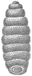
Fig. 409.—Ochromyia larva on the skin of man, South Africa. 3/1. (After Blanchard.)
Ochromyia anthropophaga, E. Blanch.; Cordylobia arthrophaga, Grünberg.
Indigenous to the Senegal and neighbouring districts; in the district of Cayor (between the mouth of the Senegal and Cape Verde) the larva is known as the “ver de Cayor.” It lives under the skin, especially at the lower extremities and the lower region of the trunk, producing small boils, which cause pain, but after about eight days, when the larva leaves the body to enter the pupal stage, the pain discontinues. Besides man the larva occurs in dogs, goats, cats, and in the jackal. It is still questionable whether the fly deposits its eggs direct or on the ground, from whence the larvæ as they emerge gain access to animals and man. Larvæ yellowish-white, 14 mm. long, 4 mm. wide, eleven segments414; head with two globular antennæ-like appendages, two black curved mouth hooks, and two wart-shaped, finely spinous structures at their base. Body evenly covered to the seventh segment with small black prickles, which are stronger at the sides and the anterior borders of the segments; from the seventh they increase in size, on the two hindermost they are wanting; on the last segment two deep yellow spiracles, each with three markedly curved fissures; in addition two stigmata on the posterior border of the first segment. Duration of the larval stage about eight days. Upon the construction of roads in Guinea the larva is spread by dogs far into the interior.
Auchmeromyia (Bengalia) depressa (Walker).415
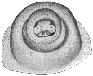
Fig. 410.—Head end of “larva of Natal.” Magnified. (After Gedoelst.)
Distributed in the region of Natal and apparently over the whole of South Africa. The “larva of Natal,” as one may still term the species provisionally, as its identity is not certain, possesses on its head (besides the mouth hooks) lateral protuberances beset with a row of chitinous spines. The cuticle of the body is spinose. The spines are difficult to recognize on account of their transparency and want of colour; they are longest over the anterior segments, from the fifth they become smaller, and over the hindermost they are very small. Apart from the foremost segment, the position they take is that of rows running transversely or obliquely, two to four generally in juxtaposition; the number of spines in the groups gradually increases posteriorly, attaining the number of eight to twelve on the sixth segment, and this number is maintained to the end of the body. Isolated spines are found over the head; over the second, third and fourth segments single ones are still found adjoining the groups of spines, from the fifth onward they are wanting. From here the spines cover the whole free surface of the segments; over the fourth the anterior three-quarters, over the third two-thirds and over the first and second only the anterior half. The stigmata found at the anterior end also serve as distinguishing characters. The parasitic stage appears to last about fourteen days. [Fuller (Agric. Journ., Dept. Agric. and Mines, Natal, 1901, iv, p. 606) refers to this as Bengalia depressa also.—F. V. T.]
Genus. Cordylobia, Grünberg, 1903.
Cordylobia grünbergi, Dönitz.
Syn.: Ochromyia anthropophaga, Grünberg, nec Blanch.; Cordylobia anthropophaga, Grünberg.
Endemic in German East Africa and neighbouring regions. Larva up to 14 mm. long, 4 to 5·5 mm. wide, of cylindrical shape, slightly narrowed behind, truncated, gradually tapering in front; antennæ-like processes, cone-shaped, blunt. Smaller cylindrical formations at the base of the mouth hooks surrounded by a circle of chitinous hooks. Body from the first segment covered with small brown squamous spines which are disposed in numerous irregular transverse rows. The spines are small over the two first segments, the two posterior thirds of all the segments, as well as from the eighth; over the third to the seventh they are larger, but between these there are very small spines. The breathing pores of the stigmata at the anterior end are kidney-shaped; the orifices are elongated and very tortuous, each divided into three. The larval period appears to last several weeks.
Cordylobia anthropophaga, Grünberg.
This well-known cutaneous African parasite seems to have been the cause of much confusion in regard to names. It belongs to the genus Cordylobia of Grünberg, and is one of the family Muscidæ, and differs from Auchmeromyia in that the second abdominal segment of the female is of normal size, whilst in Auchmeromyia it is more than half the length of the whole abdomen, and in the male the eyes are holoptic or close together, whilst in Auchmeromyia they are wide apart. The flies of this genus (three so far described) attack man in their larval stage (anyway two of the three), and also dogs and other animals, by burrowing into the skin and producing painful boils.
[C. anthropophaga, Grünberg, is widely distributed in Africa, extending from Senegal, where its maggot is known as the “ver de Cayor,” and is referred to on p. 590 as Ochromyia anthropophaga, E. Blanchard, to Natal, where it is known as the “Natal worm,” and referred to erroneously on p. 591 as Bengalia depressa, Walker.
[It is a thick-set Muscid of a general straw-yellow colour, with blackish markings on the dorsum of both thorax and abdomen, about 9·5 mm. long. The larva is fat and when mature about 12 mm. long, bluntly pointed in front, truncate behind; from the third to eleventh segments it is thickly covered with minute recurved spines of a brownish colour, arranged in transverse series of groups of two or more, which form more or less distinct irregular transverse rows. On each of the two posterior stigmatic plates, the respiratory slit on either side of the median one is characteristically curved, resembling an inverted note of interrogation. The puparium is brown to ferruginous or black and about 10 mm. long. The maggots are found in both natives and white men, and occur as a severe pest in dogs, also in monkeys, rats, and other mammals. In Sierra Leone it is called the “tumba fly.” The larvæ have been frequently found as true subcutaneous parasites, each larva living singly and forming a boil or warble in the skin, with an opening just as in an ox-warble, through which the maggot breathes and eventually escapes. Although they more usually occur as isolated specimens, Marshall found in Salisbury, South Rhodesia, that sixty were extracted from one lady, and Bérenger-Féraud, in Senegal, that more than 300 occurred in a single spaniel puppy.
[Neave (Bull. Ent. Res., 1912, iii, p. 217) records it from ulcers in a native at Lourenço Marques in 1908, and at the same time from ulcers in a dog, and that it is a severe pest to man in Mozambique and parts of the Transvaal. It seems to be more abundant in North Rhodesia and Nyasaland than to the north (Neave, Bull. Ent. Res., 1912, iii, p. 310). It is also recorded in Zanzibar, German East Africa, Uganda, East Tropical Africa (Neave).
[Simpson (Bull. Ent. Res., iii, p. 170) records a Muscid larva taken from the breast of a European in South Nigeria that was probably Cordylobia.
[It is not known how infection takes place. Neave (Bull. Ent. Res., iii, p. 310) says: “Many instances in human beings would preclude the possibility of eggs having been laid direct on the skin: in these cases they have probably been laid on the clothing put out to dry.”
[Gedoelst has described another species, C. rodhani, and Austen a third species, C. prægrandis, from Nyasaland, Cape Colony, Transvaal, Natal, North-west Rhodesia, and German East Africa.
[The following are some papers dealing with this subject: Proc. Ent. Soc., London, for year 1907, p. xlvii; Journ. R.A.M.C., 1908, pp. 5–11, figs. 1 and 2, by Austen; Journ. R.A.M.C., 1908, pp. 1 and 2, by Major F. Smith; Trans. Soc. Trop. Med. and Hyg., 1910, iii, pp. 223–225, by Austen.—F. V. T.]
Lund’s Larva.
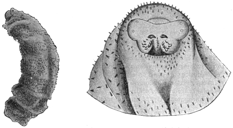
Fig. 411.—Lund’s larva: on the left, the whole larva, magnified six times. On the right, the head end, much enlarged. (After Gedoelst.)
Endemic in the region of the Congo State; called after Commander Lund, from the skin of whose arm it was extracted; 12·5 mm. long, 4·5 mm. broad; colour yellowish, with brown rings, on account of the division of the brown spines; head cone-shaped, with two hemispherical smooth antennæ, two thick black mouth hooks and wart-shaped bodies, between which are situate two to three longitudinal rows of dark brown chitinous laminæ. The body segments are covered over their whole surface with irregularly distributed triangular yellow spines, the points of which are coloured dark brown. Its size increases from the second to the sixth segment, diminishes from the seventh to the ninth, at the tenth it is reduced, and at the eleventh quite small. The posterior stigmata are bean-shaped, each with three markedly tortuous openings. Duration of the larval stage unknown; the same applies to the pupal and imago stages.
Auchmeromyia luteola, Fabricius.
[This fly, the parent of the so-called Congo floor maggot,416 belongs to a nearly allied Muscid genus to Cordylobia, but which can at once be told by the great length of the second abdominal segment. The maggot occurs in numbers in the native huts in the Congo region and is fairly common in central and northern parts of Mozambique; it is also recorded from the Zambesi River and the vicinity of Barberton in the Eastern Transvaal (Bull. Ent. Res., 1912, iii, p. 216), in German East Africa, in Nyasaland, and British East Africa. It is also recorded from Bara, Kordofan,417 where they occurred on the floor of the men’s prison and bit the prisoners. They were destroyed by sprinkling Jeyes’ fluid on the floor. Neave states (ibid., p. 310) that it occurs in the more neglected huts in native villages throughout tropical Africa, and frequently enters a tent when pitched near a village. It is also found in West Africa. The fly is thick-set and about the size and build of a bluebottle fly; length 10 to 12 mm.; tawny in colour to dirty yellowish-brown, with dusky hairs, giving it a smoky appearance; the flattened thorax has long dark stripes and the abdomen a dusky line in the centre of the second segment, which meets a dark line on its posterior border; the dusky third segment has a narrow yellowish anterior line; the fourth segment is also dusky; legs buff with black hairs; the fifth tarsal segment black. The larvæ are whitish, becoming reddish after a feast of blood, with much wrinkled skin and rather flat and broad. They live in crevices of the mud floor, under sleeping mats during the daytime, and come out at night and suck the blood of sleepers and then retire to shelter again. Dutton, Todd, and Christy noticed that where people slept on beds or platforms raised above the floor the maggots were not so numerous as under the sleeping mats laid on the ground. They turned up many of the maggots from a depth of three inches or more.418—F. V. T.]
Family. Oestridæ.
[The family of Oestridæ or warble flies are all parasitic in their larval stage, usually termed the “bot” stage. They are found as parasites in warm-blooded animals, and man is frequently attacked by them. The members of this family have the mouth rudimentary, many of them are hairy and bee-like, with large eyes and the head large, the lower part more or less swollen. The thorax is large with a distinct transverse suture, and the abdomen short and stumpy or very slightly elongated. The male genitalia are hidden, whilst the female ovipositor is often elongated. The wings may be transparent (Hypoderma) or mottled (Gastrophilus), and have muscid-like venation; the tegulæ usually large, the legs moderately long.
[As a rule each species is confined to a particular host, but as we see recorded here those that attack animals may also attack man. The flies occur in warm weather and usually during the warmest part of the day, and have a strong dislike to shade and water. The genus Hypoderma attack oxen, sheep, goats, antelope and musk deer; Oestrus, sheep, antelope and horses; Gastrophilus, the horse and ass; Cephenomyia, the deer; Cepholomyia, the camel and buffalo; Dermatobia, dogs, cats, oxen, deer, apes and man; Cuterebra and Rogenhofera, rodents and opossums.
[Some live as parasites in the stomach and intestines (Gastrophilus); others infest the skin (Hypoderma, Dermatobia and Oestromyia, the latter on Lagomys and Hypodæus); Œdemagena tarandi also infests the skin of the reindeer in Siberia and boreal America. Oestrus lives in the nasal sinus, and Cephalomyia in the throat as well, Cuterebra and Rogenhofera, the skin or scrotum, so that we have really three groups of parasitic oestride larvæ: (i) cutaneous, (ii) intestinal, and (iii) facial.
[No species seems confined to man, but the so-called “creeping disease,” caused by Hypodermæ, and the attack of sheep nasal fly are comparatively common, as also is the Dermatobia attack.—F. V. T.].
Cutaneous Oestridæ.
The eggs are deposited on the surface of the body; the larvæ burrow in the skin, which they reach after somewhat long peregrination.
Genus. Hypoderma, Latreille.
Hypoderma bovis, de Geer.
The cattle fly or warble fly, which swarms during the hot season, settles on the head or on the hair of grazing cattle: through the young being licked off they gain access to the mouth and are swallowed.419 The larvæ appear first in the commencing portion of the stomach, to escape, as some state, into the preceding sections of the alimentary canal; at any rate, they are found from July onward regularly in the submucous tissue of the pharynx, in which they travel about for several months (up to November, and in isolated cases up to February); they then penetrate the muscularis and migrate by way of the subserosa along the mediastinum, the crura of the diaphragm, the renal capsules, and the intermuscular connective tissue of the psoas muscle in the direction of the spinal canal, into which they penetrate by way of the muscles and nerves, through the intervertebral foramina. Here they stay for about two to three months, then they leave the spinal canal again through the vertebral foramina and make their way (from January to March) through the intermuscular connective tissue of the muscles of the back to the skin of the back, where sooner or later (from January to June) they arrive and enter a resting stage, which commences with penetration of the skin and terminates with outward migration from the boils due to the wound set up by the maggot. At the commencement of this period the larvæ cast their skin, and their form, hitherto cylindrical, becomes oval. After about a month, a second moulting of the skin takes place—the third larval stage, which lasts about two and a half months (up to June). The approaching end of the same is indicated by a change of colour on the part of the larva from the hitherto yellowish-white to brown and finally to blackish-brown. When they have become mature the larvæ leave the warbles, drop on to the ground and pass into the pupal stage in the superficial layers of the soil within twelve to thirty-six hours. After about a month the flies emerge. Irregularities with regard to the time and direction of the migrations of the larvæ take place (Jost, H., in Zeitschr. f. wiss. Zool., 1907, xxxvi, p. 644).
In a number of cases the larva of the cattle fly has been observed in the human integument, usually in the winter months, that is, during the migration period; consequently, it is not surprising that the larvæ before they enter on the resting stage and produce a warble undergo migrations. But that this takes place subcutaneously—which does not appear to be so in the case of cattle—is perhaps explained by the fact that in man, on account of the short space that has to be traversed, the larvæ are not sufficiently developed to enter on the resting stage simultaneously upon having obtained access to the integument. Whether the Oestrid larvæ in Bulgaria that similarly migrate beneath the skin in man belong to the cattle fly or to another species, or even another genus, has not yet been ascertained. (Doctorow, in Arch. de Par., 1906, x, p. 309; Spring, A., in Bull. Acad. sci. Belg., 1861 (2), iv, p. 172; Walker, R., in Brit. Med. Journ., 1870, i, p. 151; Kjelgaard, in Ugeskr. f. Laeger, 1904, p. 535; Condorelli, M., in Bull. Soc. Zool. Ital., 1904, xiii, p. 171.)
Hypoderma lineata, de Villers.
The larvæ of this species, that occurs not only in Europe but in North America, live under similar conditions in the skin, very rarely in man; also migrating subcutaneously (Topsent in Arch. de Par., 1901, iv, p. 609).
[In Sweden, the ox warble fly (H. bovis) is well known to attack man. Schoyen states “that over 100 years ago up to the present time cases of travelling grubs under the human skin in some districts of Sweden were well known.” The species appeared to be H. bovis, many of which he had examined. They accomplished long ramblings under the skin, always in an upward direction, previous to their appearance through an opening in a tumour on the upper part of the body, on the head, neck, or shoulders. An interesting case is recorded in Insect Life, ii, pp. 238–239. A bot similar to H. diana was taken from the eye and cheek of a child at Kane, McKean County, Pa., U.S.A. It was said to have travelled in five months from the elbow to the eye. Riley later (Insect Life, iv, p. 310) was inclined to think the maggot was that of H. lineata, the common American ox warble, which is also found in Europe in great numbers. I have recorded another case in England (Rept. Econ. Zool. for year ending September 30, 1910, p. 128), where Dr. Menzies removed the larva of H. bovis from the upper eyelid of a patient. It caused considerable swelling of the face, much pain and distress; but the case did well, and the wound healed at once. The larva was nearly mature. Numerous other references to this so-called creeping disease will be found in the Supplement.
[It is quite probable that bovis and lineata are confused in the latter accounts. The larvæ are, however, easily distinguished if carefully examined.—F. V. T.]
Hypoderma diana, Brauer.
In its larval stage it lives like other species of Hypoderma, attacking the red deer (Cervas elaphas) and roe deer (Cervas capreolus); it is occasionally also found in man (Joseph, in “Myiasis externa dermatosa,” Hamburg, 1800; Völkel, in Berl. klin. Wochenschr., 1883, xx, p. 209).
Genus. Dermatobia, Brauer.
Dermatobia cyaniventris, Macq.
Syn.: Dermatobia noxialis, J. Goudot.
The genus Dermatobia represents the subcutaneous Oestridæ of Europe in warmer parts of America. Both domesticated and wild mammals are attacked, according to one statement birds also (Ramphastus), and man with fair frequency.420 It is assumed that in all cases one and the same species is concerned, for which recently a name originating from C. Linné, jun. (Oestrus hominis), has been employed. Three larval stages are recognized in the skin; the two first appear to resemble one another in the club-shaped or tadpole-like appearance (called macaque in Cayenne, mayacuil [mayoquil] in Mexico), the third is swollen spindle-shaped (Berne, called torcel). Segments 2 to 4 in the club-shaped larvæ are closely beset with small black spines, segments 5 to 7 bear at the anterior border a complete ring of strong black hooks, segments 4 to 6 a similar ring, which, however, is interrupted at the ventral surface. The four last segments forming the tail are smooth, only at the posterior end are there small spines. The arrangement of spines of the third stage differs from this. Italian workmen that have been employed in Brazil show the presence of Dermatobia larvæ on their return (Blanchard, in Bull. Soc. Ent. France, 1893, p. 24; Bull. Soc. centr. de Méd. vet., 1896; Ann. Soc. Ent. France, 1894, lxiii, p. 142; Ward, H. B., in Mark Annivers. Vol., Article 25, p. 483, New York, 1903).
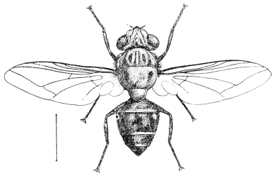
Fig. 412.—Dermatobia noxialis, Goudot.
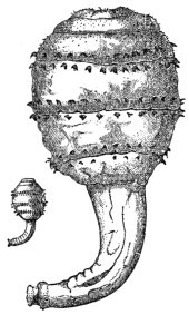
Fig. 413.—Larva of Dermatobia cyaniventris in its natural size and magnified. (After Blanchard.)
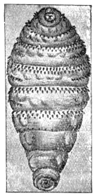
Fig. 414.—Larva of Dermatobia cyaniventris. Enlarged. (After Blanchard.)
[Dermatobia cyaniventris, Macquart, 1843, is said not to be the same as noxialis (vide Brauer, “Mono. Oestriden,” 1863, p. 266). It is known by various other names, as nuche or gusano in New Granada, the ura in Brazil, and the macaw fly in Cayenne. It occurs in Central and South America and the West Indies. According to Goudot the fly is found in great numbers on the borders of large woods and lands covered with underwood.
[It is seldom that more than one larva is found in each individual. It is generally found in the arm and leg, but now and then the face. The perfect insect has never been bred from a larva removed from a human being, so that there is still uncertainty as to the actual species. D. cyaniventris is 11 to 12 mm. long, has an ochraceous buff-coloured face, dark grey thorax, metallic dark blue to purple abdomen, and brownish wings. D. noxialis is somewhat larger.
[In the Journal of Tropical Medicine and Hygiene, January 15, 1905, viii, p. 23, reference is made to this Oestrid in Trinidad, where it is called the “mosquito worm.” One case here recorded showed no fewer than four worms on the chin and one on the hand. It is here stated that the fly never attacks man or animals directly, as it is said to do by Scheube, but that the eggs are deposited on leaves and branches in wooded lands and forests, and thus man, hunting dogs and wild animals in passing through get the larvæ deposited on them accidentally. The affection is common in Trinidad. Mention is made that a little 1 in 40 carbolic lotion syringed into the aperture in the skin over the worm quickly killed it.
[The cattle worm, or founzaia ngómbe, is the name given to a larva which develops beneath the skin of oxen and men in Central Africa, especially amongst the natives and stock of Unyamonezi. According to P. Dutrieux, the egg is laid by a large fly that accompanies cattle. It is unknown between the central plateau or the Ugogo and the East Coast.—F. V. T.]
Cavicolous Oestridæ.
The forms belonging to this group inhabit as larvæ the nasal and frontal sinuses of ruminants, Equidæ and Proboscidæ, which they leave for the pupal stage. The larva of—
Genus. Oestrus, Linnæus,
Oestrus (Cephalomyia) ovis, L.,
occurring in sheep, has also been observed in man in six cases in the nose and larynx (Saitta in Gaz. d. Osp. d. Clinic, 1903, No. 128). So far as is known, the eggs are deposited in the nasal cavity.
[Oestrus ovis frequently occurs in man. MM. Sergent (Ann. de l’Inst. Pasteur, 1907, pp. 392–399) mention that they lay their ova on the noses, eyes and mouth of humans in Algeria whilst flying, but that they disappear after three to ten days or the inflammation produced by them. Portschinsky (Mem. Bur. Ent. Sci. Com. Cent. Bd. Land Adm. and Agric., 1913, x, No. 3, p. 63) also gives cases. He doubts that ova are laid on the nose; evidently the Russian habit is anomalous, for the Sergents, Collings and myself find ova laid as a common occurrence. I have often seen them on the nose of sheep. This fly also occurs in the Argentine (Serres, in Gaceta Rural, April, 1913, vi, pp. 759–761).
[The tamné or thimni of the Kabyles, a human myiasis of the Tuareg mountains in the Sahara, is caused by Oestrus ovis. Here the larvæ are said to be ejected on to the conjunctival and nasal mucous membrane of humans.
[Ed. and Lt. Sergent (Bull. Soc. Path. exot., 1913, vi, No. 7, pp. 487–488) report their attack from the Ahaggar mountains, in Central Sahara. The Tuareg name for the fly, tamné, is the Targui form of the word thimni used by the Kabyles.—F. V. T.]
Gastricolous Oestridæ.
The eggs are deposited on the hairs of Equidæ, and the larvæ escaping from them are licked up and swallowed. They pass their larval stage, according to the species, in various parts of the intestine and stomach, and when mature, pass out per anum in order to undergo the pupal stage.
Genus. Gastrophilus, Leach.
One of the most frequent species is Gastrophilus equi, Fabr.; the eggs are laid on the hairs; the larvæ live some ten months in the stomach, living attached to the inner surface. The eggs of G. hæmorrhoidalis, L., are deposited on the lips or the long hairs on them. The larvæ adhere to the cardiac end of the stomach, to the stomach itself, and finally to the terminal portion of the intestine. Here, however, and elsewhere in the intestine, the larvæ of G. pecorum, Fabr., are also met with, whilst the larvæ of G. nasalis (so called because the eggs are deposited in the nasal orifices) almost exclusively inhabit the anterior section of the duodenum.
Cholodkowsky attributes the “wormlet” observed by Samson and Sokolew (Wratsch, 1895, Nos. 48 and 57) and others (ibid., 1896–98) to Gastrophilus larvæ. It burrows into the epidermis of man by minute passages. This observation should, however, be verified. The phenomenon is designated as skin-mole, larva migrans, and creeping eruption.
Other Papers on Dipterous Larvæ, etc., in Man.
(1) “Ein Fall von lebenden Fliegenlarven im menschlichen Magen,” Deutsch med. Wochenschr., Leipz. and Berl., xxiv (12), pp. 193–194. Bachmann, and review of same, “Living Fly Larvæ in the Human Stomach,” Philadelphia Med. Journ., 1898, i, 18, p. 773.
(2) “Sudi una larva di dittero parassita della congiuntiva umana,” Ann. di ottal., Paira, 1895, xxiv (4), pp. 329–336, 1 fig., E. Baquis.
(3) “Sur quelques diptères suceurs de sang, observé à Terre-Neuve,” Arch. de Par., Paris, 1900, iii (1), pp. 202–204, E. Barret.
(4) “An Account of the Larvæ of two Species of Insects discharged from the Human Body,” Edin. Med. and Surg. Journ., January 1, 1811, vii (25), pp. 41–48, 1 pl., figs. 1 to 8, T. Bateman.
(5) “Un cas de myiase par la Sarcophaga magnifica en Roumanie,” Bull. Soc. Zool. de France, Par., 1891, xvi (2), pp. 25–26, R. Blanchard.
(6) “Sur les oestrides américains dont la larve vit dans la peau de l’homme,” Ann. Soc. ent. de France, 1892, v, pp. 109–154, figs. 1–12, R. Blanchard.
(7) “Note additionnelle sur les oestrides américans dont la larve vit dans la peau de l’homme,” Bull. Soc. ent. de France, Paris, 1894, xiv, pp. 209–211, R. Blanchard.
(8) “Note sur des larves de Dermatobia provenant de Brésil,” Bull. Soc. ent. de France, Paris, 1893 (2), pp. 24–27, R. Blanchard.
(9) “Larven der Wohlfahrtfliege (Sarcophila wolfahrtii) im Zahnfleische eines Menchen,” Wratsch., St. Petersburg, 1888, 5–6, E. K. Brandt.
(10) “Ueber den sogenannten Oestrus hominis und die oftmals besichteten Verirrungen von Oestriden der Säugetheiere zum Menchen,” Verhandl. d. k. zool.-bot. Gesellsch., 1860, x Abhandl., pp. 57–72, Brauer.
(11) “Ueber die Larven der Gattung Cuterebra, Clk.,” Verhandl. d. k. zool.-bot. Gesellsch., 1860, x Abhand., pp. 777–786, Brauer.
(12) “Des désordres produits chez l’homme par les larves de la Lucilia hominivorax,” Thèse, Paris, 1864, 43 pp., V. Audouit.
(13) “Note on the ‘Flesh Worm,’ ” Med. Press and Circ., London, April 12, 1882, lxxxii (N.S. xxxiii), p. 314, P. S. Abraham.
(14) “Larvas de la Calliphora limensis en fosas nasalis,” 1855, 18 pp., F. Aguirre.
(15) “Raro caso di parasitismo nell ’uomo dovuto alla larva di una mosca (Sarcophaga affinis, Meigen),” Boll. d. Soc. Rom. per gli Stud. Zool., Roma, 1893, iv (5–6), pp. 278–289, 1 pl., 3 figs., Giulo Alessandrini.
(16) “Observations sur l’espèce de ver nommé Macaque (Oestrus),” Mém. Acad. Sci. par Hist., 1753, p. 72, F. Artur.
(17) “Contribuição ao estudo da biologia da Dermatobia cyaniventris,” Trav. do Inst. de Manguinhos, 1908.
Biting-mouthed and other Noxious Diptera which may be Disease Carriers.
[Amongst the division Brachycera (as meant in this work) we get several groups of flies which, like the fleas and mosquitoes, are partially parasitic on man, the adults, mainly in the female sex, being provided with a piercing mouth with which they extract the blood of man and animals. The importance of these parasites is not the mere fact that they feed upon our blood, but that they often carry germs from man to man (tsetse-flies and trypanosomiasis, Tabanidæ and anthrax). Amongst the most important biting-mouthed Diptera in this section are the following: Tabanidæ, or gad-flies; Glossinæ, or tsetse-flies; and certain other Muscidæ. Some of the exotic Asilidæ and a few Leptidæ also bite man.
Family. Tabanidæ (Gad-flies).
[The Tabanidæ have a broad, rather flattened body and a large head; eyes united in the male (except in some Chrysops). The antennæ are composed of three segments, have the third joint composed of five to eight annuli—in Chrysops they are fairly long. The proboscis is projecting, and sometimes much elongated. The legs are moderately stout. The venation of the wings is shown in fig. 415.
[This family of gad or horse flies contains a great number of genera, all of which may bite animals and man more or less severely. The female alone is blood-sucking, the males feed upon the juices of flowers. The females deposit their spindle-shaped white, black, or brown eggs on leaves, stems of plants that either overhang or stand in water, and amongst rushes; they are at first white, but become brown or black. The eggs are laid in rounded, flattened or conical masses composed of layers one upon the other. The larvæ are carnivorous, feeding upon snails, worms, other larvæ, etc., and have a distinct head; they are cylindrical, composed of eleven segments, the last with a vertical breathing pore, or the last two segments may form a breathing tube. The majority taper to a point at each end, in colour shining white or dull grey to yellowish, many of the larger specimens mottled or banded with dark brown or black. The first seven abdominal segments are encircled near the anterior margin with a ring of fleshy protuberances consisting of a transverse dorsal ridge which may be divided by a depression into two. The young larvæ burrow into any soft vegetable substance; they live both in the water and under damp soil surrounding water, also in damp earth generally. The larvæ are not only carnivorous, but they are cannibals, frequently devouring their own species. They may take more than a year to mature.
[The pupæ are found close to the surface of mud and earth, and are mostly dull yellowish to brown in colour, with rows of spines on the distal third of each abdominal segment; the thorax bears a pair of ear-shaped spiracular structures, and there are also six denticles at the apex of the abdomen.
[A habit common to the adults of most of the Tabanidæ of considerable economic importance is that of the adults coming to water to drink. Portschinsky421 has found that by applying kerosene to the pool they frequent the adults are killed, and Hine422 that the same oil kills the larvæ that fall into the water from eggs laid on plants above.
[Tabanidæ are not only of importance as purely biting insects, for they may often convey pathogenic organisms from one animal to another, such as the bacillus of anthrax, which they are known to carry, and possibly also trypanosomes in regard to man. Chrysops also acts as a host of Filaria loa in South Nigeria (Leiper, Brit. Med. Journ., January, 1912, pp. 39–40). Two species are incriminated, viz., C. silacea and C. dimidiata. With animals these flies play a more important part, for MM. Sergent, in Algeria, have proved that species of Tabanus are able to transmit three forms of animal trypanosomes by biting a healthy animal as long as twenty-two hours after having bitten an unhealthy one. In India they have also been shown to transmit the parasite of “surra” in dogs and rabbits by Rogers. Other observers have since corroborated these results, and Mitzmain, who has recently performed valuable work in this connection, states that T. striatus is undoubtedly the carrier of this disease in the Philippine Islands. Certain members of the genus Hæmatopota have also been shown to be capable of the direct transmission of Trypanosoma evansi. Martoglio (Ann. d’Ig. sper., 1913, xxiii, N.S., No. 3, pp. 363–366) states that the trypanosome disease of dromedaries known as salaf is transmitted by Tabanidæ, especially Pangonia (P. magretti and P. beckeri) in Italian Somaliland. It is quite likely that these flies play a much greater part in the spread of such diseases than is imagined at the present time.
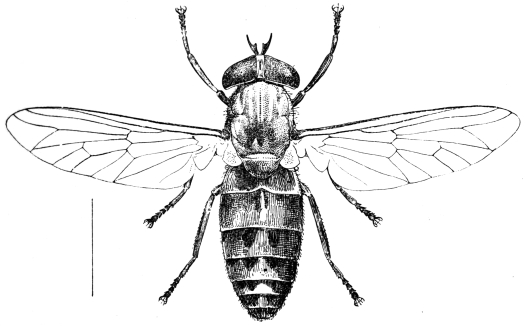
Fig. 415.—The ox gad fly (Tabanus bovinus, Linn.).
[The Tabanidæ are divided into two groups or subfamilies: (1) The Pangoninæ, and (2) the Tabaninæ; the former have spurs on the hind tibiæ and usually ocelli; the latter have neither tibial spurs nor ocelli.
[The Pangoninæ contain two main genera, Pangonia and Chrysops. In the former the proboscis is much elongated, and the third antennal segment is composed of eight rings, and is never angulated or ungulated at the base. The proboscis is often very long.
[In Chrysops, the so-called blinding storm flies, all the three segments of the antennæ are long, the third having only five annulations, and the proboscis short but very strong.
[There are many genera in the Tabaninæ, which are found in all parts of the world, of which two only are shown here—viz., Tabanus and Hæmatopota. The former has the first two segments of the antennæ short, the third angulated at the base, sometimes spurred and composed of five annulations; the second has the second segment short, and the third composed of four annulations—never angulated nor spurred at the base—and the wings are adorned with grey or brown markings. These latter are usually called “brimps” and “clegs” in Britain, the former gad or horse flies, the seruts and mangrove flies of tropical countries.
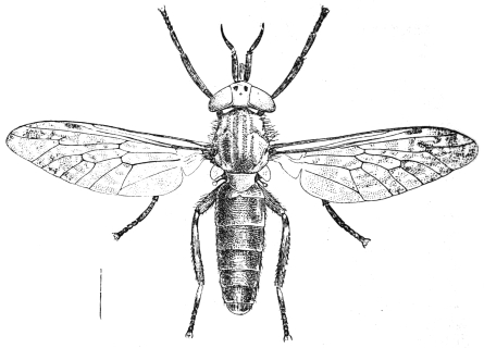
Fig. 416.—The brimp (Hæmatopota pluvialis, Linn.).
Family. Asilidæ (Wolf Flies).
[These flies are of little importance in regard to the subject dealt with in this book; but I have notes sent concerning the biting habits of one or more species belonging to this family from the Malay States and Africa.
[Asilidæ, or wolf flies, are easily told by the following characters: Large or moderate-sized flies, thickly hairy; head separated from thorax by a narrow neck; eyes separated in both sexes; proboscis firm and horny, adapted for piercing; abdomen long, pointed, and composed of eight segments. Legs strong and bristly, of moderate length. Wings sometimes mottled, lying parallel over the abdomen when at rest. There are nearly 3,000 species. They live mostly upon insects, but some are said to bite animals and man. They are, however, of little importance in this respect.
Family. Leptidæ.
[This widely distributed family of flies has a few species which suck the blood of man, and the writer has been personally badly bitten in Norway by a Leptis which was apparently Leptis scolopacea.
[The Leptidæ have usually blotched wings and similar venation to Tabanus; they are elongated flies of moderate or large size, and of dull colours. The antennæ are varied and consist of three segments, either with or without a terminal bristle or with the third segment compound, and in a few they may be almost nematocerous. The wing veins are distinct, very crowded anteriorly, the third long vein is furcate, basal cells large, and there are usually five posterior cells, the anal cell being open in some; the squamæ are always small, sometimes only rudimentary.
[Four are known to be blood-suckers, namely the American Symphoromyia, Trichopalpus obscurus in Chili, and Leptis strigosa and L. scolopacea in Europe. The genus Symphoromyia has a single spur on the hind tibiæ, none on the fore or mid tibiæ, the third segment of the three-ringed antennæ kidney-shaped, and a short proboscis. In the genus Leptis the hind tibiæ have two spurs, and the third antennal segment is not reniform.
[The other biting genus Trichopalpus can be told at once by the elongated proboscis. Most of this family live upon other insects. The larvæ live in earth, decaying wood, sand, stagnant waters, and the nests of wood-boring beetles; they are usually cylindrical and may have fleshy abdominal legs; the anal segment has a transverse cleft, and often two posteriorly directed processes and two stigmata between them. They are all predaceous, and in one genus (Vermileo) make pitfalls in sand like the ant lions (Myrmeleon).
Blood-sucking Muscidæ.
[The blood-sucking Muscidæ are mainly contained in the following genera: Glossina, Stomoxys, Hæmatobia, Lyperosia, Stygeromyia, Philæmatomyia and Bdellolarynx.
[The first is the most important genus on account of the part it plays in the spread of trypanosome diseases. Stomoxys may also serve as a disease carrier. The remainder and a few more genera cause considerable annoyance by their bites, and may also act as occasional carriers of pathogenic organisms. All these flies have their mouth parts elongated to some extent, forming a distinct proboscis, which becomes more or less strongly chitinized; the labella are usually serrated or spiny, and thus form a structure easily capable of piercing the skin. Unlike the Culicidæ, the blood-sucking Muscidæ have the sanguinary habit common to both sexes.
Genus. Glossina, Westwood.
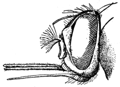
Fig. 417.—Head of Glossina longipalpis, Wied. (After Grünberg.)
[This genus contains sixteen species,423 all of which are confined to the Ethiopian region. Glossina may be distinguished from other allied genera by the proboscis, the antennæ, wings, and male genitalia. The proboscis projects forwards and has a swollen bulb-like base to the slender labium which holds the two structures, the needle-like epipharynx and the thread-like hypopharynx; the whole proboscis is ensheathed in the maxillary palpi. The antennæ have the first two segments small, the third large with a marked pore, the orifice of the sense organ near the base; from the base of the third segment also arises the three-jointed arista, the first two segments being, however, minute; the third bears a series of from seventeen to twenty-one fine branched hairs on one side. The male genitalia or hypopygium is more or less oval and tumid, its long axis lying in the antero-posterior direction, with a vulviform median groove (the anus) running from the anterior margin to beyond the middle.
[Newstead has shown the importance of the study of the genitalia in separating species (vide Bull. Ent. Res., ii, pp. 9–36 and 107–110, and iii, pp. 355–360; and Ann. Trop. Med. and Par., vii, No. 2, pp. 331–334).
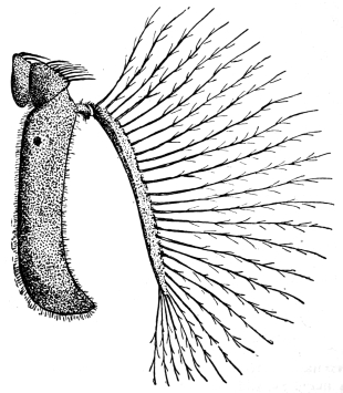
Fig. 418.—Antenna of Glossina pallidipes, male. (After Austen.)
[The tsetse-flies reproduce differently from all other Muscidæ. The female produces at each birth a single full-grown larva, which is retained within the oviduct and there nourished by the secretion of special glands, and on being born crawls to some hiding place and at once becomes a puparium.
[The larva is a yellowish footless maggot nearly as large as the mother’s body, the skin shagreened and the anal extremity having a pair of large, black, granular prominences separated by a depression containing the breathing pores.
[The puparium is brown of various shades, the tumid lips of the larva being conspicuous, the size and shape of the lips enabling the puparia to be identified.
[These puparia are often found in masses at the base of trees, in hollows in trees and rocks just buried under vegetal debris. These insects are generally confined to definite tracts known as “fly-belts.” They usually occur in damp, hot places on the borders of rivers and lakes, and never far from water in the case of the palpalis group, although others of the morsitans group may be found a considerable distance from water. They are usually absent on grass plains, but may now and then occur there (Kinghorn, vide Hindles’ “Flies and Disease, Blood-sucking Flies,” 1914, p. 274); cover of trees, shrubs, or thick reeds is essential to them.
[Their range in Africa extends roughly from 18° N. to 31° S.
[Glossina palpalis is the chief carrier of the more prevalent type of sleeping sickness. Two distinct types of parasites can produce this disease, viz., Trypanosoma gambiense, which produces the ordinary sleeping sickness, transmitted by G. palpalis, and Trypanosoma rhodesiense the Rhodesian or Nyasaland sleeping sickness, transmitted by G. morsitans, and possibly identical with T. brucei, the parasite of N’agana. Koch has also shown that G. pallidipes, Austen, and G. fusca, Walker, can be artificially infected with the human trypanosome. It appears probable that Koch used G. brevipalpis, not G. fusca, in his transmission experiments, as at that time fusca included nearly all the large tsetses, but brevipalpis is its Eastern representative.
[A Table of Species (modified after Austen) is appended here:—
I.
Glossina palpalis Group.
1.Dorsum of abdomen ochraceous buff or buff; third and following segments exhibiting sharply defined, dark brown or clove brown, interrupted transverse bandstachinoides, Westwood.
Dorsum of abdomen not so marked2.
2.Third joint of antennæ pale (cream buff to ochraceous buff), clothed with long and fine hair, forming a conspicuous fringe on front and hind marginspallicera, Bigot.
Third joint of antennæ entirely dark (mouse-grey) except at extreme base on outer side, and without a conspicuous fringe of long and fine hair3.
3.Dorsal surface of abdomen dark sepia brown; median paler area on second segment broad, and more or less quadrate or irregular in outline; hypopygium of ♂ buff or ochraceous buffcaliginea, Austen.
Dorsal surface of abdomen blackish-brown; median paler area cuneate (i.e., triangular in outline); hypopygium of ♂ greypalpalis, Rob. Desv.
II.
Glossina morsitans Group.
1.Hind tarsi entirely dark; small slender species; abdomen bright ochreous or reddish ochreous with dark lateral markingsaustenii, Newstead.
Hind tarsi not entirely dark; abdomen drab-grey, buff or ochreous buff with conspicuous dark interrupted transverse bands2.
2.Last two joints of front and middle tarsi with sharply defined clove brown or black tips3.
Last two joints of front and middle tarsi without sharply defined clove brown or black tips (front and middle tarsi either entirely pale or, at most, two joints of front tarsi faintly brownish at the tips), and last joint and distal half of penultimate joint of middle tarsi light brown, never so dark as to form a sharp contrast with the remaining jointspallidipes, Austen.
3.Third joint of antennæ with a distinct fringe of fine hair on front margin; dark brown or clove-brown bands on abdominal segments extending close to hind margins (i.e., pale ground colour, apart from the median interspace, confined to a very narrow hind border)longipalpis, Wiedeman.
Third joint of antennæ without a distinct fringe of fine hair on front margin; dark brown or clove-brown bands on abdominal segments not extending close to hind marginsmorsitans, Westwood.
III.
Glossina fusca Group.
1.Third joint of antennæ fringed with fine hair on anterior and posterior margins; fringe on anterior margin conspicuous under a hand lens magnifying 15 diameters (nominal) when head is viewed in profile2.
Third joint of antennæ with fringe of fine hair on anterior margin so short as to be scarcely noticeable under a hand lens magnifying 15 diameters (nominal) when head is viewed in profile (longest hairs in fringe in length not exceeding one-sixth of width of third joint); palpi long and slender3.
2.Longest hairs in fringe on front margin of third joint of antennæ, in length equal to from one-fourth to one-third (not exceeding one-third) of width of third joint; palpi of moderate lengthtabaniformis, Westwood.
Longest hairs in fringe on front margin of third joint of antennæ in length equal to from one-half to three-fourths of width of third joint; palpi noticeably long and slendernigrofusca, Newstead.
3.Pleuræ drab-grey or isabella-coloured, hind coxæ buff or greyish-bufffusca, Walker.
Pleuræ dark grey; hind coxæ mouse-greyfuscipleuris, Austen.
IV.
Glossina brevipalpis Group.
1.Dorsum of thorax with four sharply defined brown, more or less oval or elongate spots, arranged in a parallelogram, two in front and two behind the transverse suture; proboscis bulb with a sharply defined brown or dark brown tiplongipennis, Corti.
Dorsum of thorax without such spots; proboscis bulb not brown or dark brown at tip2.
Wings with upper thickened portion of anterior transverse vein much darker in colour than adjacent veins and thus standing out conspicuously against the rest of the wingbrevipalpis, Newstead.
Wings with upper, thickened portion of anterior transverse vein not much darker in colour than adjacent veins, and thus not standing out conspicuously against the rest of the wings (wings practically unicolorous)medicorum, Austen.424
Glossina palpalis, Rob. Desv.
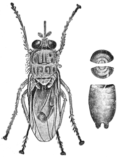
Fig. 419.—Glossina palpalis and puparium. (After Brumpt.)
[This is the chief carrier of sleeping sickness in Nature. It is found in places over the whole of West Africa from the mouth of the Senegal River to Angola, and extends eastwards into the Bahr-el-Ghazal. The eastern boundary follows the valley of the Nile and includes the eastern shores of Lakes Victoria and Tanganyika; from the southern end of the lake the boundary tends south-west, approximately following the frontier between North-eastern Rhodesia and the Congo Free State, and passing through the Katanga district of the latter country into Angola (Austen). It may occur up to 3,000 ft.; but, according to Bagshawe, it has not been recorded above 4,000 ft. It feeds on the blood of many animals, including reptiles, amphibia, birds, and even amphibious fishes, as well as all the wild mammals. It seems, however, to possess a decided predilection for man, and undoubtedly thrives better upon mammals and birds than upon cold-blooded animals.
[It is not usually found far from water, requiring a humid atmosphere and temperature of about 85° F. (shade). But a marked seasonal distribution is shown, the flies considerably extending their range during the rainy season, and thus visiting districts which are dry for the greater part of the year; as the rains diminish the fly gradually leaves the temporary haunts and returns to the more permanent ones. It bites only by day, and then only in sunny weather, and usually lives in shade.
[Roubaud has shown that the first larva produced is about three weeks after copulation, and that others are produced at an interval of nine or ten days. The puparium stage is rapidly produced after the expulsion of the larva, often in three-quarters of an hour. The puparium stage lasts from thirty-two to thirty-five days. The puparia occur in well-drained humus close to water, sheltered by trees or bushes, in crevices in rocks, and between the exposed roots of trees, sometimes in sand.
[Bruce has shown that only a very small percentage of flies fed experimentally on infected animals ultimately become infective, and that the infectivity of this small percentage depends upon a delayed infection of the salivary glands.
[A variety, wellmani of Austen, is found in Angola, Gambia, the Katanga district of the Congo Free State, the Matondwi Islands of Tanganyika, etc.
Glossina morsitans, Westwood.
[This species has been shown by Kinghorn and Yorke, and also by Bruce, to be responsible for the transmission of Trypanosoma rhodesiense, the micro-organism producing sleeping sickness in man in Rhodesia and Nyasaland and also in parts of German and Portuguese East Africa. Fisher and Taute have demonstrated experimentally that Trypanosoma gambiense—the sleeping sickness parasite of other parts of Africa—may also be transmitted by this fly, and in addition it is known to be capable of disseminating several species of trypanosomes pathogenic to animals. Of these, T. brucei (=? T. rhodesiense), the parasite of tsetse disease, first incriminated by Bruce, is perhaps the most important.
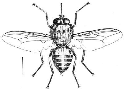
Fig. 420.—The tsetse-fly (Glossina morsitans, Westwood).
[It is the most widely spread of all tsetse-flies; its range extends from Senegambia in the north-west to Southern Kordofan and Southern Abyssinia in the north-east, and then southwards to the Bechuanaland Protectorate, North-eastern Transvaal and Zululand. The actual localities given by Austen are Gambia, French Guinea, Gold Coast, Togoland, Dahomey, Northern Nigeria, Congo Free State, the Bahr-el-Ghazal, the Uganda Protectorate, German East Africa, and Portuguese East Africa.
[This species is confined to “belts,” often of very limited extent, and appears to prefer regions where there is sufficient vegetation for moderate but not excessive cover and a hot, moderately dry climate. It is not nearly so dependent upon water as is G. palpalis, and generally is most active in a dry atmosphere; some observers, however, state that in certain districts it is more common along the banks and edges of rivers. This tsetse-fly has been taken as high as 5,500 ft. altitude. It infests native villages as well as the bush. Like other tsetse-flies it bites not only during the hottest part of the day, but also on bright warm moonlight nights, and it feeds on the blood of all mammals.
[The structure of the male genitalia of those representatives of G. morsitans occurring on the West Coast of Africa and in parts of the Soudan presents certain constant differences from that of the typical form of this species; this form is known as G. morsitans, race submorsitans, Newst.
Genus. Stomoxys, Geoffroy.
[The members of this genus which occur in temperate and tropical countries are provided with a hard, slender, shiny black proboscis which projects horizontally from beneath the head; by means of this structure they can bite severely. In general appearance they resemble house flies, but the proboscis at once distinguishes them. In many parts of Britain they are known as storm flies on account of their frequent appearance indoors previous to a storm of rain or wind, which I have invariably found to be correct; they are also called stinging flies. In colour they are greyish, dusky or brownish-grey or black, varying from 5 to 7 mm. in length; the thorax has dark longitudinal stripes and the abdomen dark spots or bands. In the male the eyes are closer together than in the female. These flies usually occur in stables and farmyards, along woods and in lanes, and mainly attack mammals.
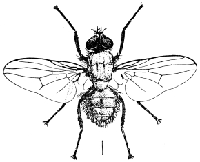
Fig. 421.—The stinging fly (Stomoxys calcitrans, Linn.).
[One species (Stomoxys calcitrans, Linnæus) occurs practically all over the world. The female lays her eggs in moist, warm, decaying vegetation; as many as eighty may be laid by a single female. The ova are white, banana-shaped, with a broad groove on the shorter curvature; they may hatch in two or three days. The creamy-white larva tapers to a point at the head end, and is truncated at the tail end. Two black mouth hooks are plainly visible at the cephalic extremity. There are two plates on the posterior surface of the last segment which bear the respiratory pores, nearly circular in outline. It reaches maturity in fourteen to twenty-one days; when mature it is 11 mm. long. The pupal stage is passed in the old larva skin and lasts from nine to thirteen days; it is barrel-shaped, 5 to 8 mm. long, and of a bright reddish-brown to dark chestnut-brown colour.
[This insect may act as a carrier of anthrax, and has been proved to be the agent of an extensive epidemic of malignant pustule in the Isle of Pines, New Caledonia.425
[Noè’s426 experiments tend to show that it is an intermediate host and transmitter of Filaria labiato-papillosa of the ox.
[Surra is generally stated to be transmitted by Stomoxys as well as Tabanus, and yet Nitzman in the Philippines obtained uniformly negative results in exhaustive experiments. Others have also been unsuccessful. Certainly Stomoxys can transmit the disease in French West Africa (Bonet and Roubaud), and mechanically has been proved to be capable of disseminating other trypanosomes (experimentally): sleeping sickness (T. gambiense); nagana (T. brucei); souma (T. cazalboui); and el debat (T. soudanense).
[S. calcitrans may also be a carrier of poliomyelitis (Rosenau and Brues, Harvard Alumni Bulletin, 1912, xv, No. 9, pp. 140–142). Several species are now known (S. brunnipes, Grünb.; S. inornata, Grünb.; S. nigra, Macq.; S. omega, Newst.; S. ochrosoma, Speiser, etc.).
Genus. Lyperosia, Rondani.
[A genus of small flies which bite man and animals, but are not so far connected with the transmission of any disease in man, but in Java it appears to carry surra (P. Schat, Meeledeel Praefstation Oost-Java, 1903, 3e ser., No. 44), the species being Lyperosia exigua, Meijere. These flies can be told from Stomoxys by the palpi being broader, flattened laterally, and as long, or nearly so, as the proboscis. When not feeding the palpi enclose the proboscis, as in Glossina. They are usually about half the size of Stomoxys, and are the smallest blood-sucking Muscidæ. They frequently swarm around and upon domesticated animals.
[The life-history of the horn fly in America (L. irritans, Linn.) is well known. It lays its ova singly in freshly dropped cow-dung, and there the maggots feed, pupating in the soil beneath.
[Patton and Cragg also give some details as to the life-history of Liperosia exigua (“Medical Entomology,” p. 375) as follows: “L. exigua, whose habits have been observed in Madras, usually lays twelve eggs at a time. The flies immediately return to the cow and the process is repeated when the dung is again dropped. The larvæ migrate from the dung when about to pupate, and the puparia are always found in the earth at some distance away or under the sides of the patch of dung. The fly usually hatches out in five days, though sometimes as late as the eighth. Weiss has studied the life-history of irritans var. weisii from Algeria; its larval stage lasts five days, and the flies hatch out of the puparia in another five days.”
[The other biting genera of Muscidæ, Hæmatobia, Hæmatobosca, Bdellolarynx, Stygeromyia, and Philæmatomyia, although sometimes annoying to man, have not in any way been connected with any disease.
[The horse fly (Hæmatobia irritans, L.427) attacks cattle chiefly, but now and then man is bitten. The different species can be told from Stomoxys by the palpi being nearly as long as the proboscis.
[The genus Philæmatomyia, Austen, is intermediate between Stomoxys and Musca in structure, and between the non-blood-sucking Musca, as M. domestica, and the blood-sucking Musca pattoni, Austen, which feeds on the blood exuding from the bites of true blood-suckers. They occur in Central Africa and India, Ceylon and Cyprus (vide “The Life-history of Philæmatomyia insignis, Austen,” Ann. Trop. Med. and Par., 1912, v, p. 515).
[Two flies belonging to the family Anthomyidæ also attack man, namely:—
[Hydrotæa meteorica, L. (the meteoric fly). This fly attacks man as well as animals. They especially bite around the eyes and nostrils of animals, but are not so particular with man; the head, however, is usually chosen. Linnæus called it the meteoric fly because it often forms clouds around horses’ heads at the approach of rain. The Hydrotæas are usually black or blue-black in colour with bare eyes and simple abdomen, the front femora peculiarly constructed. H. meteorica, L., occurs in Britain.
[The members of the genus Hydrophoria, Desvoidy, also bite man.
Pupipara or Eproboscidæ.
[The Pupipara are all blood-suckers, the majority occurring as parasites on mammals and birds, where they are more or less permanent parasites. Occasionally some may attack man. They all produce their young fully formed, and they assume the pupal stage immediately after extrusion. The puparia are large. They are mostly flat, louse-like flies which may or may not be winged. In the case of Melophagus I have found the puparia are often passed by the female. The winged forms have a short quick flight, and when disturbed will seek shelter in man’s hair or beard. Two main families occur: (1) the Hippoboscidæ, and (2) the Nycteribiidæ. The former occur on animals and birds, the latter on bats only, but may invade man. Two other families are known—the Braulidæ (bee parasites) and the Streblidæ (bat parasites).
[The mouth of the Hippoboscidæ is long and sharp, forming a proboscis. The thorax and abdomen are flat and leathery. The legs are stout and strong, and terminate in large dentate claws and other structures of use in holding on to the hair or feathers of their host when blood-sucking.
[Austen says it is probable that the Hippoboscidæ are descended from ancestors belonging to the Muscidæ, which underwent modification in bodily structure as the consequence of the adoption of a parasitic mode of life.
[Two wings are present in the true Hippoboscæ, Hippobosca equina (of the horse), H. camelina (of the camel), H. maculata (of oxen), and H. capensis (of dogs), but are absent in Melophagus, the sheep tick or ked fly (M. ovinus).
[In two genera, Lipoptena and Echestypus, wings are at first present, but are lost as soon as the fly finds its permanent host.
[With regard to their biting man, such is only occasional. I have known sheep shearers to be badly bitten by Melophagus ovinus, and have more than once been attacked myself when standing where shearing is taking place. Sharp records the grouse parasite, Ornithomyia lagopodis, as once biting severely a gamekeeper in Scotland. There are also records of H. maculata biting man in Africa and India.
[Although so far not connected with any human disease, it is interesting to note Theiler has shown that Hippobosca rufipes, v. Olfers, and H. maculata, Leach, are capable of transmitting Trypanosoma theileri, Laveran, the cause of gall sickness amongst cattle in the Transvaal. It is now considered, however, that Trypanosoma theileri is non-pathogenic, and that the cause of gall sickness is a piroplasma-like organism known as Anaplasma marginale. Theiler, Laveran and Mesnil all hold this view (vide Laveran and Mesnil, “Trypanosomes and Trypanosomiases,” second edition, 1912, p. 330).
[Lynchia.—Three members of this genus have been shown to transmit the non-pathogenic (?) organism, Hæmoproteus columbæ amongst pigeons in Algeria and S. America.
Insects and Epidemic Poliomyelitis.
[In a recent number of the Journal of Economic Entomology,428 Brues and Sheppard point out the possibility of acute epidemic poliomyelitis (infantile paralysis) being an insect-borne disease. They summarize as follows:—
[Many facts connected with the distribution of cases and the spread of epidemics of this disease with histories of insects bites, suggest at least that the disease may be insect-borne. Field work during the past summer, together with a consideration of the epidemiology of the disease so far known, points strongly towards biting flies as possible carriers of the virus. It seems probable that the common stable fly (Stomoxys calcitrans, L.) may be responsible to a certain extent for the spread of acute epidemic poliomyelitis, possibly aided by other biting flies such as Tabanus lineola. No facts which disprove such a hypothesis have as yet been adduced, and experiments based upon it are now in progress.
[If the disease should prove to be common to any species of domestic animals, as is now strongly suspected, a secondary connection of ticks in spreading the disease among such animals seems probable, as has been mentioned.
[The following is some of the more important literature on Diptera in general: Meigen, J. W., “Syst. Besch. d. bek. europ. zweiflügligen insecten,” 1818–1838, 7 vols.; Brauer, F., “Monographie der Oestriden,” Wien, 1863; Idem, “Nachtr. hiersu,” Wien. ent. Zeit., 1887, vi, pp. 4, 71; Schiner, J. R., “Fauna austriaca: die Fliegen,” Wien, 1860–64; Löw, Fr., “Ueber Myiasis und ihre Erzeuger,” Wien. med. Wochenschr., 1882, xxii, p. 247; 1883, xxxiii, p. 972; Joseph, G., “Ueb. Fliegen als Schädlinge und Parasiten des Menschen,” Deutsch. med. Zeit., 1885, i, p. 37; 1887, iii, pp. 713 and 725; Peiper, E., “Fliegenlarven als gelegentl. Paras. d. Mensch.,” Berlin, 1900; Theobald, F. V., “Monograph of the Culicidæ of the World,” 1901–1911, 5 vols. and 1 atlas, plates; Austen, E., “A Monograph of Glossina Tsetse-flies,” 1903, 1 vol.; Van der Wulp, “Diptera neerlandica,” 1877; Walker, “Insecta Britannica: Diptera,” 1851–53 and 1856; Lundbeck, “Diptera danica,” 1907–12; Zetterstedt, “Diptera scandinaviæ,” 1850; Theobald, “British Flies,” 1892; Aldrich, “N. American Diptera,” 1905; Loew and Osten Sacken, “Monographs of the N. American Diptera,” 1862–63 and 1869; Macquart, “Diptera exotique,” 1830–47; Rondani, “Diptera exotica et Italica,” 1863–68; Williston, “Manual of Families and Genera of N. American Diptera,” second edition; Verrall, “British Flies.” A fuller literature will be found in Peiper, as well as in Huber’s “Bibliographie d. klin. Ent.,” 1899, iii, Jena, in the Bibliography at the end of this work and in the Rev. of App. Ent. (Dulau and Co., London), where all references to modern research can be found.—F. V. T.]
ADDENDA.
Akamushi or Kedani Sickness (vide also p. 487).—Schuffner (Far East. Assoc. Trop. Med., Compt. rend. Trois. Cong. Biennial, 1913, Saigon, 1914, pp. 309–315) states he observed a peculiar fever in Deli, Sumatra, somewhat resembling typhoid. This he traced either to a mite or tick. He figures the possible carriers, namely, a Trombidium and Cheyletidæ. He calls this disease pseudo-typhus—a variant of Japanese kedani sickness, which, he says, also occurs in the Philippines.
Ticks.—African Tick Fever: Marzinovsky (Proc. of Conference of Bacteriologists and Representatives of Medical Sanitary Authorities on the Campaign against Infectious Diseases in connection with the War, Soc. Russ. Physicians in mem. Pirosov, Moscow, 1915, pp. 56–68), states that African tick fever has been imported into Persia, and that it is there carried by Ornithodorus tholosani.
Tick Paralysis: Todd (“Paralysis and Tick-bite,” Can. Med. Assoc. Journ., 1914, iv, No. 9, pp. 825–826) refers to paralysis ascribed to the bites of ticks in children, and possibly adults, in America, British Columbia and Australia. He states that a young child, perfectly well one day, has more or less complete paresis or paralysis on the next, fever, a rapid pulse, and other constitutional symptoms. The child may be dull and stupid, and may have convulsions. If the tick is not found and removed the child may die, but if it is removed, the symptoms disappear and recovery is complete in a few hours. The tick must be entirely removed.
Diptera.—Psychodidæ: Bolt (China Med. Journ., Shanghai, xxix, No. 2, pp. 78–86) states that sand-flies (Plebotomus) and the fever due to them are common in North China, May and June being the worst months. The natives of the region appear to be immune, but all others suffer. Old ruined buildings are the favourite haunts of the Phlebotomus. The species of Phlebotomus has not been determined.
Pulicidæ.—Dermatophilus (Sarcopsylla) penetrans, or the “Jigger.”—This flea (vide p. 544) is believed by Lama (Giorn. Ital. Mal. Ven., Milan, 1914, xlix, pp. 465–472) frequently to carry leprosy and he points out that the early lesions of leprosy usually appear on the uncovered parts of the body. This flea also attacks rats.
Brachycera.—Leptidæ (vide p. 603): White, A. (“The Diptera-Brachycera of Tasmania,” part I, Papers and Proc. Roy. Soc. of Tasmania for 1914, 1915, pp. 35–74), describes a new blood-sucking Leptid, Spaniopsis tabaniformis, which resembles a small gad fly (Tabanus) in appearance.
Pycnosoma putorium: This is believed by Roubaud (“Les Producteurs de Myiases et Agents similaires chez l’homme et les animaux,” Paris, 1914, part I) to be largely concerned in the spread of amœbic dysentery in French West Africa.
Lucilia argyrocephala, Macquart: This green-bottle fly is described by Roubaud as producing myiasis in Africa (“Les Producteurs de Myiases et Agents similaires chez l’homme et les animaux,” 1914, Paris, part I). It attacks ulcers and sores in man and animals.
Auchmeromyia luteola, Fabr.: Schwetz (Ann. Trop. Med. and Par., 1914, viii, No. 3, pp. 497–507), collected a large quantity of this insect at Kabinda. He placed them in flasks with sand and a few days later they pupated, and in fifteen days several flies hatched out. The larval period varies from an unknown minimum up to several months. The larva may live for at least two months without food. A female oviposited on the 17th, and on the 18th one larva hatched. The pupal stage seems to last eight to fifteen days. The larvæ appear to bite by day as well as night according to native information.
Cordylobia anthropophaga, Grünb.: Roubaud (“Etudes sur la Faune parasitaire de l’Afrique occidentale française,” part I, “Les Producteurs des Myiases et Agents similaires chez l’homme et les animaux,” Paris, 1914) gives the life-history of this species. One fly laid 150 ova in a glass vessel, on the sides, and on some rotten fruit, and died the following day. He found that fifteen larvæ just hatched placed on sand in a glass vessel with a guinea-pig gave rise to characteristic tumours on the ventral surface of the body and the anus. Other experiments failed. It thus seems that infection takes place from larvæ which have hatched apart from the host. Infection of man is regarded as accidental; no positive infection of horses, oxen, sheep or pigs is known—it is rare in goats, and poultry never seem to be attacked. The result of experiments tends to show that the apparent choice of a host is mainly a question of body temperature. The larva, whether freshly emerged or eight to ten days old, penetrates the skin immediately, boring obliquely between the epidermis and dermis. Once removed from the tumour the maggot cannot bore again. The first moult takes place about three days after penetration, and the total period of residence in the host is seven to eight days. Upon emerging the larva falls to the ground and buries itself. In two or three days it pupates and this stage lasts no longer than twenty days. High temperatures, such as 95° F., appear to be fatal.
Myiasis.—Coates, G. M., “A Case of Myiasis Aurium accompanying the Radical Mastoid Operation,” Journ. Amer. Med. Assoc., Chicago, Ill., 1914, lxiii, pp. 479–480: Apparently C. macellaria, forty to fifty coming away with the gauze after the operation.
Huber, G. U., and Flack, F. L., “An Unusual Case of Screw-worms in the Nose and Nasal Accessory Sinuses,” Journ. Amer. Med. Assoc., Chicago, 1914, lxiii, pp. 2288–2289.
Auricular Myiasis.—Francaviglia, M. C., “An cora sulla myiasi auricolare,” Boll. Sedute Accad. Gioenia, Catania, 1914, No. 31, pp. 15–23. This writer mentions the following parasites in the human ear: Sarcophaga carnaría, L.; Wohlfartia magnifica, Schiner; Chrysomyia macellaria, F.; Calliphora vomitoria, L.; and Anthomyia pluvialis, L. He refers to a severe myiasis in Russia, due to a fly variously recorded as Sarcophaga wohlfarti, Rond.; S. ruralis, Meig.; or Sarcophila meigeni, Portsch. These are all probably synonyms of W. magnifica. Chrysomyia macellaria, in Central America and South America, is quite as harmful as S. carnaria, causing perforation of the tympanum and meningitis. Lucilia nobilis and L. cæsar have also been incriminated. Of the sub-family Anthomyinæ, the larvæ of Fannia scalaris, Meig., F. canicularis, Meig., F. incisurata, Zett, and Hydrotæa meteorica, L., are chiefly associated with myiasis. He recommends, if the larvæ are outside the tympanum, an injection of chloroform vapour by a few drops of water saturated with chloroform, by an emulsion of 5 per cent. carbon bisulphide or with benzine. When detached they may be removed with forceps or a solution of boric acid. If the tympanum has been perforated, the larvæ must be removed at once.
Francaviglia also records the larva of Oestrus ovis in the human ear (Boll. Sedute Accad. Gioenia, Catania, 1914, No. 31, pp. 23–27).
Body, Head, and Clothes Lice.—Lobaczewski (Wien. klin. Wochenschr., Vienna, 1915, xxviii, pp. 373–374) recommends the impregnation of body linen with a 30 per cent. solution of oleum betæ in 96 per cent. alcohol as an efficient method of keeping the body free of lice. But the process must be renewed each time the linen is washed and it takes fifteen minutes to carry out. On adding the oil to the alcohol, a portion of the former is precipitated, the supernatant fluid is decanted and poured over the linen, which is wrung out in it and dried. The garments retain their lice-proof properties until washed. Three days after wearing the clothes thus treated no lice remain on the body.
Portnikov, Proc. of Conference of Bacteriologists and Representatives of Medical Sanitary Authorities on the Campaign against Infectious Diseases in connection with the War, Soc. Russ. Physicians in mem. Pirosov, Moscow, 1915, p. 131.
Pediculus capitis and Phthirus pubis are shown to be successfully controlled by applying spirit extract of sabadilla and both white and grey mercury ointment, solution of corrosive sublimate of a strength of 1 in 250 to 1 in 100, amyl and ethyl alcohol, benzine, chloroform, carbon tetrachloride, methane, birch tar, liquid of malinin, etc. The control of Pediculus vestimenti by the mixture of tartaric acid and sodium sulphite slightly moistened with water is advised. It is placed in small linen bags underneath the shirt; the heat of the body produces a reaction which continues for two days, giving off a large amount of SO2, which spreads beneath the shirt and kills all the parasites but does not affect the skin. Marzinovsky, in the same Proceedings (pp. 56–68), gives a number of remedies for Pediculus vestimenti (called humanus), and mentions quinine or mercury, which latter the natives in Turkestan carry on their hands and legs in bracelets soaked in mercury compounds. He also mentions ethereal oils, the most effective being clove oil, eucalyptus, oil of anise and camphor. He recommends for disinfecting clothing for army purposes the chamber used by the Japanese on a large scale. Kummerfelds’ wash is advised, and is prepared as follows: 20 parts of precipitated sulphur are incorporated in a mortar with 50 parts of glycerine; 2 parts of camphor are separately ground with 50 of eau-de-Cologne and 20 of borax, and 870 parts of distilled water are added; the whole is mixed together and 3 drops of an extract of musk are added; shake in order to prevent the sulphur settling down; 50 parts of ether are added to the mixture. This sounds an expensive and troublesome preparation to make.
Shipley A. E., “Flowers of Sulphur and Lice,” Brit. Med. Journ., 1915, p. 295. It is here stated by Dr. Lounsbury that the South African troops were supplied by the Government with bags of flowers of sulphur sewn in small calico bags and secured to the underclothing next the skin as a preventive of lice. The bags were 2 in. square, one on the trunk and one against each leg. This is a generally accepted preventive, but is best mixed with equal parts of creosote and naphthalene.
Shipley, A. E., “Insects and War,” Brit. Med. Journ., September 19 to November 14, 1914. General advice given re lice.