ON PARASITES IN GENERAL. (continued 12)
Reading edition
Ceratixodes, Neumann, 1902 (Ixodes, Cambridge, 1879; Hyalomma, Cambridge, 1879).
Eschatocephalus, Frauenfeld, 1853 (Sarconyssus, Kolenati, 1857).
Amblyomma, Koch, 1844 (Ixodes, Latreille, 1795).
Hæmaphysalis, Koch, 1844 (Rhipistoma, Koch, 1844; Gonixodes, Dugès, 1888; Opitodon, Canestrini, 1897).
Rhipicephalus, Koch, 1844 (Acarus, Linnæus, 1758; Ixodes, Latreille, 1795; Phanloixodes, Berlese, 1889; Boophilus, Curtice, 1890).
Dermacentor, Koch, 1844 (Ixodes, Latreille, 1795; Pseudixodes, Haller, 1882).—F. V. T.]
Genus. Ixodes, Latreille.
Ixodes reduvius, L., 1758.338
Syn.: Acarus reduvius and ricinus, L.; Ixodes ricinus, Latreille, 1806.
The males are oval; their length 1·2 to 2 mm.; they are brownish-red or black in colour; the females are yellowish-red, 4 mm. long; when gorged they are lead-coloured, and may attain 12 mm. in length by 6 to 7 mm. in breadth.
The dog tick (fig. 360) lives in thickets on leaves, etc., and attacks sheep and oxen, and more rarely dogs, horses, and human beings, into the skin of which the female bores with the rostrum in order to suck blood; the bite is not dangerous, and sometimes is not even felt. Inflammation, however, is set up if the creatures are forcibly removed from the wound, as the rostrum as a rule is torn off in the process. If left alone or smeared over with some grease—vaseline, oil, butter, etc.—the creatures drop off spontaneously. Sometimes the entire tick bores itself into the skin; they also appear to be permanent inmates of kennels.
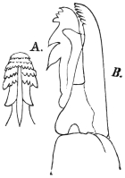
Fig. 359.—A., the rostrum of Ixodes ricinus (male); B>., the terminal joint of the maxillary palpi of the female. Enlarged. (After Pagenstecher.)
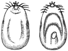
Fig. 360.—Female of Ixodes ricinus, gorged full, dorsal and ventral surfaces. 2/1. (After Pagenstecher.)
[The species I. reduvius is the same as I. ricinus, Latreille. The male is 2·35 to 2·80 mm. long; the body is dark brown, almost black, with a pale, almost white, margin; there are also traces of reddish mottling. Coxæ of the first pair of legs with a short spine. Rostrum much shorter than that of the female; shield oval; anal shield small, about one-third the length of ventral shield. The adult female varies from 2·80 to 3·5 mm. when not distended, but when gorged may reach 10 mm. long. The shield and legs are dark blackish-brown, body deep orange-red with four dark longitudinal lines, paler beneath and light grey in front. When distending it is pale red to grey or white; when fully gorged olive-green, or dark red to black, with irregular yellow streaks on the back and sides just before egg-laying. Sexual orifice opposite fourth pair of legs. The nymph varies from 1·60 to 1·70 mm. long when fasting; the body is olive-white, opaque, with four distinct brown posterior markings and similar anterior ones, leaving a pale centre to the shield. When fully gorged it is 3 mm. long. As the nymph distends, it changes from opaque white to blue-black, and finally black. The little larva is 0·80 to 1·50 mm. long, transparent with olive-green intestinal markings; as it becomes inflated it changes to blue-black, and then black. There are no eyes. It is widely distributed, and chiefly attacks sheep; sometimes it occurs on dogs and also attacks man. Mégnin records it from horses in the nymph stage. Amongst its other numerous hosts are goats, cattle, deer, hedgehogs, moles, bats, birds, and lizards. It is usually known as the grass tick and bottle-nosed tick. This species occurs in Europe, Asia, North Africa, and North America.
[Synonyms.—Considerable confusion exists over the name of this and other common ticks, owing to the same species having been described under a great many names. Observers have taken the same species on different animals and in various stages to be distinct, and have described them accordingly.
[The name Ixodes reduvius, Leach, does not stand, as Leach was describing quite a different parasite. The name I. ricinus, Latreille, 1806, is now substituted by Neumann and Wheler.
[The synonyms given by Wheler are as follow: Reduvius, Charleton, 1668; Ricinus caninus, Ray, 1710; Acarus ricinoides, de Geer, 1778; Acarus ricinus, Linnæus, 1788; Cynorhæstes reduvius, Hermann, 1804; Cynorhæstes ricinus, Hermann, 1804; Ixodes megathyreus, Leach, 1815; Ixodes bipunctatus, Risso, 1826; Cynorhæstes hermanni, Risso, 1826; Crotonus ricinus, Dumeril, 1829; Ixodes trabeatus, Audouin, 1832; Ixodes plumbeus, Dugés, 1834; Ixodes reduvius, Hahn, 1834; Ixodes fuscus, Koch, 1835 (?); Ixodes lacertæ, Koch, 1835 (?); Ixodes pustularum, Lucas, 1866; Ixodes fodiens, Murray, 1877; Ixodes rufus, Ixodes sulcatus, and Ixodes sciuri, Koch.—F. V. T.]
Ixodes holocyclus, Neumann, 1899.
[Under the name I. holocyclus, Cleland (Journ. Trop. Med. and Hyg., 1913, xvi, No. 3, pp. 43–45) says that: “This tick is common in man where there is dense scrub and tropical jungle along the east coast of Australia at certain times of the year. It may cause severe symptoms in children resulting in death.” He records a child being attacked in 1884 which died, and another case from which 200 ticks were removed, the symptoms being weak heart, collapse, syncope, but the patient recovered under treatment; again, in the same journal (pp. 188, 189), the case of a 4 1/2-year-old girl who was bitten showed widespread muscular paralysis, and other cases resembling conium poison.
[Taylor (Rep. Ent. Aust. Inst. Trop. Med., 1911, p. 21, 1913) refers to this species as the scrub tick of New South Wales. The partially fed female has a dark reddish-yellow scutum and is almost as broad as long, punctations very numerous, equal and confluent in places, long white hairs on the lower half of each coxa. He records it as attacking man commonly, mentioning Kamerunga, Cairns district, Queensland, and Sydney, N.S.W., as localities.—F. V. T.]
Ixodes hexagonus, Leach, 1815.
Syn.: Ixodes sexpunctatus, Koch, 1897; I. vulpis, Pagenstecher, 1861.
Lives in the same manner as the foregoing; especially attacks hounds, but also other mammals and even birds. The difference consists in the shape of the legs, the shorter rostrum, and the larger size of the male. It also occasionally attacks man, but is usually confused with the previously mentioned species.
[The synonyms of this species are as follow:339 Ixodes autumnalis, Leach, 1815; I. erinacei, Audouin, 1832; I. reduvius, Audouin, 1832; I. crenulatus, Koch; I. erinaceus, Murray, 1877; I. ricinus, Mégnin, 1880. Two other synonyms are given above by Braun.
[The female when fully replete is 11 mm. long, when fasting 3·86 mm.; the shield is heart-shaped and punctate, body finely hairy; palpi short and broad; labium shorter, and tarsi of all the legs more truncate than in I. ricinus. The colour of the distended body is drab and somewhat waxy; rostrum, shield and legs light testaceous. The male varies from 3·5 to 4·0 mm. long, and is reddish-brown in colour with lighter legs; the shield is punctate and leaves a narrow margin around the body; the body is elliptical, almost as large in front as behind. There is a spine on the coxæ of the first pair of legs, which is shorter than in the male I. ricinus and longer than in the female. The genital orifice is opposite the interval between the second and third pair of legs. The fasting nymph is 1·76 mm. long, light bluish-grey, margined and transparent, with four large posterior intestinal marks joined together behind the shield and smaller ones extending to the front and sides. When fully distended it is uniformly brownish-white; shield, legs and rostrum pale testaceous. The larva varies from 0·88 mm. when fasting to 1·76 mm. when gorged. Its body is light, but gradually becomes darker, with similar intestinal marks to ricinus.
[This tick is very common, especially on ferrets, stoats and hedgehogs. It is also found on sheep, cattle, etc. The males do not generally occur in company with the females on the host. Pairing probably takes place on the ground.—F. V. T.]
Genus. Amblyomma, Koch.
Amblyomma cayennense, Koch, 1844.
Syn.: Amblyomma mixtum, Koch, 1844; Ixodes herreræ, Dugés, 1887; Amblyomma sculptum, Berlese, 1888.
Characterized by the possession of eyes. The male measures 3·8 mm. in length by 3 mm. in breadth; the female 4 mm. in length by 3 mm. in breadth, but when full of blood may become 13 mm. in length and 11 mm. in breadth. They are common in the whole of Central America (Carrapatas), and attack mammals, amphibious animals and man.340
[This species was described by Fabricius. It occurs in Cayenne, Guiana, in Southern Texas, Florida, California, Mexico, Guatemala, Honduras, Nicaragua, Costa Rica, Panama, Bermuda, Cuba, Jamaica, Trinidad, Colombia, Venezuela, French Guiana, Brazil, Paraguay and the Argentine. It is called the silver tick. It frequently attacks man. Schwarz and Bishopp (Bull. 105, U.S. Dept. Agric., p. 158) heard of one man whose legs were well covered with suppurating sores and who was ill from the attack of these ticks and the wounds produced by scratching, and records other cases of their swarming on man. Newstead (Ann. Trop. Med. and Par., 1909, iii, No. 4, p. 442) records it as the worst pest to man in Jamaica.—F. V. T.J
Amblyomma americana, Linnæus.
The so-called long star tick, from the silvery spot on the apex of the scutum of the female. It will attack any mammal and even birds and also man. It occurs in North America, and also in Brazil, Guiana and Guatemala. Its punctures frequently end in suppuration. In the Eastern and Southern States man is more frequently attacked by this species than any other. Moss-gatherers in Louisiana are badly attacked by it.341 It also attacks the milkers in dairies. Attempts to transmit Texas fever failed with this species.
Amblyomma maculatum, Koch.
The so-called Gulf Coast tick, of the Gulf Coast, occurs on birds, mammals and man, especially cattle, and attacks the ears.
Genus. Hyalomma, Koch.
Hyalomma ægyptium, L., 1758.
Syn.: Acarus ægyptius, L., 1758; Ixodes camelinus, Fischer, 1823.
A species frequently found in Africa, particularly in Egypt and Algeria, and which also occurs in France and Italy, as well as in Asia. Male 8 mm. in length, 4·5 mm. in breadth. Female up to 24 mm. in length and 15 mm. in breadth. It infests large and small animals as well as human beings.342
[This is one of the largest ticks, nearly reaching the size of the bont tick. It is known in Africa as the bont leg-tick; all farm stock is attacked, but sheep and goats suffer most. Only one generation appears to occur each year. The male is almost black with a pale marginal stripe; the replete female brown with irregular light blue stripes. It is abundant in parts of South Africa.]
Genus. Hæmaphysalis, Koch.
Hæmaphysalis punctata, Canestrini and Fanzago, 1877–1878.
Syn.: Hæmaphysalis sulcata, Canestrini and Fanzago, 1877–1878; Rhicocephalus expositicius, Koch, 1877; Hæmaphysalis peregrinus, Cambridge, 1889; Herpetobia sulcata, Canestrini, 1890.
[This species does not appear to be common. It occurs on sheep, goats, horses and cattle. I have seen a female taken from man in Britain. The female when fasting is 3·44 mm. long, when gorged 12 mm. long. Colour, reddish-brown, leaden-grey when gorged; dorsal shield deeply indented in front; rostrum, shield and legs brownish; body finely punctate, both above and below; sexual opening opposite the coxæ of the second pair of legs in both sexes. Palpi a little longer than the labium; first segment short and narrow, second and third widened on the dorsal face. Coxæ with a short, broad blunt spine; tarsi short, terminated with a spur on the first pair. The male is 3·10 mm. long. Body rather narrow, yellowish to reddish-brown; dorsal shield nearly covers the whole body; numerous punctures over the whole surface. Eleven indentations on the posterior margin of the body; peritremes lighter in colour, large and comma-shaped. The three anterior pairs of legs with a short spine on the haunches, the fourth with a very long one directed backwards. The nymph varies from 2·5 to 3·0 mm., is oval, and light yellow to dark red in colour. Dorsal shield rounded with few punctations. No spur on tarsi, and sexual orifice nearly obsolete. Larva short and oval. Length 1·20 mm.—F. V. T.]
Genus. Dermacentor, Koch.
Dermacentor reticulatus, Fabricius, 1794.
Syn.: Acarus reticulatus, Fabr., 1794; Ixodes reticulatus, Latreille, 1806; I. marmoratus, Risso, 1826.
This tick is provided with eyes, but it is distinguished from Ixodes and analogous genera by the lack of the abdominal plastron in the male, which measures 5 to 6 mm. in length by 3·5 mm. in breadth. The female may attain 16 mm. in length and 10 mm. in breadth. It is found in the South of Europe, in Asia, and in America; it attacks chiefly oxen, sheep and goats, and occasionally man.343
[This tick sometimes causes much annoyance to human beings. It was once most troublesome at Revelstoke. Specimens have recently been found on fowls, turkeys and pheasants in Kent.
[Other synonyms are as follows: Cynorhæstes pictus, Hermann, 1804; Crotonus variegatus, Dumeril, 1829; I. pictus, Gervais, 1844; Dermacentor albicollis, Koch, 1844–1847; D. pardalinus, Koch, 1844–1847; D. ferrugineus, Koch, 1844–1847; Ixodes holsatus, Kolenati, 1857; Pseudixodes-holsatus, Haller, 1882; Hæmaphysalis marmorata, Berlese, 1887.
[The female when fasting is 3·86 mm. long by 2 mm. wide. The body is depressed, larger behind and reddish-brown in colour. The shield is very large and extends to the level of the third pair of legs, with a few large and many small punctations, milky white, variegated with reddish-brown. Sexual orifice opposite the coxæ of the second pair of legs. Coxæ of the front legs are deeply bifid, the others with a moderate spine. When gorged light brown, and may reach 16 mm. When depositing eggs the female is mottled with dark brown above and below. The male is like the female. The shield is reddish-brown, variegated with a milky white pattern. Coxæ of the fourth pair of legs three times the size of the third. There is a sharp backwardly pointing spine on the second palpal segment, also seen (but smaller) in the female. Length 4·20 mm.
[According to Mr. Wheler this is a very variable species both in size and colour. It occurs in England on sheep, but not commonly. It has probably been introduced into Britain. Besides the animals mentioned above it is also found on deer.—F. V. T.]
Dermacentor venustus, Banks.
[The Rocky Mountain tick fever tick. This species has been wrongly called Dermatocentor reticulatus var. occidentalis. The correct name of the carrier of Rocky Mountain tick fever is Dermacentor venustus, Banks (Hooker, Bishopp and Wood, Bull. 106, U.S. Dept. Agric., Div. Ent., p. 165).
[The female is from 13·8 by 10 by 6·4 mm. to 16·5 by 11·4 by 6·9 mm. when gorged; the male from 2·1 by 1·5 mm. to 6 by 3·7 by 1·4 mm. The male reddish-brown; scutum with an extensive pattern of white lines, usually but little white on the mid-posterior region, legs slightly lighter than scutum, joints tipped with white. Female with scutum mostly covered with white, abdomen reddish-brown, legs as in male. The nymph when unengorged reddish-brown, when gorged dark bluish-grey; the larva is yellowish-brown when unengorged, slate blue when engorged. The ova light brown, shiny and smooth.
[The chief wild hosts are the brown bear, coyote, woodchuck, rabbit, wild cat, badger and mountain goat for the larvæ; practically all small mammals act as hosts for larvæ and nymphæ, whilst the adults are seldom found on other than large domestic animals; horses and cattle are preferred. It occurs in British Columbia, southward to Northern New Mexico, and from the foothills of the Rocky Mountains in Colorado to the base of the Cascade Range in Oregon and California; abundant in Western Montana, Idaho, Eastern Washington, Oregon, North Utah, West Wyoming and North-west Colorado.
[Of great importance in the Bitter Root Valley of Montana, where a number of cases of fever occur each year, with a mortality of about 70 per cent. In British Columbia this tick causes tick paralysis in man and sheep. Only the adults seem to attack man and animals there (Hadwen and Nuttall, Parasitology, 1913, vi, No. 3, pp. 288–297 and 298–301) according to the Canadian Medical Association Journal, December, 1912. The symptoms are unlike spotted fever. For full details of this tick vide Bulls. 105 and 106, U.S. Dept. Agric.]
Dermacentor occidentalis, Neumann.
This tick only occurs in the Pacific Coast region of the United States. Owing to the fact that it frequently attacks man as well as occurring in great abundance in Oregon and California, it is of considerable economic importance. It is spoken of as the wood tick, and in the regions where found is the most common tick to attack man. Hooker, Bishopp and Wood (Bull. 106, U.S. Dept. Agric., Div. Ent., 1912, p. 189) state that a number of cases have been brought to their notice where the bite of this tick has caused considerable local inflammation, which in some cases required physicians’ attention. It has been supposed to be connected with Rocky Mountain spotted fever, but it is doubtful if it is concerned in its transmission. The engorged female is steel grey, the dorsum with an olive-green surface colour, which covers the grey except in small spots, giving a mottled appearance. The unengorged males and females are reddish-brown, scutum covered with a whitish bloom, interrupted by many red punctures. The female is 9 by 6·1 by 3·3 mm. to 11·8 by 7·6 by 5·6 mm.; the male 2·8 by 1·6 mm. to 4·2 by 2·3 mm. The larvæ are bluish-grey when engorged, reddish-brown when unengorged. The nymph is light brown, sides of scutum darker, and the intestines dark brown. It is confined to the Coast Range and Sierra Nevada Mountains in California and Oregon and the small mountain range to the south-west.
Dermacentor variabilis, Say.
The American dog tick has also been found on man, but it is of little economic importance as it is easily removed from its host.
Genus. Margaropus, Karsch.
Margaropus annulatus australis, Fuller.
The so-called Australian cattle tick. Newstead344 reports this as a great pest to man in Jamaica in its larval stage. Its chief hosts are cattle, horses, goats, sheep, dogs and rabbits.
Margaropus microplus, Canestrini.
Recorded by Aragão (Mem. Inst. Oswaldo Cruz, 1911, iii, fasc. 2, p. 163) as occurring in larval stage on man in Brazil.
Genus. Rhipicephalus, Koch.
Rhipicephalus sanguineus, Latreille, 1804.
Syn.: Ixodes sanguineus, Latr., 1804; I. rufus, Koch, 1844; Rhipicephalus limbatus, Koch, 1844; Rh. siculus, Koch, 1844; Rh. stigmaticus, Gerstäcker, 1873.
Spread over almost the entire tropical and sub-tropical regions, occurring in Europe in the South of France and in Italy; it infests dogs and more rarely sheep; oxen, cats, foxes and human beings are also attacked.345
Neumann’s Table of Species of Argas.
1Body elliptical (sides curved)2.

Body oblong (sides straight), ending anteriorly in a point7.
2Body transversely ovalvespertilionis.
Body elongate oval3.
3Margin of body striated4.
Margin of body formed by quadrangular areolæPERSICUS.
4Body flat, integument plainly wrinkled5.

Body tumid, elongate; integument finely wrinkled; coxæ of fourth pair of legs near anterior third of bodyhermanii.
5Body oval, narrowed anteriorlyREFLEXUS.
Body elliptical, blunt, hardly narrowed anteriorly6.
6Body twice as long as broadcucumerinus.
Body hardly longer than broadtransgariepinus.
7Dorsal integument with large polygonal depressions; tarsi appearing bifidBRUMPTII.

Dorsal integument almost smooth; tarsi not appearing bifidæqualis.
The Argantinæ are distinguished from the Ixodinæ by the head, which in the former is situated on the inferior aspect of the cephalothorax, while in the Ixodinæ it projects freely; also by the very short proboscis, the small club-like palpi, the lack of suckers on the legs, as well as by the scutellum, which covers the entire back and is bent up round the borders. Two genera are distinguished: Argas, Latreille, 1796 (Rhynchoprion, Hermann, 1804), and Ornithodorus, Koch, 1844. The species live on mammals, but more especially on birds.
Genus. Argas, Latreille.
Argas reflexus, Fabricius, 1794.
Syn.: Acarus reflexus, Fabricius, 1794; A. marginatus, Fabricius, 1794; Rhynchoprion columbæ, Hermann, 1804.
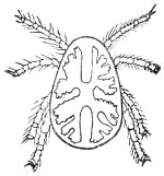
Fig. 361.—Argas reflexus: from the dorsal surface, the intestine showing through the integuments. (After Pagenstecher.)
The European marginated tick, Argas reflexus (length of male 4 mm., breadth 3 mm., length of female 6 to 8 mm., breadth 4 mm.), is of a yellowish colour and has yellowish-white legs. The ingested blood shows red or brown through the intestine, which is provided with blind sacs. It lives in dovecots. It remains hidden during the day and at night crawls on to the sleeping pigeons to suck their blood. It has been observed in France, England, Italy, Germany, and Russia. Persons sleeping near infected dovecots, or in apartments formed from pigeon-lofts, are also attacked, even when the room in question has not been used for sheltering pigeons for years, as “marginated ticks” can live in a fasting condition for a very long time. The bite sometimes gives rise to serious symptoms, such as general erythema and sudden œdema.
[This pest more often feeds on the blood of man than is imagined. Blanchard states that he has received them from men’s clothes in Strasburg. Boschulte, of Westphalia, records these parasites in a bedroom inhabited by children and connected with a pigeon-house. The children were bitten during sleep on the hands and feet. The result of the bite was intense itching along the nerves, the bite only being marked by a red spot. In a girl of 14 or 15, vesicles were formed similar to those produced by burns, and in an old man an ulcer formed. Others record painful punctures and persistent œdema produced by this pigeon pest. It was once abundant in Canterbury Cathedral, and often caused much annoyance, I am told, to the worshippers; the ticks falling down from the roof, where they were living, derived from the numerous pigeons that breed in the towers. This Acarus has enormous powers of vitality, living without food for months at a time.—F. V. T.]
Argas persicus, Fischer de Waldheim, 1824.
Of oval form and brownish-red colour. The male measures 4 to 5 mm. in length by 3 mm. in breadth; the female 7 to 10 mm. in length by 5 to 6 mm. in breadth. It frequents the entire north-west and north-east of Persia (the gerib-gez or malleh of the Persians, the miana bug of travellers). It lives concealed in houses and attacks man at night to suck his blood. Its bite is much dreaded, but the serious results may probably be attributed to unsuitable treatment of the wound or its invasion by bacteria.
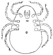
Fig. 362.—Argas persicus: ventral aspect. 7/1. (After Mégnin.)
[This tick, sometimes called the tampan and wandlius in South Africa, is mainly a fowl parasite. Fowls and ducks frequently die under its attack, particularly young ones, death being due to loss of blood. This tick remains attached to its host during its larval stage for about five days; it then leaves and moults in concealment. In its subsequent stages it visits its host by night and remains for about an hour only, during which time it distends itself fully with blood. As a nymph it moults twice, not once as do the cattle ticks. This tick and other Argas become larger with each moult, but retain their same general appearance. The female visits the host every now and then, and, between, deposits eggs in sheltered crevices. About fifty to 120 are deposited at once. Four weeks seems a necessary period to intervene between visits to the host, and the interval may be extended to upwards of a year according to Lounsbury.346
[It is found in the Sudan, where Balfour has found granules derived from the segmentation of spirilla in their digestive tract. Fantham and Hindl have confirmed this. It has been assumed that these granules carry infection.
[This so-called Persian tick, the miana, which is such a scourge to travellers in Persia, appears to infest the huts of natives in that country. It has been sent me from Quetta, where it has invaded houses to such an extent the natives cannot live in them. The virulence of its bite is probably due to the tick transmitting fever germs from natives, probably inured, to strangers, who would be susceptible.—F. V. T.]
Argas brumpti, Neumann.
[Found in Somaliland, by Brumpt, and in the Sudan. This tick attacks man as well as wild animals and produces a painful swelling according to King,347 but as pointed out by that naturalist it probably relies on other than human food.—F. V. T.]
Argas chinche, Gervais, 1844.
This Acarus, a native of the temperate parts of Colombia, is very troublesome to man. It is probably identical with A. americanus, Packard, which infests domestic fowls and turkeys, and occasionally also cattle, and is differentiated from A. reflexus by the sculpturing of the cuticle.
Genus. Ornithodorus, Koch.
Neumann’s Synopsis of the Genus Ornithodorus is as follows:—
1Hypostome unarmed; integument in nymph stage and partly in adult spinuloseMÉGNINI.
Hypostome armed with recurved teeth; integument not spinulose2.
2Camerostome with movable lateral flapsTALAJE.
Camerostome without movable lateral flaps3.
3Anterior border of distal segments of legs with tubercles or festoons4.
Anterior border of segments of legs without tubercles or festoons8.
4Body not much contracted anteriorly5.
Body pointed anteriorly7.
5Tubercles of distal segments of legs higher than broad, distant6.
Festoons of distal segments of legs as broad as high, contiguouspavimentosus.
6Eyes presentSAVIGNYI.
No eyesMOUBATA.
7Eyes presentcoriaceus.
No eyesTURICATA.
8Integument with fine radiating wrinkleslahorensis.
Integument granular9.
9Tarsi appearing bifid at apexfurcosus.
Tarsi not appearing bifid at apex10.
10Tarsi of first pair of legs with three dorsal tubercles, of other legs with onecanestrinii.
Tarsi without dorsal tubercles or with only one11.
11Tarsi of last three pairs of legs with pronounced dorsal protuberancetholozanii.
Tarsi of legs with indistinct dorsal protuberanceerraticus.
Ornithodorus moubata, Murray, 1877.
An abundant African tick which is one of the carriers of the spirillum of African relapsing fever and can also carry Filaria perstans (Christy). Its body is oval, yellowish-brown when young, greenish-brown when mature. The integument is covered with mamillose tubercles. No eyes and the stout legs granular above, the tibiæ and tarsi fringed with tubercles on the upper side. Pocock348 records it from Uganda and German East Africa, Congo and Angola, to Namaqualand and the Transvaal in the south. It is called bibo in Uganda, moubata in Angola, and tampan on the Lower Zambesi. It feeds on animals and birds as well as man. Its bite is very painful. This tick is found in native huts, living in cracks and crevices and in the thatch roofs.
The female tick infected with the spirillum transmits the infection to the eggs and the next generation. They appear to be able to live without food a long time, and probably live for years. They lay their eggs in masses on the ground or in crevices, and when they hatch they are in the nymph stage with four pairs of legs. O. moubata also occurs in Madagascar with recurrent fever (Lamoureux, Bull. Soc. Path, exot., 1913, vi, No. 3, pp. 146–149).
Ornithodorus savignyi, Audouin, 1827.
At one time considered the same as the preceding species, but can be easily separated by the presence of two pairs of eyes. It is widely spread over Africa and has been found in South India and at Aden. In the Sudan it occurs in large numbers. King349 records that a few miles N.N.E. of Khartoum 370 specimens were collected in two hours under a single tree by a well. It is found in Somaliland, where relapsing fever occurs and no O. moubata, which it probably replaces as a transmitter (Drake-Brockman, “Rep. Col. Office,” April 6 and April 16, 1913). It also occurs in Tunis, where the natives call it “tobbiah” (Weiss, Arch. de l’Inst. Pasteur de Tunis, 1912, pt. 4, p. 226).
Ornithodorus coriaceus, Koch.
Found in Mexico, Paraguay and California. Attacks man.
Ornithodorus talaje, Guerin, 1849.
An eyeless species with somewhat elongate pentagonal body found in Mexico and South America, called the “chinche.” A variety of it (coniceps) is found at Venice, etc., and another variety on various islands in the Indian Ocean and South Atlantic. Its bite is very painful to man.
Ornithodorus turicata, Dugès, 1876.
Without eyes. Indigenous in Central America; attacks human beings and pigs. The bite is painful and is often followed by serious consequences.
[So virulent is this species that pigs put in an infested sty often die in a night. This “turicatas” of Mexico often reaches 6 mm. in length.—F. V. T.]
Ornithodorus tholozani, Laboulbène and Mégnin, 1882.
Syn.: Argas tholozani, Lab. and Még., 1882.
Without eyes. Males 4 to 6 mm. in length and 2 to 4 mm. in breadth; females 8 to 10 mm. in length and 4 to 5 mm. in breadth. It especially attacks sheep. Native of Persia and Asia Minor.
[This species is reputed as being very dangerous to man. It is locally know as the kéné, or sheep-bug. In its fully gorged state it is deep violet.—F. V. T.]
Ornithodorus mégnini, Dugès, 1883.
Syn.: Argas mégnini, Dugès, 1883.
Length 8·5 mm., breadth 5·5 mm. Native of Mexico.
[Another synonym for this species is Rhynchoprion spinosum, Marx. The adult males and females are grey to dark brown, the male somewhat the smaller; female 5 by 3·5 by 2·5 mm. to 10 by 6 by 3·5 mm. The larvæ at the seed tick stage are dark grey, turning to pink, then to a whitish grey when engorged. The nymph when young is blood-red in front, rest pearly white; later they turn reddish-brown.
[Intense pain may be caused by its presence in and around the ears.
[Two specimens in the nymphal state were taken from the ears of a visitor to Cambridge by Dr. J. Christian Simpson. They were supposed to have entered the ears when the gentleman was camping out in Arizona (Lancet, 1901, i, No. 4,052, p. 1198, fig. 3).
[This species attacks the horse, ass, dog, cats and oxen, generally around the ears, and also attacks man. It is well known in the United States as infesting the ears of children (New York Ent. Soc. Journ., 1893, pp. 49–52).
[It occurs in Texas, Arizona, New Mexico and California as well as Mexico, Brazil, and possibly many parts of South America; and recently Bedford (“Sec. Report Div. Vet. Res., S. African Union,” 1912, pp. 343, 344) has shown it to occur at Vryburg and Fauresmith, in the Transvaal, on stock. It also occurs in the Sudan.—F. V. T.]
Other Literature on Ixodidæ.
(1) “Pénétration de l’Ixodes ricinus sous la peau de l’homme,” Compt. rend. Soc. de Biol., 1891, xliii, ser. 9, iii, pp. 689–691, R. Blanchard.
(2) “Notas sobre Ixodidas brazileiros,” Mem. Inst. Oswaldo Cruz, 1911, iii, fasc. 2, pp. 145–195, pls. 11 and 12, Dr. H. de Beaurepaire Aragão. Table of Brazilian Species.
(3) “Contribuicão para a sistematica e biolojia dos Ixodidas,” Mem. Inst. Oswaldo Cruz, 1912, iv, fasc. 1, pp. 96–120, pls. 2 and 3, Dr. H. de Beaurepaire Aragão.
Family. Tyroglyphidæ.
Very small mites without eyes and without tracheæ, with smooth skin.
The males usually have a suctorial pore on either side of the anus, which is used during copulation, or suckers may be found in both sexes near the sexual orifice. The mouth parts form a cone with chelate cheliceræ, and three-jointed pedipalpi; the legs are usually short, have five segments with a terminal claw and suckers, or either one or other of these organs. The numerous species and genera live free and from choice in slowly decomposing vegetable and animal matter (cheese, cereals, flour, sugar, preserves, dried anatomical preparations, bacon, dried fruits and fungi), also in the corners of dwellings, etc.; they incidentally get into or on to man, or are found in chamber utensils and in spittoons; actual parasites are rarely found amongst them.
[The chief genera are Tyroglyphus, Rhizoglyphus, Glyciphagus, Aleurobius and Histiogaster. The first three have typical characters referred to, but are distinguished from each other by the two former having the hairs on the dorsum smooth, whilst in Glyciphagus they are hairy, plumose, or feathered. Rhizoglyphus can be told from Tyroglyphus by having claws on the tarsi without any suckers; Tyroglyphus has both claws and suckers. The larvæ are hexapod and may become adult in the usual way by repeated moults, or they enter the so-called hypopial stage. In this the eight-legged nymph becomes quiescent, and during this stage it fixes itself to some insect or other animal by a patch of suckers on the lower surface of the hind end of the body, and is so carried from place to place. The hypopus does not feed and has a hard shell and short legs. When it has reached a new home it moults and development proceeds in the normal way. Canestrini and Kramer treat the Tyroglyphidæ as a sub-family of the Sarcoptidæ, calling them sub-family Tyroglyphinæ, the other sub-families being Sarcoptinæ, Canestriniinæ and Analsinæ.—F. V. T.]
Sub-family. Tyroglyphinæ.
Genus. Aleurobius, Canestrini.
Aleurobius (Tyroglyphus) farinæ, de Geer (part), Koch.
The male measures 0·33 mm. in length by 0·16 mm. in breadth; the female 0·6 mm. in length by 0·3 mm. in breadth. These mites possess five pairs of suctorial organs of a light colour; the legs are reddish. Moniez observed them in Lille on the skin of labourers who had been unloading Russian corn. A few of the species generally mentioned under the designation of Tyroglyphus siro are probably the common flour-mite, which also occurs on dry cheese.
[The farinæ of de Geer is an Aleurobius described by him in 1778 (“Mém. Hist. Ins.,” vii, t. 5, f. 15, p. 97) as Acarus farinæ.—F. V. T.]
Genus. Tyroglyphus, Latreille.
Tyroglyphus siro, L., 1756. (Defined by Gervais, 1844.)
Male 0·5 mm. in length by 0·25 mm. in breadth; female 0·53 mm. in length by 0·28 mm. in breadth; the males have two suckers on the tarsi of the fourth pair of legs. Penis straight, colour whitish or reddish.
Tyroglyphus longior, Gervais, 1844.
White or yellowish, with two black spots on the abdomen. Male 0·55 mm. in length, 0·28 mm. in breadth; penis bent. Female 0·61 mm. in length and 0·28 mm. in breadth.
T. siro and T. longior live on dry cheese, in flour, on dried fruits, etc., and have been occasionally observed in the stools, urine, or pus of human beings, and also on their skin. The so-called vanillismus is to be attributed to these species.
[T. siro and T. farinæ of Schrank (non Geer) are the same. They are described under other names, such as Acarus lactis, Linn.; A. favorum, Herm., etc.; A. lactis in milk, farinæ in flour, and siro in cheese; and as A. dysenteriæ, Linnæus (“Syst. Nat.,” ed. 12, pp. 1024–1767).]
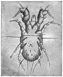
Fig. 363.—Tyroglyphus farinæ: male. Enlarged. (After Berlese.)
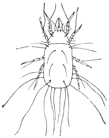
Fig. 364.—Tyroglyphus longior, Gerv. (After Fum. and Robin.)
It is to these species that a case of dysentery was referred. Rolander, who studied under Linnæus, was attacked by what was called dysentery. The complaint soon gave way to treatment, but eight days after it returned, soon disappeared, but again came a third time. All the time Rolander had been living like the other inmates of the house, who all escaped. Linnæus, aware that Bartholemy had attributed dysentery to insects which he said he had seen, advised his student to examine his stool. The result was that innumerable mites were found to be present. Their presence was easily accounted for by the fact that they were found in numbers in a cup made of juniper wood from which the student alone drank of a night, and they were found to be of the same species. What this species is we do not know. Linnæus called it Acarus dysenteriæ, but it was the same as his Acarus siro. No records have occurred since. It cannot be, as Latreille supposed, the cheese mite, for they have been eaten by millions since, and it is strange no such case has occurred again.
[Tyroglyphus minor var. Castellani, Hirst,
causes the copra itch in persons employed in the copra mills in Ceylon. The skin of the hands, arms, legs and even body becomes covered with pruriginous papules, papulo-pustules and pustules near the head. The eruption begins as a rule on the hands. The mites live in the copra dust. They produce dermatitis. Castellani produced the disease experimentally by rubbing copra dust containing mites on the skin of healthy people. Beta-naphthol ointment (5 to 10 per cent.) proved useful in treatment (Journ. Trop. Med. and Hyg., December 16, 1912, Castellani and Hirst).—F. V. T.]
Genus. Glyciphagus, Hering, 1838.
Glyciphagus prunorum, Her., and G. domesticus, de Geer.
The Glyciphagi are differentiated from the Tyroglyphi in that the chitinous hairs on the body are fringed or feathered, and that they lack a furrow dividing the cephalothorax from the abdomen. They live under similar conditions to the Tyroglyphi and are occasionally found on man or in fæces.
[Sugar merchants and grocers are frequently troubled by swarms of G. domesticus, which leave the stores when being handled, and especially shopmen, who handle sugar kept in small stores for some time. These are the Acari that cause that irritating temporary affection known as “grocer’s itch.”—F. V. T.]
Glyciphagus cursor, Gervais.
Under this name Signor Moriggia figures a horny excrescence of great length growing from a woman’s hand, and containing in its cavities quantities of Acarus. This species is really G. domesticus, de Geer. G. domesticus has also been described by Gervais (Ann. Sci. Nat., 1841, ser. 2, xv, p. 8) as G. hippopodes.
Glyciphagus buski, Murray.350
[This is a mite found by Busk and named after him by Murray. It was taken from beneath the cuticle of the sole of the foot of a negro in the Seamen’s Hospital Ship on the Thames in 1841, in large sores of a peculiar character confined to the soles of the feet. It appeared that the disease was caused by its burrowing beneath the thick cuticle. The disease was attributed to the wearing of a pair of shoes which had been lent to another negro whose feet had been similarly affected for nearly a year. The negro to whom the shoes were lent came from Sierra Leone. Mr. Busk stated that some water brought by Dr. Stranger from the River Sinoe, on the coast of Africa, contained one nearly perfect specimen, and fragments of others very similar to if not identical with this Acarus. Mr. Busk adds that he had been informed by Staff-Assistant Surgeon P. D. Murray that at Sierra Leone there is a native pustular disease called craw-craw—a species of itch breaking into open sores.
[From Busk’s original figure I see no reason to doubt that this is a Glyciphagus.—F. V. T.]
Genus. Rhizoglyphus, Claparède, 1869.
Rhizoglyphus parasiticus, Dalgetty, 1901.
The Rhizoglyphii are to be recognized by their short legs, which are beset with spines, and by the tarsi, which terminate in a claw. They live on plants, roots and bulbs, especially the bulbs of lilies.
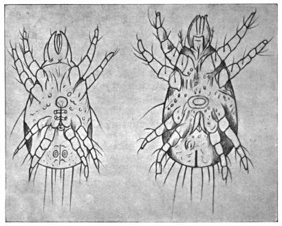
Fig. 365.—Rhizoglyphus parasiticus. a., male; b., female. Enlarged. (After Dalgetty.)
This species has been observed on the feet of Indian coolies working in the tea plantations; they produce a skin disease which always commences with blebs between the toes, and which almost always extends to the malleoli, but not beyond. The Acari have an elliptical body, which is grey, but varies from greenish-yellow to greenish-brown when the stomach is full. Eyes are absent. The legs are composed of five segments and terminate with a claw. The males measure 0·18 mm. in length by 0·08 mm. in breadth, and possess genital and anal pores; the females measure 0·2 mm. in length by 0·09 mm. in breadth.351 [This is also known as coolie itch and is common in Indian tea plantations.—F. V. T.]
Genus. Histiogaster, Berlese, 1883.
Histiogaster (entomophagus?) spermaticus, Trouessart, 1900.
The genus Histiogaster, which also approaches the Tyroglyphinæ, is characterized by the circumstance that the males possess suctorial pores used in copulation, as well as leaf-shaped appendages at the posterior end of the body. They feed on vegetables, especially on small fungi.
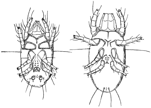
Fig. 366.—Histiogaster (entomophagus?) spermaticus: on left, male; on right, female—both from the abdominal aspect. 200/1. (After E. Trouessart.)
This species has been described by Trouessart,352 who found numerous specimens, some adult, others in the developmental stage (larvæ, nymphs), and ova, in the fluid removed by puncture from a cyst of the right testis. The males measure 0·25 mm., the females 0·32 mm., and the larvæ 0·1 mm. in length. The author is of opinion that the animal—perhaps a fertilized female—was introduced by a catheter, and, as a matter of fact, it was afterwards found that the patient had once had the catheter passed in India while suffering from pernicious fever.
It would here rather appear to be the case of a facultative parasitism of an otherwise free-living species. Histiogaster entomophagus, Laboulbène, is found occasionally in collections of insects feeding on larger species containing much fat; the species also occurs on dry cantharides; it appears to belong to the region of South Europe, where, however, it is widely spread.
[Entomophagus occurs all over Europe and in America. It has been described under the following names: Acarus malus, Shimer, 1868 (Trans. Illinois Hort. Soc.); Dermaleichus mali, Riley, 1873 (Rep. Ins. Missouri, v, p. 87); Tyroglyphus mali, Murray, 1877 (“Eco. Ent. Apt.,” p. 275); T. corticalis, Michael, 1885 (Trans. Roy. Micros. Soc., ser. 2, v, 3, p. 27, figs. 1 to 14); Histiogaster corticalis, Canestrini, 1888 (Prosp. Acarof., iii, p. 397); H. aleurophagus, Sicherin, 1894, Canestrini, Prosp. Acarof., vi, p. 815. Trouessart’s species is evidently distinct.—F. V. T.]
Genus. Cheyletus.
Cheyletus mericourti, Lab.
Acaropsis mericourti, Moq. Tand.
[This mite has been described from three specimens found in pus which flowed from an abscess in the ear of a naval officer, produced by inflammation of the auditory passage. Where the mites came from we do not know, as they were found near the Bank of Newfoundland. This genus of Acari has enormous mandibles and a peculiar tracheal system; two ungues and appendages to the tarsi.—F. V. T.]
Family. Sarcoptidæ (Itch Mites).
Small mites without eyes and tracheæ, and with delicate, transversely striated cuticle. The mouth parts form a cone, over which the shield-shaped upper lip protrudes; the cheliceræ are chelate; the pedipalpi (or maxillary palpi) have three joints; the legs are short and compact, and composed of five segments; the terminal joints have pedunculated suckers (ambulacra) or a long bristle. The larvæ are six-legged. They live on or under the skin of birds and mammals, on which they produce the skin disease known as scabies, or itch.
[The Sarcoptidæ attack the hairs, feathers or epidermis of birds, animals and man, living as permanent parasites. The punctures they produce are followed by the formation of more or less thick crusts or scabs, beneath which the mites live and breed (so called scab, mange and itch). Most are oviparous, some ovoviviparous. The eggs are minute, ovoid, with a thin semi-transparent shell. They incubate in a few days, varying from two to ten or eleven, as a rule. Generally sarcoptic diseases lie dormant in winter and revive in spring and summer in man; but in animals with long wool, such as sheep, they are most active during winter, although revival of active reproduction takes place in spring.
[Speaking generally, for the Sarcoptidæ there are three distinct stages in the development of the male, four in the female, as follows:—
[(1) The larva. In this stage only three pairs of legs occur.
[(2) The nymph, in which a fourth pair of legs appear, and which thus approaches the adult; but so far no sexual organs occur. Nymphs are of two sizes—the smaller being future males, the larger females.
[(3) The next stage in the female is the age of puberty, the female now being provided with a vulvo-anal slit; this so-called pubescent female is fertilized by the male. The male then dies. But the female again casts her skin and enters another stage—
[(4) The ovigerous female—the egg-laying female—which has differently modified legs.
[The rate at which these Acari breed is very great. Gerlach has found that roughly, in each Sarcopt gallery, a female produces fifteen individuals—ten females and five males—and that the progeny reproduce again in fifteen days. The table given below thus shows that one pair may produce the enormous number of 1,500,000 descendants in three months:—
First generation after 15 days 10 females 5 males
Second" " 30 " 100"50"
Third" " 45 " 1,000"500"
Fourth" " 60 " 10,000"5,000"
Fifth" " 75 " 100,000"50,000"
Sixth" " 90 " 1,000,000"500,000"
= 1,500,000 individuals.
[These Acarinæ are divided into three distinct sub-families, namely the Cytolichinæ, Sarcoptinæ, Canestriniinæ.
[The Sarcoptinæ alone interest us here, and of the nine genera the three following are the most important:—
[(1) Sarcoptes, Latreille; Eusarcoptes.
[(2) Psoroptes, Gerv.; Dermatodectes, Gerlach; Dermatocoptes, Fürstenberg.
[(3) Chorioptes, Gerv.; Symbiotes, Gerlach; Dermatophagus, Fürst.; Sarco-dermatocedes, Del.
[The following are the main characters of these three genera:—
[Sarcoptes—round or slightly oval; the two posterior pairs of legs being nearly or quite concealed beneath the body; the tarsi end in simple long pedicles, with ambulatory suckers.
[Psoroptes—oval; the legs are all visible outside the margin of the body; the ambulatory suckers are carried on long triangulated stalks; the male has copulatory suckers and abdominal prolongations.
[Chorioptes—oval; legs long, thick, all visible; ambulatory suckers very wide, carried at the end of simple, short pedicles.
[Sarcoptes make channels or furrows beneath the epidermis, and in these the female lays her eggs. This form of acariasis is thus difficult to cure. It is the cause of human itch (vide Sarcoptes scabiei).
[Psoroptes do not make sub-epidermic galleries; they live and breed in colonies beneath crusts or scabs formed by the changes they produce in their host’s skin. Sheep scab is a common type of disease produced by Psoroptes. This genus is of little importance as a parasite to man.
[Chorioptes live as Psoroptes; they also do not affect man. Otodectes, Can., affecting cats and dogs, and others occur, but do not affect man as far as we know at present (“Demodicidae und Sarcoptidae,” von Professor G. Canestrini und P. Kramer, Das Tierreich, 1899).—F. V. T.]
Sub-family. Sarcoptinæ.
Genus. Sarcoptes, Latreille.
Sarcoptes scabiei, de Geer, 1778.
Syn.: Acarus scabiei, de Geer, 1778; A. psoricus, Pallas, 1760; A. siro, L., 1778; Sarcoptes exulcerans,? Linn., 1758, Nitsch, 1818; S. hominis, Raspail, 1834, and Hering, 1838; S. galei, Owen, 1853; S. communis, Delaf. et Bourg., 1862; S. scabiei var. hominis, Mégnin, 1880.
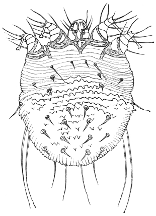
Fig. 367.—Sarcoptes scabiei: female, dorsal aspect. 200/1. (After Fürstenberg.)
The body is oval or nearly circular and whitish in colour, with transverse rows of striæ partly interrupted on the back. There are transverse rows of small bristles on the dorsal surface, and groups of trichomæ on the front, sides and back. There are chitinous hairs at the base of the legs; the two first pairs are provided with pedunculated ambulacra in both sexes, the two posterior pairs terminate each with a long bristle in the female; in the male the third pair of legs terminate in a bristle, the fourth pair with a pedunculated ambulacrum. The anus is situated at the posterior border of the dorsal surface.
At one time numerous species were differentiated, according to the form of the Acarus, the number, position and size of the hairs and spines, even according to the hosts, etc. All these characteristics, however, fluctuate so considerably that absolute differentiation is impossible; the supposed species may be regarded in the same light as Mégnin did, as varieties. It is also hardly possible to distinguish the mite of human scabies (S. hominis) from that of a number of domestic animals (S. squamiferus). It is best, therefore, to accept one single species (S. scabiei), which may give rise to different races or castes by living in the skin of man and mammals, but can pass from one host to the other.
[Canestrini and Kramer, in their monograph of the Sarcoptidæ, enumerate eighteen distinct species of this genus, from the dog, goat, camel, horse, ferret, lion, wolf, sheep, pig, etc., and two species parasites of man (scabiei and scabiei-crustosæ). There is no doubt that they are distinct species.—F. V. T.]
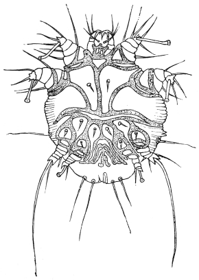
Fig. 368.—Sarcoptes scabiei: male, ventral aspect. 200/1. (After Fürstenberg.)
The S. scabiei of man (S. scabiei var. hominis) (length of male 0·2 to 0·3 mm., and breadth 0·145 to 0·190 mm.; length of female 0·33 to 0·45 mm., and breadth 0·25 to 0·35 mm.) lives in the tunnels that it excavates in the epidermis, and attacks by preference places with thin skin, such as between the fingers, in the bend of the elbows and knees, in the inguinal region, on the penis, on the mammæ, but may also affect other parts. The tunnels, which vary from a few millimetres to a centimetre and more long, do not run straight, but are somewhat tortuous; the female is found at the terminal end. The tunnels contain the excrement and oval eggs (0·14 mm. in length) of the parasite; the males are rarely met with, as they die off after copulation; the females die after depositing their eggs. The six-legged larvæ hatch out after four to eight days, and after about a fortnight, during which time they change their skins three times and undergo metamorphosis, they begin themselves to burrow. Transmission from person to person rarely is effected through linen, but by direct contact (as in coitus); transmission can be artificially effected on horses, dogs and monkeys, but not on cats.
The smaller S. scabiei-crustosæ, Fürstenberg, is the cause of the itch that occurs chiefly in Norway; it is not certain whether this is a distinct species of itch mite.
[This is quite a distinct species, which is recorded from Germany and France. Mégnin (Parasitology, 1880, p. 165) described this as S. scabiei var. lupi. The female is 140 µ long, 340 µ broad; the male is 170 µ long by 150 µ broad. In Science (March 3, 1893, p. 125) is recorded that at the Indiana Academy of Science Dr. Robert Hessler referred to “a case of that extremely rare and almost extinct form of itch known as ‘Norway itch,’ the scabies norvegica of Hebra, 1852.” The afflicted man was covered with thick, creamy white, leathery scales; some of these scales measured over an inch in diameter and 1/10 in. thick. A constant shedding of scales went on, a handful being gathered daily. They were found full of mites and eggs and riddled with passages. Under treatment the mites were killed and the skin became normal. Dr. Hessler made a calculation of the number of eggs and mites, amounting to ova and shells 7,004,000, mites in all stages 2,009,000.—F. V. T.]
The following forms may be transmitted from DOMESTIC ANIMALS to MAN:—
(1) S. scabiei var. equi. Male, 0·2 to 0·23 mm. long, 0·16 to 0·17 mm. broad. Female, 0·40 to 0·42 mm. long, 0·28 to 0·32 mm. broad. The horse is the normal host.
(2) S. scabiei var. ovis. Male, 0·22 mm. long, 0·16 mm. broad. Females, 0·32 to 0·44 mm. long, 0·24 to 0·36 mm. broad. This mite lives on sheep, and passes over to goats and human beings; it may also be artificially transferred to horses, oxen and dogs.353
(3) S. scabiei var. capræ. Male, 0·24 mm. long, 0·188 mm. broad. Female, 0·345 mm. long, 0·342 mm. broad. On goats, passing from them to horse, ox, sheep, pig and man. On the latter, in contradistinction to the varieties (1) and (2), it produces a severe affection.
(4) S. scabiei var. cameli. Frequently observed in man, chiefly in Africa. A few cases have been observed in Europe; the affection induced by it is severe.
(5) S. scabiei var. aucheniæ. Male, 0·245 mm. long, 0·182 mm. broad. Female, 0·34 mm. long, 0·264 mm. broad. It lives on the llama, and may be transmitted to man.
(6) S. scabiei var. suis. Male, 0·25 to 0·35 mm. long, 0·19 to 0·3 mm. broad. Female, 0·4 to 0·5 mm. long, 0·3 to 0·39 mm. broad. In the domestic pig and wild boar; occasionally also in man. The settlement, however, is usually of short duration.
(7) S. scabiei var. canis. Male, 0·19 to 0·23 mm. long, 0·14 to 0·17 mm. broad. Female, 0·29 to 0·38 mm. long, 0·23 to 0·28 mm. broad. In the house-dog, and also, not unusually, in human beings.
(8) and (9) S. scabiei var. vulpis and S. scabiei var. leonis of the fox and lion have likewise been observed on man.
These are all distinct species and should read as follows: S. canis, Gerl.; S. ovis, Mégn.; S. equi, Gerl.; S. dromedarii, Gerv. (cameli, Mégn.); S. aucheniæ, Raill.; S. suis, Gerl.; S. vulpis, Fürst.; S. leonis, Can.
Sarcoptes minor, Fürstenberg, 1861.
Anus situated on the back, legs short, pedunculated ambulacra broad; living on cats (S. minor var. cati) and rabbits (S. minor var. cuniculi). In cats this mite usually lives in the cervical region, and thence spreads to the ears and head; it usually causes the death of the infected animals; it is easily transferable from cat to cat, is difficult to transmit to rabbits, but once settled on them can easily infect other rabbits. On the other hand, the transmission of the itch mite of the rabbit to the cat does not succeed. In man S. minor induces an eruption that disappears after about a fortnight.
[S. minor, Fürstenberg, 1861 (“Krätzm.,” viii, p. 218), comes in Railliet’s sub-genus Notoedres, 1893 (“Zool.,” ed. 2, p. 660). Canestrini raised this to generic rank in 1894 (Prosp. Acarof., vi, p. 724).
[There are three species: (1) N. notoedres, Mégnin = Sarcoptes alepis, Railliet and Lucet (Compt. rend. Soc. de Biol., 1893, xlv, p. 404), and Sarcoptes notoedres var. muris, Mégnin (Parasitology, 1880, pp. 172–174). This occurs on the black and brown rats and the water-vole.
[(2) N. cati, Hering, 1838 (N. acta. ac. Leop., ii, 18, xliv, p. 605, figs. 9 and 10), = Sarcoptes minor, Fürstenberg (“Krätzm.,” 1861, viii, p. 215). Found on the cat in Germany, France, Italy, and Britain.
[(3) N. cuniculi, Gerlach, 1857, “Krätzm.,” iii, figs. 20, 21. It lives on the rabbit and is found in Germany and France.—F. V. T.]
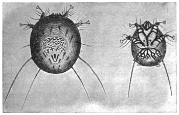
Fig. 369.—Sarcoptes minor var. cati: on the left, female (lying on its abdomen); on the right, male (lying on its back). (After Railliet.)
The itch mites of domestic animals, which belong to the genera Psoroptes (= Dermatodectes = Dermatocoptes) and Chorioptes (Symbiotes = Dermatophagus), as a rule do not infest and live on man, even when artificially transmitted. It is, however, possible for this to occur. Moniez (“Traité de par.,” 1896, p. 559) mentions that a species of Chorioptes—probably Ch. bovis—had been found on man, as had also Demodex folliculorum. This author also includes Dermatophagoides scheremetewskyi, Bogdanoff (Bull. soc. imp. d. natural., Moscou, 1864, xxxvii, p. 341), which has repeatedly been found on man in Moscow and Leipzig (Zürn, Ber. d. med. Ges., Leipzig, 1877, p. 38), as Chorioptes bovis.
Other References to Scabies crustosæ AND norvegica, ETC.
(1) “Ein Fall von Scabies crustosa norvegica,” Würzb. med. Zeitschr., l, pp. 134–139, pl. 3, H. Bamberger.
(2) “Ueber die Krätzmilbe (Acarus scabiei),” Notiz. a. d. Geb. d. Nat. u. Heilk., Weimar (1913), xlii (11), Oct., pp. 161–166 (1834), de Blainville.
(3) “Rapport sur le ciron de la gale (Acarus scabiei),” Ann. de Mus. d’Hist. nat., 1831; Parasitology, iv, pp. 213–232, de Blainville.
Family. Demodicidæ (Mites of the Hair-follicles).
Small Acarina, elongated in worm-like fashion, with annulated abdomen, and without eyes or tracheæ. The mouth parts consist of a suctorial proboscis and three-jointed palpi; the legs are short, and have three segments with small terminal ungues. The anus is situated on the anterior border of the abdomen; oviparous; the larvæ have six stumpy legs. These mites live in the hair-follicles of mammals.
Genus. Demodex, Owen.
Demodex folliculorum, Simon, 1842.
Syn.: Acarus folliculorum, Sim., 1843; Demodex folliculorum, Owen, 1843; Macrogaster platypus, Miescher, 1843; Simonea folliculorum, P. Gervais, 1844; Steatozoon folliculorum, Wilson, 1847.
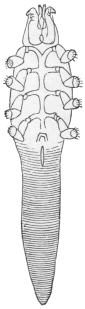
Fig. 370.—Demodex folliculorum of the dog. (After Mégnin.)
As in Sarcoptes scabiei, numerous varieties of this species are known; the form parasitic on man lives in the hair-follicles, the meibomian and sebaceous glands, and hardly ever causes inconvenience; the male measures 0·3 mm. in length and the female about 0·4 mm. in length. The eggs 0·06 to 0·08 mm. in length, 0·04 to 0·05 mm. in breadth, and are thin-shelled. The creatures are always attached with the head end downwards in the parts mentioned; they are most frequent in the sebaceous glands of the face, by the nose, lips and forehead, but they may be present on the abdomen and on other parts of the body. They may occasionally obstruct the excretory gland ducts, thus causing inflammation of the gland (comedones); their agglomeration in the meibomian glands sets up inflammation of the margins of the eyelids. There are generally only a few specimens in a gland. According to some statements Demodex occurs in 50 per cent. of mankind and even in children; they survive the death of their hosts by several days.
The variety living in the dog (D. folliculorum var. canis) is smaller than the variety living in man, and produces a skin disease resembling scabies in these animals. According to Zürn they may also live on man; nevertheless, no other investigator has recorded a similar observation, and attempts at artificial infection have proved negative.354
[Ten distinct species of Demodex are given by Canestrini and Kramer (“Demodicidae und Sarcoptidae,” Das Tierreich, 1899, vii). The species are certainly distinct.
[The species living on the dog (D. canis, Leydig, 1844) is cosmopolitan. According to the British Medical Journal (February 22, 1913, p. 407), dog mange may be caught by humans. Whitfield and Hobday describe in the Veterinary Journal seventeen cases which have come under their observation.—F. V. T.]
Order. Pentastomida.
Family. Linguatulidæ.
Arachnida greatly altered in consequence of their parasitic manner of life; for a long time they were regarded as helminthes. The body is elongated, vermiform, flattened or cylindrical, and more or less distinctly annulated. The head, thorax, and abdomen are not defined from each other (fig. 371). The elliptical mouth, surrounded by a chitinous ring, is situated at the anterior end, on the ventral surface, and the intestine leading straight through the body opens at the posterior end. Two retractile hooks are at the sides of the mouth (fig. 372); these are usually considered to be the terminal joints of two pairs of legs, but it appears to be more correct to regard them as the remains of the antennæ and palpi (Stiles). According to this opinion, the legs in the adult state are completely degenerated.
The nervous system is reduced to an œsophageal ring. No organs of sense are recognizable except the papillæ at the anterior end. There are neither organs of circulation nor of respiration.355
The sexes are distinct. In the small male the sexual orifice is situated ventrally in the anterior part of the body; in the female it is placed near the anus. The Linguatulidæ lay eggs, and from each egg, after being conveyed into an intermediate host, a four-legged larva, with rudimentary mouth parts, hatches out. It goes through a series of metamorphoses, and passes through a second larval condition, which, however, possesses the essential characteristics of the fully developed form. Sooner or later it migrates during this stage, and reaches its final host, mammal or reptile, in the nostrils or lungs of which the adult Linguatulidæ live.
[As adults they live as internal blood feeders in various birds, reptiles and mammals, especially in the nasal and respiratory passages. The larval stage occurs in another host in an encysted condition; this host is usually an animal preyed upon by the species in which the sexual forms are found. The larvæ bore through the walls of the host’s stomach and enter liver and spleen or brain, where they encyst; here they grow until they assume almost the appearance of the adult. These encysted larvæ on being eaten later make their way into the nasal passages and lungs, where they mature. Both adults and larvæ occur in man, as mentioned later.
[Three genera are recognized in this family:—
[(1) Linguatula.—Body flat, annulated. Adults live in the nasal sinus.
[(2) Porocephalus.—Body cylindrical, elongate, with often deeply cut rings. Adult in respiratory organs of snakes, larvæ in animals and man.
[(3) Reighardia.—Cylindrical, but not ringed. Not found in humans.—F. V. T.]
Genus. Linguatula, Fröhlich.
Linguatula rhinaria, Pilger, 1802.
Syn.: Tænia rhinaria, Pilger, 1802; Polystoma tænioides, Rud., 1810; Linguatula tænioides, Lam., 1816; Pentastoma tænioides, Rud., 1819.

Fig. 371.—Linguatula rhinaria: female. Natural size.
The male is white in colour, 18 to 20 mm. in length, anterior portion 3 to 4 mm. in breadth, posterior part 0·5 mm. in breadth. The female is of a yellowish colour, 8, 10, or 13 cm. long, anterior part 8 to 10 mm. and posterior part 2 mm. wide. The brownish eggs can be seen in the median line. The body is elongated, rather flat, and exhibits about ninety rings or segments with crenellated borders. The hooks round the mouth are strongly curved and are articulated to a basilar support. Eggs oval, 0·09 µ in length, 0·07 µ in breadth.
L. rhinaria, in the adult condition, lives in the nasal cavity and frontal sinus of the dog, wolf, fox, horse, goat, and occasionally of man; it causes severe catarrh, epistaxis and suppuration.
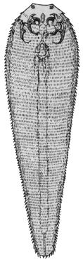
Fig. 372.—Larva of Linguatula rhinaria (Pentastoma denticulatum). Enlarged. (After Leuckart.)
Development.—The ova, which are found in masses in the nasal mucus, already possess an embryo; they are expelled with the nasal secretion, and are swallowed by herbivorous mammals with their food, mostly by hares and rabbits, but also by sheep, goats, oxen, horses, antelopes, fallow deer, pigs, cats, and occasionally also by human beings. The young larvæ hatch out in the stomach; they possess a thickened anterior body with rudimentary mouth parts and two pairs of limbs; the body gradually tapers to a short tail.
The larvæ of the Linguatulidæ bore through the intestinal wall and reach the liver, more rarely the mesenteric glands, etc.; they here become encysted and enter a sort of pupal stage in which they lose their limbs; after several moultings and gradual growth the second larval stage, having the appearance of the adult Linguatula, sets in. About five to six months after infection the creatures have become 4 to 6 mm. long, possess eighty to ninety rings, which have a series of fine points on their posterior border; the mouth and intestine are formed, the sexual organs mature and the two pairs of hooks are near the mouth. This larval stage (fig. 372) has been known for a long time, but it was regarded as an independent species of animal, and therefore had a separate name (Linguatula serrata, Fr.; Pentastoma denticalatum, Rud., etc.).
Later these Linguatula larvæ make an attempt to escape from their hosts, and this, of course, can only be effected by means of an active migration; they leave the cysts, and according to their respective positions in the abdominal or pleural cavities they reach the bronchi or the intestine, and finally pass out; they may be again sniffed up by dogs and settle in their nasal cavities. Still this outward migration does not appear to be necessary for further development. A portion of the larvæ gain access to the nasal cavities directly through the trachea, and thus herbivorous mammals certainly become directly infected. In most cases the infection of dogs, wolves and foxes, that is, of carnivorous mammals, takes place through consuming the bodies of mammals, or parts of them, such as the liver and lungs, which are affected with the second larval form; in any case most larvæ obtain access first to the stomach of their host, from here they make an active migration through the œsophagus to the oral and nasal cavities, in which they settle. It is possible also that the same larvæ which are free in the oral cavity when the food is being eaten migrate into the nasal cavities. After being stationary a fresh skin is formed and the spine-bearing cuticula are thrown off. The male attains its full size in the fourth, and the female in the sixth month. The duration of life is stated to be from fifteen months to several years.
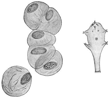
Fig. 373.—Linguatula rhinaria: on left, eggs in gelatinous covering, 110/1. On right, first larval stage. 300/1. (After M. Koch.)
L. rhinaria has been observed in man in the adult as well as in the larval condition (Pentastoma denticulatum). Zenker first called attention to the occurrence of the larva in man, having found it nine times in the liver in 168 autopsies. Heschl found it twice in Vienna in twenty autopsies, Virchow found it in Würzburg and Berlin, Wagner in Leipzig (10 per cent.), and Frerichs in Breslau five times in forty-seven autopsies. The parasite is much less frequent in Switzerland. According to Klebs, one case occurs in 900 autopsies, and according to Zaeslin two cases occurred in Basle to 1,914 autopsies. In the Seamen’s Hospital in Kronstadt P. denticulatum has been found six times in 659 autopsies. It was almost always the liver that contained one or a few specimens. The parasite was very rarely found in the kidney or spleen, or encysted in the intestinal wall. The adult L. rhinaria is far more rarely observed in man.
A case reported by Landon that related to a blacksmith of Elbing is particularly interesting. This man accompanied the campaign of 1870; he soon, however, fell ill with pains in the liver, accompanied by icterus and intestinal disorders. Soon after the war, and after the symptoms were reduced to icterus and weakness, bleeding of the nose set in and continued with slight intermissions for seven years; an unpleasant sensation of pressure in the left nasal cavity set in, with inflammatory swelling of the mucous membrane. At last, in the summer of 1878, when the pressure in the nose had considerably increased, a Linguatula was expelled from the nose with a violent attack of sneezing, and lived for three days longer in water. The bleeding of the nose then ceased and the patient soon recovered. There can be no doubt that the first illness was connected with the invasion in the liver of numerous larvæ of Pentastoma, and disappeared after their encystment; one or a few of these must subsequently have found its way to the nose and settled there.
Genus. Porocephalus.
Porocephalus constrictus, v. Siebold, 1852.
Syn.: Nematoideum hominis, Diesing, 1851; Pentastomum constrictum, v. Sieb., 1852; Porocephalus constrictus, Stiles, 1893.
Porocephalus is distinguished from Linguatula by its cylindrical body and by certain internal structures. Porocephalus constrictus is at present only known in its larval stage. It is milk white in colour with golden-yellow hooklets. Number of rings, twenty-three. Length 13 mm., breadth 2·2 mm. There are no prickles on the posterior border of the annulations of the body.
This species was first discovered by Pruner encysted in the livers of two negroes in Cairo. Bilharz reported two further cases in which the parasites were encysted in the liver and in the mucosa of the intestine; a few other observations have been made by Fenger, Aitken, Giard and Chalmas. Aitken’s report deals with soldiers of the British Colonies in Africa. The parasites were discovered in the liver as well as in the lung, and appear to have been the cause of death in one case (pneumonia, peritonitis).
Pruner has found the same parasite also in the liver of the giraffe.
It has recently been assumed that Porocephalus constrictus is the larva of Pentastoma moniliforme, Diesing, 1835, that attains a length of 70 mm. and lives in the lungs of African Pythonides. The larva is known to have been ejected from monkeys (Cercopithecus albogularis, Cynocephalus maimon), from the giraffe (Camelopardalis giraffa), from a species of hyæna (Proteles cristatus), and should be expected to occur frequently in smaller mammals which have been swallowed by African serpents of enormous size.
[The three species of Pentastomidæ, or tongue worms, found in man are Linguatula serrata, Frölich; Porocephalus armillatus, Wyman; and Pentastoma moniliformis, Diesing.
[(1) Linguatula serrata has been referred to under a great number of names.356 It is a frequent parasite in dogs, oxen and sheep; as an adult in the dog and also in the fox and wolf. The nymphal stage is found in rats, hares, rabbits, the horse, oxen, sheep, goats, pigs, camels, deer, the African and long-eared hedgehogs, porcupine, guinea-pig and peccary. In man it is found in both adult and nymphal stages. Sambon says the nymphal stage is of frequent occurrence, but is usually overlooked. Zenker, who first found it in man, obtained it in nine out of 160 post-mortems, usually encysted in the liver. It is then said to be harmless. Landon, in 1878, found the adult in man, the patient suffering from epistaxis for about seven years; in the end during a fit of sneezing the living parasite was ejected through the nostril. This case is of particular interest as it appears to suggest that this Acarid may now and then pass its entire development in the same host, or at any rate may actively migrate from the liver to the nasal cavities after a period of encystment in the liver or elsewhere, which has also been observed in herbivorous animals (vide also p. 526).
[It is recorded from man in Central America (Darling, Bull. Soc. Path. exot., 1912, v, p. 118; and again Arch. Int. Med., 1912, v, p. 401), also from Rio de Janeiro (Mem. Inst. Oswaldo Cruz, 1913, fasc. ii, p. 125) by Faria and Travassos.
[(2) Porocephalus armillatus, Wyman, is also known under a variety of names.357 This species is widely spread over tropical Africa. The adult stage is found in pythons and puff-adders, the nymphal in the chimpanzee, Sykes monkey, mandrill and other monkeys, the lion, leopard, banded ichneumon, Aard wolf, dog, black rat, South African reedbuck and the giraffe. The adult has never been found in man or any mammal. No fewer than sixteen cases of the nymphal form, Sambon tells us, have been found in man, and it is probably much more widespread than at present known. So far it has only been found in the African natives. This species has sixteen to seventeen body rings in the male, eighteen to twenty-two in the female, and the body does not taper as much as in the next species.
[(3) Pentastoma moniliformis, Diesing,358 is an Oriental species, found in India, Indo-China and South China, and the Malay Archipelago. The adult occurs in both the Indian and reticulated pythons. The nymphal stage has been found in monkeys, the tiger, the civet and the Indian otter.
[The nymph has twice been found in man; in one case in the liver of a Filipino, the other in the serous coat of the small intestine of a native of Sumatra.
[This species can be told by the female having twenty-nine to thirty-three body rings, the male twenty-six, and the annulations are more bead-like and less prominent than in the African species.
[In addition to these three, Sambon thinks it probable that others occur in man.—F. V. T.]
Other References to Pentastomidæ.
(1) “Die Wanderung des Pentastomum denticulatum beim Rinde,” Centralbl. f. Bakt. u. Parasitenk., Jan. 2, 1889, v (1), pp. 1–5, V. Bates.
(2) “Il Pentastoma moniliforme, Dies., nella pantera,” Med.-vet. Torino, 1877, 4 s., vi (12), pp. 529–532, R. Bassi.
(3) “On the Organization and Development of Linguatula (Pentastoma), accompanied with the description of a new species from the abdominal cavity of the mandrill,” Ann. and Mag. Nat. Hist., 1848, 2 s. ii (7), 2, pp. 69–70, v. Beneden.
(4) “De la Linguatula ferox (Pentastoma denticulatum aut serratum),” Bull. Acad. roy. d. Sci. d. Belg., 1855, xxii, pt. 1 (1), pp. 4–10, v. Beneden.
(5) “Note sur quelques pentastomes,” Bull. Acad. roy. d. Sci. de Belg., 1857, 26, 2 s., ii (5), pp. 29–30, v. Beneden.
(6) “Ueber das Pentastoma in de gekrösdrusen den Schafe,” Repert. d. Thierh. Stuttg., 1861, xxii, pp. 37–38, Collin.
(7) “Eine Linguatula aus der Mesenterialdrüse des Schafes und Dromedars als zweites ungesche. Stadium von Pent. taenioides,” Notiz. u. Tagsber. u. d. Geb. d. Nat. u. Heilk. Jena, 1862, v, pp. 127, 128, Colin.
B. INSECTA (Hexapoda).
Three separate regions can always be distinguished in the body of insects, namely, the head, thorax and abdomen. The HEAD is a roundish unsegmented capsule and possesses four pairs of appendages. The first pair are the various shaped feelers (antennæ), which are placed on the superior surface of the head next to the eyes; then more ventrally placed a pair of upper jaws (mandibles) without palpi and without articulations; they are powerful masticatory organs.359 The first pair of lower jaws (maxillæ) are jointed and bear a palpus (palpus maxillaris); the second pair of maxillæ are soldered together and form the lower lip (labium), and likewise carry a palpus labialis on each side. The upper lip (labrum), as well as the other parts (which, however, are only appendages), belong to the mouth, which is really formed of a number of closely united pieces. The mouth parts are modified according to the functions required of them. Coleoptera, Neuroptera, and Orthoptera have biting or masticatory mouth parts which conform with the scheme described above. In the licking mouth parts of the Hymenoptera the maxillæ and under lip are considerably elongated, while the mandibles retain their form and are used for triturating the food; in the Lepidoptera nearly all the mouth parts are shortened except the maxillæ, which form a long and sometimes spirally rolled suctorial proboscis; the Diptera and Rhynchota have piercing and sucking mouth parts. The mandibles and maxillæ are metamorphosed into needle-like structures, while the suctorial apparatus is formed by the labrum.
The thorax consists of three segments, which are frequently united; ventrally it carries three pairs of legs, which consist of a definite number of articulated pieces joined together. Their form also changes according to their function, so that legs for running, walking, digging, swimming, jumping, and preying are seen. A pair of wings are respectively attached to the last and last but one thoracic rings, and these may be traced back, not to metamorphosed appendages, but to tracheal branchia. They are composed of chitinous membranes supported by branched structures (veins or ribs). Their size and formation vary; they are seldom of equal size and form (Neuroptera); often the posterior wings are larger than the anterior wings, the former then only serving as protective coverings for the latter (Coleoptera), or the anterior wings are larger (Lepidoptera), or the posterior wings are shortened or are entirely absent (Diptera); and finally there are insects in which both pairs of wings are lacking.360
The abdomen retains its segmentation, but, with the exception of a few groups related to the primitive forms of insects, has no appendages in the imago condition; the abdomen usually consists of ten segments, on the last of which the anus is situated.
We need only observe the following characters in considering the anatomy of insects:—
The EPIDERMIS consists of the chitinous cuticle, which is separate from the cellular layer beneath (hypodermis); the various appendages are supported by the chitinous layer.
The INTESTINAL CANAL usually consists of the anterior, median and terminal intestine, and as a rule passes straight through the body; salivary glands discharge into the anterior part, and, in some cases, yield a stiffening secretion which serves for spinning webs; numerous or scanty hepatic tubes are appended to the median intestine, while on the border between the median and terminal intestine open four to six long tubes (vasa malpighiana), which act as urinary organs. Finally the end portion of the intestine carries various glands (anal and rectal glands, etc.).
The CENTRAL NERVOUS SYSTEM agrees in structure with that of the Annelids, but is more highly developed. The pharyngeal ring surrounds the front part of the intestine; the sensory nerves originate from its SUPERIOR PHARYNGEAL GANGLIA and are the seat of the higher psychical functions; the INFERIOR PHARYNGEAL GANGLIA govern the mouth parts, and in addition appear to regulate the movements (cerebellum) of the vertebrates.
The chain of GANGLIA lying on the ventral side of the abdomen consists primitively of pairs of ganglia corresponding with the twelve segments, which are connected by longitudinal and transverse commissures. But many changes in the ganglia may be seen in insects caused by partial or entire amalgamation of single ganglia, so that in a few cases only one abdominal ganglion is present. In conclusion, a definite INTESTINAL NERVOUS SYSTEM is always present.
Of the organs of sense the FACETTED EYES, situated at the sides of the head, deserve special mention, as do also the ORGANS OF TOUCH and SMELL, situated on the antennæ, and the ORGANS OF HEARING and taste, or finer sensations, situated at the mouth and in the buccal cavity.
The sounds emitted by insects are, as a rule, produced by the friction or beating of certain chitinous parts, but sounds are also produced in breathing (flies).
The ORGANS OF RESPIRATION, the so-called tracheæ, are highly developed; there are openings (stigmata) at the sides of the body which draw in air by means of the active participation of the muscles of the body. The number of stigmata varies between two and ten pairs; the tracheæ themselves branch off from the trunks in the most varied manner, and carry air to the internal organs.
The colourless BLOOD circulates between the tissues and organs, and is kept circulating by the contraction of a chambered dorsal vessel provided with ostia, and which terminates in a short aorta opening at the anterior end.
Insects are SEXUALLY DISTINCT; their sexual glands are in pairs and have a tubular structure, but the testicular tubules are united together by a capsule into an oval testicle; exceptionally, also, the excretory canals are double, as also the sexual orifices; usually the paired canals unite into a single oviduct or spermatic duct which terminates at the posterior end of the body after receiving the products of various glands.
As to the HISTORY OF THE DEVELOPMENT of insects, all that is necessary to mention here is that the young hatched from eggs only exceptionally (as in Apterygota) resemble the adult parent (insecta ametabola); as a rule they differ from them not only in the shape of the body, but also more or less by their manner of life, and only attain the form of the parent through METAMORPHOSIS. This is a gradual process (insecta hemimetabola) in the Rhynchota and Orthoptera, or a sudden one with a stage of inanition (insecta metabola) in the other orders. This stage of rest or inanition, the PUPA, concludes the larval life (caterpillar, maggot, etc.); during the pupal stage no nourishment at all is taken, but the internal organs undergo changes; in some forms the rest is not absolute, as voluntary local movements may take place (pupæ of gnats).
The insects are divided into numerous orders according to the form of the mouth parts, the structure of the wings, as well as the manner of the development; with the exception of the lowest group (Apterygota), which is most nearly related to the ancestors of the insects, and which has no wings and undergoes no metamorphosis, all the remaining orders, which are termed Pterygota, have wings on the thorax, though there are, of course, a few species and families of this group which have lost their wings.
The Pterygota include—
(1) Orthoptera.—Biting mouth parts, anterior wings leathery, posterior wings thin, folded longitudinally; metamorphosis incomplete (grasshoppers, crickets, cockroaches).
(2) Pseudoneuroptera.—Biting mouth parts, wings of equal size, thin, not folded up (dragon-flies, hair and feather lice, termites).
(3) Rhynchota or Hemiptera.—Mouth parts formed for puncturing and sucking; wings alike, or the anterior wings may be thickened, parchment-like at their base (plant lice, cicadæ, bugs and true lice).
(4) Neuroptera.—Biting mouth parts; wings alike, thin; metamorphosis complete (ant-lions, lace-wing flies, etc.).
(5) Trichoptera.—Licking mouth parts; anterior wings narrow, posterior wings longitudinally folded, both ornamented with little hairs; the larvæ are worm-like in form, live in water, and breathe through tracheal gills (may flies, etc.).
(6) Lepidoptera.—Suctorial mouth parts; wings covered with scales (butterflies).
(7) Coleoptera.—Biting mouth parts; anterior wings thickened and differ in colour, appearance and function from the thin, folded posterior wings (beetles).
(8) Hymenoptera.—Mouth parts for licking and biting; the wings alike, membranous (ichneumon flies, ants, wasps, bees, humble bees).
(9) Diptera.—Mouth parts formed for puncturing, sucking or licking; posterior wings degenerated (gnats, flies, gadflies, fleas).
(10) Strepsiptera.—Anterior wings shortened; the female without wings and living parasitically (fan-wings).
The parasites of man occur amongst the Rhynchota, Coleoptera, and amongst the Diptera.
[The most usual and recent classification of the Hexapoda is the following:—
(1) Aptera.—Wingless insects; scarcely any metamorphosis.
(2) Neuroptera.—Four membranous wings, frequently with much network; the front pair not much, if at all, harder than the under pair; the latter with but little or no fan-like action in closing; mandibulate; metamorphosis variable, but rarely complete.
(3) Orthoptera.—Four wings; front pair coriaceous or leather-like, usually smaller than the other pair, which are of more delicate texture and contract in repose like a fan; mandibulate; metamorphosis complete.
(4) Thysanoptera.—Four very narrow fringed wings; mouth imperfectly suctorial; metamorphosis slight.
(5) Hemiptera.—Four wings; the front pair either all transparent or with the basal half leathery; mouth suctorial; metamorphosis slight.
(6) Diptera.—Two membranous wings only; mouth suctorial, very varied; metamorphosis complete.
(7) Lepidoptera.—Four large wings covered with scales; mouth suctorial, metamorphosis great.
(8) Hymenoptera.—Four membranous wings; front pair larger than hind, which do not fold up in repose; mandibulate, sometimes with a tubular proboscis; metamorphosis complete.
(9) Coleoptera.—Four wings, the front pair hard and horny (elytra), meeting in a line over the back and covering the delicate hind pair; mandibulate; metamorphosis complete.
[There are two other well-known arrangements, namely, Packard’s and Brauer’s, of recent date, but the one given here, which is based on Linnaeus’ grouping by Dr. Sharp, is by far the simplest.—F. V. T.]
Order. Rhyncota.361
The lower lip forms a long thin tube that can be turned back (rostrum), and within which lie the setaceous mandibles and maxillæ; the first thoracic segment is not united with the two posterior ones; the anterior wings are usually leathery as far as the centre.
(a) Rhyncota aptera parasitica.
Family. Pediculidæ (Lice).
The lower lip is transformed into a projecting rostrum provided with barbed hooklets in which the hollow extensile sucker (maxillæ and mandibles) is situated; no wings; no metamorphosis; only simple eyes; the antennæ are five-jointed, the feet possess hook-like terminal structures; the barrel-shaped eggs (nits) are deposited on the hair of the host.
[The lice or Pediculidæ are also known as Anoplura and Siphunculata.
[They have been split up into a number of families and sub-families and a number of genera, but as far as this work is concerned it is best to retain the single family Pediculidæ.
[Only the three species mentioned here are common parasites of man, but now and then horse and cattle and sheep lice, Hæmatopinus, may cause transitory annoyance.—F. V. T.]
Genus. Pediculus, Linnæus.
Pediculus capitis, de Geer, 1778.

Fig. 374.—Mouth parts of Pediculus vestimenti. Enlarged. (After Denny.)
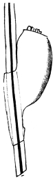
Fig. 375.—Ovum of the head louse. 70/1.
Male 1 to 1·5 mm. in length, female 1·8 to 2·0 mm. in length. The colour varies from light grey to black according to the colour of the hair of the human race upon which they are parasitic. The abdomen has eight segments, of which the six central ones are each provided with a pair of stigmata. The thorax is as broad as the abdomen. Eggs 0·6 mm. in length; about fifty are deposited by a female head louse. The young can propagate when eighteen days old.
The head louse lives especially in the hairy parts of the head of human beings; more rarely it is found on other hairy parts of the body. It is spread over the entire surface of the globe, and was present in America before the arrival of Europeans. Quite exceptionally it is said that it bores itself deep into the epidermis and can live in ulcers.
[The eggs are pear-shaped and are attached to the hairs near the roots by means of a clasping collar. They hatch in about seven days. The young are like the adults and mature in a month. Its general colour varies with that of its host. In West Africans nearly black, in Hindoos dark and smoky, on Chinese and Japanese yellow, on Hottentots orange, on South American Indians dark brown (Murray).—F. V. T.]
Pediculus vestimenti, Nitzsch, 1818.
The head in front is somewhat rounded. Antennæ longer than in the head louse; 2 to 3·5 to 4 mm. in length; whitish-grey; the abdomen is broader than the thorax; stigmata as in P. capitis. Eggs 0·7 to 0·9 mm. in length; about seventy are deposited.
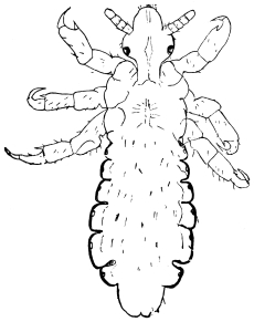
Fig. 376.—Head louse, male. 15/1.
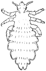
Fig. 377.—Pediculus vestimenti, Burm.: adult female. 15/1.
P. vestimenti lives on the neck, throat and trunk of persons, and the clothing next the body, in which also the eggs are deposited. The louse of so-called pedicular disease (P. tabescentium) is, according to Landois’ researches, only the usual P. vestimenti; moreover, many cases of phthiriasis are attributable to mites or fly maggots.
[This parasite has often been a great pest amongst soldiers during long campaigns, especially amongst the Russians during the Crimean War. Vide also notes in Addenda (p. 615) under “Body, Head and Clothes Lice.”—F. V. T.]
Genus. Phthirius, Leach.
Phthirius inguinalis, Redi, 1668.
Syn.: Pediculus pubis, L.
Male 0·8 to 1·0 mm. in length; female 1·12 mm. in length; colour greyish-white; form subquadrate; the two posterior pairs of legs are strong; the abdomen has nine segments and six pairs of stigmata; and still another pair of stigmata is situated between the two anterior limbs. Eggs pear-shaped, 0·8 to 0·9 mm. in length, 0·4 to 0·5 mm. in breadth, and are deposited in rows of about ten on the hairs.
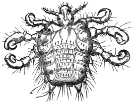
Fig. 378.—Phthirius inguinalis, Leach: they are distinguished by the larger tracheal trunks originating from the stigmata. Enlarged.
Pediculus pubis, which is found almost exclusively in the Caucasian race, lives on hairy parts of the body, but hardly ever on the skin of the head; the pubic region is its favourite place of abode.
[This species reproduces more rapidly than other lice, and is communicated much more freely. The eggs are often laid singly attached to the hairs near their apex. It is known as the “crab louse.”—F. V. T.]
(b) Rhyncota hemiptera.
Family. Acanthiadæ.
Body flattened, antennæ four-jointed, rostrum three-jointed, wings atrophied.
[This family, the Cimicidæ, includes the bed bugs; the proboscis, which lies in a groove, is of three segments; the front wings are shown by two small elytra, there is no trace of hind wings. Two species are known commonly to attack man.—F. V. T.]
Genus. Cimex, Linnæus.
Cimex lectularius, Linnæus.
Syn.: Acanthia lectularia, Fabricius, 1794.
It measures 4 to 5 mm. in length, 3 mm. in breadth; brownish-red; eight abdominal segments. The female deposits fifty whitish eggs at a time (1·12 mm. in length) three or four times a year; the entire development up to complete maturity takes about eleven months. [They will breed all the year round, but less so in cold weather.—F. V. T.]
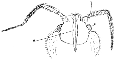
Fig. 379.—Head of the bed bug from the ventral surface. a, the rostrum; b, the antenna; and c, the eye. 70/1.
The bed bugs live in the cracks and fissures of human habitations, under carpets, behind pictures, in furniture, bedsteads, etc.; hidden during the day, they attack persons at night to suck their blood. The alkaline secretion of the salivary glands dropped into the wound around the separate bites causes the so-called “wheals.”
The bed bugs were known in bygone days by the Greeks (κάριο) and the Romans (cimex). They were first mentioned from Strasburg in the eleventh century, and in England about 1500.
[This is the common bed bug of northern latitudes and must not be confused with the tropical bed bug (C. rotundatus). The bed bug can migrate from one house to another; this especially takes place when a house is uninhabited. They escape from windows and pass along walls, water-pipes and gutters, and so reach adjoining houses. This noxious pest accompanies man wherever he goes; ships and trains become infested, especially the former.
[A characteristic feature in this animal is the peculiar odour it produces, like many others in the same group of insects. This odour comes from a clear, oily volatile liquid secreted by glands in various parts of the body. Although the normal food is man’s blood, the bed bug can subsist upon moist wood, dust and dirt that collects in crevices in floors, walls, furniture, etc. The puncturing mouth consists of a fleshy under lip, within which lie four thread-like hard filaments which pierce the flesh, the blood being drawn up through the beak.
[The eggs are oval, white, with a projecting rim around one end, with a lid which is pushed off when the young hatch; they are laid in cracks and crevices in batches of from twelve to fifty. The egg stage lasts from seven to ten days. The larval stage so gradually passes into the adult that one scarcely notices the change; during its growth the skin is cast five times, and at the change the little wing-pads are seen, showing that the adult stage is reached. The young larva is at first pale yellowish-white. It resembles the parent, but has no trace of elytra. Although eleven weeks is said to be necessary for their development, the stages may be gone through much more rapidly; Howard and Marlatt362 give seven weeks in some instances. It seems pretty certain that these Cimex only take one meal of blood between each moult and another preceding egg laying.—F. V. T.]
Cimex rotundatus, Signoret, 1852.
[This bug is common in warm climates; it is an abundant insect in India, and King has found it in the Sudan, where C. lectularius is, however, the common species. It is usually known as the tropical bed bug. Signoret’s bug can be told from the other common species by the shape of the pronotum. In C. rotundatus it is uniformly convex, whilst in C. lectularius the lateral edges are flat and sometimes even concave. The abdomen of rotundatus is also rather more elongate.
[This species is of considerable importance, as according to Patton it may act partly as the intermediary host of the piroplasma of kala-azar.
[Wenyon found at Bagdad that Cimex sp. would take up Leishmania from Oriental sore, and that the parasite developed into flagellate form. Patton came to the conclusion that the bed bug transmitted Oriental sore in Cambay, India, but Wenyon contests this view (vide Journ. Lond. School Trop. Med., 1912, ii, pt. 1, pp. 13–26). Franchini (Bull. Soc. Path, exot., 1912, v, No. 10, pp. 817–819) was unable to connect Cimex with this disease. At present nothing seems proved. Besides their possible connection with kala-azar, it has been shown by Howard and Clark (Journ. Exp. Med., 1912, xvi, No. 6, pp. 850–859) that they can carry the virus of poliomyelitis.
[This bed bug was originally described from the Island of Réunion in 1852 by Signoret. A similar insect was described from Burma by Fieber, in 1861, as C. macrocephalus. This is the same as Signoret’s species.
[The distribution given by Patton363 is as follows: India, Burma, Assam, Malay, Aden, Islands of Mauritius and Réunion. Patton in this paper refers to an erroneous statement made in a recent edition of this book (the last English edition). As I have personally kept lectularius in moist dirt, wood and refuse for over two years, the statement as far as I am concerned is not erroneous. Moreover, since his doubting this fact the same experiment has been twice repeated with the same results. What they did and do persist on I cannot say.—F. V. T.] whilst collecting them. It is rounder and has shorter antennæ than
Cimex columbarius, Jenyns.
[This is common in parts of Europe in pigeon nests, and also amongst poultry (vide Report Econ. Zool. for year ending September 30, 1913, pp. 142–144, Theobald). It occurs in Britain on the latter and will attack man. I have personally been badly bitten C. lectularius. Jenyns also described a more pubescent species from swallows as C. hirundinis. I have recently received an account of the swallow bug invading a house in Kent and causing much annoyance.—F. V. T.]
Cimex ciliatus, Eversmann, 1841.
3·3 mm. in length, yellowish-red, thickly covered with hair; indigenous in Russia (Kasan).
[From a single specimen seen it is evidently distinct.—F. V. T.]
Family. Reduviidæ.
Head long, narrowed behind into a neck; eyes large, prominent; rostrum thick and curved; antennæ moderately long, slender at the tip; legs long and stiff; carnivorous.
Amongst the Reduviidæ one genus is of particular importance, namely the genus Conorhinus, which has a long head and the first segment of the beak very much shorter than the second, and the posterior tibiæ longer than the femora.
These large bugs have a wide distribution, the Oriental region, North and South America, and the West Indies, Madagascar and West and Central Africa.
These large bugs may cause very nasty wounds by their bites, but beyond that it has recently been shown that one interposes in the life-cycle of a trypanosome, namely—
Genus. Conorhinus, Lap.
Conorhinus megistus, Burm.
This large bug has recently been shown by Chagas to be the agent in the development of the trypanosome (T. cruzii) which is the cause of the well-known disease in many parts of Brazil called Barbeiro (Barbier). This insect is about 1 in. long, black, with four red spots on the pronotum, and six red lateral lines on the abdomen, black legs, head and beak. The insect is figured in a coloured plate (No. 9) in Mem. Inst. Oswaldo Cruz, 1909, i, fasc. 2, pp. 158–218.
A further account is given by Neiva.364
Conorhinus sanguisuga, Lec. (Blood-sucking Cone-nose).
This bug is also known as the Texas or Mexican bed bug, also as the big bed bug. It is particularly troublesome in the Mississippi Valley in bedrooms. The bite is very severe and results in more pronounced swelling and inflammation than that of the Cimex. Normally this genus feeds upon the blood of mammals and insects. Its fondness for human blood appears to be quite a new habit, and appears limited to the mature insect only. It is nearly an inch long, flat, head very narrow and long, the rostrum short and thick. In colour it is dark brown with pink markings. They are fully winged when adult, and they fly with ease, entering houses on the wing, especially being attracted by lights in windows; they also run swiftly. Like the bed bug they conceal themselves during the day and come out at night and bite the sleeper. The effect of the bite is very varied, but as a rule a sore, itching wound, accompanied by burning pain and swellings, which may extend over a good deal of the body, occur. A specific poison is undoubtedly injected into the puncture; but no doubt serious results are also due to the beak being contaminated through the insects feeding upon foul carrion. Mr. Lembert, when bitten by a Conorhinus sp. (?) on the Pacific slope, exhibited the following symptoms: an itching sensation extending up the leg, large blotches manifesting themselves on the upper part of the limb and extending up to the hands and arms; his lips swelled and the itching and swelling extended over the head; there was also much nausea. Similar results are recorded from other regions.365
The eggs of the C. sanguisuga are at first white, then become yellow, then pink; the young hatch in twenty days. There are two larval and two pupal stages, the latter showing wing-pads. The eggs are laid and the young feed out of doors, chiefly upon insects. It is particularly abundant in April and May indoors.
Conorhinus, sp. novum (Monster Bug).
Another species; acts in a very similar way in California, the bite being very poisonous.
Conorhinus rubrofasciatus, de Geer366 (Malay Bug).
This large bug attacks man in Malaysia and elsewhere. It is recorded as inflicting “a very nasty sting, which is done by the huge proboscis.” Acute pain and inflammation follow in a few minutes. In one case the whole leg became swollen. This species occurs over the whole Oriental region, in Madagascar and Sierra Leone. It is dark brown in colour with dusky yellow or brick-red markings on the pronotum and elytra. Donovan suggests that it may be connected with the kala-azar piroplasma.