ON PARASITES IN GENERAL. (continued 4)
Reading edition
S. phagedenis was found by Noguchi in a ten days old ulcerated swelling of the labium. The organism shows much variation in size, being 4 µ to 30 µ in length.
S. refringens (Schaudinn, 1905) occurs in association with Treponema pallidum in syphilitic lesions, but is non-pathogenic. It is 20 µ to 35 µ long and 0·5 µ to 0·75 µ broad, being larger than T. pallidum and more easily stained.
Various spirochætes have also been notified in vomits, chiefly in Australia; others from the human intestinal tract, e.g., S. eurygyrata; S. stenogyrata (Werner); S. hachaizæ (Kowalski), in cholera motions; S. buccalis (Cohn, 1875) and S. dentium occurring in the human mouth and in carious teeth (S. dentium, Koch, 1877, being the smaller); S. acuminata and S. obtusa found by Castellani in open sores in cases of yaws.
Animal spirochætes of economic importance include:—
S. anserina, highly pathogenic to geese.
S. gallinarum (= S. marchouxi) in fowls. (See p. 119.)
S. theileri in cattle and S. ovina in sheep also occur in Africa; their pathogenicity is not clear.
S. laverani (= S. muris), occurring in the blood of and pathogenic to mice, is probably the smallest spirochæte from the blood, being only 3 µ to 6 µ long.
Numerous spirochætes have been recorded from the guts of various mammals, birds, fishes, amphibia and insects.
Cultivation of Spirochætes.—Cultures of spirochætes have been made with little success or with great difficulty until comparatively recently, when Noguchi (1912) devised a means whereby he has cultivated most of the pathogenic spirochætes as well as some Treponemata.
Noguchi has now cultivated S. duttoni, S. recurrentis, S. rossii, S. novyi and S. gallinarum from the blood; S. phagedenis157 from human phagedænic lesions; S. refringens158 and spirochætes from the teeth.
His method is as follows:—
A piece of fresh, sterile tissue, usually rabbit kidney, is placed in a sterile test-tube. A few drops of citrated blood from the heart of an infected animal, e.g., rat or mouse, is added, and about 15 c.c. of sterile ascitic or hydrocœle fluid is poured quickly into the tube. Some of the tubes are covered with a layer of sterile paraffin oil, others are left uncovered. The tubes are incubated at 37° C. The best results are obtained if the blood is taken from an animal forty-eight to seventy-two hours after it has been inoculated, that is, before the spirochætes reach their maximum multiplicative period in the blood. The presence of some oxygen seems indispensable for these blood spirochætes, and they fail to develop in vacuo or in an atmosphere of hydrogen.
For subcultures, 0·5 c.c. of a culture is added to the medium instead of citrated blood, and it is useful to add a little fresh, normal blood, either human or from an animal, such as a rat.
Noguchi found that the events in cultures were:—
S. duttoni,159 maximum multiplication on the eighth to ninth day; disintegration beginning on the tenth day, spirochætes disappeared after about the fifteenth day. No diminution of virulence was found at the ninth day.
S. rossii (= S. kochi).160 Maximum development on the ninth day, after which the virulence diminishes. The incubation period is also prolonged.
S. recurrentis161 (= S. obermeieri). Maximum growth on the seventh day.
S. novyi.162—Maximum development on the seventh day. It is more difficult to grow than the preceding forms.
All the above spirochætes showed undoubted longitudinal division and transverse division was observed in part.
S. gallinarum163 can be cultivated as above, but transverse division was usual here. Maximum growth occurred in the culture about the fifth day.
Treponemata.
The genus Treponema (Schaudinn, 1905), includes minute, thread-like organisms, with spirally coiled bodies, the spirals being preformed or fixed. No membrane or crista is present, according to Schaudinn, though a slight one is said by Blanchard to be present in the organism of yaws. The ends of the organisms are tapering and pointed. Multiplication is by longitudinal and transverse division. The most important members of the genus are T. pallidum, the agent of syphilis, and T. pertenue, which is responsible for frambœsia or yaws.
Treponema pallidum, Schaudinn, 1905.
Syn.: Spirochæta pallida.
Treponema pallidum was first described by Schaudinn and Hoffmann in 1905 under the name of Spirochæta pallida. It has also been described under the names of Spironema pallida, Microspironema pallida and Trypanosoma luis. Siegel in 1905 described an organism which he called Cytorhyctes luis and considered to be the agent of syphilis. Schaudinn reinvestigated Siegel’s work and found T. pallidum, which he considered to be the causal agent of the disease, and pronounced against Cytorhyctes luis. It is probable now that both workers were correct, for Balfour (1911) has seen the emission of minute granules or “coccoid” bodies from T. pallidum and these granules probably correspond to the C. luis of Siegel. Recently E. H. Ross, having observed a spirochæte stage in the development of Kurloff bodies, thinks that T. pallidum is a stage in the life-history of a Lymphocytozoon. MacDonagh has also described a complicated and somewhat similar cycle, but these observations require further study and confirmation.
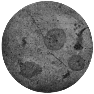
Fig. 56.—Treponema pallidum. (After Bell, from Castellani and Chalmers.)
T. pallidum varies from 4 µ to 10 µ in length, its average length being 7 µ, while its width is usually about 0·25 µ. Longer individuals of 16 µ to 20 µ have been recorded. The body has from eight to ten spiral turns and forms a tapering process at each end (fig. 56). The organism is most difficult to stain, and its internal structure is little known. It is possibly like that of Spirochæta duttoni or S. balbianii, as the “granule shedding” observed by Balfour is strongly suggestive of the formation of resistant bodies by those spirochætes. Hoffmann (1912) has seen the formation of spores in T. pallidum.
The Treponemata occur in the primary and secondary sores, but are difficult to find in the tertiary eruptions of syphilis. Noguchi and Moore (1913) and Mott164 (1913) have demonstrated T. pallidum in the brain in cases of general paralysis of the insane. Marie and Levaditi (1914), however, consider that the treponeme found in the brain in such cases is different from T. pallidum.
Cultivation of T. pallidum.—This has been accomplished successfully by Noguchi,165 using a modification of his method for spirochæte cultivation, for T. pallidum is much more difficult to grow than spirochætes, being a strict anaerobe.
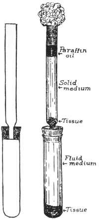
Fig. 57.—Diagram of apparatus for cultivation of Treponema pallidum by Noguchi’s method. (After Noguchi.)
The apparatus consists of two glass tubes, the upper being connected to the lower by a narrower tube passing through a rubber cork (fig. 57). Both tubes are carefully sterilized.
A piece of fresh, sterile rabbit’s kidney is placed in the lower tube, which is filled with ascitic fluid, or ascitic fluid and bouillon mixture. The tube is inoculated with syphilitic material and corked by inserting the upper tube. In the bottom of the upper tube a piece of sterile rabbit’s kidney is placed and syphilitic material poured over it. A mixture of one part ascitic fluid and two parts of slightly alkaline agar is then poured over the tissue and allowed to solidify. When solid, a layer of sterile paraffin oil is poured on top of it, and the top plugged with cotton wool (fig. 57). The whole is then incubated at 37° C. for two or three weeks. The tissue removes traces of oxygen from the lower levels of the medium and also probably provides a special form of nourishment. At first T. pallidum grows in the solid medium, and then when the cultural conditions in the lower fluid portion become favourable, the organisms migrate thither and multiply abundantly. At first the culture is impure, but after several transferences a pure culture is obtained readily.
The syphilitic material for culture is prepared by cutting off pieces of tissue from the lesions, washing in sterile salt solution containing 1 per cent. sodium citrate, and then emulsifying the tissue in a mortar with sodium citrate.
Good cultures show rapid multiplication, which is invariably by longitudinal division.
In his various cultivation experiments Noguchi166 found morphological and pathogenic variations in T. pallidum. Three forms of the organism were found, namely, thicker, average and thinner types. The lesions caused in the testicle of the rabbit differ according to the variety inoculated, but more work is necessary on the subject.
Noguchi167 has cultivated a separate organism, T. calligyrum, from the surface of human genital or anal lesions, either syphilitic or non-syphilitic. It is apparently non-pathogenic, and is 6 µ to 14 µ long.
Hata (1913)168 has modified the Noguchi technique for the cultivation of spirochætes and treponemes, with a view to simplification and convenience. Hata substitutes normal horse serum for ascitic fluid and the “buffy coat” of the clot of horse blood in place of the small pieces of rabbit’s kidney. It is unnecessary to place sterile paraffin on the surface of the medium.
The horse serum is mixed with twice its volume of physiological saline solution. The mixture is placed in tubes which are heated on a water-bath at 58° C., the temperature being raised gradually until it reaches 70° or 71° C. in three hours. The tubes are then heated at 71° C. for half an hour. After cooling, the contents will consist of an opaque semi-coagulated mass. This semi-coagulated serum and saline mixture may be substituted for Noguchi’s ascitic fluid.
The buff coagulum is cut into small pieces, about 1 c.c. in volume. They must be forced with a sterile glass rod to the bottom of the semi-coagulated serum and saline mixture. The medium is inoculated with a small quantity of infected blood and kept at 37° C. In the case of S. recurrentis, growth of spirochætes is observed on the second day, reaching a maximum in five to seven days. The growth of the organisms proceeds rather more slowly, they live for a longer period and maintain their virulence better than in Noguchi’s medium.
Treponema pertenue, Castellani, 1905.
Syn.: Spirochæta pertenuis; S. pallidula, Castellani, 1905.
Castellani discovered the organism in 1905, in scrapings of yaws pustules. He first described it under the name of Spirochæta pertenuis.
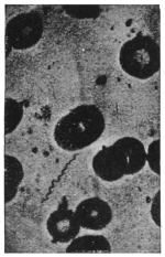
Fig. 58.—Treponema pertenue. (After Castellani and Chalmers.)
Treponema pertenue (fig. 58), though delicate and slender, shows great morphological variation both in length and thickness. It may be short, e.g., 7 µ, but can attain 18 µ to 20 µ in length and may be even larger. In cultures made by Noguchi, thick, medium and thin forms were found, each giving rise to a different type of frambœsial lesion when inoculated into the testicles of rabbits, thus suggesting the possibility of the occurrence of varieties of T. pertenue.
The organism is difficult to stain, but occasionally deeper staining granules are found along its body. They may represent a diffuse nucleus. Granule formation similar to that of T. pallidum has been observed by Ranken, using dark-ground illumination.
Many experiments have been made with a view to establishing the identity of the organism of yaws and also of differentiating between the causative agents of yaws and syphilis. Both monkeys and the human subject have been experimentally inoculated with yaws material and have developed the disease.
In an early experiment, negroes were inoculated with the secretion from lesions of yaws. All of them developed the disease, nodules appearing, chiefly at the seat of inoculation, in from twelve to twenty days, followed by the usual eruption. Similar results were obtained with thirty-two Chinese prisoners, who were inoculated with yaws, twenty-eight becoming infected.
A naturally infected yaws patient when inoculated with syphilis, contracted that infection, thus showing that yaws does not confer immunity to syphilis. This has also been observed naturally, when yaws patients have contracted syphilis.
Experiments with monkeys have been successfully performed. The incubation period varies from sixteen to ninety-two days. Lesions appear first at the seat of inoculation, and in some monkeys the eruption is localized to this spot, though the infection is general, T. pertenue occurring in the spleen, lymphatics, etc. Monkeys inoculated with splenic blood of a yaws patient, and also sometimes with blood from the general circulation, have become infected.
Castellani and others have shown that monkeys successfully inoculated with syphilis do not become immune to yaws, and vice-versâ.
Craig and Ashburn, using the monkey Cynomolgus philippinensis, found these animals susceptible to yaws but not to syphilis.
The ulcerated lesions of frambœsia are rapidly invaded by numerous bacteria as well as by different spirochætes, of which Castellani has described three distinct species. One is identical with Spirochæta refringens, Schaudinn, the other two are thin and delicate. One, S. obtusa, has blunt ends; the other S. acuminata, has pointed ends. T. pertenue is also present.
The reasons for considering T. pertenue to be the specific cause of frambœsia are:—
(1) T. pertenue is the only organism present in non-ulcerated papules, in the spleen and in the lymphatics of yaws patients, or of monkeys artificially infected with the disease. By no method has any other organism been obtained.
(2) Extract of frambœsia material, free from all organisms other than T. pertenue, reproduces the disease if inoculated.
(3) Extract of frambœsia material deprived by filtration of T. pertenue is no longer infective on inoculation.
The method of infection is contaminative, by direct contact. Women in Ceylon are frequently infected by their children. Any slight skin abrasion is sufficient to admit the parasite. In some cases, insects may carry the disease from person to person, and even in hospitals, when dressings are removed, it has been noticed that flies greedily suck the secretion from the ulcers. T. pertenue has been recovered from flies that have fed on yaws, and monkeys have contracted the disease when flies were placed and retained on them for a short time, after the insects had fed on yaws material.
Cultivation.—T. pertenue has been cultivated by Noguchi, who finds three types of parasites in his cultures, as before mentioned. Its multiplication is by longitudinal division.
Noguchi169 (1912), has cultivated species of Treponema from the human mouth, e.g., T. macrodentium, T. microdentium and T. mucosum, the latter from pyorrhea alveolaris. These parasites in the past may have been confused under the name Spirochæta dentium.
Class III. SPOROZOA, Leuckart, 1879.
The third group of the Protozoa consists entirely of parasitic organisms forming the class known as the Sporozoa or spore-producing animals. The members of this class are characterized by possessing very great powers of multiplication, coupled with a capacity for producing forms that serve for the transference of the organisms to other hosts. These reproductive bodies, whether for increase of numbers within one host or for transmission to another host, are called spores. But, strictly, the term spore should be used only in the latter connection, when a protective or resistant coat known as a sporocyst envelops the body of the spore.
The Sporozoa are widely distributed, occurring in various tissues and organs of Annelids, Molluscs, Arthropods, and Vertebrates. Their food, which is fluid, is absorbed osmotically. The life-cycle of a Sporozoön may be completed within one host or may be distributed between two different hosts.
The Sporozoa were divided by Schaudinn into two groups or sub-classes, called (1) the Telosporidia, and (2) the Neosporidia.
The Telosporidia are Sporozoa in which the reproductive phase of the parasites is distinct from the growing or trophic phase, and follows after it. The Neosporidia include Sporozoa in which growth and spore-formation go on simultaneously. This classification is not final, for certain exceptions and difficulties are already known with regard to it. It is possible that the class Sporozoa is not a natural entity, but should be replaced by two classes of equal rank, corresponding in most respects with the Telosporidia and Neosporidia.
The Telosporidia comprise the Gregarinida, the Coccidiidea, and the Hæmosporidia. Doflein combines the two latter orders into one known as the Coccidiomorpha.
The Neosporidia comprise the Myxosporidia, the Microsporidia, the Actinomyxidia, the Sarcosporidia, and the Haplosporidia. Doflein combines the first three orders into one, the Cnidosporidia.
Sub-Class. TELOSPORIDIA, Schaudinn.
Sporozoa in which the reproductive phases follow completion of growth.
Order. Gregarinida, Aimé Schneider emend. Doflein.
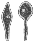
Fig. 59.—Monocystis agilis from seminal vesicles of Lumbricus × 250. (After Stein.)
Knowledge of the Gregarinida probably goes back as far as the year 1684, when Redi observed gregarines in the crab, Cancer pagurus. Von Cavolini (1787) found them in Cancer depressus. The name Gregarina was created by L. Dufour (1828), who observed masses of these organisms in the gut of insects of different orders. Hammerschmidt (1838) and von Siebold found rich infestations in insects, while Dujardin (1835) and Henle described various genera from segmented worms. Henle (1835) also observed cysts containing “navicellæ” in the sperm-sacs of segmented worms, and attention was drawn to his researches by the discovery by von Siebold (1839) of “pseudonavicellæ” in the gut of Sciara nitidicollis. Up to this time many workers considered the gregarines to be worms, but Kölliker (1845) investigated many of them and maintained their unicellular nature, while Stein’s work (1848) showed the interrelation of the pseudonavicellæ and the gregarines. The discovery of amœboid germs in the pseudonavicellæ by Lieberkühn (1855) and the demonstration of myonemes further aided in the elucidation of their true systematic position. The entire process of conjugation, of which Dufour had seen one phase, was followed by Giard under the microscope.
From 1873 onwards Aimé Schneider made important additions to the knowledge of the morphology, life-history, and systematic position of numerous gregarines. Bütschli (1881) and L. Léger (1892) also contributed much work on the subject. The discoveries of Schaudinn with regard to the life-cycle of Coccidia gave a fresh stimulus to the study of the Gregarines, whereby the life-cycles of numerous forms and the phases thereof have been elucidated.
Asexual multiplication is not common among the Gregarines, but is known to occur in the sub-order Schizogregarinea, formerly known as the Amœbosporidia.
Although the Gregarinida are not known to be parasitic in man or other vertebrates, they are of great interest, inasmuch as they are among the earliest known Sporozoa, and therefore will be briefly described here.
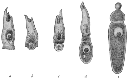
Fig. 60.—Gregarina longa from larva of crane-fly (Tipula). a, in epithelial cell of host; b, c, gradually leaving host-cell; d, adhering to host-cell; e, fully developed free trophozoite.
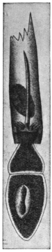
Fig. 61.—Xyphorhynchus firmus with epimerite in intestinal epithelial cell of host. (After Léger.)
The Gregarines are usually elongate, somewhat flattened organisms (figs. 59, 60), whose bodies are enclosed in an elastic and often thick cuticle. The enclosed living substance shows a separation into ectoplasm and endoplasm, as is common among Protozoa. The cuticle is sometimes regarded as the outer portion or epicyte of the ectoplasm. A single, vesicular, spherical, or elliptical, large nucleus, with its chromatin concentrated to form a spherical karyosome, is present. The body of some gregarines may be divided by ingrowing ectoplasmic partitions or septa, and are then said to be “septate” or “polycystid” (fig. 61). Other gregarines remain simple and non-septate, and are termed “monocystid” (fig. 59). The monocystid gregarines occur especially in the body cavity of Chætopoda and Insecta, more rarely in Echinodermata, in the parenchyma of Platyhelminthes, also in the gut of Tunicata and Insecta (fig. 60) and in the seminal vesicles of Annelida. In the polycystid gregarines a single septum only is present as a rule, and thus the body presents two portions: (1) an anterior portion termed the protomerite; (2) a posterior, larger portion, known as the deutomerite, which generally contains the nucleus. The protomerite is often modified anteriorly to form an organ of attachment, termed the epimerite (fig. 61), which is developed from the pointed rostrum of the sporozoite or primary infecting young gregarine. The structure of the epimerite may be complicated, being provided with hooks, spines, knobs, and other appendages. An extension of the polycystid condition is seen in Tæniocystis mira Léger (from the dipteran larva, Ceratopogon solstitialis), whose body shows a number of partitions, giving the organism a superficial resemblance to a tapeworm.
The ectoplasm of a gregarine exhibits three layers: (1) An epicyte (cuticle) externally of which the epimerite is composed; (2) a sarcocyte which forms the septa if present; (3) the deeper myocyte layer containing contractile elements in the form of fibrils or threads termed myonemes (fig. 62).
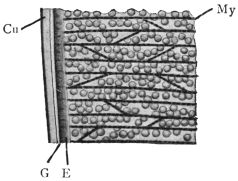
Fig. 62.—Gregarina munieri (from the beetle, Chrysomela hæmoptera). Section through surface layers. Cu, cuticle; E, ectoplasm proper; G, gelatinous layer; My, myonemes in myocyte layer. × 1500. (After Schewiakoff.)
The endoplasm is fluid and granular, containing many enclosures, which are of the nature of reserve food materials. They consist of fat droplets or of paraglycogen, and give the organisms an opaque appearance. Lithocystis contains crystals of calcium oxalate in its endoplasm.
Many gregarines are capable of active movements, though they do not possess obvious locomotor organs. The movement is of a smooth, gliding character and two suggestions have been put forward to explain it. According to Schewiakoff, a gelatinous substance is secreted between the layers of the ectoplasm. This is extruded posteriorly and thus the animal is pushed forward. On the other hand, Crawley considers that the movements are produced by contractions of the myonemes. These two explanations are probably correct as far as each goes, and are to be regarded as supplementary to one another.
Occasionally, temporary associations of gregarines are formed by a number of individuals adhering to one another end to end. Such temporary associations are examples of syzygy. Such syzygies must not be confused with true associations which form an essential part of the life-cycle.
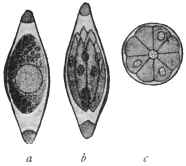
Fig. 63.—Monocystis agilis. Spores from vesicula seminalis of the Earthworm. a, Sporoblast with single nucleus, enclosed in sporocyst; b, mature spore containing sporozoites; c, diagrammatic cross-section of spore, showing eight sporozoites round residual protoplasm. (After Bütschli.)
The life-cycle of a relatively simple gregarine, such as Monocystis agilis (fig. 59), parasitic in earthworms, may now be considered. The gregarines, being members of the Sporozoa, produce spores at one phase of the life-cycle. Each gregarine spore (fig. 63) develops within itself a number of minute, sickle-shaped or vermicular bodies, known as sporozoites or primary infecting germs. Eight sporozoites are often formed within each spore. When absorbed by a new host, the spore softens and the sporozoites issue from it. They are capable of active movement and may or may not enter a cell, such as one of those of the digestive tract, or, as in Monocystis, a cell lining the vesicula seminalis which becomes a sperm-cell aggregate (sperm morula). When the sporozoite has reached the place of its choice in the host it ceases active movements and proceeds to feed passively on the fluid substances around it, whether they be those of tissues or body fluids. This passive, growing and feeding form is known as the trophozoite. After a trophic existence of longer or shorter duration, the trophozoite ceases to feed and prepares for reproduction. Two trophozoites associate together, each of them first becoming somewhat rounded. The two trophozoites, now known as sporonts or gametocytes, become invested in a single common envelope or cyst (fig. 64, a). The nucleus of each gametocyte then divides by a series of binary fissions (fig. 64, b), and the daughter nuclei thus produced arrange themselves at the periphery of the parent cells (fig. 64, c). Cytoplasm collects around each of these nuclei, and thus a number of gametes are formed within each gametocyte. The gametes for a time exhibit active movements, and ultimately ripe gametes of different parentage fuse in pairs, that is, conjugation occurs (fig. 64, d). In this way zygotes are produced, the nucleus of each zygote being formed by the fusion of two gamete nuclei.
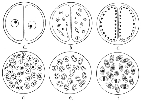
Fig. 64.—Schematic figures of conjugation and spore formation in Gregarines. For details see text. (After Calkins and Siedlecki, modified.)
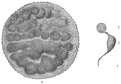
Fig. 65.—Stylorhynchus oblongatus. a, cyst containing two sporonts or gametocytes, each full of gametes, those in the upper one being male. b, ripe male and female gametes. × 1,600. (After L. Léger.)
The zygote grows slightly and becomes oval or elongate, and at this period is often called the sporoblast. It then secretes an external membrane, the sporocyst. Nuclear division occurs inside the sporocyst by a series of three binary fissions (fig. 64, e), so that each sporocyst, now usually referred to as a spore, contains eight nuclei. The cytoplasm collects around each nucleus and eight vermicular sporozoites are produced within each spore (fig. 64, f), thus completing the life-cycle.
It will be noticed that in the above life-cycle no asexual multiplication occurs. These organisms, such as Monocystis, are known as the Eugregarines, and include the majority of the gregarines. The remainder, which have introduced schizogony into their life-cycle, are known as the Schizogregarines.
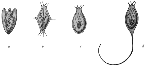
Fig. 66.—Spores of various Gregarines. a, Xiphorhynchus. b, Ancyrophora. c, Gonospora. d, Ceratospora. (After Léger.)
There are variations in the morphology and life-cycle of gregarines besides those that have been mentioned. It is not within the scope of this book to discuss them in detail, but the following may be noted:—
Morphological differentiation of gametes may occur as in Stylorhynchus oblongatus (fig. 65), which differentiation is probably of a sexual nature.
The sporocyst really consists of two layers, an epispore and an endospore. Externally the spores of different gregarines show great variety in shape and markings, and spines, or long processes may be present (fig. 66).
The resistant spores serve for the transmission of the gregarines from host to host. The mode of infection is contaminative, the spores expelled with the dejecta of one host being absorbed with the food of a new host.
The Gregarinida may be classified as follows:—
Sub-order I.—Eugregarinea, without schizogony.
Tribe 1.—Acephalina.—Without an epimerite and non-septate; often “cœlomic” (body-cavity) parasites. E.g.: Monocystis, with several species parasitic in the seminal vesicles of earthworms. Many other genera parasitic in Echinodermata, Tunicata, Arthropoda, etc.
Tribe 2.—Cephalina.—With an epimerite, either temporarily or permanently, in the trophic phase. Usually septate (except Doliocystidæ). Many families, genera and species. Common in the digestive tracts of insects. E.g.: Gregarina, with several species, Gregarina ovata in the earwig, Gregarina blattarum in the cockroach, Stylorhynchus in beetles, Pterocephalus in centipedes, etc.
Sub-order II.—Schizogregarinea, with schizogony.
Tribe 1.—Endoschiza.170—With schizogony occurring in the intracellular phase, e.g., Selenidium (from Annelida and Gephyrea), Merogregarina (from an Ascidian).
Tribe 2.—Ectoschiza.—In which the schizont is free, and schizogony is extracellular, e.g., Ophryocystis (from Blaps, a beetle), and Schizocystis (from Ceratopogon larva).
Order. Coccidiidea.
Hake (1839) first saw the organisms now termed Coccidia during his investigations on the so-called coccidial nodules of rabbits. The opinions as to the nature of these peculiar formations were very diverse. The discoverer considered them to be a sort of pus corpuscle; Nasse (1843) took them for epithelial cells of the biliary passages, others for helminthes, especially the ova of trematodes (Dujardin, Küchenmeister, Gubler, etc). Remak (1845) was the first to draw attention to their relation to the Psorospermia (Myxosporidia), and this investigator found them also in the small intestine and vermiform appendix of rabbits. Lieberkühn (1854), who examined not only the coccidia of rabbits, but found similar forms in the kidneys of frogs, likewise called them definitely psorosperms. To differentiate Müller’s psorosperms, which are found in fishes, from those of rabbits, etc., the latter were called egg-shaped psorosperms (Eimer), until R. Leuckart (1879) named them Coccidia and placed them in a group of the Sporozoa analogous to that of the Gregarinida, Myxosporidia, etc. Numerous works confirmed the occurrence of coccidia, not only in all classes of vertebrate animals, but also in invertebrates (Mollusca, Myriapoda, Annelida, etc.). A large number of genera and species have in the course of time been described which inhabit the epithelium of the intestine and its appendages for choice, but are also found in other organs (kidneys, spleen, ovaries, vas deferens, testicles). Some also live in the connective tissue of various organs, more particularly of the intestine.
The knowledge of the development of the coccidia was of particular importance in determining their classification. By means of encysted coccidia from the liver of rabbits, Kauffmann (1847) first confirmed the fact that the cyst, which was partly or entirely filled with granular contents, divided into three or four pale bodies (fig. 71) after a long sojourn in water. Lieberkühn observed the same process in the host in the case of the coccidia of the kidney of the frog. Stieda (1865) studied more minutely the changes that occur within the encysted coccidia of the liver of rabbits after the death of the host. He discovered that the bodies now known as “spores” were oval formations pointed at one pole, and surrounded by a delicate membrane, which exhibited a certain thickness at the pointed extremity and enclosed a slightly bent rodlet, expanding at either end into a strongly light-refracting globule; a finely granular globule was present in the middle of the spore. Waldenburg (1862) saw the same phenomenon in coccidia from the epithelium of the villi and Lieberkühn’s glands of the intestine of the rabbit; but the process in this case took place in a much shorter time.
According to the discovery of Kloss (1855), the spores of the coccidia of the urinary organ of the garden snail were formed in far greater numbers: the round spores also harboured several (five to six) rodlets, which after the bursting of the spore-envelope became free. Eimer’s researches (1870) afforded information regarding a Coccidium from the intestine of the mouse, which was transformed in toto into a “spore,” containing small sickle-shaped bodies. The fact was, moreover, established that the little bodies left the delicate envelope when in the intestine, made movements of flexion and extension, and were finally transformed into amœboid organisms, which apparently penetrated the epithelial cells; at all events, similar bodies of various sizes were seen in these cells. Taking the immense number of these parasites into account and the lack of any other cause, Eimer attributed the sudden death of his mice to the Gregarina falciformis, as the parasite was then called, just in the same way as a few years previously Reincke ascribed the acute and fatal intestinal catarrh of rabbits to the invasion of intestinal coccidia.
All that had become known about coccidia up to 1879 was then compiled by Leuckart, and completed by his own observations on the coccidia of the liver of the rabbit. Experimental infections had already been conducted by Waldenburg (1862) with intestinal coccidia of rabbits, and by Rivolta (1869–73) with the coccidia of fowls, which experiments confirmed the importance of the spores, or bodies enclosed in them, in the transmission of the parasites to other animals. Accordingly, it was assumed that after the entry of the spores into the intestine the sporozoites were set free, actively penetrated into the intestinal cells, where they grew into coccidia, and finally became encysted. The further development, i.e., the formation of spores, took place outside the host’s body in these cases; in other cases (Kloss, Eimer) it took place within the host. Although much regarding the cycle of development was still hypothetical, the ideas coincided with the observations, and were therefore universally regarded as established. Further research confirmed this view in numerous new forms.
L. Pfeiffer’s statements (1891) on the part that certain coccidia or their sporozoites played, or seemed to play, as causes of disease gave a renewed impetus to the investigation of the coccidia. The ingestion of even very numerous spores did not appear to account for the mass infection so frequently observed, even after Balbiani had confirmed the fact that there were two, and not one, sporozoites contained in every spore of the coccidia of rabbits (fig. 72). The hypothesis was therefore advanced that the sporozoites or young coccidia were able to divide once again by sporulating. The question was finally solved quite differently. R. Pfeiffer (1892) first confirmed the fact that in addition to the well-known method of sporulation in the coccidia of the rabbit that causes the infection of fresh hosts (“exogenous sporulation”), an enormous increase may follow in the already infected host in a manner that Eimer first observed in the coccidia of the intestine of the mouse (“endogenous sporulation”). It had hitherto been believed that some of the species of coccidia increased like the rabbit parasite, then known as Coccidium oviforme, and others like Eimeria falciformis, and this difference had been made the foundation of a classification. R. Pfeiffer was successful in observing the occurrence of both kinds of development in the same species, and expressed the opinion that endogenous sporulation (fig. 73), which takes place within the host, was the cause of the mass-infection that is mostly accompanied by serious consequences (fig. 74). L. Pfeiffer sought, especially, to demonstrate the correctness of this view as regards other species of coccidia and for this purpose he utilized the experiences already published. Coccidia were known to exist in a number of different hosts, and to propagate in some according to the Coccidium type, in others according to the Eimeria type. It therefore was reasoned that in this case it was not a question of two species belonging to different genera living side by side, with a different mode of development, but of one species, in the life of which both forms of development occurred alternately.
This interpretation of facts was combated especially by A. Schneider (1892) and by Labbé, but has, nevertheless, proved true, apart from the circumstance that Schuberg succeeded in discovering the hitherto unknown Coccidium form in the intestine of the mouse; and that, moreover, Léger confirmed the fact that there are no Arthropoda in which Eimeria are not found together with coccidia. The question was finally settled by experiments made by Léger with the coccidia of Scolopendra cingulata, by Schaudinn and Siedlecki with those of Lithobius forficatus, and by Simond with the coccidia of the rabbit. On Simond’s suggestion the sickle-shaped germs corresponding to the sporozoites, which are formed by endogenous sporulation, are generally termed merozoites; and in accordance with Schaudinn’s suggestion, those individuals which form merozoites are termed schizonts, and those which produce spores are called sporonts. In contradistinction to sporogony (exogenous sporulation), the term schizogony (endogenous sporulation) is used.
The more minute examination of these processes at last led to the discovery of sexual dimorphism, of copulation and of alternation of generations in the coccidia. Schuberg was the first to consider the possibility of copulation in coccidia; in addition to the formations which now are termed merozoites, he observed very diminutive bodies (“microsporozoites”) in the coccidia of the intestine of the mouse, which were able eventually to copulate. Labbé confirmed this observation in some of the species, and Simond expressed the opinion that bodies termed “chromatozoites” occurred in all coccidia. Copulation itself was then observed by Schaudinn and Siedlecki (1897). The copulating bodies were termed gametes. As, however, they differed considerably one from the other, the males were called microgametes (i.e., the microsporozoites of Labbé and the chromatozoites of Simond) and the females macrogametes. After copulation was completed sporogony took place, and in the cycle of development of one species this regularly alternated with schizogony (asexual multiplication). Schaudinn in 1900 described in detail the life-cycle of Eimeria (Coccidium) schubergi.
The recognition of this unsuspected complicated process was bound to effect reforms in the classification of the coccidia; and all the forms that had been regarded as developmental stages (Eimeria, etc.) had to be reconsidered.
Occurrence.—The Coccidiidea in their mature condition usually live within the epithelial cells of various organs, and by choice inhabit those of the intestine and of its associated organs. They also occur frequently in the excretory organs of mammals, birds, amphibia, molluscs, arthropods, and may also be found in the testes and vas deferens, but the statement that they live in hen’s eggs, as well as in the oviducts of fowls, has not been confirmed.171 Some species inhabit the nuclei of cells, others live in the connective tissue, but their presence in the latter situation is probably only secondary, that is, they have only reached it from the epithelium of the affected organs.
The size of the Coccidiidea, corresponding as a rule to the capacity of their habitat, is usually small, but there are said to be species that attain a diameter of 1 mm. Their form172 is globular, oval, or elliptical. External appendages are lacking, at least during the trophic or vegetative period of their life, which is spent in epithelial cells, within which they increase in size. Frequently one only is present in each cell, but more can occur. The body substance is composed of a more or less finely granular or distinctly alveolar protoplasm which exhibits no differentiation into ecto- and endoplasm. All species possess a nucleus that enlarges with their growth; sometimes it only shows through the cytoplasm as a lighter spot, or may even be quite concealed. It is vesicular, and besides containing very delicate threads of chromatin in the clear nucleoplasm, it contains generally only one large karyosome.
The infected epithelial cells degenerate sooner or later as the parasite feeds on them (fig. 67, II-IV). After their form has been changed by the action of the growing parasite, they finally perish. The cell membrane then alone surrounds the coccidia, which, at least in the species sufficiently known, begin to propagate by an asexual process (schizogony), the parasites themselves becoming schizonts, as the initial stage is usually called. They differ from later stages (sporonts or gametocytes), which resemble them in form, by the absence of granulations in the cytoplasm, as well as by the vesicular nucleus. The form is not always the same, for in some cases, at least, many schizonts assume a globular form.
Schizogony (fig. 67, V-VII) commences with a division of the nucleus, which takes place in different ways in the various species. This finally leads to the formation of numerous daughter nuclei which become smaller and smaller, and which collect beneath the surface of the schizonts. In some species the daughter nuclei collect only in one half of the schizont. A part of the protoplasm of the periphery now divides around each daughter nucleus, the remaining part (residual body) being left in the centre or on one side. The segments of the divided cytoplasm, each containing a nucleus, assume a fusiform shape and become merozoites, which very soon become free (fig. 67, VIII) and leave the residual body. They are distinguishable from the very similar sporozoites (fig. 67, I), as the merozoites possess a karyosome.
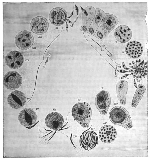
Fig. 67.—Life-cycle of Eimeria (Coccidium) schubergi, Schaud., from the intestine of Lithobius. (After Schaudinn.) The infection is caused by a cyst (XX), containing spores, which reaches the intestine of a Lithobius, where it discharges the sporozoites (I). II, A sporozoite invading an intestinal epithelial cell; III, intestinal epithelial cell with young trophozoite; IV, intestinal epithelial cell with a globular schizont; V, nuclear segmentation within the schizont; VI, the daughter nuclei arranging themselves superficially; VII, formation of the merozoites; VIII, merozoites that have become free, and which, penetrating into other epithelial cells of the same intestine, repeat the schizogony (II-VIII); IX and X, merozoites which, likewise invading the epithelial cells of the same intestine, become sexually differentiated; xia, young macrogametocyte; XIb, older macrogametocyte; XIc, mature macrogametocyte (discharging particles of chromatin); XIIa, young microgametocyte; XIIb, older microgametocyte; XIIc, increase of nuclei in the microgametocyte; XIId, the globular residual body around which numerous microgametes have formed; XIIe, an isolated microgamete; XIII, the mature macrogamete surrounded by numerous microgametes and forming a cone of reception or fertilization prominence; XIV, shows the nucleus of a microgamete that has penetrated and fused with the nucleus of the macrogamete (fertilization)—the latter forms a membrane and becomes an oöcyst; XV, XVI, XVII, nuclear segmentation in the oöcyst; XVIII, oöcyst with four sporoblasts; XIX, the sporoblasts transformed into spores, each containing two sporozoites; XX, the cyst introduced into the intestine and liberating the sporozoites by bursting.
The merozoites move in a manner similar to that of the sporozoites. The movements consist either of slow incurvations with subsequent straightenings, or annular contractions along the entire extent of the body. In addition, there are gliding movements similar to those of many gregarines, and brought about in a like manner by the secretion at the posterior extremity of a gelatinous substance that hardens rapidly.
The merozoites do not gain the open in the usual way, but are destined to infect still further the same host by actively penetrating into other epithelial cells of the affected organ. Here they continue their growth and may again and again undergo schizogony. In the Infusoria the repeated segmentations finally cease and are renewed only after a conjugation. This is likewise the case with the Coccidia, with the difference that in the latter the two conjugating individuals (gametes) are differently constituted one from the other, whereas in the Infusoria they are almost always similar.
When the schizogony ceases, the merozoites, that had penetrated the epithelial cells and become trophozoites there, consist of two kinds of differently constituted individuals. One kind possesses a clear cytoplasm (fig. 67, XII), the other an opaque, richly granular cytoplasm (fig. 67, XI), while both possess a vesicular nucleus with a karyosome. In order to continue their development, the more granular individuals must be fertilized, and are therefore termed either female gametes or, on account of their size, macrogametes. The male individuals (microgametes) necessary to conjugation, are formed in greater numbers from the less dense microgametocytes or male mother-cells (fig. 67, XIId). They are slender bodies consisting chiefly of nuclear substance, and in most species bear two flagella of unequal length directed backwards, the place of insertion of which varies according to the species (fig. 67, XIIe).
While the development of the microgametes is rapidly advancing a change occurs in the nucleus of the female parent forms or macrogametocytes. Parts of the karyosome are extruded (fig. 67, XIc), and the nucleus loses at the same time its vesicular form. One macrogamete results, after nuclear maturation, from one macrogametocyte. By this time the macrogametes are capable of conjugation, and the process takes place within the host, generally, however, outside the affected and degenerated host cells. The microgametes that have now become free from the very large residual body, crowd around the mature macrogametes, which often send out a small prominence (“cone of reception” or fertilization protuberance) for their reception (fig. 67, XIII). As soon as a microgamete comes in contact with this and penetrates into the cytoplasm of the macrogamete, the latter surrounds itself with a membrane which prevents the intrusion of other microgametes. The nucleus of the microgamete that has gained entry unites with the nucleus of the macrogamete, which latter is afterwards capable of forming the well-known spores. The parasite is now called an encysted zygote or oöcyst. The oöcysts of some other members of the Coccidiidea, e.g., Eimeria avium, can form their walls prior to fertilization. In such cases, a weak spot is left at one place in the cyst wall, forming a micropyle, that permits of the entry of the male, immediately after which the micropyle is closed.
The reduced nucleus of the macrogamete elongates on the entry of the microgamete, and becomes a fertilization spindle to which the male pronucleus (from the microgamete) becomes attached (fig. 67, XIV and XV). Thereupon the spindle divides into two daughter nuclei (fig. 67, XVI) which assume a round shape. The protoplasm at this stage may at once divide, or another segmentation of the daughter nuclei may first occur. In the former case the two halves divide again, so that finally four nucleated segments, the sporoblasts, are formed, whereas in the latter case the four sporoblasts appear simultaneously (fig. 67, XVII). In both cases a residual body of varying size is separated from the protoplasm of the oöcyst. As a rule the oöcysts have already been discharged from the body of the host, and in the manner described above, form the sporoblasts after a longer or shorter period of incubation.
The sporoblasts are originally naked, but each soon secretes a homogeneous membrane, the sporocyst, in which it becomes enveloped (fig. 67, XVIII). After the segmentation of the nucleus the contents divide into two sickle-shaped sporozoites, in addition to which there is generally also a residual body (fig. 67, XIX).
This terminates the development. The spores are intended for the infection of other hosts. If they reach the intestine of suitable hosts, either free or enclosed in the oöcyst wall, the action of the intestinal juices causes them to open and permits the escape of the sporozoites (fig. 67, XX). The latter move exactly like the merozoites and soon make their way into epithelial cells (fig. 67, I), where they become schizonts, and thus repeat the life cycle.
Although our knowledge of the development of the coccidia is but of recent date, yet it already extends to a large number of species, which exhibit various deviations from the cycle of development described above. For instance, in addition to differences in the gametocytes, the schizonts of Adelea and Cyclospora also show differentiation and give rise to macromerozoites and micromerozoites, whilst in Adelea and Klossia a precocious association of the gametocytes precedes the true copulation of the ripe gametes.
The classification of the Coccidiidea is based chiefly on the number of sporozoites found in each spore, and the number of sporocysts (spores) found in one oöcyst. Léger173 recognises two great legions, the Eimeridea and the Adeleidea, the former comprising the greater number of genera, including the genus of most economic importance, Eimeria. It must be noted that, though a member of this genus may be frequently referred to as Coccidium, strictly it should be termed Eimeria, that name having priority. The name of the disease resulting from the action of such parasites is, however, established and remains as coccidiosis.
Certain of the more important of the Coccidiidea may now be considered.
Genus. Eimeria, Aimé Schneider, 1875.
Syn.: Psorospermium, Rivolta, 1878; Cytospermium, Rivolta, 1878; Coccidium, R. Leuckart, 1879; Pfeifferia, Labbé, 1894; Pfeifferella, Labbé, 1899.
The Eimeria belong to Léger’s old family, the Tetrasporocystidæ, which comprises forms producing oöcysts with four sporocysts, each containing two sporozoites. The cysts are spherical or oval, as are also usually the schizonts. The members of the genus are confined chiefly to vertebrate hosts, the more important economically occurring in mammals and birds. From the mammalian hosts very rarely the parasites may reach man. Eimeria (Coccidium) avium of wild birds and poultry, and Eimeria stiedæ parasitic in rabbits, may be considered. There is a general similarity in their life-cycles and each is of great practical importance.
Eimeria avium, Silvestrini and Rivolta.
Eimeria avium is responsible for fatal epizoötics among game birds such as grouse, pheasants and partridges, and domestic poultry such as fowls, ducks, pigeons and turkeys, and can pass from any one of these hosts to any of the others with the same effect. The organism is parasitic in the alimentary tract of the host, affecting more especially the small intestine (duodenum) and the cæca, but in some cases penetrating to the liver and multiplying there (as in turkeys), producing necrotic cheesy patches, that ultimately become full of oöcysts. The gut is rendered very frail by the action of the parasites, its mucous membrane is greatly injured, and is often reduced to an almost structureless pulp, riddled with parasites (fig. 68). Infection is conveyed from host to host by the ingestion of food or drink contaminated with the oöcysts voided in the fæces of infected birds. Oval oöcysts from 24 µ to 35 µ long and from 14 µ to 20 µ broad are the means of infection. The oöcysts develop internally four sporocysts or spores, from each of which two sporozoites are produced. The life-history174 presents two phases: (1) The asexual multiplicative phase, schizogony, for the increase in numbers of the parasites within the same host; (2) the reproductive phase, following the formation of gametes (gametogony), leading to the production of resistant oöcysts, destined for the transference of the parasite to new hosts (sporogony).
The oöcysts usually reach the duodenum unharmed, with food or drink. Under the influence of the powerful digestive juices (especially the pancreatic) now encountered, the oöcysts soften, as do the sporocysts, and ultimately two sporozoites emerge from each sporocyst. The sporozoites are from 7 µ to 10 µ long, and each is vermicular with a uniform nucleus (fig. 69, A). After a short period of active movement in the gut, each sporozoite penetrates an epithelial cell (figs. 68 spz, 69, B), and once within, gradually becomes rounded (fig. 69, B, C). It grows rapidly, feeding on the contents of the host cell and living as a trophozoite (fig. 69, D). When the parasite is from 10 µ to 12 µ in diameter, usually multiplication by schizogony (fig. 69, E-H) begins. The nucleus of the parent cell, now called a schizont, divides into a number of portions that become arranged at the periphery (fig. 69, E). Cytoplasm collects around each nucleus (fig. 69, E, F) and gradually a group of daughter individuals (merozoites) is produced (fig. 69, G), the nucleus of each merozoite showing a karyosome.
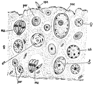
Fig. 68.—Small piece of epithelial lining of gut of heavily infected Grouse chick, showing various stages in life history of the parasite Eimeria avium; par, parasite (trophozoite); mz, merozoite; sch, schizont; spz, sporozoite; ooc, oöcyst; ♂, male gametocyte; ♀, female gametocyte. × 750. (After Fantham.)
The merozoites of Eimeria avium are arranged “en barillet,” like the segments of an orange (figs. 68 mz, 69, G), therein differing from those of E. schubergi, which are arranged “en rosace.” They separate from one another (fig. 69, H), penetrate other epithelial cells, where they may, in turn, become schizonts. Eight to fourteen merozoites are usually formed by each schizont, twenty have been found, while in cases of intense infection when space has become limited, the number may be only four.
After a number of generations of merozoites have been formed, a limit is reached both to the multiplicative capacity of the parasite and to the power of the bird to provide the invader with food. Consequently, resistant forms of the parasite are necessary, and the trophozoites begin to show sexual differentiation instead of forming schizonts, that is, gametogony commences.
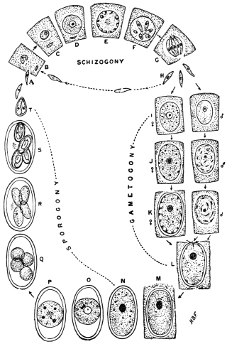
Fig. 69.—Eimeria avium. Diagram of life-cycle. For explanation see text. (After Fantham.)
Certain trophozoites store food and become large and granular. These are macrogametocytes (fig. 69, I, ♀). The microgametocytes (fig. 69, I, ♂) are smaller and far less granular. The macrogametocyte continues to grow, and becomes loaded with chromatoid and plastinoid granules (fig. 69, J, ♀), while the microgametocyte has its nucleus divide to form a number of bent, rod-like portions (fig. 69, J, ♂). The macrogametocyte gives rise to a single macrogamete, which forms a cyst wall for itself, leaving a thin spot (micropyle) for the entry of the male (fig 69, K, ♀). The microgametocyte gives rise to numerous small, biflagellate microgametes (fig. 69, K, ♂) around a large, central residual mass, from which they ultimately break free, and swim away. When a macrogamete is reached, the microgamete enters through the micropyle (fig. 69, L)—which then closes, thus excluding the other males—and applies itself to the female nucleus (fig. 69, M). Nuclear fusion occurs, the oöcyst (encysted zygote) being thus produced. Sporogony then ensues. The oöcyst (fig. 69, N) at first has its contents completely filling it. They then concentrate into a central spherical mass (fig. 69, O) which gradually becomes tetranucleate (fig. 69, P). Cytoplasm collects around each nucleus, and four sporoblasts are thus formed (fig. 69, Q). Each sporoblast becomes oval (fig. 69, R) and produces a sporocyst. Ultimately two sporozoites are formed in each sporocyst or spore, at first lying tête-bêche (fig. 69, S), but finally twisting to assume the position most convenient for emergence (fig. 69, T) when they reach a new host. The period of the life-cycle of Eimeria avium (as well as the details of the life-cycle) was determined by Fantham to be from eight to ten days, of which period schizogony occupies four to five days.
The method of infection175 is contaminative, by way of food or drink. Young birds are especially susceptible to infection. Certain birds, particularly older ones, may act as reservoirs of oöcysts, being continuously infected themselves, without showing any marked ill effects from the parasite, but being highly infectious to other birds. Much moisture retards the development of sporocysts considerably. The duration of vitality of the infective oöcysts has been determined experimentally to extend well over two years, and in certain cases longer. Eimeria avium is the causal agent of “white diarrhœa” or “white scour” in fowls, and of “blackhead” in turkeys.
Eimeria avium of birds and E. stiedæ of rabbits closely resemble one another, but are not the same parasite, for E. avium is not infective to rabbits, nor E. stiedæ to poultry.
Eimeria stiedæ, Lindemann, 1865.
Syn.: Monocystis stiedæ, Lindemann, 1865; Psorospermium cuniculi, Rivolta, 1878; Cytospermium hominis, Rivolta, 1878; Coccidium oviforme, Leuckart, 1879; Coccidium perforans, Leuckart, 1879; Coccidium cuniculi.
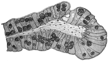
Fig. 70.—Eimeria stiedæ. Section through an infected villus of rabbit’s intestine. × 260.
Eimeria stiedæ is parasitic in the gut epithelium (fig. 70), liver, and epithelium of the bile ducts of rabbits, and is usually considered to be the parasite very occasionally found in man. The life-cycle resembles that of Eimeria avium in its general outlines (see fig. 69) and therefore will not be detailed in full here. The oöcysts (fig. 71) are large, elongate-oval, greenish in fresh preparations and vary in size from 24 µ to 49 µ long and 12·8 µ to 28 µ broad, the gut forms being usually smaller than those occurring in the liver, owing to the more confined space in which they are formed. Formerly, the parasites in the liver were described under the name of Coccidium oviforme, while those from the intestine were termed Coccidium perforans. This distinction has now broken down.
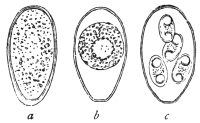
Fig. 71.—Eimeria stiedæ, from the liver of the rabbit, oöcysts in various stages of development. (After Leuckart.)
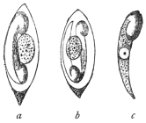
Fig. 72.—a, b, spores of Eimeria stiedæ (Riv.), with two sporozoites and residual bodies; c represents a free sporozoite. (After Balbiani.)
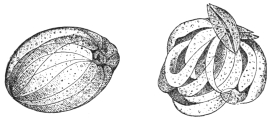
Fig. 73.—So-called swarm cysts (endogenous sporulation or schizogony) of the Coccidium of the rabbit. The daughter forms are called merozoites. (After R. Pfeiffer.)
The oöcysts176 are thick-walled, somewhat flattened at one pole, where a large micropyle is present. Four egg-shaped spores (sporocysts) are formed within, each about 12 µ to 15 µ long and 7 µ broad (fig. 72). The oöcysts are voided with the fæces. Sporogony takes, in nature, about three days in the excrement. Fæcal contamination of the food of rabbits results, and coccidian oöcysts are swallowed. Under the influence of the pancreatic juice of a new host, the sporozoites (fig. 72, a—c) are liberated from the spores and proceed to attack the epithelium and multiply within it, as in the case of Eimeria avium. From the gut, infection spreads to the liver, where multiplication of the parasite goes on actively, resulting in the formation of the whitish coccidial nodules, which may be very conspicuous (fig. 74). Proliferation of the connective tissue may occur around the coccidial nodules, which then contain large numbers of oöcysts in various stages of development. It is said that the oöcysts in the older nodules do not seem to be capable of further development. Schizogony (fig. 73) and gametogony in all stages can be found in both liver and gut.
Young rabbits often die of intestinal coccidiosis before infection of the liver occurs. The repeated schizogony of Eimeria stiedæ in the gut is sufficient to cause death.
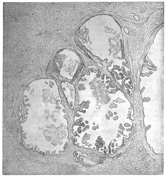
Fig. 74.—Eimeria stiedæ. Section through coccidian nodule in infected rabbit’s liver. × 55.
The disease of cattle popularly known as “red dysentery” is also ascribed to the action of Eimeria stiedæ. The fæces of infected cattle show blood clots of various sizes and in severe cases watery diarrhœa is present. Acute cases end fatally in about two days. Numerous oöcysts, considered to be those of Eimeria stiedæ, occur in the fæces, and there is a heavy infection of the gut, especially the large intestine and rectum, all stages of the parasite being found in the epithelium. It is suspected that cattle contract the disease by feeding on fresh grass contaminated with oöcysts. The disease is recorded from Switzerland and from East Africa.
As before mentioned, Eimeria stiedæ is considered to be the organism found in a few cases in man, possibly acquired by eating the insufficiently cooked livers of diseased rabbits. These cases may now be described.
(a) Human Hepatic Coccidiosis.
(1) Gubler’s Case. A stone-breaker, aged 45, was admitted to a Paris hospital suffering from digestive disturbances and severe anæmia. On examination the liver was found to be enlarged and presented a prominent swelling, which was regarded as being due to Echinococcus. At the autopsy of the man, who succumbed to intercurrent peritonitis, twenty cysts were found averaging 2 to 3 cm. in diameter, and one measuring 12 to 15 cm. The caseous contents consisted of detritus, pus corpuscles, and oval-shelled formations, which were considered to be Distoma eggs, but which, in accordance with Leuckart’s conjecture, proved to be Coccidia.177
(2) Dressler’s Case (Prague). Relates to three cysts, varying from the size of a hemp-seed to that of a pea, and containing Coccidia, found in a man’s liver.178
(3) Sattler’s Case (Vienna). Coccidia were in this case observed in the dilated biliary duct of a human liver.179
(4) Perls’ Case (Giessen). Perls discovered Coccidia in an old preparation of Sömmering’s agglomerations.180
(5) Silcock’s Case (London).181 The patient, aged 50, who had fallen ill with serious symptoms, exhibited fever, enlarged liver and spleen, and had a dry, coated tongue. At the autopsy numerous caseous centres, mostly immediately beneath the surface, were found, while the contiguous parts of the liver were inflamed. Microscopical examination demonstrated numerous Coccidia in the hepatic cells as well as in the epithelium of the biliary ducts. A deposit of Coccidia was likewise found in the spleen, which the parasites had probably reached by means of the blood-stream.182
(b) Human Intestinal Coccidiosis.
In two cadavers at the Pathological Institute in Berlin, Eimer183 found the epithelium of the intestine permeated by Coccidia. Railliet and Lucet’s case may be traced back to intestinal Coccidia, which were found in the fæces of a woman and her child, who had both suffered for some time from chronic diarrhœa.184 In other cases (Grassi, Rivolta), where only the existence of Coccidia in the fæces was known, it is doubtful whether the parasites originated in the intestine or in the liver.
(c) Doubtful Cases.
To these belong Virchow’s case185 where, in the liver of an elderly woman, a thick walled tumour measuring 9 to 11 mm. was found. Among the contents of this tumour there were oval formations 56 µ long, surrounded by two membranes and enclosing a number of round substances. Virchow considered these foreign bodies to be eggs of pentastomes in various stages of development, others consider them to be Coccidia.
The Coccidia which Podwyssotzki claims to have seen in the liver of a man, not only in the liver cells, but also in the nuclei, are also problematic.186 The parasite was called Caryophagus hominis.
Again, other explanations can be given to an observation by Thomas, on the occurrence of Coccidium oviforme in a cerebral tumour of a woman aged 40. The growth was as large as a pea and surrounded by a bony substance.187
Genus. Isospora, Aimé Schneider, 1881.
Syn.: Diplospora, Labbé, 1893.
Belonging to the section Disporea, that is, forming only two spores, each with four sporozoites.
Isospora bigemina, Stiles, 1891.
Syn.: “Cytospermium villorum intestinalium canis et felis,” Rivolta, 1874; “Coccidium Rivolta,” Grassi, 1882; Coccidium bigeminum, Stiles, 1891.
This parasite lives in the intestinal villi of dogs, cats, and the polecat (Mustela putorius, L.). According to Stiles,188 the oöcyst divides into two equal ellipsoidal portions or sporoblasts which become spores and then each forms four sporozoites. The oöcysts of this species vary from 22 µ to 40 µ in length and from 19 µ to 28 µ in breadth. Each spore is 10 µ to 18 µ long and contains four sporozoites. The parasites live and multiply, not only in the gut epithelium, but also in the connective tissue of the intestinal submucosa. Wasielewski has seen merozoites in the gut of the cat.
Isospora bigemina (fig. 75) appears to occur also in man, for Virchow published a case which was communicated to him by Kjellberg, and attributed the illness to this parasite.189 Possibly also it would be more correct to ascribe the observation of Railliet and Lucet, which is mentioned under “Human Intestinal Coccidiosis,” p. 148, to this species, as the Coccidia in that case were distinguished by their diminutive size (length 15 µ, breadth 10 µ). The case communicated by Grunow may also possibly refer to Isospora bigemina.190 Roundish or oval structures of 6 µ to 13 µ in diameter occurred in the mucous membrane of the gut and in the fæces of a case of enteritis.
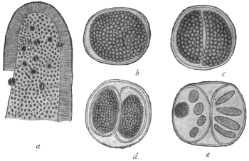
Fig. 75.—Isospora bigemina, Stiles (from the intestine of a dog). a, Piece of an intestinal villus beset with Isospora, slightly enlarged; b, Isospora bigemina (15 µ in diameter), shortly before division; c, divided; d, each portion encysted forming two spores; e, four sporozoites in each part, on the left seen in optical section, together with a residual body—highly magnified. (After Stiles.)
Doubtful Species.
In literature many other statements are found as to the occurrence of Coccidia-like organisms in different diseases of man. In some of the cases the parasites proved to be fungi. This was the case with the parasites of a severe skin disease of man, formerly called Coccidioides immitis and Coccidioides pyogenes. Other statements are founded on misapprehensions, or are still much disputed. If reference is here made to “Eimeria hominis,” R. Blanchard, 1895, this is done on the authority of the investigator mentioned. The structures in question are nucleated spindle-shaped bodies of very different lengths (18 µ to 100 µ), which either occurred isolated or were enclosed in large globular or oval cysts, alone or with a larger tuberculated body (“residual body”). These formations were found by J. Künstler and A. Pitres in the pleural exudation removed from a man by tapping. The man was employed on the ships plying between Bordeaux and the Senegal River.
Blanchard looks upon the fusiform bodies as merozoites and the cysts as schizonts of a Coccidium. On the other hand, Moniez declares the spindle bodies to be the ova and the supposed residual bodies to be “floating ovaries” of an Echinorhynchus.
Severi’s “monocystid Gregarines,” which were taken from the lung tissue of a still-born child, are also quite problematical.
No less doubtful are the bodies which Perroncito calls Coccidium jalinum, and which he found in severe diseases of the intestine in human beings, pigs, and guinea-pigs; Borini also reported another case.
Order. Hæmosporidia, Danilewsky emend. Schaudinn.
The Hæmosporidia are a group of blood parasites, comprising forms differing greatly among themselves. Some of the forms need much further investigation. However, there are certain true Hæmosporidia which present close affinities with the Coccidia, leading Doflein to use the term Coccidiomorpha for the two orders conjoined.
The Hæmosporidia present the following general characteristics:—
(1) They are parasites of either red or white blood corpuscles of vertebrates during one period of their life-history.
(2) They exhibit alternation of generations—asexual phases or schizogony alternating with sexual phases or sporogony—as do the Coccidia.
(3) There is also an alternation of hosts in those cases which have so far been completely investigated. The schizogony occurs in the blood or internal organs of some vertebrates while the sporogony occurs in an invertebrate, such as a blood-sucking arthropod or leech.
(4) Unlike the Coccidia, resistant spores in sporocysts are not generally produced, such protective phases in the life-cycle being unnecessary, as the Hæmosporidia are contained within either the vertebrate or invertebrate host during the whole of their life.
The Hæmosporidia may be considered for convenience under five main types:—
(1) The Plasmodium or Hæmamœba type. This includes the malarial parasites of man and of birds. The asexual multiplicative or schizogonic phases occur inside red blood corpuscles and are amœboid. They produce distinctive, darkish pigment termed melanin or hæmozoin. Infected blood drawn and cooled on a slide may exhibit “exflagellation” of the male gametocytes, i.e., the formation of filamentous male gametes. The invertebrate host is a mosquito. The malarial parasites of man are discussed at length on p. 155. Similar pigmented hæmamœboid parasites have been described in antelopes, dogs, and other mammals, and even reptiles.
(2) The Halteridium type. The trophozoite stage inside the red blood corpuscle is halter-shaped. Pigment is produced, especially near the ends of the organism. The parasites occur in the blood of birds. The invertebrate host of H. columbæ of pigeons in Europe, Africa, Brazil and India, is a hippoboscid fly, belonging to the genus Lynchia.
Halteridium parasites are common in the blood of passerine birds, such as pigeons, finches, stone owls, Java sparrows, parrots, etc. The Halteridium embraces or grows around the nucleus of the host red cell without displacing the nucleus. Young forms and multiplicative stages of H. columbæ have been found in leucocytes in the lungs of the pigeon (fig. 76, 8-12). Male and female forms (gametocytes) are seen in the blood (fig. 76, 3a, 3b). The cytoplasm of the male gametocytes is pale-staining and the nucleus is elongate, while the cytoplasm of the females is darker and the nucleus is smaller and round. Formation of male gametes from male gametocytes (the so-called process of “exflagellation”) may occur on a slide of drawn infected blood, also fertilization, and formation of the oökinete, as first seen by MacCallum. The correct generic name for Halteridia is, apparently, Hæmoproteus. Wasielewski (1913), working on H. danilewskyi (var. falconis), in kestrels, finds that the halteridium may be pathogenic to nestlings. The cycle of H. noctuæ described by Schaudinn (1904) lacks confirmation. The account of the life-cycle of H. columbæ given by Aragão (1908) is illustrated in fig. 76. It agrees with the work of Sergent (1906–7) and Gonder (1915). Mrs. Adie (1915) states that the cycle in Lynchia is like that of a Plasmodium.
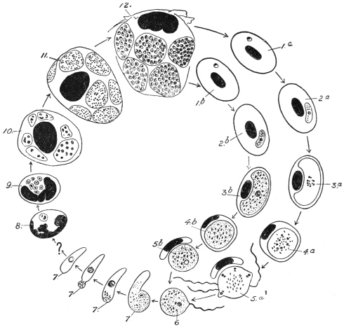
Fig. 76.—Hæmoproteus (Halteridium) columbæ. Life-cycle diagram: 1, 2, stages in red blood corpuscle of bird; 3, 4, gametocytes (3a ♂, 3b ♀); 5a, formation of microgametes; 6, fertilization (in fly’s gut); 7, oökinete; 8–12, stages in mononuclear leucocytes in lungs. (After Aragão.)
(3) The Leucocytozoön type. The trophozoites and gametocytes occur within mononuclear leucocytes and young red cells (erythroblasts) in the blood of birds. Laveran and França consider that the Leucocytozoa occur in erythrocytes. The host cells are often greatly altered by the parasites, becoming hypertrophied and the ends usually drawn into horn-like processes (fig. 77), though some remain rounded. Leucocytozoa are limited to birds, and very rarely produce pigment. Male and female forms (gametocytes) are distinguishable in the blood (fig. 77), and the formation of male gametes (“exflagellation”) may occur in drawn blood.
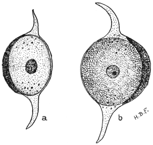
Fig. 77.—Leucocytozoön lovati. a, Male parasite (microgametocyte), within host cell, whose ends are drawn out; b, female parasite (macrogametocyte) from blood of grouse. × 1,800. (After Fantham.)
The Leucocytozoa were first seen by Danilewsky in 1884. They are usually oval or spherical. It is not easy sometimes to distinguish the altered host cell from the parasite, as the nucleus of the former is pushed to one side by the leucocytozoön. The cytoplasm of the female parasite stains deeply, and the nucleus is rather small, containing a karyosome. In the male the cytoplasm stains lightly and the nucleus is larger, with a loose, granular structure.
Many species of Leucocytozoa are recorded, but schizogony has only been described by Fantham (1910)191 in L. lovati in the spleen of the grouse (Lagopus scoticus), and by Moldovan192 (1913) in L. ziemanni in the internal organs of screech-owls.
M. and A. Leger193 (1914) propose to classify Leucocytozoa, provisionally, according as the host cells are fusiform or rounded.
(4) The Hæmogregarina type. Included herein are many parasites of red blood corpuscles, with a few (the leucocytogregarines) parasitic in the white cells of certain mammals and a few birds. They are not amœboid but gregarine-like, vermicular or sausage-shaped (fig. 78). They do not produce pigment. They are widely distributed among the vertebrata, but are most numerous in cold-blooded vertebrates (fishes, amphibia and reptiles). The hæmogregarines of aquatic hosts are transmitted by leeches, those of terrestrial hosts by arthropods.
The nucleus of hæmogregarines is usually near the middle of the parasite, but may be situated nearer one end. The body of the parasite may be lodged in a capsule (“cytocyst”). There is much variation in size and appearance among hæmogregarines. Some are small (Lankesterella); some attack the nucleus of the host cell (Karyolysus); others have full grown vermicules larger than the containing host corpuscle, and so the hæmogregarines bend on themselves in the form of U (fig. 78, b). Schizogony often occurs in the internal organs of the host, sometimes in the circulating blood.
The hæmogregarines occurring in the white cells (mononuclears or polymorphonuclears) of mammals have been referred to a separate genus, Leucocytogregarina (Porter) or Hepatozoön (Miller). Such leucocytogregarines are known in the dog (fig. 79), rat, mouse, palm-squirrel, rabbit, cat, etc. Schizogony of these forms occurs in the internal organs, such as the liver, lung and bone-marrow of the hosts. They are apparently transmitted by ectoparasitic arthropods, such as ticks, mites, and lice.
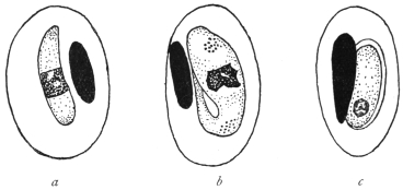
Fig. 78.—Hæmogregarines from lizards, a, H. schaudinni, var. africana, from Lacerta ocellata; b, H. nobrei from Lacerta muralis; c, H. marceaui in cytocyst, from Lacerta muralis. (After França.)
A few hæmogregarines are known to be parasitic in the red blood corpuscles of mammals. Such are H. gerbilli in the Indian field rat, Gerbillus indicus; H. balfouri (jaculi) in the jerboa, Jaculus jaculus, and a few species briefly described from marsupials. These parasites do not form pigment.
Strict leucocytic gregarines have been described from a few birds by Aragão and by Todd.
The sporogony of hæmogregarines is only known in a few cases, and in those affinity with the Coccidia is exhibited. In fact, the Hæmogregarines are now classified by some authors with the Coccidia.
(5) The Babesia or Piroplasma type. These are small parasites of red blood corpuscles of mammals. They do not produce pigment. They are pear-shaped, round or amœboid in Babesia, bacilliform and oval in other forms referred to this group. Piroplasms are transmitted by ticks. These parasites are described at length on p. 172.
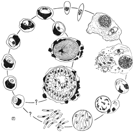
Fig. 79.—Leucocytogregarina canis. Life-cycle diagram. Constructed from drawings by Christophers. (After Castellani and Chalmers.) Schizogony occurs in the bone-marrow. The parasite is transmitted from dog to dog by the tick, Rhipicephalus sanguineus, development in which, so far as known, is shown on the right.
THE MALARIAL PARASITES OF MAN.
Malaria, otherwise known as febris intermittens, chill-fever, ague, marsh fever, paludism, etc., is the name given to a disease of man, which begins with fever. It has been known since ancient times and is distributed over almost all the world, although very unevenly, but does not occur in waterless deserts and the Polar regions. In many places, especially in the civilized countries of Central Europe, the disease is extinct or occurs only sporadically, and large tracts of land have become free from malaria.
The rhythmical course of the fever is characteristic. It begins apparently suddenly with chilliness or typical shivering, whilst the temperature of the body rises, the pulse becomes low and tense and the number of beats of the pulse increases considerably. After half to two hours the heat stage begins. The patient himself feels the rise of his temperature (shown by feeling of heat, dry tongue, headache, thirst). The temperature may reach 41°C or more. At the same time there is sensitiveness in the region of the spleen and enlargement of that organ. After four to six hours an improvement takes place, and with profuse perspiration the body temperature falls rapidly, not often below normal. After the attack the patient feels languid, but otherwise well until certain prodromal symptoms (heaviness in the body, headache) which were not noticed at first, denote the approach of another attack of fever, which proceeds in the same way.
The intervals between the attacks are of varying length which permit of a distinction in the kinds of fever. If the attacks intermit one day, occurring on the first, third and fifth days of the illness and always at the same time of day, it is termed febris tertiana; if two days occur between fever days, it is called febris quartana. In the case of the fever recurring daily, later writers speak of typical febris quotidiana. But a quotidian fever may arise when two tertian fevers differing by about twenty-four hours exist at the same time (febris tertiana duplex). The patient has a daily attack, but the fever of the first, third and fifth days differs in some point (hour of occurrence, height of temperature, duration of cold or hot stage) from the fever of the second, fourth and sixth days. Similarly, two or three quartan fevers which differ by about twenty-four hours each may be observed together (febris quartana duplex or triplex); in the latter case the result is also a quotidian fever.
Two kinds of tertian fever are differentiated—a milder form occurring especially in the spring (spring tertian fever), and a more severe form appearing in the summer and autumn in warmer districts, especially in the tropics (summer or autumn fever, febris æstivo-autumnalis, febris tropica, febris perniciosa). The latter often becomes a quotidian fever.
All the afore-mentioned infections are termed acute. They are distinguished from the very different chronic malarial infection by the frequent occurrence of relapses, which finally lead to changes of some organs and particularly of the blood. The relapses are then generally marked by an irregular course of fever.
The term masked malaria is used when any disturbance of the state of health of a periodic character shows itself and disappears after treatment with quinine.194 Generally it is a question of neuralgia.
That intermittent fever was an infectious disease, although not one which was transmitted direct from man to man, had been assumed for a long time. Therefore it was natural, at a time when bacteriology was triumphing, to look for a living agent causing infection in malaria, which search was, seemingly, successful (Klebs, Tomasi-Crudeli, 1879). Hence it was not surprising that the discovery of the real malarial parasites in November, 1880, by the military doctor A. Laveran195 in Constantine (Algeria), at first met with violent opposition, even after Richard (1882) had confirmed it and Marchiafava, Celli, Grassi and others, had further extended it. Not that the existence of structures found in the blood of malaria patients by Laveran and Richard was denied; on the contrary, the investigations of the opponents furnished many valuable discoveries, but the organisms were differently interpreted and considered to be degeneration products of red blood corpuscles. Only when Marchiafava and Celli (1885) saw movements in the parasites, which Laveran called Oscillaria malariæ and later Hæmatozoön malariæ, was their animal nature admitted and the parasites were named Plasmodium malariæ. Shortly before this, Gerhardt (1884) had stated that the disease could be transmitted by the injection of the blood of a malarial patient to a healthy person.
This supplied the starting point for further investigations, which were made not exclusively, but principally, by Italian investigators (Golgi, Marchiafava and Celli, Bignami and Bastianelli, Grassi and Feletti, Mannaberg, Romanowsky, Osier, Thayer and others). In 1885 Golgi described the asexual cycle in the blood, in the case of the quartan parasite. These investigations, after attention had been drawn by Danilewsky (1890) to the occurrence of similar endoglobular parasites in birds, were extended to the latter (Grassi and Feletti, Celli and Sanfelice, Kruse, Labbé and others).
The result was as follows: Malaria in man (and birds) is the result of peculiar parasites included in the Sporozoa by Metchnikoff, which parasites live in the erythrocytes, grow in size and finally “sporulate,” that is, separate into a number of “spores” which leave the erythrocytes and infect other blood corpuscles. Morphologically and biologically several species (and respectively several varieties) of malarial parasites may be distinguished, on which the different intermittent forms depend. Transmission of the blood of patients to healthy people produces a malarial affection which corresponds in character to the fever of the patient from whom the inoculation was made. The combined types of fever (tertiana duplex, quartana duplex or triplex) are explained by the fact that the patient harbours two or three groups of parasites which differ in their development by about twenty-four hours, whilst the irregular fevers depend on deviation from the typical course of development of the parasites. In addition to stages of the parasites which could easily be arranged in a developmental series concurrent with the course of the disease, other phases of the parasites also became known, such as spheres, crescents, polymitus forms, which seemed not to be included in the series and, therefore, were very differently interpreted.
The decision reached at the beginning of the last decade of the last century, which found expression in comprehensive statements (Mannaberg, Ziemann and others), only concerned a part of the complete development of the malarial parasites. No one could with any degree of certainty demonstrate how man became infected, nor were there reliable hypotheses based on analogy with other parasites concerning the exit of the excitants of malaria from the infected person and their further behaviour. Numerous hypotheses had been advanced, but none was able to elucidate the various observations made from time to time in dealing with malaria. One hypothesis only seemed to have a better foundation. Manson (1894), who knew from his own experience the part played by mosquitoes in the development of Filaria from the blood of man, applied this also to the malarial parasites living in the blood, whereby at least the way was indicated by which the Hæmosporidia could leave man. The parasites were said finally to get into water through mosquitoes which had sucked the blood of malarial patients, and the germ spread thence to men who drank the water. In some cases the parasites were supposed to reach man by the inhaling of the dust of dried marshes. On the other hand, Bignami believed that the mosquitoes were infected in the open air by malarial parasites which occurred there in an unknown stage and the insects transmitted the germs to man when biting. R. Koch combined both hypotheses, without, however, producing positive proof. R. Ross, then (1897–8) an English military doctor in India, was the first to succeed in this. He had been encouraged by Manson to study the fate of malarial Plasmodia which had entered the intestine of mosquitoes with malaria-infected blood, especially in the case of the Plasmodium (Proteosoma) living in the blood of birds. He showed that the Proteosoma penetrate the intestinal wall of the mosquitoes, grow and develop into large cysts which produce innumerable rod-like germs, which burst into the body cavity and penetrate the salivary glands. Ross allowed mosquitoes to suck the blood of birds affected by malaria, and some nine days later, let the infected mosquitoes which had been isolated suck healthy birds. After five to nine days Proteosoma were found to occur in the blood of the birds used. The Proteosoma and Halteridium of birds were also further investigated by MacCallum (1897–8), Koch and others, and important results followed.
In any case Ross (1898) had clearly established the importance of mosquitoes in the spread of malaria among birds. It was now only a question of proving whether, and how far, mosquitoes were concerned with human malaria. Ross himself worked to this end. Here the experiments of Italian investigators (Bignami, Bastianelli, Grassi)196 were of importance. These investigators studied the fate of malarial parasites in man, produced malaria in men experimentally by the bites of infected mosquitoes, and established that only mosquitoes belonging to the genus Anopheles were concerned, and not species of Culex. These latter are only able to transmit Proteosoma to birds. It is true that Culex can ingest the human malarial parasites, but the latter do not develop in them. Development only occurs in species of Anopheles. In Anopheles (and similarly for Proteosoma in Culex) sexual reproduction takes place; crescents, spheres and polymitus forms are necessary stages of development in the mosquito.
With these discoveries the campaign against malaria became more definite. It was directed partly against the transmitters, whose biology and life-cycle were more thoroughly investigated, instead of merely against the infection of the adult Anopheles. The latter do not, as was believed for some time, transmit the malarial germs to their offspring. They always infect themselves from human beings, whereby the relapses appearing in early summer, and the latent infection, especially of children of natives, play a principal part (Stephens and Christophers, Koch). Further, the crusade was directed against the infection of man by the bites of Anopheles. Important results have been obtained in these directions. Low and Sambon in 1900 lived in a mosquito-screened hut in a malarial part of the Roman Campagna for three of the most malarious months and did not contract the disease. In the same year Dr. P. T. Manson was infected with malaria by infected mosquitoes sent from Italy. The rôle of mosquitoes having been proved, it may be hoped that ultimately the eradication of malaria, or at least a considerable restriction of it, will be achieved.
It is of importance to record that, although malarial parasites occur in mammals (monkeys, bats, etc.) the human ones are not transmissible to mammals, not even to monkeys. The species, therefore, are specific to the different hosts (Dionisi, Kossel, Ziemann, Vassall).
An important work dealing with the modern applications of the mosquito-malaria theory in all parts of the Tropics was published by Sir Ronald Ross in 1911. It is entitled “The Prevention of Malaria” (John Murray, London, 21s.).
DEVELOPMENT OF THE MALARIAL PARASITES OF MAN.
The commencement of the developmental cycle and of the infection of man, is the sporozoites (fig. 80, 1) which are passed into the blood of a person by the bite of an infected mosquito. Prior to this the parasites collect in the excretory ducts of the salivary glands (fig. 80, 27) of the Anopheles. The sporozoites are elongate and spindle-shaped, 10 µ to 20 µ long and 1 µ to 2 µ broad, with an oval nucleus situated in the middle. They are able to glide, perform peristaltic contractions, or curve laterally. Schaudinn has studied the penetration of the red blood corpuscles (fig. 80, 2) by the sporozoites in the case of the living tertian parasite. The process takes forty to sixty minutes in drawn blood. After its entrance the parasite, which is now called a trophozoite, contracts, and becomes an active amœbula (fig. 80, 3). It develops a food vacuole and grows at the expense of the invaded blood corpuscle (fig. 80, 4), which is shown by the appearance of pigment granules (transformed hæmoglobin) in it. When the maximum size is attained, multiplication by schizogony (fig. 80, 5-7) begins with a division of the nucleus, which is followed by further divisions of the daughter nuclei, the number of which varies up to 16 or even 32, depending on the species of the parasite. Then the cytoplasm divides into as many portions as there are nuclei, the result being a structure suggestive of the spokes of a wheel or of a daisy, the centre of the resulting rosette being occupied by dark pigment. Finally, the parts separate from one another, leaving behind a residual body containing the pigment, and the daughter forms issue into the blood plasma as merozoites (fig. 80, 7). They are actively amœboid (fig. 80, 8) and soon begin to enter other blood corpuscles of their host, for the entry into which thirty to sixty minutes are necessary, according to Schaudinn’s observations.197
Here they behave like sporozoites which previously entered and again produce merozoites. This process is repeated until the number of parasites is so large that, at the next migration of the merozoites, the body of the person infected reacts with an attack of fever,198 which is repeated with the occurrence of the next or following generations.
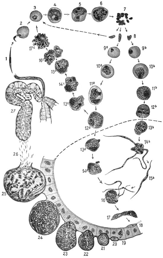
Fig. 80.—Life-cycle of the tertian parasite (Plasmodium vivax). Figs. 1 to 17, × 1,200; figs. 18 to 27, × 600. (After Lühe, based on figures by Schaudinn and Grassi.) 1, sporozoite; 2, entrance of the sporozoite into a red blood corpuscle; 3, 4, growth of the parasite, now sometimes called a trophozoite; 5, 6, nuclear division in schizont; 7, free merozoites; 8, the merozoites which have developed making their way into blood corpuscles, (arrow pointing to the left) and increase by schizogony (3–7); after some duration of disease the sexual individuals appear; 9a-12a, macrogametocytes; 9b-12b, microgametocytes, both still in the blood-vessels of man. If macrogametocytes (12a) do not get into the intestine of Anopheles they may perhaps increase parthenogenetically according to Schaudinn (12a; 13c-17c). The merozoites which have arisen (17c) become schizonts 3–7. The phases shown underneath the dotted line (13–17) proceed in the stomach of Anopheles. 13b and 14b, formation of microgametes; 13a and 14a, maturation of the macrogametes; 15b, microgamete; 16, fertilization; 17, oökinete; 18, oökinete in the walls of the stomach; 19, penetration of the epithelium of the stomach; 20–25, stages of sporogony on the outer surface of the intestinal wall; 26, migration of the sporozoites to the salivary gland; 27, salivary gland with sporozoites.
The growth and schizogony last different times, according to the species of the parasite, about forty-eight hours in the case of the parasite of febris tertiana or tropica, and seventy-two hours for the quartan parasite. The various intermittent forms produced by them depend on this specific difference in the malarial parasites.
The schizogony can, however, only be repeated a certain number of times, supposing that the disease has not been checked prematurely by the administration of quinine, which is able to kill the parasites. It appears that after a number of attacks of fever the conditions of existence in man are unfavourable for the malarial parasites, and this brings about the production of other forms which have long been known, but also long misunderstood (spheres, crescents, polymitus). The merozoites in this case no longer grow into schizonts, or at least not all of them, but become sexual individuals called gametocytes (fig. 80, 9-12), which only start their further development when they have reached the intestine of Anopheles. This does not take place in every case, nor with all the gametocytes which exist in the blood of patients with intermittent fever. Of those parasites which remain in the human blood the male ones (microgametocytes) soon perish, the females (macrogametocytes) persist for some long time, and perhaps at last acquire the capacity of increasing by schizogony. They might thus form merozoites which behave in the body as if they had proceeded from ordinary schizonts (fig. 80, 13c-17c). If their number increases sufficiently, in course of time the patient, who was apparently recovering, has a new series of fever attacks, or relapses, without there having been a new infection. This is the view of Schaudinn, who from researches of his own concluded that relapses were brought about by a sort of parthenogenetic reproduction of macrogametocytes. R. Ross, on the contrary, believes that in the relatively healthy periods the number of parasites in the blood falls below that necessary to provoke febrile symptoms; relapses then result merely from increase in the numbers of the parasites present in the individual. Ross’s view is now generally accepted.
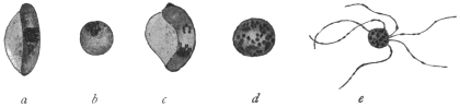
Fig. 81.—Stages of development of pernicious or malignant tertian parasites in the intestine of Anopheles macultpennis. (After Grassi.) a, macrogametocyte (crescent) still attached to human blood corpuscles; b, macrogametocyte (sphere) half an hour after ingestion by the mosquito; c, microgametocyte (crescent) attached to the blood corpuscle; d, microgametocyte (sphere) half an hour after ingestion; the nucleus has divided several times; e, microgametes attached to the residual body (polymitus stage).
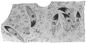
Fig. 82.—Oökinete of the malignant tertian parasite in the stomach of Anopheles maculipennis, thirty-two hours after ingestion of blood. (After Grassi.)
If the gametocytes, which are globular, or in the pernicious or malignant tertian parasite crescentic (fig. 81), gain access to the intestine of an Anopheline,199 they mature. The macrogametocytes extrude a part of their nuclear substance (fig. 80, 13a, 14a) and thereby become females or macrogametes. The microgametocytes, on the other hand, undergo repeated nuclear division, preparation for this being made apparently whilst in the blood of man. This results in the formation of threadlike bodies which move like flagella and finally detach themselves from the residual body (fig. 80, 13b, 14b). These are the males or microgametes200 (fig. 80, 15b).
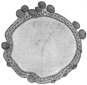
Fig. 83.—Section of the stomach of an Anopheles, with cysts (oöcysts) of the malignant tertian parasite. (After Grassi).
Copulation takes place in the stomach of the Anopheline (fig. 80, 16). A microgamete penetrates a macrogamete and coalesces with it. The fertilized females elongate very soon and are called oökinetes or “vermicules” (figs. 80, 17; 82). They penetrate the walls of the stomach, pierce the epithelium (fig. 80, 18, 19), and remain lying between it and the superficial stratum (tunica elastico-muscularis). Then they become rounded and gradually develop into cysts which grow larger and are finally visible to the naked eye, being called oöcysts (figs. 80, 20-24; 83). Their size at the beginning is about 5 µ, the maximum that they attain is 60 µ, only exceptionally are they larger.
The sporulation (figs. 80, 21-25; 84), which now follows, begins with repeated multiple fission of the nucleus. Long before the definitive number of nuclei, which varies with the individual, is attained the protoplasm, according to Grassi, begins to segment around the individual large nuclei but without separating completely into cell areas. According to Schaudinn, however, there is a condensation of the outstanding protoplasmic strands. It is certain that the number of nuclei increases with simultaneous decrease in size. They soon appear on the surface of the strands or sporoblasts, surround themselves with some cytoplasm and then elongate (fig. 84). In this manner the sporozoites are formed and break away from the unused remains of the cytoplasmic strands of the sporoblasts (fig. 80, 26). The number of the sporozoites in an oöcyst varies from several hundreds to ten thousand.
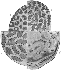
Fig. 84.—Four different sporulation stages of malarial parasites from Anopheles maculipennis, much magnified. a-c, of the malignant tertian parasite; a, four to four and a half days after sucking; b and c, five to six days after sucking; d, of the tertian parasite, eight days after sucking. (After Grassi.)
The sporulation is influenced in its duration by the external temperature (Grassi, Jansci, Schoo). In the tertian parasite it takes place quickest at a temperature of 25° to 30° C. and takes eight to nine days. A temperature a few degrees lower has a retarding effect (eighteen to nineteen days at 18° to 20° C). A still lower one has a restraining or even destructive effect. Temperatures over 35° C. also exercise a harmful effect. The malignant tertian parasite seems to need a somewhat higher temperature and the quartan parasite a lower one.
The sporozoites of the various malarial parasites show no specific differences. They were stated by Schaudinn to occur in three forms, and these were described as indifferent (neuter), female and male. There is, however, little or no evidence for this hypothetical differentiation. The last were said to perish prematurely, that is, in the oöcyst. The others after the rupture of the oöcysts enter the body cavity of the Anophelines, whence they are carried along in the course of the blood. Finally they penetrate the salivary glands (fig. 80, 27) probably by their own activity, break through their epithelia and accumulate in the salivary duct (fig. 80, 27). At the next bite by the mosquito they are transmitted to the blood-vessels of man.
The Species of Malarial Parasites of Man.
In view of the differences in opinion regarding “species” and “varieties,” the dispute whether the malarial parasites of man represent one species with several varieties, or several species is almost superfluous. If necessary two genera may be distinguished.
The parasites of the tertian and quartan fever are alike in that their gametocytes have a rounded shape (figs. 80, 12, 13), whilst the corresponding stages of the pernicious or malignant tertian parasites are crescentic (figs. 81, 88). These differences are used by some writers as the distinguishing characteristic of two genera: Plasmodium, Marchiafava and Celli, 1885, for the first mentioned species; Laverania, Grassi and Feletti, 1889, for the pernicious or malignant tertian parasite. Whether there is a genuine quotidian fever and accordingly a special quotidian parasite is still disputed.
These parasites are treated in practical detail in Stephens and Christophers’ “Practical Study of Malaria,” 3rd edition, 1908.
Plasmodium vivax, Grassi and Feletti, 1890.
Syn.: Hæmamœba vivax, Grassi and Feletti, 1890; Plasmodium malariæ var. tertianæ, Celli and Sanfelice, 1891; Hæmamœba laverani var. tertiana, Labbé, 1894; Hæmosporidium tertianum, Lewkowitz, 1897; Plasmodium malariæ tertianum, Labbé, 1899: Hæmamœba malariæ var. magna, Laveran, 1900, p.p.; Hæmamœba malariæ var. tertianæ, Laveran, 1901.
This species, P. vivax,201 is the causal agent of the simple or spring tertian fever and is, therefore, named directly the tertian or benign tertian parasite (figs. 80, 3-8; 85). During the afebrile period in the patient, the young trophozoites or amœbulæ appear on or in the red blood corpuscles as pale bodies of 1·5 µ to 2 µ diameter which at first show only slow amœboid movements. Their nucleus is difficult to recognize in the early stage. Soon the food vacuole is formed and this grows concomitantly with the trophozoite and the parasite has a ring-like appearance. Afterwards the vacuole diminishes, and at this period the first brownish melanin granule appears. From this time the activity and number of the pigment granules increase with continuous growth. When the parasite has grown to about one-third the diameter of the erythrocyte the latter shows characteristic red Schüffner’s dots or “fine stippling,” after staining with Romanowsky’s solution. Later, after about twenty-four hours, the blood corpuscles begin to grow pale, then to increase in size, and after thirty-six hours, that is, about twelve hours before the next attack of fever, schizogony of the parasite is initiated by the division of the nucleus. The parasite at this time occupies half to two-thirds of the enlarged blood corpuscle. The daughter nuclei continue dividing until sixteen, and occasionally twenty-four, daughter nuclei are produced. The pigment which, up till now lies nearer the periphery, moves to the middle, while the nuclei lie nearer the surface.
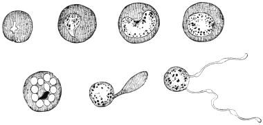
Fig. 85.—Development of the tertian parasite in the red blood corpuscles of man; on the right a “Polymitus.” (After Mannaberg.) See also fig. 80, 3—7.
Around each nucleus a portion of cytoplasm collects and thus young merozoites are produced. These separate from each other and from the little residual masses202 which contain the melanin and pass from the blood corpuscles, which now can hardly be recognized, to the blood plasma, where they soon attack new erythrocytes.
The migration of the merozoites initiates a new attack of fever and two groups of tertian parasites in the blood, differing in development by about twenty-four hours, are the conditions for febris tertiana duplex.
After a lengthy duration of fever the gametocytes (figs. 80, 9—12) appear. They are uninucleate. The microgametocytes are about the size of fully developed schizonts, the macrogametocytes are somewhat larger. Their further development takes place in Anophelines.
The chief distinctive characteristics of the simple tertian parasite, as seen in infected blood, are:—(1) The infected red-cell is usually enlarged; (2) the presence of fine red granules known as Schüffner’s dots in the red blood corpuscles, after Romanowsky staining; (3) the fragile appearance of the parasite compared with other species. Large forms are pigmented, irregular and “flimsy-looking,” sometimes appearing to consist of separate parts. Irregularity of contour is common.
Ahmed Emin203 (1914) has described a small variety of P. vivax.
Plasmodium malariæ, Laveran.
Syn.: Oscillaria malariæ, Laveran, p.p., 1883; Hæmamœba malariæ, Gr. et Fel., 1890; Plasmodium malariæ var. quartanæ, Celli et Sanfel., 1891; Hæmamœba laverani var. quartana, Labbé, 1894; Hæmosporidium quartanæ, Lewkowitz, 1897; Plasmodium malariæ quartanum, Labbé, 1899; Plasmodium golgii, Sambon, 1902; Laverania malariæ, Jancso, 1905 nec Grassi et Fel. 1890; Hæmomœba malariæ var. quartanæ; Lav., 1901.