ON PARASITES IN GENERAL. (continued 8)
Reading edition
Cysticercoid.—The bladder is but slightly developed and is usually absorbed again. The anterior portion is, moreover, retracted into the posterior, and in some cases there is a long or a stumpy tail (figs. 220, 231).
Cysticercus, or true bladder worms. (These may be divided into (1) cysticercus proper, consisting of a bladder and one scolex; (2) cœnurus, a bladder and many scolices; (3) echinococcus, a bladder in which daughter bladders or cysts are developed, and then in these multiple scolices.)
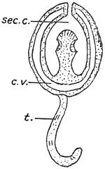
Fig. 198.—Diagram of a cysticercoid. Cf. figs. 220, 227. c.v., caudal vesicle or bladder (small); sec. c., secondary cavity caused by the growth forward of the hind-body; t., tail bearing six spines. (Stephens.)
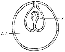
Fig. 199.—Diagram of a cysticercus. c.v., caudal vesicle or bladder; i., invagination of wall of bladder. (Stephens.)
In the case of cysticerci a papilliform invagination forms, projecting into the interior of the bladder (fig. 201). The layer of cells forming the papilla becomes divided into two laminæ, the outer279 of which forms a kind of investing membrane (receptaculum capitis) for the papilla. The head and suckers are now developed on the walls bounding the axial lumen of the papilla. The papilla eventually evaginates, so that the receptaculum capitis now forms the inner surface of the hollow head, which eventually becomes solid.
Our knowledge of the development of cysticerci in the wide sense of the word is limited almost exclusively to that of a few true “bladder worms” (cysticerci); in other cases we know either only the terminal stage, i.e., the complete larva, or, exceptionally, one of the intermediate stages, but we are not acquainted with a complete series; the description must therefore be incomplete.
We know from feeding experiments that, after the introduction of mature proglottids or of the fully developed ova of Tænia crassicollis (of the cat) into the stomach of mice, the oncospheres escape from the shell in the middle portion of the small intestine, and a few hours later penetrate into the intestinal wall by means of a boring movement; they have been found in this position twenty-seven to thirty hours after the infection. By means of this migration, for which purpose they employ their spines, they attain the blood-vessels of the intestine; indeed, already nine hours after the infection and later they are found in the blood of the portal vein, and in the course of the second day after infection they are found in the capillaries of the liver, which these larvæ do not leave.
Leuckart, in experimental feeding of rabbits with oncospheres of Tænia serrata (of the dog), found free oncospheres in the stomach of the experimental animal, but not in the intestine: however, he came across them again in the blood of the portal vein. The passage through the blood-vessels to the liver is the normal one for those species of Tænia the eggs of which become larvae in mammals; even in those cases in which the oncospheres develop further in the omentum or in the abdominal cavity (Cysticercus tenuicollis, C. pisiformis), there are distinct changes observable in the liver that lead one to the conclusion that there has been a secondary migration out of the liver into the abdominal cavity. Indeed, one must not imagine that the young stages of the Cestodes are absolutely passive; once they have invaded an organ they travel actively, and leave distinct traces of their passage.
In other cases the oncospheres leave the liver with the circulation, and are thus distributed further in the body; they may settle and develop in one or more organs or tissues. Many oncospheres may, by travelling through the intestinal wall, penetrate through it and attain the abdominal cavity direct; some, perhaps, pass also into the lymph stream. Where there are no blood and lymphatic vessels in the intestinal wall, as in insects, the oncospheres attain the body cavity or its organs direct; in short, they never remain in the intestinal lumen itself, and only rarely—as in Hymenolepis murina of the rat—do they remain in the intestinal wall.
When the infection has been intense, and the body is crowded with numerous oncospheres, acute feverish symptoms, are induced, to which the infected animals usually succumb (“acute cestode tuberculosis”); while in other cases the alterations in the organs attacked—as the liver in mice and the brain in sheep—may cause death.
Sooner or later the oncospheres of tapeworms come to rest, and are first transformed into a bladder, which may be round or oval according to the species. The embryonal spines disappear sooner or later, or remain close together or spread over some part of the bladder wall (fig. 200). Their discovery by V. Stein in the bladder worm of the “meal worm” (the larva of a beetle, Tenebrio molitor) first led to the conclusion that bladder worms (cysticerci) actually originate from the oncospheres of Tæniidæ.
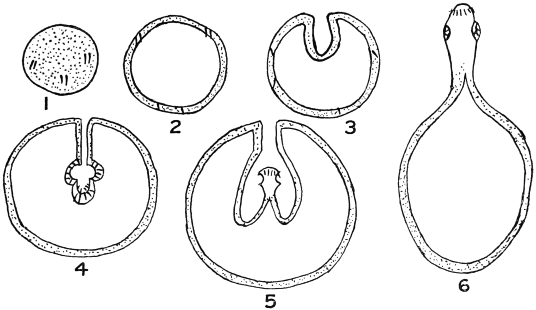
Fig. 200.—Diagram of development of a cysticercus. 1, solid oncosphere with six spines; 2, bladder formed by liquefaction of contents; 3, invagination of bladder wall; 4, formation of rostellum (with hooklets) and suckers at the bottom of the invagination; 5, evagination of head; 6, complete evagination effected by pressure. (Stephens.)
The bladder may remain as a bladder, and then by proliferation the scolex forms on its wall (fig. 202), or it may divide into an anterior so-called “cystic” portion and a solid tail-like appendage of various lengths, on which the embryonal hooks are to be found, and this is particularly the case in those larval forms (cysticercoids), e.g., those of Dipylidium caninum, that develop in invertebrate animals, such as Arthropoda.
As mentioned above one may regard the scolex as an individual that originates through proliferation of the wall of the parent cyst, mostly singly, but in those cysticerci that are termed cœnurus (fig. 201) many scolices occur, whereas in those called echinococcus the parent cyst originating from the oncosphere of Tænia echinococcus (of the dog) first produces a number of daughter cysts, which in their turn form numerous scolices. Echinococcus-like conditions also occur in cysticercoids, as, for instance, in those peculiar to earthworms; and similar conditions prevail in a larval form known as Staphylocystis, found in the wood-louse (Glomeris). Thus it happens in these cases that finally one tapeworm egg produces not one, but numerous tapeworms, for, under favourable conditions, each scolex can form a tapeworm.
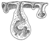
Fig. 201.—Section through a piece of a Cœnurus cerebralis, with four cephalic invaginations in different stages of development. At the bottom of the invaginations the rostellum, hooks and suckers develop. (From a wax model.)
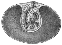
Fig. 202.—Median section through a cysticercus, with developed scolex at the bottom of the invagination. (After Leuckart.)
The rudiment of the scolex appears as a hollow bud, the cephalic invagination usually directed towards the interior of the bladder cavity; on its invaginated surface arise the four suckers, and the rostellum with the hook apparatus is formed in its blind end; we thus get a Tænia head, but with the position of the parts reversed (fig. 201). In many cysticerci the head rises up from the base of the cephalic invagination and is then surrounded by the latter. A more or less elongated piece of neck also develops, and even proglottids may appear, as in Cysticercus fasciolaris (the larva of Tænia crassicollis of the cat) of the Muridæ, a process somewhat analogous to that of Ligula, etc.
The period that elapses from the time of infection till the cysticercus is fully developed varies according to the species; the cysticercus of Tænia saginata requires twenty-eight weeks, that of T. marginata seven to eight weeks, that of T. solium three to four months, and that of T. echinococcus longer still.
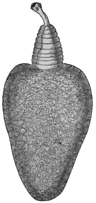
Fig. 203.—Cysticercus pisiformis in an evaginated condition, with neck, fore-body and bladder, with excretory network in its wall. 18/1.
With one single exception (Archigetes) the larvæ do not become sexually mature in the organ where they have developed; they must enter the terminal host, a matter that is usually purely passive, the carriers of the larvæ or infected parts of them being usually devoured by other animals. In this manner, for instance, the larvæ (Cysticercus fasciolaris) found in mice and rats reach the intestine of cats; those of the hare and rabbit (C. pisiformis) reach the intestine of dogs; those of the pig (C. cellulosæ) are introduced into man; those of insects are swallowed by insectivorous birds; those of crustaceans are ingested by ducks and other water fowl; perhaps, also, the infection of herbivorous mammals is caused by their accidentally swallowing smaller creatures infected by larvæ. Indeed, the researches of Grassi and Rovelli have taught us that such an intermediate host is not always necessary; Hymenolepis murina of rats and mice in its larval stage lives in the intestinal wall of these rodents, and as a larva it passes into the intestinal lumen and develops into a tapeworm in exactly the same way as the larvæ of other species that reach the intestine of the terminal host by means of an intermediate carrier. Probably this curtailed manner of transmission also occurs in many other species. In some cases the larvæ actively quit the body of the intermediate host, as in the case of Ligula and Schistocephalus, which travel out of the body cavity of infected fish and reach the water, where they may be observed in hundreds in summer, at all events in some localities. The larval stage of Calliobothrium—wrongly termed Scolex—has been observed swimming free in the sea, and the scolices of Rhynchobothrium, without their mother cysts, have been observed free within the tissues of several marine animals. In any case there is almost always a change of hosts, even in the single-jointed Cestodes, for the larva of Caryophyllæus, which lives in fishes of the carp family, is found in limicoline Oligochætes, that of Gyrocotyle (Chimæra) in shell-fish (Mactra), and different conditions can hardly be possible for Amphilina. Archigetes alone becomes sexually mature in the larval stage, but the life-history of this creature is not well known, so that it is not impossible that the attainment of sexual maturity as a larva in invertebrates (Oligochætes) is perhaps abnormal, and somewhat analogous to the maturity of some encysted Trematodes.
The METAMORPHOSIS OF THE LARVA into the tapeworm is rarely accomplished in a simple manner; the transformation, however, is not complex in the single-jointed Cestodes, nor in Ligula and Schistocephalus; the latter is swallowed by birds (Mergus, Anas, etc.), produces eggs after only a few days, and very soon quits the intestine of its terminal host. In all other cases it is the scolex only which, by proliferating at its posterior extremity, forms the proglottids, after having invaded as a larva the intestine of a suitable host. The mother cysts, or what corresponds to them, die, are digested, absorbed, or perhaps even eliminated; on the contrary, segments found on the scolex during the larval stage, also in the case of Cysticercus fasciolaris, are retained. It is not certain whether the larvæ of Dibothriocephalus lose any part.
The time required by the scolex to complete the entire chain of proglottids does not depend only on the number it has to produce, for Tænia echinococcus, which, as a rule, only possesses three or four segments, takes quite as long a time for their growth (eleven to twelve weeks) as T. solium with its numerous segments; T. cœnurus is fully developed in three to four weeks, and the same holds good for Dibothriocephalus latus, which possesses many more segments than the above-mentioned Tænia of the dog. In a number of species it has been possible to determine fairly accurately the average daily growth; for instance, in Dibothriocephalus latus the daily growth is 8 cm., in Tænia saginata 7 cm., etc.
The history of the development of the Cestodes demonstrates that persons and beasts harbouring larval tapeworms have become infected by having swallowed the oncospheres of the species of tapeworm to which they belong. In regard to Hymenolepis murina alone, it is known that the introduction of the oncospheres into those species of animals which harbour the adult tapeworm leads to the formation of the latter after the development of a larval stage in the intestinal wall; nevertheless, only young animals (rats) are capable of infection, for a previous infection, or the presence of mature tapeworms in the intestine, appears to produce a kind of immunity.
Biology.
In their adult stage, the tapeworms inhabit almost exclusively the alimentary canal of vertebrate animals, with but few exceptions the small intestine, and a few species select definite parts of it. A small number of Rhynchobothriidæ of marine fishes live apparently always in the stomach, while in rays and sharks the spiral intestine is their exclusive site. Bothriocephali generally attach themselves with their head on to the appendices of the pylorus of fishes; other species (Hymenolepis diminuta) occasionally fix their head in the ductus choledochus, and this is more frequent still in the tapeworms of the rock badger (Hyrax), which occasionally penetrate entirely into the biliary ducts. Stilesia hepatica, Wolffh., has so far only been found in the bile-ducts of its host (sheep and goat, East Africa).
In the disease of sheep induced by Cestodes, the worms have been observed also in the pancreas. Specimens found in the large intestines were probably being evacuated.
The Cestodes are looked upon as fairly inert creatures, this opinion having been formed by observing their condition in the cold cadavers of warm-blooded animals. Actually, however, they are exceedingly active, and accomplish local movements within the intestine, for they have been found in the ducts communicating with the bowel, or in the stomach, and may even make their way forward into the œsophagus.
They also invade other abdominal organs through abnormal communications, or through any that may be temporarily open between the intestine and such organs; they thus reach the abdominal cavity or the urinary bladder, or they work their way through the peritoneum.
They produce changes in the intestinal mucous membrane at the place of their attachment, the alterations varying in intensity according to the structure of the fixation organs. The mucous membrane is elevated in knob-like areas by the suckers; the epithelial cells become atrophied or may be entirely obliterated. Dipylidium caninum bores into the openings of Lieberkühn’s glands with its rostellum, dilating the lumen to two or three times its normal size, while the suckers remain fixed between the basal parts of the cells. Species with powerful armatures penetrate deeper into the submucosa, and some that are not provided with exceptionally strong armatures, or are even unarmed, may be actually found with the scolex embedded in the muscles of the intestinal walls or even protruding beyond (Tænia tetragona, Mol., in fowls, etc.). Other species, again, even cause perforation of the walls of the intestine of their hosts.
It is generally assumed that tapeworms, which almost without exception live in the gut of vertebrates, get their nutriment from the gut contents, which apparently they absorb through the whole body surface (cuticular trophopores). In favour of this view is the existence of fat drops in the proglottids, the identity in colour in certain forms between that of the fresh worm and the gut contents and the passage of certain substances derived from medicines (iron and mercury preparation) into the worms in the gut, etc. Whether the suckers are concerned in the absorption of nutriment and to what extent is still questionable.
The length of life of the adult tapeworm certainly varies; as a rule it appears to last only about a year; in other cases (Ligula) it averages only a few days, but we are likewise aware that certain species of Cestodes of man attain an age of several or many years (thirty-five). The natural death of Cestodes often appears to be brought about by alterations in the scolex, such as loss of the hooks, atrophy of the suckers and rostellum, finally the dropping off of the scolex; it is unknown whether a chain of segments deprived of its scolex then perishes or whether it first attains maturity. It has already been mentioned that in a few species the foremost proglottids are transformed into organs of fixation on the normal loss of the scolex.
Abnormalities and malformations are encountered relatively frequently in the Cestodes—such as abnormally short or long segments; the so-called triangular tapeworms, which—if belonging to the Tæniidæ—always possess six suckers; often also club-shaped segments occur between normal ones, or there may be a defect in one segment or in the centre of a number following one another (fenestrated segments); bifurcated chains of segments have likewise been observed, as well as incomplete or complete union of the proglottids, abnormal increase of the genital pores, reversion of the genitalia. Besides the above-mentioned increase of the number of suckers on the scolex (in Tæniæ), there may be a decrease in the number; in other cases the crown of hooks may be absent, or abnormally shaped hooks may be formed.
Classification of the Cestoda of Man.
Order. Pseudophyllidea, Carus, 1863.
Scolex without proboscis or rostellum. Head “stalk” absent.
Scolex never with four, generally with two (or one terminal) bothria.280 Vitellaria numerous. Uterine opening present. Genitalia do not atrophy when uterus is developed. In large majority of proglottids eggs (or, if formed, their contents) are at the same stage of development.
Family. Dibothriocephalidæ, Lühe, 1902.
Syn.: Diphyllobothriidæ, Lühe, 1910.
Genitalia repeated in each proglottid (polyzootic Cestodes). Ventral and dorsal surfaces flat. Cirrus unarmed. Cirrus and vagina if non-marginal open on the same surface as the uterus. Uterus long, convoluted, often forming a “rosette,” never dilates into a uterine cavity. Eggs thick shelled, operculated, constantly being formed in mature proglottids.
Sub-family. Dibothriocephalinæ, Lühe, 1899.
Syn.: Diphyllobothriinæ, Lühe, 1910.
Segmentation distinct. Scolex unarmed, elongated, sharply separated (generally by a neck) from the first proglottis. Cirrus and vagina open ventrally. Genital pores non-alternating. Vas deferens surrounded by a muscular bulb. Receptaculum seminis large, sharply separated from the spermatic duct.
Order. Cyclophyllidea, v. Beneden.
Four suckers always present. Uterine opening absent. Vitellarium single. Genitalia atrophy when uterus is fully developed.
Family. Dipylidiidæ, Lühe, 1910.
Rostellum if present armed. Suckers unarmed. Uterus breaks up into egg capsules. Paruterine organs absent.
Family. Hymenolepididæ, Railliet and Henry, 1909.
Segment always broader than long. Genitalia single. Longitudinal muscles in two layers. Genital pores unilateral. Testes one to four. Uterus persistent, sac-like. Eggs with three shells.
Family. Davaineidæ, Fuhrmann, 1907.
Rostellum cushion-shaped. Armed with numerous (sixty to several thousand) hammer-shaped hooks in two (rarely one) rows.
Sub-family. Davaineinæ, Braun, 1900.
Suckers armed. Uterus breaks up into egg capsules. Paruterine organs absent.
Family. Tæniidæ, Ludwig, 1886.
Suckers unarmed. Uterus with median longitudinal stem and lateral branches. Female genitalia at the hind end of the proglottis. Genital pore irregularly alternating. Testes numerous in front of female genitalia. Ovary with two lobes (wings). Vitellarium behind the ovary. Embryophore radially striated.
The Cestodes of Man.
Most of the species to be mentioned live in man in their adult stage and occupy the small intestine; man is the definite host of these parasites, but is not the specific host for all the species; some of these species, as well as others (of mammals), may occur in man also in the larval stage.
Family. Dibothriocephalidæ.
Sub-family. Dibothriocephalinæ.
Genus. Dibothriocephalus, Lühe, 1899.
Syn.: Diphyllobothrium, Cobbold, 1858; Bothriocephalus, p. p. Rud., 1819; Dibothrius, p. p. Rud., 1819; Dibothrium, p. p. Dies., 1850.
Scolex egg-shaped; dorsal and ventral bothria elongated, moderately strong, cutting rather deeply into the head; genitalia single in each proglottis; papillæ in the vicinity of the genital atrium; the testes and vitellaria are in the lateral fields, the former in the medullary layer, the latter in the cortical layer on both surfaces, and occasionally extending to the median line; the ovary ventral, the shell gland dorsal. The uterus is in the central field, taking a zigzag course, and frequently forms a rosette.
Dibothriocephalus latus, L., 1748.
Syn.: Tænia lata, L., 1748; Tænia vulgaris, L., 1748; Tænia grisea, Pallas, 1796; Tænia membranacea, Pall., 1781; Tænia tenella, Pall., 1781; Tænia dentata, Batsch, 1786; Bothriocephalus latus, Bremser, 1819; Dibothrium latum, Dies., 1850; Bothriocephalus cristatus, Davaine, 1874281; Bothriocephalus balticus, Kchnmstr., 1855; Bothriocephalus latissimus, Bugn., 1886.
Length 2 to 9 m. or more; colour yellowish-grey; after lying in water the lateral areas become brownish and the uterine rosette brown. The head is almond-shaped, 2 to 3 mm. in length, the dorso-ventral axis is longer than the transverse diameter; the head, therefore, generally lying flat, conceals the suctorial grooves at the borders; these suckers are deep and have sharp edges (fig. 205). The neck varies in length according to the degree of contraction and is very thin; there are 3,000 to 4,200 proglottids and there may be more; their breadth is usually greater than their length, but in the posterior third of the body they are almost square, and the very oldest are not uncommonly longer than they are broad. There are numerous testes situated dorsally in the medullary layer of the lateral fields; the vas deferens (fig. 192) passes dorsally in transverse loops in the central field anteriorly and forms a seminal vesicle before its entry into the large cirrus pouch.
The orifice of the vagina is close behind the orifice of the cirrus; the former passes almost straight along the median line posteriorly, and widens into a receptaculum seminis shortly before its junction with the oviduct; the ovary is bilobed, in shape like the wings of a butterfly, ventrally in the medullary layer; the shell glands lie in the posterior recess of the ovary; the uterus, forming numerous transverse convolutions, passes ventral to the vas deferens forwards. Eggs (fig. 207) large, with brownish shells and small lids, 68 µ to 71 µ by 45 µ; the ovarian cell, which is already, as a rule, in process of segmentation, is surrounded by numerous large yolk cells; the proglottids nearest the posterior extremity are frequently eggless.
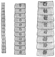
Fig. 204.—Various chains of segments of Dibothriocephalus latus, showing the central uterine rosette. (Natural size.)
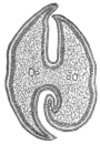
Fig. 205.—Transverse section of the head of Dibothriocephalus latus. 30/1.
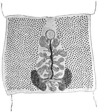
Fig. 206.—Fairly mature proglottis of Dibothriocephalus latus. The vitellaria are at the sides; the uterus, filled with eggs, is in the middle, also the vagina (the dark stripe passing almost straight from the front to the back), and the vas deferens (almost hidden by the uterus). Above in the centre is the cirrus sac, and below the shell gland and ovary are seen. 15/1. (From a stained preparation.)
The eggs, which are deposited in the intestine and evacuated with the fæces, hatch in water after a fortnight or more; the embryonal integument (embryophore) of the oncosphere is provided with cilia; after bursting open the lid of the egg the oncosphere in its embryophore (fig. 207) reaches the water and swims slowly about; often it slips out of its ciliated embryophore, sinks to the bottom and is capable of a creeping motion; sooner or later it dies in the water. The manner and means of its invasion of an intermediate host are still unknown; yet we are aware that the larval stage (plerocercoid, fig. 208), which resembles the scolex and may reach a length of 30 mm., lives in the intestine, in the intestinal wall, in the liver, spleen, genital glands and muscular system (fig. 209) of various fresh-water fish, the pike (Esox lucius), the miller’s thumb (Lota vulgaris), the perch (Perea fluviatilis), Salmo umbla, Trutta vulgaris, Tr. lacustris, Thymallis vulgaris (grayling), Coregonus lavaretus, C. albula (in Europe) and Onchorhynchus perryi (in Japan). The transmission of the plerocercoids from these fish to the dog, cat and man (Braun, Parona, Grassi and Ferrara, Grassi and Rovelli, Ijima, Zschokke, Schroeder) leads to the development of the broad tapeworm, the growth of which is rapid. In my experiments on human beings the average number of proglottids formed per diem averaged thirty-one to thirty-two for five weeks, with a length of 8 to 9 cm. According to Parona the eggs appear twenty-four days after man has been infected. Zschokke found the average growth in the experimental infection of man between 5·2 and 8·2 cm. per diem, and the person experimented upon by Ijima evacuated a piece of a Dibothriocephalus latus, 22·5 cm. in length, only twenty-one days after the infection.
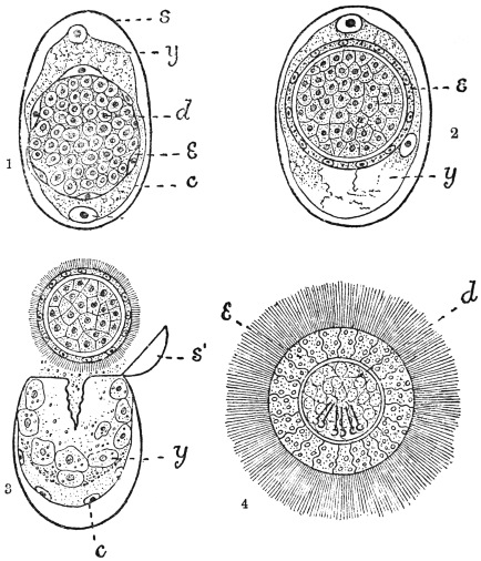
Fig. 207.—Dibothriocephalus latus: development of egg. 1, segmentation complete; some cells of the blastosphere have migrated through the yolk and have flattened to form c, the yolk envelope; others form a layer of flattened cells (e) forming the embryophore; the remaining cells (d) of the blastosphere form the hexacanth embryo. 2, embryophore (e) is becoming thicker. 3, the ciliated embryo has been pressed out of the shell; s′, the operculum; c, the yolk envelope remaining in the shell (s); y, the yolk consisting of separate cells. 4, a free-swimming larva much swollen by the water. (After Benham and Schauinsland.)
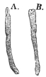
Fig. 208.—Plerocercoid of Dibothricephalus latus. A., with the head evaginated; B., with the head invaginated. From the muscle of the pike.
The “broad tapeworm” is a frequent parasite of man in some districts, but it also occurs in the domestic dog, and on rare occasions is found in the domestic cat (together with Dibothriocephalus felis, Crepl.) and fox. French Switzerland and the Baltic Provinces of Russia are the centres of distribution; from the former districts the distribution radiates to France and Italy (Lombardy, Piedmont); from the Baltic Provinces over Ingermanland to Petrograd, over Finland to Sweden (on the shore of the Gulf of Bothnia), in a southerly direction to Poland, and into the Russian Empire and across it to Roumania, and towards the west along the coast of the Baltic Sea to the North Sea, where, however, its frequency considerably diminishes (Holland, Belgium, and the North of France).
In Turkestan and Japan the “broad tapeworm” is the most frequent parasite of man; it has been reported in Africa from the vicinity of Lake N’gami as well as from Madagascar; cases, in part at least imported, have also come under observation in North America.
In Germany Dibothriocephalus latus—apart from the fact that it is undoubtedly imported from Switzerland, Russia or Italy—is particularly frequent in East Prussia amongst the inhabitants of the Courland Lagoon district, on the Baltic; it is, moreover, also found in the Province and even in the City of Königsberg. In West Prussia and Pomerania it is very much scarcer.
It is also found in Munich and in the vicinity of the Lake of Starnberg (Bollinger).
Krabbe found it in 10 per cent. of the sufferers from tapeworms in Denmark; Szydlowski found the ova of this worm in Dorpat in 10 per cent. of the fæces examined; Kruse found the worm in 6 per cent. of post-mortems; Kessler, in Petrograd, found the eggs in the fæces in 7·8 per cent.; at post-mortems he found the worms in 1·17 per cent., though Winogradoff only found it in 0·8 per cent. In Moscow, according to Baranovsky, 8·9 per cent. of the fæces examined contained the ova of Dibothriocephalus. In the interior and southern provinces of Sweden the worm, according to Lönnberg, is only found sporadically, but, on the other hand, in Angermanland about 10 per cent. of the population is affected; while again in Norbotten the majority of persons are affected, and in Haparanda the entire population (with the exception of infants) harbour this parasite. In Switzerland D. latus is very frequent in close proximity to the lakes of Bieler, Neuchatel, Morat and Geneva (according to Zaeslin 10 to 15 to 20 per cent. of the population are affected); the parasite is less frequent in districts one to four hours removed from these lakes.
Of the fish from Swiss lakes examined by Schor those from Lake Geneva were most commonly infected, and especially Lota sp. and Perea sp.
The frequency and distribution have, nevertheless, decreased perceptibly in places; at the commencement of the eighteenth century the broad tapeworm was very common in Paris, at the present date it only occurs when imported (Blanchard); in Geneva, also, according to Zschokke, it has become rarer (formerly 10 per cent., now only 1 per cent.).
The disturbances produced in man by the presence of broad tapeworms are, as a rule, very trifling; in other cases they produce partly gastric disorders and partly nervous symptoms; in a number of cases, again, they set up severe anæmia, apparently caused by toxins produced by the worms and absorbed by the host. There is no danger of auto-infection, as the larval stage lives only in fishes, not in warm-blooded animals. The case reported by Meschede (ova like those of Dibothriocephalus latus in the brain of a man who had suffered from epilepsy for six years) must be otherwise explained.
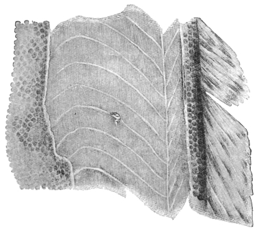
Fig. 209.—A piece of the body wall of the Burbot, Lota vulgaris. The tangential section has exposed the muscles of the trunk, with a plerocercoid of Dibothriocephalus latus. Natural size.
Human beings, like other hosts, can only acquire the broad tapeworm by ingesting its plerocercoids with the previously mentioned fresh-water fishes; the opportunity for such infection is afforded the more readily by the fact that not only do the lower classes not pay sufficient attention to the cooking of fish, so that all the larvæ that are present may be killed, but also in certain localities the custom exists of eating some parts of these fishes in a raw condition; even the mere handling of the usually severely infected intermediary hosts may occasionally cause infection. The plerocercoids are as well known as, but differ materially in appearance from, the cysticerci (Cysticercus cellulosæ) of pig’s flesh. In Germany the occurrence of the plerocercoids of Dibothriocephalus latus has been confirmed in the pike, miller’s thumb and perch of East Prussia, and more particularly in those taken from the Courland Lagoon.
The life of D. latus is a very long one (six to fourteen years), as is deduced from persons who have left D. latus regions after they have been infected.
According to the experiments of M. Schor, plerocercoids of D. latus placed in slowly warmed water completely lose their movement at 54° to 55° C.; they survive the death of their host for several days; they are killed by low temperatures -3° to +1° C. in two days; strong acids and salt solutions kill them at once, also high temperatures, but all the same at least ten minutes is required in boiling or frying fish in order to kill the plerocercoids with certainty.
Dibothriocephalus cordatus, R. Lkt., 1863.
Syn.: Bothriocephalus cordatus, R. Lkt.
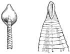
Fig. 210.—Cephalic end of Dibothriocephalus cordatus; on the left viewed sideways, on the right from the dorsal surface, showing a suctorial groove. (After Leuckart.)
Length, 80 to 115 cm.; the head is heart-shaped and measures 2 by 2 mm. The suctorial grooves are on the flat surface; the segments commence close behind the head and increase rapidly in breadth. At only 3 cm. behind the head they are already mature; the greatest breadth attained by them averages 7 to 8 mm., the length 3 to 4 mm.; the number of proglottids averages 600; the most posterior ones are usually square. The uterine rosette is generally formed of six to eight lateral loops. The eggs are operculated and measure 75 µ by 50 µ.
Dibothriocephalus cordatus is a common parasite of the seal, the walrus and the dog in Greenland and Iceland, occasionally of man also. No doubt its larva lives in fishes.
The statement that D. cordatus also occurs in Dorpat in human beings has been proved erroneous (Zool. Anzeiger, 1882, v, p. 46), as also has the report that this worm lives in hares in the neighbourhood of Berlin, whither it was supposed to have been carried by Esquimaux dogs (Rosenkranz in Deutsch. med. Wochenschr., 1877, iii, p. 620). The parasite stated by the author to be D. cordatus is Tænia pectinata, Goeze, which has been known since 1766.
Dibothriocephalus parvus, Stephens, 1908.
Largest gravid segments 5 by 3 mm. Uterus forms a central rosette with four to five loops on each side of median line. In a proglottid measuring 3·5 by 2·25 mm. the genital atrium is situated 0·4 to 0·5 mm. behind the anterior margin and the uterine opening the same distance behind the genital atrium. Calcareous corpuscles absent in the preserved specimens. Eggs operculated, 59·2 µ by 40·7 µ.
Distinguished from Dibothriocephalus latus—(1) by the size of gravid segments (the minimum width of gravid segments of D. latus is 10 to 12 mm., so that D. parvus is a much smaller worm); (2) quadrate segments of D. latus measure 6 by 6 mm., those of D. parvus 4 by 4 mm.; (3) by the eggs.
From D. cordatus it is distinguished by—(1) D. cordatus has only fifty immature segments, D. parvus has at least 200, possibly more; (2) mature segments of D. cordatus measure 7 to 8 mm., maximum width of D. parvus is 5 mm.; (3) quadrate segments of D. cordatus measure 5 to 6 mm.; (4) D. cordatus has six to eight uterine loops; (5) D. cordatus measures 75 µ to 80 µ by 50 µ.
Habitat.—Intestine of man (Syrian, in Tasmania).
Genus. Diplogonoporus, Lönnbrg., 1892.
Syn.: Krabbea, R. Blanch., 1894.
The scolex is short and has powerful suctorial grooves; no neck; the proglottids are short and broad; there are two sets of genital organs side by side in each segment, which in all essentials resemble the single one of Dibothriocephalus.
Parasitic in whales and seals, occasionally in man.
Diplogonoporus grandis, R. Blanch., 1894.
Syn.: Bothriocephalus sp., Ijima et Kurimoto, 1894; Krabbea grandis, R. Blanch.
Scolex unknown; chain of proglottids over 10 m. in length, 1·5 mm. broad anteriorly, 25 mm. broad posteriorly. The proglottids are very short (0·45 mm.), but 14 to 16 mm. broad. On either side to the right and left of the worm, along the entire ventral surface, there is a longitudinal groove; these grooves are nearer to each other than to the lateral margin; in them lie the genital pores, and they are in the same sequence as in Dibothriocephalus; corresponding to the scanty length (0·45 mm.) of the proglottids, the ovary is only developed transversely; the uterus only makes a few loops. Eggs (fig. 195) thick shelled, brown, 63 µ by 48 µ to 50 µ. This parasite has hitherto been observed twice in Japanese. Similar species are known in Cetacea and seals.
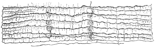
Fig. 211.—Diplogonoporus grandis, Lühe, 1899: ventral view of a portion of the strobila, showing two rows of genital pores and partially extruded cirri. (After Ijima and Kurimoto.)
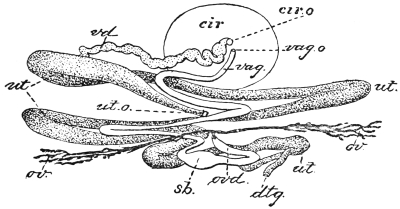
Fig. 212.—Diplogonoporus grandis: ventral view (diagrammatic) of genitalia of left side; cir, cirrus; cir.o, cirrus opening; dtg., vitelline duct; ov., ovary; ovd., oviduct; sb., receptaculum seminis; ut., uterus; ut.o., uterine pore; vag., vagina; vag.o., vaginal pore; vd, vas deferens. × 150. (After Ijima and Kurimoto.)
Sparganum, Diesing, 1854.
The term Sparganum, invented by Diesing, is used as a group name of larval bothriocephalid Cestodes whose development is not sufficiently advanced to enable them to be assigned to any particular genus.
Sparganum mansoni, Cobb., 1883.
Syn.: Ligula mansoni, Cobbold, 1883; Bothriocephalus linguloides, R. Lkt., 1886; Bothriocephalus mansoni, R. Blanch., 1886.
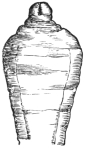
Fig. 213.—Cephalic end of Sparganum mansoni, Cobb. (After Leuckart.)

Fig. 214.—Sparganum mansoni: on the right in transverse section. Natural size. (After Ijima and Murata.)
These plerocercoids were discovered in 1882 by P. Manson during the post-mortem on a Chinaman who had died in Amoy, twelve specimens being found beneath the peritoneum and one free in the abdominal cavity. Cobbold described them as Ligula mansoni, and Leuckart, who contemporaneously reported a case in Japan, termed them Bothriocephalus liguloides. Ijima and Murata reported eight further cases, and Miyake records nine further cases, seven of which are recorded in Japanese literature.
The plerocercoid, which hitherto alone is known to us, attains a length of 30 cm. and a breadth of 3 to 6 to 12 mm. The ribbon-shaped body is wrinkled, the lateral borders are often somewhat thickened, so that the transverse section has the form of a biscuit; the anterior end is usually wider and has the head provided with two weak suctorial grooves, either retracted or protracted.
The parasite makes migrations within the body, and thus may reach the urinary passages; then it is either evacuated with the urine or has to be removed from the urethra; not rarely it causes non-inflammatory tumours on various parts of the skin, which are at times painful and at times vary in size.
Nothing is known of its development and origin.
Sparganum proliferum, Ijima, 1905.
Syn.: Plerocercoides prolifer, Ijima, 1905; Sparganum prolifer, Verdun, Manson, 1907.
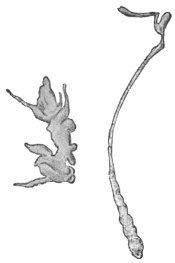
Fig. 215.—Sparganum prolifer: left with buds, right extended. × 4. (After Ijima.)
These plerocercoids produce an acne-like condition of the skin. The condition is really one of capsules in great abundance in the subcutaneous tissue and less so in the corium and in the intermuscular connective tissue. The encapsuled worms in the corium feel like embedded rice grains and raise the epidermis, giving rise to an acne-like condition. Many thousands occur scattered over the body; in Ijima’s Japanese case there were over 10,000 in the left thigh. The worms when they first appear in the skin cause itching. The capsules are ovoid, generally about 1 to 2 mm. in diameter, but they may be smaller and also much larger. The larger ones occur in the subcutaneous tissue. The capsules consist of dense tough connective tissue.
Each capsule, as a rule, contains one worm, but as many as seven may occur. The skin of areas that have been long infected is swollen and indurated or adherent, giving a somewhat elephantoid appearance. The subcutaneous tissue is thick and filled with slimy fluid or in other parts sclerosed.
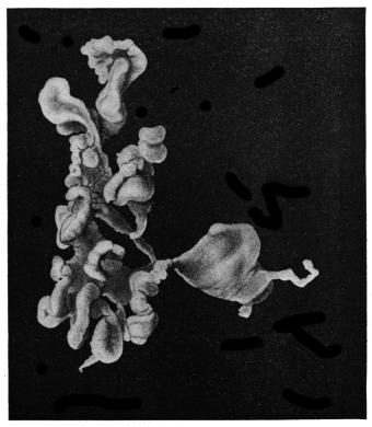
Fig. 216.—Sparganum proliferum. × 10. (After Stiles.)
The Worm.—The chief peculiarity is its irregular shape and its reproduction in the larval stage by forming supernumerary heads, which are supposed to wander about the body.
The simplest forms are thread-like bodies, flat or round, 3 mm. long and 0·3 mm. in diameter, but they may be 12 mm. long by 2·5 mm. broad. The narrow end is the head, which in life invaginates and evaginates, but there is no indication of any suckers, except an inconstant terminal depression. In addition to these simple forms the most complicated and irregular forms occur, due to the formation of buds (heads) at various parts. The detachment and growth of a head account for the presence of more than one worm in a cyst. The irregularity in form is also increased by the presence in the subcuticular tissue of the worm of reserve food bodies. These bodies are supposed to be of this nature and are spherical, 100 µ to 300 µ in diameter, but also much elongated.
Calcareous bodies in the Japanese worms were 7·5 µ to 12 µ; in the Florida worms 8·8 µ to 17·6 µ.
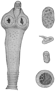
Fig. 217.—Dipylidium caninum: on the left, the scolex, neck and the first proglottids; on the right, at the top, a packet of ova; below, hooks of the rostellum, side and front views; below, an ovum. Various magnifications. (After Diamare.)
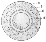
Fig. 218.—Dipylidium caninum; egg showing a, egg-shell (vitelline membrane of Moniez); b, albuminous coat; c, internal shell formed of or secreted by an outer layer of blastomeres (Moniez); d, hexacanth embryo. (After Benham and Moniez.)
Mode of Infection.—Probably from eating uncooked fish.
Distribution.—Japan, Florida.
Family. Dipylidiidæ, Lühe, 1910.
Genus. Dipylidium, R. Lkt., 1863.
Rostellum retractile, with several rings of alternating hooks; the latter with a disc-like base, having the shape of the thorns of a rose. Genital pores opposite; genitalia double. Testes very numerous in the central field; ovary with two lobes; the vitellaria, which are smaller, behind them; the uterus forms a reticulum, in the network of which the testicular vesicles lie; later on it breaks up into sacs enclosing one or several eggs. The eggs have a double shell.
Dipylidium caninum, L., 1758.
Syn.: Tænia canina, L., 1758, p. p.; Tænia moniliformis, Pallas, 1781; Tænia cucumerina, Bloch, 1782; Tænia elliptica, Batsch, 1786; Dipylidium cucumerinum, Lkt., 1863.
This worm measures 15 to 35 cm. in length and 1·5 to 3 mm. in breadth. The scolex is small, rhomboidal, and has a club-shaped rostellum on which there are, in three to four rings, forty-eight to sixty hooks resembling rose thorns, the size of those in the foremost being 11 µ to 15 µ and those in the hindmost ring 6 µ. The neck is very short, the most anterior segments broad and short, the middle as long as they are broad; the mature segments are longer than wide (6 to 7 mm. by 2 to 3 mm.), fairly thick, are frequently of a reddish colour, and when cast off resemble cucumber seeds. The genital pores lie symmetrically at the lateral margins; the roundish egg sacs, arising from the uterine reticulum, contain eight to fifteen eggs embedded in a reddish cement substance (in life). The eggs are globular (43 µ to 50 µ,); the embryonal shell (embryophore) is thin, the oncosphere measures 32 µ to 36 µ. Surrounding the embryophore is an albuminous coating, and outside this the thin vitelline envelope (fig. 218).
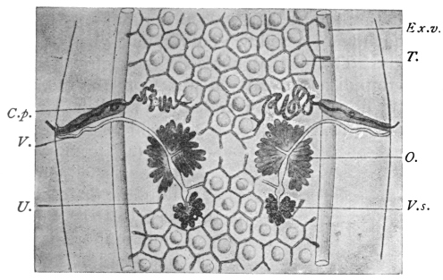
Fig. 219.—Dipylidium caninum: central portion of a proglottis. C.p., cirrus sac; V.s., vitellaria; Ex.v., excretory vessels; T., testicles lying in the meshes of the uterine reticulum which laterally forms pouches; O., ovary; U., reticulum of uterus; V., vagina and seminal receptacle (below ovary). Magnified. (After Neumann and Railliet.)
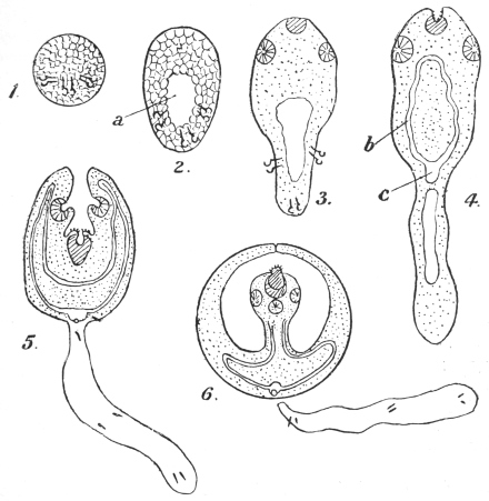
Fig. 220.—Dipylidium caninum: development of embryo. 1, solid hexacanth embryo; 2, primitive lacuna (a) in the embryo; 3, elongation of hinder part, rudiments of sucker and rostellum appearing; 4, “body” and “tail” distinct, (b) and (c) excretory system; 5, fore-body invaginates into hind-body, excretory bladder has a pore; 6, tail has dropped off; scolex growing up into secondary cavity formed by fore-body; the primitive cavity has been absorbed at stage 4. (After Benham, Grassi and Rovelli.)
Dipylidium caninum is a common intestinal parasite of dogs, in which it grows larger (Tænia cucumerina, Bloch) than in cats (T. elliptica, Batsch); it has, however, also been found in jackals, as well as in human beings, though in the latter it is of comparatively rare occurrence (twenty-four cases), and almost always affects children, generally of tender age. One-third of all the cases in children were sucklings, about a quarter of all the cases recorded were adults, and these occurred throughout all Europe with the exception of Spain and Italy.
The proglottids, which leave the intestine spontaneously, are recognizable by the naked eye on account of their form and reddish colour, as well as their two genital pores. As a rule, the presence of this parasite sets up no marked symptom in the patient.
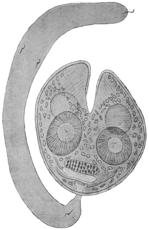
Fig. 221.—Larva (cysticercoid) of Dipylidium caninum, consisting of body and tail. The latter is solid and bears on it the embryonal spines. The bladder, which was only slightly developed, has disappeared, and the fore-part of the body bearing the rostellum is now seen invaginated into the hind portion. The hooklets are shown in front of the excretory system which has now developed. At a further stage the tail drops off; the head now evaginates, but is still enclosed in a double-walled sac formed by the prolongation upwards on each side of the topmost parts of the body shown in the figure. Cf. fig. 220, 6. Enlarged. (After Grassi and Rovelli.)
The corresponding larval form (cysticercoid) lives in the louse of the dog (Trichodectes canis), a fact that was first established by Melnikow and Leuckart; according to Grassi and Rovelli, as well as Sonsino, it also lives in the flea of the dog (Ctenocephalus canis) and in the flea of man (Pulex irritans), but not in its larva. The adult segments, which also leave the rectum of dogs and cats spontaneously, creep about around the anus and get into the hair, and are thus partly dried and disintegrated. Part of the segments, or the oncospheres released by disintegration, are then taken up by lice and fleas, within which they develop into larvæ (cysticercoids). Dogs and cats are thus infected by their own skin parasites, which they bite and swallow whilst gnawing at their fur. The infection of human beings must occur in an analogous manner, by transmission of the cysticercoids present on the lips or tongue of dogs when the latter lick them, or it may be that the vermin of cats and dogs harbouring cysticercoids are accidentally and directly swallowed by human beings.
Family. Hymenolepididæ, Railliet and Henry, 1909.
Genus. Hymenolepis,282 Weinland, 1858.
Accessory sac (opening into genital atrium) usually absent. Vas deferens with an external (outside cirrus sac) and an internal (inside cirrus sac) “seminal vesicle.” Three testes in each proglottis. The eggs are round or oval with two to four distinct envelopes. In mammals and birds.
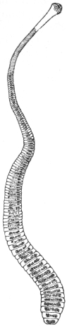
Fig. 222.—Hymenolepis nana, v. Sieb. About 12/1. (After Leuckart.)
Hymenolepis nana, v. Sieb., 1852.
Syn.: Tænia nana, v. Sieb., 1852, nec van Beneden, 1867; Tænia ægyptiaca, Bil., 1852; Diplacanthus-nanus, Weinld., 1858; Tænia (Hymenolepis) nana, Lkt., 1863.
The worm is 10 to 45 mm. in length and 0·5 to 0·7 mm. in breadth; the head is globular, 0·25 to 0·30 mm. in diameter. The rostellum has a single circlet consisting of twenty-four or twenty-eight to thirty hooks, which are only 14 µ to 18 µ in length. The neck is moderately long; the proglottids are very narrow, up to 200 in number, 0·4 to 0·9 mm. in breadth, and 0·014 to 0·030 mm. in length. The eggs are globular or oval, 30 µ to 37 µ to 48 µ; the oncospheres measure 16 µ to 19 µ in diameter, with two coats, separated by an intervening semi-fluid substance (fig. 224).
This species was discovered by Bilharz in Cairo in 1851; it was found by him in great numbers in the intestine of a boy who had died of meningitis. For several years this was the only case, until 1885, since when numerous cases have come to light. Spooner (1873) even reported a case from North America, which may, however, have related to Hymenolepis diminuta. In Europe the worm is particularly frequent in Sicily, but it has also been repeatedly observed in North Italy; it has, moreover, been reported from Russia, Servia, England, France, Germany, North and South America, the Philippines, Siam and Japan, in all over 100 cases. Notwithstanding its small size this worm causes considerable disorders in its hosts—mostly children—as it sets up loss of appetite, diarrhœa, various nervous disturbances, and even epilepsy; all these symptoms, however, disappear after the expulsion of the parasites, which are generally present in large numbers.
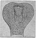
Fig. 223.—Hymenolepis nana: head. Enlarged. (After Mertens.)
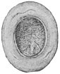
Fig. 224.—Hymenolepis nana: an egg. Highly magnified. (After Grassi.)
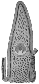
Fig. 225.—Longitudinal section through the intestinal villus of a rat, with the larva (cysticercoid) of Hymenolepis murina. Magnified. (After Grassi and Rovelli.)
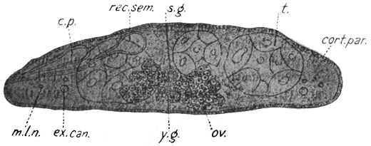
Fig. 226.—Hymenolepis nana (murina): cross section of proglottis from a rat. c.p., cirrus sac; rec. sem., receptaculum seminis; s.g., shell gland; ov., ovary; t., testis; cort. par., cortical parenchyma; m.l.n., main lateral nerve; ex. can., excretory canal; y.g., vitellarium. (After v. Linstow.)
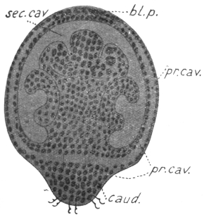
Fig. 227.—Hymenolepis nana: longitudinal section of an embryo. bl.p., anterior opening of secondary cavity; caud., caudal appendage; pr. cav., primary cavity; sec. cav., secondary cavity. Enlarged. (After Grassi and Rovelli.)
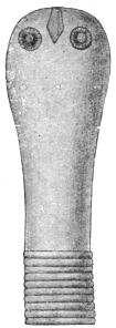
Fig. 228.—Hymenolepis diminuta: scolex. Magnified. (After Zschokke.)
The development as well as the manner of infection is still unknown; Grassi is of opinion that Hymenolepis nana is indeed merely a variety of Hymenolepis murina, Duj., which lives in rats. According to Grassi direct development takes place with omission of the intermediate host, but with the retention of the larval stage; that is to say, rats infect themselves directly with Hymenolepis murina, by ingesting the mature segments or oncospheres of this species, from which subsequently the small larvæ originate in the intestinal wall (fig. 225); when fully developed they fall into the intestinal lumen and become tapeworms. The identity of the two forms has nevertheless been disputed (Moniez, R. Blanchard, v. Linstow), though their near relationship cannot be denied. Grassi gave mature segments of Hymenolepis murina to six persons, but only one person evacuated a tapeworm. This, however, proves nothing in a district where Hymenolepis nana frequently occurs in man; it was, moreover, not possible to infect rats with segments of Hymenolepis nana (of man). Accordingly this form may represent an independent species, which, however, like Hymenolepis murina, also omits an intermediate host.
Hymenolepis diminuta, Rud., 1819.
Syn.: Tænia diminuta, Rud., 1819; Tænia leptocephala, Crepl., 1825; Tænia flavopunctata, Weinld., 1858; Tænia varesina, E. Parona, 1884; Tænia minima, Grassi, 1886.
This species measures 20 to 60 cm. in length, and up to 3·5 mm. in breadth; there are from 600 to 1,000 segments. The head is very small (0·2 to 0·5 mm.), it is club-shaped and has a rudimentary unarmed rostellum; the neck is short; the mature segments are 3·5 mm. in breadth, 0·66 mm. in length; the eggs are round or oval. The outer egg-shell is yellowish and thickened, with indistinct radial stripes; the inner embryonal shell (embryophore) double, thin; the outer layer is somewhat pointed at the poles; oncosphere 28 µ by 36 µ. Between the inner and outer shells is a middle granular layer.
Hymenolepis diminuta lives in the intestine of rats—Mus decumanus (the sewer rat), Mus rattus (the black rat), and Mus alexandrinus, rarely in mice; it is occasionally also found in human beings.
Weinland described it from specimens collected by Dr. E. Palmer in 1842, in Boston, from a child aged 19 months, as T. flavopunctata. A second case relating to a three year old child, from Philadelphia, was only reported in 1889 by Leidy; a third case was simultaneously reported of a two year old girl in Varese (T. varesina); and Grassi described another case relating to a twelve year old girl from Catania (Sicily). Sonsino and Previtera reported the same species in Italy, Zschokke in France, Lutz and Magalhães in South America, and Packard in North America: a total of twelve cases, five from America, the rest from Europe (Ransom).
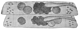
Fig. 229.—Hymenolepis diminuta: two proglottids showing testes (3), ovary and vagina. Slightly enlarged. (After Grassi.)
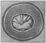
Fig. 230.—Hymenolepis diminuta: egg from man. (After Bizzozero.)
According to Grassi and Rovelli the larval stage lives in a small moth (Asopia farinalis), as well as in its larva, in an orthopteron (Anisolabis annulipes), and in coleoptera (Acis spinosa and Scaurus striatus). Experimental infections have been successful on rats as well as on human beings. In America other species of insects may be the intermediary hosts.
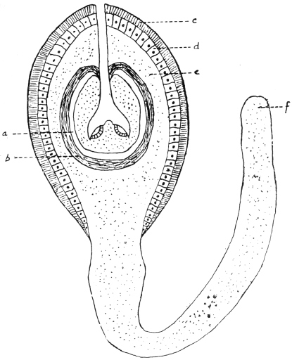
Fig. 231.—Hymenolepis diminuta: cysticercoid from the rat flea (Ceratophyllus fasciatus). a, remains of primary vesicle; b, fibrous layer; c, radially striated layer resembling cuticle; d, layer of columnar cells; e, parenchymatous layer of irregularly disposed cells; f, parenchymatous layer. (Stephens, after Nicoll and Minchin.)
Nicoll and Minchin283 found in the body cavity of 4 per cent. of rat fleas (Ceratophyllus fasciatus) the cysticercoid of Hymenolepis diminuta. That it belonged to this species was shown by its unarmed rostellum and by feeding; 340 fleas were fed to white rats and fourteen worms obtained, i.e., about 4 per cent., thus corresponding to the infection of the fleas. The development in the flea probably begins in the pupal stage, the eggs being ingested by the older flea larvæ. The larva is 0·31 by 0·25 mm.; tail 0·8 mm., scolex 0·075 by 0·09 mm., suckers, 0·055 mm. in diameter. Microscopically it shows—(1) externally a radially striated layer resembling cuticle, (2) a layer of columnar cells, (3) parenchymatous layer continuous with the tail, (4) fibrous layer around the small caudal vesicle, then the parenchymatous scolex at the bottom of the secondary cavity.
Nicoll and Minchin (loc. cit.) found a cysticercoid284 in the rat flea Ceratophyllus fasciatus which was probably that of Hymenolepis murina. Body 0·16 mm., tail 0·19 mm., scolex 0·096 mm. in diameter. Rostellum has twenty-three spines in a single row. Length 0·017 mm., handle 0·01 mm., guard 0·007 mm., prong 0·007 mm. Sucker 0·042 mm. Although this cycle, then, for H. murina also exists, it is not probable that rats (or man in the case of H. nana if this be considered distinct) infect themselves in this way, as they hardly ingest all the necessary fleas to account for the massive infection which frequently exists in rats (and man), so that Grassi’s cycle holds good as the predominant method. Xenopsylla cheopis has also been found by Johnston to harbour both cysticercoids in Australia.
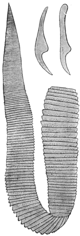
Fig. 232.—Hymenolepis lanceolata. Natural size. (After Goeze.) To the right above, two hooks. 120/1. (After Krabbe.)
Hymenolepis lanceolata, Bloch, 1782.
Syn.: Tænia lanceolata, Bloch, 1782; Drepanidotænia lanceolata, Railliet, 1892.
The parasite measures 30 to 130 mm. in length and 5 to 18 mm. in breadth; the head is globular and very small; the rostellum is cylindrical, with a circlet composed of eight hooks (31 µ to 35 µ in length). The neck is very short. The short segments increase gradually and equally in breadth, but only a little in length; the female glands lie on the side opposite to that on which the genital pore is situated; the three elliptical testes are on the same side as the pores; the cirrus is armed and slender. The eggs have three envelopes and are oval (50 µ by 35 µ), the external envelope is thin, the middle intermediate layer or envelope is not so marked as in H. diminuta, and the internal one is very thin and sometimes has polar papillæ, as in Hymenolepis diminuta and H. nana.
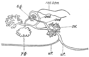
Fig. 233.—Hymenolepis lanceolata: diagram of female genitalia. ov., ovary; ovd., oviduct; rec. sem., receptaculum seminis; s.g., shell gland; ut., uterus; y.g., vitellarium. (After Wolffhügel.)
It inhabits the intestine of the following birds: Domesticated ducks and geese, the Muscovy duck (Cairina moschata), white-headed duck (Erismatura leucocephala), the pochard (Nyroca rufina), and the flamingo (Phœnicopterus antiquorum). It has been recorded from Great Britain, France, Denmark, Austria and Germany.
Zschokke reports the receipt of two specimens which a twelve year old boy in Breslau evacuated spontaneously at two different times.
The corresponding larva, according to Mrázek, lives in fresh water Cyclops; according to Dadai it is likewise found in another copepod, Diaptomus spinosus, but the hooks of Dadai’s larva differed in size.
Family. Davaineidæ, Fuhrmann, 1907.
Sub-family. Davaineinæ, Braun, 1900.
Genus. Davainea, R. Blanch., 1891.
The large scolex is more or less globular, much wider than the rostellum, which is furnished with two rings of very small and numerous hooks. Neck absent, proglottids few, genitalia single. Parasitic chiefly in birds.285
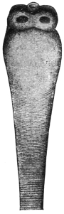
Fig. 234.—Scolex of Davainea madagascariensis. The hooks have fallen off. 14/1. (After Blanchard.)
Davainea madagascariensis, Davaine, 1869.
Syn.: Tænia madagascariensis, Dav.; Tænia demerariensis, Daniels, 1895.
This worm measures 25 to 30 cm. in length; the head has four large round suckers; the rostellum has ninety hooks (18 µ in length); there are 500 to 700 segments, of which the last 100 are filled with eggs and form half of the entire worm. The segments, when mature, measure 2 mm. in length by 1·4 mm. in breadth; genital pores unilateral; about fifty testes; the uterus consists of a number of loops, which at each side are rolled up into an almost spherical ball; when filled with eggs the convolutions unwind, permeate the segment and then lose their wall; the eggs lying free in the parenchyma become finally surrounded, one, or several together, by proliferating parenchymatous cells; this is how the 300 to 400 egg masses, taking up the entire mature segment, are formed. The globular oncosphere (8 µ) is surrounded by two perfectly transparent shells, the outer of which terminates in two pointed processes.
Davainea madagascariensis has hitherto been found in man only (eight times). Davaine described this species from fragments sent to him from Mayotta (Comoro Islands), which were found in two Creole children. Chevreau observed four cases in Port Louis (Mauritius), likewise in children; Leuckart received the first perfect specimen—it was obtained from a three year old boy, the son of a Danish captain, in Bangkok; Daniels, at the post-mortem of an adult native of George Town, Guiana, found two specimens (Tænia demerariensis); and finally Blanchard describes another perfect specimen which was in Davaine’s collection of helminthes in Paris, and which was obtained from a little girl 3 years old, of Nossi-Bé (Madagascar). The intermediate host is unknown.
Davainea (?) asiatica, v. Linst., 1901.
Syn.: Tænia asiatica, v. Linstow.
There exists only one headless specimen of this species, which is not quite adult, and which is preserved in the Zoological Museum of the Imperial Academy of Science in Petrograd. It came from a human being and was found by Anger in Aschabad (Asiatic Russia, near the northern frontier of Persia). The specimen measures 298 mm. in length. The breadth anteriorly is only 0·16 mm., the posterior part measures 1·78 mm. across. The number of segments is about 750. The genital pores are unilateral; the testes are globular and lie in a dorsal and ventral layer in the medullary layer; the cirrus pouch is pyriform, 0·079 mm. in length and 0·049 mm. in breadth; the female glands lie in the fore-part of the segments, the ovary reaching to the excretory vessels; the vitellarium is small and round. The vagina has a large fusiform receptaculum seminis; the uterus breaks up into sixty to seventy large, irregularly polyhedric eggsacs.
Family. Tæniidæ, Ludwig, 1886.
Genus. Tænia, L., 1758.286
With the characters of the family. In the genus Cladotænia recognized by some authors, the testes encroach on the mid field and the uterine stem reaches the anterior end of the segment.
Tænia solium, L., p. p., 1767.
Syn.: Tænia cucurbitina, Pall., 1781; Tænia pellucida, Goeze, 1782; Tænia vulgaris, Werner, 1782; Tænia dentata, Gmel., 1790; Halysis solium, Zeder, 1800; Tænia humana armata, Brera, 1802; Tænia (Cystotænia) solium, Lkt., 1862.
The average length of the entire tapeworm is about 2 to 3 m., but may be even more; the head is globular, 0·6 to 0·8 to 1·0 mm. in diameter. The rostellum is short with a double circlet of hooks, twenty-two to thirty-two in number, usually twenty-six to twenty-eight; large and small hooks alternate regularly; the length of the large hooks is 0·16 to 0·18 mm., of the small ones 0·11 to 0·14 mm. The rostellum is sometimes pigmented. The suckers are hemispherical, 0·4 to 0·5 mm. in diameter. The neck is fairly thin and long (5 to 10 mm.). The proglottids, the number of which averages from 800 to 900, increase in size very gradually; at about 1 m. behind the head they are square and have the genitalia fully developed. Segments sufficiently mature for detachment measure 10 to 12 mm. in length by 5 to 6 mm. in breadth. The genital pores alternate fairly evenly at the lateral margin a little behind the middle of each segment. The fully developed uterus consists of a median trunk, with seven to ten lateral branches at either side, some of which are again ramified. The eggs are oval, the egg-shell very thin and delicate; the embryonal shell (embryophore) is thick, with radial stripes; it is of a pale yellowish colour, globular, and measures 31 µ to 36 µ in diameter; the oncospheres, with six hooks, are likewise globular, and measure 20 µ in diameter (fig. 238).
Malformations are not so common as in T. saginata; they consist in two or several proglottids being partly or entirely fused, formation of single club-shaped segments, fenestration of long or short series of segments and so-called double formation, in which the head has six suckers and the segments exhibit a Y-shaped transverse section. The oncospheres occasionally also possess more than six hooklets. Very slender specimens have led to the description of a particular species or variety (T. tenella).
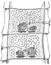
Fig. 235.—Two fairly mature proglottids of Tænia solium, showing ovaries (one bi-lobed), vitellaria, central uterine stem, cirrus and vas deferens (above), vagina (below), testes (scattered), longitudinal and transverse excretory vessels.
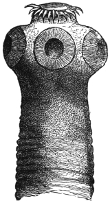
Fig. 236.—Head of Tænia solium. 45/1.
In its fully developed condition T. solium is found exclusively in man; the head is usually attached in the anterior third of the small intestine and the chain, in numerous convolutions, extends backwards; a few mature detached proglottids usually lie at the most posterior part, and these are usually evacuated during defæcation. In exceptional cases single proglottids or whole worms may reach contiguous organs if abnormal communications with them exist; thus they may reach the abdominal cavity and the urinary bladder, or they may be found in a so-called worm abscess of the peritoneum; occasionally, in vomiting, single segments or several together may be brought up. Exceptionally it induces severe anæmia.
The larval stage (Cysticercus cellulosæ) that gives rise to Tænia solium lives normally in the intramuscular connective tissue and other organs of the domestic pig, but it is known to exist also in a few other mammals, such as the wild boar, the sheep,287 the stag, dog, cat, brown bear and monkey, as well as in man. The cysticercus of the pig is an elliptical vesicle with a longitudinal diameter of 6 to 20 mm., and a transverse diameter of 5 to 10 mm.
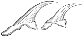
Fig. 237.—Large and small hooks of Tænia solium. 280/1. (After Leuckart.)
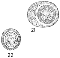
Fig. 238.—Tænia solium. 21, Egg with external membrane; 22, without (embryophore). (After Leuckart.)
Even with the naked eye a white spot may be observed in the centre of the long equator, this being the invaginated head; it is easy to make it project by pressing on the vesicle (after tearing off with the finger-nail the investing connective tissue), and on examining it under the microscope one can convince oneself that it corresponds with the head of Tænia solium.
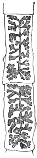
Fig. 239.—Two mature proglottids of Tænia solium with fully developed uterus. 2/1.
Numerous experiments have proved that the Cysticercus cellulosæ of the pig, if introduced into the intestine of man, grows to a Tænia solium (Küchenmeister, 1855; Humbert, 1856; Leuckart, 1856; Hollenbach, 1859; Heller, 1876); the cysticercus has frequently also been cultivated purposely by feeding pigs with mature proglottids of T. solium (P. J. van Beneden, 1853; Haubner and Küchenmeister, 1855; Leuckart, 1856; Mosler, 1865; Gerlach, 1870; etc.), but success did not attend the efforts to make Cysticercus cellulosæ establish themselves in the intestines of pigs, dogs, guinea-pigs, rabbits and monkeys (Macacus cynomolgus), and so become adult Tæniæ; the attempts, also, to infect dogs with cysticerci by means of ova were likewise, as a rule, abortive.288
The development of Cysticercus cellulosæ takes two and a half to three or four months; it is not known how long the cysticerci remain alive in animals; not uncommonly they perish at earlier or later stages, and become calcified or caseated. Extracted cysticerci die in water at a temperature of 47° to 48° C., in flesh at normal temperature they remain alive for twenty-nine days or more. On account of the present rapid means of pickling and smoking meat, the cysticerci as a rule are not killed, also the effect of cold on them for some time in cold chambers of slaughterhouses is not lethal, but freezing is fatal (Ostertag).
There is not the least doubt that human beings are almost exclusively infected with Tænia solium by eating pork containing cysticerci in a condition that does not endanger the life of the cysticerci. The infection may likewise be caused in man by eating the infected meat of other animals subject to this species of bladder worm, mainly, as a matter of fact, deer and wild boar.
The frequency of cysticerci in pigs’ flesh has considerably decreased since the introduction of meat inspection; in the Kingdom of Prussia there was on an average 1 infected pig to every 305 slaughtered between 1876 to 1882; from 1886 to 1889, there was 1 to 551; from 1890 to 1892, there was 1 to 817; in 1896, 1 to 1,470; and in 1899, 1 to 2,102; in the Kingdom of Saxony in 1894 there was 1 infected pig to every 636; in 1895 there was 1 to every 2,049, and in 1896 only 1 infected pig was found of 5,886 slaughtered. In South Germany pigs with cysticerci are very rare, but are more frequent in the eastern provinces of Prussia; in 1892 the number of infected pigs compared with the total slaughtered was as follows:—
In the district of Marienwerder1 : 28
" " Oppeln1 : 80
" " Königsberg1 : 108
" " Stralsund and Posen1 : 187
" " Danzig, Frankfort a. O. and Bromberg1 : 250
As compared with the district of Arnsberg1 : 865
" " " Coblenz1 : 975
" " " Düsseldorf1 : 1,070
" " " Münster and Wiesbaden1 : 1,900
The average for the whole of Prussia in the same year was 1 : 1,290; for the eastern provinces, on the other hand, 1 : 604. Even more unfavourable are the proportions in Russian Poland (over 1 per cent. of pigs measly), in Prague (over 3 per cent.), in Bosnia and Herzegovina (6 to 7 per cent.). The cause for this is most likely attributable to the manner in which the pigs are kept. When allowed to be in the farmyards of the small farmers for the whole day, or allowed to wander in the village streets and pasture lands, they are more liable to take up the oncospheres of the T. solium than when shut up in good pig-styes.
The geographical distribution of T. solium generally corresponds with that of the domestic pig and the custom of eating pork in any form insufficiently cooked or raw. There are, or were, some isolated districts in Germany, France, Italy and England where the “armed tapeworm” was frequent (for instance, Thuringia, Brunswick, Saxony, Hesse, Westphalia, whereas it is and was very scarce in South Germany); it is thus easily understood why it occurs very rarely in the East, in Asia and in Africa, in consequence of the Mahommedans, Jews, etc., not eating pork. In North America, also, T. solium is very rare; the tapeworm which is known there by this name is generally T. saginata, Stiles. During the last decade T. solium infection has, however, very markedly decreased in North and East Germany in consequence of the precautions exercised by the public in the choice of pork to avoid trichinosis, especially, however, because measly meat must be sold as such and must be thoroughly cooked before being placed on the market; indeed, if badly infected it may not be sold for food, but can be turned to account for industrial purposes.
The occurrence of Cysticercus cellulosæ in man has been known since 1558 (Rumler, Obs. med., liii, p. 32); there is hardly an organ in man in which cysticerci have not been observed at some time; they are most frequently found in the brain,289 where they grow to a variety known as Cysticercus racemosus; next in frequency they are found in the eye, in the muscular system, in the heart, in the subcutaneous connective tissue, the liver, lungs, abdominal cavity, etc. The number of cysticerci observed in one man varies between a few and several thousands. Of the sexes, men are most subject (60 to 66 per cent. of the number attacked). The disturbances caused in man by cysticerci vary according to the nature or position of the organs attacked; when situated in the cerebral meninges they have the same effect as tumours.
During the last decades, however, these cases have also become less common. In Rudolphi’s time 2 per cent. of post-mortems in Berlin showed cysticerci; in the ’sixties, according to Virchow, about the same; in 1875 the number fell to 1·6 per cent.; in 1881 to 0·5 per cent.; in 1882 to 0·2 per cent.; in 1900 to 0·15 per cent., and in 1903 to 0·16 per cent. Hirschberg between 1869 and 1885 discovered cysticerci in the eye seventy times in 60,000 ophthalmic cases, but during the following six years the parasite was only present twice amongst a total of 46,000 cases of ophthalmic diseases, and since 1895 no ophthalmic case has been met with.
The infection of human beings with the cysticerci can only take place by the introduction of the oncospheres of Tænia solium into the stomach with vegetable foods, salads that have been washed in impure water containing oncospheres, also by drinking contaminated water; the carriers of T. solium, moreover, infect themselves still more frequently through uncleanliness in defæcation, the privies in public localities and many private houses affording striking testimony of this. The minute oncospheres can thus easily reach the fingers and thence the mouth (as in twirling the moustache, biting the nail). More rarely a third manner of transmission or internal auto-infection may possibly take place, as when, in the act of vomiting, mature proglottids near the stomach are drawn into it; the oncospheres or segments there retained are then in the same position as if they had been introduced through the mouth.
On account of these dangers of internal or external auto-infection, it is therefore the duty of the medical attendant, after recognizing the presence of tapeworms, to expel them,290 and in doing so to employ every possible means to prevent vomiting setting in; it is, however, equally important to take the necessary steps to destroy the parasites evacuated. It may be incidentally mentioned that in using certain remedies the scolex not rarely remains in the intestine; the cure in such cases has not been accomplished, as the scolex again produces new proglottids, and after about eleven weeks the first formed ones are again mature and appear in the fæces.
Amongst the cysticerci also many malformations appear; thus absence of the rostellum and the hooks, or double formation with six suckers, or abnormalities of growth on account of the surroundings, which have had a special name given to them, viz., Cysticercus racemosus, Zenk. (= C. botryoides, Hell.; C. multilocularis, Kchnmstr.); these forms are more especially found at the base of the brain, are irregularly ramified and often without the head.
A certain interest is attached to those forms that have led to the establishment of a distinct species:—
Cysticercus acanthotrias, Weinld., 1858.
In making the autopsy of a white Virginian woman who had died of phthisis, a cysticercus was found in the dura mater, and eleven or twelve specimens in the muscles and subcutaneous tissue. Weinland and Leuckart, who examined the specimens, found that they resembled Cysticercus cellulosæ in form and size, but that they carried on the rostellum a triple crown, each consisting of fourteen to sixteen hooks, which differed from the hooks of C. cellulosæ or of Tænia solium by the greater length of the posterior root process and the more slender form of the hooks; the large hooks measured 0·153 to 0·196 mm., the medium-sized hooks, 0·114 to 0·14 mm., and the small ones 0·063 to 0·07 mm.
On account of these differences a distinct species of cysticercus was established, and this naturally presupposed a corresponding species of Tænia (T. acanthotrias, Lkt.); this could be done with justice so long as the case remained isolated, i.e., in America, as there was the possibility of the corresponding Tænia being found. In this respect, however, the position has changed; Delore first described a cysticercus the size of a nut from the biceps muscle of the arm of a silk-worker in Lyons; according to Bertolis this specimen possessed hooks of three different sizes, the dimensions of which corresponded with the figures given by Weinland and Leuckart; the correctness of the diagnosis could hardly be doubted, as Bertolis was known to be a very exact observer. A second case has become known through Cobbold, who regards a specimen of a cysticercus in Dallinger’s collection as likewise belonging to Cysticercus acanthotrias; this specimen also came from a man’s brain; finally a third case, also from France, has been published by Redon. This author, amongst numerous C. cellulosæ of a man, found one that had forty-one hooks in three rows, and he was the first to express the opinion that C. acanthotrias does not represent a distinct species, but is only an abnormality of C. cellulosæ. This view was also taken by Blanchard and Railliet, and is probably correct, as the discovery of the large corresponding Tænia furnished with three rows of hooks is not to be expected in European beasts of prey, and in Redon’s case C. acanthotrias as well as C. cellulosæ occurred simultaneously.
The duration of life of C. cellulosæ in man is very long; cysticerci of the eye have been known to persist for twenty years, and in cysticercus of the brain ten to nineteen years may elapse from the first appearance of cerebral symptoms until death. Dead cysticerci may shrivel up or become calcified, perhaps also undergo fatty degeneration and then absorption. Finally, it may be mentioned that if particular proof is required that C. cellulosæ of man belongs to the cycle of development of the Tænia solium, such proof has been furnished by Redon.
Note.—Tænia tenella, mentioned on p. 332, was ascribed by Cobbold to cysticerci of the muscular system of sheep. It has, however, been demonstrated that these cysticerci belong to the cycle of development of Tænia marginata (dog) (Cysticercus tenuicollis, from the omentum of sheep); but as already stated C. cellulosæ also occurs in sheep. Chatin himself swallowed the cysticercus, which Cobbold termed C. ovis, without causing a Tænia to develop in his intestine. Müller also vainly sought to induce infection with C. tenuicollis in his own person. On the other hand, the feeding of a dog with Cysticercus ovis resulted in the production of Tænia marginata.
Tænia bremneri, Stephens, 1908.
Characterized by the large size of the gravid segments. The largest was 32 by 9 mm. Smallest 21 by 6 mm. Average 28·6 by 8·5 mm. Mode 21 by 6 mm. Uterine branches twenty-two to twenty-four in number. Calcareous bodies numerous, 15·2 µ in diameter. Eggs maximum 45·6 µ by 41·8 µ. Smallest 34·2 µ by 30·4 µ. Mode 38 µ by 30·4 µ.
Tænia marginata, Batsch, 1786.
Syn.: T. e. Cysticerco tenuicolli, Küchenmeister, 1853.
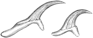
Fig. 240.—Large and small hooklets of Tænia marginata. 280/1. (After Leuckart.)
This species, which in structure resembles Tænia solium, lives in the intestine of the dog and the wolf. It attains 1·5 to 4 m. in length, possesses a double crown of thirty to forty hooks, on an average thirty-six to thirty-eight hooks, and in its larval stage (Cysticercus tenuicollis) lives in the peritoneal cavity of ruminants and the pig, occasionally in the monkey and squirrel.
It is included in this work because, according to one statement, C. tenuicollis is supposed to have been observed in man in North America; but the case is not quite certain, as the number of hooks was less than in C. tenuicollis and coincided with C. cellulosæ, although the size of the cysticercus appeared to point to C. tenuicollis. A yet earlier statement of Eschricht, that Cysticercus tenuicollis had been observed in Iceland in the liver of a man, is undoubtedly due to an error.
Tænia serrata, Goeze, 1782.
This parasite attains a length of from 0·5 to 2 m., possesses a double crown of thirty-four to forty-eight (mostly forty) hooks. It lives exclusively in the intestine of the dog, the corresponding cysticercus (Cysticercus pisiformis) living in the mesentery of the hare and rabbit. We mention this species with all reserve amongst the parasites of man, because Vital states that he has observed it twice in Constantine (Algeria) in human beings. The data, however, are not sufficient to characterize the species. It is highly probable that they relate to Tænia solium. Galli-Valerio even swallowed five specimens of Cysticercus pisiformis, but without result.
Tænia crassicollis, Rud., 1810.
I only mention this species from the intestine of the domestic cat because Krabbe regards its occurrence in man as possible. It attains a length of 60 cm. and is armed; its cysticercus (Cysticercus fasciolaris) lives in the liver of mice and rats. In Jutland, according to Krabbe, chopped-up mice (spread on bread) are eaten raw, being a national remedy for retention of urine, and this custom affords the possibility of the introduction of C. fasciolaris into the intestine of man (Nord. med. Arkiv, 1880, xii).
Tænia saginata, Goeze, 1782.
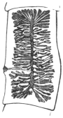
Fig. 241.—Mature segment of Tænia saginata, G., with distended uterus. 2/1.
Syn.: Tænia solium, L., 1767 (pro parte); Tænia cucurbitina, Pallas, 1781 (p.p.); Tænia inermis, Brera, 1802. Moquin-Tandon, 1860; Tænia dentata, Nicolai, 1830; Tænia lata, Pruner, 1847; Bothriocephalis tropicus, Schmidtmuller, 1847; Tænia mediocanellata, Küchenmeister, 1855; Tænia zittavensis, Küchenmeister, 1855; Tænia tropica, Moquin-Tandon, 1860; Tænia (Cystotænia) mediocanellata, Leuckart, 1863.
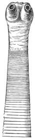
Fig. 242.—Cephalic end of Tænia saginata in the contracted condition. 8/1.
The length of the entire tapeworm averages 4 to 8 to 10 m. and more, even up to 36 m. According to Bérenger-Feraud it attains a length of 74 m. (?) The head is cubical in shape, 1·5 to 2 mm. in diameter; the suckers are hemispherical (0·8 mm.) and are frequently pigmented; there is a sucker-like organ in place of the rostellum, and this also is frequently pigmented. The neck is moderately long and about half the breadth of the head; the proglottids, the number of which averages more than 1,000, gradually increase in size; the mature detached segments are shaped exactly like pumpkin-seeds, and are about 16 to 20 mm. in length and 4 to 7 mm. in breadth. The genital pores alternate irregularly and are situated somewhat behind the middle of the lateral margin. There are twenty to thirty-five lateral branches at each side of the median trunk of the uterus, and these again ramify. The eggs are more or less globular, the egg-shell frequently remains intact and carries one or two filaments; the embryonal shell (embryophore) is thick, radially striated, is transparent and oval; it is 30 µ to 40 µ in length, and 20 µ to 30 µ in breadth. Several segments simultaneously are usually passed spontaneously with defæcation.
Malformations are not uncommon, and resemble those of Tænia solium; a triangular form has been termed T. capensis by Küchenmeister, and T. lophosoma by Cobbold, names that naturally possess as little value as does the term T. fenestrata for fenestrated specimens. Moreover, T. solium, var. abietina, Weinld., 1858, which was evacuated by an Indian, was probably a T. saginata with somewhat close uterine branches. It is regarded by Stiles and Goldberger as a doubtful subspecies.
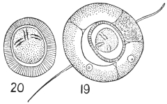
Fig. 243.—Tænia saginata. 19, egg with external shell. 20, without (embryophore). (After Leuckart.)
T. saginata in its adult condition lives exclusively in the intestinal canal of man.291 The corresponding cysticercus is Cysticercus bovis, and is found almost exclusively in the ox; it is small, 7·5 to 9 mm. in length and 5·5 mm. in breadth, may easily escape notice, and requires from three to six months for its development. Numerous experiments have confirmed the connection of Cysticercus bovis with Tænia saginata; indeed, the cysticercus was only discovered by feeding experiments after attention had been called to the ox as the probable intermediary host of this Tænia.
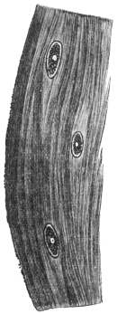
Fig. 244.—A piece of the muscle of the ox, with three specimens of Cysticercus bovis. Natural size. (After Ostertag.)
Medical men observed that weakly children who were ordered to eat raw scraped beef to strengthen them contracted T. saginata. It was found, moreover, that Jews, who are prohibited from eating pork from religious motives, suffered especially from T. saginata; when T. solium was found to occur in a Jew he often confessed to having eaten pork; and finally it was found that certain nations—for instance, the Abyssinians—frequently harbour T. saginata, and only eat beef—raw by preference.
These observations led Leuckart, in 1861, to feed young calves with the proglottids of T. saginata in order to discover the corresponding cysticercus, which was then not known. This experiment was successful. Similar experiments, with similar results, were then conducted by Mosler (1863), Cobbold and Simonds (1864 and 1872), Röll (1865), Gerlach (1870), Zürn (1872), Saint Cyr, Jolicœur (1873), Masse and Pourquier (1876), and Perroncito, in 1876. The attempts to infect goats, sheep, dogs, pigs, rabbits and monkeys were unsuccessful. Only Zenker and Heller were able to infect kids, and Heller infected one sheep, but these are exceptions.
Artificial infections of human beings with Cysticercus bovis to obtain the tapeworm were less numerous, and indeed quite superfluous, yet this was also done by Oliver (1869) in India, and Perroncito (1877) in Italy. The experiments of the latter prove that the extracted cysticerci of the ox certainly perish in water at 47° to 48° C.
It is a remarkable circumstance that, at least as regards Central Europe, C. bovis in the ox, after natural infection, was so seldom found that almost every case was published as a rarity; whereas the Tænia is very frequent in man. The reason for this is that in Germany cattle are not severely infected, and that the small, easily dried-up cysticerci easily escape notice in the large body of the host. Hertwig, the late director of the town cattle market in Berlin, in 1888, pointed out that the cysticercus of the ox is found chiefly in the musculi pterygoide externi and interni, and since that time a far greater number of infected oxen have been found in Berlin.
YearNumber of oxen slaughteredInfectedProportion
1888–89141,8141131 : 1,255
1889–90154,2183901 : 395
1890–91124,5932631 : 474
1891–92136,3682521 : 541
1892–93142,8742141 : 672
Since 1892 an increase has taken place in the number of oxen infected with cysticercus, but this is probably attributable to the more general and searching examinations. In the slaughter-houses of Prussia the number of infected beasts was as follows:—
1892 567
1893 686
1894 748
18951,143
18961,981
18972,629
The condition was most frequent in Neisse (3·2 to 4 per cent.), Eisenach (1·91 per cent.), Ohlau (1·57 per cent.), Oels i. Schles. (1 per cent.), Marienwerder (0·34 to 1·2 per cent.). The flesh of oxen only slightly infected (containing not more than ten living cysticerci) is sold in pieces of not more than 5 lb. to consumers after having been rendered innocuous by cooking, or by pickling for twenty-one days in 25 per cent. salt brine, or hanging for twenty-one days in suitable refrigerators; oxen in which only one cysticercus is found are allowed free commerce, and those strongly infected (i.e., containing more than ten living cysticerci) may only be used for industrial purposes.
It is a striking fact that more bulls than cows are infected (according to Reissmann, in Berlin, from 1895 to 1902, 0·446 per cent. bulls, 0·439 per cent. oxen, and 0·262 per cent. cows), the explanation of which, according to Ostertag, is that most oxen are killed when young, when also infection most readily takes place, and, further, that the larva later on in life can be completely atrophied.
The cysticercus of the ox has hitherto been found in man on very rare occasions. Arndt (Zeitschr. f. Psychiat., xxiv) mentions a case in the brain, Heller in the eye, and Nabiers and Dubreith also in the brain (Journ. méd. Bordeaux, 1889–1890, p. 209); but the diagnoses are not quite certain, as absence of hooks occasionally occurs in Cysticercus cellulosæ.
Tænia saginata is the most frequent tapeworm of man (with the exception of Dibothriocephalus latus in a few districts), and the parasite is widely distributed over the surface of the globe; it has been known in the East for ages, so far as data are available; it is frequent in Africa, America, and Europe. Its frequency has perceptibly increased during the last few years, but a decrease should soon take place in consequence of the extent and improvement of the official inspection of meat.
The following table shows the relative frequency of the Cestodes of man:—
AuthorYearNumber of casesT. saginataT. soliumDibr. latusDipyl. canin.Unde- termined
Parona (Milan)1899150 12111 4—14
Parona (Italy)1868–99513 3977126—19
Krabbe (Denmark)1869100 3753 91—
" "1869–86200 15324168—
" "1887–95100 89— 56—
" "1896–1904 50 41 1 53—
Blanchard (Paris)1895?1,00021———
Stiles (United States)1895more than 300more than 300— 3——


Schoch (Switzerland)1869 19 16 1 2——
Zaeslein (Switzerland)1881? 18019?——
Kessler (Petrograd)1888? 221647——
Mosler (Greifswald)1894181 11264 5——
Bollinger (Munich)1885 25 16 1 8——
Vierordt (Tübingen)1885121 113 8———
Mangold (Tübingen)1885–94128 120 6 8——
Tænia africana, v. Linst., 1900.
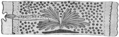
Fig. 245.—Mature segment of Tænia africana. The ovary is in the middle, and behind it are the shell gland and vitellarium; at the sides are the testicles, and externally the excretory canals; the cirrus pouch, the vas deferens and the vagina are on the left. Magnified. (After v. Linstow.)
This worm measures over 1·3 m. in length. The segments are all broader than they are long. The scolex is unarmed and is provided with an apical sucker (0·16 mm.). The parasite measures 1·38 mm. in breadth, 1·03 mm. in width; the suckers measure 0·63 mm. in diameter. The neck is very short and somewhat broader than the scolex; number of segments about 600; the hindmost segments measure 7 mm. in length and 12 to 15 mm. in breadth. The genital pores alternate irregularly in the middle of the lateral margin; the testes are very numerous and occupy the entire medullary layer; the vas deferens is much convoluted; the cirrus pouch is pyriform and thick walled; the cirrus and vagina are beset with bristles directed outwards; the receptaculum seminis is fusiform; the ovary is large and double, and consists of radially placed club-shaped tubes that do not anastomose and do not branch; the vitellarium is at the posterior border of the proglottids, the round shell gland in front of it; the uterus consists of a median trunk and fifteen to twenty-four non-ramified lateral branches on each side; the embryonal shell is thick and has radial stripes—it may be round (31·2 µ to 33·8 µ) or oval (39 µ, by 33·8 µ); the spines of the oncospheres measure 7 µ to 8 µ in length (fig. 197).
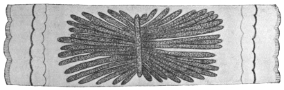
Fig. 246.—Proglottis of Tænia africana, with uterus. Magnified. (After v. Linstow.)
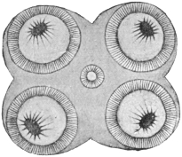
Fig. 247.—Head of Tænia africana; apical surface. Magnified. (After v. Linstow.)
At present only two specimens are known; they came from a black soldier from the vicinity of Lake Nyasa. The cysticercus is unknown; perhaps it lives in the zebu, the flesh of which the Askaris are in the habit of devouring uncooked.
Tænia confusa, Ward, 1896.
Length 8·5 m., breadth about 5 mm. The scolex is unknown; there is no neck; number of proglottids 700 to 800, always longer than they are broad; the hindmost measure 35 mm. in length, 4 to 5 mm. in breadth; the genital pores alternate irregularly behind the middle of the lateral margin; testicles numerous; vas deferens not much coiled; the cirrus pouch thick walled, elongated and club-shaped, with globular vesicula seminalis; the cirrus is beset with little hairs; the receptaculum seminis is globular; ovary small, double; each half is bean-shaped; vitellarium narrow, triangular; shell gland globular; uterus with median trunk and fourteen to eighteen short ramified lateral branches on either side. The embryophores are oval (39 µ by 30 µ), thick and radially striated.
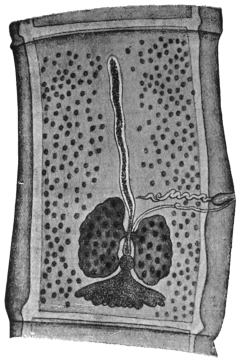
Fig. 248.—Tænia confusa: mature segment showing central uterine stem, bilobed ovary, globular shell gland, triangular vitellarium, scattered testes, cirrus, vas deferens, and vagina. 15/1. (After Guyer.)
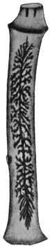
Fig. 249.—Tænia confusa: gravid segment. 25/1. (After Ward.)
Of this species only two specimens have been recorded; they occurred in human beings and were sent at different times to the first describer of them by a doctor in Lincoln (Nebr.). Perhaps Tænia solium, var. abietina, Weinld., which was found in a Chipeway Indian, is of the same species in spite of the shorter segments.
Tænia echinococcus, v. Sieb., 1853.
Syn.: Tænia nana, v. Ben., 1861 (nec v. Sieb., 1853); Echinococcifer echinococcus, Weinld., 1861.
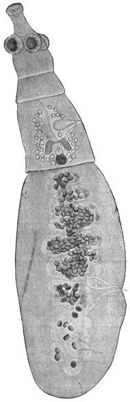
Fig. 250.—Tænia echinococcus: the cirrus sac, the vagina, uterus, ovary, shell gland and vitellarium, and the testicles at the sides are recognizable in the second proglottis; the posterior proglottis shows the uterus partly filled with eggs, as well as the cirrus sac and the vagina. 50/1.
Tænia echinococcus measures 2·5 to 5 or 6 mm. in length; the head is 0·3 mm. in breadth, and has a double row of twenty-eight to fifty hooklets (on an average thirty-six to thirty-eight) on the rostellum.
The size and form of these hooklets vary (the larger ones are 0·040 to 0·045 mm. in length, the smaller ones are 0·030 to 0·038 mm. in length). The suckers measure 0·13 mm. in diameter; the neck is short; there are only three or four segments, the posterior segment being about 2 mm. in length and 0·5 mm. in breadth. The genital pores alternate; there are forty to fifty testicles; the vas deferens is spirally coiled; the cirrus pouch is pyriform. The ovary is horseshoe-shaped with the concavity directed backwards; the vitellarium double, each half almost bean-shaped, at right angles to the plane of the segment; the shell gland is round. The median trunk of the uterus is dilated when filled with eggs and (instead of lateral branches) has lateral diverticula. It is not unusual for the eggs to form local heaps. The embryonal shell (embryophore) is moderately thin, with radial striæ, almost globular, 30 µ to 36 µ in diameter.
When mature this parasite lives in the small intestine of the domestic dog, the jackal, and the wolf, and apparently also in Felis concolor, and is usually present in great numbers; it can also be transmitted experimentally to the domestic cat, one successful result out of seven (Dévé).292 The larval stage (Echinococcus polymorphus) lives in various organs—chiefly in the liver and lungs—of numerous species of mammals (twenty-seven), especially in sheep, ox and pig, and it is even not uncommon in man, though the Tænia itself has never been found in a human being; accordingly man can only acquire the echinococcus by ingesting the eggs of the “dog worm.” The dogs disseminate the eggs of Tænia echinococcus wherever they go, or carry them to their mouths and coats by biting up the evacuated segments, and are thus able to transmit them directly to human beings (by licking them or making use of the same crockery, etc.). In other cases the oncospheres, enclosed in the embryophores, must withstand desiccation for a time and then (as when the dogs are “kissed” or otherwise caressed) are transmitted into or on to man. As echinococcus disease in man is always very dangerous, it would be a matter of general interest to prevent dogs being infected by destroying the echinococci,293 and all measures would be justifiable which would diminish the superfluous number of house-dogs (for instance, high taxes); measures should also be adopted to limit the association of men with dogs, particularly in such frequented places as restaurants, railway carriages and tram-cars.
Echinococcus is very common in slaughtered animals; in Germany, however, the figures in the reports of the abattoirs present an erroneous view in so far as, besides the total number of animals slaughtered, only the numbers of those organs (liver and lungs) are published that were so severely infected with echinococci that, even when the parasites were “shelled” out, the flesh could not be placed upon the market and was therefore “condemned.”
In Berlin the following animals were slaughtered:—
Year1889–901890–911891–921892–931896–971902
Oxen154,218124,593136,368142,874146,612153,748
Sheep430,362371,943367,933355,949395,769434,155
Pigs442,115472,859530,551518,073694,170778,538
During the same years the following were condemned in consequence of being infected with echinococci:—
LungLiverLungLiverLungLiverLungLiverLungLiverLungLiver
Oxen7,2662,4185,7921,9384,4971,7212,563 7393,2841,156 2,507 791
Sheep5,4792,7424,5952,0594,4351,6693,3311,1614,5611,93911,1384,437
Pigs6,5235,0785,0833,7356,0374,3746,7854,3127,8885,398 9,5449,233
Nevertheless there are statistics that give the total number of animals infected with echinococcus:—
AuthorPlaceOxenSheepPigs
LängrichRostock i. M.26·2 per cent.37·0 per cent.5·4 per cent.
OltStettin7·1 "25·8 "7·3 "
SteudingGotha24·6 "35·4 "21·4 "
PrettnerPrague23·2 "5·5 "?
In Güstrow, in Mecklenburg, half of the animals slaughtered are said to be infected with echinococcus; in Wismar 25 per cent. of the oxen, 15 per cent. of the sheep and 5 per cent. of the pigs are infected; according to Mayer, in Leipzig, 3·79 per cent. native pigs, 24·47 per cent. Hungarian pigs, and 13·09 per cent. of sheep were infected with echinococcus; at the same time it was stated that in regard to the native pigs the liver was more frequently affected than the lungs (3·81 per cent. as compared with 0·26 per cent.); in sheep the lungs were more frequently infected (12·71 per cent. to 3·73 per cent.), whereas in the Hungarian pigs both organs were almost equally infected (14·78 per cent. to 12·03 per cent.).
The data of Lichtenheld, in Leipzig, give the frequency with which various organs were affected, as shown in the following table:—
CattlePigsSheepHorses
♂♀
Lungs69·3 per cent.16·2 per cent.21·4 per cent.52·2 per cent.5·5 per cent.
Liver27·0 "74·2 "72·0 "44·9 "94·5 "
Spleen2·2 "3·2 "2·7 "2·9 "—
Heart0·75 "3·2 "1·3 "——
Kidneys0·75 "3·2 "1·3 "——
Subperitoneal tissue——1·3 "——
Structure and Development of Echinococcus (Hydatid).
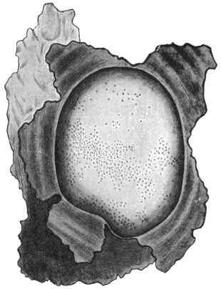
Fig. 251.—Echinococcus veterinorum: the fibrous sac enclosing the echinococcus has been opened and laid back in five parts, so that the surface of the bladder worm may be seen, with the brood capsules, visible to the naked eye, showing through it. Natural size. (After Leuckart.)
An echinococcus is a spherical or roundish bladder full of a watery liquid, which originates by liquefaction of the oncosphere, and in man may attain the size of a child’s head, but remains smaller in cattle (the size of an orange or apple). The thin wall of the bladder is composed of an external laminated cuticle (ectocyst) and an internal germinal or parenchymatous layer (endocyst). The latter again exhibits two layers: an outer layer of small cells that are less sharply defined, and an inner layer of larger cells. It contains, in addition, calcareous corpuscles, muscular fibres and excretory vessels. It is rich in glycogen.
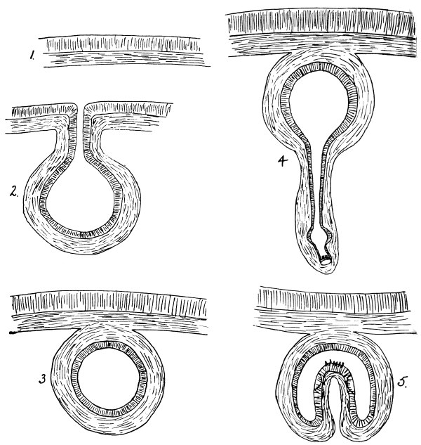
Fig. 252.
Figs. 252 and 252A.—Diagrams of mode of formation of brood capsule and scolices. (1) Wall of mother cyst, consisting of ectocyst and endocyst; (2) theoretical stage of invagination of wall; (3) a brood capsule with the layers of the wall in the reverse position to that in the mother cyst; (4) evagination of wall; (5) invagination; (6) fusion to form the solid scolex; (7) invagination of fore-part of scolex into hind-part. (Note.—The size of the scolex is much out of proportion to the brood capsule.) (Stephens.)
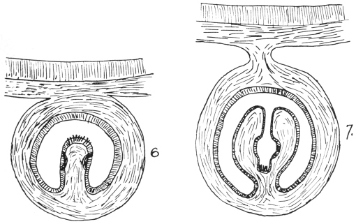
Fig. 252a.
The development in cattle often remains stationary at the bladder stage, and they are then called “acephalocysts,” or Echinococcus cysticus sterilis. According to Lichtenheld, sterile cysts occur in 80 per cent. of cases in cattle, in 20 per cent. in pigs, and in 7·5 per cent. in sheep. In other cases large numbers of small, hollow BROOD CAPSULES are formed in the germ layer, but are not arranged in any particular order. The order of the layers is just the reverse in them to what it is in the parent cyst, that is to say, they have inside a thin non-laminated cuticle and the parenchymatous layer on their external surface. These, theoretically at least, may be regarded as invaginations of the bladder wall giving rise to a cavity with the cuticle internal and the parenchymatous layer external. If we suppose the orifice to close, we should then get an isolated cavity with cuticle internal and parenchymatous layer external, as in the brood capsule (fig. 252). If we next suppose an evagination of the wall of the brood capsule to occur at one point we should get a hollow process lined with cuticle; at the bottom of this we get the scolex and hooklets formed, and a little higher up the tube the suckers (fig. 252, 4). If this hollow scolex is now pictured as being invaginated we get a hollow scolex covered with cuticle and lined by a parenchymatous layer projecting into the cavity of the brood capsule. The two sides of this hollow scolex now fuse and we get a solid scolex projecting into the cavity. Finally, if we imagine once more the rostellum and suckers invaginated into the posterior part of the scolex we get the condition as frequently found in the brood capsules, i.e., a scolex covered with cuticle projecting into the cavity, with the rostellum and suckers invaginated into the posterior portion of the scolex (fig. 252A, 7).
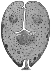
Fig. 253.—Section through an invaginated echinococcus scolex. Cf. fig. 252A, 7. × 300. (After Dévé.)
A large hydatid may contain many thousands of brood capsules. Each brood capsule is about as big as a pin’s head, and may contain ten to thirty or more scolices. The delicate wall of the brood capsules may rupture, so that the scolices are now free in the mother cyst. These free scolices and also free brood capsules constitute what is known as “hydatid sand,” which settles at the bottom of a glass when hydatid fluid is poured into it. This form occurs chiefly in domesticated animals and is termed E. veterinorum, Rud., or E. cysticus fertilis.