ON PARASITES IN GENERAL. (continued 7)
Reading edition
Testes.—Anterior testis 0·496 by 0·44 mm.; posterior testis 0·52 by 0·48 mm., usually ovoid, though both may be regularly lobed. The anterior testis is usually on the left side.
Ovary—multilobular, the lobes 6 to 8 being irregular in size and shape.
Shell Gland—extensive and diffuse, occupying an area which approximately corresponds with the loop of the transverse vitelline ducts.
Seminal Receptacle—globular, to the right of and dorsal to the posterior lobe of the ovary.
Laurer’s Canal—generally runs from the end of the receptacle with a single curve medially and backwards.
Uterine Coils—form loosely packed transverse coils terminating slightly in front of the level of the first vitelline acini. From here the uterus passes forwards into the pedicle to the left and ventral to the seminal vesicle.
Seminal Vesicle—commences about the level of the first vitelline acini. The coils displace the uterus ventrally and to the left. In the pedicle the vesicle diminishes in extent and lies in its dorsal (anterior) side.
Habitat.—Liver of pariah dogs, India. In North-Western Provinces about 40 per cent. are infected. This fluke appears to be different from Amphimerus (Opisthorchis) noverca in man, as the latter has not the pedicle on the summit of which lie the sucker and common genital pore.
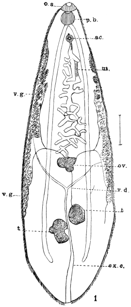
Fig. 156.—Amphimerus noverca, Braun. o.s., oral sucker; p.b., pharynx; ac., ventral sucker; ut., uterus; v.g., vitellarium; ov., ovary; v.d., vas efferens; ex.c., excretory canal; t., testis. (After McConnell.)
Genus. Amphimerus, Barker, 1912 (?).
Structure as in Opisthorchis, except that the vitellaria are separated into two portions, an ant-ovarial and a post-ovarial.
Amphimerus noverca, Barker, 1912 (?).
Syn.: Distomum conjunctum, McConnell, 1876 (nec Cobbold, 1859); Opisthorchis noverca, M. Braun, 1903 pro parte.
At the autopsy of two Mahommedans who died in Calcutta, McConnell found a large number of Distomata in the thickened and dilated bile-ducts. The worms were lancet-shaped, covered with spines, and measured 9·5 to 12·7 mm. in length and 2·5 mm. in breadth. The two suckers lie very close to one another, the anterior one being larger than the ventral; the genital pore opens immediately in front of the ventral sucker; pharynx spherical; intestinal cæca extending far back. At the commencement of the posterior third of the body the two testes, somewhat apart, the anterior one roundish, the posterior one distinctly lobed. The transverse and slightly lobed ovary in front of the bifurcation of the Y-shaped excretory bladder, whence the uterus, in convolutions barely spreading beyond the central field, extends to the pore; the vitellaria in the lateral areas commence behind the ventral sucker and extend to the testes. Cirrus pouch absent. Eggs oval, 34 µ by 21 µ.
Genus. Clonorchis, Looss, 1907.
Structure as in Opisthorchis, distinguished, however, by the branched testes situated one behind the other, the branches of which ventrally encroach upon the gut forks; dorsal to the testes the S-shaped excretory bladder, the main branches of which, arising at the level of the bifurcation of the gut, open into the bladder below its anterior end. Parasitic in the bile-ducts of mammals and man.
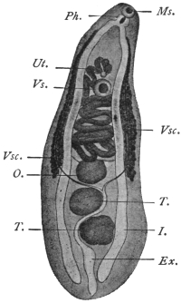
Fig. 157.—Metorchis conjunctus,272 (Syn.: Distomum conjunctum, Cobb., nec Lew. and Cunn., nec McConn.): from Canis fulvus. Vs., ventral sucker; I., intestine; Vsc. vitellaria; Ex., excretory bladder; T., testes; O., ovary; Ms., oral sucker; Ph., pharynx; Ut., uterus. (After Cobbold.)
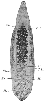
Fig. 158.—Clonorchis sinensis. C.L., Laurer’s canal; Dst., vitellaria; Ex., excretory bladder; H., testes; K., ovary; R.s., receptaculum seminis; Vd., terminal section of vas deferens. Magnified 4 1/2 times. (After Looss.)
Clonorchis sinensis, Cobbold, 1875.
Syn.: Distoma sinense, Cobbold, 1875; Distoma spathulatum, R. Leuckart, 1876 (nec Rudolphi, 1819); Distoma hepatis innocuum, Baelz, 1883.
In shape resembles Opisthorchis felineus, 13 to 19 mm. long, 3 to 4 mm. broad, at the beginning of sexual maturity 12 to 13 mm. long, 2·5 to 3 mm. broad. Oral sticker 0·58 to 0·62 mm., ventral sucker 0·45 to 0·49 mm. in transverse diameter. In the parenchyma numerous yellowish or brownish granules, especially behind the oral sucker and at the posterior end. Testicular branches very long, in the anterior testis often four, in the posterior testis five branches. Ovary generally with three large lobes and a smaller lobe. Vitellaria not always symmetrical, generally extending laterally from the ventral sucker to the ovary, interrupted in parts.
Eggs 26 µ to 30 µ by 15 µ to 17 µ. Average 29 µ by 16 µ.
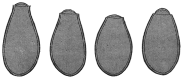
Fig. 159.—Ova of Clonorchis sinensis. The knobs on the ends of the eggs are not shown. 900/1. (After Looss.)
This (?) species was discovered in 1874 by McConnell, in Calcutta, in the bile-ducts of a Chinaman who died shortly after being admitted into hospital.
Habitat.—Bile-ducts of man, dog and cat.
Distribution.—Especially in China, apparently rare in Japan.
Clonorchis endemicus, Baelz, 1883.
Syn.: Distoma sinense s. spathulatum p.p.; Distoma hepatis endemicum s. perniciosum, Baelz, 1883; Distoma japonicum, R. Blanchard, 1886.
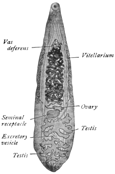
Fig. 160.—Clonorchis endemicus. × 6 about. (After Looss.)
Very similar to the previous species and consequently generally confused with it. Length between 6 and 13 mm., width varying between 1·8 and 2·6 mm. Oral sucker 0·37 to 0·5 mm., usually 0·43 to 0·45 mm. in transverse diameter; ventral sucker 0·33 to 0·45 mm., usually 0·37 to 0·40 mm. No pigment in parenchyma; anterior testis with four, posterior testis with five branches. Vitellaria continuous, ova 26 µ by 13 µ to 16 µ.
Habitat.—Bile-ducts of man, dog, cat and pig.
Distribution.—This species occurs very frequently in man, in certain districts of Japan, especially in the province of Okayama, Central Japan, in particular localities of which above 60 per cent. of the population are infected. The worms are sometimes found in enormous numbers in the liver (upwards of 4,000), also in the pancreas and rarely in the duodenum. It is common in Tonkin and Indo-China. Léger in Tonkin found 50 per cent. of people apparently in normal health infected, so that probably symptoms only arise when the infection is intense. [The exact distribution of these two species is, however, not precisely defined at present, as commonly no distinction is made between them.—J. W. W. S.]
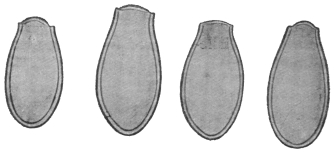
Fig. 161.—Clonorchis endemicus: eggs. The knobs on the eggs are not shown. × 900. (After Looss.)
Verdun and Bruyant deny, in opposition to Looss, the possibility of being able to distinguish within the genus Clonorchis the two species described, but they admit the justification for the new genus. They also report the occurrence of Opisthorchis felineus in man in Tonkin (Compt. Rend. Soc. de Biol., lxii, 1907).
Pathology.—Both species of Clonorchis give rise to grave symptoms. The liver is generally enlarged, though when the infection has lasted some time it begins to contract. The surface of the organ is studded with white vesicles, and on cutting into it one sees numerous cavities with thickened walls (distended bile-ducts) filled with a brownish fluid containing innumerable eggs, which cause its colour. Microscopically, the epithelium of the bile-ducts is either (1) entirely destroyed, or (2) actively proliferates, forming an adenomatous outgrowth. Occasionally this proliferation is not limited by the wall of the bile-duct but penetrates it and leads to a growth of numerous new ducts, forming a malignant biliary adenoma. The bile-ducts have their connective tissue wall greatly sclerosed. These fuse with one another, forming areas of sclerosis devoid of liver tissue. As a result of these changes the liver cells atrophy and undergo fatty pigmentary and granular degeneration. Besides these changes, due probably to the toxic action of the flukes, mechanical obstruction due to the actual plugging of the ducts by the flukes causes retention of bile and icterus, and through pressure on veins, ascites and hypertrophy of the spleen.
To what extent blood or bile respectively forms the food of the flukes is uncertain.
Life-history.—(Kobayashi, 1911, Mitteilungen aus dem kaiserlichen Institut für Infektions-Krankheiten zu Tokio, pp. 58–62.)
It results from the work of Kobayashi in Japan that fresh-water fish form the second intermediate host for Clonorchis endemicus. He fed cats with encysted flukes (cercariæ) from various fish and easily succeeded in infecting them, e.g. a kitten, proved to be uninfected by repeated examination of its fæces, was fed on infected fish; a month later innumerable flukes were found in the bile-ducts, gall-bladder, pancreas and even in the duodenum. The fish infected were Leucogobis güntheri, Pseudorasbora parva, and to a less extent Acheclognathus lanceolata, Acheclognathus limbata, Paracheclognathus rhombea, Pseudoperilampus typus, Abbottina psegma, Biwia zezera and Sarcocheilichthys variegatus. The cysts occur throughout the muscles and subcutaneous tissue of the fish. Length 0·13 mm., breadth 0·1 mm. The cercaria lies folded in the cyst, length 0·5 mm. breadth 0·1 mm. It tapers posteriorly. Skin at first covered with fine spines, disappearing as they grow older. Body dotted with fine pigment.
The first intermediate host is still unknown.
Sub-family. Metorchiinæ, Lühe, 1909.
Genus. Metorchis, Looss, 1899, emend. auctor.
Hind end rounded. Gut forks reach extreme end. Testes only slightly lobed, filling the hind end.
Metorchis truncatus, Rud., 1819.
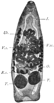
Fig. 162.—Metorchis truncatus, Rud.: from the biliary ducts of the domestic cat. V.s., ventral sucker; I., gut; V.sc., vitellaria; T., testes; O., ovary; R.s., receptaculum seminis; Ut., uterus. 25/1.
This species, which attains a length of 2 mm., is slender and conical, the anterior end is pointed and the posterior truncated, and provided with a muscular tuberosity that resembles a terminal sucker; for this reason the discoverer of the species (Rudolphi) classed it with the Amphistomes. The cuticle in the young, as well as in the adult specimens, is entirely and closely covered with spines. Suckers about equal in size (0·134 to 0·172 mm.); the ventral sticker lies somewhat in front of the middle of the body. The pharynx is small (0·09 mm.), the œsophagus minute, the intestinal cæca reach to the posterior extremity. Between them, and in front of their blind ends, lie the two elliptical testes, one generally a little in front of the other. In front of them, either in the median line or somewhat laterally, the spheroidal ovary is situated; in front, again, is the uterus, the coils of which usually extend beyond the median field. The vitellaria are at the sides of the central third of the body, thus commencing in front of the ventral sucker; cirrus pouch absent; the genital pore is close in front of the acetabulum. The excretory pore is terminal (?). Eggs 29 µ by 11 µ.
Metorchis truncatus lives in the bile-ducts of the seal, cat, dog, fox, and glutton (Gulo borealis). The source of infection is unknown, although one would suspect fish. Askanazy did not succeed in getting this fluke in his feeding experiments, but another species, Metorchis albidus, not uncommon in cats by feeding them on roach (Leuciscus rutilus).
Family. Heterophyiidæ, Odhner, 1914.
Genus. Heterophyes, Cobbold, 1866.
Syn.: Cotylogonimus, Lühe, 1899; Cænogonimus, Looss, 1899.
No crown of spines on head. Body divided into a narrow, movable, anterior part (neck), and a broader, less movable, posterior portion, which contains the genitalia. The suckers separated from one another by a space equal to half the length of the body or more; the pharynx is close behind the oral sucker; the œsophagus is long; the intestinal cæca extend to the posterior border; the genital pore is placed laterally, and behind the ventral sucker. Genital sucker provided with a circlet of chitinous rodlets, shaped like stags’ horns. The testes are at the posterior end, the ovary in a median position in front of them. Laurer’s canal with receptaculum seminis present; the small vitellaria are at the sides of the posterior part of the body. Parasitic in the intestine of mammals and birds.
Heterophyes heterophyes, v. Sieb., 1852.
Syn.: Distomum heterophyes, v. Siebold, 1852; Heterophyes ægyptica, Cobbold, 1866; Mesogonimus heterophyes, Railliet, 1890; Cœnogonimus heterophyes, Looss 1900; Cotylogonimus heterophyes, Braun, 1901.
Length up to 2 mm., breadth 0·4 mm.; the neck not sharply defined; in life it stretches to double the length of the hind body. The scales are rectangular, 5 µ to 6 µ by 4 µ, their posterior margin serrate with seven to nine teeth. Cuticular glands are numerous on the ventral surface, especially in the fore part of the body, and partly discharge at the anterior border of the oral sucker. The oral sucker is 0·09 mm., the ventral sucker 0·23 mm. in diameter; the pharynx measures 0·05 to 0·07 mm. in length; the œsophagus is about three times as long; posteriorly the intestinal cæca are directed one towards the other and terminate beside the excretory bladder. Close in front of the posterior ends of the intestinal branches are the two elliptical testes, which are not exactly on the same level. In the middle in front of them is the receptaculum seminis, and in front of the latter lies the spherical or elliptical ovary. The two vasa efferentia unite to form the vas deferens, which after a short course passes over into the angularly bent seminal vesicle; after the entry of the prostatic glands it becomes united with the metraterm (vagina), and the common duct opens into the genital sucker. The latter is somewhat smaller than the ventral sucker, lateral to and close (0·15 mm.) behind it, and bears a not entirely closed ring of from seventy-five to eighty chitinous rods (20 µ in length). The vitellaria on either side consist of about fourteen acini. The uterus is spread almost throughout the entire posterior part of the body. The eggs have thick shells with a knob resembling that of Clonorchis eggs but not so prominent, and measure 30 µ by 17 µ; they contain a completely ciliated miracidium with a rudimentary intestinal sac.
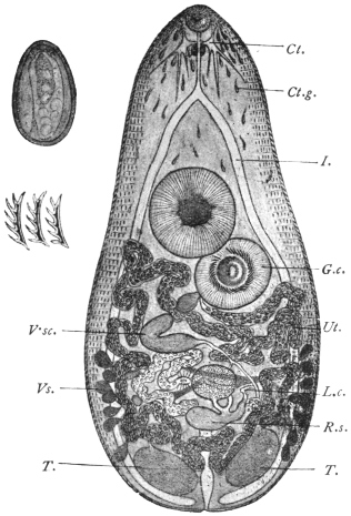
Fig. 163.—Heterophyes heterophyes, v. Sieb. C., cerebral ganglion; I., intestinal cæca; Ct.g., cuticular glands; V.sc., vitellaria; Ut., genital sucker; T., testes—the excretory bladder between them; L.c., Laurer’s canal; R.s., receptaculum seminis, with the ovary in front of it; G.c., ventral sucker; Vs., vesicula seminalis, 53/1. On the left side above, an egg, 700/1, is depicted, and below it three chitinous rodlets from the genital sucker. 700/1. (After Looss.)
This species was discovered in 1851 by Bilharz in the intestine of a boy who died in Cairo; a second case was only found in 1891 and published by R. Blanchard, so that it appeared as if the species were very scarce. According to Looss, this is, however, not the case, but the species easily escapes notice on account of its small size. Looss found it in Alexandria twice in nine autopsies, and once in Cairo, and has recently stated that in man “it is not at all uncommon to meet with the parasite in cadavers, and the eggs of the worm in the stools of the patients.” Leiper records one case from Japan and one from China. The parasites occupy the middle third of the small intestine, and even when present in large numbers appear to be harmless.
This small species, according to Looss, frequently occurs in Egyptian dogs, less so in cats, and has also been found in the fox, as well as once in Milvus parasiticus; Janson also reports this species from the intestine of the dog in Japan.
Metagonimus, Katsurada, 1913; Yokogawa, Leiper, 1913.
Resembles in general structure Heterophyes. In the arrangement of its ventral genital suckers resembles but differs from that of Tocotrema,273 Looss. The ventral and genital suckers lie laterally and on the right.
Metagonimus yokogawai. Katsurada, 1913.
Syn.: Yokogawa yokogawai, Leiper, 1913.
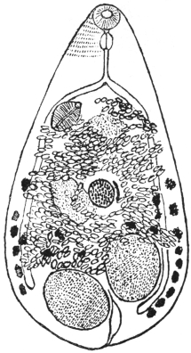
Fig. 164.—Metagonimus yokogawai, Katsurada, 1913: the spines are only shown over a small part of the skin. (After Leiper.)
One to 1·5 mm. long, seldom 2·5 mm., and 0·4 to 0·7 mm. broad; elliptical in shape. The body is thickly covered with nail-shaped spines about 10 µ long. Oral sucker 77 µ, to 85 µ in diameter. Ventral sucker characteristic and peculiar 0·12 to 0·14 mm. by 0·08 to 1 mm. It is a sac-like organ placed deeply in the body, but does not open as in other flukes on the ventral surface. Testes elliptical, not quite symmetrically placed at the hind end of the body. Vesicula seminalis retort-shaped, situated transversely, internal to the ventral sucker. Pars prostatica present. Ejaculatory duct opens with the uterus into a genital sinus, which, together with the internal opening of the ventral sucker, opens into a pit at the front of the ventral sucker. The opening of the genital sinus and that of the ventral sucker are furnished with a complex muscular apparatus. Ovary spherical, 0·12 to 0·13 mm. in diameter, lies in the middle of the hind body. Receptaculum seminis and Laurer’s canal present. Vitellaria in the hind half of the body, consisting of about ten acini on each side. Shell gland to the left of the ovary. Uterus forms three to four transverse coils. Eggs elliptical, double contoured, yellowish-brown in colour. There is no shoulder below the operculum as in the eggs of Cl. sinensis. At the rounder end there is a thickening or knob different from the spine-like or hook-like process seen in Cl. sinensis. Dimensions 28 µ by 16 µ.
Habitat.—Mainly in upper or middle portion of jejunum, rarely in cæcum. They penetrate deep into the mucosa, but not into the submucosa, and post mortem appear as a number of small brown points. They frequently occur in the solitary glands, which they destroy. They cause chronic catarrh of the gut. Parasitic in man and mammals.
Geographical Distribution.—Japan.
Life-history.—The cercarial stage occurs in a trout (Plecoglossus altivelis) and seldom in Crassius sp. and Cyprinus sp. Infection takes place through the eating of the fish raw. Seven to sixteen days later eggs appear in the fæces (of dog).
Family. Dicrocœliidæ, Odhner, 1910.
Genus. Dicrocœlium, Dujardin.
Dicrocœliidæ, with leaf-shaped bodies, pointed posteriorly and anteriorly. Greatest width behind the mid-line. Vitellaria double. The testes smooth or indented, lying symmetrically or obliquely beside or behind the ventral sucker. The ovary approaches the median line behind one testis. Parasitic in the liver and gall-bladder (rarely in the intestine) of members of all classes of vertebrate animals—by preference in birds and mammals.
Dicrocœlium dendriticum, Rud., 1819.
Syn.: Dicrocœlium lanceatum, Stil. and Hass., 1896; Fasciola lanceolata, Rud., 1803 (nec Schrank, 1790); Distomum lanceolatum, Mehlis, 1825; Dicrocœlium lanceolatum, Dujardin, 1845.
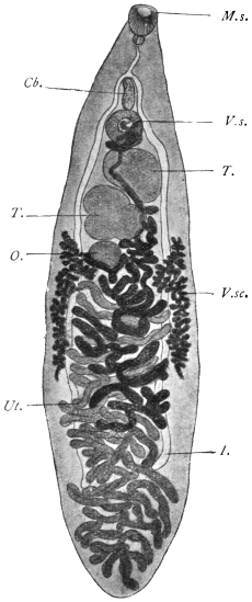
Fig. 165.—Dicrocœlium dendriticum, Rud. V.s., ventral sucker; Cb., cirrus pouch; I., intestinal cæca; V.sc., vitellaria; T., testicles; O., ovary; M.s., oral sucker; Ut., uterus. 15/1.
Body lancet-shaped, narrowing especially at the anterior extremity; length 8 to 10 mm., breadth 1·5 to 2·5 mm., the greatest breadth usually behind the middle of the body. Suckers distant from each other by about one-fifth the length of the body; oral sucker about 0·5 mm., ventral sucker about 0·6 mm. Pharynx globular, adjoining the oral sucker; œsophagus 0·6 mm. in length; intestinal cæca reach to four-fifths of the body length. Genital pore at the level of the bifurcation of the intestine; cirrus pouch small and slender. The large, slightly lobed testes lie obliquely one behind the other behind the ventral sucker; the ovary, which is considerably smaller, is placed behind the posterior one; the vitellaria, commencing at the level of the posterior testis, terminate far before the cæca. The uterus, situated behind the ovary, extends throughout the posterior end, not confined to the central field, but overlapping the lateral fields with its transverse coils; at the posterior edge of the body it turns back again and winds forwards to the ovary in transverse loops, then between the testes, and finally, dorsal to the ventral sucker, terminates in the genital pore. The thick-shelled eggs when young are yellowish, when older dark brown. They measure 38 µ to 45 µ by 22 µ to 30 µ. They contain an oval or roundish miracidium, only the anterior part of which is ciliated, and which possesses a rudimentary intestinal sac with a boring spine. The miracidia do not hatch out in water spontaneously, but, according to Leuckart, in the intestines of slugs (Limax, Avion), but they do not develop either in these (slugs) or in water-snails.
The lancet fluke inhabits the biliary duct of herbivorous and omnivorous mammals (sheep, ox, goat, ass, horse, deer, hare, rabbit, pig), and is often found associated with the liver fluke; it is not, however, so common nor so widely disseminated, nevertheless, it has been met with outside of Europe, namely, in Algeria, Egypt, Siberia, Turkestan, and North and South America.
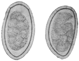
Fig. 166.—Eggs of Dicrocœlium dendriticum, Rud. To the left seen flat, to right lying on one side. 600/1.
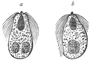
Fig. 167.—Miracidia of Dicrocœlium dendriticum. a, from the dorsum; b, from the side. (After Leuckart.)
In man it is still more uncommon than the liver fluke, and has hitherto only been observed seven times (Germany, Bohemia, Italy, France, and Egypt); it may, however, have occurred more frequently, and have been overlooked, as in slight infections it produces no special symptoms.
The intermediate host is still unknown. Leuckart for some time held the opinion that small species of Planorbis from fresh water, which contain encysted Distomata, were to blame, and he supported his views by a feeding experiment which seemingly yielded positive results; this, however, is not definitely proved. Piana’s statement that small land snails are the intermediate hosts has also not been proved.
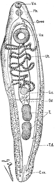
Fig. 168.—Echinostoma ilocanum. Vo., oral sucker; Ph., pharynx; Cirre, cirrus; V.v., ventral sucker; Ut., uterus; G.c., ovary; Ov., shell gland; T., testes; T.d., vitellarium; C.ex., excretory vesicle. (After Brumpt.)
Family. Echinostomidæ, Looss, 1902.
Sub-family. Echinostominæ, Looss, 1899.
Genus. Echinostoma, Rud. 1809; Dietz, 1910.
Fore-body not bulging. Greatest width at or behind the ventral sucker. Oral sucker not atrophied. Collar kidney-shaped with a double dorsally unbroken row of spines, terminating in four to five angle spines. The border spines of the aboral series not larger than the oral. Skin spined or smooth. Body elongated. Uterus long with numerous transverse coils. Ventral sucker in the anterior quarter of body. Cirrus sac small, almost completely in front of the ventral sucker. Testes round or oval, smooth incurved or lobed, in the hinder half of body. Ovary median or lateral in front of testes. Vitellaria from hinder margin of ventral sucker to end of body. Eggs oval, 84 µ to 126 µ by 48 µ to 82 µ.
The spines placed most ventrally, or those placed most medially on ventral surface, are from differences of position or form termed “angle” spines, the rest “border” spines.
Type.—Echinostoma echinatum, Rud.
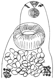
Fig. 169.—Echinostoma ilocanum, Garrison, 1908: head end showing collar of spines, ventral view. (After Leiper.)
Echinostoma ilocanum, Garrison, 1908.
Length 4 to 5 mm., breadth 1 to 1·35 mm., thickness 0·5 to 0·6 mm. The circum-oral disc 0·3 mm. broad, separated by a shallow groove from the body. Crown of forty-nine spines and five to six angle spines on each side continuous with an irregularly alternating series of fourteen spines on the dorsum. Largest spines are 34 µ long, 8 µ thick at the base. The remainder of the dorsal spines are 24 µ by 6 µ. Skin thickly covered with scales on the margins of the body as far back as the level of the hind testis. Oral sucker, 0·18 mm.; ventral sucker, 0·4 to 0·46 mm. Its anterior border about 0·07 mm. from the anterior end. Pharynx 0·17 mm. long, 0·11 mm. broad. Testes about mid-line of the body, much lobed; the lobes of the anterior testis run transversely, while the axis of the posterior testis is longitudinal, as often occurs in the Echinostomidæ. Cirrus sac reaches to the centre of the ventral sucker. Ovary transversely oval in front of the testes. Vitellaria commence about half-way between the ventral sucker and ovary and extend to the posterior end. Eggs numerous, 92 µ to 114 µ by 53 µ to 82 µ.
Average.—99·5 µ by 56 µ.
Habitat.—Gut of man (Filipinos), Philippine Islands.
Echinostoma malayanum, Leiper, 1911.
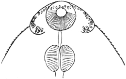
Fig. 170.—Echinostoma malayanum, Leiper, 1912: anterior end showing collar of spines, ventral view. (After Leiper.)
Twelve millimetres long, 3 mm. broad, 1·3 mm. thick. Ends bluntly rounded. At the anterior end a ventral furrow on either side, one-third the width of the body, marking off the circum-oral collar. Along its edge is a row of forty-three spines extending across the middle line dorsally but not ventrally. The spines vary in size from 0·07 mm. in length (ventrally) to 0·05 to 0·016 mm. (dorsally). Cuticular spines also exist on the ventral side as far back as posterior end of body, but dorsally limited to a triangular area ending in front of the ventral sucker. Oral sucker 0·07 mm. thick, occupying the middle third of the circum-oral disc; pharynx 0·25 mm. in diameter; œsophagus 0·04 mm. long; gut cæca simple, extending to end of body; ventral sucker 0·9 mm. long by 0·75 mm. broad by 0·7 mm. deep; wall about 0·25 mm. thick. The sucker is inclined at an angle of 40° to the ventral surface. Testes lobed, one behind the other, behind the ventral sucker. Cirrus pouch well developed, reaching to the posterior edge of the sucker. Genital pore in the angle between neck and anterior lip of ventral sucker. Ovary smooth, 0·3 mm. in diameter, 0·85 mm. behind ventral sucker. Vitellaria very numerous, extending from posterior margin of sucker to posterior end of body, where they intermingle. Eggs few in number, brown and large.
Habitat.—Gut of man (Tamils), Malay States.
Sub-family. Himasthlinæ, Odhner, 1910.
Genus. Artyfechinostomum, Clayton-Lane, 1915.
Crown of thirty-nine spines, continuous over dorsum. Two corner spines long. Vitellaria extend from posterior margin of sucker to posterior end of fluke. Eggs without filament. [Although the possession of strong rose-thorn hooks is given by Odhner as a sub-family characteristic, yet in this genus assigned to this sub-family they have not been seen.—J. W. W. S.]
Artyfechinostomum sufrartyfex, Clayton-Lane, 1915.
Spirit specimens: 9 by 2·5 by 0·8 mm. thick. Ventral sucker conspicuous, 1 mm. in diameter. Cirrus sac 2 mm. long. Testes lobed, about 1·5 mm. in diameter. Posterior border of posterior testes 1 mm. from posterior end. Vitellaria meet posteriorly behind the posterior testis.
Family. Schistosomidæ, Looss, 1899.
Genus. Schistosoma, Weinl, 1858.
Syn.: Gynæcophorus, Dies., 1858; Bilharzia, Cobb., 1859; Thecosoma, Moq. Tandon, 1860.
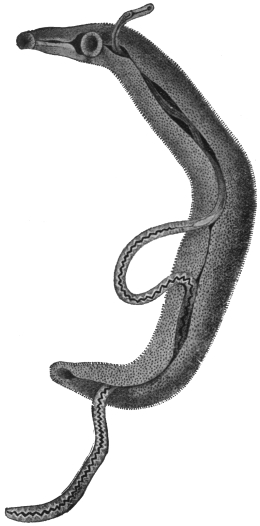
Fig. 171.—Schistosoma hæmatobium, Bil.: male carrying the female in the canalis gynæcophorus. 12/1. (After Looss.)
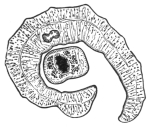
Fig. 172.—Transverse section through a pair of Schistosoma hæmatobium in copulâ. In the male the point of reunion of the intestinal forks has been cut across. (After Leuckart.)
The males have bodies that widen out considerably behind the ventral sucker, the lateral parts of which in-roll ventrally, forming the almost completely closed canalis gynæcophorus, within which the female is enclosed. There is no cirrus pouch. The male has five or six testes, the females are filiform; the uterus is long. There is no Laurer’s canal. The ova almost equally attenuated at either extremity; they have a small terminal spine, and are not provided with a lid. They contain a miracidium, ciliated on all sides, which is characterized by the possession of two large glandular cells, which discharge anteriorly beside the gastric sac. They live in the vascular system of mammals. (An allied genus [Bilharziella] lives in the blood-vessels of birds.)
Schistosoma hæmatobium, Bilharz, 1852.
Syn.: Distoma hæmatobium, Bilh.; Distoma capense, Harley, 1864.
The Male is whitish, 12 to 14 mm. in length, but is already mature when 4 mm. long. The anterior end is 0·6 mm. or a little over in length. The suckers are near each other, the oral sucker is infundibular, and the dorsal lip is longer than the ventral one. The ventral sucker is a little larger, 0·28 mm., and is pedunculated. A little behind the ventral sucker the body broadens to a width of 1 mm., decreasing, however, in thickness; the lateral edges in-roll ventrally, so that the posterior part of the body appears almost cylindrical, 0·4 to 0·5 mm. in diameter; the posterior extremity is somewhat more attenuated. The dorsal surface of the posterior part of the body is covered with spinous papillæ. There are delicate spines on the suckers, and larger ones invest the entire internal surface of the gynæcophoric canal, as well as a longitudinal zone at the edge of that side of the external surface that is covered by the other side rolling over it. The œsophagus is beset with numerous glandular cells (fig. 173), and presents two dilatations; the intestinal bifurcation is close in front of the ventral sucker, the two branches uniting sooner or later behind the testes into a median trunk, which may again divide at short intervals. The excretory pore is at the posterior end, but placed somewhat dorsally; the genital pore is at the beginning of the gynæcophoric canal, thus behind the ventral sucker; into it opens the vas deferens which, posteriorly, broadens into the seminal vesicle and then continues as the vasa efferentia of the four or five testes (fig. 173).
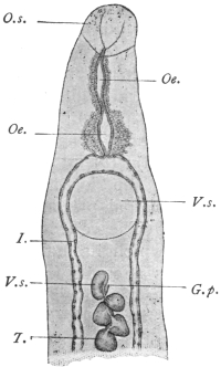
Fig. 173.—Anterior end of the male Schistosoma hæmatobium, Bilh. V.s., ventral sucker; I., gut cæca; G.p., genital pore; T., testes; O.s., oral sucker; Oe., œsophagus with glandular cells; V.s., vesicula seminalis. 40/1. (After Looss.)
The Female—filiform, about 20 mm. in length, pointed at each end, and measuring 0·25 mm. in diameter in the middle. Their colour varies according to the condition of the contents of the intestine. (Posteriorly they are dark brown or blackish.) The cuticle is smooth except in the sucker, where there are very delicate spines, and at the posterior end, where there are other larger spines. The oral sucker is a little larger than the pedunculated ventral sucker (0·07 and 0·059 mm. respectively). The anterior part of the body, 0·2 to 0·3 mm. in length; the œsophagus is as in the male. The intestinal bifurcation is in front of the ventral sucker, the two branches uniting behind the ovary and the trunk running in a zigzag manner to the posterior border. There are indications of diverticula at the flexures. The ovary is median. In young females it is of an elongated oval shape; in older females the posterior end becomes club-shaped, whereas the anterior end becomes attenuated; the oviduct originates at the posterior end, but immediately turns forwards and joins the parallel vitelline duct in front of the ovary (fig. 174), where the shell gland cells open; the common canal becomes dilated to form the oötype, and then proceeds as the uterus, with only slight convolutions, along the central field to the genital pore, which lies in the middle line immediately behind the ventral sucker. The single vitellarium starts behind the ovary and extends to the posterior end. The acini are situated at the sides of the excretory duct, which runs a median course. The eggs are compact spindles, much dilated in the middle; they have no lid, and are provided with a terminal spine (rudimentary filament) at the posterior end, measuring 120 µ to 150 µ in length and 40 µ to 60 µ in breadth, but vary in size and shape (fig. 175).
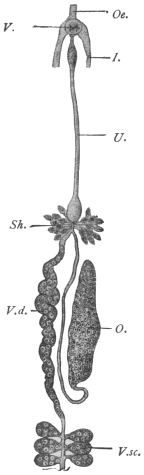
Fig. 174.—Schistosoma hæmatobium, Bilh.: genitalia of the female. V.s., ventral sucker; I., gut cæca; V.d., vitelline duct; V.sc., vitellarium; O., ovary; Oe., œsophagus; Sh., shell gland; U., uterus. Magnified. (After Leuckart.)
Distribution.—In order to understand the distribution of the worms and eggs in the body, it may be well to recall the blood supply of the abdominal and pelvic organs. It is generally assumed that the early life (? cercarial stage) of the worms occurs in the liver, and that the young worms travel from here, where they are invariably found, to their various sites along the portal vein and its tributaries and so against the blood stream. The tributaries of the portal vein are:—
(1) Superior mesenteric, the tributaries of which are: (a) the veins of the small intestine; (b) ileo-colic; (c) right colic; (d) middle colic; (e) right gastro-epiploic; and (f) inferior pancreatic. By these paths infection of the small intestine, ascending and transverse colon and pancreas would occur.
(2) Splenic. (Ova have been recorded by Symmers in the spleen.)
(3) Inferior mesenteric, the tributaries of which are (a) superior hæmorrhoidal veins from the upper part of the hæmorrhoidal plexus; (b) sigmoid veins from sigmoid flexure and lower portion of descending colon; (c) left colic vein draining descending colon.
The superior hæmorrhoidal veins form a rich plexus in the rectum, and below this level in the upper and middle parts of the anal canal. The plexus forms two networks, an internal plexus in the submucosa and an external on the outer surface. The internal plexus opens at the anal orifice into: (a) branches of the inferior hæmorrhoidal vein (from the pudic); (b) the external plexus. The external plexus gives off: (a) inferior hæmorrhoidal opening into internal pudic (of internal iliac vein); (b) mid-hæmorrhoidal into internal iliac or its branches; and (c) superior hæmorrhoidal opening into inferior mesenteric. The external plexus further communicates with the vesico-prostatic plexus. The vesico-prostatic (vaginal) plexus opens into the vesical veins, which drain into the interior iliac vein. This plexus also receives afferents from the pudendal plexus, the chief tributary of which is the dorsal vein of the penis. The pudendal plexus also receives branches from the inferior pudic and the anterior surface of the bladder.
There is thus a communication between the portal vein and the vena cava by means of these plexuses, viz., through the inferior and middle hæmorrhoidals, and by the inferior hæmorrhoidals to the bladder and thence by the vesical veins or the pudic to the caval system (interior iliac).
It is thus by the inferior mesenteric and its tributaries that the worms reach the descending colon, rectum, anal canal, and eventually the bladder, and in some cases the caval system.
Before considering what is actually found post mortem in these veins and the organs drained by them, we may further recall the fact that the calibre of “medium” veins is 4 to 8 mm., “small” veins less than 40 µ in diameter and capillaries 8 µ to 20 µ. Further, the maximum diameter of the male worm is 1 mm., that of the female 280 µ and eggs in utero 80 µ to 90 µ long by 30 µ to 40 µ.
Liver and Portal Vein.—Here worms are most easily found post mortem. Often only males are found and these of the same size, and if females occur only a few worms are found in copulâ. The worms are frequently not full size and the males may contain no free spermatozoa in their testes, and as regards the females some may be fertilized, others not, as shown by the presence or absence of spermatozoa in the seminal receptacle or uterus. In either case they may contain eggs—lateral-spined—usually one, less often two, but there may be as many as five or six. These eggs may also show some abnormality, which takes the form of: (1) abnormal contents, viz., disintegrating yolk cells with or without an ovarian cell; (2) abnormal shape but with normal contents and probably represented by the collapsed and empty egg-shells which are found in the tissues.
As to the interpretation of these facts, Looss believes that these lateral-spined eggs are products of young females whose egg-laying is not at first properly regulated. The shape that the eggs take, viz., with a lateral spine, is determined by an excess of material—ovarian and yolk cells—being present in the oötype. The shape of eggs depends upon the position they have in the oötype during their formation. In young females an excess of cells—yolk cells especially—accumulates, distending not only the dorsal wall but a portion also of the short duct joining the oötype to the uterus. The result of this is that the axis of the oötype and egg is almost transverse to the body, and the posterior funnel-shaped portion of the oötype, instead of being terminal, has now a lateral or rather a ventral position, so that the spine which occupies this portion, instead of being terminal, is now lateral. It is noteworthy that these lateral-spined eggs are thicker, owing to the excess of material present, and not uncommonly have a curved anterior border, due to a projection of the anterior end into the anterior opening of the oötype.
As these eggs are being laid by females in the portal vein they are carried back to the liver by the blood stream. The liver is one of the commonest sites for these eggs; also terminal-spined eggs may be found here for the same reason.
Hæmorrhoidal Veins.—Mature worms, generally in copulâ, are usually found here, though young not fully grown females may also occur. The tissues of the rectal wall (or colon) show, as a rule, large quantities of lateral-spined eggs, though less often only terminal-spined eggs may be found.
Vesico-prostatic Plexus.—Worms in copulâ are found in the veins of the submucosa in the bladder, and the eggs in the mucosa, and those voided are usually terminal-spined, though lateral-spined eggs are not so rare as generally thought. The problem next arises as to how the eggs get to the lumen of the gut or bladder.
The female worm is 280 µ in diameter. Veins in the submucosa of the rectum less than 178 µ in diameter are not affected with endophlebitis. It is probable that the female even by stretching could not penetrate much beyond this. Eggs are probably then laid in the submucosa as near the muscularis mucosa as possible. Now if the eggs are laid in a vein of larger calibre than the worm fills, the eggs would be carried back to the inferior mesenteric vein, so that presumably the worm must succeed in blocking the vein already narrowed by endophlebitis, so that by the stasis which ensues the eggs may escape from the veins. How this occurs is not exactly known; it is not necessarily due to the spine, as the same escape into the tissues occurs in spineless eggs, such as those of Schistosoma japonicum. The eggs, then, pass as foreign bodies through the tissues. Another hypothesis is that the worms leave the veins in order to lay their eggs, but the evidence is against this.
Caval System.—Occasionally worms that have passed through the vesical plexus may be found in the iliac vein, inferior vena cava, and even the lungs. If the worms are young they contain a lateral-spined egg; if adult, numerous (50 to 100) terminal-spined eggs.
Lungs.—When the liver is strongly infected with (terminal-spined) eggs it is possible that by passive movements some may pass into the intralobular veins, and thence by the inferior vena cava to the lungs.
Gall-bladder.—Similarly terminal-spined eggs pass into the bile-capillaries and gall-bladder (where they may be abundant), and so into the fæces.
Detection of Eggs.—Occasionally eggs may be found in various other parts of the body. They are best detected by macerating pieces of the tissue in question in about 1/4 per cent. hydrochloric acid at 50 to 60°C. (Looss).
Pathological changes:—
Rectum.—These have been studied thoroughly by Letulle in the case of an apparently pure infection of the rectum.274 They take the form of a chronic diffuse inflammation, which may result in—(1) ulceration, or (2) hyperplasia of the mucosa, producing adenomata.
Ulcerative Form.—The mucosa is transformed into a mass of vascular connective tissue. The connective tissue spaces next become invaded by numerous mononuclear cells. The tissue itself undergoes diffuse sclerosis, becoming hard and fibroid. Eventually ulcerative necrosis sets in. During these changes the Lieberkühn glands are destroyed. The process does not extend to the submucosa, in this respect differing from that in chronic dysentery.
Hyperplastic Form.—The Lieberkühn glands of the mucosa at first hypertrophy; then there is an actual hyperplasia resulting in adenomata. The interstitial tissue of the glands is also greatly hypertrophied, giving rise to very vascular granulations. These growths are often hollow and contain worms. Many eggs are found in the mucosa on their way to the lumen of the gut.
The muscularis mucosa is thickened up to twice or even ten times the normal. Its vessels are dilated (36 µ to 80 µ), but they do not allow of the passage of worms.
The submucosa is profoundly changed; rigid and hard instead of supple. It is here that the greatest number of eggs occur. A remarkable condition of endophlebitis exists in the veins of the submucosa, not only in the smaller ones but also in the larger ones (370 µ by 270 µ). This endophlebitis results in a more or less complete occlusion of the vessels of the lumen.
The muscular coats are free from change, also their veins.
The Serous Coats.—The veins about 1,900 µ, also show endophlebitis. Besides the rectum, in extreme cases even the transverse colon, the cæcum and small intestine may be affected.
Bladder.—In the early stages the mucosa is deep red and swollen like velvet, or there may be localized patches of hyperæmia or extravasation. The subsequent changes take two chief forms:—
(1) Sandy Patches.—The mucosa looks as if it were impregnated with a fine brownish or yellowish powder (myriads of ova). This is accompanied by a gradual hypertrophy and new formation of connective tissue, so that dry, hard or plate-like patches with this sandy appearance arise; the thickening eventually affects all the coats of the bladder. In the older patches many of the eggs are calcified. These patches sooner or later break down, ulcerate and necrose. Phosphatic deposits are abundant and stone is common. These patches are not found in the rectum.
(2) Papillomata.—Where the inflammatory change produced by the eggs gives rise to hypertrophy and hyperplasia of the mucosa, papillomata result, the axis of which is formed by connective tissue of the submucosa. These are most variable in shape and form and bleed readily, and sometimes contain cavities of extravasated blood.
As in the rectum, it is in the submucosa that eggs are most abundant, and worms in copulâ occur in the veins of this layer, but endophlebitis is not as general as described in the rectum. Malignant disease of the bladder is not an uncommon sequela of bilharziasis. Besides the bladder, the ureters and kidneys may in advanced cases be involved. The prostate and vesiculæ seminales are commonly diseased. Eggs have been recorded in the semen. The urethra is frequently attacked; the vagina in the female.
Eggs also occur in the lymphatic glands of the gut.
Geographical Distribution.—East Africa: Nile Valley, Red Sea Coast, Zanzibar, Portuguese East Africa, Delagoa Bay, Natal, Port Elizabeth.
South Africa: Cape Colony, Orange Free State, Transvaal, Mauritius, Bourbon, Madagascar.
West Africa: Angola, Cameroons, Gold Coast, Gambia, Senegal, Sierra Leone, Lagos, Nigeria.
North Africa: Tripoli, Tunis, Algeria, parts of the Sahara.
Central Africa: Sudan, various portions. Uganda, Nyasaland.
It occurs with varying frequency in these regions. It is probably more widely spread than this list implies, as undoubtedly many cases are seen which are not recorded.
Isolated cases have been recorded from Arabia, India,275 Greece, Cyprus.
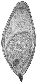
Fig. 175.—Ovum of Schistosoma hæmatobium, Bilh., with miracidium, which has turned its anterior end towards the posterior end of the egg. 275/1. (After Looss.)
The means by which infection is brought about are still uncertain; we only know that the miracidia (fig. 175) enclosed in the discharged eggs do not hatch if the eggs remain in the urine, but after cooling perish. As soon, however, as the urine is diluted with water the shell swells, generally bursting lengthways, and releases the miracidium from its investing membrane, so that it can swim about with the aid of its cilia. In its structure it differs but little from the miracidium of Fasciola hepatica, as, for instance, in the lack of eyes; the two large gland cells situated on either side of the intestinal sac are also present in the miracidia of Fasciola hepatica.
Sarcode Globules.—This is a term applied to certain globules which at times appear in the miracidium and are later ejected. Some authors consider them as indicative that the miracidium has developed into a sporocyst, but Looss considers them to be degeneration products.
The Bilharzia mission, under R. T. Leiper, sent to Egypt by the War Office early in 1915, reports that cercariæ of bilharzia type were recognized in four of the commonest fresh-water molluscs around Cairo.
With material obtained from naturally infected Planorbis boissyi acute bilharziosis was experimentally produced in rats, mice, and monkeys. Infection takes place experimentally through the skin and also through the mucous membrane of the mouth and œsophagus. The miracidium, after entering the mollusc, develops into a sporocyst. This gives rise not to rediæ, but to secondary sporocysts, which, in turn, produce cercariæ. These, like the adult worm, differ from other distomes in lacking a muscular pharynx.
Schistosoma mansoni, Sambon, 1907.
According to Manson, Sambon and others, the eggs with lateral spines belong to a species different from Schistosoma hæmatobium. Infections with this species only are said to occur in the Congo, Southern States of North America, West Indies (Guadeloupe) and Brazil (Bahia). The following characters, according to Flu, differentiate this species: (1) In the male the transition from the anterior portion of the worm to the lateral fields (the infolded portions which form the gynæcophoric canal) is not a gradual one as in Schistosoma hæmatobium, but in this case the lateral fields rise suddenly, almost at right angles to the anterior portion. (2) The ovaries have a well-marked convoluted course as in no other schistosome. (3) The oötype is symmetrical in reference to the long axis of the body, its duct being lateral on the ventral side (Looss’ explanation of this we have already given). (4) The worms live exclusively in portal vein and tract. (As lateral-spined eggs occur also in the bladder, this is not exactly true.)
Schistosoma hæmatobium, Bilharz, 1852.
Male, four or five large testes. Gut forks unite late, so that the single gut stem is short. Female, ovary in posterior half of body. Uterus very long, voluminous, with many terminal-spined eggs, some lying in pairs. Vitellaria in posterior fourth of body. Cercariæ in Bullinus contortus and Bullinus dybowski (syn.: Physa alexandrina) in Egypt.
Schistosoma mansoni, Sambon, 1907.
Male, eight small testes. Gut forks unite early, so that the single gut stem is very long. Females, ovary in anterior half of body. Uterus very short; usually only one lateral-spined egg at a time in utero. Vitellaria occupy posterior two-thirds of body. Cercariæ in Planorbis boissyi in Egypt.
The above morphological descriptions are founded on worms of each species, derived from experimentally infected mice (Leiper, R. T., Brit. Med. Journ., March 18, 1916, p. 411).
Schistosoma japonicum, Katsurada, 1904.
Syn.: S. cattoi, Blanchard, 1905.

Fig. 176.—Schistosoma japonicum: anterior end with testes; posterior end with point of union of cæca. Length of worm about 10 mm. (After Katsurada.)
Male.—Eight to 19 mm., but extreme limits are 5 to 22·5 mm. Consists of a short fore-body, separated by the ventral sucker from the hind-body. The ventral sucker is stalked and somewhat larger than the oral sucker. Both suckers are larger than the corresponding ones in S. hæmatobium. Body usually smooth, but in the fresh state numerous fairly evident spines along the margin of the canal. Œsophagus: two bulbs. The junction of the gut forks more posterior than in S. hæmatobium, the median united gut stem occupying a quarter to one-fifth to one-sixth of the body length. An excretory canal runs along each side of the body, opening into the dorsal excretory pore. Testes irregularly elliptical, six to eight in number, in the anterior part of hind-body. The vasa efferentia unite into a common vas deferens which opens directly behind the ventral sucker. The seminal vesicle lies just behind this.
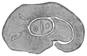
Fig. 177.—Schistosoma japonicum, male and female in copulâ. × 60. (After Katsurada.)
Female.—Up to 26 mm., generally thinner than the male. Surface smooth. Suckers armed with fine spines. Ventral sucker larger than oral. Body thicker behind the region of the ovary. The gut forks unite immediately behind the ovary. The united gut much thicker than in S. hæmatobium. Ovary elliptical, almost in the mid-body, its hinder portion dilated. The oviduct arises from its posterior end and then runs sinuously forward, where it is joined by the vitellarian duct; the vitellarium well developed, extending from behind the ovary almost but not quite to the posterior end as in S. hæmatobium. Shell gland ducts enter at the junction point of oviduct and vitelline duct. The canal here forms an oötype and then proceeds as the uterus to open directly behind the ventral sucker. The uterus occupies almost half the hind-body. In S. hæmatobium this is not so. The uterine canal is cleft-like, i.e., its dorso-ventral diameter is much greater than its lateral diameter. The number of eggs varies from about 50 to 300 from observations made in various hosts.
Eggs.—In utero assume various shapes, as they are soft; the lumen of the uterus is narrow. Outside they are oval, faint yellow, double contoured. In fæces the eggs measure 83·5 µ, by 62·5 µ (man); 85 µ by 61·5 µ (cattle); 98·2 µ by 73·8 µ (dog). The eggs have either small lateral spines or thickenings, and Looss at the opposite side has described cap-like thickenings. The eggs in the tissues undergo various deformities, and may contain a miracidium, as also the eggs in fæces do; or the contents may consist of granular matter or amorphous masses or they may be calcified. Lymphocytes and giant cells may also invade the eggs.
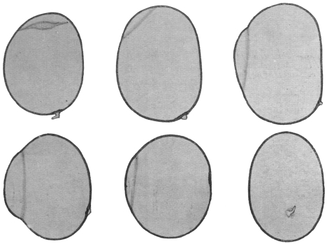
Fig. 178.—Schistosoma japonicum: eggs from human liver, showing “spines” and “hoods” at opposite pole. (After Looss.)
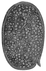
Fig. 179.—Schistosoma japonicum: from dog. Uterine egg. × c. 800. (After Katsurada.)
Mode of Infection.—The miracidia hatch in water in as little as fifteen minutes, but the majority in one to three hours. They will live in water for about twenty-four hours. In water they undergo a transformation into “larvæ,” which then penetrate the skin, as has been shown by Japanese writers to hold good for man, cattle, dog and cat. The penetration of the skin is attended with an eruption on the legs, “Kabure.” The exact route by which the worms reach the portal vein is uncertain. Infection in Japan takes place from spring to autumn, especially May to July, when the soil is contaminated with manure of cattle infected with S. japonicum. They also appear to develop in molluscs. Leiper and Atkinson found cercariæ (in sporocysts) in the liver of a mollusc, Katayama nosophora. They infected mice by immersing them in water containing liver emulsion and so free cercariæ, thus confirming the similar results of Miyairi and Suzuki.
Habitat.—The worm occurs in Japan, China, and the Philippines. The normal host is man and mammals. Cattle, dog and cat are often found naturally infected. Mice can also be experimentally infected. Their seat of election is the portal vein and its branches, especially the mesenteric veins. They either swim free in the blood or remain fixed by their suckers to the intima of the vessels. They have also been found in the vena cava and right heart of a cat, but not so far in the vesical plexus.
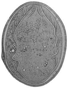
Fig. 180.—Schistosoma japonicum: from dog. × c. 800. (After Katsurada.)
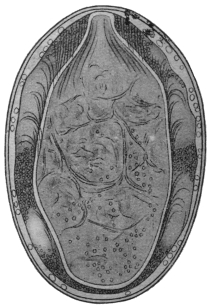
Fig. 181.—Schistosoma japonicum: from dog. Egg from fæces. × c. 800. (After Katsurada.)
Eggs are found in the submucosa and mucosa of the gut, especially the colon, and at times in the serosa and subserosa of the small intestine, where they give rise to new growths. Occasionally eggs are found in the brain. The life of the worms is at least two years.
Pathogenic Effects.—Anæmia through loss of blood due to worms; enlarged spleen, toxic in origin (?); phlebitis, thrombosis, due to portal stasis; the eggs, however, cause the greatest mischief. They are carried by the circulation to various organs where they produce inflammation, granulation tissue, and later connective tissue.
Liver.—The eggs reaching this organ give rise to granulomata and hence enlarged liver, and later, when connective tissue is formed, to contraction. The surface is rough and irregularly granular, “parasitic embolic cirrhosis” of Yamagiwa.
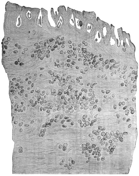
Fig. 182.—Schistosoma japonicum: section through the gut of a Chinaman showing eggs. × 58. (After Catto.)
Gut.—The eggs in the mucosa and submucosa cause catarrh and destruction of tissue or new growth. In the small intestine the eggs are mainly in the serosa and subserosa, where they give rise to polypoid or branched growths.
Spleen.—Enlarged, at first due to toxin (?) and later due to portal stasis. Eggs in the spleen are uncommon.
Ascites also arises from the portal stasis, and is generally present in advanced cases.
Eggs may be found in many other situations: glands (numerous), mesentery, stomach, pancreas, kidney, etc. The bladder remains free.
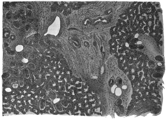
Fig. 183.—Schistosoma japonicum: liver showing eggs in the intra- and interlobular connective tissue. × c. 80. (After Katsurada.)
Class III. CESTODA, Rud., 1808.
Tapeworms have been known from ancient times—at all events, the large species inhabiting the intestines of man—and there has never been a doubt as to their animal nature. The large cysticerci of the domestic animals (occasionally of man also) have been known for an equally long period, but they were generally regarded as growths, or “hydatids,” until almost simultaneously Redi in Italy, and Hartmann and Wepfer in Germany, concluded from their movements and organization that they were of animal nature. From that time the cysticerci have been included amongst the other intestinal worms, and Zeder (1800) established a special class (Cystici, Rud., 1808) for the bladder worms. Things remained in this condition until the middle of the last century, when Küchenmeister, by means of successful feeding experiments, demonstrated that the cysticerci were definite stages of development of certain tapeworms. Before Küchenmeister, E. Blanchard, van Beneden, and v. Siebold had held the same opinion in regard to other asexual Cestodes.
Since the most remote period another question has again and again occupied the attention of naturalists, the question of the morphological nature—that of the INDIVIDUALITY OF THE TAPEWORM. The ancients, who were well acquainted with the proglottids (Vermes cucurbitani) that are frequently evacuated, were of the opinion that the tapeworm originated through the union of these separate proglottids, and this view was maintained until the end of the seventeenth century. In 1683 Tyson discovered the head with the double circlet of hooks in a large tapeworm of the dog; Redi (1684) was also acquainted with the head and the suckers of several Tæniæ. Andry (1700) found the head of Tænia saginata, and Bonnet (1777) and Gleichen-Rusworm (1779) found the head of Dibothriocephalus latus. Consequently most authors, on the ground of this discovery, considered the tapeworm as a single animal, that maintains its hold in the intestine by means of the head, and likewise feeds itself through it. The fact was recognized that there were longitudinal canals running through the entire length of the worm, and it was thought that these originated in the suckers, and that the entire apparatus was an intestine. As, moreover, the segments form at the neck, and are cast off from the opposite extremity, the tapeworm was also compared with the polyps, which were formerly regarded as independent beings.
Steenstrup, in his celebrated work on the alternation of generations (1841), was the first to give another explanation. This has been elaborated still further by van Beneden, v. Siebold and Leuckart, and until a few years ago all authorities adopted his views. According to this view, the tapeworm is composed of numerous individuals, something like a polyp colony, and, in addition to the proglottids—the sexual individuals which are usually present in large numbers—there is ONE individual of different structure, the scolex, which not only fastens the entire colony to the intestine, but actually produces this colony from itself, and therefore is present earlier than the proglottids. The scolex is a “nurse,” which, though itself produced by sexual means, increases asexually like a Scyphistoma polyp; the tapeworm chain has therefore been termed a strobila. Consequently the development of the tapeworms was explained by an alternation of generations. In support of this opinion it was demonstrated not only that the adult sexual creatures, the proglottids, can separate from the colony and live independently for a time, but that in certain Tæniæ, and especially in many Cestodes of the shark, the proglottids detach themselves long before they have attained their ultimate size, and thus separated continue to develop, grow and finally multiply; the scolex also exhibits a certain independence in so far as, though not, as a rule, capable of a free life, yet it in some cases lives as a free being, partly on the surface of the body of marine fishes and partly in the sea. With the more intimate knowledge of the development of the cysticerci, the independent nature of the scolex was recognized. It is formed by a budding of the bladder that has developed from the oncosphere, in some cases (Cœnurus) in large numbers, in other cases (Echinococcus) only after the parent cyst has developed several daughter cysts. Released from its mother cyst and placed in suitable conditions, it goes on living, and gives rise at its posterior end by budding to the strobila, the proglottids of which eventually become sexual individuals.
In order to make this clearer we will briefly summarize what takes place in the jelly-fishes.
By metamorphosis is meant a developmental change in the same individual, while alternation of generations, or metagenesis, implies a stage in which reproduction of individuals takes place by a process of budding or fission. This asexual reproductive stage alternates with the sexual mode of reproduction. Thus in the development of the Scyphozoa (jelly-fishes) we have:—
(1) The fertilized egg cell divides regularly and forms a morula.
(2) By accumulation of fluid in the interior this becomes a closed sac with a wall formed of a single layer of cells, forming the blastosphere or blastula.
(3) One end of the sac is invaginated, forming a gastrula.
(4) The gastrula pore or mouth closes, forming again a sac, the walls of which have two layers, forming a planula.
(5) This becomes fixed to a rock, an invagination forms at one end, a depression—the stomodæum—communicating with the enteric cavity. Tentacles grow out and we have a Scyphozoön polype, Scyphistoma or Scyphula. It is to this stage that Steenstrup gave the name “nurse” (“wet-nurse”), because it nourished or produced asexually the succeeding forms.
(6) Asexual reproduction by transverse fission occurs in this, forming a pile of saucer- or pine-cone-like animals which before this time had been considered to be a distinct animal, which was called strobila from its resemblance to a pine-cone. This is the alternate generation.
(7) The individuals of the strobila become free and are called Ephyrulæ.
(8) These develop finally into adult sexual jelly-fish, Scyphozoa, so that comparing a tapeworm with this we have (a) egg, (b) scolex (= Scyphula or “nurse”), (c) asexual reproduction of the tapeworm chain (= strobila), (d) development of the individuals of the chain (proglottids) into sexual adults.
Van Beneden’s terminology for these stages is the following: Ciliated embryo = protoscolex; scyphistoma = deutoscolex (or scolex); free Ephyrula = proglottis. According to this view, as is the case in many endoparasitic Trematodes, asexual reproduction by budding occurs at two stages of the whole cycle of development, viz. (1) in the formation of the scolex by budding from the bladder (“nurse”), (2) in the formation of the strobila by budding from the scolex (“nurse”).
But in cysticercal larval forms it appears that the scolex does not arise in this way but is simply a part of the proscolex (hexacanth embryo), becoming invaginated into it for protection, so that there is no asexual gemmation here. It has been questioned also whether the strobila also arises by gemmation. If it does, the tapeworm is a colony of zoöids produced by budding from the asexual scolex; if it is not produced in this way, then the tapeworm is to be regarded as an individual in which growth is accompanied by segmentation. Against the “colony” view are the facts that the muscular, nervous, and excretory systems are continuous throughout the worm, and that some tapeworms, such as Ligula, are unsegmented.
Finally, if the tapeworm is an individual the question arises which is the head end. As new segments are formed at the neck, and as this point in annelids is the antepenultimate segment, the scolex must be the last or posterior segment. The caudal vesicle or bladder of larval forms is consequently anterior. According to this view, in tapeworms as among many endoparasitic flukes, an asexual multiplication occurs at two points of the whole cycle of development, which is as follows: (1) egg, (2) oncosphere or hexacanth embryo, (3) bladder (cysticercus or hydatid), (4) (after digestion of the bladder) by budding, the scolex, (5) by budding from the scolex the sexual proglottids, (6) the egg; (4) and (5) being the two asexual stages.
Anatomy of the Cestoda.
If we except the tapeworms with only one proglottis, the Cestoidea Monozoa, Lang = Cestodaria, Monticelli, we can always distinguish in the Cestodes, in the narrower sense, one scolex or head and a large or small number of segments (proglottids). The SCOLEX serves the entire tapeworm for fastening it to the internal surface of the intestinal wall, and therefore carries at its end various organs which assist in this function, and which are as follows: (i) Suctorial organs, i.e., the four suckers (acetabula), which are placed crosswise at the circumference of the thickened end of the scolex; further, the double or quadruple groove-like suckers (bothridia), which are diversely shaped in the various genera and families.276 (2) Fixation organs (hooklets)277 that likewise occur in varying numbers and different positions; they may be in the suckers, or outside them on the apex of the scolex; for instance, in many of the Tæniidæ they appear in a circle around a single protractile organ, the rostellum, or the latter may be rudimentary, and is then replaced by a terminal sucker. (3) Proboscis. One family of the Cestodes, the Rhynchobothriidæ, carries four proboscides, moved by their own muscular apparatus, on the scolex, and they are beset with the most diverse hooks. (4) Tentacle-like formations are only known in one genus (Polypocephalus).
The thickened part of the scolex that carries the suckers is usually called the head; the following flat (unsegmented) part connecting it with the proglottids is called the neck, and is sometimes quite small. In a few cases the entire scolex (or head) disappears, and its function is then undertaken by the contiguous portion of the chain of proglottids, which is transformed into a variously shaped PSEUDO-SCOLEX.
The proglottids are joined to the scolex in a longitudinal row, and are arranged according to age in such a manner that the oldest proglottis is farthest from the scolex, and the youngest nearest to it.
The number of segments varies, according to the species, from only a few to several thousands; they are either quadrangular or rectangular; in the latter case their longitudinal axis falls either longitudinal or transverse to that of the entire chain, according as the segments are longer than broad or broader than long. When the number of segments is very large, the youngest ones are, as a rule, transversely oblong, the middle ones are squarish, and the mature ones longitudinally oblong. The posterior border of the segments, as a rule, carries a longitudinal groove for the reception of the shorter anterior border of the following proglottis. The two lateral borders of the segment are rectilinear, but converge more or less towards the front, or they are bent outwards. In most of the Cestodes the segments, just as the neck, are very flat; in rare cases their transverse diameter is equal to their dorso-ventral diameter. As a rule the segments, singly or several united together, detach themselves from the posterior end, in many cases only after complete maturity is attained, and in others much earlier; they then continue to live near their parent colony, to still call it by that name, in the same intestine and continue their development. Even when evacuated from the intestine the proglottids under favourable circumstances can continue to live and creep about, until sooner or later they perish.
The first proglottis formed, and which in a complete tapeworm [i.e., sexually complete] is the most posterior, is as a rule smaller and of different shape, it also frequently remains sterile, as likewise happens in the next (younger) segments in a few species; otherwise, however, sooner or later the generative organs develop in all the segments, mostly singly, sometimes in pairs; in the latter case they may be quite distinct from each other or possess some parts in common. The term “mature” is used for a proglottid that has the sexual organs fully developed, while “gravid” is used for one containing eggs. Most of the species combine male and female genitalia in the same segment, only a few are sexually distinct (Diœcocestus). In the hermaphrodite species one male and one female sexual orifice are always present, and, in addition, there may be a second female orifice, the uterine opening; as a rule, however, this is lacking, and in one sub-family, the Acoleinæ, to which also the genus Diœcocestus belongs, the other sexual orifice, the opening of the vagina, is also absent. The position of these orifices varies; the cirrus and vagina usually open into a common atrium on one lateral border or on a surface of the segments; the orifice of the uterus may be on the same surface or on the opposite one.
The surface on which the uterus opens is termed the VENTRAL SURFACE; if this orifice is absent, one must depend on the ovary, which almost always approaches one of the two surfaces; this surface is then called the ventral.
The length of the Cestodes—independently of their age—depends on the number and size of the segments, as well as on their contraction; the smallest species (Davainea proglottina) is 0·5 to 1·0 mm. in length; the largest may attain a length of 10 m., and even more.
The entire superficial surface of the tapeworms is covered with a fairly resistant and elastic layer, which exhibits several indistinctly limited layers and which is usually called a cuticle, which also covers the suckers, and is reflected inwardly at the sexual orifices. In some species fine hairs appear, either on the entire body or only in the region of the neck, on the external surface. In the cuticle there can be recognized, besides the pores, which no doubt are concerned with nutrition, spaces in which lie the ends of sensory cells. Close under the cuticle lies the external layer of the parenchyma (basal membrane), and below this the circular and longitudinal muscles forming the dermo-muscular coat. The matrix cells of the cuticle occur as in the Trematodes, only on the inner side of the peripheral muscles in the external zone of the parenchyma; they are fusiform cells, forming one or two layers, but are not arranged in the manner of epithelial cells (fig. 184, Sc.c.). They have fine branching processes which run between the dermal muscles, pass through the basal membrane and penetrate the internal surface of the cuticle with small pistil-like enlargements, expanding on the internal surface of the cuticle into a thin plasma layer.
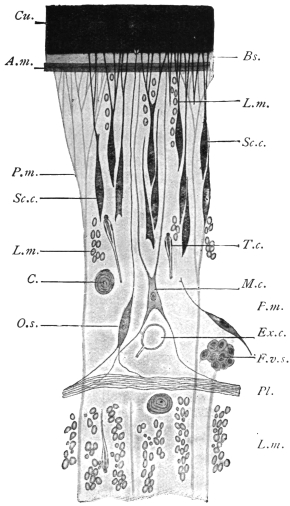
Fig. 184.—Schematic representation of a small part of a transverse section of Ligula sp. Bs., basal membrane; Cu., cuticle; at its base are the endplates of the subcuticular (epithelial) cells; in the centre a cuticular sense organ, O.s.; F.v.s., vitelline follicle; Exc., excretory vessel; C., calcareous corpuscle; L.m., longitudinal muscles; M.c., myoblast; P.m., parenchymatous or dorso-ventral muscles; Pl., plexus of nerve fibres; A.m., circular muscles; Sc.c., subcuticular or matrix cell; T.c., terminal flame cell. 500/1. (After Blochmann.)
In addition to the above mentioned, there are other cuticular formations occurring on the cuticle of some Cestodes, such as immobile hairs and variously formed hooks, such as are seen principally on the scolex. Their development is only roughly known in a few species; they are usually already present in the larval stage, and of the same arrangement and shape as in the fully developed tapeworms; a matter of importance, because by these structures larvæ can be recognized as being those of a certain species of tapeworm.
The CUTICULAR GLANDS in Cestodes are scarce.
The PARENCHYMA forms the chief tissue of the entire body, and in all essentials its structure is similar to that of the Trematodes.
The same doubt exists here also as to the nature of the parenchyma. Recent authors consider that it consists of highly branched cells, the processes of which ramify in all directions. These cells lie in a non-cellular matrix containing fluid vacuoles. This matrix spreads in between and so breaks the continuity of the epidermal cells.
In the parenchyma of almost all the Cestodes there are found in adult specimens, as well as in larvæ, light-refracting concentrically striated structures, of a spherical or broad elliptical shape, which, on account of their containing carbonate of lime, are termed CALCAREOUS CORPUSCLES (fig. 184, C.). Their size, between 3 µ and 30 µ, varies according to the species; their frequency and distribution in the parenchyma also varies, but they are chiefly found in the cortical layer. They are the product of certain parenchymatous cells, in the interior of which they lie like a fat globule in a fat cell, but according to others they are intercellular in origin.
The MUSCULAR SYSTEM of the proglottids is composed of—(1) the subcuticular muscles (figs. 184 and 185), as a rule consisting of a single layer of annular muscles; (2) longitudinal muscles; (3) dorso-ventral fibres extending singly from one surface to the other, and at both ends expanding in a brush-like manner, and inserted into the basal membrane, consisting of an outer, more numerous, and an inner, less numerous but more powerful layer (the number of bundles in this layer being in certain cases of specific importance); (4) transverse fibres, the elements of which penetrate to the borders of the segments, thus passing through the longitudinal muscles and reaching the cuticle. In the region of the septa the transverse and dorso-ventral muscles form a kind of plate.
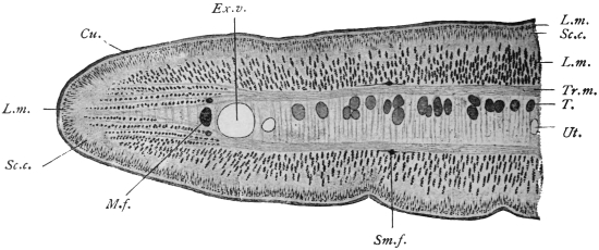
Fig. 185.—Half of a transverse section through a proglottis of Tænia crassicollis. Cu., cuticle; Ex.v., external excretory vessel, to the right of which there is the smaller internal one; T., testicular vesicles; L.m., longitudinal muscles (outer and inner); M.f., lateral nerve with the two accessory nerves; Sc.c., subcuticular matrix cells; Sm.f., submedian nerve; Tr.m., transverse muscles; Ut., the uterus, and the middle of the entire transverse section. 44/1.
The mass of parenchyma bounded by the transverse muscles is termed the MEDULLARY layer, while the mass lying outside them is termed the CORTICAL LAYER.
It was known long ago that the myoblasts adhere to the dorso-ventral fibres as thickenings, but it is only recently that large star-shaped cells (fig. 184), separated from but connected with them by processes, have been recognized as the myoblasts of other fibres (Blochmann, Zernecke).
Within the scolex the direction and course of the muscular layers change.
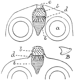
Fig. 186.—Dipylidium caninum: from the cat. In the upper figure the rostellum is retracted, in the lower protruded, a, sucker; b, hooks of rostellum; B, enlarged hook; c, apical aperture on scolex; d, longitudinal muscles; e, circular muscles. (After Benham.)
The SUCKERS are parts of the musculature, locally transformed, with a powerful development of the dorso-ventral muscles, now become radial fibres.
The ROSTELLUM of the armed Tæniæ, like the proboscis of the Rhynchobothriidæ, also belongs to the same category of organs.
In the simplest form, the rostellum, or top of the head (as in Dipylidium caninum), appears as a hollow oval sac, the anterior part of which, projecting beyond the upper surface of the head, carries several rows of hooks (fig. 186). The entire internal space of the sac is occupied by an elastic, slightly fibrous mass, while the anterior half of the surface of the rostellum is covered by longitudinal fibres and the posterior half by circular fibres. On contraction of the latter the entire mass is protruded through the apical aperture, the surface of the rostellum becomes more arched, and the position of the hooks is, in consequence, altered. The rostellum of the large-hooked Tæniidæ, which inhabit the intestine of man and beasts of prey, is of a far more complicated structure, for, in addition to the somewhat lens-shaped rostellum carrying the hooks on its outer surface, there are secondary muscles grouped in a cup-like manner (fig. 187). Every change in the curvature of the surface of the rostellum induces an alteration in the position of the hooks. In the hookless Tæniidæ the muscular system of the rostellum is altered in a very different manner; in a few forms a typical sucker appears in its place.
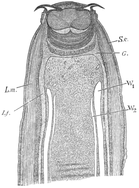
Fig. 187.—Longitudinal section of the head and neck of Tænia crassicollis, showing the lens-shaped muscular rostellum, with two hooks lying in the concentric cup-like mass of muscles. L.m., longitudinal muscles of the neck; L.f., left lateral nerve; G., ganglion; S.c., subcuticular layer; W1, external, W2, internal excretory vessel. 30/1.
The NERVOUS SYSTEM commences in the scolex and runs through the neck and the entire series of proglottids. Within the proglottids it consists of a number of longitudinal nerve fibres of which those at each lateral border are usually the largest. In the Tæniæ the lateral nerves are accompanied both dorsally and ventrally by a thinner nerve (accessory nerve) (fig. 185); on each surface, moreover, between the lateral nerve and the median plane, there are two somewhat stronger bundles (sub-median), so that there is a total of ten longitudinal nerve bundles. They lie externally to the transverse muscle plates, and the lateral and accessory bundles lie externally to the principal excretory vessels, and are everywhere connected by numerous anastomoses and secondary anastomoses; one typical ring commissure is usually found at the posterior border of the segments. In the Bothriocephalidæ the distribution of the nerve bundles is different (for instance, two lie in the medullary layer), or they are split up into a larger number of branches. In the scolex the nerve bundles are connected in a very remarkable manner by commissures with that which is generally termed the central part of the entire nervous system. There occurs normally a commissure between the two lateral nerves; at the same level, the dorsal and ventral median nerves are also connected at each surface as well with each other as with the lateral nerves, so that a hexagonal or octagonal figure is formed. The so-called apical nerves pass from this commissural system anteriorly, embrace the secondary muscular system of the rostellum semicircularly, and form an annular commissure (rostellar ring) at the inner part of the rostellum.
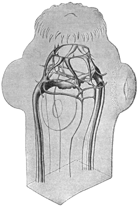
Fig. 188.—Tænia cœnurus, head and part of neck showing nervous system. Enlarged. (After Niemiec.)
The peripheral nerves arise from the nerve bundles as well as from the commissures situated in the scolex; some go direct to the muscles, while others form a close plexus of nerves external to the inner longitudinal muscles, which plexus likewise sends out fibres to the muscles, but principally to numerous fusiform sense organs (fig. 184, Pl.); they lie internal to the subcuticular cells and, piercing the cuticle with their peripheral processes, end as projecting “receptor” hairs. Higher organs of sense are not known.
The EXCRETORY APPARATUS of the Cestodes is similar to that of other flat worms. The terminal (flame) cells, which hardly differ in appearance from those of the Trematodes, are distributed throughout the parenchyma, but are more common in the cortical than in the medullary layer (fig. 184, T.c.). Before opening into a collecting tube, the capillaries run straight, tortuously, or in convolutions, anastomosing frequently with one another or forming a rete mirabile. The collecting tubes, which have their own epithelial and cuticular wall, and which also appear to be provided with muscular fibres, occur typically as four canals passing through the entire length of the worm (fig. 189); they lie side by side, two (a wider thin-walled ventral, and a narrower thick-walled dorsal one) in either lateral field; in the head the two vessels on each side unite by means of a loop, at the posterior extremity they open into a short pyriform or fusiform terminal bladder which discharges in the middle of the posterior edge of the original terminal proglottis.
This primitive type (fig. 189) of arrangement of collective tubes is subject to variation in most Cestodes, in the scolex as well as in the segments. Indeed, even the lumen of the four longitudinal tubes does not remain equal, as the dorsal or external tubes are more fully developed and become thicker, whereas the ventral or internal ones remain thin, and in some species quite disappear in the older segments (figs. 185, 187). Moreover, very frequently connections are established between the right and left longitudinal branches, as in the head, where a “frontal anastomosis” develops, which in the Tæniidæ usually takes the form of a ring encircling the rostellum (fig. 190), and in the segments of a transverse anastomosis at each posterior border, especially between the larger branches, and more rarely between the smaller collecting tubes also (fig. 191).
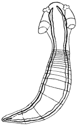
Fig. 189.—Young Acanthobothrium coronatum, v. Ben., with the excretory vessels outlined. Slightly enlarged. (After Pintner.)
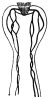
Fig. 190.—Scolex of a cysticercoid from Arion sp., with the excretory vessels outlined. (After Pintner.)
The so-called “island” formation is another modification, i.e., at any spot a vessel may divide and after a longer or shorter course the two branches reunite, and this may appear in the collecting tubes themselves as well as in their anastomoses. The above-mentioned ring in the frontal commissure of the Tæniidæ is such an island; similar rings also frequently encircle the suckers (fig. 190). In extreme cases (Triænophorus, Ligula, Dibothriocephalus, etc.) this island formation extends to all the collecting tubes and their anastomoses. Instead of two or four longitudinal canals only, connected by transverse anastomoses at the posterior border of the segments, there is an irregular network of vessels, situated in the cortical layer, from which the longitudinal branches, having again subdivided, can only be distinguished at intervals, and even then not in their usual number.
The opening of the longitudinal branches at the posterior end requires more accurate investigation; it is true that a single terminal bladder is mentioned as being present in many species, but this is also disputed; when the original end proglottis has been cast off, the longitudinal branches discharge separately. Some species possess the so-called foramina secundaria, which serve as outlets for the collecting tubes; they are generally at the neck, but may be situated on the segments.
The contents of the excretory vessels is a clear fluid, the regurgitation of which is prevented by the valves present at the points of origin of the transverse anastomoses. The fluid contains in solution a substance similar to guanine and xanthine.
Genital Organs.—With the exception of one genus (Diœcocestus, Fuhrm.), in which the species are sexually differentiated, all the Cestodes are hermaphroditic; the genitalia develop gradually in the segments (never in the scolex), the male organs, as is usual in hermaphroditic animals, forming earlier than the female. The youngest proglottids generally do not exhibit even traces of genitalia: these, as a rule, develop first in the older segments, and the development proceeds onwards from segment to segment. In a few exceptional cases (Ligula) the sexual organs are already developed in the larval stage, but are only functional after the entry of the parasite into the final host.
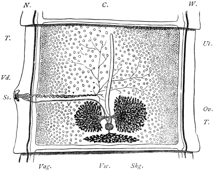
Fig. 191.—Proglottis of Tænia saginata, Goeze, showing genitalia. C., transverse excretory canal; N., lateral longitudinal nerve; W., longitudinal excretory canal; T., testicles scattered throughout the proglottis; Ut., opposite the central uterine stem (a closed sac); Ss., genital pore leading into the genital sinus; above the cirrus and coiled vas deferens (V.d.), below the vagina (Vag.), bearing near its termination a dilatation, the seminal receptacle; Vsc., the triangular vitellarium, and above it (Shg.) the shell gland; leading from this to the uterus is seen the short uterine canal, on either side of this the two lobes of the ovary (Ov.). 10/1.
With the exception of the end portions of the vagina, cirrus and uterus, all the parts of the genital apparatus lie in the medullary layer, except only the vitellaria, which in many species are in the cortical layer. The male apparatus consists of the testes, of which, as a rule, there are a large number,278 and which lie dorsal to the median plane (fig. 185, T.); a vas efferens arises from each testis, unites with contiguous vasa, and finally discharges into the muscular vas deferens that is situated in about the middle of the segment. According to the position of the genital pore, the vas deferens opens on the lateral margin or in the middle line in the front of the segment; it is much convoluted or twisted, and frequently possesses a dilatation termed the vesicula seminalis. It finally enters the cirrus pouch, which is usually elongated; within the cirrus pouch lies the protrusible cirrus, which is not uncommonly provided with hooklets.
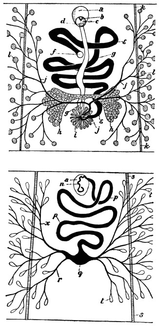
Fig. 192.—Dibothriocephalus latus. Upper figure: female genitalia, ventral view. Lower figure: male genitalia, dorsal view. The central portion only of the proglottis is shown. a, cirrus sac; b, partly everted cirrus; c, genital atrium and pore; d, vaginal pore; e, uterus; f, uterine pore; g, vagina; h, ovary; i, shell gland; j, vitelline duct; k, lateral nerve; l, vitellarium; n, vas deferens (muscular portion); p, vas deferens; q, seminal vesicle; r and x, vasa efferentia; s, lateral excretory canal; t, testicular follicles. (After Benham and Sommer and Landois.)
The male sexual orifice almost always opens with that of the vagina into a genital atrium, the raised border of which rises above the edge of the segment and forms the genital papilla (fig. 191).
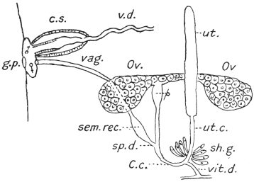
Fig. 193.—Diagram of genitalia of a Cestode. g.p., genital pore; ♀ ♂, male and female ducts opening into genital sinus; c.s., cirrus sac; v.d., coiled vas deferens (“outer seminal vesicle”); vag., vagina; sem. rec., seminal receptacle; sp. d., spermatic duct; C.c., fertilization canal; vit. d., vitelline duct; sh. g., shell gland; ut. c., uterine canal; ut., uterus; Ov., ovary; p, pumping organ. Cf. figs. 191 and 233. (Stephens.)
The vagina, like the vas deferens, usually runs inwardly and posteriorly, where it forms a spindle-shaped dilatation (receptaculum seminis); its continuation, the spermatic duct, unites with the oviduct, the common duct of the ovaries (fig. 191). The ovaries, usually two in number, are compound tubular glands in the posterior half of the proglottis, which extend into the medullary layer, but ventral to the median plane.
At the origin of the oviduct there is frequently a dilatation provided with circular muscles (suction apparatus), which receives the ovarian cells and propels them forward. After the oviduct has received the spermatic duct the canal proceeds as the fertilization canal, and after a very short course receives the vitelline duct or ducts, and then the numerous ducts of the shell glands (oötype). [Although the nomenclature of these parts varies, we may consider the oviduct as extending from the ovary to the shell gland and as receiving the spermatic duct and then the vitelline duct and the ducts of the shell gland. The short piece into which the shell gland ducts open corresponds to the oötype in the flukes, but in the tapeworms this portion of the canal is seldom dilated. From this point the oviduct is continued as a shorter or longer tube, the uterine canal or true oviduct opening into the uterus proper.—J. W. W. S.] The vitellarium may be single, but often exhibits its primitive duplication more or less distinctly, in which case it is situated at the posterior border of the segments in the medullary layer (fig. 191). The original position of the double organ is, moreover, the same as in the Trematodes, i.e., at the sides of the proglottids, and thence eventually extending more or less on both surfaces (figs. 192 and 194); the gland is then distinctly grape-like and the follicles lie mostly in the cortical layer.
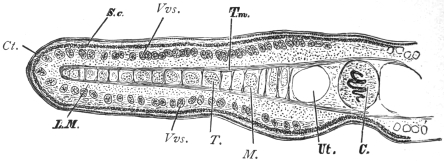
Fig. 194.—Part of a transverse section through a proglottis of Dibothriocephalus latus. Ct., cuticle; C., cirrus; Vvs., vitelline follicles; L.M., longitudinal muscles; T., testicles; M., medullary nerve; S.c., subcuticle; T.m., transverse muscles; Ut., uterus. 20/1.
The egg cell that has been fertilized and supplied with yolk cells receives the shell material at the point of entry of the shell gland ducts, and, as a complete egg, then moves onward to the uterus. In those cases in which the uterus in its further course presents a convoluted canal, and may form a rosette (pseudo-phyllidea), there is an external opening which is usually separate from the genital pore, and lies on the same or the opposite surface. In all other cases, however, the uterus terminates blindly and is represented by a longer or shorter sac lying in the longitudinal axis (fig. 191), but in many forms transversely. With the accumulation of eggs it becomes modified in various ways: (1) it sends out lateral branches (fig. 241), or (2) forms numerous isolated sacs (PARENCHYMAL CAPSULES) containing single eggs or groups of eggs (fig. 217); further, (3) in some cases at the blind end one or more special thick-walled cavities are formed (PARUTERINE ORGANS or UTERINE CAPSULES), in which all or most of the eggs are collected, the uterus then undergoing atrophy.
In species in which the uterus lacks an opening, simultaneously with the growth of this organ an atrophy of the male apparatus, at least of the testes and their excretory ducts, takes place; this atrophy also frequently occurs in the female glands, so that the entire mature segments have besides the uterus only traces of the genitalia left.
In the Acoleïnæ the vagina is more or less extensively atrophied, and in any case has no external opening.
A number of genera are distinguished by the duplication of the genitalia in every segment; the genital apparatus in its entirety, or with the exception of the uterus, is double, or the genital glands and the uterus are single, but the cirrus, vas deferens and vagina are double.
On comparing the genitalia of the Trematodes and Cestodes the parts will be found to agree, but the vagina of the Cestodes corresponds with the uterus of the Trematodes, and the uterus of the tapeworms to Laurer’s canal of the Trematodes, which in most of the Cestodes has lost its external orifice.
Development of the Tapeworms.
Copulation.—As each proglottis possesses its own genital apparatus, and male as well as female organs are present, the following processes may occur: (1) self- or auto-fecundation (without immissio cirri); (2) self- or auto-copulation (with immissio cirri); (3) cross-copulation between proglottids of the same or different chains (of the same species); and (4) cross-copulation in the same proglottis in species with double genital pores. These various modes have actually been observed.
In those species which lack the vagina (Acoleïnæ) it appears that the cirri, which are always furnished with hooks, are driven into the tissues and for the most part reach the receptaculum seminis.
The eggs of all Cestodes are provided with shells, but the shells, like their contents, vary. In genera that possess a uterine pore the mature eggs frequently do not differ from those of the Distomata; they have a brown or yellow shell of oval form provided with an operculum, and contain a number of yolk cells in addition to the fertilized ovarian cell (fig. 128), but in other genera (with a uterine pore) the lid is absent and the egg-shell is very thin, the eggs of these genera resembling those of Cestodes in which the secretion of the vitellarium is a light albumin-like substance that contains only a few granules, and in which the egg-shell is very delicate and without operculum.
The eggs of Tæniidæ, for example, at first consist of egg-shell (oötype), ovum and yolk cells. The egg-shell is as a rule soft, colourless and frequently deciduous, and the yolk is scanty in amount and contains few granules. The eggs are, moreover, more complicated than this. They enlarge and change their shape and various envelopes are developed around the embryo. The egg-shell proper often disappears, and one or more embryonal envelopes, or protoplasmic layers, arise, so that eventually it is difficult to say whether the whole egg is present, and, if not, what the layers that remain really are.
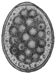
Fig. 195.—Egg of Diplogonoporus grandis, showing the morula surrounded by yolk cells and granules. 440/1. (After Kurimoto.)
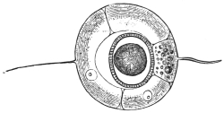
Fig. 196.—Uterine egg of Tænia saginata, G. Uterine shell with filaments; the oncosphere with embryonal shell (embryophore) in the centre. 500/1. (After Leuckart.)
The embryonal development in most species takes place during the stay of the eggs in the uterus; in other species it takes place after the eggs have been deposited and are in water. Separate cells or a layer of cells always separate from the segmentation cells, as well as from the cells of the developing embryo, and form one or more envelopes round the embryo; usually two such envelopes are formed, the inner one of which stands in intimate relationship with the embryo itself and is often erroneously termed the egg-shell, but more correctly the embryonal shell or embryophore. In some species it carries long cilia, as in Dibothriocephalus latus, by aid of which the young swim about when released from the egg-shell; as a general rule, however, there are no cilia and this envelope is homogeneous, or is composed of numerous rods and is calcified, as in Tænia spp. (fig. 197). The second outer envelope (“yolk envelope”) (fig. 207, 3) lies close within the true (oötype) egg-shell, and remains within it when the embryo hatches out, and in many species, as in Tænia spp., it perishes at the end of the embryonal development with the delicate egg-shell which was formed in the oötype, so that one observes not the entire egg with egg-shell but only the embryo in its embryonal shell, viz., the embryophore (fig. 197, a.).
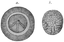
Fig. 197.—a., oncosphere, in its radially striated embryophore (erroneously termed egg-shell) of Tænia africana. Greatly magnified. (After von Linstow.) b., freed oncosphere of Dipylidium caninum. (After Grassi and Rovelli.) Both oncospheres show six spines.
The embryo (the ONCOSPHERE) enclosed within the embryonal shell (embryophore) is of spheroidal or ovoid form (fig. 197, b.), and is distinguished by the possession of three pairs of spines, a few terminal (flame) cells of the excretory system, and muscles to move the spines.
No further development of the oncosphere takes place, either in the parent organism or in the open; in fact, in all cases in which the oncospheres are already formed within the proglottids they do not become free, but remain in their shell; it is only when the oncospheres are provided with a ciliated embryophore that they leave the egg-shell, and they even cast this ciliated envelope after having swum about in water by its means for a week or so. Sooner or later, however, all the oncospheres leave the host that harbours the parental tapeworm and reach the open, either still enclosed in the uterus of the evacuated proglottids, after the disintegration of which they then become free, or after being deposited as eggs in the intestine of the host; they then leave it with the fæces. In the former case also, the slightest injury to the mature proglottids while still in the intestine suffices to allow a part of the oncospheres in their embryophores to be released and mingled with the fæces. Here they are the generally, but falsely, so-called Tæniæ “eggs.” For, as stated above, the “yolk” envelope and the true shell deposited in the oötype have before this disintegrated.
In other cases, e.g., Hymenolepis spp., the uterine (oötype) shell persists in fæces (fig. 230).
In any case the oncospheres must be transmitted into suitable animals to effect their further development; in only very rare cases might an active invasion be possible, as, for instance, takes place with the miracidia of many Trematodes. The entry into an animal is, as a rule, entirely passive, that is to say, the oncospheres are swallowed with the food or water. Many animals are coprophagous and ingest the oncospheres direct with the fæces; others swallow them with water, mud, or food contaminated by such fæces. Infection is easily produced artificially by feeding suitable animals with mature proglottids of certain Cestodes or introducing the oncospheres with the food. As the mature tapeworm frequently finds the conditions suitable for its development in only one species of host, or in species nearly related, and perishes when artificially introduced into other hosts, experiment has taught us that to succeed in cultivating the oncospheres certain species of animals are necessary. Thus we are aware that the oncospheres of Tænia solium, which lives in the intestine of man, develop only in the pig, and only quite exceptionally develop into the stage characteristic of all Cestodes—the cysticercus in the wide sense of the word—in a few other mammals. The oncospheres of T. saginata develop further only in the ox; those of T. marginata (of the dog) in the pig, goat, and sheep; those of T. serrata (of the dog) in hares and rabbits; those of Dipylidium caninum (of the dog and cat) in parasitic insects of the dog and cat, etc. It is not unusual that young animals only appear to be capable of infection, while older animals of the same species are not so.
Once introduced into a suitable animal, which is only exceptionally the same individual or belongs to the same species as the one which harbours the adult tapeworm, the oncosphere passes into the larval stage common to all Cestodes, but varying in structure according to the species. In the simplest case—as, e.g., in Dibothriocephalus—such a larva resembles the scolex of the corresponding tapeworm, only that the head, provided with suckers, is retracted within the fore-part of the neck. Such a larval form is known as a plerocercoid (πλήερης, full; κέρκος, tail). They differ from the cysticercoids in being solid larval forms, elongated, tape-like or oval, with the head invaginated. The conditions appear to be similar in Ligula, Schistocephalus, Triænophorus, but here the larvæ are very large, indeed as large in the first-mentioned genera as the tapeworms originating from them, and the sexual organs are already outlined; doubtless, however, this stage is preceded by one that corresponds to the scolex of the genus in question, and which represents the actual larval stage. In such cases the development of the body of the tapeworm from the scolex has already begun within the first or intermediate host; in other cases, except in the single-jointed (monozootic) Cestodes, this only takes place in the definitive host. The direct metamorphosis of the oncosphere into the larval forms termed PLEROCERCOID has hitherto not been investigated, although Ligula, Schistocephalus and Bothriocephalus are very common parasites, but many circumstances point to the conclusions arrived at by us and by other observers. In the larval stages of other tapeworms we can always distinguish the scolex and a caudal-like appendage, vesicular in the cysticerci (fig. 200), compact in the cysticercoids (fig. 231). The scolex alone forms the future tapeworm, the variously formed appendage perishing.
It has now been proved that the appendage, the caudal vesicle, originates direct from the body of the oncosphere, and therefore is primary, and that the scolex only subsequently forms through proliferation on the surface of this appendage. On account of this origin the scolex is generally regarded as the daughter, and the part usually designated as the appendage as the mother, originating from the oncosphere.
Accordingly, two modes of development of the larval stage may be distinguished; in the one case, plerocerci and plerocercoids, the oncosphere changes directly into the scolex, thus forming the body of the tapeworm within the primary host; in the other case, cysticerci and cysticercoids, the scolex only forms secondarily in the transformed body of the oncosphere, which later on perishes, the scolex alone remaining as the originator of the tapeworm colony.
We may summarize briefly what has been said regarding these larval forms. We have, firstly, solid larval forms without any bladder. These arise directly from the oncosphere and are of two kinds, plerocercus and plerocercoid. Plerocercus is a solid globular larva with the head invaginated into the posterior portion. Plerocercoid (fig. 208) is a solid elongated larva also with the head invaginated into the posterior portion, which is sometimes very long. Secondly, we have larval forms with bladders from which the scolices arise thus indirectly from the oncosphere. They are of two kinds, cysticercoid and cysticercus.