ON PARASITES IN GENERAL. (continued 6)
Reading edition
The Chlamydozoa are parasites of epiblastic tissues (e.g., epidermal cells, nerve cells, conjunctival cells).
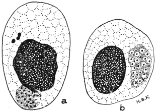
Fig. 119.—Chlamydozoa. Trachoma bodies in infected epithelial cells of the conjunctiva. (a) initial bodies (above) and cluster of elementary bodies (touching the nucleus); (b) cluster of granules surrounded by mantles. × 2,000 approx. (Original. From preparation by Fantham.)
The life-history of a Chlamydozoön (fig. 119), such as that of vaccinia, is, according to Prowazek, Hartmann and their school, as follows:—
1. The infection begins with elementary bodies or elementary corpuscles which live at first extracellularly. An elementary body is a minute speck of chromatin, apparently devoid of cytoplasm, which can pass through a bacterial filter. It can enter a host cell, but the entry is not a process of phagocytosis.
2. Inside the host cell the elementary body grows in size, and becomes an initial body (fig. 119, a).
3. A reaction on the part of the host cell results, for nucleolar, plastin substance is extruded from the cell-nucleus and surrounds the parasitic initial body. The latter is thus enveloped in a mantle (hence the name Chlamydozoa, from χλαμὑς, a mantle), and the characteristic cell-inclusion (Guarnieri’s body, Negri’s body, etc.) is produced. The nucleolar, mantle substance probably represents the “cytoplasm” of Cytoryctes, described by Calkins.
4. The body next breaks up into a number of smaller bodies known as initial corpuscles. These, in their turn, divide by simple division (in the manner already described) into numerous elementary bodies (fig. 119). Thus, the life-cycle is completed.
The Chlamydozoa are, then, the minute granules inside the body of the Cytoryctes variolæ or the Neuroryctes hydrophobiæ, so that the whole body of the Cytoryctes or Neuroryctes corresponds to the mantle and parasite of the Chlamydozoön. The Cytoryctes group is said to cause destruction of the host cell. The Cytoöikon group (e.g., trachoma bodies) causes proliferation of the host cell.
In September, 1913, Noguchi252 described the cultivation of the parasite of rabies in an artificial medium, similar to that used by him for the cultivation of Spirochæta recurrentis. The cultures were stated to be infective to dogs, rabbits and guinea-pigs. Levaditi, in December, 1913, stated that he had succeeded in cultivating spinal ganglia of rabid monkeys in monkey plasma.
Noguchi and Cohen (November, 1913)253 have succeeded in cultivating the so-called trachoma bodies, or at any rate bodies very closely resembling them morphologically. The medium employed was Noguchi’s ascitic fluid and rabbit kidney medium, as used for spirochætes. The coarser cultural forms stained blue with Giemsa’s solution, the finer ones stained red. Attempts to infect monkeys from the culture tubes failed.
From their behaviour on treatment with such reagents as saponin, bile and sodium taurocholate, Prowazek considers that the Chlamydozoa approach the Protozoa.
PROTOZOA INCERTÆ SEDIS.
Sergentella hominis, Brumpt, 1910.
Et. and Ed. Sergent in 1908 found vermiform bodies about 40 µ long by 1 µ to 1·5 µ broad in the blood of an Algerian suffering from nausea and cold sweats, without other symptoms. The bodies were pointed at each end, with a somewhat ill-defined nucleus in the middle. Their systematic position is doubtful.
Note.—An Appendix on Protozoology will be found on pp. 733–752. This has been prepared in order to incorporate a number of new additions to knowledge made since the body of the book was printed off.
B. PLATYHELMINTHES, or Flat Worms. BY J. W. W. STEPHENS, M.D., B.C., D.P.H.
Definition: Bilaterally symmetrical animals without limbs, the form of which is leaf or tape-like, rarely cylindrical, and whose primary body cavity (segmentation cavity) is absent, the cavity being filled by a mesenchymatous tissue (parenchyma).
The mouth is either situated at the anterior end of the body, or is shifted more or less backwards on to the flat ventral surface. The alimentary canal consists of a short fore-gut, which is frequently provided with a muscular pharynx, and of a simple forked or branched mid-gut; there is neither a hind-gut nor an anus; in one class, the Cestodes, the alimentary canal has entirely disappeared except for muscular remnants in the scolex.
The INTEGUMENT OF THE BODY consists either of a ciliated epithelium of only one layer (Turbellaria), or of a cuticle and gland-like cells embedded in the parenchyma, or subcuticular layer (Cestodes, Trematodes). The dermo-muscular layer consists of annular, longitudinal, and even diagonal fibres, while the parenchyma is traversed by dorso-ventral fibres.
The central NERVOUS SYSTEM, which is embedded in the parenchyma of the body, consists of cerebral ganglia, united together in the shape of dumb-bells, and of two or more longitudinal MEDULLARY FASCICLES, often forming transverse anastomoses. Organs of sense usually occur only in the free-living species, more rarely during the free-living stages of a few parasitic species and in a few ectoparasitic forms.
[In Platyhelminthes simple eye-spots frequently occur, and in a few an auditory vesicle.]
Blood-vessels and definite RESPIRATORY ORGANS are lacking [except in Nemertinea]; the EXCRETORY APPARATUS (formerly termed water-vascular system) is typical of the entire class. It commences in the interstices of the parenchyma, with peculiar terminal cells (ciliated funnels), which will be described later (p. 219), the capillary processes of which go on uniting into larger branches, and finally form two large collecting vessels, which, sometimes separately and sometimes united, open to the exterior through one, two, or numerous pores.
Nearly all the Platyhelminthes are HERMAPHRODITIC, and in nearly all there are, in addition to the ovaries producing ova, other glands attached to the female genital apparatus, namely, the vitellaria or yolk glands, which provide a substance termed yolk, which serves as nourishment for the embryo. The fully formed eggs have shells and are “compound,” i.e., composed of the egg or ovarian cell, which is surrounded by numerous yolk cells or their products of disintegration. The two sexual openings usually lie close together, frequently in the fundus of a genital atrium; they are rarely separated from one another. Shell glands also usually occur (p. 221).
Reproduction is sexual, often, however, combined with asexual methods of propagation (segmentation, budding). The Platyhelminthes live partly free in fresh or salt water, exceptionally also on land. The greater part, however, live as parasites on or in animals.
Classification of the Platyhelminthes.
Class I.—Turbellaria (or Eddy Worms). Flat worms for the most part, free living, and always covered with a ciliated epithelium.
Order 1.—Rhabdocœlida, gut unbranched.
Order 2.—Tricladida, gut with three main branches.
Order 3.—Polycladida, a central gut with lateral cæca. Development direct or through metamorphosis. They live in fresh and salt water or on land; very seldom as parasites.
Class II.—Trematoda (Sucking Worms254). [Usually known as Flukes.—F. V. T.] Flat worms, living as ecto- or endoparasites, that are only ciliated in the larval condition, and in their adult state are covered with a cuticle, the matrix cells of which lie in the parenchyma. They have either one, a few, or several suckers,255 and frequently also possess chitinous fixation and adhesive organs. The intestine is single, but generally bifurcated, and not uncommonly there are transverse anastomoses between the forks or diverticula on them. Excretory organs double, with two orifices at the anterior extremity or a single one at the posterior end. Development takes place by a metamorphosis or alternation of generations (p. 283). These worms are almost always hermaphroditic, with two or more female and one male sexual orifice. They live, almost without exception, as parasites on vertebrate animals, but the intermediate generations are passed in molluscs.
Class III.—Cestoda (Tapeworms). Endoparasitic flat worms without an alimentary canal. The larval stages are rarely ciliated, but are usually provided with six spines; the adult worm is covered with a cuticle, the matrix cells of which are embedded in the parenchyma. The body consists of a single segment (Cestodaria) or a chain of segments, in which case it consists of the scolex and the segments containing the sexual organs (proglottides) (Cestodes s. str.). The scolex is provided with various adhesive and fixation organs, and there are calcareous corpuscles in the parenchyma. Excretory organs symmetrical, opening at the posterior end. These worms are always hermaphroditic, and then possess one or two female and one male sexual orifice. During development a larval intermediate stage (“measle”) occurs and almost always in a different host to that in which the adult sexual worm lives. The adult stage is parasitic in vertebrate animals; but the larval stage may occur in invertebrates.
Class II. TREMATODA, Rud.
These worms are usually leaf- or tongue-shaped, but also barrel-shaped or conical; they vary from 0·1 mm. to almost 1 m.256 in length; most of them, however, are small (5 mm. to 15 mm.). The surface on which the orifice of the uterus and the male sexual opening are situated is termed the ventral surface; the oral aperture, which also acts as anus, is always at the anterior end in the sub-order Prostomata (p. 230), but in the sub-order Gasterostomata it is ventral.
Suckers are always present and occur in varying numbers and positions at the anterior extremities as well as on the ventral surface, and occasionally on the lateral margin and on the dorsum; the beginning of the intestine (mouth) is always surrounded by a sucker in the Prostomata.
In or near the suckers there may be chitinous hooks, claws or claspers, or the surface of the body is more or less covered with spines, scales or prickles; in one genus (Rhopalias) there are projectile tentacles beset with spines on the sides of the anterior part of the body.
The body of adult Trematodes is covered by a homogeneous layer of varying thickness, which either lies directly over the external layer (basement membrane) of the parenchyma, or over the muscles embedded in the parenchyma. This investing membrane (cuticle) arises from pear-shaped or spindle-shaped cells arranged singly or in groups (which lie between or internal to the diagonal muscles), and is connected with them by processes; these cells one may regard as epithelial cells which have sunk down, or possibly as parenchymatous cells. An epithelium of one layer is also found on the body of young stages, but it disappears during growth, and only occasionally do its nuclei persist until adult life. In its place we then find the cuticle, which, moreover, extends into all the body openings more or less deeply.
It is thus a debatable point whether the “investing layer” of flukes is a cuticle—that is, consists of modified epithelial cells—or whether it is a basement membrane, i.e., compressed and modified connective tissue cells; in this latter case the true epidermis and cuticle have been cast off. In the former case the epidermal cells are the pear-shaped cells referred to above. According to recent authors it consists of two parts, an outer true cuticle and an inner basement membrane. There are also unicellular cuticular glands, lying isolated or in groups, which are termed cephalic, abdominal, or dorsal glands according to the position of their orifice.
The PARENCHYMA is a connective substance, the structure of which is still a matter of dispute. It consists, according to some authors, of multipolar cells, the offshoots from which anastomose with each other so that a network, permeating the entire body and encompassing all the organs, is produced. There exists also, as part of it, a homogeneous matrix, in the form of lamellæ and trabeculæ that border small cavities communicating with each other and filled with fluid. According to other authors, the parenchyma of the Trematodes consisted originally of cells, of which, however, only the cell membranes remain, while the protoplasm has been liquefied except for small residua around the nucleus. Between these cells an intercellular mass has appeared. By partial absorption of the walls, adjoining spaces unite, and the originally flat cell walls become transformed into trabeculæ. According to this view the cavities filled with fluid are intra-cellular, according to the former view inter-cellular. Pigment cells occur only in a few species.
The MUSCULAR SYSTEM of the Trematodes is composed of (1) a dermo-muscular tube, (2) the dorso-ventral or parenchymal muscles, (3) the suckers, and (4) the special muscles of certain organs.
The dermo-muscular tube, which lies fairly close to the cuticle, consists of annular, diagonal, and longitudinal fibres which surround the entire body in one or several layers, and as a rule are more strongly developed on the ventral surface as well as in the anterior part of the body. The MUSCLES OF THE PARENCHYMA are found chiefly in the lateral parts of the body and pass through the parenchyma in a dorso-ventral direction; their diverging brush-like ends are inserted on the inner surface of the cuticle (fig. 120).
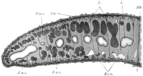
Fig. 120.—Half of a transverse section through Fasciola hepatica, L. 25/1. Cu., Cuticle with scales; under the cuticle are circular muscles, and adjoining them the longitudinal and diagonal muscles; internal to the latter are the matrix cells of the cuticle; I., gut; the other similarly contoured cavities are gut diverticula that have been transversely or obliquely sectioned; F.v.s., vitellaria; Ex.v., excretory vessels; T., testes; Md., median plane; the fibres passing from the ventral to the dorsal surface are the muscles of the parenchyma. The parenchyma itself is omitted.
The suckers are specially differentiated parts of the dermo-muscular tube. Their concave inner surface is lined by the continuation of the cuticle and their convex external surface is covered by a more dense tissue that frequently takes the form of a refractive membrane, thus separating them from the parenchymal muscles.
The principal mass of the suckers consists of muscular fibres which run in three directions—equatorial, meridional and radial. The equatorial fibres correspond to the annular muscles, the meridional fibres to the longitudinal muscles, and the radial fibres to the muscles of the parenchyma; the radial fibres are always the most strongly developed. The function of these muscles is evident from their position; the meridional fibres flatten the suctorial disc and diminish the depth of its cavity, so that the internal surface may adhere to the object to be held; if the equatorial fibres now contract, the sucker rises by elongating longitudinally, and its inner surface is drawn in by the contraction of the radial muscles. Thus the sucking disc becomes adherent. Usually also there is a sphincter at the border of the suckers, which plays its part during the act of adhesion by constricting in a circular manner that part of the mucous membrane to which it is attached. The loosening of the fixed sucker is effected by relaxation chiefly of the radial fibres, by the contraction of the meridional fibres and certain bundles of muscles situated at the base and at the periphery of the suckers. The connective and elastic tissues between the muscles of the suckers probably also take part in the process.
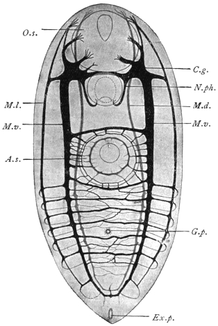
Fig. 121.—Harmostomum leptostomum, Olss., an immature specimen from Helix hortensis. Nervous system, according to Bettendorf. A.s., ventral sucker; C.g., cerebral ganglion; Ex.p., excretory pore; G.p., genital pore; O.s., oral sucker; M.d., dorsal medullary nerve; M.l., lateral medullary nerve; N.ph., pharyngeal nerve; M.v., ventral medullary nerve. Magnified.
Of the muscles of the organs which have developed from the parenchyma muscles we may briefly mention those bundles that are attached to certain parts of the genital apparatus, to the suckers, to the hooks and claws, and also, at all events in Fasciola hepatica, to the spines. The sheaths used for the projection of the tentacles of the Rhopaliadæ are also muscular.
The contractile elements consist of fibres of various lengths that are mostly parallel to one another, and frequently anastomose; a cortical substance finely fibrillated can usually be distinguished from an internal homogeneous mass; large nucleated cells of uniform size are always connected with them; these have been variously interpreted, but have been proved to be myoblasts, one or more of their processes constituting the muscular fibres.
The MOVEMENTS of the Trematodes consist in alterations of form and position of the body, as well as in creeping movements.
In the NERVOUS SYSTEM (fig. 121) can be distinguished a cerebral portion as well as strands (medullary strands) running from it, and peripheral nerves. The cerebral portion always consists of two large ganglia situated in the anterior end of the body which pass dorsally over the œsophagus and are connected by means of a broad and thick commissure composed of fibres only. From each ganglion three nerves run anteriorly—the inner and dorsal nerve for supplying the anterior dorsal part of the body; the median and ventral for the oral sucker; and the exterior and lateral likewise for the supply of the sucker.
In a similar manner three strands run backwards from each ganglion—one dorsal, one lateral and one ventral. The dorsal and ventral strands become united and curve backwards; the symmetrical lateral strands are connected by means of transverse commissures, the number of which vary according to the species. Such commissures also exist between the lateral and the two other strands on each side. There are ganglion cells along the entire course of the posterior cords, more particularly at the points of origin of the commissures. There also appears to be in addition a fourth anterior and posterior pair of nerves, the front pair for the oral sucker and the hind pair for the pharynx.
The peripheral nerves, which spring from the posterior strands as well as from the commissures, either pass directly to the muscular fibres or to the sensory cells that are situated at the level of the subcuticular cells, or they reach these after the formation of a plexus situated immediately beneath the dermo-muscular layer; the processes directed outwards terminate in small vesicles in the cuticle.
As to other ORGANS OF SENSE, simple eyes, two or four in number, are known in several ectoparasitic species as well as in a few free-living larval stages (Cercariæ) of endoparasitic forms. In the adult stage, however, they usually undergo complete atrophy.
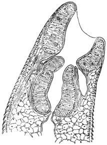
Fig. 122.—Median section through the anterior part of Fasciola hepatica: the oral sucker, pharyngeal pouches, pharynx, œsophagus, cuticle with spines, and the body parenchyma.
The ALIMENTARY CANAL commences with an oral aperture, generally terminal or sub-terminal (ventral) at the anterior extremity, which leads into an oral cavity usually surrounded by a sucker; the œsophagus, of various lengths, is directed backwards and is generally surrounded by a muscular pharynx (fig. 122). In some cases there exists between the sucker and pharynx, pharyngeal pouches (præpharynx). Sooner or later the intestine divides into two lateral branches directed backwards, both of which end blindly (cæca) at the same level.257 In many ectoparasites (Monogenea [p. 222]) a connection exists between the genital glands and one of the intestinal branches (ductus vitello-intestinalis [fig. 123]).
The oral cavity, pharyngeal pouches, pharynx, and œsophagus are lined with a continuation of the cuticle of the body; the gut cæca are lined with tall cylindrical epithelium (fig. 120). The œsophagus and intestinal branches often have also one layer of annular and longitudinal muscles; the pharynx has essentially the structure of a sucker (fig. 122).
The accessory organs of the alimentary canal consist of groups of unicellular SALIVARY GLANDS that discharge into the œsophagus in front of or behind the pharynx, or even into the pharynx itself.
The food of the Trematodes consists of mucus, epithelial cells, the intestinal contents of the hosts, and often also of blood, and this not only in those species living in the vascular system, but also in species living as ectoparasites or in the intestine or biliary passages of their hosts.
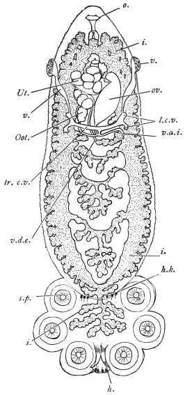
Fig. 123.—Polystomum integerrimum, a monogenetic fluke from the urinary bladder of the frog. i., intestine; h., large hooks of the sucking disc; h.k., smaller hooklets; l.c.v., longitudinal vitelline ducts; o., oral orifice; Oot., oötype; ov., ovary; s.p., suckers of the disc; tr.c.v., transverse vitelline ducts; Ut., uterus with ova; v., entrance to the vagina; v.d.e., vas deferens; v.d.i., ductus vitello-intestinalis; the vitellaria and testes are not shown. Magnified. (After Zeller.)
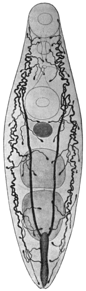
Fig. 124.—Allocreadium isoporum, Looss. Excretory apparatus. Of the other organs, the oral sucker, pharynx, genital pore, ventral sucker, ovary and testes are shown; the cylindrical excretory bladder is in the posterior end. 38/1. (After Looss.)
The final products of assimilation dissolved in the fluids of the body are distributed throughout the parenchyma and are thence expelled by a definite tubular system (excretory apparatus, proto-nephridia, formerly also termed the water-vascular system). This system, which is distributed throughout the entire body (fig. 124), is symmetrically developed, and, in the ectoparasitic Trematodes, it opens, right and left, at the anterior end on the dorsal surface; in all other flukes, however, it opens singly into the excretory pore (foramen caudale) at the centre of the posterior border; in those cases, however, where a sucker is present at the posterior end, as in the Amphistomata, the excretory pore is situated on the dorsal surface close in front of the sucker.
The EXCRETORY SYSTEM258 consists of several parts: (1) of the more or less numerous terminal “flame” cells or funnel cells (figs. 124, 125); (2) of the capillaries ending in them; (3) of larger vessels receiving the capillaries; and (4) of the excretory bladder. Terminal cells and capillaries may be compared to unicellular glands with long excretory ducts; the cellular body (fig. 125) is comparatively large, stretched longitudinally, more rarely transversely, and provided with numerous processes, that are lost in the parenchyma; within is a conical cavity (analogous to the secretory cavity of unicellular glands) which is continued directly into the structureless capillary; at its blind end is a bunch of cilia projecting into the cavity, and which, during life, shows a flickering motion (ciliary flame). The nucleus is situated in the protoplasm of the terminal cell at its blind end.
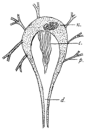
Fig. 125.—Terminal flame cell of the excretory system. n., nucleus of cell; c., bundle of cilia forming the “flame”; p., processes of cell extending into parenchyma; d., excretory capillary. (Stephens.)
The entire apparatus thus begins blindly—i.e., within the terminal cells, to which must be ascribed the capacity of taking up from the fluid that permeates the parenchyma the products which are first collected into their own cavities and thence excreted by means of the capillaries and vessels.
The vessels possess definite walls, consisting of a membrane and a nucleated protoplasmic layer. They unite at many points on either side, and again pass into other canals (COLLECTING TUBES), which finally, travelling towards the posterior end, discharge into the excretory bladder (fig. 124).
The form and size of the bladder vary much according to the different species, but it always possesses its own flattened epithelium, surrounded by circular and longitudinal muscles, the circular muscles forming a sphincter around the opening. Frequently also the structure of the bladder extends to the tubules discharging into it, which therefore are not to be regarded as separate “vessels,” but rather as tubular diverticula of the bladder, directed anteriorly. In some few species the diverticula also branch and the branches anastomose, so that a network of tubules ensues which receives the vessels or capillaries. In such cases there are also ciliary tracts in the tubules.
The contents of the entire apparatus usually consist of a clear or sometimes reddish fluid; in some species there are larger or smaller granules, and occasionally also concretions occur.
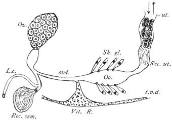
Fig. 126.—Diagram of female genitalia. Ov., ovary; ovd., oviduct; L.c., Laurer’s canal; Rec. sem., receptaculum seminis; Vit. R., vitellarian reservoir; t.v.d., transverse vitelline duct; Oo., oötype; Sh. gl., shell gland; Rec. ut., receptaculum uterinum; ut., uterus. (The various parts are not to the same scale.) (Stephens.)
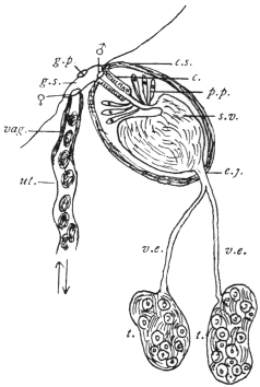
Fig. 127.—Diagram of male and part of female genitalia. ut., uterus; vag., vagina; ♀, opening of vagina; g.s., genital sinus; g.p., genital pore; ♂, opening of ejaculatory duct or vas deferens; c.s., cirrus sac; c., cirrus; p.p., pars prostatica; s.v., seminal vesicle; e.j., ejaculatory duct or vas deferens; v.e., vas efferens; t., testis. (Stephens.)
Sexual Organs.—Nearly all the Trematodes are hermaphrodites, and only a few (Schistosomidæ, Koellikeria) are sexually differentiated. The sexual organs usually lie in the “central field” limited by the gut cæca; the vitellaria, on the other hand, are, as a rule, external to the gut cæca in the “lateral fields.”
The male apparatus259 is composed of two variously formed testes (fig. 127) (globular, oval, indented, lobed, or ramified), which may lie side by side or one behind the other; from each testicle a tube (vas efferens) originates; sooner or later, both tubes as a rule unite to form the ejaculatory duct or vas deferens, which is frequently enclosed in a muscular CIRRUS SAC, or more rarely passes directly into the genital pore. The cirrus, which is the thick muscular terminal portion of the vas deferens, can be everted and protruded from the cirrus sac and serves as an organ of copulation. The walls of the muscular portion of the tube (the cirrus) are attached to the walls of the cirrus sac, and hence when the sac contracts the cirrus cannot be protruded except by evagination of its lumen. Opening into the middle portion of the vas deferens, and as a rule enclosed in the cirrus sac, is found a mass of unicellular glands (prostate), the vesicula seminalis (which is likewise within, or may also be outside the sac) being the dilated first portion of the vas.
The female genitalia (fig. 126) consist of an ovary, usually situated in front of the testes, the form of which varies according to the species, the usually double vitellaria, the ducts and a number of auxiliary organs; the short oviduct directed towards the centre arises from the ovary, and is connected in the median line with the excretory duct of the vitelline glands. These grape-like glands possess longitudinal excretory ducts, which assume a transverse direction behind the ovary, unite together at the median line and form a single duct, often dilated into a vitelline receptacle, that unites with the oviduct. Near this point, moreover, there frequently opens a canal (Laurer’s canal) which begins on the dorsal surface, and on the inner end of which a vesicle filled with sperm (receptaculum seminis) usually occurs (fig. 126). Moreover, there are also numerous radial unicellular glands (shell glands) at or beyond the point of junction of the oviduct, vitelline ducts and Laurer’s canal. In this portion of the duct (oötype), which is usually dilated, the ovarian cells are fertilized, surrounded with yolk cells and shell material, and as ova with shells they pass into the uterus (a direct continuation of the oviduct), which, with its many convolutions, occupies a larger or smaller portion of the central field, and runs either direct to the genital pore or, forming convolutions, first runs posteriorly and then bends forward (descending and ascending limbs). In both cases the terminal part lies beside the cirrus pouch and discharges beside the male orifice either on the surface of the body or into a genital atrium. The terminal portion of the uterus, which is often of a particular structure, serves as a vagina (METRATERM).
The cirrus sac may include (1) the genital atrium (i.e., the common sinus, into which the vas deferens and vagina may open), or (2) a variable extent of the vas from cirrus to seminal vesicle. Thus the latter may be outside the sac. In the absence of a sac, the genital sinus may be surrounded by a pseudo-sucker, as in Heterophyes (in some cases the ventral sucker itself, from its close proximity to the genital pore, serves as an accessory copulatory organ). In other cases copulatory organs are formed by hooks projecting into the lumen of the terminal portion of the vas.
The GENITAL PORE, which is the opening from the genital sinus on to the surface, is generally situated at or near to the median line on the ventral surface and in the anterior region of the body; in most of the Distomata it is in front of the ventral sucker, in other cases, e.g., in the Cryptocotylinæ, it is behind.260
The spermatozoa do not differ essentially in their structure from those of other animals; the ovarian or egg cells are cells without integument and contain a large nucleus and a little protoplasm; the vitellaria also produce nucleated cells, in the plasm of which there are numerous yellow yolk granules; the yolk cells detach themselves, like the ovarian cells, from the ovarium, and pass into the oviduct to surround each ovarian cell in the oötype. They disintegrate sooner or later in the completely formed egg and are utilized as food by the developing embryo.
Development of the Trematodes.
(1) Copulation.—Observation has demonstrated that the one or two vaginæ occurring in the ectoparasitic Trematodes are utilized as female organs of copulation, and that the copulation is cross; it is also known that Laurer’s canal, which was formerly generally regarded as the vagina, has only quite exceptionally, if at all, served the digenetic Trematodes as such—it appears to be homologous with the canalis vitello-intestinalis of the Monogenea261—but the terminal portion of the uterus, termed the metraterm, is used for copulation. Cross-copulation occurs as well as auto-copulation and auto-fecundation. The spermatozoa subsequently pass through the entire uterus, which is still quite short at the time the male organs are matured; the maturation of which, as usually is the case in hermaphrodites, precedes that of the female organs. It is only later with the onset of egg formation that the uterus is fully developed. Copulation, however, takes place also in the case of fully grown forms with completely developed uteri.
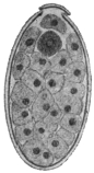
Fig. 128.—Ovum of Fasciola hepatica, L., cut longitudinally. The lid has been lifted in the process. Within the egg are numerous yolk cells, and at the lid end there is the still unsegmented ovum (dark). 240/1.
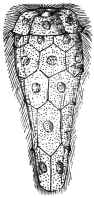
Fig. 129.—Miracidium of Fasciola hepatica that has just hatched from the egg, with a distinct cuticular ciliated epithelium. Magnified. (From Leuckart.)
(2) Formation of the Ova.—The ovarian cells arising from the ovary first become mature after their entry into the oötype by the formation of three polar bodies, fertilization then taking place. At the same time as the ovarian cell a number of yolk cells from the vitellarium and secretion, drop by drop, from the shell gland reach the oötype.262 The shell is then formed during the generally active contractions of the oötype walls and then passes on into the uterus. In the uterus of the endoparasitic trematodes the eggs accumulate more and more, often in large quantities, while in ectoparasitic species generally only one or some few eggs can be found. The completed ova are of various forms and sizes. They are mostly oval, at all events in the digenetic trematodes, and the yellowish or brown shell is provided with an opening at one pole which is closed by a watch-glass-shaped lid (operculum). Appendages (filaments) on the shell—at one or both poles—are uncommon, but are the rule in the ova of the Monogenea (ectoparasitic species).
(3) Deposition of the Ova.—Soon after their formation, the Monogenea (ectoparasitic trematodes) deposit round the place of their attachment on the skin or the gills or other organs of their hosts, eggs which attach themselves by means of their filaments. The embryonic development thus takes place outside the parent. This also holds good for the eggs of many endoparasitic species, although as a rule in these the eggs are always retained for a longer time in the uterus. Moreover, they usually here undergo a part or a whole of their development, and are eventually deposited in those organs in which the adult forms are parasitic, but this is not always the case, as the egg, e.g., of F. hepatica appears in bile (and fæces) quite unchanged. By the natural passages they eventually get out of the body, and in cases where such do not exist, as in the case of the blood-vessels, the eggs pass out by means of the kidneys.
(4) The embryonic development, after irregular segmentation of the ovum into a number of blastomeres, leads to the formation of a solid blastosphere or morula, which is surrounded by a cellular investing membrane (yolk envelope), while the principal mass of the cells forms the embryo, which uses for its nourishment the yolk cells, which have in the meantime disintegrated (cf. footnote, p. 223). Usually, after the ova have reached water the embryos hatch out, leaving the yolk envelope in the egg-shell; in other cases, however, the embryos only hatch out after having been subjected to the influence of the intestinal juices, that is to say, in the intestine of an intermediate host which has ingested with its food the ova that have escaped from the primary host.
(5) The post-embryonic development of the Trematodes is accomplished in various ways; the process is the most simple in the ectoparasitic species (Monogenea), the young of which should certainly be regarded as larvæ, because they possess characteristics (cilia, simple gut, etc.) that are lacking in the adult worms, but which, nevertheless, pass into the adult state direct after a relatively simple metamorphosis. In the Holostomata,263 a group found chiefly in the intestine of aquatic birds, and which rarely occur in other vertebrates, the ova develop in water. The young are ciliated all over, and, after having entered an intermediate host (leeches, molluscs, arthropods, amphibians, fishes) living in the water, they undergo a metamorphosis into a second larval stage; they then encyst and await transmission into the final host, where they become adult Metastatic trematodes, i.e., trematodes without asexually produced generations (p. 229).
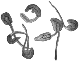
Fig. 130.—A group of cercariæ of Echinostoma sp. (from fresh water). 25/1.
In the remaining so-called digenetic trematodes (p. 230) one or two asexual generations interpose between the miracidium and terminal stage, so that quite a number of adult worms may originate from one egg. Usually the young, which are termed MIRACIDIA264 (fig. 129), hatch in water, where they move with the aid of their cilia. Sooner or later they penetrate into an intermediate host, which is always a snail or a mussel, and while certain of their organs disappear, they grow into a gutless germinal tube (SPOROCYST, fig. 131). These are simple elongated sacs with a central body cavity. They may or may not have excretory tubules. In these, according to the species, the larval stages (CERCARIÆ) that will ultimately become adult worms are produced, or another intermediate generation is first formed, viz., that of the REDIÆ265 (figs. 132, 133), which are always provided with an intestine, and these then give rise to cercariæ (figs. 130, 134). The cercariæ, as a rule, leave their host and move about in the water with the assistance of their rudder-like tails. After a little time, however, they usually again invade an aquatic animal (worms, molluscs, arthropods, fishes, amphibians), then they lose their tails and become encysted (fig. 135); here they wait until they attain, together with their host, the suitable terminal host, and in this new situation they establish themselves and reach maturity. Or, again, the cercariæ may themselves encyst in water or on foreign bodies (plants) and wait until they are taken up directly by the terminal host, e.g., sheep.
Accordingly the following conditions are necessary for the completion of the entire development: (1) The terminal host in which the adult stage lives; (2) an intermediate host into which the miracidia penetrate and in which they become sporocysts; (3) a second intermediate host in which the cercariæ become encysted. In certain species, as in Fasciola hepatica, this second host is omitted, as the cercariæ spontaneously encyst on plants, or again (in other species) encystment may occur within the first intermediate host, when, in fact, the cercariæ (which in this case do not acquire an oar-like tail) do not swarm out of, but encyst themselves within their sporocysts. The development, moreover, may be further complicated by rediæ appearing in addition to the sporocysts, though this occurs in the first intermediate host and not in a second one.
Animals that harbour adult digenetic Trematodes thus become infected by ingesting encysted cercariæ, which either occur (1) in certain animals (second intermediate hosts) on which they feed, or (2) in water, or (3) on plants, or finally (4) in the first intermediate host; whereas animals harbouring encysted cercariæ have been directly infected by the corresponding tailed stage, and animals harbouring germinal tubes (sporocysts or rediæ) have been infected by the miracidia.
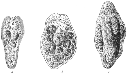
Fig. 131.—Development of Fasciola hepatica, L. a, the miracidium in optical section showing cephalic lobe, x-shaped eye-spot resting on the cerebral ganglion, two germ balls; below each of these a flame cell, and still lower germ cells lying in a cavity (primitive body cavity). b, young sporocyst with two eye-spots, and germ balls; the cells lining the cavity are not shown. c, older sporocyst with a young redia. Magnified. (After Leuckart.)
Thus certain species of ducks and geese become infected with Echinostoma echinatum by devouring certain water-snails (Limnæus, Paludina) in which the encysted cercariæ occur. Oxen become infected with Paramphistomum cervi (= Amphistomum conicum) by swallowing with water, cysts of this species which occur at the bottom of puddles and pits. Sheep are infected with Fasciola hepatica by eating grass to which the encysted cercariæ of the liver-fluke are attached; our song-birds infect themselves or their young with Urogonimus macrostomus by tearing off pieces containing the corresponding sporocysts which are full of encysted cercariæ from snails (Succinea amphibia), which act as the first intermediate hosts, and eating, or offering their young these pieces.
(1) The MIRACIDIA of the digenetic Trematodes are comparatively highly organized, and the mode of their formation from the segmentation cells of the ovum is only imperfectly known. They have a cuticular epithelium (fig. 129) entirely or partly covered with cilia, beneath this a dermo-muscular tube composed of circular and longitudinal muscles; also, a simple gut sac with an œsophagus, occasionally also with pharynx, salivary glands and boring spine, also a cerebral ganglion on which, in some species, there are eyes (fig. 131, a). As to the excretory organs, they are represented by two symmetrically placed terminal flame cells, with excretory vessels opening separately; there is a more or less ample (primary) body cavity between the parietes of the body and the gut; from the cellular parietal lining of this cavity single cells (germ cells) become free (fig. 131, a, b), and become rediæ or cercariæ.
[The germ cells of the miracidium and the germ balls of the sporocyst arise, according to some observers, by further division of undifferentiated blastomeres; according to others from the cells of the lining wall of its body cavity. It is from these free germ balls that the redia stage is developed.
[In the germ ball or morula appears an invagination, giving rise to the cup-shaped gastrula stage. This elongates and forms the REDIA (fig. 131, c).
[In the interior of the redia cells are budded off and develop into gastrulæ, as in the case of the sporocyst. These become a fresh generation of rediæ or give rise to the third stage (CERCARIA).]
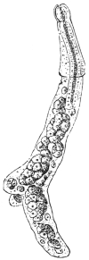
Fig. 132.—Young redia of Fasciola hepatica, with pharynx and intestine, with a circular ridge anteriorly and a pair of processes posteriorly and masses of cells (germ balls) in the interior. Magnified. (From Leuckart.)
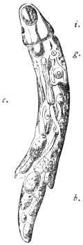
Fig. 133.—Older redia of Distoma echinatum, with rudimentary intestine i.; cercariæ, c.; germ balls, b.; and birth pore, g. Magnified.
(2) The SPOROCYSTS, on the contrary, which are produced direct from the miracidia, are very simple, as all the organs of the latter disappear, even to the muscles and excretory organs, during or after penetration into the intermediate host, whereas the budded and still budding cells of the wall of the (primary) body cavity continue to develop rapidly and form germ balls. The sporocysts when fully developed have the appearance of tubes or fusiform bodies with rounded edge; they are frequently of a yellow colour. Their length rarely exceeds a few millimetres; in some species their size increases exceedingly through proliferation, and they then occupy a large portion of the body of the intermediate host.
(3) The REDIÆ (figs. 132, 133), on the other hand, are more cylindrical and always have a simple intestine of varying length, provided with a pharynx; they likewise possess, situated near the circular ridge, a “birth pore” which serves for the exit of the cercariæ originating within them.
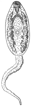
Fig. 134.—Cercaria of Fasciola hepatica; the cutaneous glands at the side of the anterior body. Magnified. (After Leuckart.)
(4) The CERCARIÆ266 are very different; typically they consist of the anterior body and the oar-like tail at the posterior end (fig. 134). The former, even to the genitalia, has the organization of the adult digenetic Trematodes, and thus allows the easy recognition of at least the characters of that large group to which the species in question belongs. On the other hand, however, there are also organs that are lacking in the adult form, such as, in many, the boring spine in the oral sucker, or the eyes situated on the cerebral ganglion; moreover, also, cutaneous glands (fig. 134), the secretion of which forms the cyst membrane. The oar-like tail may be long or short (stumpy-tailed cercaria) or entirely absent; its free end may be partly split (furcate cercaria), or split to its base (bucephalus); in various forms also the anterior end of the tail is hollow, and has enclosed within it the anterior body, which is otherwise free. The size also of the cercaria belonging to the different species is very diverse; in addition to forms swimming in the water that have the appearance of minute milky-white bodies, there are forms which measure as much as 6 mm. in length.
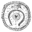
Fig. 135.—Encysted cercaria of Fasciola hepatica. Magnified. (After Leuckart.)
The encysted cercariæ (fig. 135) are globular or oval, and are surrounded by a homogeneous membrane, which may be striated or contain granules. The tail is always cast off when encystment occurs, and organs peculiar to the cercaria stage (boring papilla, eyes) almost entirely disappear. On the other hand, the genitalia appear or become more or less highly developed, in extreme cases to such an extent that they become functional, and after autocopulation the creatures produce ova within the cysts.
The cycle of development of the digenetic Trematodes has hitherto been generally explained as a typical ALTERNATION OF GENERATIONS, one sexual generation regularly alternating with one or two asexually reproducing generations. Recent authors, however, regard the cells in the sporocysts from which rediæ or eventually cercariæ arise as parthenogenetically developing ova, and the sporocysts as well as the rediæ as generations propagating parthenogenetically. In this case, however, it is an alternation of a sexual not with an asexual but with firstly a parthenogenetic generation (the sporocyst), the central cells of which are regarded as ova which develop parthenogenetically into the redia, and this the second parthenogenetic generation finally produces larvæ (cercariæ) capable of developing into the sexually mature form.
Other authors, again, regard the development of the Digenea as only a complicated metamorphosis (p. 283), which is distributed over several generations before it is concluded.
Biology.
Endoparasitic Trematodes, as fully developed organisms, occur in vertebrate animals only, with very few exceptions; they inhabit almost all the organs (with the exception of the nervous and osseous systems and the male genitalia), but by preference the intestine in all its extent from the oral cavity to the anus; and, further, certain species or groups inhabit only quite restricted parts of the intestine. Besides in the intestine other species live in the liver, or in the bile-ducts, or in the gall-bladder; other accessory organs of the intestine, such as the pancreas, bursa Fabricii (of birds), are only infected by a few species. Many inhabit the lungs, or the air sacs in fowls, a few the trachea. Trematodes have also been known to occur in the urinary bladder, the urethra and the kidneys of all classes of vertebrates; they are also present in the vascular system of a few tortoises, birds and mammals; in birds they even penetrate from the cloaca into the oviducts, and are occasionally found enclosed in the laid eggs; one species is known to occur in the cavum tympani and in the Eustachian tube of a mammal (Dugong), another in the frontal sinus of the polecat; several species infest the conjunctival sac under the membrana nictitans of birds, one species even lives in cysts in the skin of song-birds. In an analogous manner the ectoparasitic Trematodes are not entirely confined to the surface of the body or the trachea of the lower vertebrate animals; a few species appear exclusively in the urinary bladder, in the œsophagus, and in the case of sharks in an accessory gland of the rectum.
Trematodes live free and active within the organs attacked, though they may attach themselves by suction for a longer or shorter period; in other cases, however, they bore more or less deeply into the intestinal wall with their anterior end, or lie in cysts of the intestinal wall which only communicate with the lumen through a small opening; in those species living in the lungs of mammals the host likewise produces a cyst, which usually encloses two specimens; such association of a pair is also observed in other situations, and, though this is the rule in species sexually distinct, it is not entirely confined to these.
As regards the AGE attained by endoparasitic Trematodes, there are but few reliable records, and these differ considerably; the overwhelming majority of species certainly live about a year, or perhaps a little longer, but there are some whose term of life extends to several or many years.
Trematodes are but rarely found encysted in the higher vertebrate animals; the condition, however, is more frequent in amphibians, and especially in fishes, as well as in numerous invertebrate animals.
Classification of the Trematodes of Man.
The following classification, partly artificial, partly natural, embraces only the flukes found in man:—
Order. Digenea, v. Beneden, 1858.
Anterior sucker single and median, present. Eggs few. The (specialized) terminal portion of the uterus serves as a vagina. Development indirect, i.e., an intermediate host is required.
Sub-order. Prostomata, Odhner, 1905.
Mouth surrounded by the anterior sucker.
Group. Amphistomata, Rudolphi, 1801, ep., Nitzsch, 1819.
Gut forked, two suckers, the posterior sucker (acetabulum) terminal or ventro-terminal behind the genitalia, or at most embraced by the vitellaria. Skin with no spines. Excretory bladder a simple sac opening dorsally near hind end. Testes in front of ovary. Genital pore, median in anterior third of body. Thick flukes, almost circular in cross section.
Family. Paramphistomidæ, Fischoeder, 1901.
Amphistomata: Body not divided into a conical anterior portion and disc-like caudal portion. Ventral pouch absent.
Sub-family. Paramphistominæ, Fisch., 1901.
Paramphistomidæ: Oral sucker without evaginations. Not in man.
Sub-family. Cladorchiinæ, Fisch., 1901.
Paramphistomidæ: Oral sucker with evaginations; testes, two, deeply cleft (fig. 137). Genera: Watsonius, Cladorchis, etc.
Family. Gastrodisciidæ, Stiles and Goldberger, 1910.
Amphistomata: With body divided into a conical cephalic and disc-like caudal portion (fig. 138). Posterior sucker ventro-terminal. Oral sucker with evaginations. Genera: Gastrodiscus and Homalogaster.
Group. Distomata, Retzius, 1782.
Gut forked, two suckers, the posterior sucker (acetabulum) ventral. It is always separated from the hind end by at least a part of the genitalia.
Family. Fasciolidæ, Railliet, 1895.
Large flat forms, genital pore in front of ventral sucker, the latter powerful. Vitellariæ of numerous follicles, united by branching vitellarian ducts, at the sides of the body meeting posteriorly and extending ventrally and dorsally. Cirrus and vagina without spines. No crown of strong spines around sucker. Testes much branched. Uterus not well developed. Excretory bladder much branched. Eggs large.
Sub-family. Fasciolinæ, Odhner, 1910.
Large or median forms, gut much branched. Body has a shoulder separating head from body. Receptaculum seminis absent. Ovary branched, ventral sucker in anterior part of body. Genus: Fasciola.
Sub-family. Fasciolopsinæ, Odhner, 1910.
Shoulder absent. Receptaculum seminis present. Ovary branched, gut takes a zig-zag course with kinks on it, ventral sucker in anterior part of body. Genus: Fasciolopsis.
Family. Opisthorchiidæ, Braun, 1901, emend. auctor.
Ovary in front of testes. Small to medium flukes, very transparent, tapering anteriorly. Vitellaria moderately developed not extending in front of sucker. Cirrus absent. Seminal vesicle a twisted tube free in parenchyma. Testes near hind end one behind the other, lobed or branched, but not dendritically. Excretory bladder Y-shaped, the two limbs short, the stem S-shaped passing between the testes. Receptaculum seminis well developed. Laurer’s canal present. Uterine coils transverse, numerous. Eggs small.
Sub-family. Opisthorchiinæ, Looss, 1899, emend. auctor.
Opisthorchiidæ in which the excretory pore is terminal. Excretory bladder long, dorsal to testes. Uterine coils not overlapping gut forks. Genera: Opisthorchis, Paropisthorchis, Clonorchis, Amphimerus, etc.
Sub-family. Metorchiinæ, Lühe, 1909.
Opisthorchiidæ in which the excretory pore is ventral. Excretory bladder short, ventral to testes. Uterine coils partly overlapping gut forks and extend anteriorly beyond the sucker. Vitellaria compressed on the sides of the body. Genus: Metorchis.
Family. Dicrocœliidæ, Odhner, 1910.
Ovary behind testes. Testes behind the ventral sucker, between it and the ovary. Body thin and transparent. Cirrus sac encloses the pars prostatica and seminal vesicle. Skin smooth. Gut forks do not reach posterior end. Receptaculum seminis and Laurer’s canal present. Vitellaria, moderate, lateral in mid-body slightly overlapping the gut. Uterus with an ascending and descending branch and numerous transverse coils extending to hind end. Eggs dark brown, 25 µ to 60 µ. Excretory bladder tubular in posterior third or half of body. Parasitic in bile-ducts of mammals and birds. Genus: Dicrocœlium.
Family. Heterophyiidæ, Odhner, 1914.
Ovary in front of testes. Genital pore behind or on a level with ventral sucker. Genital pore surrounded by a pseudo-sucker (i.e., its muscle is not sharply separated from but blends with the body muscles). Cirrus sac absent, consequently vesicula seminalis and pars prostatica lie free. Vagina and ejaculatory duct unite into a common duct before opening. Small and very small forms. Body covered with scales. Genera: Heterophyes, Metagonimus, etc.
Family. Troglotremidæ, Odhner, 1914.
More or less flattened Distomes of compact form, 2 to 13 mm. long. Ventral surface flat or somewhat hollowed, dorsal surface arched. Skin completely covered with pointed spines. Musculature weakly developed also in the suckers in those forms that inhabit cysts. Gut with pharynx and a not very long œsophagus and cæca, which end more or less shortly before the hind end. Excretory bladder Y-shaped or tubular. Pars prostatica and seminal vesicle always distinct. Testes elongated, symmetrically placed in or behind the middle of the body. Ovary directly in front of the testes, right-sided, generally much lobed. Receptaculum seminis and Laurer’s canal present. Vitellaria generally well developed, exclusively or for the most part confined to the dorsal surface, leaving only a median band unoccupied. Uterus either very long, coiling here and there, or shorter and more convoluted. Eggs in first case small 17 µ to 25 µ, in the second much larger 63 µ to 85 µ or even 120 µ (?) long. Parasitic in carnivora or birds, generally occurring in pairs in cyst-like cavities. Genera: Paragonimus, Pholeter, Collyriclum, Troglotrema.
Family. Echinostomidæ, Looss, 1902.
More or less elongated flukes, small or very large, much flattened anteriorly, less so posteriorly, or even round. Suckers near one another, the anterior small and weak, the posterior large and powerful directed obliquely backwards. Surrounding the oral sucker dorsally and laterally but not ventrally is a fold or “collar” bearing a row or rows of pointed spines which are continued round laterally on to the ventral corners, the number being constant for each species, the corner spines large or specialized, skin anteriorly scaled or spiny. Alimentary canal consists of a pharynx, epithelial “pseudo-œsophagus” and gut cæca reaching to posterior end. Testes behind one another in hind body. Ovary on right side or median directly in front of the testes. Vitellaria lateral, usually extending to the hind end and not beyond the ventral sucker anteriorly. Genital pore just in front of ventral sucker. Uterus in transverse loops. Genital sinus absent or present. Receptaculum seminis and Laurer’s canal present. Eggs thin shelled and large, bright yellow, 65 µ to 120 µ long. Excretory bladder Y-shaped. Parasitic in gut of vertebrates, especially birds.
Sub-family. Echinostominæ, Looss, 1899.
Cirrus sac usually reaching to centre of ventral sucker, but not beyond. Cirrus long, usually without spines, coiled when retracted. Seminal vesicle tubular, twisted. On the head a ventral uniting ridge between the angles of the collar. Dorsal circlet of spines, single or double, not interrupted unless the collar itself is dorsally divided. Genera: Echinostoma, etc.
Sub-family. Himasthlinæ, Odhner, 1910.
Cirrus sac reaching far beyond ventral sucker. Cirrus armed with strong rose-thorn-shaped hooks. Vesicula seminalis tubular not coiled. Cervical collar not continued across ventral aspect. Spines on collar in one row. Body armed with fine needle-shaped spines.
Family. Schistosomidæ, Looss, 1899.
Sexes separate. Genital pore behind the ventral sucker. Ventral sucker elevated above the surface. Pharynx absent. Gut forks reunite to form a single stem. In ♂ four or more testicular follicles. In ♀ a single ovary, just in front of the union of the gut forks. Vitellaria on either side of the united gut stem.
The Trematodes Observed in Man.
Family. Paramphistomidæ, Stiles and Goldberger, emend. 1910.
Sub-family. Cladorchiinæ, Fisch., 1901.
Genus. Watsonius, Stiles and Goldberger, 1910.
Cladorchinæ.—Body pyriform. Ventral pouch absent. Acetabulum ventral or (?) ventro-subterminal, very large, margins projecting, aperture small. Genital pore in front of bifurcation of gut, not surrounded by a sucker; ductus hermaphroditicus apparently absent. Excretory pore at posterior end of excretory vesicle, behind Laurer’s canal. Oral sucker with a pair of irregularly globular suctorial pouches; œsophagus thickened distally; cæca long, not wavy; end in acetabular region.
Male Organs.—Testes two lobed, smaller than acetabulum; longitudinally, nearly or quite coinciding; transversely they abut or slightly overlap; preovarial in equatorial and caudal thirds. Pars musculosa not largely developed; cirrus pouch absent.
Female Organs.—Ovary and shell gland post-testicular. Vitellaria extend from gut fork to slightly beyond gut ending; uterus intercæcal, partly post-testicular. Laurer’s canal in front of excretory vesicle.
Type Species.—Watsonius watsoni, Conyngham, 1904.
Watsonius watsoni, Stiles and Goldberger, 1910.
Syn.: Amphistomum watsoni, Conyngham, 1904; Cladorchis watsoni, Shipley, 1905.
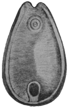
Fig. 136.—Watsonius watsoni: ventral view. 4/1. (After Shipley.)
Body, 8 to 10 mm. long, by 4 to 5 mm. broad, by 4 mm. thick; tapers anteriorly to 2·5 mm. Caudal extremity bluntly rounded, venter surrounded by an elevated ridge, surface with transverse ridges best defined ventrally. Genital pore median about one-quarter of body length from anterior end at level of suctorial pouches. Acetabulum 1 mm. in diameter, margin projecting, aperture small. Mouth in a groove with digitate papillæ. Oral sucker very large, one-fifth of length of body, with a pair of irregularly globular pouches. Œsophagus somewhat longer than sucker. Excretory pore at the level of the acetabular aperture. The vesicle extends from the plane of the transverse vitelline ducts to centre of acetabulum.
Male Organs.—Testes deeply notched adjoining one another. Vesicula seminalis much coiled and dilated, pars musculosa not coiled. Pars prostatica (?) dilated, ejaculatory duct long and narrow, opening on a papilla; genital atrium papillated.
Female Organs.—Ovary dorso-posterior of posterior testis. Shell gland dorsal to ovary. Vitellaria ventral and lateral to gut cæca extending from gut fork to equator of acetabulum. Uterus dorsal to testes, ductus hermaphroditicus absent. Laurer’s canal opens in dorso-median line slightly behind anterior border of sucker.
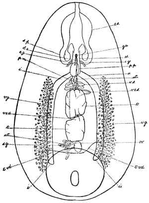
Fig. 137.—Watsonius watsoni: ventral projection composed from a series of transverse sections. o.s., oral sucker; s.p., suctorial pouch; ga., genital atrium; d.e., ejaculatory duct; es., œsophagus; e.g., œsophageal ganglion; p.p., pars prostatica; p.m., pars musculosa; i., gut; ut., uterus; v.e., vas efferens; v.e.s., left vas efferens; v.e.d., right vas efferens; v.g., vitellarium; t., testes; ov., ovary; s.g., shell gland; t.vd., transverse vitelline duct. (After Stiles and Goldberger.)
Eggs.—123 µ to 133 µ long by 75 µ to 80 µ broad.
Habitat.—Jejunum and duodenum of man, German West Africa. The parasite has only been found once in man. The patient, a negro from German West Africa, died at Zola, Northern Nigeria. The symptoms were persistent watery diarrhœa without blood or mucus. The parasites were also passed in the stools. It occurs also in monkeys.
Family. Gastrodisciidæ.
Genus. Gastrodiscus, Lkt., 1877.
Acetabulum small, caudal and ventral margin raised, aperture relatively large. Genital pore without sucker. Excretory pore post-vesicular, posterior to opening of Laurer’s canal. Œsophagus with muscular thickening; cæca not wavy, long, end post-equatorial and post-testicular.
Male Genitalia.—Testes two, branched pre-ovarial.
Female genitalia.—Ovary and shell gland post-testicular. Vitellaria extracæcal; uterus intercæcal; Laurer’s canal entirely prevesicular.
Type.—Gastrodiscus ægyptiacus, Cobbold, 1876.
Gastrodiscus hominis, Lewis and McConnell, 1876.267
Syn.: Amphistomum hominis, Lew. and McConn.
Body, reddish in the fresh, 5 to 8 mm. long; posteriorly, 3 to 4 mm. broad. The disc has incurved edges which are interrupted in front where it joins the anterior cylindrical portion and posteriorly behind the ventral sucker. The disc itself and ventral surface are covered with a number of (microscopic) papillæ. Pharynx provided with two diverticula or pouches. The bifurcation of the gut lies sometimes above, sometimes below the level of the genital pore. The gut cæca end about the level of the centre of the acetabulum.
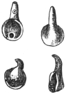
Fig. 138.—Gastrodiscus hominis. Slightly magnified. (After Lerckart.)
Genital Pore.—About the middle of the conical anterior portion. (It appears to be surrounded by a muscular sucker.) Leiper (1913) describes the ducts as discharging at the tip of a large fleshy papilla, the surface of which bears cuticular bosses.
Testes much lobed, the anterior is smaller than the posterior and lies at about the level where the anterior conical portion joins the disc. The posterior testis just in front of the anterior margin of the acetabulum separated from it by the ovary. The ovary, somewhat oval in shape or slightly constricted in the middle, lies slightly to the right of the median line. Dorsal to it lies the well-developed shell gland, Laurer’s canal opening in front of the excretory bladder. The excretory bladder is a long sac with its opening at its posterior extremity about the level of the middle of the acetabulum. The vitellaria are restricted in extent. They do not extend forward beyond the anterior border of the posterior testis. They are best developed in the area between the acetabulum and the termination of the gut cæca.
The eggs are oval and measure 150 µ in length by 72 µ in breadth.
Habitat.—Cæcum and large intestine of man. Also in the pig (5 per cent.) in Annam.
Distribution.—This parasite has been recorded from Assam (not uncommon), British Guiana (Indian immigrants), and Cochin China.
Gastrodiscus ægyptiacus, Cobbold, 1876, and G. secundus, Looss, 1907, occur in the horse; G. minor, Leiper, 1913, in the pig in Nigeria and Uganda.
Family. Fasciolidæ, Raill., 1895.
Sub-family. Fasciolinæ, Odhner, 1910.
Genus. Fasciola, L., 1758.
The ventral sucker is situated at the level of the junction of the cone with the body, viz., at the level of the “shoulder,” and is large and powerful. The cuticle is covered with strong spines; the gut cæca run in the mid-line to the hind end, and are provided with numerous long lateral and fewer and shorter median branches. The ovary lies on one side in front of the transverse vitelline duct; the testes lie obliquely one behind the other. The uterus, in the shape of a rosette, lies in front of the genitalia. Laurer’s canal is present; the vesicula seminalis lies in the cirrus pouch; the ova are large, not very numerous, and only develop after they have been deposited. Parasites of the biliary ducts of herbivorous animals.
Fasciola hepatica, L., 1758.
Syn.: Distomum hepaticum, Retz., 1786; Fasciola Humana, Gmel., 1789; Distomum caviæ, Sons., 1890; Cladocœlium hepaticum, Stoss., 1892.
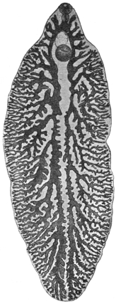
Fig. 139.—Fasciola hepatica, L. From a specimen that is not yet mature, showing the gut and its branches. 5/1.
Length 20 to 30 mm., breadth 8 to 13 mm., cephalic cone 4 to 5 mm. in length and sharply differentiated from the body by a shoulder on each side. Spines in alternating transverse rows and extending on the ventral surface to the posterior border of the testes, and on the dorsal surface not quite so far. The spines are smaller on the cephalic cone than on the posterior part of the body, where they are discernible with the naked eye. The suckers are hemispherical, and near each other; the oral sucker is about 1 mm. and the ventral sucker about 1·6 mm. in diameter. The pharynx, which includes almost the entire œsophagus, measures 0·7 mm. in length and 0·4 mm. in breadth. The intestine bifurcates at the limit of the cephalic cone and the branches are even here furnished with diverticula directed outwardly. The ovary is ramified and situated in front of the transverse vitelline duct, usually on the right side; the shell gland lies near the ovary in the median line; posterior to the transverse vitelline ducts are the greatly ramified testes, which occupy the greater portion of the posterior part of the body, with the exception of the lateral and posterior border; the long vasa efferentia only unite as they enter the cirrus pouch. The vitellaria occupy the sides of the posterior part of the body, commencing at the level of the ventral sucker and uniting behind the testes. The ova are yellowish-brown, oval, operculated, 130 µ to 145 µ in length, 70 µ to 90 µ in breadth (average size 132 µ by 70 µ).
The Liver Fluke inhabits the bile-ducts of numerous herbivorous mammals (sheep, ox, goat, horse, ass, rabbit,268 guinea-pig, squirrel, beaver, deer, roe, antelope, camel, kangaroo, and others), and is distributed over the whole of Europe, though not to an equal extent. It is further known in North Africa, in North and South America, as well as in Australia; it is also found in Asia, as it has been reported from Japan, China, and Tonkin (Gaide, two cases in man). In some districts of Germany it is very frequent, and the slaughter-house statistics of various places show that it is of daily occurrence. Fasciola magna occurs in herbivora in America.
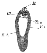
Fig. 140.—Fasciola hepatica. M., mouth; Ut., uterine rosette; Tr.c., transverse vitelline ducts uniting to form a vitelline receptacle in the mid-line; E.d., longitudinal vitelline ducts; V.s., vitellaria. The clear space in the centre represents the position of the ramifying testes and part of the gut. Natural size. (Mull. fluid, alcohol, creosote, Canada balsam.)
The liver fluke, however, is by no means a harmless parasite, for it produces in domestic animals, more especially in sheep, a disease of the liver that appears epidemically in certain years and districts, and commits great ravages amongst the flocks.
[The following records show the enormous loss caused in sheep by this parasite. In 1812, in the Midi, principally in the Departments of the Rhône, Herault, and Gard, the disease was rampant; 300,000 sheep perished in the Arles territory, and 90,000 in the Arrondissements of Nîmes and Montpellier. In 1829 and 1830, in the Department of the Meuse and near localities, not only sheep but oxen died in enormous numbers; for instance, in the Arrondissement of Verdun out of 50,000 sheep 20,000 died, and out of 20,000 cattle 2,200 died. In England, in 1830, 2,000,000 sheep were carried off; whilst in 1862 60 per cent. of the sheep died in Ireland; and in 1879 over 300,000 were lost in England; whilst as late as 1891 one owner in the same country lost over 10,000 sheep (Live Stock Journal, October 30, 1891).—F. V. T.]
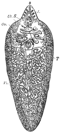
Fig. 141.—Fasciola hepatica, L. I., intestine; Vs., vitellaria; Ov., ovary; O., oral aperture; Ut., uterus; S., ventral sucker; T., testes. In front of the testes are seen the transverse vitelline ducts uniting to form the pyriform vitelline receptacle. Immediately in front of this the spherical shell gland. The two vasa efferentia can also be seen running up in the mid-line. The branches of the gut are only shown in the cephalic cone. (After Claus.)
The disease usually commences towards the end of summer with an enlargement of the liver, induced by the invasion of numerous young flukes; in the autumn and winter the animals suffer from the consequences of disordered biliary secretion; they become feverish, emaciated, and anæmic, and lose their appetite. In consequence of the consecutive atrophy of the liver, œdema and ascites set in, and many animals succumb to this “liver rot.” On examination the liver is found to be shrunken, the bile-ducts are enormously dilated and in parts saccular and full of flukes. Should the animals survive this stage, spontaneous recovery ensues in consequence of the flukes commencing to leave the liver in the spring, but the liver remains changed and its sale is prohibited269 when the changes are extensive.270
[The following stages may be noticed in sheep suffering from fascioliasis. Gerlach recognized four stages, based on the varied relations that the flukes contract with the liver of their host. These periods are sometimes very marked, but at others, owing to subsequent infections, the features become merged and so obliterated. But when a single infestation occurs they are very marked.
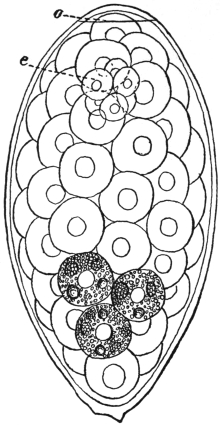
Fig. 142.—Fasciola hepatica: egg from liver of sheep. o, operculum, e, segmenting ovum. The rest of the space is occupied by yolk cells, the granules in three only being shown. × 680. (After Thomas.)
[The first period is called the PERIOD OF IMMIGRATION. This occurs at the fall of the year and generally passes unperceived, as the young flukes do little harm to the liver. It varies from four to thirteen weeks. Gerlach has remarked upon cases of death from apoplexy at this period.
[The second period is the PERIOD OF ANÆMIA. This occurs in November and December. The sheep at first fatten rapidly, but later the mucous membranes become pale and of a yellowish hue, and the sheep become sluggish and cease to feed. The fæces are normal, but may contain fluke ova.
[The third period is the PERIOD OF WASTING. This corresponds with the beginning of January—about three months after the entry of the larvæ. Emaciation now becomes very marked, the skin and mucous membranes blanched, temperature variable and marked by an irregular curve; respiration laboured and quick; appetite regular; abortion frequently occurs in pregnant ewes; pressure on the back causes the animals to fall; local œdemas occur, the most perceptible in the submaxillary space, extending below the larynx and over the cheeks and parotids (called “bourse,” “boule” in France; “watery poke” or “cockered” in England). Death usually occurs at this period, but a fourth stage may occur.
[The fourth period is the PERIOD OF MIGRATION OF THE FLUKES. This is a period of convalescence and recovery, generally in May and June.—F. V. T.]
Oxen suffer less in general, but even in these animals “stray” hepatic flukes are occasionally found in the lungs, enclosed in thick-walled cysts.
Pathological Anatomy.—The bile-ducts are conspicuous on the surface of the liver. They are thickened and much dilated and in parts saccular, and considerable atrophy of the liver cells accompanies the condition. Histologically there is immense proliferation of the epithelium of the bile-ducts leading to “adenomata.”
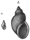
Fig. 143.—Limnæus truncatulus, Müll., the intermediate host of Fasciola hepatica. a., natural size; b., magnified. (From Leuckart.)
The LIFE-HISTORY of the liver fluke was discovered by R. Leuckart and P. Thomas. According to these investigators the elongated miracidium (fig. 131, a) ciliated all over develops from the eggs a few weeks after the latter (fig. 142) have reached the water, and after it has become free the embryo penetrates and becomes a sporocyst (fig. 131, b) in a water-snail (Limnæus truncatulus, Müll. = L. minutus, Drap.) that is common in fresh water, and can live in the smallest collection of water as well as in fields that have been flooded. The sporocyst first of all produces rediæ, which remain in the same host (and under certain circumstances, e.g. in summer, these develop a second generation of rediæ), and these finally form cercariæ (fig. 134). The latter become encysted on blades of grass and are taken up by the respective hosts with their food; this takes place towards the end of summer, while the sheep feeding on the pasture land in the spring spread the eggs of the fluke, and sometimes the fluke itself, by passing them with their fæces.
In districts where Limnæus truncatulus is absent, analogous species act as the intermediary hosts, of which one example according to Lutz is Limnæus oahuensis in the Sandwich Islands.
[The host in Europe is Limnæus truncatulus. This snail extends from Siberia to Sicily and Algeria, and according to Captain Hutton is a native of Afghanistan. It also occurs in Thibet, Amoor, Morocco, Tunis, Canary Islands and the Faroe Islands. It deposits its eggs or spawn upon the mud around ponds, ditches and streams. The eggs are laid in batches of thirty to a hundred, each snail laying as many as 1,500 eggs; they are united into strips of a gelatinous substance. In about two weeks young snails appear. It is amphibious, being more frequently met with out of the water than in it. It occurs in elevated spots as well as in low-lying districts. Moquin-Tandon found it at 4,000 feet in the Pyrenees. In the allied species, L. peregra, the fluke will develop up to a certain stage, but never completes all its varied phases.
[In South America the host is probably Limnæus viator, Orb., and in North America Limnæus humilis, Say.—F. V. T.]
In human beings as well as in some of the mammals quoted above, the liver fluke is only a casual parasite, and hitherto only twenty-eight cases have been observed in man; the infection was mostly a mild one and there were no symptoms, or only very trifling ones; a few isolated cases were only discovered post mortem. Occasionally, however, even when the infection was inconsiderable, severe symptoms were set up, which in isolated cases led to death. The symptoms (enlargement and painfulness of the liver, icterus) merely pointed to a disease of the liver.
Diagnosis can only be established by finding eggs in the fæces. Care should be taken not to confuse them with those of Dibothriocephalus latus.
Halzoun.
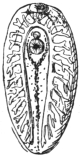
Fig. 144.—Young Fasciola hepatica, soon after entry into the liver. The intestinal cæca have lateral diverticula. Magnified. (From Leuckart.)
In North Lebanon, the liver fluke is, according to A. Khouri, a frequent parasite of man, not in the liver, however, but in the pharynx. The occurrence in this unusual site is effected by the eating of raw infected livers, especially those of goats (Capra hircus). The flukes thus taken in do not all reach the stomach, where they would be soon killed, but some of them attach themselves to the pharyngeal mucosa and to the adjoining parts, and there cause inflammation and swelling, which lead to dyspnœa, dysphagia, dysphonia and congestion of the head, sometimes even to still more severe symptoms, and even death. The affection termed “Halzoun” lasts some hours or several days, and after vomiting recovery sets in. In other cases man becomes infected in the usual way by ingesting cysts attached to grass or the underside of leaves of plants (e.g., Rumex sp.), where they are overlooked from their scanty size (0·2 to 0·3 mm.).
As the liver fluke feeds on blood it is possible that it also reaches, particularly when young, the circulatory system, and cases have been known in which it has been carried by the blood into organs far from its original situation. Such cases also have been repeatedly observed in men. Probably the parasite described by Treutler, 1793, as Hexathyridium venarum, which protruded from the ruptured anterior tibial vein of a man, was a young liver fluke. A few adult specimens were found by Duval in the portal and other veins post mortem at Rennes (1842) in a man, aged 49, and a similar statement is reported by Vital from Constantine (1874). Giesker, in 1850, found two hepatic flukes in a swelling on the sole of the foot of a woman. Penn Harris states that he observed six specimens in Liverpool in a spontaneously ruptured abscess of the occiput of a two months old infant. Another case which, like the previous one, is reported by Lankester,271 relates to a sailor who suffered from an abscess behind the ear, and from which a liver fluke was expelled. Finally, Dionis de Carrières reports the case of a man, aged 35, in whose right hypochondriac region a tumour the size of a pigeon’s egg had formed, and from which a young liver fluke was extracted.
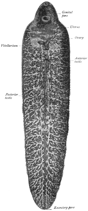
Fig. 145.—Fasciola gigantica. × 6 1/2 (After Looss.)
From such records it is not impossible that Distomum oculi humani, Ammon, 1833, as well as Monostomum lentis, v. Nordm., 1832, may have been very young hepatic flukes that had strayed. Ammon found four specimens (length 0·5 to 1 mm.) of his species (named Distomum ophthalmobium by Diesing in 1850) between the opaque lens and the capsule of a five months old child in Dresden, and von Nordmann discovered his Monostomum lentis to the number of eight specimens (only 0·3 mm. in length) in the opaque lens of an old woman. Minute white bodies which Greef found in the cortex of the lens of a fisherman, aged 55, removed on account of cataract, were with some reserve regarded as Trematode larvæ. The fact that Ammon found that the intestinal cæca of the worm discovered by him had no lateral branches does not negative the above opinion, as in the liver fluke the intestinal cæca are originally unbranched, and according to Lutz they only develop lateral ramifications later, between the twelfth and twenty-second day of infection (fig. 144).
Fasciola gigantica, Cobbold, 1856.
Syn.: Distomum giganteum, Diesing, 1858; Fasciola gigantea, Cobbold, 1858; Cladocœlium giganteum, Stoss., 1892; Fasciola hepatica var. angusta, Raill., 1895; Fasciola hepatica var. ægyptiaca, Looss, 1896.
This species is closely allied to Fasciola hepatica, but is distinguished by its elongated body, short cephalic cone, almost parallel sides, larger ventral sucker, which is also closer to the oral sucker, and by its larger eggs. Length up to 75 mm., width up to 12 mm. Oral sucker 1 to 1·2 mm., ventral sucker up to 1·7 mm. in diameter. Eggs 150 µ to 190 µ long by 75 µ to 90 µ broad.
Habitat.—Bile-ducts of Camelopardalis giraffa, Bos taurus, Bos indicus, Bos bubalis, Ovis aries and Capra hircus.
Distribution.—Africa.
This species has once been observed in man by Gouvea, in Rio de Janeiro, in a French naval officer who became ill with fever, cough and slight blood-spitting. The lungs were normal except for a sharply circumscribed spot at the base of the left lung. Twenty days later during a fit of coughing the patient spat up a fluke 25 mm. long, characterized by its slender aspect and by the size of its ventral sucker, and its close proximity to the oral sucker. Considering the fact that Gouvea’s patient had spent many weeks in July of the same year in Dakar (Senegambia), where according to Railliet Fasciola gigantica is common in slaughtered animals, and considering also the characters of the fluke, Railliet rightly assumes that one had to do with the African giant fluke and that the patient had infected himself in Dakar.
Sub-family. Fasciolopsinæ, Odhner, 1910.
Genus. Fasciolopsis, Looss, 1898.
Ventral sucker large, and elongated posteriorly into a sac. Cirrus pouch long and cylindrical, its greatest length being occupied by the sinuous tubular seminal vesicle, on which exists a peculiar cæcal appendage. Laurer’s canal present.
Fasciolopsis buski, Lank., 1857.
Syn.: Distomum buski, Lank., 1857; Dist. crassum, Cobbold, 1860, nec v. Sieb., 1836.
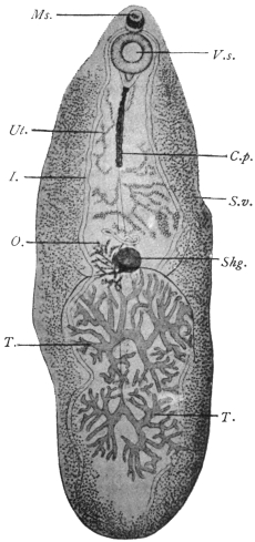
Fig. 146.—Fasciolopsis buski, Lank. V.s., ventral sucker; C.p., cirrus pouch; I., intestinal fork; S.v., vitellaria; T., testes; O., ovary; Ms., sucker; Shg., shell gland; Ut., uterus. Magnified. (After Odhner.)
The length of the body varies; it may measure 24 to 37 or even attain 70 mm.; the breadth is from 5·5 to 12 to 14 mm. In the pig the fresh parasites measure, smallest, 12 to 8 mm.; largest, 35 to 16 mm. (Mathis and Léger). Skin without spines, but according to Heanly always present in man and pig specimens. The oral sucker measures 0·5 mm. in diameter; the ventral sucker is three to four times as large; the pharynx is globular, 0·7 mm. in diameter; the prepharynx is provided with a sphincter; the intestinal cæca extend to the posterior border with two characteristic curves, one at the anterior border of the anterior testis, the other between the two testes. The genital pore is at the anterior border of the ventral sucker; the cylindrical cirrus pouch extends from behind the ventral sucker to half-way to the shell gland. The seminal vesicle extends forwards within the cirrus pouch as a convoluted tube. From its anterior portion is given off the cæcal appendage, which has itself short lateral diverticula. It runs backwards, ending blindly about 0·5 mm. from the posterior end of the cirrus sac. The seminal vesicle is continued as the pars prostatica (?) 0·5 mm. long, and this by the very short ejaculatory duct (13 µ), and finally by the fairly long cirrus, which is beset with very fine spines except at either extremity. The ovary and shell gland are situated at about the middle of the body with the testes behind them, and the uterus in front. The vitellaria extend from the ventral sucker to the posterior border. The eggs measure 120 µ to 130 µ in length and 77 µ to 80 µ in breadth, and resemble those of Echinochasma sp. in dogs. The larval stages are said to occur in shrimps.
Habitat.—Intestine of pig and man.
Distribution.—In man: India, Siam, China, Assam, Sumatra. It is common in Cochin China (16 out of 133 Annamites, Noc.), in Tonkin very rare. Dr. J. Bell has sent me [J. W. W. S.] human specimens from Hong Kong. In pigs: very common in South China (Heanly). Common in pigs in Hong Kong. Sixteen out of 248 pigs (i.e., 6 per cent.) infected in Hanoi.
Fasciolopsis rathouisi, Ward, 1903.
Syn.: Distomum rathouisi, Poirier, 1887.
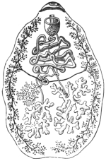
Fig. 147.—Fasciolopsis rathouisi, Poir.: the mouth at the top, and under it the genital pore and ventral sucker, behind which again is the uterus. The vitellaria are at the sides, and posteriorly in the central field the ramified testes; the ovary is in front of the right testis. (After Claus.)
Fifteen to 19 mm. long by 8·5 to 10·5 mm. broad by about 3 mm. thick. Skin with spines (Leiper). Bluntly oval or elliptical with short cephalic cone which is absent in Fasciolopsis buski. Oral sucker subterminal, 0·25 to 0·29 mm. broad by 0·2 mm. in antero-posterior diameter. Distant from ventral sucker by about twice its diameter. Ventral sucker 1·32 to 1·38 mm. broad by 0·68 to 0·7 mm. in antero-posterior diameter. Œsophagus extremely short. Cirrus sac not conspicuous and straight as in Fasciolopsis buski, but is convoluted. Testes one behind the other (according to Poirier they lie beside one another), more compactly branched, broader and denser than in Fasciolopsis buski. Ovary on right side, small, coarsely branched. Uterus in broad, closely grouped coils, packed with ova anterior to ovary. Vitellarian acini more numerous and somewhat differently distributed. Eggs 150 µ by 80 µ, thin shelled. [H. B. Ward, who has examined this species, and from whose account the above is mainly taken, considers that it is a good species, although the differences between it and Fasciolopsis buski are slight, while Odhner, who examined the original species, is of the opposite opinion.—J. W. W. S.] The parasite appears to cause diarrhœa, wasting and occasionally jaundice.
Habitat.—Intestine of man.
Distribution.—China, common in some parts (Goddard).
Fasciolopsis goddardi, Ward, 1910.
Twenty-one to 22 mm. long, 9 mm. broad. Skin with spines (Leiper). Uterus very closely coiled, most striking character is the large size of the vitelline acini. Imperfectly known.
Distribution.—China (Shanghai).
Fasciolopsis fülleborni, Rodenwaldt, 1909.
The fully extended fluke is tongue-shaped, 50 by 14 mm.; two contracted specimens measured 40 by 15 mm. and 30 by 16 mm. respectively. Skin without spines, with according to Leiper cephalic cone not clearly defined. Oral sucker circular, 0·75 mm. in diameter, slightly larger than that of Fasciolopsis buski. Ventral sucker 2·6 mm. in diameter (that of Fasciolopsis buski 1·6 to 2 mm.). Length 2·9 mm. (as in Fasciolopsis rathouisi), the excess of length over breadth being due to the posterior elongated sac-like prolongation of the sucker. Prepharyngeal sphincter present. Pharynx 0·7 mm. in diameter. Œsophagus practically absent. Gut cæca similar to those of Fasciolopsis buski.
Testes—regularly branched, separated by an incurving of the cæca, the anterior occupying a smaller area than the posterior.
Ovary—very small, as in Fasciolopsis buski, on the right side.
Shell Gland—almond-shaped, 2·3 by 1·2 mm. In Fasciolopsis buski it is round and smaller, 1 to 1·5 mm. in diameter.
Vitellaria—similar in distribution to those of Fasciolopsis buski, but the acini are strikingly small.
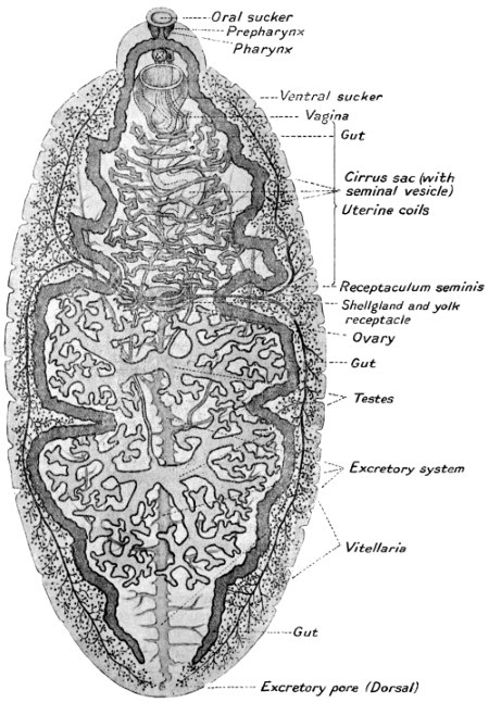
Fig. 148.—Fasciolopsis fülleborni, ventral aspect. (After Fülleborn.)
Cirrus Sac—is the most characteristic feature of this species. It is a powerfully built, convoluted sac standing out clearly on the body. It is not a uniform, straight cylinder 0·25 to 0·33 mm. in diameter, as in Fasciolopsis buski, but even in fully extended flukes is typically convoluted. It is 1 mm. thick in the middle, but in other parts varies much from this. The posterior end of the cirrus sac is at two-thirds or more of the distance from ventral sucker to shell gland. In the case of Fasciolopsis buski the posterior end of the sac only extends half-way.
Seminal Vesicle—has a peculiar convoluted, saccular and angular course, but the cæcal appendage characteristic of the genus appears to be absent!
Excretory System.—The main stem gives off very regular transverse branches which are well seen posteriorly.
Eggs.—100 µ by 73 µ. Thin shelled.
Habitat.—Intestine. Mahommedan from Calcutta.
[It is evident that a re-examination of fresh material is required before the validity of all these species can be accepted.—J. W. W. S.]
Family. Troglotremidæ, Odhner, 1914.
Genus. Paragonimus, Braun, 1899.
Body egg-shaped or somewhat elongated, generally more broadly rounded in front than behind. Covered all over with spear-shaped spines arranged in groups. Gut cæca winding with dilatations or constrictions in parts. Ventral sucker in or in front of the middle of the body. Excretory bladder cylindrical, very long and broad, reaching in front to the bifurcation of the gut. The lateral excretory canals join the bladder only a little in front of the excretory pore. Genital pore median just behind the ventral sucker. Genital sinus duct-like. Cirrus sac absent. Male terminal organs very small. Ejaculatory duct present. Testes and ovary deeply lobed, the testes in or just behind the middle, the ovary somewhat laterally placed just behind the ventral sucker. Uterus forms a coil behind the ventral sucker. Eggs rather large, thin shelled, the ovarian cell still unsegmented on deposition. Receptaculum seminis, small.
Parasitic in the lungs of mammals, enclosed in cyst-like cavities, generally in pairs.
Type Species.—P. westermanii in the tiger.
Paragonimus ringeri, Cobb., 1880.
Syn.: Distoma ringeri, Cobb., 1880; Distoma pulmonale, Baelz, 1883; Distoma pulmonis, Suga, 1883.
Fig. 149.—Paragonimus ringeri, Cobb.: to the right, dorsal aspect; to the left, ventral aspect. Natural size. (After Katsurada.)
The body is of a faint reddish-brown colour and plump oval shape. The ventral surface a little flattened; 7·5 to 12 mm. in length, 4 to 6 mm. in breadth, and 3·5 to 5 mm. thick (in man). The oral sucker (0·75 mm.) is subterminal; the ventral sucker (0·8 mm.) somewhat in front of the middle of the body. Pharynx spherical, 0·3 mm. in diameter, or 0·4 by 0·3 mm.; œsophagus, 0·02 mm.; intestinal cæca convoluted, asymmetrical, the first part having the same structure as the œsophagus. The cuticle is covered with spines in groups; the excretory pore opens at the posterior end rather on the ventral surface, the excretory ducts open into the elongated bladder at the hind end near the pore. Genital pore behind the ventral sucker and median. Genital sinus 0·2 mm. long with thick wall, ejaculatory duct 0·13 mm., pars prostatica 0·2 mm., seminal vesicle duct-like of irregular outline. Behind the sucker the ovary on the left, and the closely packed uterine coil on the right (though amphitypy of these two organs is common); the two irregularly lobed testes lie side by side posteriorly. Vitellaria extensive, leaving only a median dorsal and ventral space free. Seminal receptacle probably absent; Laurer’s canal present. The eggs are oval, brownish-yellow, fairly thin shelled, and measure on an average 81·2 µ by 49·2 µ.
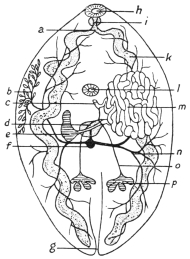
Fig. 150.—Paragonimus ringeri, Cobb.: diagram of the internal organs. a, œsophagus; b, vitellaria (a portion only shown); c, common genital duct; d, shell gland with oviduct, Laurer’s canal and vitelline duct; e, ovary; f, vitelline receptacle; g, excretory pore; h, oral sucker; i, pharynx; k, gut; l, ventral sucker; m, uterine coils; n, vitellarian ducts; o, vas efferens; p, testis. (After Kubo.)
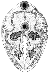
Fig. 150a.—Paragonimus westermanii, Kerb.: seen from the ventral surface. Mouth, pharynx, intestinal cæca, at the sides of which the vitellaria are observed. The genital pore is behind the ventral sucker, and next to it, on the left, the ovary; on the right, the uterus; the two testes posteriorly; the excretory vessel in the middle, 10/1. (After Leuckart.)
The following species are also known:—P. westermanii, Kerb., 1878, in the tiger, and P. kellicotti, Ward, 1908, in the pig, dog, and cat (N. America). Ward and Hirsch give the following differences between the spines of the three forms:—
P. ringeri.P. westermanii.P. kellicotti.
ShapeChisel-shaped, mod- erately heavy.Lancet-shaped, very slender.Chisel-shaped, heavy.
DistributionCircular rows, in groups.Circular rows, in groups.Circular rows, singly.
Two other species, P. rudis, Diesing, 1850, in a Brazilian otter (Lutra brasiliensis) and P. compactus, Cobbold, 1859, in the Indian ichneumon, are but little known.
Habitat.—Lungs, pleuræ, and especially the bronchi of man and dog. The alleged occurrence (of eggs) in other organs may be due to confusion with those of Schistosoma japonicum.
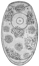
Fig. 151.—Egg of Paragonimus ringeri, Cobb., from the sputum. Showing the ovarian cell and vitelline cells and granules. 1,000/1. (After Katsurada.)
Distribution.—China, Korea, and especially in Japan, where, according to Katsurada, there are no districts that are entirely free from pulmonary flukes. The mountainous provinces of Okayama, Kumamoto, Nagano and Tokushima are the principal centres.
Pathology.—The number present in the lung varies from two to twenty, about. Usually one cyst contains one worm, but in the dog each cyst contains two. The cysts admit the tip of the finger, and have a fibrous wall 1 mm. thick. They originate partly from dilatation of bronchi and bronchioles. Others arise from the inflammatory reaction of lung tissue into which the worms have wandered. The worms and their eggs cause bronchitis and peribronchitis, catarrhal, hæmorrhagic, or purulent, and areas of consolidation. Areas containing eggs in their centre resembling tubercle nodules are not uncommon, and extensive cirrhosis of the lung may be found. As a result of these changes, emphysema and bronchiectasis also occur.
As to the development, only the following details are known: that the eggs, which before segmentation of the ovum reach the open in the sputum and through being swallowed also in the fæces, develop in water into a miracidium ciliated all over, which hatches and swims about freely. According to Manson this takes place in four to six weeks.
Sub-family. Opisthorchiinæ, Looss, 1899.
Genus. Opisthorchis, R. Blanch., 1845.
Opisthorchiinæ with lobed testes. Laurer’s canal present. Parasitic in the bile-ducts of mammals and birds.
Opisthorchis felineus, Riv., 1885.
Syn.: Distoma conus, Gurlt, 1831 (nec Creplin, 1825); Distoma lanceolatum, v. Sieb., 1836, v. Tright, 1889 (nec Mehlis, 1825 = Fasciolo lanceolata, Rud., 1803); Distoma sibiricum, Winogr., 1892; Distoma tenuicolle, Mühl., 1896.
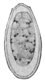
Fig. 152.—Egg of Opisthorchis felineus, Riv. 830/1.
This parasite is yellowish-red in the fresh condition, and almost transparent. The body is flat, with a conical neck at the level of the ventral sucker marked by a shallow constriction; this, however, is only noticeable in fresh and somewhat contracted specimens. Posteriorly to the ventral sucker the lateral borders run fairly parallel; the posterior end is either pointed or rounded off. The length and breadth vary according to the contraction, being usually 8 to 11 mm. by 1·5 to 2 mm. The suckers are about one-fifth to one-sixth of the length of the body distant from each other, and of about equal size (0·23 to 0·25 mm.). The œsophagus is hardly any longer than the pharynx, which lies close behind the oral sucker; the intestinal cæca reach almost to the posterior border and are often filled with blood. The excretory pore is at the posterior extremity, and the excretory bladder forks in front of the anterior testis. The testes in the posterior fourth of the body lie obliquely one behind the other; the anterior one has four lobes, the posterior one five lobes; the ovary is in the median line transversely, simple or slightly lobed; behind it lies the large pear- or retort-shaped receptaculum seminis and Laurer’s canal. The uterus is in the median field. The vitellaria occupy the fairly broad lateral areas, in about the central third of the body, beginning behind the ventral sucker and terminating at about the level of the ovary; the acini are small and arranged in groups of seven to eight, separated by interstices. The genital pore is close in front of the ventral sucker. The eggs are oval with sharply defined operculum at the pointed pole, 30 µ, by 11 µ.
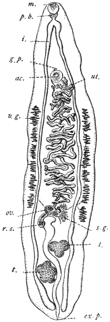
Fig. 153.—Opisthorchis felineus: from the cat. m., mouth; p.b., pharynx; i., gut; g.p., genital pore; ac., ventral sucker; ut., uterus; v.g., vitellarium; ov., ovary; s.g., shell gland; r.s., receptaculum seminis; t. testes; ex. p., excretory pore. (After Stiles and Hassall.)
This species, which is frequently confused with others, inhabits the gall-bladder and bile-ducts of the domestic cat especially; but is also found in the dog, in the fox, and in the glutton (Gulo borealis). It has been observed in France, Holland, North Germany (being particularly frequent in East Prussia), in Russia, Scandinavia, Siberia, Japan, Tonkin, Hungary, and Italy. The North American form (from cats and Canis latrans) is a distinct species (Opisthorchis pseudofelineus).
In man this species was first found by Winogradoff in Tomsk (nine cases), then by Kholodkowsky in a peasant from the neighbourhood of Petrograd who had travelled a great deal in Siberia, and finally by Askanazy in five persons who were natives of the East Prussian district of Heydekrug. In Tomsk, Opisthorchis felineus is the most frequent parasite of man that comes under observation at post mortem (6·45 per cent.), whereas Tænia saginata has only been found in 3·2 per cent., Echinococcus in 2·4 per cent., Ascaris lumbricoides in 1·6 per cent., and Oxyuris vermicularis in 0·8 per cent. of the autopsies. In the district of Heydekrug, however, the species in question is also frequent, as in a few years five cases came to our knowledge (of which three were diagnosed by the discovery of the eggs in the fæces).
In none of Winogradoff’s nine cases had the death of the patient been caused direct by the parasites, yet more or less extensive changes in the liver were found in all of them; such as dilatation of the bile-ducts with inflammation and thickening of their walls, and foci of inflammation or atrophy in the liver substance; icterus was present five times and atrophy of the liver an equal number of times; ascites was observed three times, and in two cases, probably of recent date, the organ was enlarged. The number of parasites found fluctuated between a few and several hundreds.
In two of Askanazy’s cases, which he examined more closely, carcinoma which had developed at the places most invaded by flukes was found at the post-mortem, so that perhaps there may be grounds for the connection which the author seeks to establish between cancer of the liver and the changes induced by the parasites; these changes consist of numerous and even ramified proliferations of the epithelium of the biliary duct into the connective tissue, which is likewise proliferated. The number of worms found in one case amounted to over 100; in a second case, in which the parasites had also invaded the pancreatic duct, their number was even larger.
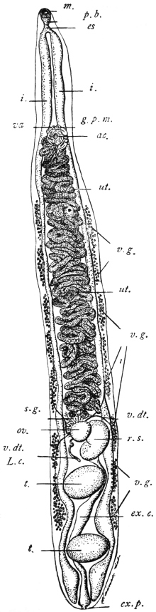
Fig. 154.—Opisthorchis pseudofelineus: from the bile-duct of the cat (Iowa), m., oral sucker; p.b., pharyngeal bulb; es., œsophagus; i., intestine; va., vagina; g.p.m., male orifice; ac., ventral sucker; ut., uterus; v.g. vitellarium; s.g., shell gland; v.dt., vitelline duct; ov., ovary; r.s., receptaculum seminis; L.c., Laurer’s canal; t., testis; ex.c., excretory bladder; ex.p., excretory pore. (After Stiles.)
Winogradoff as well as Askanazy found isolated flukes in the intestine also.
Unfortunately, nothing much is known of the history of the development of Opisthorchis felineus; we only know that when deposited the eggs already contain a ciliated miracidium, which, however, according to my experience, does not hatch out in water, but only after the entry of the eggs into the intestine of young Limnæus stagnalis; no further development, however, occurs. Winogradoff states that he has seen the miracidia hatch after the eggs had been kept in water for a month at 37° C.; and has even observed free miracidia in the bile of man and of a dog respectively. Although the whole post-embryonal development of the cat fluke remains yet to be investigated, Askanazy by a series of experiments on cats and dogs has discovered the mode of infection. The intermediate hosts are fish, and mainly the ide, in this country called Tapar (Idus melanotus, H. and Kr.), and of subsidiary importance the roach (Leuciscus rutilus). Both species of fish as well as others are readily eaten raw by man on the Courland lagoon (Baltic). It is, moreover, significant that those persons whom Askanazy found infected with the cat fluke were also infected with Dibothriocephalus latus, the intermediate host of which is also fish (Lota sp., Esox sp., Perca sp.).
In one of his nine cases Winogradoff also saw a small fluke covered all over with spines, which he conjectured to be the young stage of Opisthorchis felineus; as, however, according to my experience, this species, even in smaller specimens, is always without spines, the above hypothesis cannot be accepted. It is much more probable that one of the other species that also invade the liver of cats may accidentally be introduced into man; we know, in fact, that Metorchis albidus, Braun, and Metorchis truncatus, Rud., are both covered with spines. As, however, the spines of the first-named species are rather apt to fall off, and also as it possesses a different shape (spatula-shaped), it may be assumed that probably Winogradoff had found Metorchis truncatus, Rud., 1819, in his patient.
Genus. Paropisthorchis, Stephens, 1912.
Structure as in Opisthorchis, except that the ventral sucker and genital pore occur on the apex of a process or pedicle projecting from the anterior portion of the body. This process is about 1/2 mm. long, and is retractile.
Paropisthorchis caninus, Barker, 1912.
Syn.: Distoma conjunctum, Lewis and Cunningham, 1872; Opisthorchis noverca, M. Braun, 1903 (pro parte); Opisthorchis caninus, Barker, 1912 (?).
Length varies from 2·75 to 5·75 mm. in preserved specimens, average 3·6 to 5·2 mm. Body uniformly spinose, though as a rule spines are not present on the pedicle. Body slightly concavo-convex, the concavity being ventral. Oral sucker 0·28 mm. Pharynx 0·224 by 0·184 mm. Œsophagus 0·04 mm. Ventral sucker 0·176 mm. in diameter. Pedicle about 1/2 mm. long, may be completely retracted.
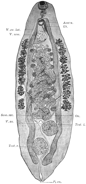
Fig. 155.—Paropisthorchis caninus: from the bile-ducts of the pariah dog, India. Acet. v., ventral sucker; Ut., uterus; V. ex. lat., longitudinal excretory duct; V. sem., seminal vesicle; Sem. rec., seminal receptacle; Ov., ovary; V. ex., excretory bladder; Test. l., left testis; Test. r., right testis; P. ex., excretory pore. × 40. (After Stephens.)
Genital Pore—opens on the apex of the pedicle in front of the ventral sucker. Its exact position varies with the state of contraction of the parts. In certain cases it actually opens within the cuticular border of the sucker, in other cases it opens externally to the sucker and anterior to it. The opening is covered with scales. The vas deferens and uterus run alongside one another until they merge near the apex of the pedicle into a common sinus.
Vitellaria—consist of eight acini on each side, extending from slightly behind the base of the pedicle to the anterior border of the ovary, or as far back as a line separating the posterior border of the ovary from the anterior border of the anterior testis.