ON PARASITES IN GENERAL. (continued 10)
Reading edition
Azur Staining:—
(4) G1 cell small, easily overlooked.(4) G1 cell large, stains deep blue, cell protoplasm = twice width of larva, easily seen.
(5) Excretory cell close to excretory pore, 2 per cent. of length.(5) Excretory cell farther from pore, 4 per cent. of length.
Vital Staining with Neutral Red:—
(6) Internal body or reserve material clearly shown.(6) Not shown.
Life History.—In the stomach of the mosquito the larvæ cast their sheath in the thickened blood in one to two hours. In twenty-four hours the majority have reached the thoracic muscles, where development proceeds. They are at first immobile and of a “sausage” form (110µ by 13 µ), with a short spiky tail. In three to five days the œsophagus is formed, the larva now being 0·5 mm. long. The larva appears to moult at this time. After the gut is formed papillæ, three or four in number, appear at the tail end. In two to three weeks the larvæ are 1·5 mm. long. They now leave the thorax and reach the labium, but they may be found in various parts of the body, e.g., the legs. They bore through Dutton’s membrane and so arrive on the surface of the skin, which they rapidly enter. Their development in man is unknown, but it may be very long, as children are not infected till 4 to 5, or even 10 years old, but this may be due to unknown causes.
Development takes place in numerous mosquitoes. Anophelines: Myzomyia rossii, Pyretophorus costalis, Myzorhynchus sinensis, Myzorhynchus barbirostris, Myzorhynchus peditæniatus.
Culicines: Culex pipiens, Culex fatigans, Culex skusei, Culex gelidus, Culex sitiens, Culex albopictus, Stegomyia fasciata, Stegomyia pseudoscutellaris, Stegomyia gracilis, Stegomyia perplexa, Mansonioides uniformis, Mansonioides annulipes, Scutomyia albolineata, Tæniorhynchus domesticus.
Partial development takes place in other species.
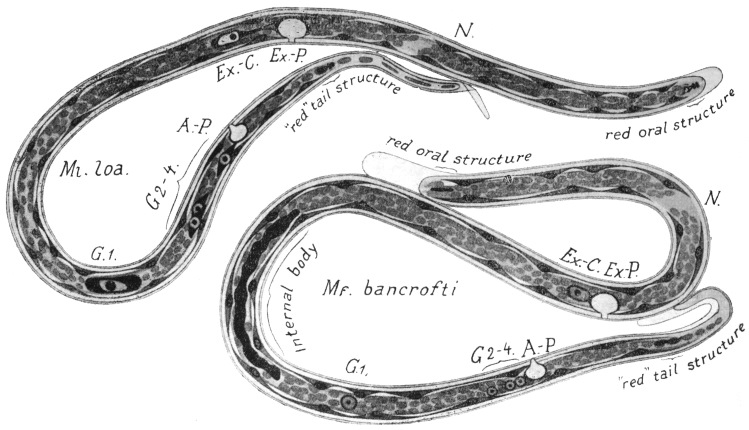
Fig. 283.—Schematic drawings of the anatomy of Ml. loa and Mf. bancrofti combined from specimens stained in different ways. The position of the organs has not been based on the average values of a large series of specimens, but on that of a single specimen. G1, chief genital cell; G2–4, other genital cells; Ex.-C., excretory cell; Ex.-P., excretory pore; A-P., anal pore; N., nerve ring. Magnification circa 1,000. (After Fülleborn).
Pathology.—Among the conditions which Filaria bancrofti is believed to produce are lymphangitis, varicose glands, especially inguinal and epitrochlear, chyluria, chylocele, lymph scrotum, orchitis, abscess, and elephantiasis. The evidence that these so-called “filarial diseases” are produced by F. bancrofti is (1) geographical and statistical; (2) pathological. Bahr has contributed evidence of the former kind from his researches in Fiji, on which we may base the following statements:—
(1) The prevalence of filarial diseases is proportional to the prevalence of Mikrofilaria bancrofti in the blood. Thus in four villages examined by him he got the following figures:—
Village AVillage BVillage CVillage D
Mf. bancrofti12·5 per cent.25 per cent.31 per cent.33 per cent.
Filarial diseases29"39"58"34"
Total population168114425222
(2) Out of 257 people with Mf. bancrofti in the blood, 153 were suffering from filarial diseases, i.e., 59 per cent.
(3) Whereas of 672 people without Mf. bancrofti in the blood, only 263 were suffering from filarial diseases, i.e., 37·6 per cent.
(4) Again out of 416 people suffering from filarial disease, 153 showed Mf. bancrofti in their blood, i.e., 36·7 per cent.
It is generally assumed that all people suffering from filarial disease show at some (presumably early) stage larvæ in the blood; but we do not consider that this must necessarily be so. It appears to us quite possible that living adult filariæ may be present in the body, producing disease, without their larvæ appearing in the blood. The absence of larvæ from the blood in 63·3 per cent. of persons suffering from filarial disease is, however, generally explained otherwise. The adults which occur in enlarged glands, etc., get eventually destroyed by inflammatory reaction, so that larvæ are no longer being produced, while the enlarged gland, etc., which the adults have produced remains. This explanation assumes that the larvæ of the original worm die in the circulation or elsewhere, e.g., kidney, but we have no evidence as to the duration of life of larvæ in the human body; but also it assumes that a person cannot be reinfected with filaria, for otherwise there is no reason why the diseased should not be infected in the same proportion as the non-diseased. But assuming the explanation to be true, it would explain why a diseased population show larvæ in only about one-third of the cases. It must be borne in mind also that the figures are rather small.
Pathology.—In order to explain the effects which do or may be expected to occur from obstruction of lymphatics, it is necessary to have an accurate knowledge of the distribution and connections of lymphatic vessels (and glands) and the anastomoses of these vessels. We can only briefly summarize our knowledge here.
We should recall also that considerable destruction or obstruction of lymphatics or glands may occur without necessarily producing any lymphatic obstruction, at least, of a permanent nature, e.g., when a mass of lymphatic glands is destroyed by a bubo in the groin or, again, when a carcinomatous mass of glands is removed from the axilla. Again, to take the case of chyluria—where it is generally assumed that obstruction must occur higher up than the point at which the intestinal lacteals enter the juxta-aortic glands—this disease may occur, e.g., in temperate regions, quite apart from such obstruction. It is true that some of these cases of chyluria are not cases of chyle in the urine, but, as little or no fat is present, lymphuria. These do not require the above assumption, but seeing that true chyluria may apparently occur without such obstruction, we should be cautious about explaining this and other symptoms on the basis of obstructions which theory may demand, for only too often there are no post-mortem facts at our disposal.
Lymphangitis: What this is due to is unknown. There is no actual evidence of the occurrence of adults in the inflamed vessel. Complete disappearance, not to reappear, of (non-periodic) mikrofilariæ from the blood has been shown by Bahr and others to occur within twenty-four hours after an attack of lymphangitis, orchitis adenitis or simply a high temperature. This mysterious phenomenon requires explanation. If the mikrofilariæ were being killed by the attack, their dead bodies should still be found in the blood; or if the adults were being killed, for all we know to the contrary, the larvæ might well survive. We consider there is no evidence that either are affected, but that for some reason, as little understood as in periodicity, the larvæ now remain in the organs.
Abscess: In Fiji, by Bahr, they have been found in the substance of various muscles, e.g., quadriceps extensor, latissimus dorsi, serratus magnus, in the popliteal space, groin, axilla, and over the internal condyle of the humerus, and in the upper extremity they are frequently infected with cocci. They not infrequently contain fragments of dead adult filariæ. Their mode of origin is not clear. They form nearly 30 per cent. of cases of filariasis in Fiji. Of 95 cases, 41 showed mikrofilariæ in blood, 54 did not.
Hydrocele and enlarged testis: In Fiji they form about 10 per cent. (36 out of 343) of cases of filariasis. The fluid is usually sterile; mikrofilariæ were present in the fluid in 1 out of 11 cases. In the wall numerous calcified adult filariæ may be found. The walls consist chiefly of hypertrophied muscle with fibrous tissue, dilated blood-vessels and lymphatics, the lining epithelium of which appears to be absent; of 38 cases 14 had mikrofilariæ in the blood, 24 had not. Most of the cases are associated with elephantiasis of the scrotum (11 out of 12 cases).
Enlarged glands form over 40 per cent. (153 out of 343) of cases of filariasis, so that they are the commonest expression of filariasis met with in Fiji. The glands are enlarged, fibrotic, and the trabeculæ are thickened. The lymphatics are thickened or represented merely by fibrous tissue. The gland also shows dilated blood-vessels and numerous spaces filled with lymph. Giant-cells are common in those glands which contain remnants of filariæ. Masses of lymphocytes enclosed by inflammatory or fibrous tissue are common. Eosinophile cells are also extremely common, not only in the fibrous tissue of the glands, but in other inflammatory or fibrotic conditions: in other organs living or calcified filariæ are “usually” present. Only about 33 per cent. show mikrofilariæ in the blood. The epitrochlear gland is frequently enlarged in Fiji.
Breinl has examined enlarged glands and finds loose vascular fibrous tissue with lymphocytic invasion. In parts, the lymphocytes collect into areas 200 µ to 800 µ in diameter. The lymph tissue surrounding the spermatic cord showed abundance of vessels—(1) large, (2) small. The large had thick walls and wide lumina. In other cases the lumina were nearly filled by a thrombus of newly formed, fine, loose connective tissue.
Varicose glands: In about 7 per cent. (24 out of 343 cases) of filariasis, mikrofilariæ are found in the blood in 50 per cent. (12 out of 24).
Elephantiasis.—Elephantiasis scroti is associated with hydrocele in 50 per cent. of cases (12 out of 23); in 65 per cent. of cases (15 out of 23) there are associated enlarged glands in one or both groins, though also hydrocele and enlarged glands occur without elephantiasis scroti. In 13 out of 27, i.e., about 50 per cent., cases of elephantiasis in various regions, no associated enlargement of glands is found. Elephantiasis forms in Fiji less than 10 per cent. of cases of filariasis. Mikrofilariæ are present in the blood in 36 per cent. (12 out of 33) of cases.
Chyluria.—Exceedingly rare in Fiji. Theory would demand an obstruction above the point of entry of the lacteals, viz., the pre-aortic lymphatic glands, but in cases in temperate regions it may occur without any such lesion. In some of these cases the fluid is not chyle (fat absent), but presumably lymph. A discussion of the mode of production of chyluria, lymph scrotum, elephantiasis, etc., is at present premature; theory has far outrun fact. Too much stress had been laid on the mechanical action of the worms to the almost total exclusion of their (or possibly their larval) toxic action. The above analysis has been made in the hope of acquiring more extended observations similar to those made by Bahr.
Geographical Distribution.—Filaria bancrofti is known in nearly all tropical countries. It occurs in India, China, Indo-China, Japan, Australia, Queensland, the Islands of Polynesia (with the exception of the Sandwich Islands), Egypt, Algeria, Tunis, Madagascar, Zanzibar, Sudan, etc., the south of the United States of America, Brazil, the Antilles, etc. Whether it is the same species in all cases is questionable.
Filaria demarquayi, Manson, 1895.
Syn.: F. ozzardi, Manson, 1897.
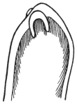
Fig. 284.—F. demarquayi: tail, showing paired large fleshy papillæ. (After Leiper.)
The adult female F. demarquayi measures from 65 to 80 mm. in length by 0·21 to 0·25 mm. in breadth. The head has a diameter of from 0·09 to 0·1 mm. The mouth is terminal. The genital pore opens at 0·76 mm. from the head. The alimentary canal is nearly straight and terminates in an anus, which is subterminal. The opening of the anus is marked by a slight papilla. The tail is curved. It rapidly diminishes in size just below the anal papilla. A characteristic pair of fleshy papillæ project from the tip of the tail. The diameter near the tip of the tail before its termination is 0·03 mm. F. demarquayi is a thicker worm than Ac. perstans. It differs from F. bancrofti in the greater size of the head, in the smaller tail, and particularly in the marked fleshy papillæ at the tip of the tail. These papillæ are knobby, and not simply cuticular as in Ac. perstans.
The male of Filaria demarquayi has still to be found.
The adult female form of F. demarquayi was found by Dr. Galgey in the body of a native of St. Lucia in whose blood the larvæ had been found during life. Five adult females were found in the connective tissue of the mesentery.
The larva measures 200 µ in length by 5 µ in breadth; it is sharp-tailed, and has no sheath. Its movements are very active, and the absence of a sheath enables it to glide along freely all over the slide. It observes no periodicity, being present in the peripheral circulation both by day and by night. As a rule, some eight or ten parasites are found in an ordinary preparation. Sometimes hundreds of these larval filariæ may be counted on every slide.
The intermediate host has not been discovered.
Geographical Distribution.—St. Vincent, Dominica, Trinidad, and St. Lucia (West Indies), British Guiana, New Guinea (?).
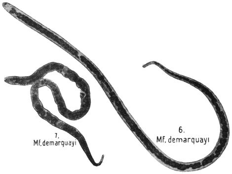
Fig. 285.—Mf. demarquayi in thick film, dried and stained with hæmatoxylin. 6, unshrunken; 7, shrunken. × 1,000. (After Fülleborn.)
Filaria taniguchi, Penel, 1905.
Female 68 by 0·2 mm. in breadth. Cuticle non-striated. Mouth two pairs of papillæ. Anus 23 mm. from extremity. Vulva 1·3 mm. from mouth. Larva 164 µ by 8 µ, sheathed. Tail truncated. Periodicity nocturnal.
Habitat.—Lymphatic glands of man. Japan.
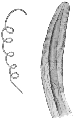
Fig. 286.—Filaria (?) conjunctivæ: to the left, life size; to the right, the anterior extremity magnified. (After Addario.)
Filaria (?) conjunctivæ, Addario, 1885.
Syn.: Filaria peritonei hominis, Babes, 1880; Filaria inermis, Grassi, 1887; Filaria apapillocephata, Condorelli-Francaviglia, 1892.
The female only of this species is known. It measures 16 to 20 cm. in length and 0·5 mm. in breadth, and is of a whitish or brownish tint. The cuticle is striated with fine transverse and more marked longitudinal striæ with the exception of a small field surrounding the mouth, which is terminal and has neither papillæ nor lips. The œsophagus measures 0·6 mm. in length. The anus is 3 mm. in front of the rounded posterior extremity, and behind it there are two (glandular?) sacs. The vulva is close behind the oral aperture; the vagina soon divides into two convoluted uteri, which are filled with eggs and embryos. Embryos 350 µ by 5·5 µ.
This species (115 mm. long) was first observed in Milan by Dubini in the eye of a man; subsequently it was observed, encysted and calcified (190 mm. long), by Babes in the gastro-splenic omentum of a woman in Budapest, and finally one (95 mm. long) was extracted by Vadela from a tumour the size of a pea in the ocular conjunctiva of a woman in Catania (Sicily), which case has been described by Addario. Possibly Agamofilaria palpebralis, Pace, 1867 (nec Wilson, 1844), and A. oculi humani, v. Nordm., 1832, are the same species.
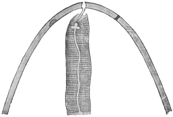
Fig. 287.—Filaria (?) conjunctivæ: anterior end greatly magnified; the mouth with the pharynx in the middle; in the cuticle on the right side the opening of the vagina, and behind it the excretory pore. (After Grassi.)
Filaria (?) conjunctivæ is certainly only an incidental parasite of man; the horse and ass are its normal hosts, but it is not common in these animals, or is frequently confused with Hamularia equi, Gmelin, 1789.
Group. Agamofilaria, Stiles, 1906.
Not a generic but a group name for immature Filariidæ the development of which does not admit of generic determination.
Agamofilaria georgiana.
Adult unknown, length from 32 to 53 mm. Maximum diameter 560 µ to 640 µ. Head no cephalic cone. Mouth small, circular, surrounded by six papillæ (two small latero-median and four sub-median). The larger papillæ are 24 µ from base to tip. Excretory pore about 0·5 mm. from head. Anus 64 µ to 128 µ from tip. Cuticle fine striæ near anus, occasionally elsewhere. Lateral lines clearly marked. Œsophagus 2·5 to 2·9 mm. Rectum 200 µ long.
Habitat.—Superficial sores on the ankle of a negress, Georgia, U.S.A.
Agamofilaria palpebralis, Pace, 1867 (nec Wilson, 1844).
100 by 1·5 mm., removed from a cyst in the left upper eyelid of a boy by Pace, in Palermo.
Agamofilaria oculi humani, v. Nordmann, 1832.
Syn.: Filaria lentis, Diesing, 1851.
The sexless Nematodes observed in the lens of the human eye were termed Filaria oculi humani. Only three cases are known. v. Nordmann observed very small round worms in the lens of a man and woman with cataract, and Gescheidt once found three specimens in the lens of a woman similarly affected.
The demonstration of nematode-like formations in the vitreous remains uncertain even when movements are observed, and when they cannot be extracted and examined microscopically the doubt may occur that one may have mistaken the remains of the hyaloid artery for a worm, which it resembles in form, size and colour; the slightest movement of the eye also causes it to move so that it simulates a living organism.
Accordingly it would be more correct to exclude all the cases known only ophthalmoscopically (Quadri, 1857; Fano, 1868; Schoeler, 1875; Eversbusch, 1891). There then remains only one positive case, described by Kühnt in 1891. In this case it was possible to follow the gradual growth of the parasite for some time, and the worm, which measured only 0·38 mm. in length, was finally extracted.
Agamofilaria labialis, Pane, 1864.
The parasite measures 30 mm. in length; the anterior extremity is pointed; the terminal oral aperture is surrounded by four papillæ; the anus opens 0·5 mm. in front of the posterior extremity; the vulva is 2·5 mm. in front of the anus; the uterus is double; the anterior one passes with convolutions forward to the cephalic end; the posterior one is directed backwards and remains rudimentary.
Extracted from a small pustule on the inner surface of the upper lip. Also found in Naples by Pierantoni in 1908.
The position of many of these worms is doubtful, and still more so is that of many other imperfectly described “Filariæ,” which are hardly more than useless and confusing names. These include the following:—
Filaria (?) romanorum-orientalis, Sarcani, 1888.
Observed in the blood of a Roumanian woman; 1 mm. in length, 0·03 mm. in breadth; tail end pointed, a tongue-like appendage on the head. Eggs the size of a red cell with developed embryo, apparently viviparous.
Filaria (?) kilimaræ, Kolb, 1898.
Several female specimens, 10 to 20 cm. long by 0·5 to 1 mm. broad, were once found free in the abdomen of a fallen Kitú warrior; according to Spengel, who examined them, the oral papillæ of these worms were similar to those of Dracunculus medinensis. Moreover, Kolb classifies together Nematodes that probably have no connection with each other.
Filaria (?) sp.?
Cholodkowsky calls attention to Filariæ that are still unknown which cause tumours resembling whitlows on the fingers of peasants of the Twer Government.
Mikrofilaria powelli, Penel, 1905. In Bombay.
Mikrofilaria philippinensis, Ashburn and Craig, 1906. In the Philippines.
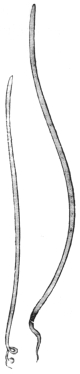
Fig. 288.—Setaria equina: left, male; right, female. Natural size. (After Railliet.)
Genus. Setaria, Viborg, 1795.
Syn.: Hamularia, Treutler, 1793; Tentacularia, Zeder, 1800 (nec Bosc, 1797).
Mouth with projecting peribuccal armature deeply notched on the lateral margins, less so dorsally and ventrally. Tail in both sexes with peculiar caudal appendages.
Parasitic in serous cavities, especially of ruminants.
Setaria equina, Abildg., 1789.
Syn.: Gordius equinus, Abildg., 1789; Filaria equi, Gmelin, 1789; Hamularia lymphatica, Treutler, 1793; Tentacularia subcompressa, Zedder, 1800; Filaria papillosa, Rud., 1802; Filaria hominis bronchialis, Rud., 1819; Filaria hominis, Dies., 1851; Strongylus bronchialis, Cobb., 1879.
The body is whitish, filiform, pointed posteriorly. The cuticle presents a delicate transverse striation. The mouth is small, round, and surrounded by a chitinous ring, the border of which carries, at the sides, two semilunar lips, and there is on the dorsal as well as on the ventral surface a papilliform process; on the tail, corresponding with each sub-median line, is a conical papilla. The male measures 6 to 8 cm. in length; the posterior extremity ends in a corkscrew spiral; there are on each side four pairs of pre-anal and four or five post-anal papillæ; the spicules are unequal. The female measures 9 to 12 cm. in length and is viviparous; the embryos measure 0·28 mm. in length and 0·007 mm. in breadth.
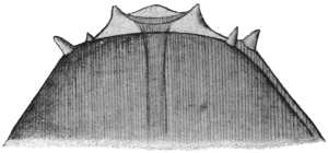
Fig. 289.—Setaria equina: anterior end, magnified. (After Railliet.)
Setaria equina is a frequent parasite of horses and asses; it inhabits the peritoneal cavity, and from there occasionally invades the female genitalia or even the liver; it is found more rarely in the pleural cavity or in the cranium. The statement that it also occurs in the subcutaneous connective tissue is probably due to confusion with Setaria (Filaria) hæmorrhagica, Raill., 1885 (Filaria multipapillosa, Cond. et Drouilly, 1878). Setaria labiata papillosa (immature form) occurs in the eye of the horse, adults in the peritoneal cavity.
Treutler, in 1790, found a filaria in the enlarged bronchial lymphatic gland of a patient suffering from phthisis. It measured 26 mm. in length and had two spicules, which Treutler mistook for mouth hooks, hence the name Hamularia. Blanchard mentions another case from Geneva, Brera a third and v. Linstow a fourth. As shown by the synonyms, a few authors consider this form to be a distinct species, which is hardly probable.
Genus. Loa, Stiles, 1905.
Characterized by the possession of cuticular bosses in both sexes (fig. 294).
Loa loa, Guyot, 1778.
Syn.: Filaria oculi, Gerv. et v. Ben., 1859; Dracunculus oculi, Diesing, 1860; Dracunculus loa, Cobbold, 1864; Filaria subconjunctivalis, Guyon, 1864.
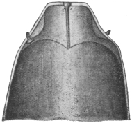
Fig. 290.—Loa loa: the anterior end of the male, magnified. (After R. Blanchard.)
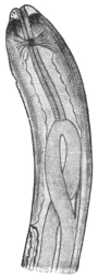
Fig. 291.—Loa loa: anterior portion of the female as far as vulva. (After Looss.)
The male measures 25 to 35 mm. in length, and 0·3 to 0·4 mm. in breadth; the cuticle is not striated, but, with the exception of the anterior and posterior extremities (1·5 mm.), is beset with numerous irregularly distributed bosses (4 µ to 12 µ high by 12 µ to 27 µ broad). The anterior extremity is somewhat attenuated, and in front is conical and transversely truncated. At the anterior limit of the conical part is a small papilla corresponding with the dorsal and ventral median lines, and a little in front six non-projecting sensory papillæ (two lateral, four sub-median). Excretory pore 0·65 mm. from the anterior end. The posterior extremity is attenuated and somewhat curved ventrally; the anus is 0·082 mm. distant from the rounded posterior border. In front of the anus on each side are three globular and pedunculated papillæ of different sizes, set close one behind the other but asymmetrically; behind the anus on either side are two smaller papillæ of a different shape; the anterior one resembles the pre-anal papillæ in form, but is smaller; the posterior one is conical, and rests with a broad base on the cuticle. The spicules are 0·113 and 0·176 mm. long.
The female measures 45 to 63 mm. in length by 0·5 mm. in breadth. It is also beset with irregularly distributed bosses, which in places lie close to each other, and extend to the anterior extremity; posteriorly they become less frequent, but are not entirely absent. The anterior extremity is conical, the posterior one straight, attenuated, rounded off, 0·17 mm. from the anus. The uteri contain eggs in the most various stages of development, as well as hatched-out larvæ, 253 µ to 262 µ in length and 4·7 µ to 5 µ in breadth. The vulva lies about 2 mm. from the head end. The vagina, 9 mm. long, divides into two branches, which at first run posteriorly and parallel to one another for about 18 mm. One then bends forward, runs as far as the œsophagus, bends here again and runs backward to end at the point of its first bending. The other branch at first runs straight backward and then bends forward, but before reaching the point of the first bend of the anterior tube bends backward again, forms again a loop and ends at the level of the anus. The tubes consist in the main of the uterus, then a club-shaped swelling, the receptaculum seminis, then the oviduct 2 mm. long, and finally the ovary.
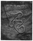
Fig. 292.—Loa loa in situ. Natural size. (After Fülleborn and Rodenwaldt.)
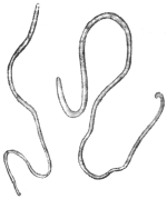
Fig. 293.—Loa loa: male on the left, female on the right. × 2. (After Looss.)
Unsegmented eggs measure 32 µ by 17 µ, in the morula stage 40 µ by 25 µ, and when containing embryos 50 µ by 25 µ. The vitelline “shell” of the egg is, according to most authors, stretched by the embryo and becomes the sheath of the hatched larva. While still in the vulva, the larva measures 217 µ to 274 µ (average 246 µ) in fresh, 146 to 226 µ (average 192 µ) stained.
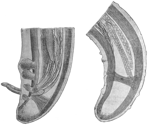
Fig. 294.—Loa loa: on the left, the hind end of a male; on the right, of a female. Note the cuticular bosses shown in the figure of the female. × 285. (After Looss.)
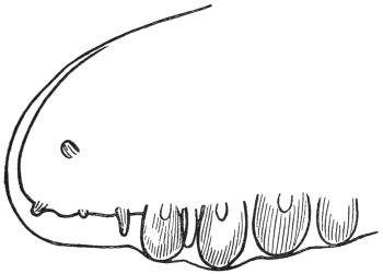
Fig. 295.—Loa loa: lateral view of tail of male showing papillæ. (After Lane and Leiper.)
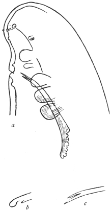
Fig. 296.—Loa loa. a, ventro-lateral aspect of tail showing papillæ and one spicule; b and c, terminations of the two spicules. (After Leiper.)
Site of Worms.—In various localities; under the muscular aponeuroses on extensor surfaces of arms and legs, fingers, trunk, eyelid, conjunctiva, frænum linguæ, penis, pericardium, anterior chamber of eye, and, according to some authorities, in lymphatic vessels, e.g., those of spermatic cord. As many as thirty adults may be found. The worms appear to be frequently immature, and it has been stated that worms in superficial parts are immature, those situated deeply are mature, but the data are few.
The first accounts of Loa loa—long since forgotten—were reported by Pigafetta, and are contained in a book of travels on the Congo printed in 1598. In an accompanying illustration is depicted, not only the ancient method of extraction of the Medina worm, but also the operative removal of the filaria from the conjunctiva. Subsequently the presence of the worm in negroes was confirmed by Bajon in Guiana (1768) and by Mongin in Mariborou (San Domingo), likewise in a negro (1770). At about this time a French ship’s doctor, Guyot, was cruising on the West Coast of Africa; he observed the parasite termed “loa” by the natives, and learned that it was frequent in the negroes of the Congo district. Since that time numerous observations have been reported. It was formerly common in South America, where the parasite was imported by slaves, but it disappeared when the traffic ceased; it was particularly prevalent in the Congo, where it occurs not only in natives, but also in Europeans. During recent times it has repeatedly been observed in Europe in negroes as well as in white men who have lived on the West Coast of Africa.
Nematodes of different size have been repeatedly observed in the eye of man, in the anterior chamber, lens and vitreous. For example, Mercier, in 1771 and 1774, removed a filaria out of the anterior chamber of two negroes in St. Domingo. One was 36 mm. long. Barkan, in 1876, in San Francisco, removed one from the eye of an Australian. Again, Cappez and Lacompte, in Brussels, in 1894, observed for some weeks immature Nematodes in the eye of a negro girl, aged 2 1/2 years, and then removed them. What these Nematodes actually were in these cases it is impossible to say.
Structure of Larvæ.—In dried films the larva varies in size from 140·5 µ to 166·5 µ, average, 152·5 µ; while another set of measurements gave the values 131 µ, to 150 µ, average, 143·6. In films fixed with hot alcohol the dimensions were 208 µ to 254 µ, average, 231 µ.
The nerve ring 21·4 to 21·8 per cent. Excretory pore 30·4 to 31·8 per cent. Excretory cell 34·8 to 37·3 per cent. G1 cell 68·2 to 68·5 per cent. Anal pore 81·6 to 82·4 per cent. of total length. For other details cf. Filaria bancrofti.
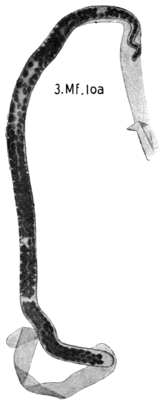
Fig. 297.—Mf. loa: in thick film, dried and stained with hæmatoxylin. × 1,000. (After Fülleborn.)
Larvæ in Blood.—These from their diurnal periodicity are known as Mikrofilaria diurna. The evidence that these larvæ are the young of the adult worm Loa loa is: (1) They are identical in structure with larvæ taken from the uterus of L. loa; (2) their geographical distribution is the same as that of L. loa; (3) they eventually occur in the blood of patients suffering from Calabar swellings, a condition due to L. loa. Their occurrence in the blood in this latter condition and in L. loa infections we shall consider later.
Periodicity.—Here, as in the case of the larvæ of Filaria bancrofti, the larvæ that appear in the blood are probably the overflow simply of the larvæ which we assume, on analogy, to have their principal site in the lungs. They appear in the blood about the time of getting up, 6 to 8 a.m. (10 in 20 mm.3), at 12 noon there are twenty-four, at 8 p.m. the number has fallen to eighteen, and at midnight to one, while from 2 a.m. to 6 a.m. none, or one only, may be found. This periodicity is, as a rule, a very constant one, but there are exceptions, and in certain cases more have been found at midnight than at 9 a.m. The periodicity is also lost in pathological conditions, e.g., sleeping sickness (vide also under Filaria bancrofti). The possibility of non-periodic Loa loa larvæ should also be considered.
Pathology.—The parasite wanders about the body, and may be seen under the skin in thin parts. Their advance is in some cases at the rate of an inch in two minutes. During their progress they give rise to creeping sensations and to a condition of transient œdematous areas known as Calabar swellings on various parts of the body, e.g., arm. These vary in diameter from 1 to 10 cm., and often shift their position an inch or so a day. They give rise to a certain amount of redness, tension and heat, and their development is promoted by muscular action of the part. They disappear to reappear elsewhere. The condition is associated with a high eosinophilia, 50 per cent. being not uncommon. Patients known to harbour L. loa, e.g., native children, frequently show no larvæ in their blood, but they may do so after years of infection. Again, in patients having an infection of Mikrofilaria diurna, there is frequently at the time no evidence of the presence of Loa loa adults. Here again they may appear later, but the conditions which determine whether persons infected with L. loa show larvæ in the blood, or persons infected with Mikrofilaria diurna also show L. loa, are unknown, though explanations unsupported by facts abound. Likewise also the mode of production of the swellings is unknown.
Not uncommonly Mikrofilaria perstans occurs in the blood together with M. diurna.
Duration of Life.—This is long, as some cases have been observed five to six years after leaving Africa. The incubation period is about a year.
Life-history.—Development of the larvæ takes place in the salivary glands of Chrysops sp. as shown by Leiper.
Geographical Distribution.— West Africa, especially in Congo.
Genus. Acanthocheilonema, Cobbold, 1870.
Cuticle striated longitudinally. Œsophagus divided into two portions. Tail in both sexes with short lateral conical cuticular appendages. Spicules unequal, the larger membranous distally, the smaller hooked. Vulva in œsophageal region.
Acanthocheilonema perstans, Manson, 1891.
Syn.: Filaria perstans, P. Manson, 1891; Filaria sanguinis hominis var. minor, Manson, 1891.
The adult female Ac. perstans measures 70 to 80 mm. in length by 120 µ to 140 µ in breadth. The head is club-shaped and measures 0·07 mm. in diameter. The vulva opens at 0·6 to 1·0 mm. from the head. The tail is curved and presents a cuticular thickening which forms two triangular appendages. The anus opens at the apex of a papilla situated in the concavity of the curve formed by the tail 150 µ from the end. The diameter of the tail just before termination is 0·02 mm.
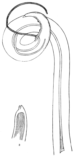
Fig. 298.—Acanthocheilonema perstans. 1, tail of male; 2, tail showing cuticular flaps devoid of fleshy contents. (After Leiper.)
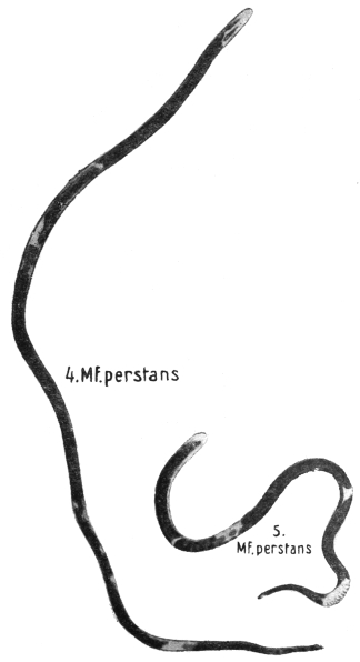
Fig. 299.—Mf. perstans in thick film, dried and stained with hæmatoxylin; 4, unshrunken; 5, shrunken. × 1,000. (After Fülleborn.)
The adult male measures 45 mm. in length by 60 µ to 80 µ in breadth. The diameter of the head is 0·04 mm. The tail is much curved. There are four pairs of pre-anal papillæ and two pairs of post-anal papillæ. Spicules very unequal in size. Cloaca 121 µ from the tail end. At the tail end two triangular cuticular appendages.
The adult worms inhabit the connective tissue at the base of the mesentery, especially in the region of the pancreas, abdominal aorta and suprarenals. To find them the mesentery should be removed, placed in a 2 per cent. solution of formalin, and then carefully examined at leisure.
Mikrofilaria perstans.—160 µ to 210 µ by 5 µ to 6 µ broad. Has no sheath. Cuticle transversely striated. Tail rounded off, not pointed. Nerve ring at 34 µ. Excretory pore 49 µ, genital pore 125 µ from head. Smaller larvæ 90 µ to 110 µ by 4 µ broad. A “fang” is also described on the head.
Mf. perstans.Mf. demarquayi.
(1) Tail stumpy.(1) Tail pointed.
(2) Column of nuclei extends to tip of tail.(2) Does not extend to tip.
Periodicity.—None.
Life-history.—Unknown.
Geographical Distribution.—Very common in many parts of Africa: Sierra Leone, Dahomey, Northern Nigeria, Southern Nigeria, Cameroons, Ivory Coast, Gold Coast, Old Calabar, Congo, Uganda. Absent from Zululand, Basutoland. On the East Coast of Africa it is not found in the towns of Zanzibar and Mombasa, neither is it found in the country of the Masi, nor amongst the Kavirondo, who dwell along the north-east shores of Lake Victoria.
In South America, Ac. perstans is very common amongst the aboriginal Indians in the interior of British Guiana. However, it is not found in Georgetown and in New Amsterdam, neither is it found in the cultivated strip of coast lying between these two towns, but it is common on the coast farther north near the Venezuelan boundary, where the forests stretch to the sea. The Waran Indians, who live at the mouth of the Waini river, harbour this parasite. It is absent in the West Indies.
Topographically, Ac. perstans is found only in areas covered by dense forest growth and abounding in swamps. In Kavirondo, where the forest disappears and the land is covered with scrub and short grass, it is not found; likewise it is not found on the grassy plains of the highlands of British East Africa. Towns and cultivated areas are free from it.
Genus. Dirofilaria. Railliet and Henry, 1911.
Body very long, thread-like, cuticle transversely striated. Mouth with six papillæ. Male tail spiral with voluminous pre-anal and some large post-anal papillæ; spicules unequal. Vulva near the anterior hundredth of body; viviparous. Parasitic in heart or blood-vessels and subcutaneous tissue.
Dirofilaria magalhãesi, R. Blanchard, 1895.
Syn.: Filaria bancrofti, v. Linstow, 1892; Filaria bancrofti, P. S. de Magalhães, 1892 (nec Cobbold, 1877).
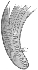
Fig. 300.—Dirofilaria magalhãesi: posterior extremity. (After v. Linstow.)
The male measures 83 mm. in length by 0·28 to 0·40 mm. in breadth. The anterior extremity is rounded, and has no papillæ (?6); the posterior extremity exhibits a double curve, with four pre-anal and four post-anal papillæ on each side. These are large and have a villous appearance. The mouth is round and unarmed, the pharynx measures 1 mm. in length, is cylindrical, very muscular, and its hinder part is dilated. The anus is situated 0·11 mm. in front of the hind end. There are probably two unequal spicules; one only, however, is known—apparently the shorter one—the length of which is given as 0·17 to 0·23 mm.
The female measures 155 mm. in length and 0·6 to 0·8 mm. in breadth; the rings of the cuticle are 0·005 mm. apart (in the male 0·003 mm. apart); the anterior extremity is slightly thickened and club-like, the posterior extremity is slender, and terminates obtusely; the lateral line is 0·127 mm. in breadth (that of the male 0·007 to 0·008 mm.); the anus opens 0·13 mm. in front of the hind end, the vulva is 2·5 mm. distant from the mouth, the ovaries are two much convoluted tubes. The eggs measure 38 µ by 11 µ.
This species was first discovered at a post-mortem, in the left ventricle, by J. P. Figueira de Saboia in Rio de Janeiro, and has been described by P. S. de Magalhães.
D. immitis occurs in the right ventricle of the heart of the dog in Europe and the Tropics.
D. repens is also a common subcutaneous Nematode in dogs in Annam.
Sub-family. Onchocercinæ, Leiper, 1911.
Cuticle with spiral thickenings.
Genus. Onchocerca, Diesing, 1841.
Male with four pre-anal papillæ. Female with vulva situated anteriorly.
Onchocerca volvulus, R. Leuckart, 1893.
Syn.: Filaria volvulus, R. Leuckart, 1893.
The adult male measures 30 to 35 mm. in length by 0·14 mm. in breadth. The body is white, filiform, attenuated at both ends. The head is rounded and has a diameter of 0·048 mm. The cuticle is distinctly transversely striated. The mouth is unarmed. The alimentary canal is straight, the anus opening 0·07 mm. from the tip of the tail. The tail is strongly curved and somewhat flattened on the concave surface. There are three papillæ, one large and two small, on each side of the cloaca and one large and two post-anal small papillæ. Two curved spicules, 0·166 and 0·08 mm. respectively.
The adult female is of uncertain length, but much longer than the male, probably about 10 to 12 cm. The head is rounded and truncated; it measures 0·065 mm. in diameter. The tail is curved. The vulva opens 0·55 mm. from the head. The hand-like cuticular thickenings are well marked. Eggs ovoid with a prolongation at each pole “like an orange wrapped in tissue paper.” The larva measures about 300 µ by 7 µ to 8 µ; it has no “sheath.” The body tapers from about the last fifth of its length, and terminates in a sharply pointed tail. At about the anterior fifth of the body there is a V spot.
O. volvulus is found in peculiar subcutaneous tumours, the size of a pea to that of a pigeon’s egg. The same patient may present one or several of these tumours. The regions of the body most frequently affected are those in which the peripheral lymphatics converge. Thus they are usually found in the axilla, in the popliteal space, about the elbow, in the sub-occipital region and in the intercostal spaces. The tumours are never adherent to the surrounding structures, and can be easily enucleated. They are formed of a dense connective tissue wall and internally a looser fibrous meshwork. This is traversed by a series of canals in which the worms lie, but they are also partly embedded in the denser wall. The canals apparently dilate into cavities filled with slimy pus-like fluid consisting largely of larvæ. According to Brumpt the posterior extremity of the male, and the anterior extremity of the female with its vaginal opening, are free in one of the spaces for the purpose of copulation and parturition. If a tumour be cut into and placed in salt solution, Rodenwaldt states that the undamaged males wander out into the solution.
The formation of the tumours is elucidated by Labadie-Lagrave and Deguy’s case. The authors found an immature female Onchocerca volvulus in a lymphatic vessel partly obstructed by an infiltration of fibrin and leucocytes. It appears, therefore, that the presence of the parasites within the lymphatics gives rise to an inflammatory process, and that the consequent fibrinous deposit envelops the parasites, obliterates the lumen of the vessel, and ultimately isolates the affected tract. At any rate, in young tumours the worms appear to lie in a structureless substance permeated by leucocytes in which connective tissue is gradually organized from the periphery, thus isolating the worms.
In cases of infection with O. volvulus larvæ have been found by Ouizilleau, Fülleborn, and Simon in lymph glands, and in the finger blood if considerable pressure is used so as to squeeze lymph out of the tissues. They are sheathless, and the following are the dimensions in ordinary dried films: Length, 274 µ; nerve ring, 23·7 per cent.; G1 cell, 69·6 per cent.; end of last tail cell, 96·3 per cent. The dimensions of larvæ of O. volvulus taken from the uterus and prepared in the same way are: Length, 224·5 µ; nerve ring, 24·3 per cent.; G1 cell, 68·9 per cent.; end of the last tail cell, 95·5 per cent. In all probability the larvæ in the glands and blood are those of O. volvulus.
According to the natives, the tumours may last indefinitely and never ulcerate. Some old patients told Brumpt that their tumours had been present since childhood. Probably Onchocerca volvulus, like some other Filariidæ, may live for many years.
O. volvulus occurs in various parts of West Africa: Gold Coast, Sierra Leone, Dahomey, Lagos, Cameroons. Brumpt, on the banks of the Welle between Dongon and M’Binia (Belgian Congo), found about 5 per cent. of the riverine population affected.
Family. Trichinellidæ, Stiles and Crane, 1910.
Sub-family. Trichurinæ, Ransom, 1911.
Male with a single long spicule, with sleeve-like sheath. One ovary. Eggs with an opening at each pole closed by a plug-like operculum. Eggs hatch on being swallowed by a new host. Genera: Trichuris, Capillaria.
Genus. Trichuris, Röderer and Wagler, 1761.
Syn.: Trichocephalus, Goeze, 1782 (nec Trichiurus, L., 1758); Mastigodes, Zeder, 1803.
The anterior part of the body is very long and thread-like; the posterior, much shorter part, is thicker, rounded posteriorly, and the anus is terminal. The males have the posterior extremity spirally rolled; the vulva is situated at the commencement of the posterior part of the body. The Trichocephali live in the large intestine of mammals, the cæcum by predilection; their development is direct, infection occurs through the ingestion of embryo-containing eggs.
Trichuris trichiura, Linnæus, 1761.
Syn.: Trichocephalus trichiurus, L., 1771; Ascaris trichiura, L., 1771; Trichocephalus hominis, Schrank, 1788; Trichocephalus dispar, Rud., 1801.
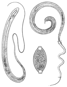
Fig. 301.—Trichuris trichiura: on the left, male; on the right, female with the anterior extremity embedded in the mucous membrane of the intestine; below, egg.
The male measures 40 to 45 mm. in length, the spicule is 2·5 mm. long, its retractile sheath is beset with spines. The female measures 45 to 50 mm. in length, of which two-fifths appertain to the posterior part of the body. The ova are barrel-shaped and have a thick brownish shell which is perforated at the poles. Each opening is closed by a light-coloured plug. The eggs measure 50 µ to 54 µ in length and 23 µ in breadth; they are deposited before segmentation. Trichuris trichiura usually lives in the cæcum of man, and is also occasionally found in the vermiform appendix and in the colon, exceptionally also in the small intestine; usually only a few specimens are present, and these do not cause any particular disturbance, although, as Askanazy found, they feed on blood; in other cases cerebral symptoms of more or less severity are observed when Trichocephali are present in large numbers. At post-mortems performed soon after death the filiform anterior extremity of the worm is frequently found embedded in the mucous membrane (Askanazy).
The whip worm is one of the most common parasites of man and appears to be distributed over the entire surface of the globe; it is, however, more frequent in the warmer regions. It is found in persons of both sexes and all ages with the exception of infants. In autopsies it is found in the following numbers: In Dresden in 2·5 per cent., in Erlangen in 11·1 per cent., in Kiel in 31·8 per cent., in Munich in 9·3 per cent., in Petrograd in 0·18 per cent., in Göttingen in 46·1 per cent., in Basle in 23·7 per cent., in Greenwich in 68 per cent., in Dublin in 89 per cent., in Paris in about 50 per cent., and in Southern Italy in almost 100 per cent. On examining the fæces the eggs of the whip worm were found as follows: In Munich in 8·26 per cent., in Kiel in 45·2 per cent., in Greifswald in 45 per cent., in North Holland in 7 per cent., in Novgorod in 26·4 per cent., in Petrograd in 5 per cent., in Moscow in 5·3 per cent.
The development of the eggs is completed in water or in moist soil, and occupies a longer or shorter time according to the season; the eggs possess great powers of resistance, as do the larvæ, which, according to Davaine, may remain as long as five years in the eggshell without losing their vitality. Leuckart proved by experiment that direct infection with Trichuris ovis (Ovis aries) and T. crenata (Sus scrofa dom.) was produced by embryo-containing eggs; Railliet obtained the same results with T. depressiuscula of dogs, and Grassi subsequently, by means of two experiments, demonstrated the direct development of Trichuris trichiura. In one case embryo-containing eggs were swallowed on June 27, 1884, and on July 24 the ova of Trichocephali were found in the fæces for the first time.
Trichuris trichiura is found not only in man, but also in various monkeys (T. palæformis, Rud.), as well as in lemurs (T. lemuris, Rud.).
Other species are T. crenata in pig; T. ovis in cattle, sheep, goat, and pig (?); T. depressiuscula in dog; T. campanula in cat; T. unguiculata in rabbit and hare; T. cameli in camel; T. discolor in humped cattle; T. nodosus in mouse; T. alcocki in the thamin (India); T. globulosa in camel; T. giraffæ in giraffe.
Sub-family. Trichinellinæ, Ransom, 1911.
Male without spicule; females ovoviviparous. Larvæ penetrate muscles of host and become encysted. Genus: Trichinella.
Genus. Trichinella, Railliet, 1895.
Syn.: Trichina, Owen, 1835 (nec Meigen, 1830).
Very small Trichinellinæ, the males of which have two conical appendages at the caudal extremity; the vulva is situated at the border of the anterior fifth of the body. There is only one species.
Trichinella spiralis, Owen, 1835.
Syn.: Trichina spiralis, Owen, 1835.
The male measures 1·4 to 1·6 mm. in length and 0·04 mm. in diameter. The anterior part of the body is narrowed, the orifice of the cloaca is terminal and lies between the two caudal appendages; internal to these are two pairs of papillæ, dorsal one behind the other. The cloaca is evertible for copulation. The females measure 3 to 4 mm. in length and 0·06 mm. in diameter; anus terminal.
Trichinella spiralis in its adult stage inhabits the small intestine of man, pig, wild boar, rat. The young do not leave the body of the host but become encysted in the muscles. Experimentally it develops in the black rat (Mus rattus), the sewer rat (M. decumanus), the domestic pig (Sus scrofa dom.), the wild boar (Sus scrofa ferox), the domestic dog (Canis familiaris), the fox (C. vulpes) the badger (Meles taxus), the polecat (Putorius fœtidus), the marten (Mustela foina), the raccoon (Procyon lotor), the hippopotamus and the cat, and many other mammals (rodents and carnivora); Trichinellæ have been artificially introduced, by administering the encysted stage, into the dog, the mole (Talpa europæa), the mouse (Mus musculus), the hare (Lepus timidus), the rabbit (L. cuniculus), the hedgehog (Erinaceus europæus), the marmot (Cricetus vulgaris), the vole, the dormouse, the sheep, the calf, the horse, etc. Human beings and the pig, rat, mouse, guinea-pig and rabbit are most easily infected; less easily the sheep, calf and horse; with difficulty the cat, dog and badger. Trichinella can also be reared in birds (fowl, pigeon and duck), but the young do not encyst in the muscular system, but are expelled with the fæces. By cold-blooded animals as well as by insects (Calliphora vomitaria), encysted Trichinellæ are evacuated without undergoing any change, but they will still develop if subsequently ingested, say, by rabbits. According to Gujon, however, Trichinella can develop in salamanders, because he has found Trichinella of the muscles in these animals after they had been fed on encysted specimens. A high temperature (30°C.) must be provided in which to keep the experimental animals to ensure the success of the infection.
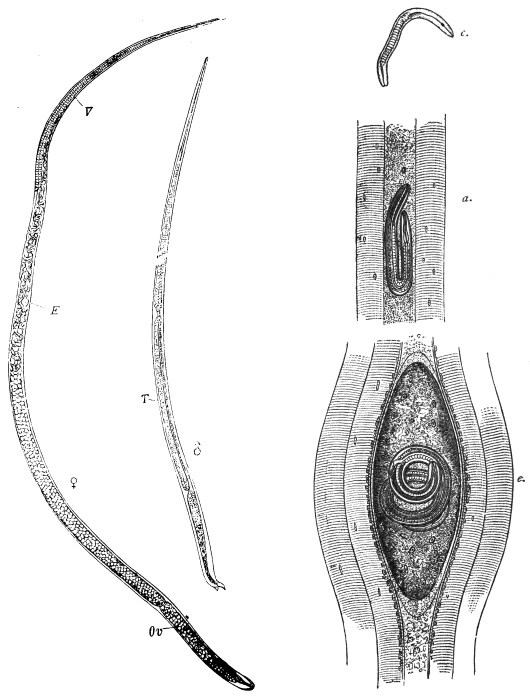
Fig. 302.—Trichinella spiralis. ♀, mature female: E, embryos; V, vulva; Ov, ovary. ♂, mature male: T, testes. c., newly born larva. d., larva in the muscles. e., encapsuled larva in the muscles. Magnified. (After Claus.)
History.—Encapsuled Trichinellæ had been observed in London by Peacock (1828) and by J. Hilton (1833) in the muscular system of man; soon after (1835), Paget found them in London in an Italian who had died of tuberculosis, and recognized them to be encysted entozoa, which R. Owen described as Trichina spiralis. Soon after, some further observations were reported on the occurrence of encysted Trichinellæ, in man, in England, Berlin, Heidelberg, Denmark, North America; they were also found in the pig (Leidy, Philadelphia) and the cat (Herbst, Göttingen, and Gurlt, Berlin). Herbst even succeeded in infecting a badger with encysted Trichinellæ, and subsequently infected two dogs with the flesh of this badger (1850). In 1855 R. Leuckart (Giessen) also commenced feeding experiments, and, like Küchenmeister and Virchow (1859), first went on the wrong track because it was believed at that time that Trichinellæ were the larvæ either of Trichocephalus or Strongylus. Nevertheless, these experiments yielded some important results; they showed that Trichinellæ become adult in the intestine within a few days, and that the females are viviparous (Leuckart). Until that time Trichinellæ had been regarded as fairly harmless guests of man, but opinions soon changed when Zenker in Dresden (January, 1860), in performing the autopsy of a girl, aged 10, who had entered the hospital with typhoid symptoms and there died, found Trichinellæ (not yet encysted) in the muscles; the intestinal lesions characteristic of typhoid were lacking, but numerous adult Trichinellæ were found in the intestine. Inquiries elicited the fact that at about Christmas time the girl had been taken ill after eating pork, and at the same time the butcher from whom the meat was bought as well as several of his customers fell sick: the pickled pieces of the same meat were full of Trichinellæ. In the face of this information it was not difficult to ascertain the cause of the disease and the manner of infection in Zenker’s case, and it was not long before Leuckart, Virchow and Zenker were able by renewed experiments to demonstrate the cycle of development of Trichinella spiralis. Similar investigations followed by Claus in Würzburg, Davaine in Paris, Fuchs and Pagenstecher in Heidelberg, etc.
Hardly had Zenker’s case been published than numerous observations on trichinosis in man appeared, some referring to isolated cases, others to small or great epidemics, and nearly all from North Germany. The worst epidemic was that of Hadersleben (1865), in which place, numbering hardly 2,000 inhabitants, 337 persons were taken ill within a short time, and of these 101 died. The source of infection proved to be a single pig, the flesh of which had been mixed with that of three other pigs; 200 of the badly infected persons had exclusively eaten raw pork.
Moreover, it soon became clear that epidemics of trichinosis had been observed in Germany prior to 1860, but that their nature had not been recognized, although in a few cases Trichinellæ had been found in the muscles of those who had succumbed.
History of the Development of Trichinella spiralis.
Shortly after their introduction into the intestine of experimental animals the encysted Trichinellæ escape from their capsules, which are destroyed by the gastric juices, and they then enter the duodenum and jejunum, where they become adult. During this period they do not grow much, the males from 0·8 to 1·0 to 1·2 to 1·5 mm.; the females to 1·5 to 1·8 mm. Soon after copulation, which takes place in the course of two days, the males die; the females, which during the following days attain a length of 3 to 3·5 mm., either bore more or less deeply into the villi or, by means of Lieberkühn’s glands, into the mucous membrane (Askanazy, Cerfontaine, Geisse), and thus usually attain the lymph spaces. A few also pierce the intestinal wall and are then found in the mesentery and glands. The females deposit their young, the number of which, according to Leuckart, averages at least 1,500, in the lymph spaces; the newly born larvæ measure 90 µ to 100 µ in length, 6 µ in diameter, and they do not appear to increase in size during their migrations. The migrations are mostly passive, that is to say, the larvæ are carried along mainly by the lymph stream to the heart, but sometimes they are active, as may be inferred from the fact that young Trichinellæ are found in various parts of the intestinal wall beyond the chyle and lymph spaces, as well as in abundance in the abdominal cavity. Trichinellæ occur in the heart’s blood of artificially infected animals seven to twenty-three days after infection. If scanty, dilute the blood with about ten times the amount of 3 per cent. acetic acid and centrifugalize.
The young brood is distributed from the heart throughout the entire body, but the conditions necessary to its further development are found only in striated muscle; the young Nematodes penetrate the capillaries, attain the intramuscular connective tissue and then invade the fibres (Virchow, Leuckart, Graham304). On the ninth or tenth day after infection the first Trichinellæ have reached their destination; but further invasions are constantly taking place because the intestinal Trichinellæ live from five to seven weeks, and continue to produce their young.
Symptoms.—(1) Period of invasion: Gastro-intestinal symptoms—nausea, vomiting, watery diarrhœa, colic. Muscular pains may occur even at this period. Recurrent abdominal pains about the eighth day, a temporary œdema. Embryos are abundant in the serous cavities.
(2) Period of dissemination: Second week. Myositis, variable in amount, is the predominant symptom. The biceps and calf may be hard and tender. Mastication, speech, respiration, etc., may be difficult and painful. Dyspnœa may be intense. Temperature 104° to 105° F.
(3) Period of encystment: Symptoms of marked cachexia. Third week: Second period of œdema, especially of face. Delirium, somnolence, lung affections. Death or gradual subsidence of symptoms in mild cases.
Eosinophilia (50 per cent. or more) is present.
In consequence of the new batches of young produced during several weeks, the above-mentioned symptoms of disease are often considerably aggravated; the fever increases, delirium may arise, and infiltration of the lungs, fatty degeneration of the liver and inflammation of the kidneys may ensue; the initial slight œdema may extend, the strength dwindles, and in many cases the patients succumb to the trichinosis. In severe cases improvement of the condition is only apt to occur in the fourth or fifth week; the convalescence is always protracted.
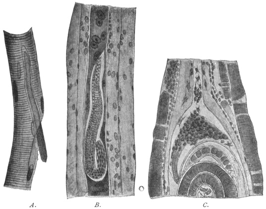
Fig. 303.—A., isolated muscular fibre of a rat, invaded by Trichinella. 510/1. B., section through the muscle of a rat; the infected fibre has lost its transverse striation; its nuclei are enlarged and multiplied. 310/1. C., portion of a Trichinella capsule, at the pole of which connective tissue cells are penetrating the thickened sarcolemma. (After Hertwig-Graham.)
The muscular fibres attacked degenerate, the transverse striation at first disappearing; the fibres then assume a granular appearance, the nuclei multiply and become enlarged, and are surrounded by an area of granular material, which stains more deeply than the remaining contents of the sarcolemma. Two or three weeks after infection, the spirally rolled-up Trichinellæ have grown to 0·8 to 1·0 mm., and in their vicinity the muscular fibre is swollen, spindle-shaped, and the sarcolemma is glassy and thickened. The inflammation also extends to contiguous fibres, especially to the intramuscular tissue, which proliferates greatly, especially in the vicinity of the degenerated fibres. While the latter become more and more absorbed, the capsule is formed by the inflamed connective tissue, which, penetrating into the glassy and thickened sarcolemma from the poles of the spindle, forms the cystic membrane. According to other authorities, the larvæ settle in the intermuscular connective tissue which forms the cyst and not in the muscular fibres within the sarcolemma. The cysts are lemon-shaped and usually lie with their longitudinal axis in the direction of the muscular fibres; on an average they measure 400 µ in length by 250 µ in breadth.
Later on fat cells appear at their poles, and after about six or nine months they commence to calcify, the process starting at the poles (fig. 305). Finally, sometimes after the lapse of years, the captive Trichinellæ themselves become calcified.
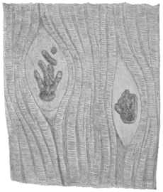
Fig. 304.—Calcified Trichinella in the muscular system of a pig; the capsules are not calcified. (After Ostertag.)
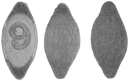
Fig. 305.—Various phases of the calcification of Trichinella of the muscles, which starts at the poles of the capsule.
According to experience, Trichinellæ are not evenly distributed in the muscular system of pigs; the diaphragm, the muscles of the larynx, tongue, abdomen and intercostal spaces are their favourite positions; this predilection for the respiratory muscles is explained by their regular contractions, owing to which regular narrowings of the capillaries take place, thus favouring the settling of the circulating Trichinellæ. The same circumstance probably explains the frequency of the parasites in the tongue.
Possibly also the Trichinellæ that bore direct through the intestine may, from the abdominal cavity, penetrate the muscles in the vicinity. Frequently also encysted Trichinellæ are found in remarkable numbers in the vicinity of the points of insertion of the tendons, this proclivity being probably connected with the fact that the Trichinellæ first of all wander into the muscular fibres and find a natural barrier at the points of insertion of the tendons.
The Trichinellæ, in their encysted condition, may remain alive and capable of development for many years—in the pig eleven years and in man as much as twenty-five to thirty-one years. Encystment, however, is not a necessary condition for the development of the brood, that is to say, Trichinellæ which reach the gut of suitable animals become sexually mature and multiply provided that they have developed so far as to possess a rudimentary genital spot, which occurs when the body is 0·5 to 0·75 mm. long, but all the same a great part of non-encapsuled Trichinæ perish on their passage through the stomach.
The black rat (Mus rattus), and more particularly the sewer rat (Mus decumanus305), are the normal hosts of Trichinella spiralis. These animals, especially the last-named species, infect themselves very easily, as they are cannibalistic, and they also transmit trichinosis to other species by which they are devoured, such as pigs, dogs, cats, foxes, bears and martens. Rats are infected also by the ingestion of fæcal matter from infected animals which contains trichinæ (Höyberg). Man becomes infected with Trichinella by eating the flesh, insufficiently cooked, of infected pigs, also, but more rarely, by eating the infected flesh of wild boars, dogs, cats, bears and foxes.
The infection of pigs may likewise take place by their having access to the offal of trichinous pigs, or being actually fed on it. These are, however, exceptions, which, as a matter of course, are of great importance in certain places. As a matter of fact, the rats examined for Trichinella were always found to be severely infected. Thus Billings, in the knackers’ yard at Boston, found that 76 per cent. of the rats were infected, and in an export slaughterhouse 100 per cent. were found to harbour the parasite; in the city of Boston 10 per cent. of the rats had trichinosis. Heller found that of 704 rats, from twenty-nine different places in Saxony, Bavaria, Würtemberg and Austria, 8·3 per cent. were infected with Trichinellæ; of the rats caught in the knackers’ yards, 22·1 were diseased; of those taken in slaughterhouses, 2·3 were infected, and of rats from other localities only 0·3 per cent. harboured the parasite. Leisering found almost the same figures, but in rats from slaughterhouses 5·3 per cent. were infected.
The geographical distribution of T. spiralis does not correspond with the occurrence of trichinosis in man; local customs are an important factor; for instance, the custom of eating pork in a condition that does not affect the life of the enclosed trichinella. In places where such customs do not prevail, epidemics do not occur—at the most there are isolated cases of the disease, although there be a great number of infected pigs. The following conditions prevail in North America: In Boston, Billings found that 4 to 5·7 per cent. of the pigs examined were trichinous; Belfield and Atwood found that 8 per cent. were infected in Chicago; Salmon found on an average that 2·7 per cent. were infected (but at various places the percentage fluctuated between 0·28 to 16·3 per cent.), yet epidemics of trichinosis hardly ever occur in North America, and only isolated cases of the disease are met with in German immigrants, who keep to their native customs.
This report, according to the researches of H. U. Williams, must be considerably modified. This author has examined the muscular system of human cadavers according to the method employed by inspectors of meat for pigs. The investigations were conducted in the Pathological Institute of the University of Buffalo, and the observer has examined 505 bodies since 1894, of which 27 (= 5·34 per cent.) were invaded by Trichinella. The cases, according to the nationality, are divided as follows:—
ExaminedTrichinellaPercentage of positive results
AbsentPresent
Americans:
(a) Whites20720162·89
(b) Negroes706557·14
British and Irish625758·06
Canadians1210216·66
Germans4943612·24
Italians1210216·66
Other nationalities272700
Nationality unknown666511·51
Total50547827 5·34
It is worthy of remark that half of all the positive cases were mental patients, who were found to be affected with Trichinella to well-nigh 12 per cent. Trichinosis was not, however, the cause of death in any case. Very frequently the Trichinellæ were found calcified and dead.
Conditions are similar in most countries of Europe, where, of course, the number of infected pigs is considerably smaller, but the disease depends less on this than on the way in which the pork is prepared.
Cases of trichinosis have been known to occur in nearly all the countries of Europe; further, in Egypt, Algeria, East Africa, Syria, India, Australia, and America. North Germany, more especially the Saxe-Thüringian states, is the classical land for epidemics of trichinosis; the mortality varies, but it may be very high.306
Prophylaxis.—The grave nature of the disease and the comparatively high mortality relating to trichinosis led the authorities to adopt certain preventive measures, which are the more necessary as national customs cannot be altered in a short time. As the usual process of pickling and smoking, even when long continued, does not certainly ensure the death of the Trichinellæ contained in the meat, and also because in roasting and boiling large pieces of pork a considerable time is necessary to permit the temperature required to kill off the parasites (62° to 70° C.) to penetrate to the middle of the joint, it appeared to be most practical to have all pigs microscopically examined for Trichinellæ before they, or parts of them, were placed on the market, and all infected meat condemned, no matter whether the Trichinellæ were present in large or small numbers, still undeveloped or calcified. Since 1877 obligatory examination of pork has been introduced in Prussia, though as yet it is not thoroughly carried out; other states of North Germany as well as the larger towns of South Germany soon followed; a complete army of trichina inspectors, officially examined and periodically controlled by experts, and whose number in Prussia amounted to 27,602 in 1896, this being even increased to 28,224 in 1899, have the charge of examining pork on certain lines laid down. These are at the present time uniformly administered. The proceeding is usually that the trichina inspector himself goes to the slaughterhouses, or special samplers take pieces of the muscles that are known to be the favourite seats of the parasite (pillars of the diaphragm, the costal part of the diaphragm, muscles of the tongue and larynx, intercostal and abdominal muscles); six small portions are separated from each piece, pressed between slides or special compressors, and carefully gone through by examining them with a low power of the microscope. The pigs free from Trichinellæ are passed for commerce; trichinous pigs, on the other hand, in Prussia, are only allowed to be used for industrial purposes, i.e., the hide and bristles are used, the fat is allowed to be melted down, or certain parts are used for the manufacture of soap or glue. In Saxony, however, it is still permitted to place trichinous flesh on the market, fully declaring its nature, and after having been heated to its deepest strata at a temperature of 100° C. in a suitable apparatus, and under the supervision of a veterinary surgeon.
As to the proportion of trichinous pigs to healthy ones, the following tables give the figures for Prussia:—
YearNumber of pigs examinedNumber of trichinous pigsProportion
18782,524,1051,2221 : 2,065
18793,164,6561,9381 : 1,632
18813,118,7801,6951 : 1,839
18823,808,1421,8521 : 2,056
18834,248,7672,1991 : 1,932
18844,611,6892,6241 : 1,741
18854,421,2082,3871 : 1,852
18864,834,8982,1141 : 2,287
18875,486,4162,7761 : 1,988
18886,051,2493,1111 : 1,945
18895,500,6783,0261 : 1,818
18905,590,5101,7561 : 3,183
18916,550,1822,1871 : 2,996
18926,234,5592,0851 : 2,992
18968,759,4901,8771 : 4,666
18999,230,3531,0211 : 9,040
19029,093,210 7251 : 12,397
The proportion, however, is not only subject to variation in separate years, but differs according to the district; thus, in 1884, in the state district of Minden there was one trichinous pig to 30,146 healthy animals, in Erfurt 1 to 14,563, in the district of Gnesen 1 to 101, in Schrimm 1 to 86, and in Schroda 1 to 68.
In Germany Trichinella is becoming LESS COMMON in pigs (Ostertag):—
(a) Prussia.
YearPigs found to be trichinous
1878–18850·061–0·048 per cent.
1886–18920·033–0·043"
18960·021"
18990·014"
19020·011"
(b) Saxony.
YearNumber of pigs found to be trichinous
18910·014 per cent.
18920·011"
18930·008"
18940·007"
18950·012"
18960·0102"
18990·004"
19020·0056"
(c) City of Berlin.
YearNumber of pigs found to be trichinous
1883–18930·035–0·064 per cent.
1893–18970·022–0·015"
19020·006"
There is no doubt that the excellent preventive measure of official inspection for Trichinella has led to the avoidance of grave disasters; its introduction has not yet caused an entire cessation of trichinosis in man, because inspection of pork is not obligatory everywhere, so that human beings may become infected by unexamined trichinous pigs from their own country or from abroad, and also because an infection may occasionally escape notice. For these reasons the meat imported into Berlin from abroad as free from Trichinæ is examined again and not always in vain; finally, also, gross negligence may at times occur, or fatal errors may be made.
In addition private prophylaxis must not be neglected, and its chief aim should be directed to the suitable preparation of pork.
Family. Dioctophymidæ.
Genus. Dioctophyme, Collet-Megret, 1802.
Syn.: Eustrongylus, Dies., 1851.
Large worms. Anterior extremity unarmed; the mouth is surrounded by six papillæ. One ovary. The vulva is in the anterior region of the body.
Dioctophyme gigas, Rudolphi, 1802.
Syn.: Dioctophyme renale, Goeze, 1782; Ascaris canis et martis, Schrank, 1788; Ascaris visceralis et renalis, Gmelin, 1789; Strongylus gigas, Rud., 1802; Eustrongylus gigas, Dies., 1851; Strongylus renalis, Moq. Tand., 1860; Eustrongylus visceralis, Raill., 1885.
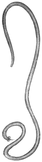
Fig. 306.—Dioctophyme gigas, male. Natural size. (After Railliet.)
Colour blood-red; the anterior extremity somewhat slender; there is a series of about 150 papillæ along the lateral lines; the sub-median lines are strongly developed, and from them spring the radial muscles for the intestine.
The males attain a length of 40 cm. and a diameter of 4 to 6 mm.; the posterior extremity is transversely truncated; the anal orifice is within the base of the collar-like bursa, the thickened edges of which are beset with papillæ; the spicule measures 5 to 6 mm. in length.
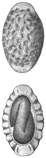
Fig. 307.—Eggs of Dioctophyme gigas; above seen from the flat, below in optical section. 400/1. (After Railliet.)
The females attain a length of 100 cm. and a breadth of 12 mm. The anus is crescent-shaped and terminal. The vulva is 50 to 70 mm. distant from the anterior extremity. The eggs are oval and have a thick shell presenting numerous depressions; the shell itself is brownish, but it is colourless at the somewhat thickened poles; it measures 60 µ in length by 40 µ in breadth. The larva measures 240 µ by 14 µ.
Dioctophyme gigas lives in the pelvis of the kidney, more rarely in the abdominal cavity of the seal, otter, dog, wolf, fox, horse, marten and polecat, exceptionally also in human beings. It also occurs in tumours of the mamma and perinæum. Most of the cases in which this parasite has been reported as occurring in man may be traced back to unrecognized Ascaris lumbricoides or to clots of fibrin; seven certain cases, eight more or less doubtful, however, remain.
The source of infection is unknown, but according to Balbiani the eggs develop an embryo in water or moist soil, and this embryo may remain alive several years without hatching; the infection of dogs with embryo-containing eggs did not succeed; an intermediate stage in fishes is conjectured, but still the infection of cattle and horses is unintelligible.
Family. Strongylidæ.
Sub-family. Metastrongylinæ, Leiper, 1908.
Buccal capsule absent or slightly developed, vagina elongate, uteri convergent307 and have a simple musculature. Parasitic in the respiratory or circulatory system. Genera: Metastrongylus, Synthetocaulus.
Genus. Metastrongylus, Molin, 1861.
Mouth with six lips, of which the two lateral are the largest. Postero- and postero-external rays308 of bursa thin, the rest thick. Only the median ray double. Spicules very long and slender, striated. Vulva immediately in front of anus. Eggs contain an embryo when laid.
Metastrongylus apri, Gmelin, 1789.
Syn.: Gordius pulmonalis apri, Ebel, 1777; Ascaris apri, Gmelin, 1789; Strongylus suis, Rud., 1809; Strongylus paradoxus, Mehlis, 1831; Strongylus elongatus, Duj., 1845; Strongylus longevaginatus, Dies., 1851.
The male measures 12 to 25 mm. in length; the bursa is bilobed; there are five rays in each lobe; the spicules are thin and up to 4 mm. in length. The females measure 20 to 50 mm. in length, the anus is close in front of the posterior extremity, which has a recurved, hook-like process; the vulva is close in front of the anus. The eggs are elliptical, 57 µ to 100 µ in length, 39 µ to 72 µ in breadth; when the eggs are deposited the embryo is already formed, 220 µ to 350 µ by 10 µ to 12 µ.
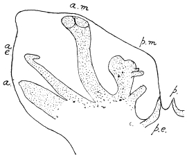
Fig. 308.—Metastrongylus apri: one side of bursa. a., anterior; a.e., antero-external; a.m., antero-median; p.m., postero-median; p.e., postero-external; p., one division of posterior ray. (Stephens.)
Metastrongylus apri frequently lives in the bronchial tubes—usually the smaller ones—of the pig309 and wild boar; it is also found occasionally in sheep and in man; in young pigs it is apt to set up a bronchitis, which frequently causes death.
The first communication as to the occurrence of this species in man was that of Diesing, who, in 1845, in Klausenburg, had the opportunity of examining Strongylidæ found by Jortsits in the lung of a little boy, aged 6, in Transylvania; probably also the Nematodes found in the trachea and larynx of man, and described by Rainey and Bristowe as specimens of Filaria trachealis, belong to this group; according to Chatin, Metastrongylus apri may also occur in the intestine of man; this occurrence, however, may in all probability have been due to an accidental introduction of adult worms into the intestine, and should not be attributed to an infection by the larval stage.
No experiments to induce infection have been made; it is probable, however, that infection is direct and without the aid of an intermediate host.
Sub-family. Trichostrongylinæ, Leiper, 1908.
Strongylidæ with buccal capsule absent or slightly developed, vagina short, uteri divergent (i.e., anterior and posterior), ovejectors differentiated. Parasitic in the alimentary canal. Contains the genera Trichostrongylus, Hæmonchus, Ostertagia, Nematodirus, Cooperia, Dictyocaulus.310
Genus. Trichostrongylus, Looss, 1905.
Very small Strongylidæ. Mouth with three small lips and nodular or punctiform papillæ. Cervical papillæ absent. Bursa entirely closed, with large lateral lobes, and median lobe not distinctly defined. Anterior311 rays double, the branches widely divergent, one thin, the other thick, and close to the antero-median. The postero-median ray is thin and close to the postero-external. Posterior ray bifurcate, each branch bifid at the tip (fig. 311). Spicules short, spoon or spatula-like, with on the broad anterior end a lateral knob or disc and in front of the point an angular projection. Gubernaculum of a peculiar canoe or shoe shape in profile. Vulva in the hinder half of the body. Tail with two minute papillæ just in front of tip. Eggs thin shelled; when laid they show eight to thirty-two segments. Parasitic in duodenum, seldom in the stomach of herbivora.
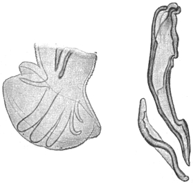
Fig. 309.—Trichostrongylus instabilis: left, posterior end of male; right, spicule and gubernaculum, side view. Cf. fig. 311. Magnified. (After Looss.)
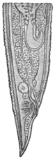
Fig. 310.—Trichostrongylus instabilis: posterior end of female. Magnified. (After Looss.)
Trichostrongylus instabilis,312 Railliet, 1893.
Syn.: Strongylus instabilis, Railliet, 1893; Strongylus subtilis, Looss, 1895.
Male 4 to 5·5 mm. long, 0·08 mm. thick in front of bursa. Spicule 0·135 to 0·145 mm. long, accessory piece (gubernaculum) 0·07 mm. thick. Antero-external ray usually thickest of all, occasionally only as thick as the antero-median; postero-median far more slender than the antero-external and antero-median and nearer to the postero-external than to the antero-median. Female 5 to 6 mm. long, vulva 1·05 to 1·2 mm. distant from the tip of the tail, placed longitudinally, 50 µ to 55 µ long, always shorter than the unpaired portion of the canal formed by the union of the two ovejectors; anus 0·055 to 0·07 mm. distant from tip of the tail; ova 73 µ to 80 µ by 40 µ to 43 µ.
This species lives in the duodenum, exceptionally also in the stomach of Ovis aries, O. laticauda, Antilope dorcas, Camelus dromedarius (Egypt), Cynocephalus hamadryas (North Africa), sheep and goats (France), and has been found by Looss in bodies of fellaheen at Alexandria and in the stomach of a Japanese female by Ijima.
Trichostrongylus probolurus, Railliet, 1896.
Syn.: Strongylus probolurus, Railliet, 1896.
Male 4·5 to 5·5 mm. long, in front of bursa 0·08 mm. thick; spicule 0·126 to 0·134 mm. long, gubernaculum 0·075 to 0·08 mm. long. Bursa: latero-anterior rib thickest; antero-external thicker than antero-median, postero-median and postero-external very short and close together. Female 4·5 to 6 mm. long, vulval opening 1·08 to 1·25 mm. from tip of tail, placed longitudinally, and slightly curved, 76 µ long, always longer than the unpaired portion of the ovejector; anus 0·040 to 0·05 mm. distant from tip of tail. Posterior end thick, point of tail short. Ova 76 µ to 80 µ by 43 µ to 46 µ.
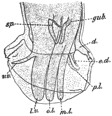
Fig. 311.—Trichostrongylus probolurus: tail of male from left side. d., posterior; e.d., postero-external; p.l., postero-median; m.l., antero-median; e.l., antero-external; l.v., latero-anterior; v.v., antero-anterior; gub., portion of gubernaculum; sp., portion of spicules. × c. 300. (After Looss.)
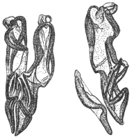
Fig. 312.—Trichostrongylus probolurus: spicules and gubernaculum of male; on left, ventral view; on right, lateral view. × c. 300. (After Looss.)
Habitat.—In the duodenum of Ovis aries, O. laticauda, Antilope dorcas, Camelus dromedarius (Egypt) and occasionally also in man (Egypt).
Trichostrongylus vitrinus, Looss, 1905.
Male 4 to 5·5 mm. long, in front of bursa 0·085 mm. thick. Bursa larger than in the other two species, antero-external rib thickest, antero-anterior and postero-median equally thick, straight. Spicule 0·16 to 0·17 mm. long, gubernaculum 0·085 to 0·095 mm. long. Female 5 to 6·5 mm. long, vulval opening 1·15 to 1·25 mm. distant from tip of tail, crescent shaped, oblique to body axis, and around it irregular thickenings. Ova 84 µ to 90 µ by 46 µ to 50 µ.
In duodenum of Ovis aries, O. laticauda, occasionally in Camelus dromedarius and in man (Egypt).
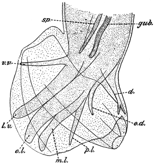
Fig. 313.—Trichostrongylus vitrinus: tail of male from left side. d., posterior; e.d., postero-external; p.l., postero-median; m.l., antero-median; e.l., antero-external; l.v., latero-anterior; v.v., antero-anterior; gub., portion of gubernaculum; sp., portion of spicule. × c. 300. (After Looss.)
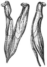
Fig. 314.—Trichostrongylus vitrinus: spicules and gubernaculum; on left, ventral view; on right, lateral view. × c. 300. (After Looss.)
Genus. Hæmonchus, Cobb., 1898.
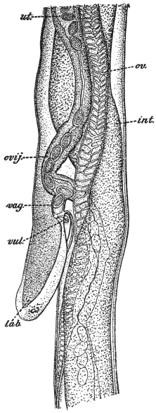
Fig. 315.—Hæmonchus contortus: vulval region of female viewed from left side. int., intestine; lab., linguiform process covering vulva; ov., ovary; ovij., ovejector; ut., uterus; vag., vagina; vul., vulva. × 75. (After Ransom.)
Small mouth cavity contains a “tooth” or “lancet” arising from the dorsal side. Cuticle of head and neck not inflated. Cervical papillæ well marked. Bursa bilateral, with large lateral lobes and a small dorsal lobe not median, but lateral, attached to the base of one of the lateral lobes (fig. 316). Posterior ray bifurcate, each branch bifid apically. Each lateral lobe six rays. Anterior rays separated distally, curving forward. Antero-median and postero-median rays distally curve away from the antero-external. Postero-external ray long and slender. Spicules less than 1 mm. Gubernaculum present. Vulva in posterior part of body covered by a prominent tongue-like flap. Eggs ellipsoidal.
Hæmonchus contortus, Rudolphi, 1803; Cobb., 1898.
Dorsal “tooth” or “lancet” 10 µ to 15 µ long. Cervical papillæ 0·3 mm. from head.
Male 20 mm. long by 400 µ thick (maximum). Asymmetrical lobe of bursa 150 µ by 125 µ attached to left lateral lobe. Posterior ray bifurcate; each branch bifid. Stem of ray less than twice as long as its branches. Spicules 300 µ to 500 µ with knobbed tips, and the left spicule with a barb 20 µ from the tip, right spicule with a barb 40 µ from tip. Gubernaculum 200 µ by 25 µ to 35 µ, fusiform with thickened edges.
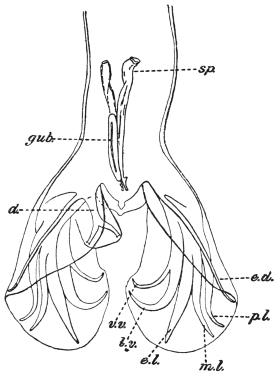
Fig. 316.—Hæmonchus contortus: tail of male, dorsal view, d., posterior ray of the asymmetrically placed posterior lobe; e.d., postero-external; p.l., postero-median; m.l., antero-median; e.l., antero-external; l.v., latero-anterior; v.v., antero-anterior; gub., gubernaculum; sp., spicule. × 75. (After Ransom.)
Female 18 to 30 mm. by 500 µ (maximum). Vulva 3 to 4·5 mm. from tip. Linguiform flap 0·5 mm. (a second one exists, according to Brumpt). Anus 400 µ to 630 µ from tip. Tail acutely pointed. Eggs 75 µ to 95 µ by 40 µ to 50 µ.
Habitat.—Fourth stomach of cattle, sheep, antelope.
Distribution.—Europe, America, Africa, Asia, Australia, New Zealand. Once in man in South America by de Magalhães.
Pathology.—Produces anæmia, emaciation, dropsy in sheep; and in the human case the symptoms were mistaken for those of ancylostomiasis.
Life-history.—Rhabditic embryos easily hatch in water, then moult several times, becoming eventually “filariform” larvæ enclosed in the moulted skin. These crawl up blades of grass and are swallowed by sheep, etc.
Genus. Nematodirus, Ransom, 1907, emend. Railliet, 1912.
Head over 50 µ in diameter. Cuticle may be slightly inflated and often transversely striated. Cuticle with eighteen distinct longitudinal ridges. Cervical papillæ absent (?). Posterior lobe of bursa reduced to short lobules each with a dorsal ray. Antero-anterior + latero-anterior (= anterior double) rays close together, parallel; antero-external ray diverges widely from antero- and postero-median, which are close together and parallel. Postero-external ray slender. Spicules more than 0·5 mm. long, at most one-twelfth of body, united by a membrane throughout their length or only distally. Gubernaculum absent. Vulva behind middle of body. Eggs ellipsoidal, shell rather thick.
Habitat.—Duodenum of ruminants.
Sub-genus. Mecistocirrus, Railliet, 1912.
Head slightly inflated, with transverse striations. Skin with eighteen longitudinal ridges, but little apparent; cervical papillæ distinct. Bursa bilobed; median ray double (= postero-median + antero-median); very large antero-external at the edge, close to the anterior. Spicules very long, slender, one-sixth length of body (3·5 mm.); tail pointed. Vulva immediately in front of anus.
Habitat.—Stomach of ruminants.
Mecistocirrus fordi, Daniels, 1908.
Syn.: Strongylus fordi, Daniels, 1908; Strongylus gibsoni, Stephens, 1909; Nematodirus fordi, Leiper, 1911.
Male 21 mm. long by 0·4 mm. thick. Cervical papillæ 0·45 mm. behind the head. Spicules about 7 mm. long, i.e., one-third of the body length. At the level of the postero-external rays of the bursa, the bursa has a projecting lobule.
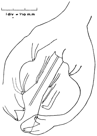
Fig. 317.—Mecistocirrus fordi: bursa of male, dorsal view. The rays are (1) postero-external, (2) median (= postero-median + antero-median), (3) antero-external, (4) latero-anterior, (5) antero-anterior. These two latter are parallel. The posterior ray is absent. (After Stephens.)
Female 25 mm. long. Anus 0·2 mm., vulva 0·5 mm. from the tip of tail. Eggs 100 µ by 53 µ.
Sub-family. Ancylostominæ, Railliet, 1909.
Strongylidæ with buccal capsule, well developed. Uteri divergent. Parasitic in the alimentary canal, exceptionally in the respiratory system.
Group. Œsophagostomeæ, Railliet and Henry, 1909.
Bursa with anterior and median ray cleft (not double), postero- and postero-external arising from a common trunk, posterior bifurcated, each limb bidigitate.
Contains at present four genera: (1) Ternidens, (2) Chabertia, (3) Œsophagostomum, (4) Agriostomum.
Genus. Ternidens, Railliet, 1909.
Buccal capsule sub-globular, opening obliquely in the dorsal surface, and having at the bottom three complex teeth resembling those of Triodontophorus.313 Two crowns of leaflets; peristomic collar moderate, edge of bursa slightly toothed.
Type.—T. deminutus, Railliet and Henry.
Ternidens deminutus, Raill. and Henry, 1905.
Syn.: Triodontophorus deminutus, Raill. and Henry, 1905.
Body relatively thick. Cervical papillæ 0·5 mm. behind the head. Buccal capsule 40 µ deep. Teeth 40 µ long.
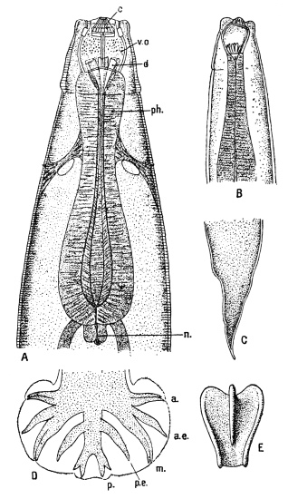
Fig. 318.—Ternidens deminutus. A, head end, ventral view: c, crown of leaflets; v.o., buccal cavity; d, pharyngeal plates; ph., pharynx; n., valve. B, lateral view. C, tail of female. D, bursa of male: a., anterior ray; a.e., antero-external; m., median; p.e., postero-external; p., posterior. E, pharyngeal plate. Enlarged. (After Railliet and Henry.)
Male 9·5 mm. long by 560 µ thick. Œsophagus 660 µ long. Bursa broader than long, the lateral lobes united by a small posterior lobe with slightly sinuous margin; edge of bursa finely toothed. Spicules about 900 µ long.
Female 12 to 16 mm. long by 650 µ to 730 µ thick. Œsophagus 860 µ long. Vulva forms a distinct projection 480 µ from tip of tail. Anus 240 µ to 270 µ from tip. Eggs 60 µ to 80 µ by 40 µ.
Habitat.—Large intestine of a negro (Comoro Islands) and in the natives of Nyasaland and Portuguese East Africa. Also in large intestine of Macacus sinensis and Macacus cynomolgus.
Genus. Œsophagostomum, Molin, 1861.
No teeth. Cuticle around the mouth dilated to form a narrow cuticular “peristomic collar.” Separated by a constriction from this is a much more extensive inflation, the “cephalic vesicle,” bounded abruptly behind on the ventral side by a transverse groove, the “ventral cleft,” which is always present even in absence of the vesicle. Buccal cavity of slight depth with a short dorsal tunnel. Internal margin of the mouth armed with chitinous leaflets (“external crown”); internal border of the buccal capsule armed with short tongue-like leaflets (internal crown). Lateral membranous wings may extend backwards from the ventral cleft. Cervical papillæ present. Bursa with two lateral lobes united by a smaller median lobe. Spicules over 5 mm. long, slender; gubernaculum inconspicuous. Vulva in front of anus. Adults usually in large intestine of ruminants, suidæ, tapirs, edentates and apes. Larvæ sometimes in nodules in intestinal wall.
Œsophagostomum brumpti, Railliet and Henry, 1905.
Female immature, 8·5 to 10·2 mm. long, 0·295 to 0·325 mm. thick. Cuticle transversely striated. The cephalic vesicle immediately behind the vestibulum oris, embracing the anterior two-fifths of the œsophagus, extending ventrally, however, towards its posterior end. Vestibulum oris formed by a cuticular band provided with a crown of twelve apical leaflets directed forward and inwards; six cephalic papillæ (two lateral, four submedian); buccal capsule in front of cervical swelling not delineated circularly behind, but provided with three wide incisions (one dorsal, two sub-ventral). Œsophagus, 0·470 to 0·500 mm. long, two cervical papillæ at five-eighths of its length. Vulva 0·350 to 0·475 mm., anus 0·170 to 0·200 mm., before tip of tail.
Habitat.—Found by Brumpt in tumours of the cæcum and colon of a native of the River Omo (Lake Rudolph), East Africa. Immature forms only were present. Adults have been found in similar tumours in monkeys.
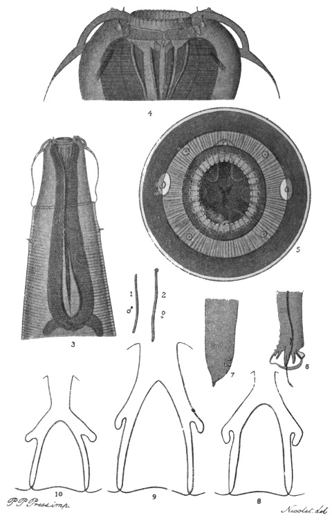
Fig. 319.—Œsophagostomum stephanostomum var. thomasi. 1, male, natural size; 2, female, natural size; 3, head of female, ventral view, showing cephalic vesicle and ventral cleft limiting it behind, × 55; 4, head of female, dorsal view, × 225; 5, head of male, end view, showing external and internal leaf crowns, × 225; 6, tail of male, lateral view (cf. fig. 318, D), × 20; 7, tail of female, lateral view, × 20; 8, Œs. thomasi, posterior ray of bursa, × 150; 9, Œs. dentigerum, from chimpanzee, posterior ray of bursa, × 150; 10, Œs. stephanostomum, from gorilla, posterior ray of bursa, × 150.
Pathology.—They occur in hæmorrhagic cysts in the submucosa or muscularis mucosæ of the gut wall. The cysts project internally and externally, and contain immature adults, which eventually escape into the lumen of the gut.
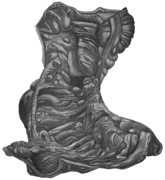
Fig. 320.—Œsophagostomum stephanostomum var. thomasi: cæcum and ascending colon. Subperitoneal cysts are seen on the top right hand, and in the lumen of the gut numerous cysts arranged transversely. The small roundish patches are areas of necrosis in the cyst walls. (After Thomas.)
Œsophagostomum stephanostomum var. thomasi, Raill. and Henry, 1909.
Body thick, pointed only at the ends. Buccal capsule much reduced. External crown of thirty-eight leaflets (the “crown” nearest the centre of fig. 319, 5). Male 17 to 22 mm. long by 750 µ thick. Spicule 1·380 to 1·475 mm., slightly curved at the tip. Female, immature, 16 to 20 mm. long by 900 µ thick, tail ending in a little conical appendage. Anus 230 µ, vulva 500 µ to 525 µ from tip. Ovejectors close together. Uteri very short in form of oblong pouch.
Œs. stephanostomum, Stossich, 1904, in the large intestine of gorilla. Œs. stephanostomum var. dentigera, Raill. and Henry, 1909, in the chimpanzee.
Habitat.—In large and small intestine of man, Brazil.
Pathology.—Nodules occur in the gut wall; 187 were found by Thomas in his, the sole case. The tumours contain each a single worm.
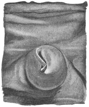
Fig. 321.—Œsophagostomum stephanostomum var. thomasi: portion of the ileum, showing a cyst with protruding worm. × 8. (After Thomas.)
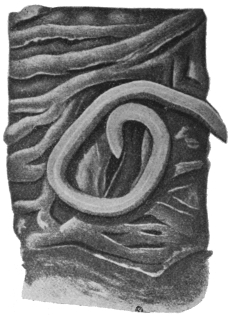
Fig. 322.—Œsophagostomum stephanostomum var. thomasi: colon with œsophagostome withdrawn from its cyst cavity. × 20. (After Thomas.)
Œsophagostomum apiostomum, Willach, 1891.
According to Leiper, Œs. brumpti is identical with, and hence a synonym of, this species. Parasitic in large intestines of monkeys, producing dysentery, and in man (Northern Nigeria).
According to Walker this species is common in Philippine monkeys. Ova are scanty in the fæces. They measure 73 µ to 84 µ by 44 µ to 57 µ and are in the morula stage. They are easily cultivated. The rhabditiform larva is 340 µ by 16 µ and has a long filiform tail. It moults twice, and at the second moult becomes a filariform larva retaining the skin of this moult, this stage being that of the mature larva. It now measures 9 mm. long by 30 µ thick. Walker suggests that the mode of infection is similar to that of ancylostomes.
Group. Ancylostomeæ, Railliet and Henry, 1909.
Bursa with anterior ray cleft, median double,314 postero- and postero-external arising from a common trunk, posterior bifurcate, each limb being tridigitate. Vulva in posterior third of body. Uteri divergent.
Contains the following genera: (1) Strongylus,315 (2) Ancylostoma, (3) Uncinaria, (4) Characostomum, etc.
Genus. Ancylostoma, Dubini, 1843, emend. Looss, 1905.
Ventral margin of mouth capsule armed with teeth, the “roots” of which are continued backwards and appear on the external surface of capsule as rib-like thickenings. Terminal third of dorsal ray cleft. Genital tubes very long, with short, closely packed diagonal convolutions.
Ancylostoma duodenale, Dubini, 1843.
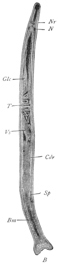
Fig. 323.—Ancylostoma duodenale, male. B, bursa; Bm, bursal muscles; Cdr, cement gland surrounding the ejaculatory duct; Glc, cervical glands; N, nucleus of cephalic gland; Nr, nerve ring; T, testes; Sp, spicule; Vs, vesicula seminalis. × 15. (After Looss.)
Buccal Capsule.—The buccal capsule is bent dorsally, 0·21 mm. long, 0·19 mm. broad. If a worm is rolled under the cover-glass so that the dorsal side is upwards, we observe the following features (fig. 325): In the dorsal edge of the chitinous capsule there is a gap as if a U-piece had been punched out. This is the “dorsal gap or incision.” The so-called “dorsal teeth” are simply the rounded edges of the tips of this gap. They project beyond the skin which covers the capsule externally. Below this gap is seen a curved line which, if followed along the sides of the capsule on each side, merges into the base of the most posterior ventral tooth. This line is the optical expression of a very shallow groove on the inside of the capsule. The skin on the outside of the capsule, which is reflected over the edge of and into the capsule, dips into this groove, which gives it a firm attachment. Below the middle (dorsally) of this curved line there is a thickening in the capsule wall, which is perforated by the opening of the dorsal œsophageal gland. This is the “dorsal ridge”; in optical section it has a conical appearance with a lumen (of the duct).
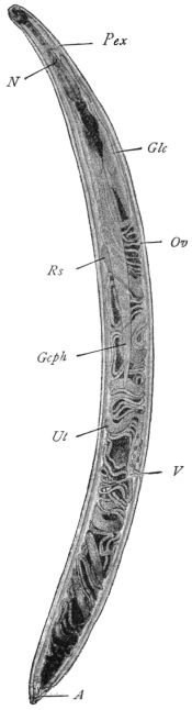
Fig. 324.—Ancylostoma duodenale, female. A, anus; Gcph, cephalic gland; N, nucleus of cephalic gland; Glc, cervical gland; Ov, ovary; Pex, excretory pore; Rs, receptaculum seminis; Ut, uterus; V, vagina. × 15. (After Looss.)
Male 9 mm. long by 0·45 mm. thick, female 12 mm. long by 0·6 mm. thick. Pale flesh colour, or an intense red in posterior third. Anteriorly may be more or less black due to (blood) pigment in the cells of the chyle intestine (= stomach + small intestine). The worm is about the same thickness all through and is plump and rigid. Cuticle striated. The body has a marked torsion, so that if the ventral side of the head is upwards the anus appears to open laterally and vice versâ. The dorsal curve of the head end is only slight and the œsophagus is roughly cylindrical.
On the ventral wall one sees the two pairs of strong teeth, their points being directed somewhat backwards. They are covered by cuticle above and below, but their points are free, piercing the cuticle. The “roots” of these teeth followed backwards appear as two thickenings or ribs on the outside of the capsule wall, so that the outside wall is not smooth—a characteristic of the genus Ancylostoma. In the space between these ribs lies the ventral nerve papilla, and lying against the outside of the outer root the lateral nerve papilla. The nerve papillæ are thus, as it were, concealed by these roots, and not conspicuous as they are in Necator. Following the ventral curve of the capsule on the inside, posteriorly we next find two triangular ventral lancets.316 These stand straight up into the capsule on either side of the longitudinal axis, converging at their summits. So that to sum up, the cutting apparatus is entirely ventral, consisting of two pairs of cutting teeth and a pair of lancets.
Cervical Papillæ.—Two, one on each side behind the head at the level of the excretory pore. They consist of “pulp,” i.e., extensions of the substance of the lateral bands covered by cuticle and supplied with a nerve (fig. 326).
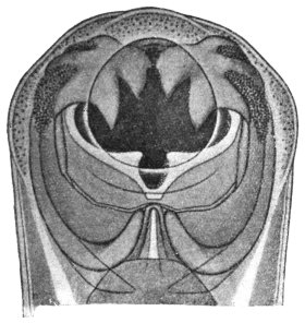
Fig. 325.—Ancylostoma duodenale: showing ventral teeth, dorsal cleft, and behind it the dorsal ridge with duct of dorsal œsophageal gland. × c. 200. (After Looss.)
Œsophageal Glands (3).—The chitin of the triradiate œsophagus is continuous with that of the buccal capsule. In its muscular walls are three glands—one dorsal, two sub-ventral. The dorsal gland opens into the buccal cavity through the dorsal ridge; the two others into the lumen of the œsophagus at the nerve ring. They branch freely amidst the muscles. They are probably digestive in function.
Cephalic Glands (2).—Lie in the lateral lines or bands on either side. They begin about the middle line of the body, and their ducts open at the base of the outer ventral tooth on the surface of the skin on each side. Each is 0·15 mm. thick in the middle, and has a single nucleus about as big as an ancylostome egg (N, fig. 323). They probably function as poison glands.
Excretory System and Cervical Glands (2).—The excretory pore lies in the mid line ventrally behind the œsophageal nerve ring (figs. 324 and 326). It opens into the excretory vesicle, a cavity in a large cell with lateral appendages which fuse with the lateral lines, this cell thus forming the “bridge” of the excretory system. Adhering to this (bridge) cell are the spindle-shaped cervical glands (Glc, fig. 324), and branches from the excretory vesicle enter the glands, which are excretory in function; the vesicle also receives branches from the lateral excretory canals (fig. 326) running in the lateral lines or bands. The cervical glands are swollen anteriorly, forming the so-called ampullæ just in front of the bridge. They extend backwards a little beyond the anterior loop of the testis.
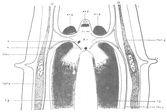
Fig. 326.—Ancylostoma duodenale: diagrammatic representation of excretory system. ex.p., excretory pore; e.c.g., excretory cervical gland; Ex. ves., excretory vesicle in B.c., bridge cell, which is connected with c.g., cervical gland, and l.l., lateral lines; ceph.g., cephalic gland; l.ex.c., lateral excretory canal passing into the bridge cell; l.l., lateral line containing excretory canal and cephalic gland; c.p., cervical papilla; n., nuclei of bridge cell. (After a drawing of Looss.)
Lateral Lines.—(1) Are broad elevations of the subcuticle, in which, here and there, a nucleus occurs. (2) Near the bursa in the male they increase in volume, and finally divide into branches which form the “pulp” of the different rays. (3) In addition to the lateral lines or bands, there is also a dorsal and ventral band. (4) The ventral band is well developed caudally, forming a large pad dorsal to the cloaca, “pulvillus post-analis.”
The bursal rays are outgrowths of the lateral lines. Beside this “pulp” they contain a nerve, and at their bases complex muscles.
The Bursa is closed on all sides with a short median (ventral) lobe, which may be tucked inwards. It is an outgrowth of the inner layer of the skin pushing the outer layer before it, so that it consists of three layers, not four, as it would be if it were a fold. The bursa is twice as broad as long. It is supported by a variety of rays, the arrangement of which is best followed from the figure (fig. 327). The different terminology for these rays as used by various authors should be noted: Ventral = anterior; externo-lateral = antero-external; medio-lateral + postero-lateral or antero-median + postero-median = median (doubled); externo-dorsal = postero-external; dorsal = posterior. All the rays end in tactile papillæ, seven, on each side; the postero-external and antero-external on the outer surface of the bursa, the five others on the inner surface.317 Of the six terminal digitations of the dorsal ray, only the external two contain tactile papillæ.
In the male there are prebursal papillæ and minute caudal papillæ in the female.
In the female the inner layer of the cuticle projects at the posterior end as a sharp spike, 20 µ long, which may sometimes be broken off.
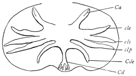
Fig. 327.—Ancylostoma duodenale: bursa enlarged. Ca, anterior ray cleft; cle, antero-external; cls, antero-median; clp, postero-median; Cde, postero external; Cd, posterior bifurcated, each bifurcation tridigitate. (After Railliet.)
Ovaries.—The anterior tube runs from the cephalic to the posterior end and back again. The posterior tube begins anteriorly, runs to the posterior end of the body, and then back to the cephalic end, forming a vulval loop before ending. The ovaries on the whole run in oblique coils. The uterus is the thicker portion of the tube, 5 mm. long. A short tube connecting the ovary and uterus is the oviduct. The two uteri unite to form a single duct, the vagina, opening 1 mm. behind the middle line. The portion of the uterus next to the oviduct functions as a seminal receptacle, whereas the part next the vagina functions as an ovejector.
Testis.—The blind end begins a little behind the beginning of the cement gland. The transverse coils occupy the middle third of the body. About the middle of the body it passes into the spindle-shaped seminal vesicle, which, with the spicular canal and rectum, opens into the cloaca. An anterior longitudinal coil pushing in between the cervical glands is characteristic of Ancylostoma. The cement gland surrounds the ejaculatory duct for practically its whole course, and it occupies nearly the posterior half of the body and secretes a brown or black cement. The spermatozoa are curved rods about 2 µ long.