ON PARASITES IN GENERAL. (continued 5)
Reading edition
Plasmodium malariæ is the parasite of quartan malaria (fig. 86). The trophozoites of the quartan parasite differ from the corresponding stages of the tertian parasite in that their motility is less and soon ceases. They differ also in their slower growth, by the early disappearance of the food vacuole, by the more marked formation of the dark brown pigment, and by the fact that the red blood corpuscles attacked are not altered either in colour or size.
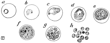
Fig. 86.—Development of the quartan parasite in the red corpuscles of man—asexual stages. (After Manson.)
When the parasites have grown almost to the size of the erythrocytes schizogony occurs. The pigment granules arrange themselves in lines radiating towards the centre and the merozoites are also radially disposed in groups of 6, 8, 10 or even 12, but are often arranged less regularly. The whole development, growth and schizogony, occupies seventy-two hours.
The appearance of quartana duplex or triplex is conditional on the presence in the blood of the patient of two or three groups of Plasmodia differing in their development by twenty-four hours.
The chief distinctive characters of the quartan parasite are: (1) The erythrocyte is unchanged in size; (2) the rings are compact and show pigment early; in the larger forms the chromatin is dense and relatively plentiful; (3) the pigment, which is relatively well-marked, may be arranged at the periphery.
Laverania malariæ, Grassi and Feletti, 1890 = Plasmodium falciparum, Welch, 1897.
Syn.: Plasmodium malariæ var. quotidianæ, Celli et Sanf., 1891; Hæmamœba malariæ præcox, Gr. et Fel., 1892 (nec H. præcox, Gr. et Fel., 1890); Hæmamœba laverani, Labbé, 1894; Hæmatozoön falciparum, Welch, 1897; Hæmosporidium undecimanæ and H. sedecimanæ and H. vigesimo-tertianæ, Lewkowitz, 1897; Hæmamœba malariæ parva, Lav., 1900; Plasmodium præcox, Dofl., 1901; Plasmodium immaculatum, Schaud., 1902; Plasmodium falciparum, Blanch., 1905.
The names most commonly used for the parasite of malignant tertian malaria are Plasmodium falciparum and Laverania malariæ.
The summer and autumn fever (febris æstivo-autumnalis), also called malignant tertian or sub-tertian, is caused by a malarial parasite which is distinguished by the small size of its schizont, while the gametocytes are crescentic (figs. 81, 88).
Most authors identify this kind of fever or the parasites which cause it (Laverania malariæ) with the pernicious malaria of the tropics. Ziemann, however, repeatedly has drawn attention to certain small but definite differences between the usual malignant tertian or pernicious parasites which occur in the tropics and the tropical parasites of some malarial districts, particularly of West Africa, and insists that at least two varieties or sub-species occur. Other investigators distinguish from this or these forms a quotidian parasite. On the other hand, the assertion is made that there are no specific differences, but that the malignant or pernicious tertian parasite which normally needs forty-eight hours for its development in the blood of man, can also develop in twenty-four hours. The establishment of the duration of the development is a matter of especial difficulty, because the stages of schizogony are far less numerous in the peripheral blood than in that of the internal organs. It is also stated that the tropical parasite very seldom forms crescentic but rather rounded gametocytes. According to such an observation the organism would belong to Plasmodium and not to Laverania. The question whether the tropical fevers are caused by two different parasites does not seem to be definitely settled.
The young trophozoite of the malignant, pernicious tertian, or sub-tertian parasite (fig. 87) are but slightly active and are very small, even after the formation of the comparatively large food vacuole, which makes the body appear annular (“signet ring” stage). Often two and even more parasites are found in one blood corpuscle.
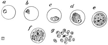
Fig. 87.—The pernicious malignant or sub-tertian parasite in the red corpuscles of man, asexual stages. (After Manson.)
Fully grown they only attain two-thirds or less of the diameter of the erythrocytes, which display an inclination to shrink and then appear darker than the normal (brass-coloured). In the early stage dots or stippling—sometimes called Maurer’s dots—appear on the blood corpuscles as in those attacked by the ordinary tertian parasite (Plasmodium vivax), but the Maurer’s dots are relatively coarse and few, and are not easily stained. These dots were first described by Stephens and Christophers in 1900, and subsequently by Maurer in 1902.
About thirty hours after the entrance into the blood corpuscles, the parasites are rarely found in the peripheral blood, but they are present in the internal organs, and especially in the spleen. The schizogony, which now begins in the internal organs, proceeds on the same lines as that of the quartan parasite, that is, usually with the merozoites radially arranged around a central agglomeration of dark brown pigment.
The number of merozoites formed is quoted differently, e.g., 8 to 24, on an average 12 to 16. However, according to the recent cultural researches of J. G. and D. Thomson204 (1913) the number of merozoites of P. falciparum is 32. D. Thomson, from examination of spleen smears at autopsy, also concludes that the number of merozoites may reach 32. During their formation the blood corpuscle which is attacked gets paler and disintegrates.
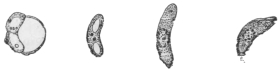
Fig. 88.—The crescents of the malignant tertian parasite. (After Mannaberg.) See also fig. 81.
The gametocytes which finally appear are attenuated, curved bodies, rounded at each end and known as crescents (figs. 81, 88), and are provided with a nucleus and with coarse pigment masses. In the males the pigment is more scattered than in the females, where it is around the nucleus. Their length is 9 µ to 14 µ, and their breadth is 2 µ to 3 µ. At first they are still in the pale blood corpuscles, later they free themselves and are found in numbers in the peripheral blood in cases of pernicious malaria of Southern Europe and the tropics, while, on the other hand, they occur much more rarely in the peripheral blood in West African malignant tertian. Their further development takes place under the same conditions as in the other malarial parasites.
D. Thomson (1914),205 from studies of autopsy smears, has shown that crescents develop chiefly in the bone-marrow and spleen, and take about ten days to grow into the adult state in the internal organs. He believes that crescents are produced from ordinary asexual spores. Quinine, he states, has no direct destructive action on crescents, but it destroys the asexual source of supply.
The sporozoites of Laverania malariæ (P. falciparum) are represented in fig. 89.
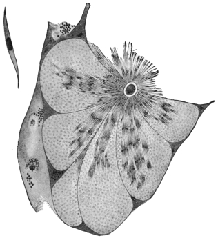
Fig. 89.—Section through a tubule of the salivary gland of an Anopheles with sporozoites of the malignant tertian parasites; on the left at the top a single sporozoite greatly magnified. (After Grassi).
The principal distinctive characters of the malignant tertian parasite are: (1) The ring forms are very small, occasionally bacilliform, and may be marginal (“accolé” of Laveran); (2) the larger trophozoites are often ovoid, and about one-third or one-half of the erythrocyte in size; (3) the infected red cells sometimes show coarse stippling (Maurer’s dots); (4) the gametocytes, or sexual forms, are crescentic in shape.
J. W. W. Stephens (1914) has described a new malarial parasite of man; it is called Plasmodium tenue. It is very amœboid, with scanty cytoplasm and much chromatin, sometimes rod-like or irregular. The parasite was described from a blood-smear of an Indian child. The creation of a new species for this parasite has been criticized by Balfour and Wenyon, and by Craig.
Plasmodium relictum, Sergent, 1907.
Syn.: Plasmodium præcox, Grassi and Feletti, 1890; Plasmodium danilewskyi, Gr. et Fel., 1890; Hæmamœba relicta, Gr. et Fel., 1891; Proteosoma grassii, Labbé, 1894.
Hæmamœboid, pigment-producing, malarial parasites are often found in birds. Like the human malarial parasites they have been variously named. Labbé created the genus Proteosoma for them, and this name is still often used as a distinctive one unofficially. The correct name is stated to be either Plasmodium relictum or P. præcox, or possibly even P. danilewskyi, assuming that there is only one species. The nomenclature of the malarial parasites is most confused. The avian malarial parasites are transmitted by Culicine mosquitoes.
The organism was discovered by Grassi in the blood of birds in Italy, and causes a fatal disease in partridges in Hungary. Sparrows are affected in India, and it was this Plasmodium in which Ross first traced the development of a malarial parasite in a mosquito. The parasite may be transmitted from bird to bird by blood-inoculation, canaries being very susceptible.
The principal stages of the avian plasmodium closely resemble those of the malarial parasites of man. In its earliest stage P. relictum is unpigmented, but soon the trophozoite grows and becomes pigmented, meanwhile displacing the nucleus of the avian red-blood corpuscle, a characteristic feature, distinguishing it from Halteridium. Schizonts are formed, each of which gives rise to about nine merozoites in the circulating blood. Sexual forms or gametocytes also occur in the blood. These develop in Culex fatigans, C. pipiens and C. nemorosus. Oökinetes or vermicules are formed in twelve to fifteen hours in the stomach of the mosquito, and in one to two days well-developed round oöcysts may be seen. In three to four days sporoblasts have formed within the oöcysts and young sporozoites begin to develop. In nine to ten days the oöcysts are mature, being filled with sporozoites. The oöcysts then burst and the sporozoites travel through the thoracic muscles to the salivary glands of the Culicine.
Neumann, experimenting with canaries, found that Stegomyia fasciata could transmit the infection, but less efficiently than species of Culex.
The Cultivation of Malarial Parasites.
The successful cultivation of malarial parasites in vitro was first recorded by C. C. Bass and by Bass and Johns (1912).206 Since then, J. G. and D. Thomson,207 and McLellan (1912–13), Ziemann208 and others have repeated the experiments.
DIFFERENTIAL CHARACTERS OF THE HUMAN MALARIAL PARASITES.
CharacterPlasmodium malariæ (Quartan)Plasmodium vivax (Benign tertian)Laverania malariæ = Plasmodium falciparum (Malignant tertian)
SchizogonyComplete in seventy-two hoursComplete in forty-eight hoursComplete in forty-eight hours or less
TrophozoiteSmaller than P. vivax, larger than L. malariæYoung trophozoite large.Young trophozoite small
Pseudopodia not marked or longLong pseudopodia
MovementsRather slow in immature formsActive amœboid movementsSometimes actively motile
PigmentCoarse granules, peripherally arranged, little movementFine granules, with active movementGranules fine and scanty, movement oscillatory
SchizontSmaller than red corpuscleLarger than red blood corpuscleSmaller than red corpuscle
Merozoites6 to 12 forming rosette15 to 20 regularly arranged8 to 32 (according to different authors) arranged irregularly
GametocytesSphericalSphericalCrescentic
Distribution of parasites in vertebrate hostAbout equal number in peripheral and visceral bloodLarger numbers in visceral bloodScanty in peripheral blood compared with the enormous numbers in the internal organs. The latter part of the cycle (schizogony) may occur in the internal organs only
Alterations in erythrocytesAlmost normalPale and hypertrophied.Corpuscle may be shrunken and dark, or may be colourless. Maurer’s coarse dots sometimes seen
Schüffner’s dots seen in deeply stained specimens
Essentially the method of cultivation, as used by Thomson, is as follows: 10 c.c. of infected blood are drawn from a vein and transferred to a sterile test tube, in which is a thick wire leading to the bottom of the tube. One-tenth of a cubic centimetre of a 50 per cent. aqueous solution of glucose or dextrose is placed in the test tube, preferably before adding the blood. The blood is defibrinated by stirring gently with the wire. When defibrination is complete the wire and the clot are removed, and the glucose-blood is transferred, in portions, to several smaller sterile tubes, each containing a column of blood about one inch in height. The tubes are plugged and capped and then transferred, standing upright, to an incubator kept at a temperature of 37° C. to 41° C. The blood corpuscles soon settle, leaving a column of serum at the top, to the extent of about half an inch in each tube. The leucocytes need not be removed by centrifugalization. J. G. Thomson (1913) and his collaborators did not find it necessary to destroy the complement in the serum, and they found that the malarial parasites developed at all levels in the column of corpuscles, and not merely on the surface layer of the corpuscles as first stated by Bass and Johns.
So far only the asexual generation of the malarial parasites has been grown in vitro. Thomson rarely observed hæmolysis in the cultures. Clumping of the malignant tertian parasites occurred. In cultures of the benign tertian parasite (Plasmodium vivax) clumping was not observed. J. G. and D. Thomson consider that this difference as regards clumping explains why only young forms of malignant tertian are found in peripheral blood, as the clumping tendency of the larger forms causes them to be arrested in the finer capillaries of the internal organs. It also explains the tendency to pernicious symptoms, such as coma, in malignant tertian malaria. Further it was found from cultures that P. falciparum was capable of producing thirty-two spores (merozoites) in maximum segmentation, while P. vivax produced sixteen spores (merozoites) as a rule, though the number might be greater than sixteen. (Quartan parasites produce eight spores or merozoites in schizogony.)
It may also be mentioned here that Babesia (Piroplasma) canis has been successfully cultivated in vitro by Bass’s method. This has been accomplished by Thomson and Fantham,209 Ziemann, and Toyoda in 1913. J. G. Thomson and Fantham used the simplified Bass technique recorded above, namely, infected blood and glucose, incubating at 37° C. In one of the B. canis cultures, starting with heart blood of a dog containing corpuscles infected with one, two, or, exceptionally, four piroplasmata, Thomson and Fantham succeeded in obtaining a maximum of thirty-two merozoites in a corpuscle. The cultures are infective to dogs and sub-cultures have been obtained.
Family. Piroplasmidæ, França.
The parasites included in this provisional family or group belong to the Hæmosporidia. They are minute organisms, sometimes amœboid, but usually possessing a definite form. They are endoglobular, being contained within mammalian red blood corpuscles, but they produce no pigment. The true Piroplasmata, belonging to the genus Babesia, destroy the host corpuscles, setting free the hæmoglobin, which is excreted by the kidneys of the cow, sheep, horse, dog, etc., acting as host. The disease produced, variously called piroplasmosis or babesiasis, is consequently characterized by a red coloration of the urine known as hæmoglobinuria, or popularly as “red-water.” One of the best known piroplasms is Piroplasma bigeminum or Babesia bovis (probably the latter name is correct), which is the causal agent of “Texas fever” or “red-water” in cattle and is spread by ticks.
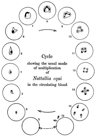
Fig. 90.—Nuttallia equi, life-cycle as seen in red blood corpuscles in stained preparations of peripheral blood. (After Nuttall and Strickland.)
Of recent years, researches on the morphology of these blood parasites has led to their separation into various genera and species. However, our knowledge is still very far from complete. The various genera recognized by França210 (1909), and placed in a provisional family, Piroplasmidæ, may be listed, though further research may lead to emendations:—
(1) Babesia (Starcovici) or Piroplasma (Patton). Pyriform parasites, dividing by a special form of budding or gemmation with chromatin forking, as well as by direct binary fission. Parasitic in oxen, dogs, sheep, horses, etc.
(2) Theileria (Bettencourt, França and Borges). Rod-shaped and oval parasites occurring in cattle and deer. T. parva is the pathogenic agent of African East Coast fever in cattle.
(3) Nuttallia (França). Oval or pear-shaped parasites, with multiplication in the form of a cross. N. equi211 (fig. 90) of equine “piroplasmosis” (nuttalliosis). N. herpestidis in a mongoose.
(4) Nicollia (Nuttall). Oval or pear-shaped parasites with characteristic nuclear dimorphism, and with quadruple division at first fan-like, then like a four-leaved clover. N. quadrigemina from the gondi.
(5) Smithia (França). Pear-shaped, single forms stretching across the blood corpuscle. Multiplication into four in the form of a cross. S. microti from Microtus arvalis, S. talpæ from the mole.
(6) Rossiella (Nuttall). This belongs to the family Piroplasmidæ of França. It is intracorpuscular and non-pigment forming, occurring singly, in pairs, or occasionally in fours. It is usually round and larger than Babesia. The parasite multiplies by binary fission. R. rossi in the jackal.
The genus Babesia is the best known and most important, and will be considered next.
Genus. Babesia, Starcovici, 1893.
Syn.: Pyrosoma, Smith and Kilborne, 1893; Apiosoma, Wandolleck, 1895; Piroplasma, W. H. Patton, 1895; Amœbosporidium, Bonome, 1895.
The organisms belonging to this genus are pyriform, round or amœboid. The characteristic mode of division is as follows: Just before division the parasite becomes amœboid and irregular in shape, (fig. 91, 1–5) with a compact nucleus. The latter gives off a nuclear bud. This nuclear bud divides into two by forking (fig. 91, 6, 7). The chromatin forks grow towards the surface of the body of the rounded parasite, and then two cytoplasmic buds grow out. The forking nuclear buds, which are Y-shaped, pass into the cytoplasmic outgrowths212 (fig. 91, 8, 9). The buds gradually increase in size at the expense of the parent form until they become two pear-shaped parasites joined at their pointed ends. The connecting strand shrinks and the two daughter forms separate (fig. 91, 10–14). The pyriform parasites after having exhausted the blood corpuscle escape from it (fig. 91, 15), and seek out fresh host corpuscles, entering by the rounded, blunt end (fig. 91, 1). It is the pyriform phase of the parasite which penetrates red blood corpuscles, not rounded forms, which die if set free. The pyriform parasite, however, becomes rounded (fig. 91, 2, 3), soon after its entry into a fresh host cell. This interesting mode of division by gemmation and chromatin forking has been made diagnostic of the genus Babesia by Nuttall.213 Rounded forms of Babesia divide by binary fission, and this direct method can also be adopted by the other forms of Babesia.
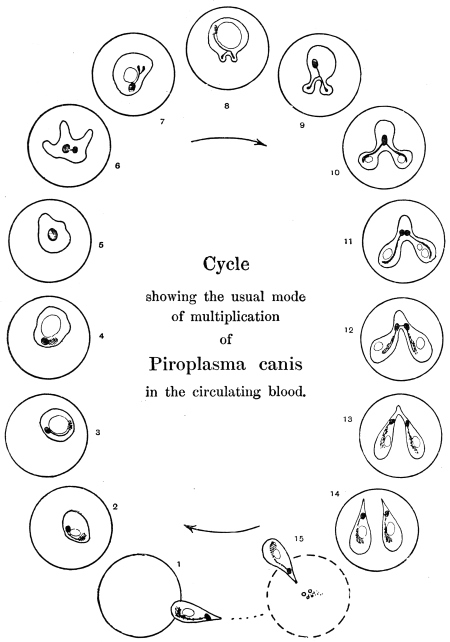
Fig. 91.—Babesia (Piroplasma) canis, life-cycle in stained preparations of infected blood of dog. (After Nuttall and Graham-Smith.)
The distribution of the chromatin in the pear-shaped Babesia, as seen in B. canis and B. bovis, is interesting. The main nuclear body consists of a karyosome surrounded by a clear area. There is also a loose (chromidial) mass of chromatin representing the remains of the chromatin forks seen during the formation of the parasite as a daughter form by gemmation. Occasionally there is a small dot or point, the so-called “blepharoplast” of Schaudinn and Lühe. This minute dot is not a flagellate blepharoplast, for there is no flagellate stage in the life-history of Babesia. These nuclear phenomena have been described by Nuttall and Graham-Smith and Christophers (1907)214 for B. canis, by Fantham (1907)215 for B. bovis, and by Thomson and Fantham (1913) from glucose-blood cultures of B. canis.
Babesia are tick borne, as was first shown by Smith and Kilborne (1893). The developmental cycle in the tick is incompletely known. The best accounts are those of Christophers (1907)216 for B. canis and Koch (1906) for B. bovis, and these accounts are supplementary. The principal stages, so far as known, may be summarized thus:—
(1) The piroplasms taken by the tick in feeding on blood pass into the tick’s stomach. The pyriform parasites, which alone are capable of further development, are set free from the blood corpuscles. In about twelve to eighteen hours they become amœboid, sending out long, stiff, slender, pointed pseudopodia. The nucleus of each parasite divides unequally into two. Similar forms have been obtained in cultures. These stellate forms may be gametes, and according to Koch fuse in pairs.
(2) A spherical stage follows, possibly representing the zygote. This grows, and a uninucleate globular mass results. This form is found in large numbers on the third day, according to the observations of Koch.
(3) A club-shaped organism is next formed. This may represent an oökinete stage. The club-shaped bodies are motile and gregarine-like, and are about four times the size of the blood forms. These club-shaped bodies and subsequent stages were described by Christophers in the development of B. canis in the dog-tick, Rhipicephalus sanguineus.
(4) The club-shaped bodies pass from the gut of the tick into the ovary, and so get into the ova. There they become globular, and later are found in the cells of the developing tick-embryo. The parasites are, then, transmitted hereditarily. Similar globular bodies are found in the tissue cells of the body of tick nymphs which have taken up piroplasms. The globular stage was called the “zygote” by Christophers, but it may correspond to the oöcyst of Plasmodia.
(5) The globular body divides into a number of “sporoblasts,” which become scattered through the tissues of the larval or nymphal tick, as the case may be.
(6) The sporoblasts themselves divide into a large number of sporozoites, which are small uninucleate bodies, somewhat resembling blood piroplasms. The sporozoites collect in the salivary glands of the tick. They are inoculated into the vertebrate when the tick next feeds.
The chief species of Babesia and their pathogenic importance may be listed thus:—
(1) Babesia bovis (Babes) produces infectious hæmoglobinuria of cattle in Europe and North Africa. It is transmitted by Ixodes ricinus. A similar parasite also occurs in deer.
(2) Babesia bigemina (Smith and Kilborne) produces Texas fever, tristeza, or red-water in cattle in North and South America, South Africa and Australia. It is transmitted by Boöphilus annulatus in North America, by B. australis in Australia, South America, and the Philippines, and by B. decoloratus in South Africa.
The parasite is from 2 µ to 4 µ long, and from 1·5 µ to 2 µ broad.
Babesia bigemina may be the same parasite as B. bovis.
(3) Babesia divergens (MacFadyean and Stockman) is a small parasite. It is found in cattle suffering from red-water in Norway, Germany, Russia, Hungary, Ireland, Finland, and France, and is transmitted by Ixodes ricinus.
(4) Babesia canis (Piana and Galli-Valerio) gives rise to malignant jaundice or infectious icterus in dogs in Southern Europe, India, and other parts of Asia and North Africa, where it is transmitted by Rhipicephalus sanguineus. In Africa generally, especially South Africa, the disease is transmitted by Hæmaphysalis leachi. Babesia canis varies from 0·7 µ to 5 µ, the size depending partly on the number of parasites within the corpuscle. It averages about 3 µ. It has been cultivated in Bass’ medium (glucose and infected blood), see p. 172.
In India Piroplasma gibsoni (Patton) infects hunt dogs and jackals. It is annular or oval in shape.
(5) Babesia ovis (Babes) produces “Carceag,” a disease of sheep in Roumania, the Balkan Peninsula, Italy, and Transcaucasia. It varies in size from 1 µ to 3 µ. It is transmitted by Rhipicephalus bursa. The parasite has recently been recorded from Rhodesia.
(6) Babesia caballi (Nuttall and Strickland) causes “biliary fever” in equines. The parasite occurs in Russia, Roumania, and Transcaucasia. It varies in size from 1 µ to 2 µ. It is transmitted by Dermacentor reticulatus.
It should be mentioned that Nuttallia equi also causes “piroplasmosis” in equines, with symptoms of hæmoglobinuria and jaundice in Italy, Sardinia, many parts of Africa, Transcaucasia, India, and Brazil. In Africa it is transmitted by Rhipicephalus evertsi. It has been shown experimentally that a horse recovered from Babesia caballi was susceptible to the inoculation of Nuttallia equi blood.
(7) Babesia pitheci (P. H. Ross) was found in a monkey, Cercopithecus sp., in Uganda. The pear-shaped forms measure 1·5 µ by 2·5 µ.
(8) Babesia muris (Fantham)217 was found in white rats. The pyriform parasites are 2 µ to 3 µ long and 1 µ to 1·5 µ broad; oval forms are 0·5 to 1·5 µ diameter.
The usual symptoms of babesiasis (piroplasmosis) are high fever, loss of appetite, hæmoglobinuria, icterus, anæmia, paralysis, and death in about a week in acute cases. In chronic cases there is anæmia, and hæmoglobinuria is less marked. When animals recover, there are still some piroplasms left in the blood. “Recovered” or “salted” animals are not susceptible to reinfection, but ticks feeding on them acquire piroplasms, and are a source of danger to freshly imported animals.
Treatment.—Trypan-blue is the best drug, as shown by Nuttall and Hadwen218 (1909). It should be administered intravenously in 1 to 1·5 per cent. aqueous solution. A dose of 5 to 10 c.c. is curative for dogs, one of 100 to 150 c.c. for horses and cattle. Unfortunately, the tissues are coloured blue by the drug. The “salted” animals, after trypan-blue treatment, still harbour the parasites in their blood for years.
Genus. Theileria, Bettencourt, França and Borges, 1907.
The organisms belonging to this genus are rod-like or bacilliform, and coccoid or round.
The best known of the species of Theileria is T. parva, the pathogenic agent of East Coast fever or Rhodesian fever in cattle in Africa.
Theileria parva, Theiler, 1903.
Syn.: Piroplasma parvum.
In the blood corpuscles of infected cattle minute rod-like and oval parasites are seen. Some are comma shaped and others are clubbed (fig. 92, 1–12). The rod-like forms measure 1 µ to 3 µ in length by 0·5 µ in breadth; the oval forms are 0·7 µ to 1·5 µ in diameter. The intracorpuscular parasites are said by R. Gonder (1910) to be gametocytes, the rod-like forms being thought to be males, the oval forms to be females. Free parasites are practically never seen in the blood. It is known that it is impossible to produce the disease in a healthy animal by blood inoculation, but only by intraperitoneal transplantation of large pieces of infected spleen (Meyer). There may be as many as eight parasites in a corpuscle. The chromatin is usually at one end of the organism. In some parasites the appearance of the chromatin suggests division, but such division, if it takes place, must be very slow, as it has not been actually seen in progress. The red blood corpuscles appear merely to act as vehicles for the parasites (Nuttall, Fantham, and Porter).219
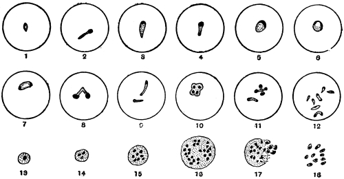
Fig. 92.—Theileria parva. 1–12, intracorpuscular parasites, stained. (After Nuttall and Fantham); 13–18, Koch’s blue bodies, from stained spleen smear; 17–18, breaking up of Koch’s body. (After Nuttall.)
In the internal organs, especially the lymphatic glands, spleen and bone-marrow, are found multinucleate bodies known as Koch’s blue bodies (fig. 92, 13–18). These are schizonts, according to Gonder.220 The actual Koch’s blue bodies are said to be extracellular, but similar multinucleate bodies, schizonts, occur in lymphocytes. The schizonts divide and the merozoites resulting probably invade the red blood corpuscles in the internal organs. Gonder considers that the sporozoites injected by the tick collect in the spleen and lymphatic glands, penetrate the lymphocytes and give rise to the schizonts.
Gonder has studied the cycle of T. parva in the tick. He states that the gametocytes leave the host corpuscles and give rise to gametes, then conjugation occurs producing zygotes. The zygotes are then said to become active to form ookinetes, and to enter the salivary glands of the tick. Multiplication is said to occur therein, producing a swarm of sporozoites. This work needs confirmation.
T. parva is transmitted by Rhipicephalus appendiculatus, R. simus, R. evertsi, R. nitens, and R. capensis. The parasites are not hereditarily transmitted in Rhipicephalus, but when taken by the transmitter at one stage of its development the tick is infective in its next stage (e.g., if the larva becomes infected, then the nymph is infective; if the nymph becomes infected, then the adult is infective).
An animal recovered from Theileria parva is incapable of infecting ticks, but few animals recover from East Coast fever. Animals suffering therefrom do not show hæmoglobinuria.
Theileria mutans, Theiler, 1907·
Syn.: Piroplasma mutans.
This is transmissible experimentally by blood inoculation. It occurs in cattle in South Africa and Madagascar and is apparently non-pathogenic. No Koch’s blue bodies are formed. It is transmitted by ticks.
Theileria annulata (Dschunkowsky and Luhs) occurs in cattle in Transcaucasia.
A Theileria (T. stordii) has been found in a gazelle (França, 1912).
Genus. Anaplasma, Theiler, 1910.
This genus221 may be mentioned here. The organisms included therein are, according to Theiler, coccus-like, consisting of chromatin, and are devoid of cytoplasm. They occur in the red blood corpuscles of cattle, causing a disease characterized by destruction of red cells, fever and anæmia, but with yellow urine. The disease is tick transmitted. The bodies now called Anaplasma marginale were formerly described as marginal points. They multiply by simple fission. They are said by Theiler to cause gall-sickness in cattle in South Africa. Some authors doubt whether these bodies are organismal.
Genus. Paraplasma, Seidelin, 1911.
Under this generic name Seidelin described certain bodies found by him in cases of yellow fever in 1909. The type species is P. flavigenum,222 and is claimed by Seidelin to be the causal agent of yellow fever.
Paraplasma flavigenum occurs in the early days of the disease as small chromatin granules with or without a faint trace of cytoplasm. The bodies are usually intracorpuscular. Also, somewhat larger forms, with distinct cytoplasm, are seen in small numbers. During the later days of the disease still larger forms are found, and these occur also in sections of organs (e.g., kidney) made post-mortem. Some of these larger forms are perhaps schizonts. In the second period of the disease possible micro- and macro-gametes may be found, some of which are extracorpuscular. Some small free bodies have been seen. Recently schizogony has been stated to occur in the lungs, and it is said that guinea-pigs can be inoculated with Paraplasma flavigenum, and show yellow pigment in the spleen.
Seidelin places Paraplasma in the Babesiidæ, with resemblances more particularly to Theileria. V. Schilling-Torgau and Agramonte have criticized these findings; the former considers them to be the resultant of certain blood conditions.
P. subflavigenum was found by Seidelin in 1912 in a man suffering from an unclassified fever in Mexico.
Further, it is now known that a Paraplasma occurs naturally in guinea-pigs. More researches are needed on these matters, as some writers (e.g., Wenyon and Low) claim that the bodies are not organismal.
Paraplasma flavigenum.—The Yellow Fever Commission (West Africa) in their third report, dated 1915, have come to the conclusion that there is no evidence that the bodies termed Paraplasma flavigenum are of protozoal nature or that they are the causal agents of yellow fever.
Sub-class. NEOSPORIDIA, Schaudinn.
Sporozoa in which growth and spore formation usually go on together.
Order. Myxosporidia, Bütschli.
These parasites, which were discovered by Johannes Müller (1841), live principally in fishes, and occasionally cause destructive epizoötics amongst their hosts. Müller first observed them in the form of whitish-yellow pustules on the skin or on the gills of various fishes. These pustules contained masses of small shell-covered bodies with or without tails (“psorosperms,” see fig. 93). Similar bodies were also found in the air bladders of certain fish. Creplin (1842) demonstrated the resemblance of the cysts (“psorosperm tubes”) harbouring the psorosperms to the “pseudonavicella-cysts” of a gregarine, as described by v. Siebold. Dujardin (1845) considered that there was possibly some connection between the protoplasmic “psorosperm tubes” and the spores they contained, and the developmental stages of monocystid gregarines from the vesiculæ seminales of earth-worms. The relationship of the “fish psorosperms” was placed on a firmer basis by Leydig (1851) and Lieberkühn. The former found numerous forms in marine fish, and he discovered in species which live free in the gall bladder of cartilaginous fishes that the psorosperms originated in a manner similar to the gregarines. Lieberkühn (1854) studied the Myxosporidia in the bladder of the pike (fig. 93, a, b, d), and observed their amœboid movements, as well as the formation of the spores, from each of which a small amœboid body escaped, a discovery that was confirmed by Balbiani. The same author also found that spiral filaments were enclosed in the so-called polar body, i.e., the polar capsule of the psorosperm spores, and that these could be protruded (fig. 93, d, and fig. 95).
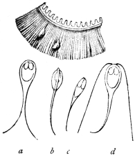
Fig. 93.—Upper figure, part of a gill of a roach, Leuciscus rutilus (natural size), with two myxosporidia. Lower figures, a, b, d, spores of myxosporidia from a pike, Esox lucius. c, Spore from Platystoma fasciatum. (After J. Müller.)

Fig. 94.—The tailless spore of Myxobolus mülleri, with the polar bodies and their nuclei and the sporozoite. (After Bütschli.)
The term Myxosporidia, which at the present day is universally applied to the “psorosperm tubes,” was introduced by Bütschli in 1881, who studied not only the structure and development of the spores, but also the protoplasmic body of the parasites (fig. 96), and confirmed the occurrence of numerous nuclei. Many authors have made important additions to our knowledge of the Myxosporidia: Perugia, Thélohan, Mingazzini, L. Pfeiffer, L. Cohn, Doflein, Mercier, Schröder and Auerbach; while the presence of this parasite outside the class of fishes has become known through Lutz, Laveran, and others. The species causing disease in fishes have been described by Ludwig, Railliet, Weltner, L. Pfeiffer, Zschokke, Hofer, Doflein, Gurley, Plehn, Schuberg, Fantham and Porter. With regard to classification the works of Thélohan (1895) and Gurley (1894) may be mentioned.
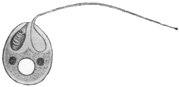
Fig. 95.—Schematic representation of a spore of Myxobolus. One polar capsule has protruded its filament; two nuclei and a “vacuole” in the sporozoite. (After Doflein.)
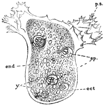
Fig. 96.—Chloromyxum leydigi. Active trophozoite (parasitic in gall-bladder of skates, rays, dog-fish). Ect, ectoplasm; ps, pseudopodia; end, endoplasm; y, yellow globules in endoplasm; sp, spores, each with four polar capsules. × 525. (After Thélohan.)
The Myxosporidia live either free on the epithelial surface of hollow organs (gall or urinary bladder, renal tubules, but never in the intestine), or are enclosed in the tissues of their host. The gills and muscular system are their favourite habitat, but other tissues or organs may be attacked. Species of Myxosporidia are also known from Amphibia, Reptilia, and a few invertebrates.
The free forms, which are often amœboid (fig. 96), move by the aid of variously shaped pseudopodia, have a constant form, or may exhibit contractions of the body. The tissue parasites often reach a considerable size, so that the integument of the host forms protuberances over them. They are of a roundish or irregular shape. Frequently they are enveloped in a connective tissue covering formed by the host.
The protoplasmic body in the trophic phase (fig. 96) shows a distinct ectoplasm which is finely granular or sometimes striated, and an endoplasm which is coarsely granular and contains many nuclei as well as cell inclusions, such as crystals, pigment grains and fat globules. The nuclei originate by division from the primitive nucleus of the amœboid germ that issues from the spore. This amœbula may or may not live intra-cellularly during the early stages of its existence.
The multinucleate trophozoite of a Myxosporidian forms spores in its endoplasm practically throughout its whole period of growth (fig. 96). Vegetative reproduction by a process of external budding or plasmotomy may also occur, as in Myxidium lieberkühni from the urinary bladder of the pike.
The myxosporidian trophozoite may produce two spores within itself, when it is placed in the sub-order Disporea, or it may produce numerous spores, which is characteristic of the sub-order, Polysporea. The phenomenon of spore formation is not simple (fig. 97), and the spore itself is surrounded by a bivalved shell or sporocyst and contains polar capsules in addition to the amœboid germ (fig. 97, G, H). The valves of the sporocyst and the polar capsules are really differentiated nucleate cells, so that each spore is an aggregate of cells rather than one cell, though only a single amœbula issues from a spore. The accounts of spore formation vary somewhat according to the different workers.
Spore formation is usually very complicated and there are differences of opinion as to the interpretation of various stages, particularly as to whether conjugation occurs therein. The process is initiated by the concentration of cytoplasm around one of the nuclei of the endoplasm, so that a small spherical mass or initial corpuscle is produced, the pansporoblast (Gurley) or primitive sphere (Thélohan). Some authors state that a pansporoblast really results from a conjugation of two initial corpuscles (fig. 97, A-D). Nuclear multiplication occurs within the pansporoblast (fig. 97, E), and sooner or later two multinucleate sporoblasts are formed within it (fig. 97, F). Each sporoblast gives rise to a single spore, which consists of a sporocyst or envelope composed of two valves each secreted by a cell, two polar capsules each secreted by a cell, and the sporoplasm or amœbula which becomes binucleate (fig. 97, G). During the process of spore formation (fig. 97) various vegetative and reduction nuclei may be produced, in addition to those which are essentially involved in spore formation, and the sporocyst cells may be developed early.
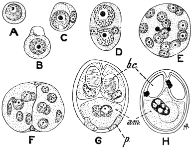
Fig. 97.—Myxobolus pfeifferi. Spore formation. A, reproductive cell from plasmodial trophozoite; B, cell divided unequally into two; C, smaller cell forming envelope to larger one; D, pansporoblast formed by union of two forms like C; E, multinucleate pansporoblast, two of the nuclei being those of the envelope; F, pansporoblast divided into two multinucleate sporoblasts; G, spore differentiation; p, two parietal cells forming sporocyst; bc, polar capsules; am, binucleate amœbula; H, ripe spore in which the two nuclei of the amœbula have fused. (After Keysselitz.)
Each spore contains two (figs. 94, 95) or more polar capsules which are clearly visible in the fresh condition. Each polar capsule is a hollow, more or less pear-shaped body, secreted by a cell and having a well defined contour. Within it, a long, delicate, elastic filament, the polar filament, is formed, and lies spirally coiled in the polar capsule until just before the emergence of the amœbula from the spore (fig. 95). The polar filament is ejected, probably under the influence of the digestive juice, when the spore reaches a new host, and serves to anchor the spore to the tissue with which it is in contact, and thus allow of the emergence of the amœbula in a situation suitable for its development. The polar capsule with its contained polar filament has been compared with the stinging cells or nematocysts of the Cœlentera, but it has a totally different function.
The spores fulfil the purpose of effecting transmission to other hosts. Infection occurs by the ingestion of the parasites per os after their escape by some means from their host. Thélohan and others have demonstrated that the valves of the spores soon open under the influence of the digestive juices, thus allowing the young myxosporidia to escape. Their further history is unknown; but it may be surmised that they either travel direct to the organs usually affected (gall bladder, urinary bladder), or are distributed in the body by means of the circulatory or lymphatic systems.
The Myxosporidia that invade tissues are often deadly to their hosts. They may be present in a state of “diffuse infiltration” when practically every organ of the body may be infected, as in barbel disease (due to Myxobolus pfeifferi). On the other hand, the parasites may be concentrated at one spot, when cysts, either large or small, are produced. Such cysts occur on the gills of many fishes. A few additional important pathogenic forms are Myxobolus cyprini, the excitant of “pockenkrankheit” of carp, and Lentospora cerebralis, parasitic in the skeleton of Salmonidæ and Gadidæ. The skeletons of the tail, fins and skull particularly are seats of infection, and from the skull the Lentospora can spread to the semicircular canals, resulting in loss of power to maintain its balance on the part of the fish. On this account the malady is termed “drehkrankheit.” Young fish are more particularly infected. Myxobolus neurobius infects the spinal cord and nerves of trout.
Myxosporidia are divided into two sub-orders—Disporea and Polysporea—according to whether they form only two or several spores during their growth. The former include two genera limited to fishes, which are easily distinguishable by the shape of the spores: Leptotheca, Thél., with a rounded spore, and Ceratomyxa, Thél., with a very elongate spore. The larger number of genera belong to the Polysporea, which are divided into three families:
(1) Amœboid germ with a vacuole the contents of which do not stain with iodine.(a) With two polar capsules.—Myxidiidæ.

(b) With four polar capsules.—Chloromyxidæ.
(2) Amœboid germ with a vacuole stainable with iodine. Spores with two polar capsules.—Myxobolidæ.
For further subdivisions the differences in the spores are principally utilized.
Order. Microsporidia, Balbiani.
These are the organisms discovered in the stickleback by Gluge in 1834, and in Coccus hesperidum by Leydig in 1853. They have since been found in numerous other arthropods, especially insects. They acquired particular importance when it was discovered that they were the cause of the “pébrine” disease (“gattina” of the Italians) which caused so much destruction amongst silkworms (Bombyx mori). Pasteur (1867–70) and especially Balbiani (1866) participated in the researches on Nosema bombycis, and it was the latter who classed the “pébrine bodies” or “psorospermia of the arthropoda” amongst the Sporozoa as Microsporidia (1882).223 The complete life cycle of N. bombycis was described in 1909 by Stempell. The Microsporidia are not confined to insects and arachnoids, they are now known to occur also in crustacea, worms, bryozoa, fishes, amphibians and reptiles. Certain tumours in fishes, similar to those formed by many Myxosporidia, are produced by Microsporidia. Fantham and Porter found that Nosema apis was pathogenic to bees and other insects, and was the causal agent of the so-called “Isle of Wight” disease in bees224 in Great Britain.
The Microsporidia, as their name implies, form minute spores which usually are oval or pear-shaped. Each spore contains a single polar capsule which is not easily visible in the fresh state (fig. 98, f) and a single amœboid germ issues from the spore (fig. 99, b).
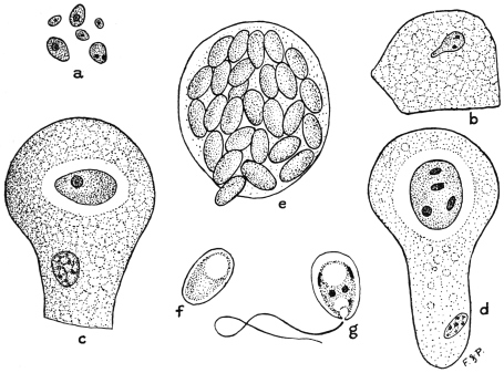
Fig. 98.—Nosema apis. Various stages in life-cycle. a, planonts or amœbulæ from chyle stomach of bee; b, amœboid planont creeping over surface of gut epithelial cell; c, uninucleate trophozoite within epithelial cell; d, meront with nucleus divided into four, about to form four spores; e, epithelial cell crowded with spores; f, young spore; g, spore showing five nuclei, polar filament ejected, and amœbula, about to issue. × 1,500, a-e; × 2,150, f-g. (After Fantham and Porter.)
The life cycle of Nosema apis, parasitic in bees, may be taken as an example of that of a microsporidian. The infection of the host is initiated by the ingestion of spores of N. apis in food or drink contaminated with the excrement of other infected bees. Under the influence of the digestive juice of the bee the spore-coat (sporocyst) softens, the polar filament is ejected and anchors the spore to the gut epithelium, and the minute amœbula contained in the spore emerges. The amœbula is capable of active amœboid movements (fig. 98, b) and so is termed the planont or wandering form (fig. 98, a). After a short time each planont penetrates between or into the cells of the epithelium of the gut, a few only passing through into the body cavity. Within the cells the amœbulæ become more or less rounded, lose their power of movement, and after a period of growth of the trophozoite (fig. 98, c) commence to divide actively, these dividing forms being known as meronts (fig. 98, d). Various forms of fission occur, and during this phase, termed merogony, the numbers of the parasite within the host are greatly increased, with concomitant destruction of the epithelium (fig. 98, e). After a time sporogony commences. The full-grown meront becomes successively the pansporoblast and sporoblast. Nuclear multiplication and differentiation ensue and five nuclei are ultimately produced. At the same time a sporocyst is secreted, and two vacuoles are produced within. One is the polar capsule, and within it the polar filament is differentiated; the other forms the posterior vacuole (fig. 98, g). Between the two vacuoles the body cytoplasm or sporoplasm forms a girdle-like mass. Of the nuclei, one regulates the polar capsule, two control the secretion of the sporocyst, and two remain in the sporoplasm. The polar capsule and polar filament are not usually visible in the fresh condition, but can be demonstrated by the use of various chemical reagents (fig. 100). The sporoplasm ultimately becomes the amœbula (fig. 98, g) which issues from the spore after the ejection of the polar filament.
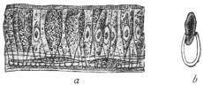
Fig. 99.—a, section through the abdominal wall of a silkworm, whose epithelial cells contain Microsporidia (Nosema bombycis); b, a spore, the contents of which are escaping. (After Balbiani.)
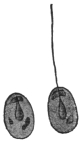
Fig. 100.—Nosema bombycis, Naeg. Spores treated with nitric acid, thus rendering the polar capsule perceptible, and the filament has protruded from one of the spores. (After Thélohan.)
A trophozoite (meront) of N. apis becomes a single pansporoblast which gives rise to one sporoblast producing one spore, and this procedure is characteristic of the genus Nosema. In other genera the trophozoite may form more than one pansporoblast and each pansporoblast may form a variable number of spores in different cases. Various attempts at classification have been based on these characteristics. It must suffice here to note that in the cases where the trophozoite becomes one pansporoblast, the latter can produce four spores in the genus Gurleya, eight spores in Thélohania and many spores in Pleistophora. In other cases, where the trophozoites give rise to many pansporoblasts, each of the latter may form many spores, as in the genus Glugea.
A few pathogenic microsporidian parasites other than N. apis may be mentioned. N. bombycis, causing pébrine in silkworms, may infect any or all the tissues of the host (fig. 99). The larvæ of the host, i.e., the “silkworms,” may become infected by eating food contaminated with spore-containing excrement of already infected silkworms. In cases of heavy infection the silkworm dies, but should the infection be less intense the larva becomes a pupa in which the parasite persists, so that the moth emerges from the cocoon already infected. Not only is the moth parasitized itself, but the Nosema reaches the generative organs of both sexes and penetrates the ovaries of the female, with the result that the ova are deposited infected. Such infected eggs are capable of developing, so that infection may be transmitted hereditarily as well as by the contaminative method. Infected eggs can be recognized by microscopic examination, as Pasteur showed, and thus preventive measures may be adopted.
A microsporidian parasite is known to occur on the roots of the spinal and cranial nerves of Lophius piscatorius, the angler fish. This parasite is variously referred to the genera Nosema and Glugea.
Thélohania contejeani, parasitic in the muscles of crayfish, is believed by some to be the causal agent of recent epizoötics among them, though others believe the disease to be really due to a bacillus. It may be that the one organism aids in the entry of the other into the host.
Order. Actinomyxidia, Stolč.
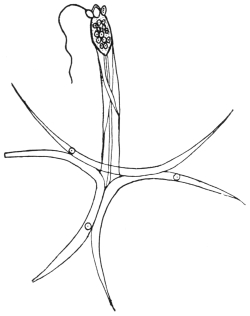
Fig. 101.—Spore of Hexactinomyxon psammoryctis. At top of figure three polar capsules, one with polar filament extended. × 450. (After Stolč.)
A brief mention may be made of the Actinomyxidia (fig. 101), which were first described by Stolč in 1899 as parasites of Oligochætes. They have also been investigated by Mrazek, and a detailed study of certain species was made by Caullery and Mesnil (1905). The trophozoite is small and amœboid. The spores are large, and exhibit tri-radiate symmetry. Spore formation is complicated and sexual processes occur therein. Many amœbulæ are set free from each spore.
Order. Sarcosporidia, Balbiani.
The first member of this group was discovered by Miescher in 1843. This author found white filaments running parallel with the direction of the fibres in the voluntary muscles of mice. They were visible to the naked eye, and proved to be cylindrical tubes tapering at each end. They were as long as the muscular fibres, were enveloped in a membrane, and contained innumerable elongate or kidney-shaped bodies and a smaller number of little spherical forms. Th. v. Hessling confirmed (1853) the occurrence of these “Miescher’s tubes” within the muscular fibres, this author having discovered the same structures in the heart muscles of deer, cattle, and sheep. Both investigators considered them to be pathological transformations of the muscles. v. Siebold, from his own experiences, regarded them as fungus-like entophytes.
Rainey (1858) discovered similar structures in the muscular system of pigs, and considered them to be early stages of Cysticercus cellulosæ, which error Leuckart rectified, simultaneously emphasizing their relationship with Myxosporidia. Both these authors found them in the muscular fibres, and both observed that they possessed a thick striated membrane. Manz (1867) published the results of more minute investigations on the structure and contents of the cylinders. This observer also recognized the disease in rabbits and attempted to cultivate the parasites. He also tried to induce experimental infection in guinea-pigs, rats, and mice, but the result was negative.
However, domestic and wild mammals are not the only hosts of Sarcosporidia; these parasites are also harboured by birds. Thus, according to Kühn, they are found in the domestic fowl; according to Rivolta in Turdus, Corvus, and other birds; according to Stiles in North American birds; while Fantham found Sarcosporidia in the African mouse-bird, Colius. Reptiles also are parasitized occasionally. Bertram found them in the gecko, Lühe in the wall-lizard. It was found also that the Sarcosporidia could develop not only in the muscles but also in the connective tissue. This led to the foundation of a new, but provisional, classification by Blanchard, using the generic name Miescheria for the parasites in the muscles and Balbiania for those in the connective tissue. Finally, Sarcosporidia have also been observed in man.
The relation of these parasites to certain diseases of domestic animals has been studied by veterinary surgeons. Sarcosporidia may cause fatal epizoötics among sheep.
There is still a wide field open for research in regard to the structure and development of these parasites, and the manner in which the hosts become infected.
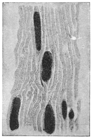
Fig. 102.—Longitudinal section of a muscle of the domestic pig, with Sarcocystis miescheriana. × 30. (After Kühn.)
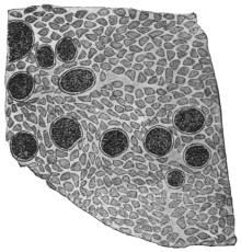
Fig. 103.—Transverse section of the muscle of a pig, with Sarcocystis miescheriana. × 38. (After Kühn.)
The Sarcosporidia usually appear as elongate, cylindrical, or fusiform bodies, rounded at both extremities and of various lengths and breadths (fig. 102). In some species they may be from 16 mm. to 50 mm. long, as in the sheep and roebuck. These bodies are the so-called sarcocysts or Miescher’s tubes. They lie in transversely striated muscular fibres which they distend more or less. The forms found in the connective tissue are apparently parasites which originally inhabited the muscular fibres, and only on disintegration of the fibres reached the connective tissue, where they grow to large oval or globular bodies (fig. 105). The mammalian muscles usually infected are those of the œsophagus, larynx, diaphragm, body-wall, and the psoas muscles. The skeletal muscles may be affected in acute cases, as well as those of the tongue and eye. The heart muscles are sometimes parasitized.
In fresh material cut into thin slices the parasites are frequently recognizable, even with the naked eye, because of their yellowish-white colour. Under the microscope they appear to be coarsely granular (fig. 103). Beginners may find some difficulty in distinguishing them from other foreign bodies, such as dead and calcified encapsuled Trichinæ, or from Cysticerci that have died and become calcified in the early stages, more particularly as the Sarcosporidia also occasionally may become calcified.
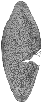
Fig. 104.—Sarcocystis miescheriana from pig. Late stage in which body is divided into numerous chambers or alveoli, each containing many spores. (From Wasielewski, after Manz.)
The Sarcosporidia are always enveloped in a membrane, which is probably formed at an early stage. In a few cases it remains thin and simple, in other cases a radially striated ectoplasmic layer is present (figs. 104, 108), which has been variously described. From the inner integument, which may be homogeneous or fibrous, thick or thin, membranes or trabeculæ pass into the interior of the body, forming anastomosing partitions, and so producing a system of chambers of various sizes that do not communicate with one another (figs. 104, 108). These chambers are occupied by sickle- or bean-shaped bodies (spores or sporozoites), or various developmental stages of them. The oldest spores are found in the centre of the Miescher’s tubes or trophozoites. If they are not liberated they die there, so that the central chambers of the tube are empty and hollow.
In the youngest Sarcosporidia (40 µ in length) from the muscles of the sheep there occur, according to Bertram, small roundish or oval cells (4 µ to 5 µ), the nuclei of which are half their size, and are embedded in a granular protoplasmic mass. In somewhat larger, and therefore older, cylinders, the investing membrane of which already shows both layers, the cells have become larger (to 7 µ) and are more sharply outlined from each other (fig. 106). These uninucleate cells may be considered as pansporoblasts. In each pansporoblast division of the nucleus occurs (fig. 107), and meanwhile the pansporoblasts become isolated within the chambers, the dividing partitions of which originate from the granular protoplasm which is present between the pansporoblasts. The numerous uninucleate daughter forms produced within the chambers become spores direct (fig. 108).
The process commences in the centre of the cylinders or sarcocysts, and then progresses towards the extremities, the parasites meanwhile increasing in size, and new pansporoblasts being continually formed at the extremities (fig. 107).
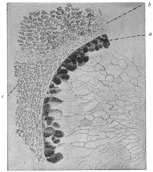
Fig. 105.—Transverse section of Sarcocystis tenella, Raill. From the œsophagus of the sheep, Ovis aries. × 38. a, marginal chambers filled with spores; b, connective tissue of the œsophagus; c, muscles of the œsophagus.
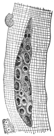
Fig. 106.—Young Sarcocystis tenella of the sheep, 47 µ in length. (After Bertram.)
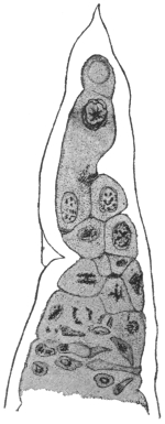
Fig. 107.—End of a trophozoite of Sarcocystis miescheriana from the diaphragm of the pig, showing division in pansporoblasts. × 800. (After Bertram.)
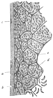
Fig. 108.—Sarcocystis blanchardi of the ox. Longitudinal section of sarcocyst or Miescher’s tube. a, substance of muscle fibre; b, envelope of sarcocyst; c, muscle nuclei; d, spores in chambers; e, ground substance. × 400. (From Wasielewski, after van Eecke.)
The spores (sometimes called Rainey’s corpuscles), vary in shape according to the species, but are also of different form individually. They are mostly kidney-, bean- or sickle-shaped (fig. 109), and of small size, sometimes reaching 14 µ by 3 µ to 5 µ. They are apparently surrounded by a thin membrane, and at one extremity (according to the discovery of L. Pfeiffer, confirmed by van Eecke, Laveran and Mesnil) contain an obliquely striated body (fig. 109) often homologized with the polar capsule, while the greater part of the spore is taken up by the nucleate sporozoite. Several authors state that they have also observed filamentous appendages (polar filaments) at one end of the spores, and have seen two kinds of spores in the same Sarcosporidium. Spores of various species of Sarcosporidia may contain metachromatic granules, often centrally placed (fig. 109). These granules may be metabolic or possibly may contain toxin (see below).
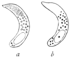
Fig. 109.—Spores of Sarcocystis tenella, Raill. a, fresh, showing the polar capsule; b, stained, showing metachromatic granules and nucleus. × 1,000. (After Laveran and Mesnil.)
The gymnospores of Sarcocystis muris, from the mouse, show active boring movements when kept in saline solution warmed to 35° or 37° C. S. muris is very deadly to its host. From their structure the spores do not appear to have great powers of resistance to external conditions. They measure 12 µ by 3 µ to 4 µ or less.
Laveran and Mesnil (1899) isolated a toxin from S. tenella of the sheep and called it sarcocystin. This substance is especially pathogenic to experimental rabbits.
The duration of life of the Sarcosporidia is a comparatively long one. The affected muscular fibres may remain intact and capable of performing their functions for a long time, but at last they perish, if the host lives long enough. Thus the Sarcosporidia of the muscles are then enveloped only by sarcolemma, and finally, when this likewise disappears, they fall into the intra-muscular connective tissue. In many cases the Sarcosporidia die off within their hosts, this, according to Bertram, being brought about by a disintegration of the spores in the central chambers. In other cases the leucocytes play a part in the destruction of the Sarcosporidia, and sometimes it happens that lime salts are deposited in and around the vacant cylinders.
In some places pigs, sheep, mice and rats are infected with sarcosporidiosis to a remarkable extent, in certain cases almost reaching 100 per cent. Young animals also are infected, and perhaps infection only takes place during youth.
Although the natural mode of transmission of the Sarcosporidia remains to be determined, yet various experimental researches on the problem are of interest and importance. Theobald Smith (1901) found that mice could be experimentally infected with S. muris by feeding them with the flesh of other infected mice. The incubation period was a long one, namely forty to fifty days. Thus, on the forty-fifth day after feeding young Sarcosporidia were found, and seventy days after feeding spore formation began. Ripe spores were found two and a half to three months after the commencement of these experiments. This mode of infection—a cannibalistic one—hardly seems likely to be the natural method for the infection of sheep and ruminants generally. Smith’s researches have been confirmed. Nègre225 (1910) found that the fæces of mice fed on infected muscular tissue were infective to other mice when ingested by them. Negri226 infected guinea-pigs with S. muris by feeding them on infected mouse flesh, and found that the parasite in guinea-pigs showed different characters from those exhibited by it in mice. Darling227 also succeeded in infecting guinea-pigs with S. muris, and Erdmann infected mice with S. tenella (from the sheep).
According to Erdmann228 (1910) the Sarcosporidian spore germinates in the intestine of the host, which has recently ingested infected material. The spore liberates its contained toxin—sarcocystin—which acts upon the adjacent intestinal epithelium, whereby the latter is shed, and an amœbula creeps out of the spore. The amœbula is able to penetrate the denuded area and get directly into the lymph-spaces of the submucous coat of the intestine. The first period of development, lasting some twenty-eight to thirty days, is said to be passed in the lymph-spaces of the intestine. Later the amœbula reaches a muscle fibre. Writing in May, 1914, Erdmann229 records the appearance of small amœboid and schizogony forms six days after infection of the host. Crawley230 (1913) controverts some of these statements and considers that the Sarcosporidian spore, still sickle-shaped, bores its way into the epithelial cells of the intestine and comes to rest there. The spore then becomes round or elliptical, and peripheral masses of chromatin appear within it, suggesting schizogony. This happens about twelve hours after feeding, and in twenty-four hours the spores appear to have left the intestine. More recently (May, 1914), Crawley231 considers that there is sexual differentiation among the Sarcosporidian spores, a few hours after their ingestion by the host.
Interesting discussions have occurred as to the site of the toxic sarcocystin within the spore. Metachromatic granules occur in the middle of the Sarcosporidian spore (fig. 109), and the toxin may be contained in these grains, as they disappear, according to Erdmann, before the amœbula penetrates the denuded intestinal wall. However, a polar capsule, containing a polar filament, may be present at one end of a Sarcosporidian spore. Laveran and Mesnil described a striated area at the more pointed end of the spore of S. tenella, which area they consider to represent a polar capsule. Fantham232 (1913) found a vacuole-like, polar capsule area in the spores of S. colii from the African mouse-bird. The sarcocystin may be contained in the polar capsule. The nucleus of the spore is generally at the opposite, blunter end.
Again, various authors have stated that Sarcosporidian spores may occur in the blood of the host at times. If so, then an intermediate host may be concerned in their transmission. Perrin suggested that Sarcosporidia might be spread by blow-flies and flesh-flies.
The classification of the Sarcosporidia as proposed by R. Blanchard, which was based on their various habitats, can no longer hold, because the same species may occur in the muscles as well as in the connective tissue. For the present, the few species that are known may be placed in one genus, Sarcocystis, Ray Lankester, 1882.
The following species of Sarcocystis are of interest:— S. miescheriana, Kühn, 1865, in the pig. S. bertrami, Doflein, 1901, in the horse. S. tenella, Railliet, 1886, in sheep. S. tenella bubali in buffaloes in Ceylon and Egypt. S. blanchardi, Doflein, 1901, in cattle. S. muris, Blanchard, 1885, in the mouse, to which it is lethal. S. hueti, Blanchard, 1885, in the seal. S. colii, Fantham, 1913, in the African mouse-bird, Colius erythromelon.
Also various Sarcosporidia from antelopes, monkeys, opossum, birds, the gecko and wall-lizard are known.
The spores of S. muris, S. bertrami, S. tenella, and S. colii can multiply by longitudinal fission.
Sarcosporidia observed in Man.
(1) Lindemann233 found on the valves and in the myocardium of a person who had died of dropsy certain brownish masses, 3 mm. in length and 1·5 mm. in breadth which he regarded as gregarines. If these were actually independent animal organisms it may be suggested that they were Sarcosporidia. Rivolta (1878) named the species S. lindemanni.
(2) Rosenberg234 found a cyst 5 mm. in length and 2 mm. in breadth in a papillary muscle of the mitral valve of a woman, aged 40, who had died from pleuritis and endocarditis. The cyst contained no scolex nor hooklets of tænia. Numerous small refracting bodies, round, oval or kidney-shaped, were found in a daughter cyst, as well as sickle-shaped bodies. The description hardly appears to indicate Sarcosporidia.
(3) Kartulis235 observed Miescher’s cylinders of various sizes in the liver (?) and in the muscular system, of a Sudanese who had succumbed to multiple abscesses of the liver and abdominal muscles. This may be considered as the first actual case of the occurrence of Sarcosporidia in man. Koch in 1887 described a case in Egypt.
(4) The case reported by Baraban and St. Remy236 was at once demonstrated as certain. It related to a man who had been executed, and in the laryngeal muscles of whom Sarcosporidia were found; the length of the parasites varied between 150 µ and 1,600 µ, their breadth between 77 µ and 168 µ. The affected muscular fibres were distended to four times their normal thickness. This species was described by Blanchard as “Miescheria” muris, but according to Vuillemin, it was more probably Sarcocystis tenella of the sheep.
(5) Vuillemin has also described a case of Sarcosporidia found in the muscles of a man who died from tubercle at Nancy. The author considered that the parasite corresponded to S. tenella.
(6) Darling237 (1909) found Sarcosporidia in the biceps of a negro from Barbados.
The Myxosporidia, Microsporidia, Actinomyxidia and possibly the Sarcosporidia may be included within the section Cnidosporidia (Doflein), since they possess spores containing polar capsules.
Order. Haplosporidia, Caullery and Mesnil.
The Haplosporidia are a group of organisms having both a simple structure and life-history. The simplicity may represent a primitive condition or may be due to degradation resultant on parasitism, and thus it is possible that the group is not a homogeneous one. The order Haplosporidia was created by Caullery and Mesnil in 1899, and includes parasites of rotifers, annelids (fig. 110), crustacea, fish, prochordates and man. They may be present in the body cavity or alimentary tract, and can also occur in the septum nasi of man, in the nervous system of Cephalodiscus, and in tumours of fish.
As the name implies, the spores of the Haplosporidia are simple, without polar capsules, and are uninucleate. In some genera, e.g., Haplosporidium, Urosporidium (fig. 111) there is a spore-coat or sporocyst which may be elongate or spiny. The developmental cycle of a Haplosporidian, such as Haplosporidium or Bertramia, begins with a small, uninucleate cell, often rounded, possessing a cell membrane that may be prolonged into processes. Growth takes place, coupled with an increase in the number of nuclei, so that a multinucleate trophozoite is produced. Later, this multinucleate trophozoite becomes segmented into a number of ovoid or spherical pansporoblasts, which give rise to few (one to four) spores. Such a spore, when set free, begins the life cycle over again.
More recently (1905–1907) two important organisms have been described and included in this group, namely, Neurosporidium cephalodisci238 (Ridewood and Fantham) from the nervous system of the prochordate, Cephalodiscus nigrescens, and Rhinosporidium kinealyi (or seeberi) from the septum nasi of man. In the case of Rhinosporidium and Neurosporidium, after the uninucleate spore has grown into a multinucleate trophozoite, the latter segments into uninucleate pansporoblasts, as in the preceding cases. A difference then occurs, for each pansporoblast enlarges, its nucleus divides and a “spore-morula” is formed. Thus a multinucleate pansporoblast or spore-morula, divided into many uninucleate sporoblasts (spore mother cells) is produced, and each sporoblast without further change becomes a uninucleate spore.
The Haplosporidia have therefore been divided by Ridewood and Fantham (1907)239 into two sections:—
(1) The Polysporulea, wherein the pansporoblast gives rise to a number of spores (nine or more), e.g., Rhinosporidium, Neurosporidium.
(2) The Oligosporulea, wherein the pansporoblasts give rise each to a few (four) spores or to only a single spore, e.g., Haplosporidium, Bertramia, Cœlosporidium, Ichthyosporidium.
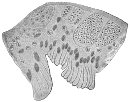
Fig. 110.—Haplosporidium heterocirri. Section throughout wall of the Polychæte worm, Heterocirrus viridis, showing various developmental stages of the Haplosporidium. × 550. (After Caullery and Mesnil.)
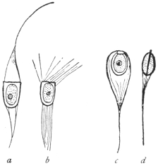
Fig. 111.—Haplosporidian spores. a, b, Haplosporidium heterocirri. a, fresh; b, after immersion in sea water; c, d, Urosporidium fuliginosum. × 1000. (After Caullery and Mesnil.)
Rhinosporidium kinealyi, Minchin and Fantham, 1905.
Rhinosporidium kinealyi, parasitic in man, must now be considered in greater detail. This organism was found in nasal polypus in India, and has since been recorded from the ear as small nodules in the external auditory meatus. The Indian cases came from the neighbourhoods of Calcutta and Madras, and the parasite has been seen in Ceylon. Similar structures have since been described from the United States and South America.
The Rhinosporidium polypus is said not to be particularly painful, though nasal forms must interfere with breathing to some extent. The first nasal polyp reported from India formed a vascular pedunculated growth on the septum nasi and was about the size of a large pea or raspberry. It was compared with a raspberry, being red in colour with a number of small whitish dots upon its surface. When the tumour was cut, a number of similar whitish dots were seen within. These were the cysts of Rhinosporidium. According to Minchin and Fantham240 (1905), they vary considerably in size and measure up to 200 µ or 250 µ in diameter. Each possesses a cyst wall which varies in thickness in different cysts. Its outer wall is always firm and distinct, the inner limit being less definite at times. Each large cyst is filled with numbers of spherical or oval bodies, showing every gradation between small ones at the periphery and large ones at the centre (fig. 112). Roughly, three zones of parasites can be distinguished in a large cyst, a peripheral set consisting of the youngest parasites, an intermediate group and a central, oldest zone. A large cyst may possess a pore for the egress of its contents. Some of the cysts show polar distribution of the zones.
The youngest forms of Rhinosporidium are difficult to detect. They are small, granular masses, round, ovoid or irregular and at times even amœboid in appearance. These are young trophozoites. They increase in size, but encystment occurs early, the outer layer becoming firm so that the organisms have a definite contour. Each is soon multinucleate and the cytoplasm segments around the nuclei. The cyst thus becomes full of uninucleate pansporoblasts or sporonts, with a peripheral layer of undifferentiated protoplasm. The pansporoblasts grow in size. In the larger cysts the formation of pansporoblasts progresses at the expense of the peripheral layer of protoplasm, which, however, continues to grow, so that the cyst as a whole increases in size. The pansporoblasts at first are uninucleate (fig. 112, a), and then undergo nuclear multiplication. This is well seen in the intermediate zone of parasites, where the pansporoblasts show first one, then two, then four or more spores (fig. 112, b), while in the oldest centrally placed pansporoblasts, about a dozen or sixteen closely packed spores (fig. 112, c), can be seen. The spore is small and rounded, and its nucleus is clear and distinct. The fully formed pansporoblast or spore morula becomes surrounded by a membrane.
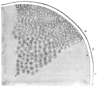
Fig. 112.—Rhinosporidium kinealyi. Portion of ripe cyst containing pansporoblasts of various ages. × 480. (After Minchin and Fantham.)
Certain of the cysts have been found in a ruptured condition, whereby the spores have been liberated into the surrounding tissue. It is almost certain that the spores serve for the auto-infection of the host, for though the tumours of Rhinosporidium seemed to have been removed entirely, it has been found that they recur, some minute fragment of the parasite having probably been left behind. The method whereby the parasite reaches new hosts has not yet been determined, and it would be of interest if its life-history could be more fully investigated.
The Asiatic specimens of R. kinealyi were first described in detail by Minchin and Fantham (1905) from material briefly reported to the Laryngological Society of London in 1903, by O’Kinealy. Material obtained by Dr. Nair, of Madras, was described by Beattie241 in 1906. This material came from Cochin. Castellani and Chalmers have found similar polypi in Ceylon.
Wright242 has described the parasite from Memphis, Tennessee. Seeber243 in 1896 described nasal polypi in Buenos Ayres, and in 1900 Wernicke named the parasite therein Coccidium seeberi. Seeber’s parasite is a Rhinosporidium, R. seeberi, and may ultimately be found to be the same as R. kinealyi. Ingram244 reports Rhinosporidium cysts, with pores in the cyst walls, in conjunctival polypus and in papilloma of the penis in India. Zschokke has reported the presence of Rhinosporidium in horses in South Africa.
Class IV. INFUSORIA, Ledermüller, 1763.
The Infusoria (or Heterokaryota, Hickson, or Ciliophora, Doflein) include the Ciliata and the Suctoria. A few authorities, including Braun, raise the Suctoria (or Acinetaria) to separate rank as a class, but this is not widely followed.
The body of the Ciliata usually is bilaterally symmetrical and is enveloped in a cuticle which has numerous openings for the protrusion of the cilia. Most kinds have a fixed shape, whilst changes in the form of others are brought about by the contractions of the body substance. The latter exhibits hyaline ectoplasm, in which myonemes, and occasionally also trichocysts (minute spindle-shaped bodies) appear, and granular endoplasm which may contain numerous vacuoles. The cilia, on whose various arrangements the classification is based, are always processes of the ectoplasm. Their form varies; they may be hair-like, or more rarely thorn-like, spur-like, or hook-shaped; undulatory membranes also may occur, which are probably composed of fused cilia.
With the exception of some of the parasitic species, an oral cavity, peristome or cytostome, is always present. It is frequently beset with cilia or provided with undulatory membranes, which help to waft the food inwards; sometimes there is an anal aperture (cytopyge) generally placed at the opposite pole of the organism. A cytopharynx fringed with cilia or sometimes with a specialized supporting apparatus is connected with the peristome. Vacuoles form round the ingested food, and in many species a constant rotation goes on in the endoplasm. Often one, and sometimes two contractile vacuoles are present, the frequency of the pulsations of which depends on the surrounding temperature. Sometimes special conducting channels lead to the vacuoles, or there are outlet channels leading to the exterior.
There is in almost every case a large nucleus (macronucleus), and lying close up to it a small nucleus (micronucleus). The form of the large nucleus varies according to the species. Numerous nuclei are not very common, but these occur in Opalina, which lives in the hind-gut of amphibia, and is also distinguished by the absence of an oral aperture.
Reproduction is effected by binary fission; less commonly, after encystment, by multiple division, or by budding. The divisions can be repeated many times, but finally cease, and then the conjugation of two specimens brings about a regeneration, particularly of the nuclei. Numerous examinations (Bütschli, Hertwig, Maupas, Calkins) have demonstrated that after two individuals have associated by homologous parts of the body, the micronucleus separates from the macronucleus, becomes larger and divides twice by mitosis, so that four micronuclei are present in each one of the two individuals forming the couple. Three of these nuclei perish and become absorbed, the fourth gradually passes to the portion of protoplasm connecting the two conjugants, which has originated by absorption of the cuticle at the point of contact of the conjugants. After a further division one micronucleus of each conjugant passes over into the other conjugant, and fusion ensues between the two micronuclei of each individual. Complicated changes and divisions may occur, but only the main principles can be noted here. A new nuclear body is thus formed in each conjugant, and soon divides into two. Of the segments thus produced one becomes a micronucleus, and one or several of the others, as the case may be, form or amalgamate into a new macronucleus, the old macronucleus usually perishing or becoming absorbed during the conjugation. Usually, sooner or later, the two conjugants separate, or may have separated already, and again multiply independently by fission until a series of divisions by simple fission is again followed by conjugation. The theoretical significance of conjugation cannot be dealt with fully here. It may be remarked, however, that the macronucleus plays no part in it, but governs entirely the metabolism of the Infusorian, whereas the micronucleus is essentially a generative nucleus from which macro- and micro-nuclei are again and again produced.
Encystment amongst the Infusoria is very general, and is essentially a means of protection when the surrounding medium dries up. Doubtless these cysts are frequently carried long distances by the wind, which explains the wide geographical distribution of most species. Also, multiplication often takes place in the encysted condition.
Some Infusoria live a free life, others are sedentary; the latter form colonies in fresh as well as in salt water. Numerous species are parasites of various lower and higher animals,245 and a few also are parasitic in man.
The Prague zoologist, v. Stein, introduced a classification of the Infusoria that has been almost universally adopted. It is founded on the different position of the cilia on the body. Though, no doubt, artificial, it is a convenient system. Bütschli has compiled a better one.246 But for our purpose Stein’s system is sufficient:—
Order 1. Holotricha, Infusoria with cilia that are evenly distributed over the entire body.
Order 2. Heterotricha, ciliated all over like the Holotricha, but having stouter cilia about the peristome.
Order 3. Hypotricha, only ciliated on the ventral surface.
Order 4. Peritricha, with only a ring of spiral cilia, mostly sedentary.
The Infusoria observed in man belong to the order Heterotricha, with few exceptions.
Genus. Balantidium, Claparède et Lachmann.
Heterotrichous Infusoria of oval or bag-like form and almost circular on transverse section; the anterior extremity narrowed, the posterior end broad and rounded off, or also narrowed; the peristome starting at the anterior end is broadest there and becomes narrower as it gradually obliquely approaches towards the posterior extremity. There are coarse cilia along the entire left border and the anterior part of the right border. Longitudinal striation is distinct and regular. There are two contractile vacuoles on the right, and occasionally also two or more to the left. The anus (cytopyge) is terminal. The macronucleus is usually horse-shoe or kidney-shaped, sometimes oval; the micronucleus contiguous. Reproduction is by binary fission and conjugation, and encystment occurs. The cysts are spherical or oval. These ciliates are parasitic in the large intestine of human beings and pigs, in Amphibia, and in the body cavity of polychæte Annelida.
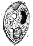
Fig. 113.—Balantidium coli. a, nucleus; b, vacuole; c, peristome; d, bolus of food. (After Leuckart.)
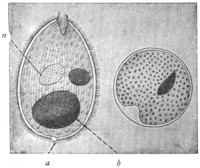
Fig. 114.—Balantidium coli, free and encysted; a, anus or cytopyge; n, macronucleus; b, bolus of food. (After Casagrandi and Barbagallo.)
Balantidium coli, Malmsten, 1857.
Syn.: Paramæcium coli, Malmsten, 1857.
The body is oval, 60 µ to 100 µ in length (up to 200 µ according to Janowski), and 50 µ to 70 µ in breadth. The peristome is funnel-shaped or contracted, the anterior end being then broadened or pointed according to the degree of contraction (figs. 113, 114). The ecto- and endo-plasm are distinct, the latter is granular, containing drops of fat and mucus, granules of starch, bacteria, and occasionally also red and white blood corpuscles. There are usually two contractile vacuoles, seldom more. The anus (cytopyge) opens at the posterior extremity. The macronucleus is bean- or kidney-shaped, rarely oval; the micronucleus is spherical.
Balantidium coli lives in the large intestine of man, in the rectum of the domestic pig, and has been found in monkeys. It propagates by transverse division, but conjugation and encystment are known to take place.247 Transmission to other hosts is effected by the cysts of the parasite (fig. 114).
Balantidium coli, first seen by Leeuwenhoek, was described by Malmsten in 1857 in a man aged 35 years, who had two years previously suffered from cholera, and since then had been subject to diarrhœa. The examination showed an ulcer in the rectum above the mid sphincter ani, in the sanguineous purulent secretion of which numerous Balantidia were swimming about. Although the ulcer was made to heal, the diarrhœa did not cease and the stools contained numerous Balantidia, the number of which could only be decreased by extensive enemas of hydrochloric acid.
The second case related to a woman who was suffering from severe colitis, and who died ten days after admission. The malodorous, watery evacuations contained innumerable Balantidia, in addition to pus, and at the autopsy the anterior portion of the large intestine was found to be infested with them.
Subsequently this parasite has often been observed in human beings, and various cases have been recorded. These occurred in Russia, Scandinavia, Finland, Cochin China, Italy, Germany, Serbia, Sunda Islands, Philippine Islands, China, and in other parts of Asia and in America. Other cases were reported by Askanazy, Ehrnroth, Klimenko, Nagel, Koslowsky, Kossler, Waljeff, Strong and Musgrave, Glaessner248 and others. Sievers found B. coli very common in Finland.
In the majority of the cases described by Sievers from Finland, and in other cases from Central Europe, the patients suffered from obstinate intestinal catarrh, which did not always cease even after the Balantidia had disappeared. On the other hand, Balantidia have occasionally still been found to persist, though in small numbers, after the catarrh has been cured. Some authors, nevertheless, do not regard Balantidia as the primary cause of the various diseases of the large intestine, which often commence with the development of ulcers, but they consider that they may aggravate these diseases and render them obstinate. According to Solowjew, Askanazy, Klimenko and Strong and Musgrave, however, the parasites penetrate the intestinal wall, and give rise to ulcerations which may extend deeply into the submucosa, and even be found in the blood and lymphatic vessels of the intestinal wall. According to Stokvis, B. coli occurs also in the lung; at all events this author states that he found one living and several dead paramæcia (?) in the sputum of a soldier, returned from the Sunda Islands, who was suffering from a pulmonary abscess. Sievers has shown that B. coli might occur in persons not suffering from intestinal complaints, but E. L. Walker249 (1913) states that every person parasitised with B. coli is liable sooner or later to develop balantidian dysentery.
Since Leuckart confirmed the frequent presence of B. coli in the rectum of pigs, and corresponding observations were made in other countries, the pig is universally considered to be the means of the transmission of Balantidium to man. The encysted stages only serve for transmission, because, according to all observations, the free parasites have a very small power of resistance. They perish when the fæces have become cool; they cannot live in ordinary, slimy, or salt water. As they are killed by acids even when much diluted, they cannot pass through the normal stomach alive except under the most unusual circumstances. The pigs, in whose intestines the Balantidium appears to cause little or no disturbance, evacuate numerous encysted Balantidia with the fæces, and their occasional transference to man brings about their colonization there, but perhaps only when a disease of the colon already exists.
Experimental transmission of the free parasites to animals (per os or per anum) yielded negative results, even in the case of pigs. Casagrandi and Barbagallo (1896), however, had positive, as well as negative, results. They employed healthy young cats, or cats in which catarrhal entero-colitis had been artificially induced (which in other experiments is apt to cause the death of the animals experimented upon in about six or seven days), or finally cats that had dilatation of the rectum with alkaline reaction of the fæces. An attempt to infect three healthy cats by injecting human fæces containing Balantidium into the rectum proved negative, in so far as the fæces of the experimental animals had an acid reaction and contained no Balantidia, but at the autopsy performed eight days after infection a few encysted parasites were found in the mucus of the ileum. In the case of four cats suffering from entero-colitis, into which human fæces containing Balantidia were introduced per os, Balantidium cysts were found in the fæces three days after the last ingestion. Great numbers, moreover, were found in the cæcum and the posterior part of the small intestine at the autopsy of the animals, which died about eight days after the commencement of the experiments. Actual colonization, therefore, was not effected in either series of experiments. Free or encysted Balantidia of pigs were used for further experiments. The experiments proved negative when fæces containing cysts were injected into the rectum of healthy cats (three experiments), or cats (two) suffering from spontaneous intestinal catarrh, or when such material was introduced per os into three healthy cats. In the case of two cats with intestinal catarrh artificially produced, a small number of the active Balantidia injected into the rectum remained alive. Larger quantities of fæces containing encysted Balantidia were introduced into two other cats affected with the same complaint. These, certainly, did not appear in the fæces, but small numbers, free and alive, were found in the cæcum. Similarly, encysted Balantidia were introduced into two cats with dilated rectum, and whose fæces had an alkaline reaction. In these cases no parasites appeared in the fæces, but three and five days later, when the two animals were examined, a very small number were discovered free in the large intestine. Klimenko did not succeed in infection experiments with B. coli on young dogs, whose intestines had been artificially affected by disease.
More recent experiments by Brumpt have shown that young sucking pigs can be infected with Balantidium from infected monkeys (Macacus cynomolgus) and suffer heavily from the same, whereas the Balantidium of the pig is rarely harmful to its host. This and previous experiments may be thought to suggest that there are perhaps several pathogenic species, and also that harmless strains of Balantidium may occur. At the same time, it must be remembered that a large proportion of the cases recorded of Balantidian colitis occur among swineherds and butchers, that is, among people in frequent contact with pigs. Morphologically, there are practically no differences between the Balantidia found in man, monkeys and pigs, and it is probable that one species only, under slightly different environmental conditions, may be responsible for the colitis observed. In any case, efficient prophylactic measures should be taken against balantidiasis in countries where it may occur, by confining the pigs and not allowing them to run in yards and dwellings.
E. L. Walker (1913) has given a good summary of work on balantidiasis. His own researches in the Philippines showed that monkeys could be infected by Balantidia both from pigs and men. Parasites may appear in the stools only at infrequent intervals. He believes that the ciliates are the primary etiologic factor in the symptoms and lesions of balantidian dysentery.
Behrenroth (1913) has given an interesting account of Balantidium coli and its pathogenic significance.
Balantidium minutum, Schaudinn, 1899.
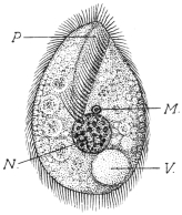
Fig. 115.—Balantidium minutum. P, peristome; N, nucleus; M, micronucleus; V, contractile vacuole. Food vacuoles are represented in the endoplasm. (After Schaudinn.)
The body is of oval form, with the anterior extremity pointed, and posterior extremity broad and rounded (fig. 115). The length is 20 µ to 32 µ, and the breadth is 14 µ to 20 µ. The peristome, which is fissure-like, extends to the centre of the body (fig. 115). The right lateral border of the peristome is beset with cilia the same length as those of the body, the left side terminates in a thin hyaline membrane that extends towards the back, and can pass over to the right side. A row of longer and stronger cilia (cirri) are on the left border of the peristome. The cuticle is refractile, the ectoplasm hyaline and the endoplasm granular, with numerous food vacuoles.
A single contractile vacuole lies dorsally and to one side at the posterior extremity. The macronucleus, which is always spherical, is central and is 6 µ to 7 µ in diameter. The micronucleus, close in front of it, only measures 1 µ (fig. 115). The cysts are oval.
These parasites were found in numbers in the evacuations of a man aged 30, who was born in Germany and had repeatedly travelled between Hamburg and North America, where he made long stays. The patient came to the Charité in Berlin to seek advice for constipation alternating with diarrhœa accompanied by abdominal pain.
A second case (the parasite of which was described as Colpoda cucullus by Schulz) was observed in a patient in the same institution.
As, in both cases, the parasites only appeared during the diarrhœa, and disappeared as soon as the fæces had assumed a normal consistency, or could only be demonstrated in a few encysted specimens, it may be assumed that the small intestine or the duodenum is their habitat.
Genus. Nyctotherus, Leidy, 1849.
Flat, heterotrichous Infusoria, kidney- or bean-shaped. The peristome commences at the anterior pole of the body and extends along the concave side to the middle, where the oral aperture is situated. The cytopharynx is oblique and is more or less curved. The cytopyge is at the posterior extremity, where a single contractile vacuole is also situated. The macronucleus is almost in the centre of the parasite. The members of this genus live parasitically in the intestine of amphibia, insects and myriapods, and at least one species is found in man.
Nyctotherus faba, Schaudinn, 1899.
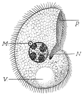
Fig. 116.—Nyctotherus faba. P, peristome; N, nucleus; M, micronucleus; V, contractile vacuole. (After Schaudinn.)
The body is bean-shaped, and a little flattened dorso-ventrally. It is 26 µ to 28 µ long, 16 µ to 18 µ broad, and 10 µ to 12 µ thick (fig. 116). The peristome is on the right border and extends to the middle; at the left there are large adoral cilia, the cilia on the right border not being larger than those on the body. The cytopharynx is short, slightly curved and turned backwards. The contractile vacuole is large, spherical, situated at the posterior extremity, and its contents are voided through the anus at its left. The macronucleus is in the centre of the body; it is globular (6 µ to 7 µ in size), and contains four or five chromatin masses. The micronucleus lies close to it, and is spherical or somewhat elongate measuring 1 µ to 1·5 µ (fig. 116). The cysts are oval.
This species has hitherto only been seen once in the same patient in whom Balantidium minutum was discovered.
Nyctotherus giganteus, P. Krause, 1906.
Under the name Balantidium giganteum n. sp., P. Krause described an Infusorian which was repeatedly observed with Trichomonas intestinalis in the alkaline evacuations of a typhoid patient in Breslau. The body is ovoid, narrower and rounded anteriorly and broader and stunted posteriorly. The peristome lies to one side; the macronucleus is bean-shaped, the micronucleus small and globular; one or two vacuoles are present. The anus is at the farther end. The organism is 90 µ to 400 µ long, 60 µ to 150 µ broad (fig. 117). After a prolonged stay outside the body, it becomes rounded and encystment occurs. In the thermostat the Infusoria remain alive at 37° C. for five weeks.
The species, however, hardly belongs to Balantidium, but to all appearances is a Nyctotherus and is distinguished from N. faba by the difference in size.
[Nyctotherus] africanus, Castellani, 1905.
In the fæces of a native of Uganda who suffered from sleeping sickness and diarrhœa and had in his intestine Ascaris lumbricoides, Trichocephalus trichiurus and Ancylostoma duodenale, Castellani found a curiously shaped Infusorian, 40 µ to 50 µ long, and 35 µ to 40 µ broad, with spherical macro- and micronucleus and a contractile vacuole (fig. 118). He included the organism in the genus Nyctotherus, perhaps wrongly, or the parasite may have been deformed. After the patient’s death the same parasite was found in the intestine and especially in the cæcum.
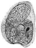
Fig. 117.—Nyctotherus giganteus. (After Krause.)
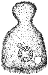
Fig. 118.—Nyctotherus africanus. (After Castellani.)
G. Lindner, in Cassel, studied certain peritrichal Infusoria (stalkless Vorticella), and connected them, probably incorrectly, with the most varied diseases of man and domestic animals, even with Sarcosporidia of pigs. It may be mentioned that according to a communication by letter from Schaudinn, Vorticella may be found in freshly evacuated fæces, but always only after the administration of a water enema. In spite of this, several other investigators mention Vorticellæ as intestinal parasites of man.
The Chilodon dentatus (Ehrenberg) recorded in 1903 by J. Guiart as a parasite of man, which may be found in all infusions, can hardly have lived in the man from whose fæces it was cultivated, but may represent a chance admixture both in the fæces and the cultivations. C. uncinatus was also found as a chance parasite of man by Manson and Sambon. According to Doflein250 (1911) certain Chilodon-like organisms have been found by Selenew in prostate secretions in gonorrhœa. Other species of the genus Chilodon are known, but only as ectoparasites (e.g., Chilodon cyprini, Moroff, 1902, from the skin and gills of diseased carp).
A number of other parasitic Ciliates are known, among which Ichthyophthirius multifiliis, destructive to fish, is important. It lives in the skin and the layers immediately below it, forming small whitish pustules which may become confluent. The pustules are most common on the head and fins, but occur also on the eyes and gills of the host. The young parasite, which is one of many formed in a cyst, is very small. At first it is free swimming, but soon attaches itself to the skin of a fish. It bores inwards and becomes surrounded by the irritated skin. There it attains a relatively large size, being 500 µ to 750 µ and occasionally more in diameter. The body has a rounded terminal mouth, short cytopharynx and a number of minute contractile vacuoles. The macronucleus is large and horseshoe-shaped; the small micronucleus is only seen in the very young animal. When full grown, the organism encysts and forces its way to the surface and bursts through, leaving a small, gaping wound behind. The cyst sinks to the bottom of the water, nuclear multiplication occurs and a number of young parasites are produced, which leave the cyst and either attack new hosts or else perish.
Opalina ranarum, parasitic in the rectum and urinary bladder of frogs and toads, shows great degradation and simplification due to parasitism, possessing no separate micronuclei, no cytostome, cytopharynx or cytopyge. It has many macronuclei, and is a large parasite. During summer and autumn nuclear multiplication followed by division of the body occurs, the process being repeated after the daughter forms have grown to the size of their parent. In spring, the Opalina divide rapidly, but do not grow much before dividing again. Finally, tiny forms, containing three to six nuclei, encyst and pass from the host with the fæces. As these latter are greedily devoured by tadpoles, the Opalina gain new hosts in which they develop.
THE CHLAMYDOZOA.
The name Chlamydozoa was proposed by Prowazek in 1907 for a number of minute, problematic organisms (fig. 119) believed to be the causal agents of certain diseases in man and animals, such as vaccinia and variola, trachoma, inclusion blenorrhœa in infants, molluscum contagiosum, and bird epithelioma contagiosum. Other diseases possibly due to Chlamydozoa251 are hydrophobia, measles, scarlet fever, foot-and-mouth disease, the “Gelbsucht” disease of silkworms, and perhaps even typhus (Prowazek, 1913). The subject is difficult and controversial and can only be briefly discussed here. It is known that the viruses in all these diseases can pass through ordinary bacterial filters, that is, they belong to the group of “filterable viruses.” At such periods the organisms are extracellular or free. It is also known that in many of these cases the virus produces definite and characteristic reaction-products or cell-inclusions in the infected cells, during the intracellular phase of the life-history of the organism. As the organisms to be considered are problematic, it will be convenient to summarize their history:—
(1) Cell-inclusions, usually named after their discoverers, have been found in certain diseases, thus: In vaccinia Guarnieri’s bodies, in scarlet fever Mallory’s bodies, in hydrophobia Negri’s bodies, in trachoma Prowazek’s bodies occur.
(2) At first these characteristic cell-inclusions were considered to be actual parasitic organisms causing the diseases in question. The bodies received zoological names and attempts were made to work out their supposed development cycles. The supposed parasites of vaccinia and variola were referred to a so-called genus Cytoryctes, those of hydrophobia to Neuroryctes, of scarlet fever to Cyclasterium, while those of molluscum contagiosum were referred to the Coccidia. Calkins in 1904 studied in detail the cell-inclusions of vaccinia and small-pox, calling them Cytoryctes variolæ, Guarnieri. Calkins considered that in the stratified cells of the epidermis they passed through two cycles, the one cytoplasmic, the other intranuclear. The first is the vaccinia cycle, the second the pathogenic (intranuclear) variola cycle. It is hardly necessary to follow all Calkins’ stages here.
Negri (1909) described a cycle for Neuroryctes hydrophobiæ. Calkins refers both Cytoryctes variolæ and Neuroryctes hydrophobiæ to the Rhizopoda.
Siegel (1905) described quite different organisms under the name Cytorhyctes. He listed several species: C. vacciniæ; of vaccinia and small-pox, C. scarlatinæ of scarlet fever, C. luis of syphilis (this is probably the granule stage of Treponema pallidum), and C. aphtharum of foot-and-mouth disease.
(3) The afore-mentioned views were criticized, and the bodies were not considered to be living organisms but merely reaction products or cell-inclusions due to the effects of the virus on the host cells. Thus Guarnieri’s bodies were stated to consist of extruded nucleolar or plastin material, having no developmental cycle. It was further asserted that infection could be produced by lymph in which Guarnieri’s bodies had been destroyed. Similar assertions have been made regarding the Negri bodies, and others. The Cytoryctes, Neuroryctes, etc., are considered, according to these views, to be degeneration products of the nucleus or to be of a mucoid nature.
(4) More recently a positive belief has gained ground that there are true parasitic organisms causing these diseases, and that the parasites are very minute, being termed Chlamydozoa by Prowazek and Strongyloplasmata by Lipschütz.
The Chlamydozoa are characterized by (a) their very minute size, smaller than any bacteria, so that they can pass through bacterial filters; (b) they pass through intracellular stages, in the cytoplasm or the nucleus of the host cell, producing therein the reaction products or inclusions in the cell already recorded as characteristic or diagnostic of the diseases produced; (c) they pass through definite developmental cycles. Such a cycle consists essentially of growth and division. The mode of division of the Chlamydozoa resembles that of the centriole of a cell, by the formation of a dumb-bell-shaped figure. Two dots are observed connected by a fine line or strand which becomes drawn out and finally snaps across the middle. Prowazek and Aragão (1909) working on smallpox in Rio de Janeiro found that the chlamydozoal granules passed through a Berkefeld filter and that the filtrate was virulent. But if an “ultra-filter” were used, i.e., one coated with agar, then the granules were retained and the filtrate was no longer virulent. The surface of the ultra-filter was found to contain many granules.