ON PARASITES IN GENERAL. (continued 11)
Reading edition
Spicules are 2 mm. long, ending in a fine point. They are moved by exsertor and retractor muscles. At first they lie free in the body cavity; next in a groove in the dorsal wall of the cloaca; then in an isolated canal, and finally in two canals. Anteriorly each has two longitudinal crests on its inner surface. These meet the corresponding crests of the other spicule, and so form a canal along which the sperm passes into the female. The gubernaculum is a thickening of the dorsal wall of the cloaca. It is not a free piece, but is moved by various muscles.
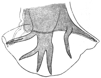
Fig. 328.—Ancylostoma duodenale: bursa of male. The rays from left to right are: (1) anterior cleft; (2) antero-external; (3) and (4) median doubled, i.e., antero-median and postero-median; (5) postero-external arising from a common trunk with the posterior. × c. 120. (After Looss.)
Genital Cone is a prominence on the floor of the bursa on the ventral side of the body, on which the genito-anal orifice opens. The cone is only slightly marked in Ancylostoma duodenale, but is much more prominent in Necator americanus.
Distribution.—Africa, Egypt, Europe, Japan, China (mainly), but in association with Necator americanus in Southern States of America, British India, Assam, Burma, Hongkong, Liberia, Jamaica, Martinique, Costa Rica, Colombia, Antigua, Guadeloupe.
Habitat.—The worms live in the jejunum, less frequently in the duodenum, of man only.
Food.—The worms feed on the mucous membrane of the gut, attaching themselves to the base of the villi, sucking these in; and when these are destroyed they attack further the submucosa. As a rule the worms have no blood in the gut, but in their attack on the submucosa a blood-vessel may be eroded, and so the gut of the worm filled with blood.
Development.—The eggs are oval with broadly rounded poles, 56 µ to 61 µ by 34 µ to 38 µ. In fresh fæces they contain four granular nucleated segmentation masses of the ovum (fig. 329) separated by a clear space from the shell.
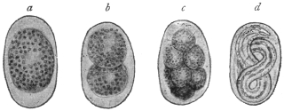
Fig. 329.—Ancylostoma duodenale: eggs in different stages of development. a to c, in fresh fæces; d, containing a larva, only in old fæces. × 336. (After Looss.)
Egg of Ancylostome appears to have a single contour. Under high powers this appears double, but they are the outer and inner surface of the true (chitinous) egg-shell. Internal to this is the extremely delicate yolk-envelope, a kind of skin secreted by the egg cell around itself for protection. The function of this is probably to absorb water to swell and burst the outer chitinous shell. The embryos when hatched are termed larvæ.
Embryos which are ready to hatch have their bodies almost free from granules; others, though apparently mature, that have granules will not hatch.318
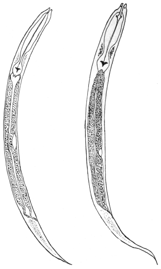
Fig. 330.—Ancylostoma duodenale larva on fourth day of culture on right; Strongyloides stercoralis larva on left. (After Leichtenstern.)
Larva.—Stage I: Average length, 25 mm. Maximum thickness in œsophageal region, 17 µ. Head end fairly blunt, from behind the anus (the tail) tapering in an uniform manner. Buccal cavity is characteristic, 10 µ to 12 µ by 1 µ to 8 µ, longer and narrower than the corresponding larvæ of Strongyloides stercoralis. Œsophagus “rhabditic” in character, i.e., it has three sections, but they are not so clearly marked off as in larvæ of the genus Rhabditis. The posterior bulb has a Y-shaped valve, the function of which, according to Looss, is to prevent regurgitation of food. The granules of the gut serve as a reserve of food, and are used up if the larvæ are starved. The genital rudiment consists of two cells half-way between the end of the œsophagus and the anus in the mid-ventral line. The larva lives on fæcal matter and grows to about 0·4 mm., then moult319 I takes place in two days or more, the skin being ruptured by the activity of the larva.
Stage II: The larva is now in this stage, which does not differ much from the previous one. It grows to 0·5 mm. The mouth opening closes. The œsophagus elongates, becoming cylindrical or “filariform”; a new skin is formed underneath the old one, and in about a week moult II takes place.
Stage III: The mature larva remains enclosed in the old skin. Its movements are now much more active and of a boring character. Length is now 0·6 mm. This mature stage has been erroneously called the encysted larva, because there is no cyst secreted from its surface by the larva, but it is simply the old skin, which is not cast off, but is retained for purposes of protection, as the larva is free living, but casts it as soon as it assumes parasitic life again. From the egg to this mature stage is thus six to ten days.
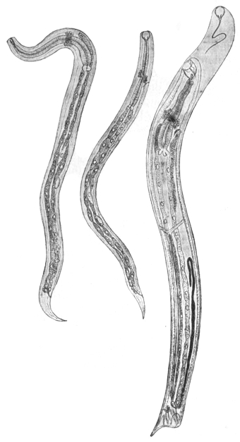
Fig. 331.—Ancylostoma duodenale: left, four days after transmission into dog, 190/1; in the centre, at the commencement of the second stage of development (five to six days), 105/1; on the right, fourteen to fifteen days after transmission. 42/1. (After Looss.)
Bionomics of Development.—Air: Eggs can develop when shut off from the air for a “comparatively long” time.
Temperature: Hatching takes from eight hours upwards. Eggs develop best at 25° to 30° C., but will not develop below 8° to 10° C. The larvæ, however, will stand freezing.
Moisture: Eggs and larvæ do not live long under water, because they suffocate or starve, but mature larvæ will live for months (six to twelve) in water; they require no food—in fact, can take none in—but live on their reserve granules, and in course of time become as clear as glass.
Thigmotropism: The mature larvæ, after casting their skin, will penetrate pith, climb up stems, stalks, etc., and creep into any pore.
It is important to recognize that this third stage of the mature larva is the only infective one.
Mode of Entry into the Body.—Infection is effected through the mouth (Leichtenstern and others), and also through the skin, as was first discovered by Looss and afterwards confirmed from the most diverse quarters, partly in the case of Ancylostoma duodenale, partly in that of A. caninum in dog, man, and monkey. The larvæ that gain access to the intestine partly through contaminated food, or through unwashed hands, or under some circumstances through water, first throw off their “sheath”—that is, they complete moult II. Moult III takes place four to five days after they have reached the gut, and they now have a mouth capsule supplied with four small teeth arranged crosswise, enabling them to fasten on to the intestinal epithelium, upon which they feed. On this food the worms grow in four to six days to 3 to 5 mm. in length, and now moult IV. takes place, thus attaining their definite shape and distinctive character. About eight days later the sexual organs commence to function; at this time the first copulation should be taking place—it will later be frequently repeated—and a few days later the first ova are laid, first in less and later in larger numbers, so that they appear in the fæces about four to five weeks after the infection.320
Infection by the Skin.—Mature larvæ, which are placed on the skin of man or suitable animals, cast their “sheath” and bore their way through delicate fissures either horizontal in the superficial scales of the epidermis, or through vertical fissures into hair follicles where these exist, and then they invade the cutis. Now according as they migrate further into the lymphatic vessels or the small vesicles, the final path to the gut differs to some extent. The blood path leads to the right heart, and from there into the lungs; here the larvæ leave the blood stream and enter the air passages, over the mucosa of which they travel further headwards, through the bronchi into the trachea and larynx, and from hence through the œsophagus to the stomach; in some cases also they are swallowed. The lymphatic path leads finally also into the blood stream, but the lymphatic glands must first be passed, and in these many larvæ are retained and perish. In the cutaneous infection seven to ten weeks elapse till the time of appearance of the first ova in the fæces.
The penetration of the skin by the larvæ also in man causes reddening and burning at the affected points, and this is followed in a few days by transitory swelling in the subcutaneous connective tissue. Skin affections can also be set up by such Ancylostoma (and Strongyloides) larvæ as do not gain access to the blood or lymphatic vessels or gut; such larvæ apparently wander further in the connective tissue, and, as Looss has in his own person observed, gain access to the cutis at different points, thus causing progressive swellings (accompanied by intense itching), which cease when the worm again penetrates into the deep tissues. Skin affections such as “ground-itch” or “pani-ghao” occurring in the tropics and only attacking the feet, or other affections (e.g., sump bunches) are now well recognized as being due to the invasion of Ancylostoma larvæ.
Other names for these skin affections are water-sore, sore feet of coolies, maza-morra, bunches, botches, quaddeln, krätze, ampoules, gourmes, taons, pitirr. Whether oral or dermal infection is the more important one further observation must decide.
The duration of life of Ancylostoma duodenale, which is a specific parasite of man and has not been observed in other mammals, amounts to about five years, as strayed larvæ according to Looss wander for this extent of time in the body.
Cultivation of Larvæ.—(1) Mix the fæces (free from drugs such as salines or thymol) with animal charcoal, adding water if necessary till a consistence of porridge is obtained. If the stools are very fluid, allow to sediment first and pour off the fluid. The best charcoal is that made from bones, and should not have an acid reaction. Charcoal is necessary in order to prevent fermentation, which kills the larvæ. Spread in layers 2 to 3 mm. thick in Petri dishes. Incubate at room temperature. To extract the larvæ from the culture allow the surface thoroughly to dry, then pour on water; the larvæ wander out and are poured off and subsequently further purified by sedimentation or filtering through blotting paper, the larvæ passing through.
(2) A funnel is plugged with cotton wool, then filled with washed sand to within a centimetre or two of the rim. Stand this in a jar of water so that the level of the water is slightly below that of the sand. On the surface of the wet sand now place layers of blotting paper, and spread the fæces, diluted if necessary, on this in layers of a few millimetres thick (vide p. 474).
Detection of Eggs.—Vide p. 473.
Dermal Infection of Dogs.—Infection with larvæ of A. caninum. In two hours most of the larvæ are free in the cutis and in four hours in the subcutaneous tissue. By scraping a few days later the mucosa of the trachea large numbers of larvæ are found there.
Ancylostoma ceylanicum, Looss, 1911.
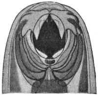
Fig. 332.—Ancylostoma ceylanicum: head end, two teeth on each side, the inner almost concealed by the outer. × c. 200. (After Looss.)
At the anterior edge of mouth capsule one large tooth; below or behind this towards the middle line a very small tooth, the tip only of which is seen. Male 5 mm. average. Lobes of bursa almost as long as broad, strongly projecting towards the ventral side. Rays short and relatively thick. Female 7 mm.
Habitat.—Intestine civet cat (Viverricula malacensis), Ceylon, and man in Bengal according to Clayton-Lane.
Other species are: A. caninum (Ercolani), in cat and dog, Europe and Africa; A. malayanum (Alessandrini), 1905, in the Malay bear (Helaretos malayanus); A. pluridentatum (Alessandrini), 1905, in Felis mitis, Brazil.
Ancylostoma braziliense, Gomez de Faria, 1910.
In cats (and dog), Brazil. Female 8·5 mm., male 7·5 mm. long. Eggs 65 µ by 32 µ. Leiper considers it to be identical with A. ceylanicum.
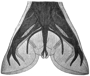
Fig. 333.—Ancylostoma braziliense: bursa of male. (After Gomez de Faria.)
Group. Bunostomeæ, Railliet and Henry, 1909.
Bursa with median double, postero- and postero-external arising from a common trunk, posterior bifurcated, each limb bidigitate (fig. 336). Vulva in middle of body or a little in front. Uteri divergent.
Contains the following genera: (1) Bunostomum (= Monodontus); (2) Necator; (3) Bathmostomum; (4) Gaigeria.
Genus. Necator, Stiles, 1903.
Mouth capsule small, narrowed anteriorly (ventrally) by chitinous plates, as in Uncinaria. On each side of the base of the dorsal cone a lateral chitinous plate or lancet with smooth edge (not serrated), ventral lancets as in Ancylostoma. No ridges on outside of ventral wall. Aperture of dorsal œsophageal gland on tip of a cone projecting freely into the buccal capsule. Bursa closed. Posterior ray cleft to its root.
Necator americanus, Stiles, 1902.
Syn.: N. africanus, Harrison, 1910.
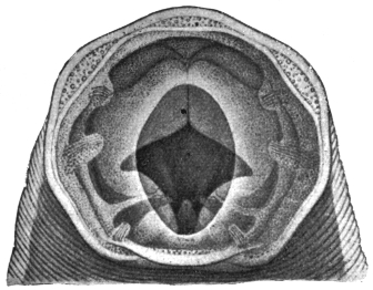
Fig. 334.—Necator americanus. Showing cutting plates and the projecting dorsal ridge, and deep in the cavity the edges of the ventral lancets. × c. 475. (After Looss.)
Male 8 mm. long, female 10 mm. The head is strongly bent dorsalwards so that almost by this character alone it can be distinguished from Ancylostoma duodenale. The buccal capsule is markedly small—in the male, 0·093 by 0·084 mm., in the female 0·11 by 0·097 mm. There are no teeth anteriorly on the ventral side of the capsule, but instead there are two cutting chitinous plates, the anterior portions of which are prominent and angular, and meet in the middle line in front. Posteriorly on each side the plate projects less, while between the anterior and posterior parts there is a deep angle. The inner (posterior) ventral lancets which also occur in A. duodenale are large, and project far into the lumen, the tips of these, of the lateral lancets, and of the dorsal cone almost meeting in the centre of the lumen. As already stated in the definition of the genus Necator, there are also lateral lancets which start from the base of the dorsal cone. This dorsal ridge, or rather in this case cone, is a striking object in the mouth, and projects right out into the cavity, and on its summit opens the dorsal œsophageal gland.
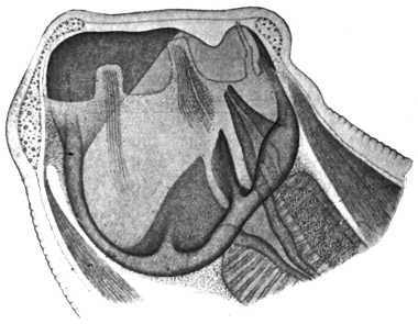
Fig. 335.—Necator americanus: lateral view, showing the dorsal ridge perforated by the duct of the dorsal œsophageal gland, the lateral lancet and ventral lancet and the nerve papillæ. × c. 475. (After Looss.)
The bursa is about as long as broad, but has the lateral lobes strikingly lengthened, giving a trilobed appearance (fig. 336), but as in Ancylostoma duodenale it is closed on the ventral side. The distribution of the rays is best understood from the figure. The genital aperture lies on a marked conical protuberance; the cement gland is bilobed in transverse section. In the female the opening of the vulva is in front of the middle line, in A. duodenale it is behind.
The spicules, 0·92 mm. long are hooked at the extremity.
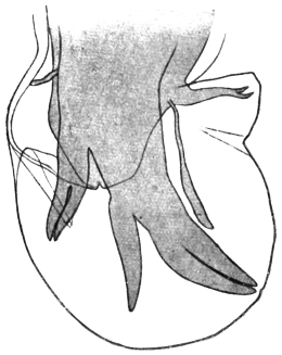
Fig. 336.—Necator americanus: bursa of male. The rays from right (top) to left are: (1) posterior, (2) postero-external, (3) and (4) median doubled, i.e., postero-median and antero-median, (5) antero-external, (6) anterior (cleft), and above it on left a pre-bursal ray. × c. 120. (After Looss.)
Eggs more pointed at the poles than those of A. duodenale, 64 µ to 72 µ by 36 µ, so that it may not be possible to distinguish single eggs owing to individual variations, yet on comparing a number they can be distinguished.
Geographical Distribution.—Brazil, Porto Rico, Cuba, Central Africa, East Africa, Victoria Nyanza, Gold Coast, Uganda, North-Western Rhodesia, Ceylon, Mysore. For other localities where A. duodenale is also found see p. 450.
Habitat.—In small intestine of man and gorilla (Troglodytes gorilla).
Necator exilidens, Cummins, 1912.
Syn.: N. africanus, Looss, 1911.
Male 7 mm., female 9 mm. long. The edges of the cutting plates are rounded, not angular, and do not meet in the middle line. Inner (posterior) ventral lancets very small. Lateral lobes of bursa broader than long. Rays thick and plump.
Habitat.—In the chimpanzee (Anthropopithecus troglodytes).
Ancylostomiasis.
Morbid Anatomy.—Organs pale and bloodless. Abdominal organs sodden, and there is fluid in the serous cavities. Lungs: œdema. Kidneys: fatty changes, especially large pale kidney. Liver and heart also show fatty changes—there is much hæmosiderin in the liver cells. Blood: early stages, a leucocytosis 20,000 upwards, and eosinophilia 50 per cent. Later, anæmia (hydræmia). The number of worms found varies from ten to 1,000. They are rare in the duodenum, but occur as far as 6 ft. from the pylorus.
Group. Syngameæ, Railliet and Henry, 1909.
Bursa with anterior and median ray cleft; antero-external, close to median; postero-external, arising separately from posterior; posterior bifurcate to base, each branch bifurcate or trifurcate. Vulva in the anterior fourth of body. Uteri divergent.
Genus. Syngamus, von Siebold, 1836.
Head thickened, not tapering; broad mouth with gaping buccal capsule.
Male and female often in permanent copulâ.
Parasitic in respiratory passages of birds and mammals.
Habitat.—S. trachealis in poultry; S. bronchialis in goose; S. laryngeus in cattle; S. vasicola in goats, etc.
Syngamus kingi, Leiper, 1913.
Buccal capsules of male and female on same level. In S. trachealis and S. laryngeus, that of male in front of that of female. In S. dispar, that of male behind that of female. Œsophagus of male one-sixth, that of female one-ninth of total length. Mouth capsule in male and female terminal; it is dorsal in S. trachealis and in mammalian species. Tail of female bluntly pointed. Ovary reaches to anus. Excretory pore opposite the middle of the bulb of œsophagus. In S. trachealis it is opposite the œsophageal valves.
Habitat.—Found in sputum of patient by King in St. Lucia. Normal host probably a carnivore.
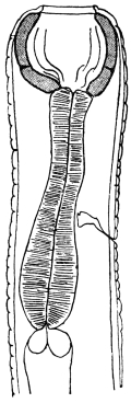
Fig. 337.—Syngamus kingi: anterior end of male. (After Leiper.)
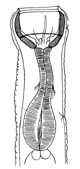
Fig. 338.—Syngamus kingi: anterior end of female. (After Leiper.)
Family. Physalopteridæ.
Genus. Physaloptera, Rudolphi, 1819.
Mouth surrounded by two large lateral lips bounded posteriorly by a cuticular band projecting anteriorly, forming a collar. Each lip bears anteriorly and inwardly a cuticular appendage, the external tooth. Immediately below and internal to the external teeth the internal teeth, one on each lip. Each lip bears two large submedian papillæ. Tail of male with four pairs of pedunculated papillæ in a row on each side external to the six pairs of unpedunculated papillæ. Spicules unequal. Vulva in the anterior region of the body. Eggs with a characteristic thick smooth shell.
Parasitic in the intestine, more especially the stomach, of mammals (twenty species), birds (twelve species), reptiles (fourteen species).
Physaloptera caucasica, v. Linstow, 1902.
The male measures 14·2 mm. in length and 0·71 mm. in breadth; the bursa is broad, rounded off in front and narrower at the back; the right spicule measures 0·62 mm. in length, the left spicule 1·76 mm.; there are two papillæ in front of the orifice of the cloaca, four behind it and six unpedunculated on the tail. The female measures 27 mm. in length, 1·14 mm. in breadth; the caudal extremity is rounded off; the vulva is on the border of the first and second sixth of the length of the body; the eggs have thick shells, and measure 57 µ by 39 µ. It has hitherto only been observed once, by Ménétriés in the intestine of man (Caucasus).
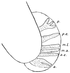
Fig. 339.—Bursa of Syngamus trachealis. a., anterior ray cleft; a.e., antero-external; m.a., antero-median; m.l., postero-median; p.e., postero-external; p., one branch of posterior (trifurcate). (Stephens.)
Physaloptera mordens, Leiper, 1907.
Large worms resembling an immature Ascaris lumbricoides.
The inner lancet-shaped teeth have a sharp cutting edge towards the lumen. Below each is a cuticular boss projecting into the mouth (fig. 340).
Male 30 to 50 mm., bursa with four pairs of pedunculated papillæ, the second and third lying external to the first and fourth on each side. Spicules unequal, one slender (4·6 mm.), the other stouter (6 mm.).
Female 40 to 55 mm. Tail sharp. Vulva opens between the anterior fourth and fifth of the body. Eggs 43·6 µ by 35·3 µ with a thick smooth shell.
Habitat.—Œsophagus, stomach, small intestine of man (several cases). Nyasaland and Portuguese East Africa.
Family. Ascaridæ, Cobbold, 1864.
Sub-family. Ascarinæ.
Without œsophageal or intestinal diverticula; spicules without flanges.
Genus. Ascaris, L., 1758.
Intermediate lips and auricles absent. Lips edged with fine teeth. Lips triangular in cross section. Not grooved on internal surface.
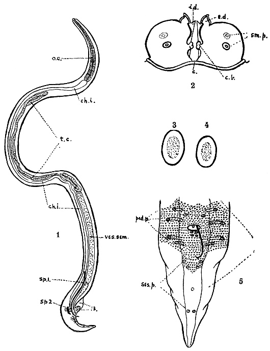
Fig. 340.—Physaloptera mordens, Leiper, 1907. (1) adult male: o.e., œsophagus; ch.i., chyle intestine; t.c., testicular coils; ves. sem., vesicula seminalis; sp. 1, long spicule; sp. 2, short spicule; B., bursa. (2) Mouth parts: c., cuticular collar embracing the two lips posteriorly; c.b., cuticular bosses guarding the mouth laterally; e.d., external tooth; i.d., internal tooth; sm.p., submedian papillæ. (3) egg of P. caucasica. (4) egg of P. mordens. (5) bursa enlarged: ped.p., pedunculated papillæ; ses.p., sessile papillæ. (After Leiper.)
Ascaris lumbricoides, L., 1758.
The colouring, in the fresh condition, is reddish-yellow or greyish-yellow; the body is of an elongated spindle shape. The oral papillæ are finely toothed. The dorsal papilla carries two sensory papillæ, the two ventral papillæ each one sensory papilla. The male measures from 15 to 17 to 25 cm. in length, and about 3 mm. in diameter; the posterior extremity is conical and bent hook-like ventrally; the spicules measure 2 mm. in length, are curved, and somewhat broadened at their free end; on each side around the orifice of the cloaca there are seventy to seventy-five papillæ, of which seven pairs are post-anal. The testicular tube is much folded, showing through the body integument, and is about eight times the length of the body. The female measures 20 to 25 to 40 cm. in length and about 5 mm. in diameter; the posterior extremity is conical and straight. The vulva is at the junction of the anterior and middle thirds of the body, which, at this point, has a slight ring-like constriction; the convoluted ovaries measure ten times the length of the body.
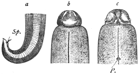
Fig. 341.—Ascaris lumbricoides. a, posterior extremity of the male with the spicules protruding from the orifice of the cloaca (Sp.); b, anterior extremity from the dorsal surface, the two lobes of the pulp of the lip separated by the “saddle”; c, anterior extremity from the ventral surface; P., excretory pore. (From Claus.)
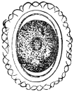
Fig. 342.—Ovum of Ascaris lumbricoides, with shell and albuminous envelope. 400/1.
The ova are elliptical with a thick (4 µ) transparent shell (fig. 342) and an external albuminous coating which forms protuberances; the ova measure 50 µ to 70 µ in length, 40 µ to 50 µ in breadth; they are deposited before segmentation; the albuminous coating is stained yellow by the colouring matter of the fæces, but is sometimes absent. The egg cell is unsegmented, it almost completely fills the shell, and its nucleus is concealed by the large amount of coarse yolk granules.
Abnormal or unfertilized eggs also occur in fæces. They are distinguished by their elongated form (80 µ by 45 µ), irregularly cylindrical, its contents consisting of refractive granules.
Ascaris lumbricoides is one of the most frequent parasites of man; it is distributed all over the inhabited parts of the world, and though it is particularly frequent in the warmer regions, yet it also occurs in Finland, Greenland, etc. In temperate climates A. lumbricoides occurs most frequently in young children; it is, moreover, more common amongst country dwellers than amongst the inhabitants of towns, but is not lacking in infants, adults and aged persons. As a rule only a few specimens are present in the intestine, but many cases are known in temperate zones in which several hundreds of worms have been found in the same patient. This species is particularly numerous in the negroes of Africa and America. It occurs also in the monkey, dog and pig (? A. suilla).
The parasite was known in ancient times; the Greeks called it ἐλμινς στρογγύλη, Plinius termed it Tinea rotunda, later on it was named Lumbricus teres. The ἄσκαρις of the Greeks is our Oxyuris.
The small intestine is the normal habitat of Ascaris lumbricoides; the worms, however, often leave this part of the intestine and wander into the stomach, whence they are frequently evacuated by vomiting, or they may creep through the œsophagus into the pharynx and crawl out through the nose or mouth; very rarely they may find their way into the Eustachian tube or into the naso-lachrymal duct, or into the excretory ducts of the liver and pancreas; exceptionally they may gain the trachea, and they have also been found in the abdominal cavity. They may bore through adhesions between the intestinal wall and the omentum (worm abscess); they occasionally penetrate the urinary apparatus and are passed with the urine; in febrile diseases A. lumbricoides usually leaves the intestine spontaneously. It is obvious that these wanderings may be accompanied by the most serious symptoms, but in sensitive persons the invasion of even only a few intestinal Ascarides gives rise to a series of almost inexplicable symptoms (hysterical, epileptiform attacks, cerebral congestion, aphonia, etc.), which cease with the expulsion of the worms, so that many authors are driven to the conclusion that these Ascarides secrete a toxin. Fortunately, the presence of A. lumbricoides in the intestine is easily demonstrated by the microscopical examination of the fæces.
Development.—Several authors (Gros, Schubart, Richter, Leuckart and Davaine) have demonstrated that the ova of Ascaris develop in water or moist earth after a long period of incubation. Freezing and desiccation (if not too long) do not injure their powers of development; the duration of the development depends on the degree of the surrounding temperature. At a medium temperature, after a varying period of incubation, it takes from thirty to forty days for the embryo to become formed. The spirally rolled up embryo, with its so-called “tooth,” formed by three papillæ close together, never leaves the egg-shell in the open, even if the eggs are kept for years under favourable conditions. Davaine proved that the larvæ hatch out in the intestine of the rat, but are again expelled with the fæces; he therefore concluded that the hatching likewise takes place within the intestine of man, but is followed by the invasion of the larvæ. In the meantime Leuckart had sought to infect himself by swallowing embryo-containing eggs, but without results; he therefore conjectured that there must be an intermediary host, and v. Linstow thought he had found it in myriapods (Julus guttulatus). Subsequently, Davaine’s opinion proved correct. First of all Grassi succeeded in infecting himself by swallowing 100 embryo-containing eggs of Ascaris lumbricoides; five weeks after ingestion the worms had attained maturity and their ova appeared in the fæces. Calandruccio also sought to infect himself, but failed, yet he succeeded in infecting a little boy aged 7. Lutz also reports a successful experiment which must have been positive, as young worms 5·5 to 18 mm. long were expelled. Lutz proved that the eggs lost their albuminous shell by long lying in water and then died when introduced into the stomach; this would explain the failure of Leuckart’s experiment; in moist earth the albuminous shell is retained. Finally, Epstein conducted unimpeachable experiments on three children which place direct infection with embryo-containing eggs beyond doubt; he, moreover, proved that the development of the eggs takes place more rapidly in the fæces when there is free admission of air, sun, and a sufficiency of moisture.
Accordingly, infection occurs partly through water, but principally direct from the soil.
Ascaris, sp.
Wellmann states that yet another species of Ascaris in man occurs in the highlands of Angola: up to the present nothing certain is known about it (Welland, “Critical Notes on Tropical Diseases of the Angola Highlands,” New York Med. Journ. and Philadelphia Med. Journ., August 12 to September 2, 1905.)
Ascaris texana, Smith et Goeth, 1914.
Female alone known; 58 to 60 mm. and upwards in length; characterized by the serration of the anterior border of the lip and by the appearance of interlabia. Evacuated by a white settler in Texas. Position of this worm doubtful.
Ascaris maritima, Leuckart, 1876.
Only one immature specimen, a female (43 mm. in length and 1 mm. in breadth), has hitherto been described, and it was vomited by a child in North Greenland in 1865. (R. Leuckart, “Die menschlichen Parasiten,” 1876, edition 2, i, p. 877.)
Genus. Toxascaris (τόξον, an arrow), Leiper, 1907.
Body anteriorly bent dorsally, cuticle finely striated. Œsophagus without a distinct bulb. Tail of male tapers to a point. Testis in anterior portion of posterior half of body. Vulva about middle of body. Eggs oval and smooth.
Toxascaris limbata, Railliet and Henry, 1911.
Syn.: Lumbricus canis, Werner, 1782; Ascaris teres, Goeze, 1782; Ascaris cati et caniculæ, Schrank, 1788; Ascaris canis et felis, Gmelin, 1789; Ascaris tricuspidata et felis, Bruguiere, 1791; Ascaris werneri, Rud., 1793; Fusaria mystax, Zeder, 1800; Ascaris marginata et mystax, Rud., 1802; Ascaris alata, Bellingham, 1839.
Striations 6 µ to 12 µ apart. Cephalic wings long, narrow, semi-lanceolate. Male, 4 to 6 cm. Spicules, 1,002 µ and 1,005 µ. Female, 0·5 to 10 cm. Eggs, 75 µ to 85 µ, shell thick and smooth. Host: dog, occasionally man.
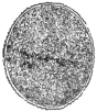
Fig. 343.—Ovum of Toxascaris limbata, with thin albuminous envelope. Magnified.
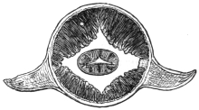
Fig. 344.—Transverse section through the head part of Belascaris cati from the cat, with the lateral wings. In addition, one may note the four fields of muscles, the longitudinal lines with the œsophagus in the centre. Magnified. (After Leuckart.)
Genus. Belascaris (Βέλος, an arrow), Leiper, 1907.
Body anteriorly bent ventrally, cuticle coarsely striated. Œsophagus with a distinct bulb. Tail of male conical. A papillæ-bearing protuberance behind the anus. Testis in anterior half of body. Vulva in anterior part of body. Eggs corrugated.
Belascaris cati, Schrank, 1788.
Syn.: Belascaris mystax, Leiper, 1907; Ascaris mystax.
Striations 12 µ to 16 µ apart. Cephalic wings lanceolate. Male 3 to 6 cm. Spicules 17 to 1·9 mm. Female 4 to 10 cm. Eggs, 65 µ to 75 µ in diameter, surface finely honeycombed. Host: domestic cat, and man, eight or nine cases.
Belascaris marginata, Rudolphi, 1802.
Striations 16 µ to 22 µ apart. Cephalic wings long, narrow, semi-lanceolate. Male, 5 to 10 cm. Spicules, 750 µ and 950 µ. Female, 9 to 18 cm. Eggs, 75 µ to 80 µ. Shell finely honeycombed. Host: dog.
Genus. Lagocheilascaris, Leiper, 1909.
Thick lips separated by a furrow from the body; between the lips small intermediate lips without “pulp.” The cutting angle of each lip bifurcated. Along each lateral line a cuticular wing extending the whole length of the body. Eggs, thick shell with a mosaic pattern.
Lagocheilascaris minor, Leiper, 1909.
Male, 9 mm., tail sharply curved. Spicules colourless, 3·5 and 4 mm. long. More than twenty-four pairs of pre-anal papillæ, at least five pairs of post-anal. Female, 15 mm. Straight posteriorly. Vulva 6 mm. from head with two lips. Eggs, 65 µ in diameter. Host: man, cutaneous abscesses. Trinidad.
Family. Oxyuridæ.
Genus. Oxyuris, Rudolphi, 1803.
Mouth unarmed. The three labial papillæ are only slightly protuberant, the œsophagus is long and presents two well-marked bulbs. The vulva is in the anterior part of the body.
Oxyuris vermicularis, Linnæus, 1767.
Syn.: Ascaris vermicularis, L.; Fusaria, Zeder, 1803.

Fig. 345.—A, male, and B, female, of Oxyuris vermicularis. 5/1.
Colour white, the striated cuticle forms projections at the anterior end which extend some distance back along the middle of the ventral and dorsal surfaces; the longitudinal lateral flanges of the skin corresponding to the lateral lines are well seen in transverse sections; there are three small retractile labial papillæ around the mouth. The male measures 3 to 5 mm. in length, and shortens on death; the posterior extremity of the body is curved ventrally and presents six papillæ. Spicule 70 µ long, hook-like. The female is 10 mm. in length and 0·6 mm. in diameter; the anus is about 2 mm. in front of the tip of the tail; the vulva is in the anterior third of the body; the eggs are oval, asymmetrical, with double-contoured shells, and measure 50 µ to 55 µ by 16 µ to 25 µ; they are deposited with clear, non-granular tadpole-like embryos already developed.
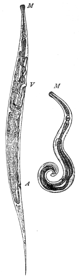
Fig. 346.—On the left, female; on the right, male. A, anus; M, mouth; V, vulva. Greatly enlarged. (After Claus.)
Habitat.—Adults in large intestine of man. Young forms in small intestine and often in the appendix.
The worm lives in the lower part of the small intestine, cæcum and vermiform appendix, and before becoming adult undergoes two or three moults (Heller). According to Wagener the worms at times live in the gut wall, giving rise to calcareous nodules. When the uterus of the fertilized females begins to fill with eggs they leave the cæcum and travel through the colon to the rectum. The uterus is now packed with eggs which contain a tadpole-shaped embryo. Egg-laying now takes place, partly in the rectum, partly outside, the mode of exit being not only passive through defecation but also an active one on the part of the worms when the patient is in bed. In this case the worms crawl out of the anus, producing a most intolerable itching as they scatter their eggs between the nates and the perinæum. From here in the case of girls they may get occasionally into the vulva and vagina, and even into the oviducts and so into the body cavity. The worms also may wander through the alimentary canal in the opposite direction, getting out occasionally through the mouth. Recently a rôle has been assigned to them, as to other gut parasites, in appendicitis and typhlitis.
It is stated that the males die after fertilizing the females, thus explaining why they are so rarely met with in fæces [but it is probable that they often escape notice from their small size.—J. W. W. S.].
Development.—The eggs, which often adhere together, contain a tadpole-like embryo, the thin tail of which is bent upwards ventrally; the embryo in a short time, given a sufficiently high temperature, passes into a second folded nematode-like embryonal stage, lying in the egg-shell, either in the fæces, with which also numerous females pass out, or in the moisture of the groove between the buttocks, and they there await the opportunity of being reintroduced into man per os. It is very improbable that infection takes place directly in the large intestine, as is occasionally stated, because although the harbourers of Oxyuris are frequently liable to auto-infection, this takes place exclusively through the mouth, and is conveyed by the fingers, on which the ova of Oxyuris, and occasionally the female worms, have clung.
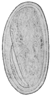
Fig. 347.—Oxyuris vermicularis: egg freshly deposited, with tadpole-like embryo. × 640.
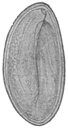
Fig. 348.—Oxyuris vermicularis: egg twelve hours after deposition, with nematode-like embryo. × 640.
The opportunity for this is afforded every evening, as naturally the troublesome itching caused by the wandering of the worms is met by scratching and rubbing with the fingers. It is therefore possible that the eggs may even thus be introduced into the nose, where the young Oxyuris are perhaps hatched out, if they get high enough up on the moist pituitary mucous membrane. As a matter of fact, the larvæ of Oxyuris have been found in the nose. Moreover, one can understand that the eggs of Oxyuris are transferred from person to person by the hand, directly or indirectly. This again explains the wholesale infections which occur in collective dwellings, after a person harbouring Oxyuris has been admitted into boarding-houses, etc. The primary infection may be also caused in other ways—by foods, fruits, vegetables and other articles that are eaten raw, and are polluted with the ova. Perhaps also flies or their excrement play a part in the distribution of the parasite, similar to that demonstrated by Grassi as taking place in the spread of the ova of Trichocephalus and Tænia.
The assumption of a direct development without an intermediary host was first substantiated by Leuckart by experiments on himself and three of his students; about fourteen days after swallowing the eggs the Oxyuris has attained 6 to 7 mm. in length; Grassi, and later on Calandruccio, infected themselves by swallowing adult female Oxyuris, with the same results. Heller found worms in the gut (appendix vermiformis) of a male child five weeks old.
Other species are: O. compar in the cat; O. curvula and O. mastigodes in horse, ass, mule; O. ambigua in the rabbit; O. poculum in the horse; O. tenuicauda in the horse. Many species occur in insects, especially in Blattidæ and Hydrophilidæ (aquatic beetles).
Family. Mermithidæ.
Genus. Mermis, Dujardin, 1845.
With characters of the family.
Mermis hominis oris, Leidy, 1850.
Fourteen centimetres in length, 0·16 mm. in breadth; mouth terminal; posterior extremity obtuse and provided with a recurved hook 50 µ long.
The parasite was “obtained from the mouth of a child.” Stiles considers it to be probably a Mermis, possibly swallowed in an apple.
Agamomermis, Stiles, 1903.
Group name for immature Mermithidæ.
Agamomermis restiformis, Leidy, 1880.
This worm measures 65 cm. in length, pointed anteriorly, the posterior extremity broadened and rounded off (1·5 mm. in breadth); the mouth is terminal, without lips. Behind the mouth six papillæ; the œsophagus measures 1·125 mm. in length; the intestine appears to terminate blindly.
This parasite was obtained in West Virginia from the urethra of a young man, aged 20, who for a few days previous to expelling the worm passed turbid and bloody urine.
TECHNIQUE.
Preservation and Examination of Flukes.
Fixation.—(Method A.) (1) Place the flukes in a test tube or small bottle a quarter full of normal saline. Shake the contents as hard as possible (the object of this is to extend the flukes) for half a minute.
(2) Add immediately an equal bulk of saturated aqueous solution of corrosive sublimate and shake again as vigorously as possible for a few minutes.
(3) Transfer when convenient to 70 per cent. alcohol. (Before staining and mounting remove the sublimate with tincture of iodine.)
(Method B.) In case of large flukes, e.g., Fasciola hepatica, Fasciolopsis buski, compress the flukes between two glass slides with rubber bands or thread. Fix in sublimate or in absolute alcohol, or in 10 per cent. formalin.
(Method C.) Place the flukes in 10 per cent. formalin solution.
Staining is successfully effected by using quite dilute solutions of carmine or hæmatein overnight. This is far preferable to using strong solutions, as it may be almost impossible to remove a too intense stain. Almost any dilute carmine solution suffices. One of the best is acetic-alum carmine (boil excess of carmine in a saturated aqueous solution of potash-alum for about fifteen minutes; add glacial acetic acid to the extent of 10 per cent.; let it stand for a week; filter). For use, dilute about thirty times with water. Place the flukes directly in the stain. Stain overnight or longer.
Differentiation.—In order to get the sharpest picture, it is best now to differentiate (but this may often be omitted) with acid alcohol (70 per cent. alcohol 100 parts, HCl 5 drops). Allow to act from one to twenty-four hours, according to the appearance of the flukes. Similarly, in staining with hæmatoxylin solution, dilute twenty to thirty times so that the water is merely tinged with the stain. Differentiate as before. After staining, dehydrate, clear, and mount in balsam if required.
Clearing and Mounting.—(1) Carbolic acid (carbolic acid 94, water 6) is a very convenient clearing agent. It may be used for stained or unstained specimens. It will clear rapidly without previous dehydration. If it is required to mount a specimen permanently, transfer from carbolic to alcohol, then cedar-wood oil (or xylol, etc.), then balsam.
(2) Creasote.—Dehydrate the specimen, stained or unstained, transfer to creasote. If it is desired to mount permanently, transfer back to alcohol, then cedar-wood oil, then balsam.
(3) Cedar-wood Oil.—Preferable to xylol or oil of cloves. Dehydrate the specimen in alcohol. To mount permanently, transfer to balsam.
(4) Glycerine.—Vide under methods of preservation of ova; to mount permanently, transfer to glycerine jelly; subsequently to harden the jelly, expose to formalin vapour.
Of these media, carbolic acid has the greatest refractive index excepting that of balsam. The latter may, in some cases, render structures too transparent, and it may be advisable to use only glycerine or glycerine jelly.
Preservation of Ova in Fæces, Urine, Bile, etc.
Heat some 70 per cent. alcohol in a basin to about 60 to 70° C. (until bubbles begin to appear). Add the fæces, etc., in the proportion of one part to about nine of fixative; keep stirring. Allow the sediment to settle. Transfer to a bottle with some fresh 70 per cent. alcohol.
Transference to Glycerine.—Prepare 5 per cent., 10 per cent., 20 per cent. solutions of glycerine in 70 per cent. alcohol. Pour off the alcohol in the bottle of fæces, etc., and replace by 5 per cent. glycerine solution. Allow to stand an hour or so. Then in the same way replace the 5 per cent. by a 10 per cent. glycerine, and finally by a 20 per cent. glycerine solution. When in this latter expose freely to the air (protecting from dust), so as to allow the alcohol and water to evaporate. Add a few drops of glycerine from time to time till eventually the ova are in pure glycerine. (In a very moist climate it may be necessary to use lime or calcium chloride to dry the air.) To mount permanently transfer some of the sediment to glycerine jelly.
Preservation and Examination of Cestodes.
Fixation.—(1) Saturated aqueous corrosive sublimate.—Add to this glacial acetic acid to the extent of 1 per cent. (Note this fixative will dissolve the “calcareous corpuscles”; 10 grammes of sublimate to 160 c.c. of water will give a saturated solution.) Warm the fixative to 70° to 80° C. (Avoid the use of needles.) Use plenty of fixative. Allow to act for a quarter of an hour or so. (a) Transfer to 70 per cent. alcohol. (It is advisable to remove the sublimate by the use of Lugol’s solution, or a solution containing tincture of iodine, adding this until the iodine colour is permanent.) Or (b) transfer for preservation to 10 per cent. formalin.
Or (2) 10 per cent. formalin.—In order to prevent contraction it is advisable to extend the tapeworm and keep it fixed by glass plates, or wind the worm around a wide glass tube or bottle, and then fix it.
Or (3) fix in hot alcohol.
Staining.—As under flukes. It is necessary to sacrifice portions of the tapeworm for this purpose, cutting out, e.g., mature segments, so as to study the topography of the genitalia.
Clearing.—As under flukes. To examine the hooks satisfactorily it is best to cut off the head with a sharp knife and mount. A certain amount of pressure is then advisable in order to view the hooks completely so as to measure them.
Preservation of Ova in Fæces, etc.
As under flukes.
Preservation and Examination of Nematodes.
Fixation.—(1) Thoroughly wash the worms to get rid of mucus, etc., by shaking up in warm saline (or water) till the washings are clean. Then transfer to 70 per cent. alcohol heated to about 70° C. It is absolutely necessary to use hot fixatives in order to extend the worms. If no alcohol or spirit is immediately available, drop the worms into hot water, or saline, and transfer later to 70 per cent. alcohol.
(2) Drop into hot 10 per cent. formalin.
Clearing.—(1) Carbolic acid, vide p. 471.
(2) Creasote, vide p. 471.
(3) Glycerine, vide p. 472.
Staining.—In case of quite small Nematodes, e.g., Anguillulidæ, carmine may be used, but as a rule staining is not advantageous.
Rolling.—In order to study the mouth parts, or bursa, etc., it is necessary to place the worm in any desired position. This is done as one would roll a penholder along the table by one’s finger placed on top of it. In the case of a worm, one edge of the cover-glass is placed over the worm, the other is supported by a strip of cardboard. By tapping the cover-glass the worm will now revolve as much as required provided it is round and straight. In certain cases it may be necessary for this purpose to cut off the head or tail. Roll these separately.
When a suitable position is got, the worm may be fixed in this position by pressure on the cover-glass, so as slightly to flatten it.
Mounting the Head.—If it is required to get an end view, it is necessary to cut off the head transversely as near the end as possible, and then mount.
Detection of Eggs (Bass and Hall).—Mix the fæces thoroughly with ten times the volume of water. Filter through gauze. Centrifugalize the filtrate. Wash the sediment and centrifugalize. Repeat twice. To sediment add CaCl2 solution, sp. gr. 1250. The eggs float to surface. Pour off surface fluid. Dilute to sp. gr. 1050. Centrifugalize. Examine sediment, which contains practically all the eggs in the stool.
Detection of Small Nematodes.—Mix the fæces thoroughly with water. Allow to settle for five minutes. Carefully decant off, or better, syphon off the fluid. Mix the sediment again with water. Allow to settle. Remove the fluid. Repeat several times. Examine the sediment in a Petri dish. As the fluid is poured off, the worms will be seen collected in the backwater. Remove them with a brush. Fix in hot 70 per cent. alcohol.
Cultivation of Larval Forms of Ancylostoma and Strongyloides.
A modification of the second method of Looss (p. 455) is that of Fülleborn. A glass filter funnel is lined with linen or with cotton wool dyed black with iron-tannin. On this is placed a layer of sterile sand, and on top of this the fæces. The whole is moistened. The larvæ hatch out and wander through the meshes of the wool, appearing on the edges of the same as white threads visible to the naked eye. With a platinum needle these can be easily removed. The glass filter can be placed on a glass cylinder, and this in another large stoppered cylinder containing caustic potash solution at the bottom, so that any larvæ escaping from the funnel are killed.
D. ACANTHOCEPHALA, Rud.
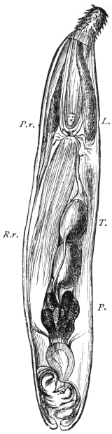
Fig. 348a.—The male of Echinorhynchus augustatus. L., lemnisci; T., testicles; P., prostatic glands; P.r., sheath of proboscis, with ganglion; R.r., retractor of sheath of proboscis. 25/1.
Gutless, nematode-like worms that carry at their anterior end a retractile rostrum beset with hooks. In their adult condition they only live in vertebrate animals. During their larval stage they are often parasitic in invertebrate animals.
The Acanthocephala are elongated cylindrical worms, with a rounded posterior end. In some species an annulation is distinctly recognizable; they are, however, not segmented. The size varies according to the species, between about 5 to 10 mm. and 40 to 50 cm.; in general, however, there is a preponderance of the small species. The sexes are separate, and the males can easily be distinguished from the females without examination of the genitalia, as the females are both larger and thicker.
The body wall of Echinorhynchus is limited by a thin cuticle, which is attached inwardly to the hypodermis. In only exceptional cases a syncytium with large nuclei, even in the adult condition, is represented by the hypodermis; and in it fibre systems, the elements of which run in layers in various directions, appear, and it is only towards the interior from these strata of fibres that the nuclei of the hypodermis are found. As a rule, these fibres, at all events the radiary fibres, are regarded as muscles. Hamann describes them as elastic fibres, which lie in a viscid gelatinous connective substance (transformed protoplasm?); a lacune system filled with a granular fluid, the central part of which are two longitudinal lacunes lying at the sides, also belongs to the cutaneous strata, as do the so-called lemnisci, two short, flat organs suspended in the body cavity, and the pedicles of which are attached anteriorly at the border between the rostrum and body; their structure as well as their origin permit them to be traced to the skin (fig. 348A).
Finally, inwardly below the skin there follows a layer of annular, and after these a layer of longitudinal muscles, the structure cells of which remain present in the residues, carrying nuclei. The motor apparatus of the rostrum, the sheath of the rostrum, and the lemnisci also belong to the muscular system. The rostrum represents a finger-shaped hollow process of the cutaneous layer; but, according to Hamann, it originates from the entoderm and passes through the skin secondarily. It is covered by a thin cuticle, and as a rule contains a large number of regularly placed chitinous hooks that adjoin a granular formation tissue. From the base of the rostrum springs a tubular hollow muscle extending into the body cavity; this is the RECEPTACULUM PROBOSCIDIS, from the base of which again bundles of longitudinal muscles originate, which pass along its axis and that of the rostrum itself, and are inserted at the inner surface of its anterior end (RETRACTOR PROBOSCIDIS). These muscles when they contract invaginate the proboscis and draw it into the receptaculum; when reversed they act again as PROTRUSOR PROBOSCIDIS. The whole of the anterior body, however, can be invaginated, and for this purpose there is a muscle that originates from the body wall at a variable distance back, and which is joined to the receptaculum (RETRACTOR RECEPTACULI); there is also a bell-shaped muscle which springs from the body wall behind the lemnisci in rings, and passes forward to the spot of attachment of the lemnisci.
The nervous system consists of a cluster of ganglia situated at the base of the rostrum, from which three nerves pass towards the front and two towards the back. No sensory organs are known.
The excretory organs, according to Kaiser, lie at the upper border of the ductus ejaculatorius in the male and at the so-called bell in the female. Here they represent the long-known villous tufts, placed on disc-like cushions. In each of the cylindrical villi—which terminate blindly towards the body cavity—there lies a cilium, which springs from the membrane lining the villus, and which lies in a space cavity of the villus, which ultimately proceeds as a little canal. There are three canals discharging into the uterus that serve to conduct the excretory materials from the body cavity; special glandular cells corresponding to the terminal cells of the Platyhelminths, at the commencement of the system, are not present in the Acanthocephala.
Sexual Organs.
(a) Male Organs.—The greatest part of the male genital apparatus is contained in a muscular sheath—the ligament—which originates at the posterior end of the receptaculum proboscidis, passes along longitudinally through the body cavity, and is inserted at the posterior end of the worm. The two oval testicles usually lie one behind the other; their vasa efferentia unite sooner or later into a vas deferens which passes backwards, and finally terminates in the penis; the terminal portion of the conducting apparatus is surrounded by six large glandular cells (prostatic glands) the excretory ducts of which open into the vas deferens. The penis itself is placed at the base of a bell-shaped invagination of the posterior end, the bursa, which is everted during copulation.
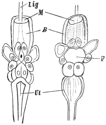
Fig. 348b.—Anterior portion of the female apparatus of Echinorhynchus acus. On the left seen from behind, on the right seen from the front. F, inferior orifice of the bell; B, bell; Lig, ligament; M, mouth of bell; Ut, uterus. Magnified. (After Wagener.)
(b) Female Organs.—There are only two ovaries present in the ligament during the larval stage. During the course of growth they divide into accumulations of cells (placentulæ, loose or floating ovaries), which finally cause the ligament to burst and they thus attain the body cavity. Thence a peculiarly constructed apparatus finally conveys the eggs out. This apparatus consists of the uterine bell and vagina, the latter discharging at the posterior extremity of the body. The bell is a muscular canal provided with apertures at both the anterior and posterior extremities. Its interior space is in direct communication with the body cavity, and the anterior orifice takes up all materials floating in the cavity—egg-balls, mature and immature eggs—and pushes these further backwards. The continuation of the bell lumen is now narrowed by a number of large cells in such a manner that only bodies of a certain form can pass through this tract and attain the uterus; everything else is conveyed back into the body cavity through the posterior opening of the bell.
The eggs are already fertilized in the body cavity, and in this position go through their development to the formation of the embryo. Completely developed eggs are surrounded by three shells, and are generally fusiform. The eggs agglomerate in masses in the uterus until they are finally deposited through the vagina and vulva. For the further development, the transmission of the eggs into an intermediary host—usually a crustacean or an insect—is necessary; the metamorphosis is very complicated; but this transmission may be very easily effected artificially by feeding suitable crustaceans (Asellus, Gammarus, etc.) with the eggs of Acanthocephala; this being the only method of inducing the larva to hatch out so that its structure may be studied. The larva appears in the form of an elongated, somewhat bent body, at the stumpy anterior end of which there is a crown of hooks or spines, whereas the posterior end is pointed. Especial retractors draw in the hook-beset anterior surface, and an elastic cushion beneath them jerks them forward again when required. In the middle of the body a roundish heap of small cells is seen, from which the entire body of the Echinorhynchus originates, even to the cutaneous layer; the latter is also the larval skin in which the small Echinorhynchus gradually grows. The development of all the organs takes place within the intermediary host, and the parasite only needs to be imported into the terminal host to attain the adult stage after a certain growth. In some cases, however, a second intermediary host is utilized.
Species of Acanthocephala only occur exceptionally in human beings.
Echinorhynchus gigas, Goeze, 1782.
Syn.: Tænia hirudinacea, Pallas, 1781.
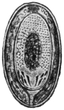
Fig. 348c.—Egg of Echinorhynchus gigas. 300/1. (After Leuckart.)
The body is elongated, gradually decreasing in thickness towards the back. The rostrum is almost spherical, and is beset with five or six rows of recurved hooks. The males measure 10 to 15 cm. in length, the females 30 to 50 cm.; the eggs are provided with three shells, of which the middle one is the thickest. The eggs measure 0·08 to 0.1 mm. in length. The giant Echinorhynchus occurs especially in the intestinal canal of the domestic pig; it is less common in other mammals. It bores deep into the mucous membrane with its rostrum, and causes an annular proliferation around the perforated spot; occasionally also it causes perforation of the intestine.
It is doubtful whether the giant Echinorhynchus occurs in man. Leuckart admitted that there were a few positive cases. According to Lindemann, Ech. gigas occurs in human beings in South Russia, and its presence is not rare. This statement, however, has not been confirmed. Its presence in man is by no means impossible, as its intermediary host, the cankerworm, or cock-chafer (Melolontha), is, according to Schneider, occasionally eaten raw by human beings. According to Kaiser, the golden beetle (Cetonia aurata) and, according to Stiles, another beetle in America (Lachnosterna arcuata) are also intermediary hosts.
Echinorhynchus hominis, Lambl, 1859.
This term is applied to an Echinorhynchus found by Lambl in the intestine of a boy who had died of leucæmia; the worm was 5·6 mm. in length, and the almost spherical head was beset with twelve transverse rows of hooks.
Echinorhynchus moniliformis, Bremser, 1819.
The male is 4 cm. in length, the female 8 cm. long. This species lives in the intestine of field-mice, rats, marmots and Myoxus quercinius. A beetle (Blaps mucronata) is the intermediary host.
This species has also once been artificially cultivated in man (Grassi and Calandruccio).
E. GORDIIDAE.
Very long thin worms similar to Filariæ, which, in their adult condition, live free in brooks, pools and springs; the mouth and the commencement of the intestine are obliterated; there are no lateral ridges, and the muscular system presents a structure different to that of the Nematoda. The posterior end of the male is split, and spicules are lacking; there are two testicles. In both sexes the genitalia discharge through the terminal gut.
The larvæ, which carry a rostrum beset with hooks, force themselves into the larvæ of water-insects; more rarely they invade molluscs, and they then become encysted within the body of the host. According to Villot, at least a part of them attain the intestine of fishes, where they again become encysted, and after a period of rest they travel into the tissues of their hosts, and finally again reach the exterior by way of the intestine, where they then become adult. In most cases, however, the gordius larvæ are taken up by predacious water insects; they live for a while in the body cavity of these insects, undergo a metamorphosis, and finally wander into the water.
A few species invade man accidentally with water, in which case they are usually vomited up:—
Gordius aquaticus, Dujardin, 30 to 90 cm. in length (Aldrovandi, Degland, Siebold, Patruban).
Gordius tolosanus, Duj., 11 to 13 cm. in length (Fiori).
Gordius varius, Leidy, 10 to 16 cm., female, up to 30 cm. in length (Diesing).
Gordius chilensis, Blanch. (Guy). Gordius villoti, Rosa (Bercutti, Camerano); Gordius tricuspidatus, L. Def. (R. Blanchard), Gordius violaceus, Baird (Topsent), and Gordius pustulosus, Baird (Parona).
F. HIRUDINEA s. DISCOPHORA (Leech).
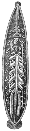
Fig. 348d.—The internal organs of the leech. The creature has been opened from the dorsal surface, and part of the intestine has been removed. The testicles, with vas deferens, may be seen between the blind ducts of the intestine; beyond these on either side the segmental organs. The female genital organs are in front of the most anterior pair of testicles. (After Kennel.)
The Hirudinea, which have been appropriately included amongst the Annelida, differ in many respects from the typical members of the group; their body is long and flat, it lacks the parapodia that are characteristic to all forms of Annelida; but, on the other hand, possesses a terminal posterior sucker, and in many species there is also an anterior sucker. The mouth is terminal at the anterior end, the anus lies dorsally above the posterior sucker (fig. 348D). The body is segmented, but this is less manifest in the body covering than it is in the arrangement of the internal organs; the segmentation, nevertheless, is also indicated exteriorly by the appearance of the cutaneous sensory organs which correspond to the segments. This shows what the condition of the ganglia in the abdominal ganglion chain has taught us, that the anterior and the most posterior segments are considerably abbreviated—a part of the latter taking part in the formation of the suctorial organs. In a great many species the skin is distinctly annulated, four or five of such rings, at least in the central region of the body, appearing on one segment of the body. The condition of their body cavity is another peculiarity of the Hirudinea; it is narrowed by the powerful development of the connective tissue and the muscular system into four tubular sinuses, which have the appearance of blood-vessels. There are usually one dorsal and one ventral median trunks, as well as two lateral trunks; in addition, a particular blood-vessel system exists.
The skin consists of a very thin cuticle that is cast off from time to time; it is secreted by the underlying cylindrical epithelium, which contains numerous goblet cells. The muscular system is strongly developed; it consists of long tubular fibres, which run circularly, longitudinally and in the dorso-ventral direction; the muscular system is subject to a particular expansion in the clinging organs and at the commencement of the intestine. On the whole, the alimentary canal represents a tube running straight from the mouth to the anus, which possesses a number of blind sac-like protuberances at the sides varying according to the species. The most anterior section, the pharynx, in the leeches with maxillæ carries three chitinous, semicircular plates furnished with teeth—the jaws—which serve to tear up the epidermis in order to open the blood-vessels; in the leeches with rostra a long protractile proboscis rises from the base of the elongated pharynx. Numerous salivary glands, the secretion from which possesses toxic properties, discharge into the pharynx. The œsophagus, which follows the pharynx, and to the exterior of which numerous radiary muscles are fixed, is a suctorial organ in its entire structure. The nutriment in the larger species consists of the blood of vertebrate animals, in smaller species and in the young stages the food consists of small invertebrate animals.
The NERVOUS SYSTEM exhibits the typical structure of other segmented worms; the sensory organs consist of the previously mentioned goblet-shaped cutaneous sensory organs, of the organs of taste, and of eyes, the latter frequently being present in large numbers.
The EXCRETORY or segmental organs exhibit many peculiarities, which cannot, however, be detailed here. They commence with funnels in the lacunes of the body cavity, and usually discharge on the ventral surface.
Almost all the Hirudinea are hermaphrodite and copulate reciprocally. The two ovaries are very small, and the oviducts that proceed from them soon unite into a common duct, which then passes into the uterus and discharges through the short vagina in the median line of the ventral surface behind the male organs into the so-called clitellar region. The male sexual apparatus consists of symmetrically arranged testicles, varying in number according to the species, the short vasa efferentia of which, one by one, run into the vas deferens, passing towards the front on each side. In front, at about the level, or a little in front, of the female genitalia, the two vessels pass into a convoluted mass of tubes to the so-called epididymis, and then discharge into the single protractile penis (fig. 348D).
All leeches deposit so-called COCOONS. These are small barrel-shaped or pouch-like bodies, which are surrounded by a thicker shell and contain a number of eggs in a large mass of albumen; the albumen originates from glands of the generative organs, the shell substance from cutaneous glands of the clitellar region.
Family. Gnathobdellidæ (Leeches with Jaws).
These are distinguished by the possession of usually three jaws in the pharynx; the body consists of twenty-six segments. The posterior sucker is large and flat; the anterior sucker is smaller. The Hirudinea have five pairs of eyes, the Nephelinæ have four pairs.
Genus. Hirudo, L., 1758.
The entire body consists of 102 annulations, five appearing on one segment in the central region of the body. The pharynx has three semicircular jaws, the arched border of which is beset with numerous teeth (50 to 100). The male sexual orifice lies between the thirtieth and thirty-first rings, the female orifice between the thirty-fifth and thirty-sixth. There are numerous species, some of which are utilized for medicinal purposes.
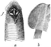
Fig. 348e.—Hirudo medicinalis. a, anterior end, with open buccal cavity, with the jaws, J, at the; b, one jaw isolated. (After Claus.)
Hirudo medicinalis, L., 1758.
It occurs in numerous colour varieties, one of which has been designated Hirudo officinalis, Moq.-Tandon. Usually the dorsal surface is greyish-green and is marked with six rusty-red longitudinal stripes. The ventral surface is olive-green, more or less spotted with black, and marked at the sides with a black longitudinal line. The length averages 8 to 12 to 20 cm. This leech lives in swamps, ponds and brooks, overgrown with plants and having a muddy bed. The cocoons are deposited in the soil at the sides. Europe, as well as North Africa, is its home. At the present day it has been exterminated from most parts of Central Europe, but it is still very common in Hungary. Its use for medicinal purposes is well known. A large leech can suck about 15 grs. of blood, and about the same amount is lost through secondary hæmorrhage.
Hirudo troctina, Johnston, 1816.
Syn.: Hirudo interrupta, Moq.-Tandon, 1826.
This species measures 8 to 10 cm. in length. The back is greenish, with six rows of black spots surrounded by red; the lateral borders are orange-coloured; the abdomen spotted or unspotted. Its habitat is in North Africa and Sardinia. It is applied medicinally in England, Spain, France, Algeria, etc.
Genus. Limnatis, Moq.-Tandon, 1826.
Nearly related to Hirudo, but is differentiated by a longitudinal groove on the inner surface of the upper lip of the anterior sucker. The jaws are furnished with over 100 very sharp toothlets.
Limnatis nilotica, Savigny, 1820.
Syn.: Bdella nilotica, Sav.; L. nilotica, Moq.-Tandon; Hæmopis (vorax), Moq.-Tandon, 1826, p. p.; Hæmopis sanguisuga, Moq.-Tandon, 1846 (nec Hir. sanguis, Bergm., 1757).
This species measures 8 to 10 cm. in length, and becomes gradually more pointed towards the front; the body is always soft. The back is brown or greenish, and has usually six longitudinal rows (rarely only two or four) of black dots. The abdomen is dark; but numerous colour variations occur.
The native place is North Africa, especially the coastal regions; it is also found in the Canaries, the Azores, Syria, Armenia, Turkestan, perhaps also Southern Europe. It is taken into the mouth with drinking water, and may settle in the pharynx, larynx, œsophagus, and nasal cavities of human beings. This species has also been observed in the vagina and on the conjunctiva. It is equally fond of attacking domestic animals.
Hirudo mysomelas (Senegambia) and Hirudo granulosa (India) are placed with this genus, and, like our leech, are also used for medicinal purposes.
Genus. Hæmadipsa, Tennent, 1861.
These leeches live on land, and measure 2 to 3 cm. in length. About a dozen species are known. They are a veritable scourge to persons in the tropics (Asia, South America), as they attack them to suck their blood. They are able to force their way even under close-fitting garments, so that it is difficult to protect oneself from their assaults (Hæmadipsa ceylonica, Bl., and other species).
Family. Rhynchobdellidæ (Leeches with Rostrum).
These are furnished with a proboscis in lieu of the jaws; the segment consists of three annulations.
Genus. Hæmentaria, de Filippi, 1849.
Hæmentaria officinalis, de Fil.
Inhabit Mexico, where they are used for medicinal purposes.
Genus. Placobdella, R. Blanch.
Placobdella catenigera, Moq.-Tandon.
Indigenous to South Russia, Hungary, Italy and South France. It is a parasite of the swamp turtle, but frequently attacks human beings.
G. ARTHROPODA (Jointed-limbed Animals). BY FRED. V. THEOBALD, M.A.
Bilaterally symmetrical segmented animals which are covered with a thick cuticle that is frequently calcareous (Crustacea), but always thinner between the segments; they carry (primitively) a pair of jointed appendages on every segment.321 The segments of the body are uniform in certain regions, but differ from those of contiguous regions, so that it is easy to distinguish three parts (head, thorax and abdomen), each composed of segments. The cephalic segments are always formed into a uniform head, the segmentation being scarcely recognizable at either end; the thoracic segments may also fuse, or part or all of them may coalesce with the head; the abdomen, as a rule, retains its segmentation, but this may possibly also be lost, in which case it is [sometimes] united to the cephalothorax. The structure of the three regions depends mostly on the varying form and function of the appendages: those on the head are primitively locomotive organs (and frequently are still so in the early stages), but they become transformed into feelers and mouth-parts (mandibles, maxillæ); the limbs of the thorax, however, usually retain their ambulatory functions, as frequently do those of the abdomen; sometimes, however, the abdominal limbs disappear, entirely or partly; in the latter case they are then utilized for other purposes.
In their organization the Arthropoda approach the segmented worms.
The Arthropoda are generally divided into five groups (Crustacea,322 Protracheata, Arachnoidea, Myriapoda,323 and Insecta or Hexapoda), of which only the Arachnoidea and the Hexapoda interest us here.
A. ARACHNOIDEA (Spiders, Mites, etc.).
The head and thorax are always united together; the abdomen is either segmented or without exterior segmentation, in which case it is united with the cephalothorax.324 The number of pairs of appendages amount to six, of which the two front pairs, the cheliceræ and the pedipalpi, are attached to the head region and the four remaining pairs to the thoracic region.325 The abdomen in the adult condition has no appendages. The Arachnoids are air-breathers, and for this purpose are either provided with tracheæ or with so-called lung-sacs, or they breathe through the surface of the body. Some aquatic forms breathe by gills.
There are eight or ten orders of Arachnoids,326 of which, however, only two, the Acarina and the Linguatulida, have to be considered here.327
Order. Acarina (Mites).
Small Arachnoids, the three parts of the body of which are, as a rule, coalesced; it is only rarely that a faint line indicates the division between a cephalothorax and abdomen. The two appendages on the head are designed for biting or puncturing and sucking, and vary according to their use. The cheliceræ328 are fang-like jaws or puncturing bristles forming a kind of rostrum, the pedipalpi are claw-like or shear-shaped, or form a suctorial proboscis.329 The four pairs of legs are usually well developed, more rarely they are rudimentary or have partly vanished; many parasitic forms are provided with pedunculated suckers [ambulacra—F. V. T.]. Respiratory organs (tracheal tufts) may be present or absent. The nervous system is reduced to a minimum, eyes are usually lacking. The intestine, situated in the central part, generally has three blind appendages; the anus is situated on the venter above the posterior end. Sexes separated; nearly all the species deposit eggs, from which six-legged larvæ hatch. The Acarina live either free in the water or in moist soil, or they are parasitic on plants and animals.330
Family. Trombidiidæ (Running Mites).
Soft-skinned Acarina with tracheæ and with two eyes, usually pedunculated; they are often brightly coloured; cheliceræ lancet- or claw-shaped; pedipalpi claw-like; legs composed of six segments, with suctorial discs between the terminal ungues.331 Larvæ six-legged. To the latter belong the larvæ of several species of Trombidium such as:—
Genus. Trombidium, Latreille (and Leptus).
Leptus autumnalis, Shaw, 1790.
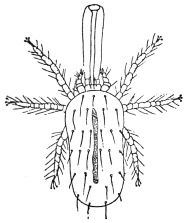
Fig. 349.—Leptus autumnalis, with so-called sucking proboscis. Enlarged. (After Gudden.)
Leptus occur as parasites in the human skin and cause a cutaneous disease known as autumn erythema, and produce a very unpleasant sensation on account of the troublesome itching; in children it is very often accompanied by fever.332
Formerly these mites were considered adult forms, but when they were recognized as mite larvæ they were taken for those of the spider-mite (Tetranychus telarius); the investigations of Hanstein, however, showed this to be a mistake. When Henking first investigated the development of Trombidium fuliginosum, parasitic in the larval stage on vine-fretters, he demonstrated the occurrence of a form very similar to Leptus autumnalis, and the “autumn, grass, or gooseberry” louse was commonly designated the Trombidium larva. Even before Henking’s work it had been described by Mégnin as the larva of Trombidium holosericeum, a red-coloured species, frequently occurring in spring and summer on the ground, trees, etc. This assumption, however, as Moniez was the first to explain, is not correct; indeed, as many as three species come under consideration: T. gymnopterosum, L., T. fuliginosum, Herm. (according to Brucker), and two species known hitherto only in the early stage, T. striaticeps, Helm. et Oudem., and T. poriceps, Helm. et Oudem., which are not only parasitic on mammals, but on birds, on Arthropods and especially on insects. Arthropods appear to be the normal hosts for the larvæ.
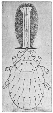
Fig. 350.—Leptus autumnalis: the so-called proboscis is formed around the hypopharynx sunk into the skin. 100/1. (After Trouessart.)
The above-mentioned forms invade the skin of man by means of their oral apparatus, by preference invading the orifices of the sebaceous glands so as to suck the blood; around the point attacked there arises a wheal about the size of a lentil, and around the inserted hypopharynx a fibrinous secretion, the “proboscis,” which, however, is a product of the host, just as chitinous secretions are provoked by Trombidia parasitic on Arthropods.
Further species, analogous in habit to Leptus autumnalis, are described by Riley from Central and South America as L. americanus and L. irritans.
[L. autumnalis attacks small mammals by preference, such as moles and hares, which are often literally covered with them. Dogs are also subject to their attack, and cats suffer similarly. This mite also frequently appears in colonies on cows; cavalry horses after autumn manœuvres often suffer from an erythematous affection about the hocks and knees due to this pest.
[A number of Leptus, so far undescribed, occur abroad which attack man in the same way as L. autumnalis in Europe. Dr. Durham has brought me specimens from British Guiana called bête rouge; this species works under the skin much as does our European species, but it is very distinct, being considerably larger.—F. V. T.]
Trombidium tlalsahuate, Lemaire, 1867.
T. tlalsahuate occurs in Mexico under conditions similar to those of Leptus here. It also frequently attacks men, and especially fastens itself on to the eyelids, in the axillae, navel, or on the prepuce; it induces itching and swelling of the parts affected, and sometimes even causes suppuration; the symptoms, however, generally disappear after a week and remain localized.333
Other species of mites which attack man are reported, mostly by travellers, from various other places; zoologically, however, there is little known about them. The pou d’agouti in Guiana, niaibi in New Granada, colorada in Cuba, mouqui in Para, and the buschmucker in New Guinea represent a few of these.
Akamushi or Kedani.
In a few districts of Japan there occurs a serious illness, with a mortality of 40 to 70 per cent. It is called river or flood fever, and the Japanese doctors have connected it with a small mite (akamushi, kedani). Baelz has opposed this opinion on the grounds that he has repeatedly observed the same species of mite in his dwelling without any subsequent illness occurring. According to Keïsuke Tanaka, however, a connection certainly does exist, inasmuch as the akamushi, like Leptus, attacks persons to suck blood. If the mite is not removed, or if the spot attacked is injured by scratching, etc., a papule surrounded by a red area forms, and a pustule ensues; and finally a black scab covers the seat of injury. The lesion becomes the point of entrance of bacteria, especially a species of proteus which produces river fever. If the mites are carefully removed no general illness takes place.
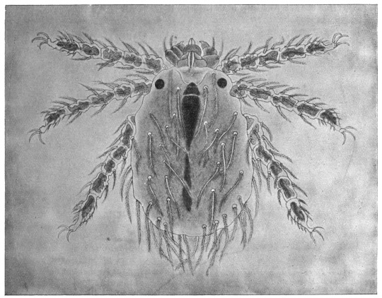
Fig. 351.—The kedani mite. Enlarged. (After Tanaka.)
The orange-red mites, which we only know in their larval condition, measure 0·16 to 0·38 mm. in length by 0·10 to 0·24 mm. in breadth. They have leg-like palpi with three joints, hirsute bodies, and very hairy legs composed of five segments, terminating with three ungues.
Family. Tetranychidæ (Spinning Mites).
These have tracheæ and eyes; the palpi are composed of four segments, of which the last but one has a powerful claw. The legs have six segments with sucker discs between the claws.
[The red spiders or spinning mites (Tetranychi) are usually placed in the family Trombidiidæ.—F. V. T.]
Genus. Tetranychus, Dufour.
Tetranychus molestissimus, Weyenbergh, 1886.334
Found in Argentine and Uruguay on the under surface of the leaves of Xanthium macrocarpum; it attacks mammals and men, producing severe itching, accompanied by fever in the latter.
It has been asserted by Haller that the Cape ailment (Port Natal sickness) is caused by mites, but this statement has been contested.
Tetranychus telarius, L., 1758,335 var. russeolus, Koch.
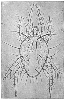
Fig. 352.—Tetranychus telarius var. russeolus, Koch. Enlarged. (After Artault.)
This common spinning mite likewise attacks human beings, but the papules produced by it very soon disappear.
Family. Tarsonemidæ.
A family distinguished by complete sexual dimorphism, the species of which are provided with tracheæ; the legs have five segments; the terminal segments of the front pair of legs of both sexes possess a claw; the terminal segment of the posterior pair of legs of the male likewise has a claw. In the female this pair of legs, like the second and third pairs of both sexes, is provided with two hooklets and a sucking disc. The cuticle of the body on the back is “annulated.”
[This family of small transparent mites live normally as plant parasites. The last two pairs of legs are widely separated from the two front pairs.—F. V. T.]
Genus. Pediculoides.
Pediculoides ventricosus, Newport, 1850.
Syn.: Heteropus ventricosus, Newport, 1850; Acarus tritici, Lagrèze-Fossot, 1851; Physogaster larvarum, Lichtenstein, 1868; Sphærogyna ventricosa, Laboulbène and Mégnin, 1885.
Males are oval in shape, 0·12 mm. in length and 0·08 mm. in breadth, flattened. There are six pairs of chitinous hairs on the dorsal surface and a lyre-shaped lamella on the posterior part. The female in the non-gravid state is cylindrical in form, 0·2 mm. in length and 0·07 mm. in breadth; when gravid the posterior part of the body becomes enlarged into a ball, which may attain 1·5 mm. in size, as in the case of Pulex penetrans and of the female Termites. On emerging the young are already provided with four pairs of legs and copulate soon after birth.
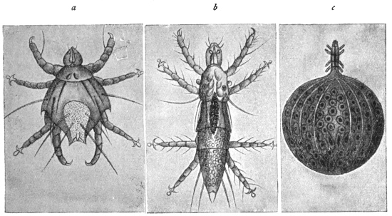
Fig. 353.—Pediculoides ventricosus. a, male; b, young female; c, gravid female. Enlarged. (After Laboulbène and Mégnin.)
These animals live on the stalks of cereals, and feed on vegetable and animal juices; they are also found on corn-infesting insects. They invade the barns and seek out the insects living in the dry grains of corn, or wait for an opportunity of obtaining food. They have been repeatedly observed on human beings, particularly labourers occupied in handling grain; their bite causes severe irritation, local elevation and reddening of the epidermis, as well as fever. It cannot be positively asserted that all cases of the occurrence of cereal mites on man relate to P. ventricosus, as the descriptions are often insufficient. Geber states that one form is Chrithoptes monunguiculosus, or Acarus hordei; Flemming mentions Tarsonemus uncinatus; Koller Oribates sp.; and Karpelles Tarsonemus intectus.
[The pregnant female Pediculoides has a large round inflated abdomen, in which the ova hatch and the young mature. Later they escape from the parent as adults.—F. V. T.]
Genus. Nephrophages.
Nephrophages sanguinarius, Miyake and Scriba, 1893.
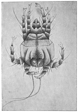
Fig. 354.—Nephrophages sanguinarius: male, ventral surface. Enlarged. (After Miyake and Scriba.)
Males measure 0·117 mm. in length and 0·079 mm. in breadth; females up to 0·360 mm. in length by 0·120 mm. in breadth. The head is provided with two very large scissors-like jaws and two large round eyes. The legs are composed of five segments and are all of equal length; the three anterior pairs of legs have pedunculated ambulacra, the posterior ones terminate in a claw. The cuticle on the back is thickened in three places, shield-like; the abdominal surface without scutellum is longitudinally striped and is beset with chitinous hairs. Colour greenish to brownish-yellow. Eggs 0·046 to 0·040 mm.
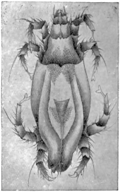
Fig. 355.—Nephrophages sanguinarius: female, dorsal aspect. Enlarged. (After Miyake and Scriba.)
The authors discovered these mites, but always dead, in the urine of a Japanese suffering from fibrinuria complicated with chyluria and hæmaturia. They surmised that they were endoparasites, probably situated in the kidney; but this view is not convincing, though they also report that for a week, day after day, the mites were found in the patient’s urine, as well as in urine drawn off by means of a catheter, and in the water used to wash out the bladder (one or two specimens and an egg). The statement that these mites have large eyes makes the discovery suspicious, to say the least. The significance of the discovery is not supported by the further statement that Disse is supposed to have found an encapsuled mite closely related to the Tyroglyphides on the wall of the vena cava.
In the case of Marpmann, who found a dead Acarid in the urine of a man suffering from chronic nephritis, and in whom later examinations proved negative, the author himself was of opinion that the mite had reached the urine from outside.
We are certainly acquainted with mites living endoparasitically, namely, the Cysticolæ, Analgesinæ, of which Laminosioptes gallinarum live in the intramuscular and subcutaneous connective tissue of fowls, and Cytoleichus sarcoptoïdes in their air sacs. Another kind of mite (Halarachne halichœri) is occasionally found in the nasal mucous membrane of the seal (Halichœrus grypus), and, quite recently, Pneumonyssus simicola, which is more nearly related to Halarachne, has been found in the lung of Cynocephalus sp. It is therefore not improbable that endoparasitic mites are found in man; but no definite discovery has yet been made.
Family. Eupodidæ.
Small tracheate mites, with moderately long or short pedipalpi, composed of four segments, of which the last segments bend; cheliceræ forceps-shaped, with serrated edges; legs with two claws, more rarely with one, and terminating in a tuft ornamented with fine hairs; genital orifices on the abdomen, surrounded by a circle of little hairs. Most species live free—one lives parasitically on the bodies of slugs.
Genus. Tydeus, Koch.
Tydeus molestus, Moniez, 1889.
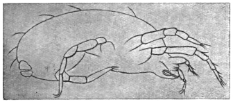
Fig. 356.—Tydeus molestus: seen in profile. Enlarged. (After Moniez.)
Male, 0·2 mm. in length, 0·125 mm. in breadth. Females, 0·225 mm. in length, 0·135 mm. in breadth; gravid female 0·315 to 0·360 mm. in length and 0·180 mm. in breadth. They were observed by Moniez on an estate in Belgium, whither the creature had apparently been imported twenty-five years previously with Peruvian guano; they appeared regularly in the summer and remained until the first frost set in; they were found on grass-plots, on trees and bushes in masses; they regularly attacked human beings, mammals and birds, tormenting their hosts in a terrible manner.
Family. Gamasidæ (Coleopterous or Insect Mites).
Cheliceræ chelate or styliform; pedipalpi filiform; the legs are composed of six segments with two terminal ungues and a bladder-like sucking disc [caruncle—F. V. T.]. Stigmata situated between the third and fourth pairs of legs; the cuticle thickened, leather-like; no eyes; the larvæ have six legs.
The Gamasidæ are predaceous on small insects and other mites; some are parasitic on insects, and one is noticeable as a pest on birds, etc.
Genus. Dermanyssus, Dugés.
Dermanyssus gallinæ, de Geer, 1778.
Syn.: Pulex gallinæ, Redi, 1674; Atarus gallinæ, de Geer, 1778; Dermanyssus avium, Dugés, 1834.
The male measures 0·6 mm. in length by 0·32 mm. in breadth; the female 0·7 to 0·75 mm. in length by 0·4 mm. in breadth. The body is somewhat pear-shaped; the colour whitish, reddish, or reddish-black, according to the contents of the intestine. The legs are fairly short and strong. During the day they live concealed in the nests, cracks, etc., of the hen-house, and at night attack the inmates in order to suck their blood; they rarely remain long on the birds. They have been repeatedly found on persons, on whose skin they produce an itching eruption.
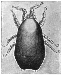
Fig. 357.—Dermanyssus gallinæ. Enlarged. (After Berlese.)
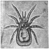
Fig. 358.—Dermanyssus hirundinis. 40/1. (After Delafond.)
Dermanyssus hirundinis, Hermann, 1804.
Syn.: Acarus hirundinis, Herm., 1804.
Of a brownish colour, 1·2 or 1·4 mm. in length; lives in the nests of swallows and is occasionally found on man.
[The red hen mite (Dermanyssus gallinæ) not only attacks poultry and man, as stated above, but is found on all birds and many mammals. The D. gallinæ is the same as D. avium. The species found in swallows’ nests is also said to be the same. This mite can remain for weeks without any food from its normal host. They only attack man when entering or cleaning dirty and neglected fowl-houses; they do not produce a true dermatosis. They chiefly attack the backs of the hands and forearms of those who constantly attend poultry and give rise to symptoms similar to the papular eczema of scabies. That they may remain some time upon the human body we know from the following cases out of many recorded: Geber observed that the Dermanyssus had caused a diffused eczema on a woman, which lasted four weeks and then disappeared. The tique of F. V. Raspail is the bird Dermanyssus; he records children and adults being attacked not only when handling pigeons, but even when walking in a garden manured with pigeons’ dung. The affection soon disappeared when the pigeons were destroyed and the excreta buried. I have frequently heard of poultrymen being seriously attacked by this pest.—F. V. T.]
Genus. Holothyrus.
Holothyrus coccinella, Gervais, 1842.
Measures 5 mm. in size; lives on birds in the Island of Mauritius; ducks and geese frequently fall victims to its bite; it also attacks human beings, on whose skin it causes severe burning and swelling, but no reddening; it may be dangerous to children, especially by settling in the oral cavity.
Other Gamasides occasionally occur in man, for instance, according to Moniez, Leignathus sylviarum, Canestr. et Fanzago; according to Neumann Lælaps stabularis. The former live normally in the nests of various species of Sylvia, Lælaps on dried vegetable substances, also in houses.
[Marchoux and Conoy (Bull. Soc. Path. exot., 1912, v, No. 10, pp. 796–798) found Leishman granules in Lælaps echidninus. It is assumed that Leishman granules may be found in most Arachnoids, and have no connection with Spirochæta.—F. V. T.]
Family. Ixodidæ (Ticks).
Comparatively large Acarines with a leathery skin; they are flattened in form, but after sucking blood the abdomen becomes spherical; the cheliceræ are rod-like and possess a serrated terminal joint, bent hook-like; the median parts of the pedipalpi (maxillæ) form a rostrum furnished with barbed hooks (fig. 359); the maxillary palpi themselves are club-like or rounded; the legs are composed of six segments with two terminal ungues, often also with “sucking discs”; the stigmata are at the sides of the body, posterior to the fourth or third pair of legs. The larvæ are six-legged.
[The true ticks (Ixodidæ) are all blood-suckers, and as far as is known they do not take vegetable food at all. Not only are the Ixodidæ important as actual parasites, but they are most so on account of the fact that they are the active agents in carrying various diseases in animals and apparently in man. It has been conclusively proved that the bont tick (Amblyomma hebræum) is the carrier of the fatal “heart-water fever” so rife amongst sheep in South Africa, that the dog tick (Hæmaphysalis leachi) is the agent by which the protozoa that cause malignant jaundice in dogs are distributed, that Texas fever in cattle is spread by Rhipicephalus annulatus, and Coast or Rhodesian fever by R. appendiculatus and R. simus. Their importance as disease carriers amongst mammals is therefore considerable, and it may prove to be so for man.336 They frequently attack man, but chiefly, according to my observations, in their early stages in Europe; this is not so, however, abroad. The life-history of a number of ticks has been clearly demonstrated. Mr. Wheler has shown that in Ixodes reduvius it is as follows: the female deposits her eggs in masses upon the ground, gradually reducing in size as the eggs pass out, until she finally remains a mere shrivelled empty bag and then dies. The eggs are oval, golden brown in colour and smooth; in length they are 0·59 mm.; as in all Ixodidæ they are covered with a glutinous secretion, by means of which they adhere together in masses. These egg masses may be deposited anywhere on the ground, but amongst rough, coarse herbage seems to be the favourite place. The egg stage may last as long as twenty-two weeks, or it may only take eight weeks. In the case of the bont tick a single female may deposit 15,000 or more eggs. The process of egg-laying is described as follows by Mr. Wheler: “When egg-laying is about to take place, the head is further depressed till it rests close against the under side of the body. In this attitude the end of the rostrum actually touches the genital orifice, the palpi being at the same time widely opened out. Behind the head and from beneath the shield, at what for the purposes of explanation may be described as the back of the neck, a white, perfectly transparent, delicate gelatinous membrane is brought down through inflation, either with air or with a transparent fluid, above the head, which it temporarily conceals. The end of this membrane terminates in two conical points which appear to be covered with a glutinous secretion, and at the same time an ovipositor of a somewhat similar character, but only semi-transparent, is pushed forward from the genital orifice. This latter is a tube, within which is the egg. As the ovipositor projects it turns itself inside out, like the finger of a glove, leaving the egg protruded at the end and lying between the two finger-like points of the membrane. The membrane and the ovipositor are then withdrawn each from the other. The egg adheres to the former, which collapses through the withdrawal of its contents, dragging the head forward and depositing it on the top of the head. Neither legs, palpi, nor the organs of the mouth take any part in oviposition, but after the collapse of the membrane the palpi are closed and the head is raised, by which action the egg is pushed forward to the front edge of the shield, forming in time an adherent mass of eggs, which are deposited in front of the tick.”
[The egg gives rise to the larval form, the so-called “seed-tick” stage. At first these minute specks are pallid and soft, but they soon harden and darken in colour. These larvæ are six-legged and crawl up grasses and various plants, and there await a passing host, waving their two front legs in the air and becoming attached by this means. The larval ticks feed upon the blood of the host, and when replete fall to the ground, the body becoming inflated in the meanwhile. These larvæ may remain on the host only two days, or they may remain much longer. Eventually they moult on the ground and change to the nymph or pupal stage, which has four pairs of legs. This pupa acts just as the larva, crawls up plants and waits to regain the host. After a time the nymphs, having gorged themselves with blood, fall off and remain on the ground for nearly three months; they then moult and become adult males and females. In about ten days they assume their normal colour and regain the host afresh; the female gradually swells until she attains that large inflated form so characteristic of ticks. The male does not swell, but nevertheless feeds upon the host and fertilizes the female.
[The act of coitus is strange: the male tick inserts its rostrum and other mouth organs into the sexual orifice of the female, between the base of the posterior pair of legs. The males then die and the females fall to the ground and deposit the ova. There are variations in the different species, of course, from those given above, which apply solely to Ixodes reduvius. The larvæ and nymphs seem to attack most animals, but the adults mainly keep to the same host. The periods in the life-cycle of ticks not only vary in the different species, but in each species according to climatic conditions; for instance, in the bont tick (Amblyomma hebræum, Koch), Lounsbury has shown that the development is rapid in summer, slow in winter. The period from the time that the female drops to the time she commences to lay eggs varied in specimens observed by him from twelve days in summer to twelve weeks in winter, and the complete period from the dropping of the female to the hatching of the eggs, from eleven weeks in summer to thirty-six weeks through the winter. Other stages vary in a similar manner.
[Ticks may live a long time away from the host provided they are supplied with a certain amount of moisture. Mr. Wheler kept dog ticks (Ixodes plumbeus) in the larval stage for ten months; the pupæ, male and female, of I. reduvius for six months.
[I have kept Ornithodorus moubata alive for eighteen months without food.
[In many species moulting takes place off the host, but in I. bovis, now known as Rhipicephalus annulatus, Say (the carrier of Texas fever), moulting takes place on the host, and in many other species also.337 Some species of ticks leave their host on its death (as the dog tick, Hæmaphysalis leachi), but others die with the host (bont tick, Amblyomma hebræum).
[Two species are of special importance, namely Ornithodorus moubata, Murray, which may infect human beings with the spirillum of African tick fever, and Dermatocentor reticulatus var. occidentalis, which is said to be the carrier of Rocky Mountain spotted fever.
Classification of Ixodidæ.
[The ticks, or Ixodidæ, are divided into two groups, known as (1) Argantinæ, (2) Ixodinæ. The Argantinæ are told from the Ixodinæ by the absence of dorsal or ventral shields in both sexes, and also by the rostrum being placed beneath the cephalothorax, which covers it over: except in the larval stage, in which it is subterminal, and in the pupal, when it partly projects. Legs nearly equal in length. The sexual orifice is situated between the two first pairs of legs. The males usually smaller than the females.
[The Ixodinæ have the legs unequal, of six segments with two false segments, making them look as if composed of eight segments. The rostrum is terminal and never hidden beneath the body. The sexual orifice is situated between the bases of the first three pairs of legs. In the males the orifice is obsolete or very rudimentary, sexual intercourse being effected by the rostrum. The males are smaller than the females. The shield in the females never covers so much as one-half of the body even when fasting, also in the larvæ and nymphs; but in the males, which do not distend, the shield covers the body entirely, or all but a narrow margin. The Ixodinæ are divided into two groups: (i) the Ixodæ, and (ii) the Rhipicephalæ. The former have a long proboscis reaching nearly to the end of the palpi or even a little longer than the palpi. The palpi are longer than broad. The Rhipicephalæ have short palpi, nearly or quite as broad as long, more or less conical or subtriangular. They were called Conipalpi by Canestrini.
Synopsis of Genera.
[A. Argantinæ: Rostrum concealed in adult, partly exposed in larvæ and nymphs. No dorsal and ventral shields.
Body flat with thin edges, finely shagreened and punctate Argas.
Body with numerous small round granules and with thick sidesOrnithodorus.
[B. Ixodinæ: Rostrum terminal. Body with dorsal shield over some part of it.
I.Rostrum and palpi longer than broad (Ixodæ).
α.A groove around anus in front.
Palpi caniculated in both sexesIxodes.
Palpi claviform, not caniculated in the male; legs very longEschatocephalus.
Palpi claviform, not caniculated in the male; anal groove absent in the femaleCeratixodes.
β.A groove around the anus behind.
No eyes; adanal shieldsAponomma.
Eyes present.
Males with no adanal shieldsAmblyomma.
Males with adanal shieldsHyalomma.
II.Labium and palpi short; palpi not, or very little, longer than broad (Rhipicephalæ).
α.No eyes.
Rostrum rectangular; no ventral shields in the maleHæmaphysalis.
β.Eyes present.
No adanal shields, but usually with greatly developed coxæ on fourth pair of legs. Capitulum quadrangularDermacentor.
Capitulum hexagonalRhipicentor.
Adanal shields in male. Stigmata comma-shapedRhipicephalus.
Stigmata oval or round; legs normalBoophilus.
Segments of legs expandedMargaropus.
[The genus Ceratixodes of Neumann, 1902, occurs on birds.
[The genus Eschatocephalus of Frauenfeld, 1853, of which seven species are known, is mostly parasitic on bats, and is found in holes, caves, and church towers.
[The genus Aponomma of Neumann, 1899, is exotic, and almost entirely confined to snakes and saurians.
[The following are synonyms of the different genera:—
Argas, Latreille, 1796 (Rhynchoprion, Hermann, 1804).
Ixodes, Latreille, 1795 (Acarus, Linnæus, 1758; Cynorhæstes, Hermann, 1804; Crotonus, Dumeril, 1822).