ON PARASITES IN GENERAL. (continued 2)
Reading edition
Within the large intestine an active increase of Entamœba histolytica must occur. Nevertheless, Jürgens did not definitely find changes that might be interpreted in this sense. Schaudinn (1903) observed division and gemmation in vivo. Both processes, in which the nucleus divides by amitosis, can only be distinguished by the fact that the daughter individuals are similar in binary fission but dissimilar in gemmation, whether they make their appearance singly or in greater numbers. Schizogony, resulting in the formation of eight individuals, which is so characteristic for Entamœba coli, was not observed. (But schizogony, into four merozoites, is now known to occur. Gemmation processes are apparently degenerative.)
Resistant stages, which serve for transmission to other hosts, are according to Schaudinn17 first formed when the diseased portions commence to heal, or more accurately, the recovery commences when the vegetative increase of the amœbæ in the intestine discontinues. The so-called spores of E. histolytica were distinguished very definitely from those of E. coli; they were said to consist of spheres of only 3 to 7 µ in diameter, which were surrounded by a double membrane, at first colourless, but becoming a light brownish yellow colour after a few hours, and possessing a protoplasmic content containing chromidia. They were said to arise by fragments of chromatin passing outwards from the nucleus of the amœba into the surrounding cytoplasm (fig. 9, a) and undergoing so marked an increase that finally the whole cytoplasm became filled with chromidia. The remainder of the nucleus underwent degeneration and became extruded. On the surface of the cytoplasm there then arose small protuberances containing chromidia. These processes had been observed in the living organisms. They gradually divided and separated from membranes which later became yellow. The remainder of the amœba perished. Craig18 had also seen phases of this process of development. It must be remarked that, according to recent researches, these processes of exogenous sporulation are degenerative in character (see p. 41). The small spores may be fungi. The “sporulation” processes are only mentioned here as a warning. They are now only of historic interest. By means of an experiment made on a cat, Schaudinn ascertained that ingestion of permanent cysts, which resist desiccation, is the cause of the infection. The animal took food containing dry fæces with amœba cysts; these fæces came from a patient suffering from amœbic enteritis in China. On the evening of the third day the cat evacuated blood-stained mucous fæces which contained large numbers of typical Entamœba histolytica. On the fourth day after the infection the animal experimented upon died, and the large intestine showed the changes previously stated.
E. histolytica also is found in the large intestine. This was originally shown to be the case by Kartulis, and the fact has recently been confirmed from many quarters. It is also present in the metastatic abscesses of which it is the cause (cf. among other authors, Rogers, Brit. Med. Journ., 1902, ii, No. 2,177, p. 844; and 1903, i, No. 2,214, p. 1315).
It should lastly be pointed out in this connection that mixed infections also take place. For instance, in addition to E. histolytica, E. coli, and, under certain circumstances, flagellates may be found together. In the same way E. coli may come under observation even in bacillary dysentery. On the other hand, Schaudinn stated that in cases of dysentery endemic in Istria, Entamœba coli, if it had hitherto been present, disappeared, to return again after recovery from the illness.
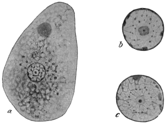
Fig. 8.—Entamœba histolytica. a, trophozoite (tetragena type) containing red blood corpuscles, × 1,300; b and c, two isolated nuclei showing different appearances of karyosome, centriole and nuclear membrane, × 2,600. (After Hartmann.)
(Entamœba tetragena, Viereck, 1907.)
This amœba must now be considered to be a part of the lifecycle of Entamœba histolytica, in fact a very important part of that cycle, especially in its tetranucleate cystic stages.
This organism, the so-called Entamœba tetragena, may occur in the human intestine in cases of amœbic dysentery, especially in mild or chronic cases. It was discovered by Viereck in 1907 in patients suffering from dysentery contracted in Africa. Soon afterwards an independent description was published by Hartmann, who called the amœba E. africana. It was also studied by Bensen and Werner. Recently (1912–13) much work has been published on this amœba by Darling and others; in this way its relationship to Schaudinn’s E. histolytica has been made known.
In general morphology it somewhat resembles Entamœba coli, and its discoverer at first mistook it for a variety of that species. According to Hartmann, a distinct ectoplasm is only clearly visible when a pseudopodium is protruded (fig. 5). The granular endoplasm may contain ingested red blood corpuscles (fig. 6). The large, round nucleus is visible in the fresh state (fig. 8, a). So-called chromidial masses (? crystalloidal substances) may occur in the cytoplasm.
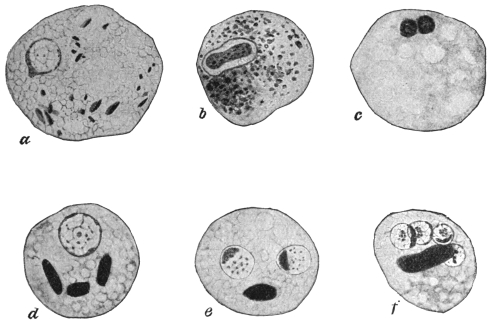
Fig. 9.—Entamœba histolytica (tetragena form). a, emission of chromatin from nucleus; b, nuclear division; c, degenerating form with two nuclei; d, e, f, cysts containing one, two and four nuclei respectively, and showing chromidial blocks. × 2,000. (After Hartmann.)
Some investigators, as Hartmann,19 lay stress on the internal structure of the nucleus (fig. 8, b, c), best seen in preparations fixed wet and stained with iron-hæmatoxylin. The nucleus is limited by a well-marked nuclear membrane, on the inside of which granules or nodules of chromatin may occur. There is a karyosome, which, in successfully stained specimens, shows, at times, a central dot called a centriole. (The nucleus of Entamœba coli does not contain such a centriole.) However, the structure of the nucleus varies at different periods during the life-cycle.
The diameter of the trophozoites or vegetative forms (fig. 8, a) is variously given as from 20 µ to 40 µ. Multiplication proceeds by binary fission and also by schizogony into four merozoites.20
Reproduction takes place by endogenous encystment (fig. 9, d-f), which is preceded by nuclear division into two, reduction and then autogamy. The interpretation of the latter phenomenon as autogamy is disputed by some authors. The round cysts, which may measure 12 µ to 15 µ in diameter, contain four nuclei, together with darkly staining masses of various shapes, the so-called “chromidial blocks” (fig. 9, f). The cyst-wall of E. histolytica (tetragena) is thinner than that of E. coli, and the diameter of the cyst is rather less. E. histolytica has not yet been cultivated.
Infection in man occurs by way of the mouth by the ingestion of cysts. A patient showing acute symptoms of dysentery is not usually infective, for he is merely harbouring the large trophozoites, which, by experiment, have been shown not to be infective to animals (kittens) when administered by the mouth. The stools of recovered patients may still contain cysts, and they may thus act as cyst-carriers or reservoirs of disease by infecting water and soil. The stools of such cyst-carriers are often solid, and so cysts of E. histolytica (tetragena) are easily overlooked. Mathis (1913)21 points out that healthy carriers of E. histolytica may be found; 8 per cent. of the natives of Tonkin examined by him were healthy carriers of cysts.
In return cases, or prolonged untreated cases of entamœbic dysentery, a generation of smaller trophozoites is associated with, or replaces the larger ones. In stools they are frequently refractile and consequently stain slowly intra vitam. These trophozoites are the “smaller, senile, or pre-cyst generation” of Darling. This pre-cyst generation is characterized by the presence of blocks of crystalloidal substance in the cytoplasm, and by the possession of a prominent, densely stainable karyosome. Darling believes this generation to be the same as that described by Elmassian as Entamœba minuta.22
Walker,23 Darling,24 Wenyon25 and others believe that Entamœba histolytica, which was only seen by Schaudinn in a single case, that of a Chinaman, is really E. tetragena. Darling states that if the published illustrations of E. histolytica and of E. tetragena are collected from the literature and compared, it will be seen that the writers have been calling E. histolytica the large trophozoites seen in dysenteric stools. These large trophozoites frequently display no karyosome, but they can be demonstrated as E. tetragena by animal inoculation, or by the history of the case. On the other hand, the illustrations of E. tetragena show that the authors have been dealing with the small generation or reduced forms (“E. minuta”), which are the direct descendants of the large trophozoites. If kittens are inoculated rectally with dysenteric material containing large trophozoites, the strain may be carried in successive kittens for four to six transfers. If, on the other hand, kittens are inoculated rectally with small trophozoites of the pre-cyst generation, the transmission cannot be carried through more than one or two kittens. Wenyon has succeeded in maintaining E. tetragena in kittens for several generations.
In some of the preparations from the last remove, pathological forms of the trophozoites may be seen. These show abnormal forms of budding, especially peripherally, such as have been described by Schaudinn and by Craig as characteristic of E. histolytica. Schaudinn’s small peripheral, exogenous buds and cysts are thus explained. Craig has latterly changed his views.
Further, Darling states that tetragena cysts fed by the mouth to kittens produce bowel lesions in which trophozoites having the characters of E. tetragena, E. histolytica and E. nipponica (Koidzumi) occur.
In view of the work of recent observers, the peculiar exogenous encystment which Schaudinn made characteristic of Entamœba histolytica has been shown to be due to degenerative changes in senile races of the amœba. E. histolytica and E. tetragena are one and the same species, and its trophozoite is subject to variation. According to some observers the histolytica type of nucleus—described by Schaudinn as being poor in chromatin and not easily seen in the fresh state—occurs frequently in patients with severe symptoms of dysentery; on the other hand, the tetragena type of nucleus—round and easily seen in the fresh state—may occur in cases presenting slight dysenteric symptoms. Intermediate types of nuclei are seen. The name of this species, the principal pathogenic amœba of man, must then be E. histolytica by priority. The cystic stages of E. histolytica are those first recorded by Viereck and formerly described as E. tetragena. The geographical distribution of E. histolytica is wide.
Noc’s Entamœba (1909).
A species of Entamœba was cultivated by Noc26 in 1909 from cysts derived from liver abscesses, from dysenteric stools and from the water supply of Saigon, Cochin China. He cultivated it in association with bacteria. It is pathogenic. It has been considered allied to E. histolytica, and shows internal segmentation or schizogony. It exhibits polymorphism. This amœba has been found by Greig and Wells (1911) in cases of dysentery in India. It is an important organism and requires further investigation.
Certain other Entamœbæ27 have been described at various times from the intestinal tract of man. Probably most, if not all, of these are not good species and in some cases much more information is needed.
Entamœba tropicalis (Lesage, 1908). This parasite is said to be non-pathogenic, and to occur in the intestine of man in the tropics. It has a general resemblance to E. coli, but forms small cysts (6 µ to 10 µ in diameter). The nucleus of the cyst is said to break up into a variable number of daughter nuclei, from three to thirteen having been noted. Lesage states that it is culturable in symbiosis with bacteria. It is probably a variety of E. coli, if not a cultural amœba.
Entamœba hominis (Walker, 1908) has a diameter of 6 µ to 15 µ. A contractile vacuole is present. Encystment is total, and small cysts are formed. It is culturable. The original strain, now lost, was obtained from an autopsy in Boston Hospital. This organism is probably a cultural amœba.
Entamœba phagocytoides (Gauducheau, 1908). This parasite was discovered in a case of dysentery at Hanoi, Indo-China. The amœba is small, 2 µ to 15 µ in diameter. It is active. It ingests bacteria and red blood corpuscles, while peculiar spirilla-like bodies are found in its cytoplasm. It multiplies by binary and multiple fission. It can be cultivated. More recently (1912) the author appears to consider the amœba to be a stage of a Trichomonas, but abandons the view later (1914). Further researches on this organism are needed.
Entamœba minuta (Elmassian, 1909)28 was found, in association with E. coli, in a case of chronic dysentery in Paraguay. It resembles E. tetragena but is smaller, rarely exceeding 14 µ in diameter. Schizogony occurs, four merozoites being produced. The encystment is total and endogenous, giving rise to cysts containing four nuclei. This amœba is considered by Darling and others to be the pre-cyst trophozoite stage of E. histolytica (tetragena).
Entamœba nipponica (Koidzumi, 1909) was found in the motions of Japanese suffering from dysentery or from diarrhœa, in the former case in company with Entamœba histolytica. Its diameter is 15 µ to 30 µ. The endoplasm is phagocytic for red blood corpuscles. The nucleus is well defined, resembling that of E. coli and of E. tetragena. Multiplication occurs by binary fission and by schizogony. Encystment is total, but has not been completely followed. Darling and others consider that this is an abnormal form of E. histolytica, while Akashi (1913) doubts if it is an amœba at all, but rather is to be regarded as shed epithelial cells.
General Remark.—It is now considered by some workers that true Entamœbæ cannot be cultivated on artificial media. Quite recently Williams and Calkins (1913)29 have somewhat doubted this opinion, and state that certain cultural amœbæ, originally obtained from Musgrave in Manila, exhibit the various morphological variations associated with true entamœbæ of the human digestive tract.
Entamœba buccalis, Prowazek, 1904.
The size varies from 6 µ to 32 µ. Ectoplasm is always present; the endoplasm contains numerous food-vacuoles. The nucleus is vesicular, with a greenish tinted membrane which is poor in chromatin. The size of the nucleus is from 1·5 µ to 4·5 µ. A contractile vacuole is not visible. The pseudopodium is broad. It was discovered in the mouths of persons with dental caries at Rovigno and also at Trieste, being most easily found in dense masses of leucocytes, also among leptothrix and spirochæte clusters. It can be easily distinguished from leucocytes by more intense staining with neutral red. Multiplication proceeds by fission. Transmission may take place through the small spherical cysts. This species (fig. 10) has since been observed in Berlin, and is also occasionally found in carcinoma of various regions of the oral cavity. (Leyden and Löwenthal, 1905).
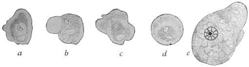
Fig. 10.—Entamœba buccalis, Prow. a-d, the same specimen observed during five minutes. × 1,000. e, amœba fixed and stained with iron-hæmatoxylin. × 1,500. (After Leyden and Löwenthal.)
Entamœba buccalis, Prow., is said to be allied to a protozoön which A. Tietze has found either encysted or free in the lumen of the orifice of the parotid gland of an infant aged 4 months. The gland had undergone pathological change, and had therefore been extirpated. The organisms, which were roundish and three to four times the size of the normal epithelial cells of the gland, were without a membrane and possessed a nucleus in which the chromatic substance appeared to be contained in a karyosome. Bass and John’s30 (Feb. 1915) and Smith, Middleton and Barrett (1914) state that E. buccalis is the cause of pyorrhœa alveolaris.
Entamœba undulans, Aldo Castellani, 1905.
Under this name a protozoön is described which A. Castellani found in addition to Entamœba histolytica and Trichomonas intestinalis in the fæces of an European planter living in Ceylon, who had suffered from amœbic enteritis and liver abscess. The shape of the body was roundish or oval, 25 µ to 30 µ in the greatest diameter. It was without a flagellum, but with an undulating membrane, and capable of protruding a long pseudopodium from different parts of its body at short intervals. The nucleus could not always be recognized in life; it was, however, always demonstrable by staining. One or two contractile vacuoles were present. The protoplasm was finely granular, showing no differentiation into ecto- and endo-plasm. According to Braun, in spite of the author declaring himself expressly against the flagellate nature of the parasite, such a nature may be assumed to be tolerably certain in view of the description and illustration.
It is now considered that Entamœba undulans is a portion of a flagellate, namely, Trichomonas.
Entamœba kartulisi, Doflein, 1901.
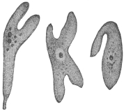
Fig. 11.—Entamœba kartulisi, Dofl., from the pus of an abscess in the lower jaw, showing different stages of movement. (After Kartulis.)
Doflein gave this name to amœbæ, from 30 µ to 38 µ in diameter, which Kartulis (1893) found on examining the pus of an abscess in the right lower jaw of an Arab, aged 43, and in a portion of bone that had been extracted. The movements of the amœbæ (fig. 11) were more active than those of “dysenteric amœbæ.” Their coarsely granular cytoplasm contained blood and pus corpuscles, and a nucleus was generally only recognizable after staining. Vacuoles were not seen with certainty. Flexner reported upon a similar case, and Kartulis published five additional cases. As in these cases dental caries was present the infection is likely to have proceeded from the oral cavity as a result of the carious teeth. Craig31 (1911) considers that this parasite is probably identical with Entamœba histolytica.
In the literature the following species have been reported as occurring in the oral cavity of man:—
Amœba gingivalis, Gros, 1849. [? identical with Entamœba buccalis.] Amœba buccalis, Sternberg, 1862. Amœba dentalis, Grassi, 1879.
Far too little, however, is known concerning these to regard them as definite species, that is, independent organisms; Grassi thinks it even possible there may have been a confusion in their case with salivary corpuscles. If they really are amœbæ they are all of them probably identical with Entamœba buccalis.
Genus Paramœba, Schaudinn, 1896.
Schaudinn established the genus Paramœba for a marine rhizopod which multiplied by division, became encysted at the end of its vegetative life and then segmented into swarm bodies with two flagella. These multiplied by longitudinal fission, and finally passed into the condition of Amœbæ. Whether the human parasite described by C. F. Craig (1906) as
Paramœba hominis.
belonged to this genus was for a time uncertain. It is now placed in a new genus Craigia, Calkins, 1912, since it possesses only one flagellum.32
In the amœbic stage it is 15 µ to 25 µ in diameter; ecto- and endo-plasm during rest are indistinguishable. The body substance is granular, with a spherical, sharply contoured nucleus and an accessory nuclear body. No vacuoles are present, but occasionally the endoplasm contains red blood corpuscles. The pseudopodia are hyaline, finger- or lobe-shaped, and are protruded either singly or in twos. Multiplication is by binary fission and by the formation of spherical cysts (15 µ to 20 µ in diameter) in which occurs successive division of the nuclei, ultimately forming ten to twelve roundish bodies each of which soon develops a flagellum. The flagellate stages have similarly a spherical shape and attain a diameter of 10 µ to 15 µ. They also occasionally contain red blood corpuscles and pass either directly or after longitudinal division into the amœboid phase.
Craig found these Amœbæ and the flagellate stage belonging to them in six patients in the military hospital at Manila (Philippine Islands), five of whom were suffering from simple diarrhœa whilst the sixth exhibited an amœbic enteritis and contained also Paramœba hominis, with Entamœba histolytica, Schaudinn. In one of the other cases, Trichomonas intestinalis was present.
B. Amœbæ from other Organs.
Entamœba pulmonalis, Artault, 1898.
Artault33 discovered a few amœboid forms with nucleus and vacuole in the contents of a lung cavity. In the fresh condition they were distinguishable from leucocytes by their remarkable capacity of light refraction. They were also much slower than the latter in staining with methylene blue or fuchsine. Their movements became more lively in a strong light. Water and other reagents killed them, and then, even when stained, they could not be distinguished from leucocytes. They have also been seen by Brumpt. R. Blanchard found amœbæ which may belong here in the lungs of sheep. A. pulmonalis is perhaps the same as Entamœba buccalis. Smith and Weidman34 (1910, 1914) described an entamœba, E. mortinatalium, from the lungs and other organs of infants in America.
Amœba urogenitalis, Baelz, 1883.
This species was found in masses in the sanguineous urine as well as in the vagina of a patient in Japan, aged 23. Shortly before the death of the patient, which was caused by pulmonary tuberculosis, hæmaturia with severe tenesmus of the bladder had set in. The amœba, which showed great motility, and had a diameter of about 50 µ when quiescent, exhibited a granular cytoplasm and a vesicular nucleus. Baelz is of opinion that these parasites were introduced into the vulva with the water used for washing the parts, and thence had penetrated into the bladder and vagina. Doflein places the organism in the genus Entamœba, and it is perhaps identical with E. histolytica.
Similar cases are also reported (1892–3) by other authors: Jürgens, Kartulis, Posner, and Wijnhoff. Jürgens found small mucous cysts, filled with amœboid bodies, in the bladder of an old woman suffering from chronic cystitis; they were also found in the vagina. The amœba observed by Kartulis in the sanguineous urine of a woman, aged 58, suffering from a tumour of the bladder, measured 12 µ to 20 µ, and exhibited slow movements by protruding short pseudopodia. The vacuoles and nucleus became visible only after staining with methylene blue.
Posner’s case related to a man, aged 37, who had hitherto been quite healthy and had never been out of Berlin. Suddenly, after a rigor, he passed urine tinged with blood. This contained, besides red and white blood corpuscles and hyaline and granular casts, large granular bodies (about 50 µ in length and 28 µ in breadth), which slowly altered their shape, and contained red blood corpuscles in addition to other foreign matter. These bodies exhibited one or several nuclei and some vacuoles. From the course of the disease, which extended over a year, and during which similar attacks recurred, Posner came to the conclusion that the amœbæ which had originally invaded the bladder had penetrated into the pelvis of the kidney, where they probably had settled in a cyst, and thence induced the repeated attacks.
Wijnhoff observed four cases of amœburia in Utrecht.
Amœba miurai, Ijima, 1898.
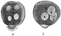
Fig. 12.—Amœba miurai, Ij. × 500. a, fresh; b, after treatment with dilute acetic acid. (After Ijima.)
Under this term the author describes protoplasmic bodies which Miura, in Tokyo, found in the serous fluid of a woman, aged 26, who had died from pleuritis and peritonitis endotheliomatosa. Two days before death these same forms had also appeared in the hæmorrhagic fæces of the patient. The bodies were usually spherical or ellipsoidal, and at one pole carried a small protuberance (fig. 12) beset with filamentous short “pseudopodia” (really a pseudopodium covered with cilia). Their size varied between 15 µ and 38 µ. The cytoplasm was finely granular, and no difference was observable in the ecto- and endo-plasm, only the villous appendage was clearer. The cytoplasm contained vacuoles more or less numerous, none of which was contractile. After the addition of acetic acid one to three nuclei could be distinguished, 8 µ to 15 µ in size. Actual movements were not observed. Taking everything into consideration, the independent nature of these bodies is, to say the least, doubtful, although it cannot be denied that they possess a certain similarity to the marine Amœba fluida, Grüber or Greeff, and to a few other species. (It is likely that cells present in serous exudation were mistaken for amœbæ.)
Appendix.
“Rhizopods in Poliomyelitis acuta.”
In three cases of poliomyelitis acuta which were investigated by Ellermann, the spinal fluid obtained by puncture of the cord contained bodies, from 10 µ to 15 µ in size, which had amœboid movements and exhibited variously shaped pseudopodia in large numbers. After staining, a usually excentric nucleus, about 1·5 µ in size, was demonstrated in them.
Order. Foraminifera, d’Orbigny.
The order is divided by Max Schultze into Monothalamia and Polythalamia. Only a few of the former can be considered here.
Sub-Order. Monothalamia. (Testaceous Amœbæ).
These forms occur frequently in fresh water, rarely in sea water. They possess a shell which is either pseudo-chitinous in character, or consists of foreign particles, or in a few cases is composed of siliceous lamellæ. There is usually an orifice for the protrusion of pseudopodia. The only representative of the order of interest here is:—
Genus. Chlamydophrys, Cienkowski, 1876.
The genus is based on a form which A. Schneider carefully investigated and considered to be the Difflugia enchelys of Ehrenberg. L. Cienkowski rediscovered this same form and created for it the genus Chlamydophrys. We agree with this view, but not with the renaming of the organism (so common at the time). If the parasite in dung, Chlamydophrys stercorea Cienk. is identical with Difflugia enchelys of Ehrenberg, the old specific name should be retained.
The genus is characterized by the possession of a hyaline, structureless, slightly flexible shell which is ovoid or reniform. At the more pointed pole there is an orifice situated terminally or somewhat laterally, serving for the emergence of the filiform pseudopodia (fig. 13, a). The protoplasm does not entirely fill the interior of the shell. An equatorial zone bearing excretory granules divides the shell internally into two almost equal portions. The anterior portion is rich in vacuoles and serves for the reception of nutriment and for digestion. The posterior part is vitreous, and contains the nucleus. One to three contractile vacuoles are situated in the equatorial zone.
Chlamydophrys enchelys, Ehrbg.
Syn.: Chlamydophrys stercorea, L. Cienkowski.
This species (fig. 13) is found in the fæces of various animals (cattle, rabbits, mice, and lizards), and also in quite fresh human fæces. According to Schaudinn, the parasite occurs so frequently in the human fæces that it must be considered of wide distribution. The species must traverse the intestine of man and animals during one stage of its life cycle, as Schaudinn showed by experiments on himself and on mice. He infected himself with cysts (fig. 14) by swallowing them, and evacuated the first Chlamydophrys as early as the following day. After the evacuation of numerous specimens on one of the following days the infection ceased.
The nucleus of a living specimen is surrounded by a hyaline, strongly refractile chromidial mass, arranged in the form of a ring. Chromatin stains colour it darkly.
Asexual multiplication (fig. 13, b), which takes place in fæces, follows a similar course to that of allied forms (e.g., Euglypha, Centropyxis). It commences by the cytoplasm issuing from the orifice of the shell and assuming the shape characteristic of the mother organism, but in a reverse position. The nucleus then divides by mitosis, when the daughter nuclei move apart from one another. The chromidial ring also divides into two portions by a process of dumb-bell like constriction. The one daughter nucleus remains in the mother organism, the other moves towards the daughter individual, which then separates from the parent.
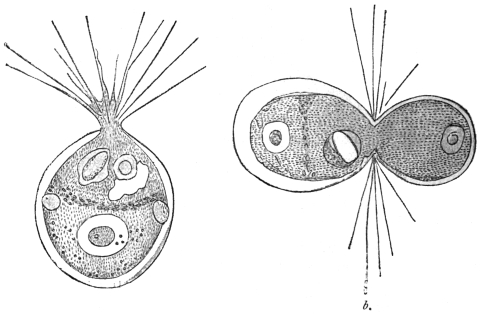
Fig. 13.—Chlamydophrys enchelys. a, free, motile form, showing nucleus, equatorial granules, vacuoles and pseudopodia; b, dividing organism. × 760. (After Cienkowski.)
In this species plasmogamic union of two or more individuals (up to twenty) is frequently observed. Such colonies may similarly divide, and in this way monstrosities frequently arise. When drying of the fæces, or deficiency of food occurs, encystment takes place apparently spontaneously. The whole body, as stated by Cienkowski, issues from the shell, assumes a spherical shape (probably with discharge of water) and becomes surrounded with a thick membrane (fig. 14). After the addition of water and the escape of the encysted Chlamydophrys, a new shell must be formed. Schaudinn, who has not given a more detailed description of the process of encystment in this species, but refers to Cienkowski and to similar observations made on Centropyxis, states of the latter that the encystment takes place within the shell.
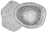
Fig. 14.—Chlamydophrys enchelys, encysted; on the left the old capsule. × 760. (After Cienkowski.)
The sexual multiplication is accompanied by shedding of all the foreign bodies and of the degenerating nucleus. The protoplasm, now contracting into a sphere, remains behind in the shell with the chromidial mass. From the latter several new nuclei arise (sexual nuclei) often eight in number. The cytoplasmic sphere then segregates into as many spherical portions as there are nuclei present. When they have assumed an oval form, two flagella develop at one pole and the flagellispores swarm out of the shell.35 The biflagellate swarm-spores, or gametes, copulate in pairs and apparently the individuals of the pairs of gametes arise from different mother organisms. The zygote secretes a thick covering which soon becomes brown and rough. These zygote cysts or resistant spores must now pass from the intestine of an animal in order to complete their development. The escape of the cyst contents does not always take place in the intestine; often it does not occur until after defæcation. These shell-less individuals (amœbulæ) soon become invested with a shell. But in the alkaline intestinal contents, shell formation may proceed even while the organism is in the intestine, and multiplication may take place.
Schaudinn’s further communication was of special interest; it was to the effect that Chlamydophrys was related to
Leydenia gemmipara, Schaudinn, 1896.
In the fluid removed by puncture from two patients suffering from ascites in the first medical clinic in Berlin, cellular bodies with spontaneous movement were found, which Leyden and Schaudinn regard as distinct organisms. They remained alive without the use of the warm stage for four or five hours, the external temperature being 24° to 25° C. In a quiescent condition they were of a spherical or irregular polygonal form. Their surface was rarely smooth, being beset with protuberances and excrescences (fig. 15). The substance of the body was thickly permeated with light refractile granules with a yellowish shimmer. The hyaline ectoplasm was rarely seen distinctly. All sizes from 3 µ to 36 µ in diameter were observed. The movements were rather sluggish, the ectoplasm in the meantime appearing in the form of one or several lamellæ, in which also strings of the granular endoplasm occurred, and frequently protruded over the border of the hyaline pseudopodia. The tendency for the joining of several individuals by means of their pseudopodia was so marked that associations ensued similar to those known in free-living Rhizopoda.
The cytoplasm enclosed blood corpuscles as well as numerous vacuoles, one of which pulsated slowly about every quarter of an hour. A vesicular nucleus the diameter of which was about equal to one-fifth of the body was present.
Multiplication took place by means of division and budding (fig. 15, c), after previous direct division of the nucleus. The buds were supposed to divide repeatedly soon after their appearance, thus giving rise to minute forms of 3 µ.
There was a suspicion in both cases that the ascites was associated with malignant neoplasms in the abdomen, and autopsy confirmed this view in one case.
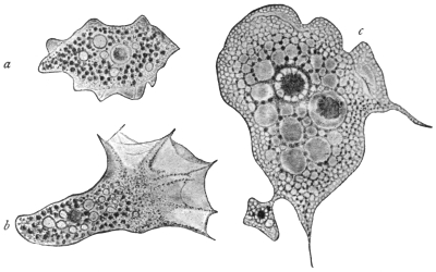
Fig. 15.—Leydenia gemmipara, Schaud. a, in a quiescent condition, × 1000; b, in the act of moving, × 1000; c, from a fixed preparation, showing a bud, × 1500.
The parasite, which has seldom been observed, has been variously interpreted; for example, it has been regarded merely as altered tissue cells. It is now known, from Schaudinn’s researches, that Leydenia gemmipara is connected with abnormal conditions of Chlamydophrys, occasionally occurring as a commensal in the ascitic fluid. The form is produced when pathological conditions of the large intestine create an alkaline reaction of its whole contents. The formation of shells then often ceases, and these naked Chlamydophrys are enabled to multiply atypically by division and gemmation. Such stages, which are no longer capable of a normal development, are the Leydenia, as Schaudinn has demonstrated.
Class II. MASTIGOPHORA, Diesing.
Sub-Class. FLAGELLATA, Cohn emend. Bütschli.
During the motile part of their life the Flagellata possess one or more flagella which serve for locomotion, and in many cases also for the capture of food. A few groups (Euglenoidinæ, Choanoflagellata) have only one flagellum, others two or several of about equal length (Isomastigoda), or of various lengths (Monadina, Heteromastigoda, Dinoflagellata). The long flagellum is the principal one; the smaller ones on the same organism are accessory flagella. The flagella directed backwards, which occur in the Heteromastigoda and are used for clinging, are termed trailing flagella or tractella. At the base of the flagellum, which is almost always at the anterior end, a Choanoflagellate possesses a cytoplasmic funnel-shaped neck or collar. In the parasitic forms an undulating membrane is often present.
The body of the Flagellata is usually small, generally elongate and of unchangeable form. It is frequently covered by a distinct cuticle, and, in certain groups, by a hard envelope, or it may be more or less loosely enveloped by a gelatinous or membranous covering. An ectoplasmic layer is thin and not always obvious. The granular cytoplasm contains a varying number of vacuoles, one of which may be contractile, and is generally situated near the area from which the flagella arise, that is, at the anterior extremity. The cytoplasm, moreover, contains the nucleus, which is nearly always single; and in many species there are also yellow, brown, or green chromatophores of various shapes, such as occur in plants. Some species feed after the manner of green plants (holophytic), or of plants devoid of chlorophyll (saprophytic); others, again, ingest solid food, and for this purpose usually possess a cytostome; the latter, however, in a few forms is not used for its original function, but is connected with the contractile vacuole. Many parasitic forms feed by endosmosis. A few species possess eye-spots with or without light-refracting bodies.
Variation in the form of the nuclear apparatus occurs. One nucleus only, which may be compact or vesicular, is known in many species. This nucleus is situated either centrally or sometimes near the flagellar end of the body, but its position is subject to variation. The flagella may arise near the nucleus. Other structures, such as an axial filament and a rhizoplast, may be present. Some flagellates are binucleate, the two nuclei—which often differ in size and shape—being separated from each other. One of these nuclei is the principal, vegetative or trophic nucleus; the other is an accessory nucleus, frequently termed the blepharoplast, flagellar or kinetic nucleus. One or more small basal granules are often present at or very near the origin of the flagella.
Multiplication is by fission, usually longitudinal, which may occur in either the free or encysted forms. Division is initiated by that of the nucleus or nuclei (especially the kinetic nucleus). The basal granule divides also. Collars and chromatophores, if present, likewise separate into two. Variation in the method of doubling the original number of flagella occurs. In most organisms, especially uniflagellate forms, the flagellum splits lengthwise, after division of the basal granule, blepharoplast and nucleus. The daughter flagella may be of the same or different lengths and thicknesses. Other flagellates at division are said to produce new flagella in the neighbourhood of the original ones. The daughter organisms in such cases are provided with one or more parental flagella in addition to newly formed ones. It has been stated that in certain cases the parent flagellate retains all its flagella, while new ones arise ab initio in the cytoplasm of the daughter forms.
Multiplication by longitudinal fission may be interrupted sooner or later by the production of gametes, which form zygotes, from which new generations of individuals arise. In many flagellates gamete formation and sporogony are unknown, and asexual reproduction by fission alone prevails.
Incomplete division results in the formation of colonies of individuals. These colonies must not be confused with the aggregation rosettes of flagellates found among the parasitic Mastigophora. The individuals of aggregation rosettes are capable of immediate separation from the rosette at will.
A number of parasitic Flagellata produce non-flagellate stages which are very resistant to external conditions, the assumption of which forms serves to protect the organisms during their transference from one host to another. Such non-flagellate forms possess one or more nuclei, are usually of an oval or rounded contour, and are capable of developing into the full flagellate on the return of more favourable conditions. These forms are often known as the post-flagellate stage of the organism. When ingested by a new host, the post-flagellate coat becomes more flexible, and the phase of the organism which now recommences growth is known as the pre-flagellate stage; it gradually develops into the typical flagellate organism.
Many Flagellata live free in fresh and salt water. They prefer stagnant water, rich in organic products of decomposition, such as puddles, swamps and pools. Those forms developing shells and colonies are, as a rule, adherent. A number of species are parasitic in man and animals, living mostly within the intestine or in the blood.
It is usual to classify the Flagellata in four orders: Euflagellata, Dinoflagellata, Choanoflagellata, and Cystoflagellata, of which only the Euflagellata are of interest to us. This is a group comprising numerous species, for the further classification of which the number and position of the flagella are utilised.
The Euflagellata observed in man belong to the Protomonadina as well as to the Polymastigina. The former possess either only one or two similar flagella, or one principal and one or two accessory flagella. The Polymastigina possess at least three flagella of equal size, or four to eight of unequal size, inserted at different points. An undulating membrane may be present in members of both groups.
It must also be pointed out that unicellular organisms with one or several flagella are not always classified with flagellates, for such forms occur in Rhizopods as well as temporarily in the lower plants. In addition, the examination of the flagellates, especially the parasitic species, is very difficult on account of their diminutive size and great activity; thus it happens that certain forms cannot with certainty be included in the group because their description is insufficient.
Order. Polymastigina, Blochmann.
The Polymastigina contains flagellates with three to eight flagella. Some of the Flagellata parasitic in man belong to the Polymastigina, and to two or three genera that are easily distinguishable.
Genus. Trichomonas, Donné, 1837.
The body is generally pyriform, the anterior part usually rounded, the posterior part pointed. There are at the anterior extremity three (? four) equally long flagella that are sometimes matted together. A blepharoplast (kinetic nucleus) and basal granule are present, together with a supporting structure known as an axial filament or axostyle. In addition there is an undulating membrane, bordered by a trailing flagellum, that commences at the anterior extremity and proceeds obliquely backwards. The nucleus, which is vesicular, is situated near the anterior extremity, and behind it are one or more vacuoles, none of which seems to be contractile. These flagellates are parasitic in vertebrate animals, and live chiefly in the intestine.
Trichomonas vaginalis, Donné.
The form of the body is very variable, and is elongate, fusiform or pear-shaped, also amœboid. The length varies between 15 µ and 25 µ, and the breadth between 7 µ and 12 µ. The posterior extremity is drawn out to a point and is about half the length of the remainder of the body. The cuticle is very thin and the body substance finely granular. At the anterior extremity there are three—some say four36—flagella of equal length which are frequently united together, at least at the base, and are easily detached.
There is an undulating membrane (fig. 16) which runs spirally across the body, arising from the place of insertion of the flagella, and terminating at the base of the caudal process. A cytostome seldom is recognizable in fresh specimens, but is apparently present. The nucleus is vesicular, elliptical and situated near the anterior extremity.37
Multiplication takes place by division (Marchand). Encysted forms are almost unknown.
Trichomonas vaginalis lives in the vaginal mucus of women of various ages, not in normal mucus, but in mucus of acid reaction. It is found in menstruating females as well as in females who have passed the menopause. It occurs in pregnant and non-pregnant women, even in very young girls, provided always that they have a vaginal catarrh with acid reaction of the secretion. Should the acid reaction change, as, for instance, during menstruation, the parasites disappear, as they do likewise on injection of any alkaline fluid into the vagina. A low temperature (below +15° C.) is also fatal to the parasites. These flagellates can pass from the vagina through the urethra into the bladder, and produce severe catarrh, and are not easily removed.
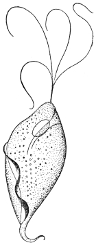
Fig. 16.—Trichomonas vaginalis, Donné. × 2,000 approx. (After Künstler.) Four flagella are represented, but usually only three are present.
T. vaginalis appeared to be a parasite specific to the female organs and not transmissible to man. However, several observations have since been made that confirm the occurrence of this species in the urethra of the male. The infection apparently takes place through coitus when changes are present in the urethral mucous membrane. At any rate, three cases observed point to this circumstance.
Attempts at experimental transmission to rabbits, guinea-pigs and dogs failed (Blochmann, Dock). So far, the manner in which women become infected is unknown.
Trichomonas intestinalis, R. Leuckart, 1879 = Trichomonas hominis, Davaine, 1854.
Some authors believe that a second trichomonad inhabiting man, Trichomonas intestinalis, R. Lkt., is identical with Trichomonas vaginalis, Donné. Leuckart’s species was based on the discoveries of Marchand (1875) and Zunker (1878), who stated that according to all appearances, and in their opinion, it was the same as Cercomonas intestinalis, Lambl, 1875 (nec 1859), which they found in the fæces of patients suffering from intestinal disorders. The organism is described by them as being pear-shaped and 10 µ to 15 µ in length and 3 µ to 4 µ in breadth. The posterior extremity terminated in a point (fig. 17).
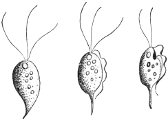
Fig. 17.—Trichomonas intestinalis, Lkt. (After Grassi.)
A row of twelve or more cilia was said to commence at the anterior end and extend over the body. Leuckart stated that this parasite, placed by the two authors in the genus Cercomonas, was a Trichomonas, and that they mistook the undulating membrane for cilia, and overlooked the flagella. Notwithstanding its striking similarity with T. vaginalis, it was said to be distinguishable from that species by differences in the undulating membrane. Lambl’s C. intestinalis38 (of 1875) which corresponds with C. hominis, Davaine39 (1854), is regarded by Leuckart as a true Cercomonad (characterized by a flagellum and the absence of an undulating membrane, see p. 61), and is thus generically distinct from Trichomonas.
The correctness of Leuckart’s judgment in regard to Marchand-Zunker’s flagellate was demonstrated by Grassi’s researches, accounts of which were published soon after. In about 100 cases of bowel complaints in North Italy and Sicily, Grassi found Flagellata in the stools, which he first named Monocercomonas and Cimænomonas, but later termed Trichomonas. However, in opposition to Leuckart, Grassi has also classified Davaine’s C. hominis (= C. intestinalis, Lambl, 1875) as Trichomonas, and most authors have followed his example. Hence arose the use of the name Trichomonas hominis. It was through Janowski (1896) that the former view was again taken up. After a review of the literature, the occurrence of Cercomonads in the intestine of human beings in addition to Trichomonads was considered by the author to have been proved, and he added a description of the Trichomonads. According to this, all morphological distinction between T. vaginalis, Donné, and T. intestinalis, Leuckart, disappeared. On the other hand, it is worthy of note that the smaller size, the more pear-shaped form, and the longer flagella differentiate T. intestinalis (= T. hominis) from T. vaginalis.40
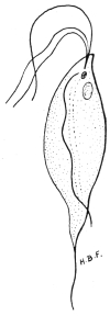
Fig. 18.—Trichomonas intestinalis from man, showing anterior flagella, cytostomic depression anteriorly, undulating membrane, nuclei, and axostyle. ×2,500. Original.
The easily deformed pear-shaped body has three free flagella anteriorly, and an undulating membrane with its flagellar border terminating in a short free flagellum posteriorly (figs. 17, 18). The undulating membrane may coil itself spirally round the body. A supporting rod or axostyle projects as a posterior spine. It appears to begin near the nucleus and blepharoplast, which are situated near the more rounded, anterior end of the body. There may be a chromatoid basal supporting line along the body for the undulating membrane. Rows of chromatoid granules are sometimes situated along one side of the axostyle. A cytostome may sometimes be seen. In mice, Wenyon (1907) found these parasites to vary in length from 3 µ to 20 µ. They occur in the cæcum and intestine of mice, where their internal structure seems more obvious than in man. The flagellates divide by longitudinal fission.
T. intestinalis, R. Leuckart, appears to be capable of settling in all parts of the human intestine in which the contents have an alkaline reaction. Trichomonads have been cited as occurring in the oral cavity by Steinberg, Zunker, Rappin and Prowazek; in the œsophagus by Cohnheim, and in the stomach by Strube, Cohnheim, Zabel, Hensen and Rosenfeld. The normal situation seems to be the small intestine. The parasites then appear in the dejecta, especially in various intestinal diseases the course of which is connected with an increased peristalsis. They are also found in healthy persons, from whom they are obtained after the administration of laxatives. They have been regarded by some workers as commensals, which, however, have the power of accelerating the onset of intestinal complaints, or at least of adding to them. They have been found in cases of carcinoma of the stomach, and in other diseases of that organ in which the acid reaction ceased.
Naturally, whether all the reports relate to the same species of Trichomonas must remain undecided. Certain authors (Steinberg, Cohnheim, van Emden) accept several species. Prowazek speaks of a variety of T. intestinalis inhabiting the oral cavity. This was distinguished by a posterior process exceeding the length of the body fourfold, and by a somewhat unusual course of the undulating membrane. The food of this form, which was found in the whitish deposit present, especially in the cavities of carious teeth, consisted almost exclusively of micrococci. Schmidt and St. Artault named the Trichomonads found in pathological products (e.g., gangrene, putrid bronchitis, phthisis) of the lungs of man, as Trichomonas pulmonalis. Trichomonads have also been found by Wieting in lobular pneumonia in the lungs of pigs.
It is still uncertain in what way the infection takes place. Experiments in the transmission of free trichomonads to mammals (per os), in which the same or allied species occur (guinea-pigs, rats, apes), have been without result. Probably encystment is necessary. Such conditions are mentioned by May, Künstler, Roos, Schurmayer, van Emden, Prowazek, Galli-Valerio and Schaudinn. According to Prowazek, intestinal trichomonads of rats become encysted for conjugation. In the cyst an accumulation of reserve food material occurs, causing distension. The nuclei of the conjugants each give off a reduction body and, after fusion, produce the nuclei for the daughter individuals. According to Schaudinn the intestinal trichomonads lose their flagella before conjugation, become amœboid and encyst in twos, the formation of a large agglomeration of reserve substance accompanying this. Galli-Valerio found double-contoured cysts in the fæces of trichomonad-infected guinea-pigs, after the fæces had been kept for a month in a damp chamber. When exposed to heat small flagellates escaped from them. Administration of such material containing cysts resulted in severe infection with trichomonads, and death of the experimental guinea-pigs followed. The cyst wall is clearly a protection against the deleterious acid reaction of the stomach contents. Alexeieff (1911) and Brumpt (1912) think that the trichomonad cysts of man are really fungi, while other workers also doubt encystment among trichomonads. Wenyon (1907) states that T. intestinalis in mice produces spherical contracted forms which escape from the body in the fæces.
Air, water, and under certain circumstances even food may be regarded as vectors for the trichomonads. The occurrence of the organisms in the oral cavity, and still more so in the lungs, is in favour of the air being the transmitting agent. An observation made by Epstein supports the idea of water transmission. Multiplication of the trichomonads, once they have gained access to the body, is effected by longitudinal division commencing at the anterior end (Künstler). “Cercomonads” with several flagella and an undulating membrane, as well as trichomonads, have been observed by Ross in some cases of cutaneous ulcers.
Mello-Leitao (1913)41 has described flagellate dysentery in children in Rio de Janeiro. He states that it is due to T. intestinalis and Lamblia intestinalis either separately or together. Flagellate dysentery, he thinks, is benign and is the most frequent form of dysentery in infants. The flagellates are pathogenic to infants under three years of age. Escomel (1913)42 found 152 cases of dysentery in Peru due solely to Trichomonas. Such cases are probably widespread.
Genus. Tetramitus, Perty, 1852.
Tetramitus mesnili, Wenyon, 1910.
Syn.: Macrostoma mesnili, Chilomastix mesnili, Fanapapea intestinalis.
The genus Tetramitus differs from Trichomonas in possessing an undulating membrane inserted in a deep groove or cytostome. There are three anterior flagella. The pear-shaped organism measures 14 µ by 7 µ, but smaller examples occur. T. mesnili occurs in the human intestine, having been described by Wenyon43 (1910) from a man from the Bahamas in the Seamen’s Hospital, London. Its occurrence is widespread. Alexeieff considers that Macrostoma and Tetramitus are synonymous. The parasite is the same as Fanapapea intestinalis, Prowazek, 1911, from Samoa. Brumpt (1912) found T. mesnili to be the causal agent of colitis in a Frenchwoman. Nattan-Larrier (1912) considers it of little pathological importance.
Gäbel44 (1914) described an interesting case of seasonal diarrhœa acquired in Tunis, in which a new Tetramitid was the causal agent. The organism was pear-shaped, without an undulating membrane, and measured 6·5 µ to 8 µ by 5 µ to 6 µ. The cytostome was large, and there was no skeletal support. Encystment occurred. Gäbel named the organism Difämus tunensis and considered that it was pathogenic.
Genus. Lamblia, R. Blanchard, 1888.
Syn., Dimorphus, Grassi, 1879, nec Haller, 1878; Megastoma, Grassi, 1881, nec de Blainville.
The body is pear-shaped, with a hollow on the under surface anteriorly. It has four pairs of flagella directed backwards, of which three pairs lie on the borders of the hollow disc, and the fourth arises from the pointed posterior extremity.
Lamblia intestinalis, Lambl, 1859.
Syn.: Cercomonas intestinalis, Lambl, 1859 (nec 1875); Hexamitus duodenalis, Davaine, 1875; Dimorphus muris, Grassi, 1879; Megastoma entericum, Grassi, 1881; Megastoma intestinale, R. Blanch., 1886; Lamblia duodenalis, Stiles, 1902.
The organism is pear-shaped and bilaterally symmetrical. It is from 10 µ to 21 µ long and 5 µ to 12 µ broad and possesses a thin cuticle. Anteriorly an oblique depression is present, which functions as a sucking disc (fig. 19, s). Its edges are raised above the general surface and are contractile. It corresponds to a peristome and acts as an adhesive organ (fig. 20, b, c). No true cytostome is present. A double longitudinal ridge, representing axostyles, extends from the sucking disc to the tapering posterior extremity, which is prolonged as two flagella from 9 µ to 14 µ long.
Lamblia intestinalis possesses eight flagella (fig. 19). The first pair of flagella, which cross one another, arise in a groove formed by the anterior edge of the sucking disc. Two pairs of flagella (lateral and median) are inserted on the posterior edge of the disc, while the posterior flagella occur at the tapering posterior extremity of the body. Basal granules are found at the bases of the flagella. The median flagella are most active in movement, the anterior and lateral flagella being less motile, as they are partially united to the body for part of their length.
The nuclear apparatus is situated in the thin, anterior, hollowed part of the body. It is at first dumb-bell shaped, the “handle” of the dumb-bell being formed by a very slight connecting strand, which eventually separates, so that the flagellate becomes binucleate, and thus completes the general bisymmetry of the organism.
There is a karyosome in each nucleus. Other bodies of unknown function, and possibly composed of chromatin, occur on or near the axostyles.
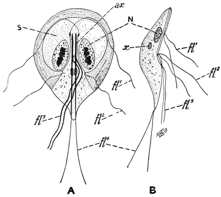
Fig. 19.—Lamblia intestinalis. A, ventral view; B, side view; N, one of the two nuclei; ax., axostyles; fl1, fl2, fl3, fl4, the four pairs of flagella; s, sucker-like depressed area on the ventral surface; x, bodies of unknown function. (After Wenyon.)
Division has not been observed in the flagellate stages of the Lamblia, but it occurs within the cysts. The resistant cysts (fig. 20, e) are oval and are surrounded by a fairly thick, hyaline cyst wall. They measure 10 µ to 15 µ by 7 µ to 9 µ, and may be tetranucleate. According to Schaudinn, the cysts arise from the conjugation of two individuals, and nuclear rearrangement occurs.
L. intestinalis occurs in its flagellate stage in the duodenum and jejunum, and rarely as such in the other parts of the intestine. Normally it is found in the large intestine as cysts, which are voided with the fæces. The hosts of Lamblia include Mus musculus, M. rattus, M. decumanus, M. silvestris, Arvicola arvensis and A. amphibius, the dog and cat, rabbit, sheep and man. Cysts voided with the fæces of infected animals reach plants or drinking water, and thence are transferred to man.
The flagellate in these different hosts exhibits some variation in size and in the problematic chromatic bodies. Bensen has suggested the species L. intestinalis from man, L. muris from the mouse and L. cuniculi from the rabbit. It is not certain whether these different species are necessary, as the variation may be due to differences of environment.
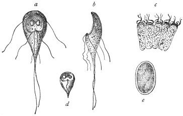
Fig. 20.—Lamblia intestinalis. a, from the surface; b, from the side; c, on intestinal epithelium cells; d, dead and e, encysted. (After Grassi and Schewiakoff.)
Like Trichomonas, Lamblia can multiply under inflammatory conditions of the alimentary tract. Thus they are found in cases of diarrhœa, carcinoma of the stomach, etc. The parasites attach themselves by their sucking discs to the epithelial cells of the gut (fig. 20, c), and though their numbers may be very great, their direct pathological significance is not fully known. Their occurrence in cases of diarrhœa has been explained as being due to the increased peristalsis, which has detached the parasites from the epithelium. Free flagellate forms perish in stools if kept, more especially if the temperature falls below 0° C. or rises above 40° C. Lamblia has often been found in dysenteric diseases, especially in the East, and is said to be the causal agent of certain diarrhœas in India. Mathis (1914)45 found Lamblia in cases of diarrhœa with dysenteriform stools in Tonkin. He also discovered healthy carriers of Lamblia cysts.
The parasite under discussion was first observed by Lambl (1859) in the mucous evacuations of children. He regarded the parasite as a Cercomonad and termed it Cercomonas intestinalis, which name as a rule is applied to Cercomonas hominis, Davaine, although Stein had already pointed out the difference between the two species. Grassi (1879) observed this species first in mice (calling it Dimorphus muris), and subsequently in human beings in Upper Italy and named it Megastoma entericum. Bütschli and Blanchard then laid stress on the identity of this species with Lambl’s C. intestinalis (1859), and consequently called it Megastoma intestinale. Later, Blanchard drew attention to the circumstance that the generic name Megastoma chosen by Grassi had already been used four times for various kinds of animals, and established the genus Lamblia. Accordingly, L. intestinalis is the valid name, and should be generally adopted.
In Upper Italy the parasite in the encysted condition has also been seen by Perroncito in man. At the same time, Grassi and Schewiakoff began a new investigation of specimens from mice and rats. In Germany, L. intestinalis was found by Moritz and Hölzl, Roos, Schuberg and Salomon. Moritz and Hölzl confirmed the relative frequency of the species. In Königsberg, Prussia, a student found encysted Lamblia in his fæces. One case was reported from Finland by Sievers, another case from Scandinavia by Müller. Frshezjesski and Ucke reported cases from Russia. Jaksch announced the occurrence of the parasite in Austria; Piccardi mentioned their presence again in Italy. They were reported from Egypt by Kruse and Pasquale, and from North America (Baltimore) by Stiles. Noc stated that 50 per cent. of the population of Tonkin harboured Lamblia. Finally, the structure of L. intestinalis has been described by Metzner (1901), and by Wenyon46 (1907) in mice.
In all these cases L. intestinalis has been observed in the small intestine, or in the evacuations of patients with intestinal diseases. It has also been found in the intestine of healthy subjects. Just as Trichomonas intestinalis may be found inhabiting the stomach in diseases of that organ, in which an alkaline reaction is present (carcinoma), so has L. intestinalis been found to occur under similar circumstances (Cohnheim, Zabel). However, in Schmidt’s case, 1 per cent. hydrochloric acid was certainly stated to be present. Infection takes place by the ingestion of cysts (fig. 20, e), as was established by Grassi, experimentally on himself. Cereal food-stuffs, contaminated with Lamblia cysts from vermin of the locality, such as rats and mice, serve to convey the infection to man. Such cysts may probably be found in street-dust, etc. Stiles induced infection in guinea-pigs, and Perroncito in mice and rabbits, by means of cysts of Lamblia from human beings. Stiles suspected that flies could transport Lamblia cysts. Mathis (1914) found that L. intestinalis was not amenable to emetine, at any rate in its cystic stage.
Order. Protomonadina, Blochmann.
The smallness of the Protomonadines and their less superficial situation than the Polymastigines, may be the cause that so far as the species occurring in man are concerned, they were formerly less well known. As regards parasitic species, this group may be divided as follows, according to the number of flagella and the presence or absence of an undulating membrane:—
(1) Cercomonadidæ, with one flagellum at the anterior extremity, without an undulating membrane.
(2) Bodonidæ, with two flagella, without an undulating membrane, except in Trypanoplasma.
(3) Trypanosomidæ, with one flagellum, and an undulating membrane along the length of the body in some genera.
Family. Cercomonadidæ, Kent emend. Bütschli.
Small uniflagellate forms, without cytostome.
Genus. Cercomonas, Dujardin emend. Bütschli.
Oval or rounded organisms, with the aflagellar end often drawn out into a tail-like process.
Cercomonas hominis, Davaine, 1854.
Davaine found flagellates in the dejecta of cholera patients. They had pear-shaped bodies, lengthening to a point posteriorly. Their length was from 10 µ to 12 µ, and a flagellum about twice as long as the body projected from one extremity (fig. 21). A nucleus was hardly recognizable. Occasionally a somewhat long structure (cytostome?) appeared at the anterior extremity. The animals moved with remarkable activity. They also attached themselves by means of their posterior extremities and swung about around the point of attachment. Davaine found a smaller variety, only about 8 µ long, in the dejecta of a typhoid patient (fig. 21, b).
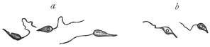
Fig. 21.—Cercomonas hominis, Dav. a, larger, b, smaller variety. Enlarged. (After Davaine.)
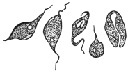
Fig. 22.—Cercomonas hominis, Dav. From an Echinococcus cyst. (After Lambl.)
The Flagellata observed by Ekeckrantz (1869) in the intestine of man belong to this form—at least to the larger variety—and Tham (1870) reported fresh cases soon after. Lambl’s publication of 1875, which was written in Russian, and became known through Leuckart’s work on parasites, also alludes to apparently typical Cercomonads, which, however, were discovered, not in the intestine, but in an Echinococcus cyst in the liver (fig. 22). The elliptical, fusiform, rarely pear-shaped or cylindrical bodies of the parasites measured 5 µ to 14 µ in length, and were provided with a flagellum at one end, while the other extremity usually terminated in a long point. An oral aperture occurred at the base of the flagellum, and there were one or two vacuoles near the posterior extremity. Longitudinal division was also observed (fig. 22).
As already mentioned, this form, which Lambl termed Cercomonas intestinalis, differs considerably from the form found by the same author in 1859, which received the same designation (cf. Lamblia intestinalis, p. 60), but it corresponds with Cercomonas hominis, Davaine. The latter, as well as C. intestinalis, Lambl, 1875, is usually classed with the Trichomonads, but, as has already been remarked (cf. Trichomonas intestinalis, p. 54), this cannot be considered correct, as only one flagellum is present.
Cercomonas vaginalis (Castellani and Chalmers, 1909) was found in the vagina of native women in Ceylon.
Other species of Cercomonas have, at various times, been recorded from man. However, the parasitic species of the genus Cercomonas require further investigation.
According to Janowski (1896–7), typical Cercomonads have also been observed in the intestine of man by Escherich, also by Cahen, Massiutin, Fenoglio, Councilman and Lafleur, Dock, Kruse and Pasquale, Zunker, Quincke and Roos, and others. However, it is an open question whether the Flagellata observed by Roos in one of his cases belonged to Davaine’s species, the size showing some deviation (14 µ to 16 µ). In his, as in many other cases, doubts have been raised as to whether the flagellates found in the stools had actually lived in the intestine, or had subsequently appeared in the fæces: for this a surprisingly short time only is necessary. Salomon also appears to have observed Cercomonads (Berl. klin. Wochenschr., 1899, No. 46).
As with T. intestinalis so with C. hominis, it appears that the parasite settles not only in the intestine but also in the air-passages. This is demonstrated by the statements of Kannenberg and Streng of the occurrence of Monads and Cercomonads in the sputum and putrid expectoration in gangrene of the lungs, which no doubt apply to C. hominis (cf. also Artault). Possibly also the Flagellata observed in the pleural exudation by Litten and Roos may be included here; this is the more probable in Roos’s case as the process ensued in the pleura after the breaking through of a vomica.
Perroncito and Piccardi have described encysted stages of Cercomonads.
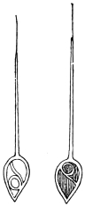
Fig. 23.—Monas pyophila, R. Blanch. (After Grimm.)
Monas pyophila, R. Blanch., 1895.
R. Blanchard thus designates a Flagellate that Grimm found in the sputum, as well as in the pus of a pulmonary and hepatic abscess, in the case of a Japanese woman living in Sapporo. The parasites resemble large spermatozoa (fig. 23). The body, 30 µ to 60 µ, has the shape of a heart or a myrtle leaf, and is surrounded by a thick cuticle which is supposed to extend into the interior of the body, dividing it into three parts. A long appendix at the rounded pole is covered for the greater part of its length by the cuticle; the extremity, however, is free and resembles a flagellum. The parasites were very active, frequently changed their shape, and were able to retract the long appendix within the body, which then assumed a round form.
[This organism requires further investigation.]
Family. Bodonidæ, Bütschli.
Protomonadina which are either free-living or parasitic, with two dissimilar flagella, while the possession of an undulating membrane and of a kinetic nucleus or blepharoplast is variable.
There are three genera:—
1. Bodo, Stein, 1878, without a kinetic nucleus and undulating membrane.
2. Prowazekia, Hartmann and Chagas, 1910, with a kinetic nucleus and without an undulating membrane.
3. Trypanoplasma, Laveran and Mesnil, 1901, with a kinetic nucleus and undulating membrane.
Of these genera Prowazekia must be discussed. Bodo does not occur in man. Species of Trypanoplasma occur in the blood and in the gut of various fishes, in the seminal receptacle of certain snails, in the gut and genitalia of a flatworm (Dendrocœlum lacteum) and in the vagina of a leech. Closely allied to Trypanoplasma is the genus Trypanophis, parasitic in the cœlenteric cavity of Siphonophores.
Genus. Prowazekia, Hartmann and Chagas, 1910.
The genus was founded for a flagellate parasite, Prowazekia cruzi, discovered in a culture of human fæces in Brazil. Various other species have been referred thereto. The genus is separated from Bodo by the possession of a second nucleus, the so-called kinetonucleus or blepharoplast. It differs from Trypanoplasma in the absence of an undulating membrane. It is heteromastigote, that is, it possesses two dissimilar flagella, one anteriorly directed and the other lateral and trailing.
The principal species are:
Prowazekia urinaria, Hassall, 1859.
Syn.: Bodo urinarius, Hassall, 1859; Trichomonas irregularis, Salisbury, 1868; Cystomonas urinaria, Blanchard, 1885; Plagiomonas urinaria, Braun, 1895.
Hassall47 in 1859 first found Bodo-like flagellates in human urine. He examined fifty samples of urine from patients suffering from albuminuria and from cholera. The reaction of the urine was alkaline or sometimes only feebly acid. The flagellates were only seen after the urine had been standing for several days. Hassall named the organism Bodo urinarius, and gave a very good description of it with illustrations. The flagellate, which was round or oval, measured 14 µ by 8 µ. The organism had “one, usually two, and sometimes three lashes or cilia.” In 1868 Salisbury described a similar flagellate in the urine under the name Trichomonas irregularis. Künstler in 1883 described the latter parasite under the name B. urinarius. In 1885 Blanchard, considering Künstler’s organism a different parasite from Hassall’s, called it Cystomonas urinaria. Braun, in 1895, gave the name Plagiomonas urinaria. Barrois (1894) considered Künstler’s and Hassall’s organisms to be identical and not to be true parasites of man. Sinton,48 in 1912, found the flagellate in the deposit, after centrifuging, of a 24-hour old specimen of alkaline urine from a Mexican sailor in the Royal Southern Hospital, Liverpool. Sinton found a kinetic nucleus or blepharoplast in the organism, and therefore placed it in the genus Prowazekia.
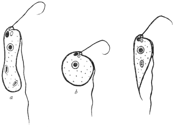
Fig. 24.—Types of Prowazekia urinaria. (a) sausage-shaped; (b) round; (c) carrot-shaped form. (After Sinton.)
The flagellate stage (fig. 24) of the organism is polymorphic, and may be either (a) sausage-shaped, 10 µ to 25 µ in length by 2·5 µ to 6 µ in breadth; (b) round or oval, varying from 4 µ in diameter to oval forms 15 µ by 10 µ; (c) a carrot-shaped form, of varying size up to 25 µ by 4 µ. The kinetic nucleus is large and pear-shaped. Near it are basal granules, closely applied to one another, from which the flagella arise. There is a small cytostome near the roots of the flagella. There is a well-marked karyosome in the nucleus. The movement is jerky. The shorter, anterior flagellum may be used in food-capture. In life, bacteria have been seen to be ingested. Food-vacuoles tend to accumulate at the posterior (aflagellar) end. A contractile vacuole may be present, near the base of the cytostome, and may really be the dilated fundus of the latter. Division occurs by binary fission. The organism can encyst (fig. 25, a), when the flagella are lost, and round or oval cysts are found, 5 µ to 7 µ in diameter. After a time flagella are formed inside the cyst, and the organism emerges therefrom in its typical flagellate form (fig. 25, b-f).
Sinton’s case is interesting. He obtained the flagellate only twice from the same patient, a Mexican then in hospital in Liverpool. The flagellate was not found in the patient’s fæces, nor was it found in the urine on later occasions when taken aseptically.
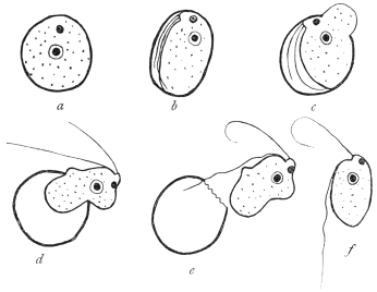
Fig. 25.—Prowazekia urinaria. Flagellate emerging from cyst. (After Sinton.)
In cultures Prowazekia urinaria was always found in association with bacteria. The cultures died at a temperature of 37° C., but grew well at 20° C. Various media were useful at the lower temperature, such as urine, salt agar, nutrient agar, serum agar, blood agar, peptone salt solution, and diluted blood serum. The flagellate was, then, considered to be an accidental contamination and not a true parasite of human urine.
Prowazekia asiatica, Castellani and Chalmers, 1910.
The flagellate was found by the discoverers in the stools of patients suffering from ankylostomiasis and diarrhœa in Ceylon. It was referred by them to the genus Bodo, but in 1911 Whitmore49 further studied it and placed it in the genus Prowazekia. In the stools the flagellate is found either as a long, slender form measuring 10 µ to 16 µ by 5 µ to 8 µ or as a rounded form 8 µ to 10 µ in diameter. Its cytoplasm is alveolar. A rhizoplast connects the basal granules to the kinetic nucleus. There is multiplication and cyst formation as before. The organism is easily cultivated, especially in the condensation water of nutrose agar and maltose agar. The pathogenicity is stated to be nil.
Prowazekia javanensis, Flu, 1912.
Found in agar cultures from the motions of patients at Weltevreden, Dutch East Indies.50 The flagellates are 12 µ long and 5 µ broad. The lateral flagellum is stated to be attached to the cell body for a short distance. Regarding the karyosome in the nucleus, the author states that the smaller the karyosome the more chromatin is deposited on the nuclear membrane. Flu mentions that the specific name javanensis is a temporary one, as in the course of time it may be shown that there is only one species of Prowazekia.
Prowazekia cruzi, Hartmann and Chagas, 1910.
Found in a culture from human fæces on an agar plate in Brazil, and considered to be a free-living form.51 The organism is oval or pear-shaped, 8 µ to 12 µ long and 5 µ to 6 µ broad. In human stools at Tsingtau, China, a Prowazekia has been found by Martini which he thinks is the same as Prowazekia cruzi. He considers it to be a cause of human diarrhœa and intestinal catarrh.
Prowazekia weinbergi, Mathis and Léger, 1910.
This species was found in the fæces of men, both healthy and diarrhœic, in Tonkin.52 It is pear-shaped, 8 µ to 15 µ long by 4 µ to 6·5 µ broad. The flagella occur at the broad end.
The discoverers think that Prowazekia weinbergi is an intestinal inhabitant, but non-pathogenic, since it was found to occur in the fæces even when obtained with aseptic precautions.
Prowazekia parva, Nägler, 1910.
A free-living form found in the slime on the stones at the biological station at Lunz. Another Prowazekia was found in 1914 in tap-water in Calcutta.
Family. Trypanosomidæ, Doflein.
The Trypanosomidæ, broadly considered, are uniflagellate organisms, the flagellum being at the anterior end. The flagellum arises near the blepharoplast (kinetic nucleus), which lies anterior, near or posterior to the nucleus.
The following genera will be considered:—
Trypanosoma—with an undulating membrane along the length of the body.
Crithidia—with a less well-developed undulating membrane anteriorly (see fig. 49).
Herpetomonas—including the so-called Leptomonas, with anterior free flagellum only, and no undulating membrane.
Leishmania—non-flagellate forms in mammalian blood, flagellate herpetomonad stages in culture, probably occurring naturally in Arthropods.
Genus. Trypanosoma, Gruby, 1843.
The members of the genus possess a single flagellum, which arises posteriorly, adjacent to a blepharoplast or kinetic nucleus. The flagellum forms a margin to an undulating membrane, and may or may not be continued beyond the body as a free flagellum. Many species are parasitic in vertebrate blood and in the digestive tracts of insects.
Historical.
The history of blood flagellates goes back to the year 1841, in which Valentin discovered in the blood of a brook-trout (Salmo fario L.) minute bodies, from 7 µ to 13 µ in length, with active movements and presenting marked changes in form. Valentin considered the parasite a new species of the old genus Proteus or Amœba, Ehrbg. This announcement led Gluge (1842) to publish a similar discovery he had made in frog’s blood. The latter forms were called by Mayer (1843) Amœba rotatoria, Paramœcium loricatum and P. costatum, while Gruby (1843) called them Trypanosoma sanguinis.53 Later it was discovered that similar organisms occurred also in the blood of birds (Wedl (1850), Danilewsky) and of mammals. Gros (1845) found them in the mouse and mole, Chaussat (1850) in the house rat, Lewis (1879) in the Indian rat, Wittich (1881) in the hamster. Danilewsky (1886–89) and Chalachnikow (1888) investigated the structure and division of trypanosomes.
In the case of all these forms, there was no discussion as to a pathogenic influence on the host. Opinion, however, as to the action of trypanosomes changed when, in 1880, Evans found flagellates in the blood of horses in India that suffered from a disease endemic there called “surra,” and associated the parasites with the disease. Steel and Evans were successful in transmitting the parasites—first known as Spirochæta evansi, Steel, then as Trichomonas evansi, Crookshank, and finally as Trypanosoma evansi—to dogs, mules and horses. They recognized that the above mentioned flagellates in the blood of the experimental animals were the causal agents of the disease.
From that time there was a considerable increase in the literature, the contents of which have been summarized by Laveran and Blanchard. In 1894 Rouget discovered trypanosomes in the blood of African horses that suffer from “stallion’s disease” (dourine). In 1894 Bruce found similar forms (T. brucei) in the blood of South African mammals suffering from “nagana,” and in consequence attention was drawn to the part which the much dreaded tsetse-fly played in the transmission of “nagana.” In 1901 Elmassian discovered trypanosomes in the blood of horses that were stricken with “mal de caderas,” which is very common in the Argentine. The disease in cattle named “galziekte” (gall-sickness), occurring in the Transvaal, was also at one time attributed to a trypanosome remarkable for its great size, and like some other species, bearing the name of its discoverer (T. theileri).
The study of the species hitherto known has been carried on partly by the above mentioned authors and in part by others, e.g., Rabinowitsch and Kempner, Laveran and Mesnil, Wasiliewski, Senn. It was greatly advanced by the method of double staining (with alkaline methylene blue and eosin) introduced by Romanowsky (1891) and elaborated by Ziemann, Leishman, Giemsa and others. By this means the presence of a terminal flagellum and of an undulating membrane at the side of the flattened and extended body was demonstrated. Laveran and Mesnil (1901) discovered allied flagellates in the blood of the fish, Scardinius erythrophthalmus. These flagellates, now placed in the genus Trypanoplasma, had a second free flagellum in addition to the one bordering the undulating membrane. Trypanoplasms have since been found in both freshwater and marine fishes. The transmission of trypanoplasms of freshwater fishes is effected by leeches. Trypanoplasma varium from Cobitis is transmitted by Hemiclepsis marginata according to Léger, while the Trypanoplasmata of Cyprinus carpio and Abramis brama reach new hosts by the agency of Piscicola according to Keysselitz.
Another ally of the Trypanosomidæ, Trypanophis, lives in the cœlenteric cavity of Siphonophores. It has also an extra terminal flagellum (Poche, Keysselitz). [Trypanoplasma and Trypanophis belong to the Bodonidæ, see p. 63].
Finally it was shown that Trypanosomes occurred in human beings. Although Nepveu’s early report of trypanosomes in the blood of malarial patients may be doubtful, subsequent researches by Forde and Dutton demonstrated trypanosomes (fig. 28) in the blood of a European, apparently suffering from malaria, living in the Gambia. Dutton (1902) called the human trypanosome, T. gambiense. The expedition despatched by the Liverpool School of Tropical Medicine (1902) to Senegambia found trypanosome infections in six cases among a thousand inhabitants examined.
About the same time attention was devoted to the disease of West African negroes known for a century as “sleeping sickness.” Castellani (1903) was the first to succeed in demonstrating the presence of trypanosomes (at first called T. ugandense) in centrifugalized cerebro-spinal fluid obtained by puncture from cases of sleeping sickness in Uganda. Similar discoveries were made by Bruce, who also found trypanosomes in the blood of those attacked with sleeping sickness. Sambon regarded a species of Glossina as the transmitter. From consideration of the geographical distribution of the disease Christy regarded Glossina palpalis as the transmitter. Brumpt first thought it was G. morsitans, but, later, supported the view of G. palpalis. Bruce, Nabarro and Greig also named the same insect as the transmitter, not only for geographical reasons but also because healthy apes became infected by the bite of certain G. palpalis. The inoculation of cerebro-spinal fluid from subjects of sleeping sickness into the spinal canal of apes (Macacus) had the same result.
Just as the discovery of the malarial parasites called forth a whole flood of research memoirs which were followed by a second series on the relation of the mosquitoes to malaria, so a similar outpouring occurred after the discovery of the pathogenic trypanosomes of mammals and men. In both cases the inquiry was not limited to the stages in man and other vertebrate hosts, but the fate of the parasites in the intermediate (invertebrate) hosts was investigated, and allied species were obtained from many different hosts.
Novy and MacNeal (1903) were the first to cultivate trypanosomes in artificial media (blood-agar).
In 1910 Stephens and Fantham recorded the presence of another human trypanosome, T. rhodesiense, from a case of sleeping sickness in Rhodesia, where G. palpalis was absent. Kinghorn has since demonstrated that T. rhodesiense is transmitted by G. morsitans. Kinghorn and Yorke believe that big game (e.g., antelope) is the reservoir of T. rhodesiense.
The output of literature on trypanosomiasis in men and animals is enormous. To cope with it the Sleeping Sickness Bureau Bulletin was founded in 1908, and it is now (since November 1912) continued as a section of the Tropical Diseases Bulletin, wherein current literature is reviewed.
General.
Trypanosomes occur in the blood of representatives of all the vertebrate classes. Often the trypanosomes occur so scantily in the blood that they are overlooked on examination. A useful aid in detecting the flagellates in such cases consists in the use of cultures of the blood of the host on artificial media. Stimulated by the medium multiplication occurs, and hence the parasites are more easily detected. [For the composition of such culture media see Appendix.]
There is a periodicity in the appearance of the trypanosomes in the peripheral blood of the host, due to alternating phases of multiplication and of rest on the part of the parasites. Such periodicity has been established both by biological and enumerative methods. Again, a seasonal variation has been observed in the occurrence of certain trypanosomes in the peripheral circulation of the hosts; for example, some trypanosomes (e.g., T. noctuæ in birds) are found only in the summer in the blood, while in the winter they occur in the internal organs.
Recent cultural researches have established that trypanosomes, e.g., T. americanum, may be present in very small numbers in hosts, such as cattle, which are quite unharmed by them, and in which the presence of these flagellates formerly was never suspected (“cryptic trypanosomiasis.”) However, the majority of the trypanosomes occurring in domestic animals are usually deleterious or even lethal to their hosts. Many wild animals, such as various species of antelope, harbour trypanosomes without being injured thereby. In such cases it is probable that the vertebrate hosts have been so long parasitized in the past, that they have become tolerant and immune to the effects of the flagellates. Should such trypanosomes of wild animals be transmitted to domesticated stock or man, they may re-acquire their initial virulence and become pathogenic to the new host. As a general statement, the newer a parasite is to its host the greater is its virulence. For example, T. gambiense, T. rhodesiense and T. brucei are innocuous to big game in Africa, but are pathogenic to man and domestic animals respectively. Pathogenic trypanosomes appear to have a wider range of hosts, that is, to be less limited to one specific host than non-pathogenic forms. Thus, T. rhodesiense is pathogenic to man and all laboratory animals, while it is non-pathogenic to antelopes and their kind.
Morphology.
The general structure of the various trypanosomes shows much uniformity, though variations in size and shape occur. Typically the body is elongate and sinuous. The flagellar end tapers gradually to a point, the aflagellar extremity usually being rounded or more blunt. In some trypanosomes there is much diversity in size, the organisms varying from long, slender forms to short, stumpy ones; in other species relative constancy of size is maintained. The former are known as polymorphic trypanosomes, the latter as monomorphic forms.
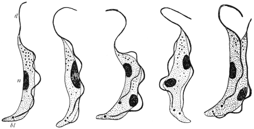
Fig. 26.—Trypanosoma brucei in division. n, nucleus; bl, blepharoplast; fl, flagellum. × 2,000. (After Laveran and Mesnil.)
Two nuclei are present. The main or principal nucleus, sometimes termed the trophic nucleus, is often situated towards the centre of the body; it is frequently of the vesicular type, containing a karyosome. The blepharoplast or kinetic nucleus is posterior to the nucleus, and usually is rod-like. The flagellum arises close to the blepharoplast, and forms an edge to the undulating membrane. It may or may not extend beyond the limits of the undulating membrane. If it does so, the unattached part is known as the free flagellum. Sometimes a small granule is found at the origin of the flagellum. This is the basal granule, and is considered by some to function as the centriole of the kinetic nucleus.
The undulating membrane is a lateral extension of the ectoplasm or periplast, and is the main agent in locomotion. It is edged by the flagellum, which forms a deeply stainable border to it. Within the membrane substance, often arranged parallel with its edge, are a number of fine contractile elements, the myonemes. These contractile elements may also occur on the body of the trypanosome. They are easily seen in some large trypanosomes, but are difficult of demonstration in others, owing to their great fineness.
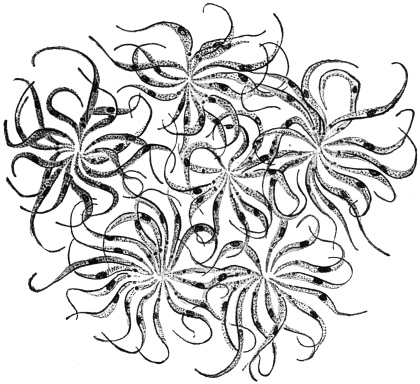
Fig. 27.—Trypanosoma lewisi. Multiplication rosettes. × 1,000. (After Laveran and Mesnil.)
Multiplication of trypanosomes in the blood is brought about by binary longitudinal fission (fig. 26). Division is initiated by that of the blepharoplast and nucleus. The division may be equal or subequal, whereby differences in size of individuals partly arise. Multiple division by repeated binary fission, without complete separation of the daughter forms, is known in some trypanosomes (e.g., T. lewisi), and rosettes of parasites thereby are produced (fig. 27).
The classification of trypanosomes is very difficult. Laveran (1911)54 has suggested the examination of the relative length of the flagellum as a diagnostic character, and so arranged these flagellates in mammals in three groups. The first group included those trypanosomes always having part of the flagellum free (e.g., T. evansi, T. vivax); the second group comprised forms without a part of the flagellum free (e.g., T. congolense), while the third group included forms some members of which have free flagella, while others have not (e.g., T. gambiense). Bruce55 (1914) and Yorke and Blacklock56 (1914) have also devised classifications.
Resting stages of some trypanosomes have been found in the internal organs of their vertebrate hosts. The formation of these oval, Leishmania-like bodies will be noted in individual cases later. Similar small oval bodies form an important phase in the life-history of T. cruzi, which multiplies normally by multiple fission or schizogony into these oval, daughter elements, and not by binary longitudinal fission in the circulating blood.
Polymorphism in trypanosomes (e.g., T. gambiense, T. rhodesiense) is now interpreted as a phenomenon resulting from growth and division.57 Long, thin forms are those about to divide. Fully mature forms are shorter and broader. Various intermediate types occur and represent growth forms. Formerly, polymorphism was interpreted in terms of sex, thin forms being regarded as males, broad forms as females, while the intermediate types were termed indifferent. Conjugation was not observed, and there is no evidence in support of the sexual interpretation.
The transmission of trypanosomes from one vertebrate host to another is usually accomplished by the intermediation of some biting arthropod in the case of terrestrial animals, while leeches are usually considered to act as transmitters in the case of the trypanosomes occurring in aquatic animals. Developmental phases of the life-histories of trypanosomes occur in the invertebrate transmitters, and will be considered in individual cases.
Trypanosoma gambiense, Dutton, 1902.
Syn.: Trypanosoma hominis, Manson, 1903. Trypanosoma nepveui, Sambon, 1903. Trypanosoma castellanii, Kruse, 1903. Trypanosoma ugandense, Castellani, 1903. Trypanosoma fordii, Maxwell Adams.
In vertebrate blood Trypanosoma gambiense is polymorphic, for long, thin forms may be seen in contrast with short, stumpy forms, as well as intermediate forms (fig. 29, a—c). This polymorphism has been interpreted in terms of sex, especially by German investigators, following Schaudinn (see above). However, there is no evidence of conjugation, and the polymorphic forms are more easily interpreted in terms of growth and division, for the long thin forms are potential dividing organisms, and the stumpy or short parasites, with little or no free flagellum, are the adult individuals.
Morphology of T. gambiense in the Circulating Blood.
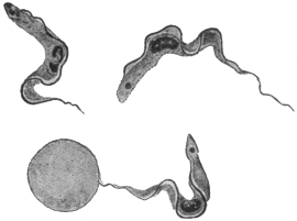
Fig. 28.—Trypanosoma gambiense. × 1,700. (After Dutton.)
T. gambiense varies from 13 µ to 36 µ in length, its average length being 24·8 µ, as was determined in 1913 by exact biometrical methods by Stephens and Fantham.58 Three forms of parasite occur. According to Miss Robertson,59 the relatively short forms from 13 µ to 21 µ long may be regarded as the mature or “adult” type of parasite in the blood. They carry on the cycle in the vertebrate. From them intermediate forms, which are longer than the “adult” but at first have the same breadth, arise by growth. They possess a free flagellum. The intermediate forms grow into long individuals, which are those about to divide. The products of division give rise, directly or indirectly, to the adult forms.
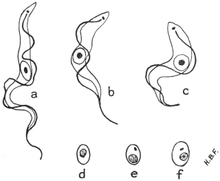
Fig. 29.—Trypanosoma gambiense. Development in vertebrate host. a, long, slender, b, intermediate and c, short, stumpy forms, found in the blood; d, e, f, non-flagellate, latent forms from internal organs. × 2,000. (Original. From preparations by Fantham.)
The organism has an elongate body with an anterior or flagellar end and a blunter posterior or non-flagellar end. The protoplasm is finely granular, large inclusions being rare. The central nucleus is oval and large, often containing most of its chromatin concentrated as a karyosome, with small granules only scattered near or on the fine nuclear membrane. The blepharoplast is either rounded or rod-shaped. The undulating membrane is thrown into folds and is bordered by the flagellum. A small basal granule may be present near, or at the actual origin of the flagellum.
Multiplication in the vertebrate is brought about by longitudinal division. According to the recent account of division by Miss Robertson, the blepharoplast doubles, then the flagellum splits for the greater part of its length, and the daughter flagella separate, one being shorter than the parent flagellum. The nucleus often shows two well marked dark granules on the membrane at opposite poles, and these appear to act as centrosomes. Nuclear constriction occurs and the halves gradually separate. Finally the two daughter organisms become free, the aflagellar end splitting last. The products of division may be equal or unequal. Repeated division goes on in the general circulation until the blood swarms with parasites. Then the trypanosomes gradually disappear, and a period occurs when it is practically impossible to demonstrate the parasite in the blood. At such a period, trypanosomes can be obtained by puncture of the enlarged lymphatic glands or of the spinal canal, or can be found in the internal organs, more particularly in the spleen, lungs, liver and bone-marrow. In the latter organs, latent bodies are produced (fig. 29, d—f) which are capable of again becoming flagellates and entering the general circulation. Their formation was described by Fantham (1911).60 The parasite contracts, the blepharoplast migrates towards the nucleus, a very thin coat differentiates around the two nuclei and a certain amount of cytoplasm, and the parts exterior to the coat disintegrate, leaving a small, oval body behind. Fuller details are given in connection with T. rhodesiense. Laveran (1911)61 considers that latent bodies are “involution” forms, but acknowledges that they can flagellate and become infective in fresh blood.
No multiplication of the trypanosomes within the cells of the lung, liver or spleen of infected monkeys was found by Miss Robertson in her recent researches.
There appear to be negative periods in infected monkeys, since, although trypanosomes may occur in their blood at such times, they are not infective to Glossina.
Development in Glossina palpalis.—The principal accounts are those by Sir D. Bruce and his colleagues (1911),62 and by Miss Robertson63 (1912), whose results will be followed. According to the latter investigator T. gambiense never enters the body cells of the fly (G. palpalis), nor does it penetrate the gut wall into the body cavity. Practically no crithidial stage occurs in the fly’s main gut, but a trypanosome facies is retained therein.
After the trypanosomes are ingested by the fly during a meal of infected blood, sooner or later multiplication occurs. This development usually begins in the middle or posterior part of the mid gut, and trypanosomes of varying sizes are produced. After the tenth or twelfth day, many long, slender trypanosomes (fig. 30, a) are found, which gradually move forwards into the proventriculus. Such long, slender forms represent the limit of development in the lumen of the main gut. The proventricular type, developed about the eighth to the eighteenth or twentieth day, is not infective; it may occur in the crop, but is not to be found permanently there. Between the tenth and the fifteenth days multinucleate forms of trypanosomes are found, and may be styled multiple forms (fig. 30, b). Some of these latter may be degenerative.
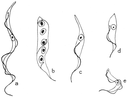
Fig. 30.—Trypanosoma gambiense. Development in the fly, Glossina palpalis. a, slender, proventricular form; b, multinucleate form; c, d, crithidial forms; e, infective type of trypanosome found in salivary gland. × 2,500. (After Robertson.)
Invasion of the Salivary Glands of the Fly.—Long, slender trypanosomes from the proventriculus pass forward into the hypopharynx. They then pass back along the salivary ducts, about sixteen to thirty days after the fly’s feed. The trypanosomes reach the salivary glands as long, slender forms. In the glands they become shorter and broader, attach themselves to the surrounding structures, and assume the crithidial facies (fig. 30, c, d). As crithidial forms they remain attached to the wall and multiply in the glands. These crithidial stages differentiate into the short, broad trypanosome forms, capable of swimming freely (fig. 30, e).
Miss Robertson considers the development in the main gut to be indifferent multiplication, and that salivary fluid seems necessary to stimulate trypanosomes to the apparently essential reversion to the crithidial type. The second development in the salivary gland is the essential feature. The short, stumpy forms of trypanosomes (fig. 30, e) finally produced in the salivary glands are alone infective. No conjugation of trypanosomes occurs in the fly. Only about 5 per cent. of captive tsetse flies fed on trypanosome-infected blood become infective, but they probably remain infective for the rest of their lives.
J. G. Thomson and Sinton (1912)64 have obtained in cultures the various trypanosome forms of T. gambiense seen in the fly’s main gut.
Duke (1912)65 found T. gambiense in a species of antelope, the situtunga (Tragelaphus spekei), on Damba Island in Victoria Nyanza. Wild G. palpalis could be infected therefrom. The antelope may then act as a sleeping sickness reservoir in that district, but men are apparently the chief reservoir.
Trypanosoma nigeriense, Macfie, 1913.66
Macfie has recently (August, 1913) described a human trypanosome from the Eket district of Southern Nigeria. It is common in young people. The disease produced does not seem to be of a virulent type in Nigeria, and does not occur in epidemic form. In the early stages the glands of the neck are enlarged. In the later stages—cases of which are rarer—lethargy appears. The parasite is a polymorphic trypanosome, morphologically almost indistinguishable from T. gambiense, though it may be slightly shorter. Macfie recorded the occurrence in his preparations of a few trypanosomes appearing to have a flagellum free during their whole length. Some of the parasites, as seen in a sub-inoculated guinea-pig, are very small (8 µ long). Other trypanosomes have their nuclei displaced somewhat anteriorly. This parasite may only be a variety of T. gambiense. The parasite is perhaps spread by Glossina tachinoides.
Trypanosoma rhodesiense, Stephens and Fantham, 1910.
The parasite was found in the blood of a young Englishman who had contracted sleeping sickness in the Luangwa Valley, North-eastern Rhodesia, in the autumn of 1909. The patient had never been in an area infested with Glossina palpalis.
(1) Morphology.—The morphology of the parasite in man and sub-inoculated rats was studied by Stephens and Fantham in 1910.67 They pointed out a morphological peculiarity in the presence of certain trypanosomes with posterior nuclei in sub-inoculated animals, that is, parasites in which the nucleus (trophonucleus) was situated towards the posterior or aflagellar end, close up to or even beyond the blepharoplast or kinetic nucleus (fig. 31, 4, 5). When the nucleus was beside the blepharoplast, the former was seen to be kidney-shaped (fig. 31, 4). The posterior nuclear forms were of the stout and stumpy variety, and about 6 per cent. of the stumpy forms were found to have their nuclei displaced from the centre. The anterior or flagellar end of these trypanosomes often contained chromatoid granules. T. rhodesiense varies in length from 12 µ to 39 µ68; short stumpy forms vary from 13 µ to 21 µ, intermediate forms from 21 µ to 24 µ, and long, slender forms from 25 µ onwards. The average length is 24·1 µ.
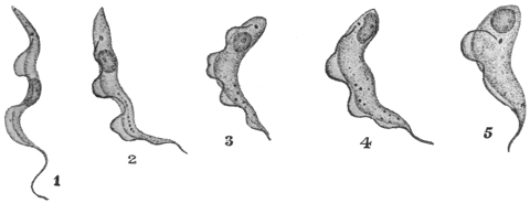
Fig. 31.—Trypanosoma rhodesiense. 1, Long narrow form; 2–4, nucleus passing to posterior (aflagellar) end; 5, nucleus quite posterior. × 1,800. (After Stephens and Fantham.)
Certain regular periods occur in the course of the trypanosomiasis when few or no flagellate trypanosomes are found in the peripheral blood of the patient or of the sub-inoculated animal. These periods can be explained in terms of morphology, for the trypanosomes are capable of assuming a non-flagellate form in the internal organs of the host, particularly in the lungs and in the spleen. Such forms are known as “latent” or “resting” forms. The term “latent body” was first used by Moore and Breinl in 190769 in connection with T. gambiense. Fantham70 (1911) has described the process of formation of latent from motile forms and the reconversion of the latent bodies into active flagellates. Fresh preparations of splenic blood or lung blood containing trypanosomes were made. A trypanosome gradually withdrew or cast off its flagellum, concentrated its cytoplasm, and became more or less elongate oval. Nucleus and blepharoplast approached one another and came to lie more or less side by side. Then an opaque line often made its appearance around the nuclear area and differentiated as a slight envelope or covering, the cytoplasm external to this merely degenerating. The small, oval, refractile body (fig. 29, d—f) thus formed was a non-flagellate latent body, 2 µ to 4 µ in diameter, like Leishmania or the non-flagellate, multiplicative forms of T. cruzi (fig. 34), and remains temporarily inactive in the internal organs of the host. After this period of inactivity, the non-flagellate body, recuperated by its rest, begins to elongate again. The nuclei separate. From a small vacuole-like portion the flagellum differentiates and forces out the ectoplasm, which assumes the form of the undulating membrane with its flagellar border. Subsequent growth results in the production of the typical trypanosome form, which re-enters the circulating blood and multiplies by longitudinal binary fission. Division of the parasite prior to the formation of a latent body may occur and division of the latent forms themselves is known, though less common. Consequently latent bodies, like the flagellate forms themselves, show diversity in size. The blepharoplast of the latent bodies is sometimes less well marked than in Leishmania (see fig. 29, d-f). Laveran’s views on these bodies have already been given on p. 74.
(2) Animal Reactions.—The posterior nuclear trypanosomes were found in all sub-inoculated animals, such as rats, guinea-pigs, dogs, mice, Macacus, rabbits and horses, but were not seen in the human patient, as few trypanosomes occurred in his peripheral blood. R. Ross and D. Thomson71 found a periodic, cyclical variation in the number of the parasites in the patient’s blood from day to day, the cyclical period being about a week (fig. 32). Fantham and J. G. Thomson72 (1911) found a similar periodic, cyclical variation in the trypanosomes in the blood of sub-inoculated rats, guinea-pigs and rabbits. On counting the parasites in the blood of similar animals inoculated with T. gambiense, they established, by enumerative methods, that T. rhodesiense was more virulent than T. gambiense, while Yorke also showed this marked virulence of T. rhodesiense in practically all laboratory animals. In other words the duration of infection in the case of T. rhodesiense was shorter. It was also found that T. rhodesiense was resistant to atoxyl. The patient, from whom the original strain was obtained, died about nine months after the probable date of infection. Some patients infected with T. rhodesiense have died in an even shorter period, such as four or five months.