PART III.—THE METAMORPHOSES OF INSECTS (continued 2)
Reading edition
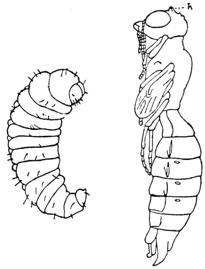
Fig. 592.—Larva and pupa of a wood-wasp (Rhopalum), enlarged: h, temporary locomotive tubercles on head of pupa.—Trouvelot del.
Our attention[106] was drawn to this subject by a rustling, cutting, and tearing noise issuing from a cocoon of Actias luna. On examination a sharp black point was seen moving to and fro, and then another, until both points had cut a rough irregular slit, through which the shoulder of the moth could be seen vigorously moving from side to side. The hole or slit was made in one or two minutes, and the moth worked its way at once out of the slit. The cocoon was perfectly dry. The cocoon-cutter occurs in all the American genera, in Samia cynthia, and is large and well marked in the European Saturnia pavonia-minor and Endromis versicolora. In Bombyx mori the spines are not well marked, and they are quite different from those in the Attaci. There are three sharp points, being acute angles of the pieces at the base of the wing, and it must be these spines which at times perform the cutting through of the threads of the cocoon described by Réaumur, and which he thought was done by the facets of the eyes. It is well known that in order to guard against the moths cutting the threads, silkraisers expose the cocoon to heat sufficient to destroy the enclosed pupa. In Platysamia the cocoon-cutters, though well developed, do not appear to be used at all, and the pupa, like that of the silkworm and other moths protected by a cocoon, moistens the silk threads by a fluid issuing from the mouth, which also moistens the hairs of the head and thorax, together with the antennæ. It remains to be seen whether these structures are only occasionally used, and whether the emission of the fluid is not the usual and normal means of egress of the moth from its cocoon. Dr. Chapman remarks that throughout the obtected moths “there are many devices for breaking through the cocoon: specially constructed weak places in the cocoon, softening fluid, applied by the moth, assisted by special appliances of diverse sorts, such as in Hybocampa[107] and Attacus,” etc.
As to the fluid mentioned above, Trouvelot states that it is secreted during the last few days of the pupa state, and is a dissolvent for the gum so firmly 636uniting the fibres of the cocoon. “This liquid is composed in great part of bombycic acid.” (Amer. Naturalist, i, p. 33.)
The pupa of the dipterous genus Sciara (S. ocellaris O. S.) resembles a tineid pupa, and before transforming emerges for about two-thirds of its length from the cocoon; the pupa-skin remaining firmly attached in this position.[108]
Certain hymenopterous pupæ are provided with temporary deciduous conical processes. Thus we have observed in the pupa of Rhopalum pedicellatum two very prominent acute tubercles between the eyes (h, Fig. 592). As the cocoon is very slight, these may be of use either in extracting itself from the silken threads or in pushing its way along before emerging from the tunnel in the stem of plants. (See also p. 611.)
c. The cremaster
Although this structure is in general confined to lepidopterous pupæ, and is not always present even in them, since it is purely adaptive in its nature, yet on account of its singular mode of development from the larval organs, and the accompanying changes in the pupal abdomen, it should be mentioned in this connection. The cremaster is the stout, triangular, flattened, terminal spine of the abdomen, which aids the pupa in working its way out of the earth when the pupa is subterranean, or in the pupa of silk-spinning caterpillars its armature of secondary hooks and curved setæ enables it to retain its hold on the threads of the interior of its cocoon after the pupa has partially emerged from the cocoon, restraining it, as Chapman well says, “at precisely that degree of emergence from the cocoon that is most desirable.” He also informs us that while in the “pupæ incompletæ the cremaster is attached to an extensible cable, which always allows some emergence of the pupa, in the pupæ obtectæ there is no doubt but that in such cases as the Ichthyuræ, Acronyctæ, and many others, it retains the pupal case in the same position within the cocoon that the living pupa occupied; this is also very usually the case in the Geometræ and in the higher tineids (my pyraloids).”
In many of the more generalized moths there is no cremaster (Micropteryx, Gracilaria, Prodoxus, Tantura, Talæporia, Psychidæ, Hepialidæ, Zeuzera, Nola, Harrisina), though in Tischeria and Talæporia (Fig. 590, but not in Solenobia) and Psychidæ, two stout terminal spines perform the office of a cremaster, or there are simply curved setæ on the rounded, unarmed end of the abdomen, as in Solenobia.
In the obtected Lepidoptera, for example in such a group as the Notodontidæ, where the cremaster is present, though variable in shape, it may from disuse, owing to the dense cocoon, be without the spines and hooks in Cerura, or the cremaster itself is entirely wanting in Gluphisia, and only partially developed 637in Notodonta. In the butterflies whose pupæ are suspended (Suspensi), the cremaster is especially well developed. Reference might here be made to the temporary pupal structures in certain generalized moths, which take the place of a cremaster, such as the transverse terminal row of spines in Tinea, the two stout spines in Tischeria, and the dense rough integument and thickened callosities of the pupal head and end of abdomen of Phassus, which bores in trees with very hard wood; also the numerous stout spines at the end and sides of the abdomen in Ægerians. These various projections and spines, besides acting as anchors and grappling hooks, in some cases serve to resist strains and blows, and have undoubtedly, like the armature in the larvæ and imagines of other insects, arisen in response to intermittent or occasional pressure, stresses, and impacts.
Mode of formation of the cremaster and suspension of the chrysalis in butterflies.—We are indebted to Riley[109] for an explanation of the way the cremaster has originated, his observations having been made on species of over a dozen genera of butterflies (Suspensi).
He shows that the cremaster is the homologue of the suranal plate of the larva.[110] The preliminary acts of the larva have been observed by various authors since the days of Vallisneri, i.e. the larva hanging by the end of the abdomen, turning up the anterior part of the body in a more or less complete curve, and the skin finally splitting from the head to the front edge of the metathoracic segment, and being worked back in a shrivelled mass toward the point of attachment. The critical feat, adds Riley, which has most puzzled naturalists, is the independent attachment of the chrysalis and the withdrawal from and riddance of the larval skin which such attachment implies. Réaumur explained this in 1734 by the clutching of the larval skin between sutures of the terminal segments of the chrysalis, and this is the case, though the sutures act in a somewhat different way.
Before pupation the larva spins a mass or heap of silk, the shape of which is like an inverted settee or a ship’s knee, and “one of the most interesting acts of the larva, preliminary to suspension, is the bending and working of the anal parts in order to fasten the back of the (suranal) plate to the inside of the back of the settee, while the crotchets of the legs are entangled in the more flattened position or seat.”
In shedding the larval skin, the following parts are also shed, and have some part to play in the act of suspension: i.e. 1st, the tracheal ligaments (Fig. 593, tl), or the shed tracheæ from the last or 9th pair of spiracles; 2d, the rectal ligament (Fig. 593, rl), or shed intestinal canal; 3d, the Osborne or retaining 638membrane (membrana retinens, Fig. 593, mr), which is the stretched part of the membrane around the rectum and in the anal legs, and which is intimately associated with the rectal ligament.
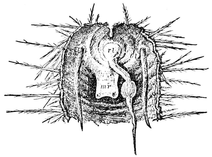
Fig. 593.—Shrunken larval skin of Vanessa antiopa, cut open from the back and showing (mr) the retaining membrane, (rl) the rectal ligament, and (tl) the tracheal ligaments.
The structures in the chrysalis are, first, the cremaster, with its dorsal (Fig. 594, dcr) and ventral (vcr) ridges, and the cremastral hook-pad (chp), said by Riley to be “thickly studded with minute but stout hooks, which are sometimes compound or furnished with barbs, very much as are some of our fishing-hooks, and which are most admirably adapted to the purpose for which they are intended.”
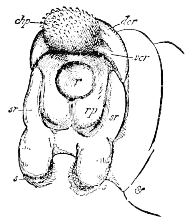
Fig. 594.—Ideal representation of the anal subjoint of Vanessa antiopa, from behind, with the spines removed, and all parts forced apart by pressure so as to show the homologies of the parts in the chrysalis which are concerned in pupation: homologies indicated by corresponding letters in Fig. 595, except that r (the rectum) corresponds with pr in Fig. 595.
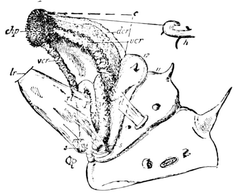
Fig. 595.—Anal parts of chrysalis of Vanessa antiopa, just prior to final extraction from shrunken larval skin: c, cremaster; chp, cremastral hook-pad; h, one of the hooks, more enlarged; vcr, ventral cremastral ridge; dcr, dorsal cremastral ridge; lr, larval rectum; pr, pupal rectum; rp, rectal plate; sr, sustentor ridges; mr, membrana retinens; rl, rectal ligament; tl, tracheal ligament; the 11th or last spiracle-bearing joint and the 12th joint being numbered.
Secondly, there are the other structures, viz., the sustainers (sustentors), two projections which Riley states “homologize with the soles (plantæ) of the anal prolegs, which take on various forms (3), but are always directed forward so as easily to catch hold of the retaining membrane.” These sustentors are, 639however, as Jackson[111] has shown, and as we are satisfied, the vestiges of the anal legs.
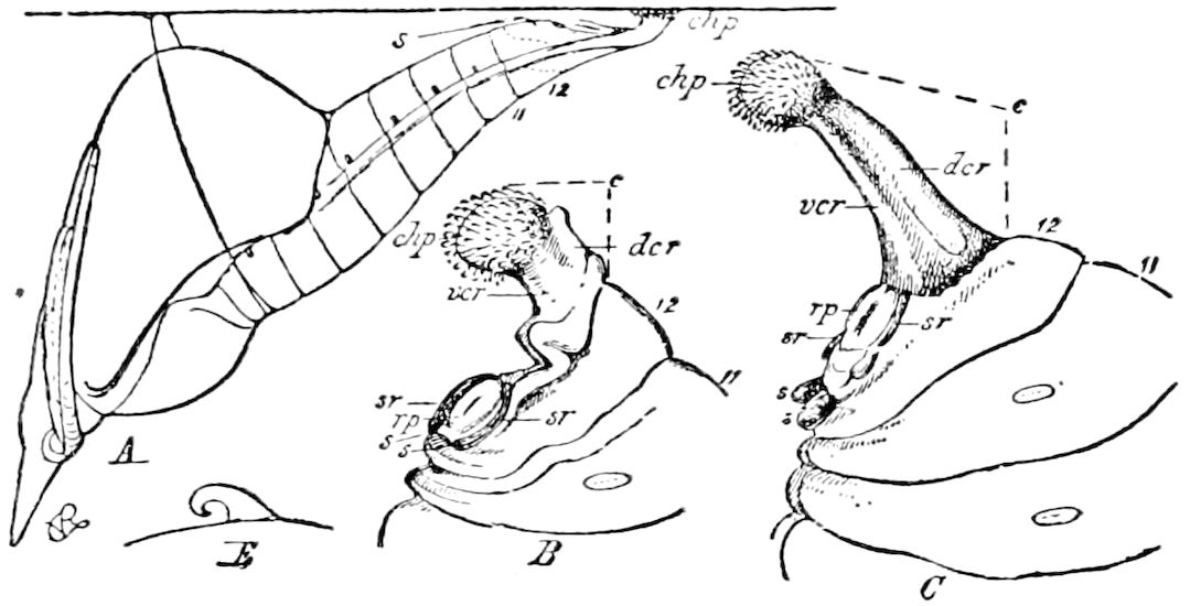
Fig. 596.—A, chrysalis of Terias. B, posterior end of chrysalis of Paphia. C, posterior end of chrysalis of Danais. E, one of the sustainers of Terias, greatly enlarged to show its hooked nature. All the parts of subjoint lettered to correspond with Fig. 595.
Thirdly, the sustentor ridges, which, as Riley states, may be more or less obsolete in some forms, in Paphia (Fig. 596, B) and Limenitis form “quite a deep notch, which doubtless assists in catching hold of the larval skin in the efforts to attach the cremaster.”
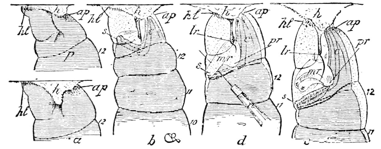
Fig. 597.—Pupation of butterflies: a, attachment of larva of Danais archippus; p, attachment of larva of Paphia glycerium; b, ideal larva soon after suspension; d, ideal larva a few hours later, the needle (n) separating the forming membrane from the sustainers; l, ideal larva just before splitting of larval skin, with retaining membrane loosened from the sustainers and showing its connection both with the larval and pupal rectum. In all the figures the joints of the body are numbered; the forming chrysalis is shaded in transverse lines; the intervening space between it and larval skin is dotted: h, is the hillock of silk; hl, hooks of hind legs; ap, anal plate; lr, larval rectum; pr, pupal rectum; mr, retaining membrane; c, cremaster; s, sustainers.—This and Figs. 593–596 after Riley.
“It is principally,” adds Riley, “by the leverage obtained by the hooking of the sustainers in the retaining membrane, which acts as a swimming fulcrum, 640that the chrysalis is prevented from falling after the cremaster is withdrawn from the larval skin. It is also principally by this same means that it is enabled to reach the silk with the cremastral hook-pads.”
“Dissected immediately after suspension, the last abdominal segment of the larva is found to be bathed, especially between the legs and around the rectum, in an abundance of translucent, membranous material.”
“An hour or more after suspension the end of the forming chrysalis begins to separate from the larval skin, except at the tip of the cremaster (Fig. 597, b). Gradually the skin of the legs and of the whole subjoint (10th segment) stretches, and with the stretching, the cremaster elongates, the rectal piece recedes more and more from the larval rectum, and the sustentor ridges diverge more and more from the cremaster, carrying with them, on the sustainers, a part of the soft membrane.” The rectal ligament will sustain at least 10 or 12 times the weight of the chrysalis. That of Apatura seems to rely almost entirely on the rectal ligament, assisted by the partial holding of the delicate larval skin.
FORMATION OF THE PUPA AND IMAGO IN THE HOLOMETABOLOUS INSECTS (THE DIPTERA EXCEPTED)
We have seen that in the incomplete metamorphosis, although there may be as many as five, and possibly seven moults, and in Chloëon as many as 20, and in Cicada septemdecim perhaps 25 or 30, there is but a slight change of form from one stage to another, and no period of inactivity. And this gradual outer transformation is so far as yet known paralleled by that of the internal organs, the slight successive changes of which do not differ from those observed in the growth of ametabolous insects. With the growth of the internal organs there probably goes on a series of gradual regenerative processes, and Korschelt and Heider state that we may venture to assume that each changed cell or group of cells which have become exhausted by the exercise of the functions of life are reabsorbed and become restored through the vital powers of the tissues, so that as the result there goes on a constant, gradual regeneration of the organs.
While the Hemiptera have only an incomplete metamorphosis, the males of the Coccidæ are, as shown by O. Schmidt, remarkable for passing through a complete or holometabolous development, with four stages, three of which are pupal and inactive. Hence, as Schmidt observes, there is here a hypermetamorphosis, like that of the Meloidæ, Stylopidæ, etc.
Shortly before the end of the larval stage of the male appear the imaginal buds of the eyes, legs, and wings. In the 2d or 1st pupal 641stage there is an atrophy of the antennæ and legs. On the other hand, at this stage the female completes its metamorphosis.
The rudiments of the wings arise on the edge of the dorsal and ventral side of the 2d thoracic segment, and this, we would remark, is significant as showing a mode of origin of the wings intermediate between that of the manometamorphic and holometamorphic insects. (See pp. 137–142.) While Schmidt could not ascertain the exact structure of the imaginal buds, he says “in general the process of formation of the extremities is exactly as Weismann has described in Corethra.” The two later pupal stages are “as in other metabolic insects.” (See p. 690, Fig. 637.)
Thus far the internal changes in the metamorphosis of the Coleoptera have not been thoroughly studied. They are less complete than in the other holometabolous insects, the differences between the larva and imago being much less marked than in the more specialized orders, and so far as known all the larval organs pass, though not without some great changes, directly into the imaginal ones, the only apparent exception being the mid-intestine, which, as stated by Kowalevsky, undergoes a complete transformation during metamorphosis. The following account, then, refers almost wholly to the Lepidoptera, Hymenoptera, and Diptera.
a. The Lepidoptera
The first observations on the complete metamorphosis of insects which were in any way exact were those of Malpighi, in 1667, and of Swammerdam, in 1733. While the observations of Swammerdam, as far as they extended, were correct, his conclusions were extraordinary. They were, however, accepted by Réaumur and by Bonnet, and generally held until the time of Herold in 1815, and lingered on for some years after. The rather famous theory of incasement (“emboîtement”) propounded by Swammerdam was that the form of the larva, pupa, and imago preëxisted in the egg, and even in the ovary; and that the insects in these stages were distinct animals, contained one inside the other, like a nest of boxes, or a series of envelopes one within the other, or, to use his own words: “Animal in animali, seu papilio intra erucam reconditus.”
This theory Swammerdam extended to the whole animal kingdom. It was based on the fact that by throwing the caterpillar, when about to pupate, in boiling water, and then stripping off the skin, the immature form of the butterfly with its appendages was disclosed. Malpighi had previously observed the same fact in the 642silkworm, perceiving that before pupation the antennæ are concealed in the head of the larva, where they occupy the place previously taken by the mandibular muscles; also that the legs of the moth grew in those of the larva, and that the wings developed from the sides of the worm.
Even Réaumur (1734) remarked: “Les parties du papillon cachées sous le fourreau de chenille sont d’autant plus faciles à trouver que la transformation est plus proche. Elles y sont neanmoins de tout temps.” He also believed in the simultaneous existence of two distinct beings in the insect. “Il serait très curieux de connaître toutes les communications intimes qui sont entre la chenille et le papillon.... La chenille hache, broye, digere les aliments qu’elle distribué au papillon; comme les mères préparent ceux qui sont portés aux fœtus. Notre chenille en un mot est destineé à nourrir et à defendre le papillon qu’elle renferme.” (T. i, 8e Mémoire, p. 363.)
Lyonet (1760), even, did not expose the error of this view that the larva enveloped the pupa and imago, and, as Gonin says, it was undoubtedly because he did not use for his dissections of the caterpillar of Cossus any specimens about to pupate. Yet he detected the wing-germs and those of the legs, stating that he presumed the bodies he saw to be the rudiments of the legs of the moth (p. 450).
Herold, in his work on the development of the butterfly (1815), was the first to object to this erroneous theory, showing that the wings did not become visible until the very end of larval life; that as the larval organs disappear, they are transformed or are replaced by entirely new organs, which is not reconcilable with a simple putting off of the outer envelope. The whole secret of metamorphosis, in Herold’s opinion, consisted in this fact, that the butterfly in the larva state increases and accumulates a supply of fat until it has reached the volume of the perfect state; then it begins the chrysalis period, during which the organs are developed and take their definite form.[112] (Abstract mostly from Gonin.) Still the old ideas prevailed, 643and even Lacordaire, in his Introduction à l’Entomologie published in 1834, held on to Swammerdam’s theory, declaring that “a caterpillar is not a simple animal, but compound,” and he actually goes so far as to say that “a caterpillar, at first scarcely as large as a bit of thread, contains its own teguments threefold and even eightfold in number, besides the case of a chrysalis, and a complete butterfly, all lying one inside the other.” This view, however, we find is not original with Lacordaire, but was borrowed from Kirby and Spence without acknowledgment. These authors, in their Introduction to Entomology (1828), combated Herold’s views and stoutly maintained the old opinions of Swammerdam. They based their opinions on the fact, then known, that certain parts of the imago occur in the caterpillar. On the other hand, Herold denied that the successive skins of the pupa and imago existed as germs, holding that they are formed successively from the “rete mucosum,” which we suppose to be the hypodermis of later authors. In a slight degree the Swammerdam-Kirby and Spence doctrine was correct, as the imago does arise from germs, i.e. the imaginal disks of Weismann, while this was not discovered by Herold, though they do at the outset arise from the hypodermis, his rete mucosum. Thus there was a grain of truth in the Swammerdam-Kirby and Spence doctrine, and also a mixture of truth and error in the opinions of Herold.
The real nature of the internal changes wrought during the process of metamorphosis was first revealed by Weismann in 1864. His discovery of the germs of the imago (imaginal buds) of the Diptera, and his theory of histolysis, or of the complete destruction of the larval organs by a gradual process, was the result of the application of modern methods of embryology and histology, although his observations were first made on the extremely modified type of the Muscidæ or flies, and, at first, he did not extend his view to include all the holometabolous insects. Now, thanks to his successors in this field, Ganin, Dewitz, Kowalevsky, Van Rees, Bugnion, Gonin, 644and others, we see that metamorphosis is, after all, only an extension of embryonic life, the moults and great changes being similar to those undergone by the embryo, and that metamorphosis and alternation of generations are but terms in a single series. Moreover, the metamorphoses of insects are of the same general nature as those of certain worms, of the echinoderms, and the frog, the different stages of larva, pupa, and imago being adaptational and secondary.
While the changes in form from the larva to the pupa are apparently sudden, the internal histogenetic steps which lead to them are gradual. In the Lepidoptera a few days (usually from one to three) before assuming the pupa stage, the caterpillar becomes restless and ceases to take food. Its excrements are now hard, dry, and, according to Gonin, are “stained carmine red by the secretions of the urinary tubes.” Under the microscope we find that they are almost exclusively composed of fragments of the intestinal epithelium. These red dejections were noticed by Réaumur, and afterwards by Herold, and they are sure indications of the approach of the transformations. It now wanders about, and, if it is a spinner, spins its cocoon, and then lies quietly at rest while the changes are going on within its body. Meanwhile, it lives on the stores of fat in the fat-body, and this supply enables it to survive the pupal period.
The amount of fat is sometimes very great. Newport removed from the larva of Cossus ligniperda 42 grains of fat, being more than one-fourth of the whole weight of the insect, he adds that the supply is soon nearly exhausted during the rapid development of the reproductive organs, “since, when these have become perfected, the quantity that remains is very inconsiderable.”
Although the larval skin of a lepidopterous insect is suddenly cast off, the pupa quickly emerging front it, yet there are several intermediate stages, all graduating into each other. If a caterpillar of a Clisiocampa, which, as we have observed, is much shortened and thickened a day or two before changing to a pupa, is hardened in alcohol and the larval skin is stripped off, the semipupa (pro-nymph, pro-pupa of different authors) is found to be in different stages of development, and the changes of the mouth-parts are interesting, though not yet sufficiently studied.
Newport attributes the great enlargement and changes in the shape of the thoracic segments of the larva of Vanessa urticæ at this time, to the contraction or shortening of the muscles of the interior of those segments, “which are repeatedly slowly extended and shortened, as if the insect were in the act of laborious respiration.” This, he adds, generally takes place at short intervals during 645the two hours immediately preceding the change to the pupa, and increases in frequency as that period approaches. He thus describes the mode of moulting the larval skin: “When the period has arrived, the skin bursts along the dorsal part of the 3d segment, or mesothorax, and is extended along the 2d and 4th, while the coverings of the head separate into three pieces. The insect then exerts itself to the utmost to extend the fissure along the segment of the abdomen, and, in the meantime, pressing its body through the opening, gradually withdraws its antennæ and legs, while the skin, by successive contortions of the abdomen, is slipped backwards, and forced towards the extremity of the body, just as a person would slip off his glove or his stocking. The efforts of the insect to get entirely rid of it are then very great; it twirls itself in every direction in order to burst the skin, and, when it has exerted itself in this manner for some time, twirls itself swiftly, first in one direction, then in the opposite, until at last the skin is broken through and falls to the ground, or is forced to some distance from it. The new pupa then hangs for a few seconds at rest, but its change is not yet complete. The legs and antennæ, which when withdrawn from the old skin were disposed along the under surface of the body, are yet separate, and do not adhere together as they do a short time afterwards. The wings are also separate and very small. In a few seconds the pupa makes several slow, but powerful, respiratory efforts; during which the abdominal segments become more contracted along their under surface, and the wings are much enlarged and extended along the lateral inferior surface of the body, while a very transparent fluid, which facilitated the slipping off of the skin, is now diffused among the limbs, and when the pupa becomes quiet dries, and unites the whole into one compact covering.”
The changes in the head and mouth-parts.—The changes of form from the active mandibulate caterpillar to the quiescent pupa, and then to the adult butterfly, are, as we have seen, in direct adaptation to their changed habits and surroundings, and they differ greatly in details in insects of different orders. In many Lepidoptera and certain Diptera the pupa and imago are without the mandibles of the larva, and, instead, the 1st maxillæ in the former order, and the 2d maxillæ in the latter, are highly developed and specialized. The changes in the shape of the head, with the antennæ, the latter rudimentary in the larvæ of the two orders named, are noteworthy, and will be referred to under those orders. The same may be said of the thorax with the legs and wings, and the abdomen with the ovipositor. Every part of the body undergoes a profound change, though 646in the Coleoptera, Trichoptera, and the more generalized and primitive Diptera, each segment and appendage of the larva are directly transformed into the corresponding parts of the pupa, and subsequently of the imago. We shall see, however, beyond, that this general statement does not apply to the Hymenoptera, in which there is a process of cephalization or transfer of parts headward, peculiar to that order.
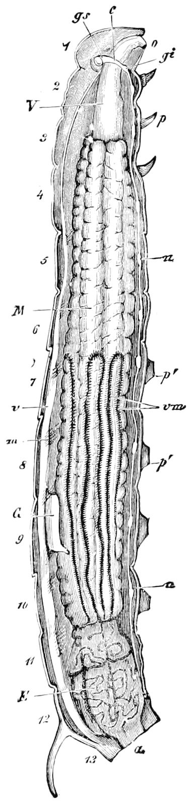
Fig. 598.—Internal organs of Sphinx ligustri: 1, head; 2–4, thoracic, 5–13, abdominal segments; V, fore-, M, mid-, E, hind-intestine; gs, brain; gi, infraœsophageal ganglion; n, ventral ganglion; vm, urinary tubes; c, heart; G, testis; o, œsophagus; a, anus; m, alary muscles of the heart.
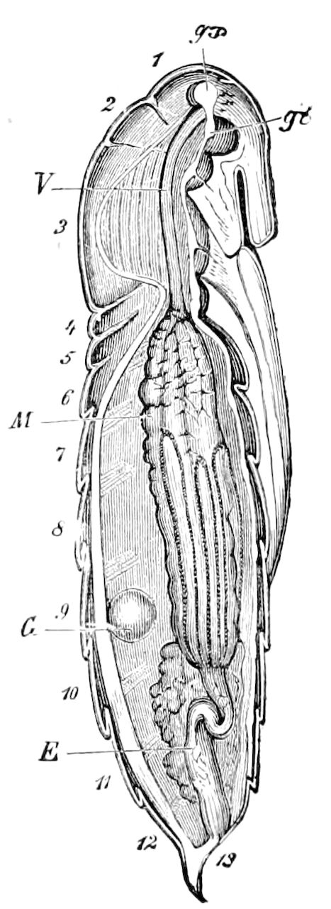
Fig. 599.—Pupa of the same.
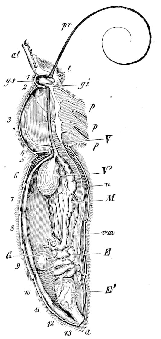
Fig. 600.—Imago of the same.—This and Figs. 598 and 599 after Newport, from Gegenbaur.
647
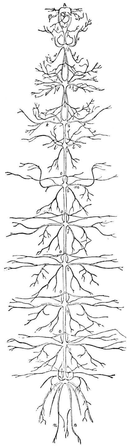
Fig. 601.—Nervous system of the larva of Sphinx ligustri.
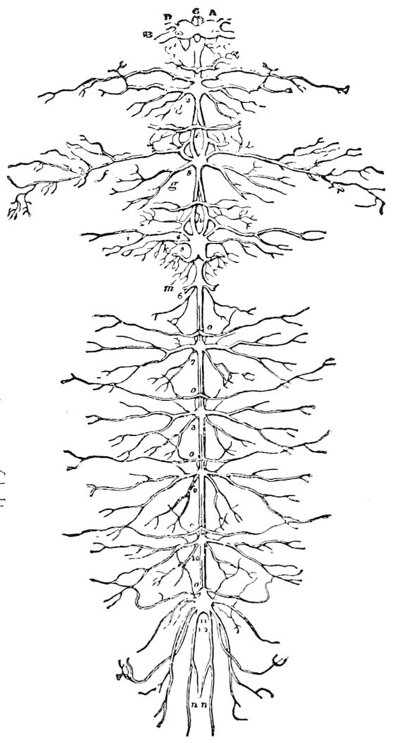
Fig. 602.—Nervous system of the pupa of Sphinx ligustri, soon after pupation.—This and Fig. 601, after Newport.
The change in the internal organs.—These were especially, as regards the nervous system, first carefully examined and illustrated by that great English entomotomist, Newport, and those of the reproductive organs by Herold as early as 1815. A glance at the figures (598–604), reproduced from Newport’s article Insecta, will show the changes wrought especially in the digestive and nervous systems of Sphinx and Vanessa, his account of the alterations in the muscles having already been quoted. As the pupal form is much nearer to that of the imago than of the larva, so the digestive canal is seen to be nearly as much differentiated in the pupa as in the imago, though the reservoir (“sucking-stomach”) of the imago is not indicated in the pupa. These changes 648are such as occur in an insect which is enormously voracious as a larva, and which often, passing through a period of complete inactivity, taking no food at all, finally becomes an insect which needs to suck in only a minimum quantity of water or nectar, and which practically abstains from all food. The head and genital glands also, as well as the urinary vessels, are nearly the same. On the other hand, the salivary glands have undergone, in the imago, a thoroughgoing reduction.
The changes undergone by the nervous system of Sphinx ligustri and Vanessa urticæ have been described by Newport with fulness of detail. An abstract of his observations on Vanessa urticæ, which undergoes its changes in June in 14 days, and in August in eight days, we will now give, in part verbatim, the subject being rendered much clearer by his figures, which are reproduced.
During the last larval stage, certain changes have already taken place in different parts of the cord, which shows that they had been a long time in progress. Besides the lateral approximation of the cords, the first change consists in a union of the 11th and 12th ganglia, the latter one being carried forwards; these two ganglia being entirely separate before the 3d moult.
Two hours after the larva of Vanessa urticæ has suspended itself in order to pupate, the brain is not yet enlarged, but the subœsophageal ganglion is nearly twice its original size and the ganglia behind are nearer together. “A little while before the old larval skin is thrown off there is great excitement throughout the body of the insect.” About half an hour (Fig. 603, 2) before this occurs the alary nerves and the cerebral, 2d, 3d, 4th, and 5th ganglia are slightly enlarged, and the 1st subœsophageal ganglion very considerably. Immediately after the insect has entered the pupa state (Fig. 603, 3), all the ganglia are brought closer together. One hour after (Fig. 603, 4) pupation the cerebral ganglia are found to be more closely united, the 4th and 5th ganglia are nearer, and the distance between the remaining ganglia is also reduced.
Seven hours after pupation there is a greater enlargement of the cerebral ganglia, optic nerves, and ganglia and cords of the future thoracic segments.
At 12 hours (Fig. 603, 5) the 5th pair of ganglia has almost completely coalesced with the cord and the 4th; at 18 hours (Fig. 603, 6) the whole of the ganglia, cords, and nerves have become more enlarged, especially those of the wings, while the 4th and 5th ganglia of the cords have now so completely united as to appear like an irregular elongated mass. At 24 hours (Fig. 604, 7) the 4th and 5th ganglia are completely united, the 5th being larger than the 4th. At 36 hours (Fig. 604, 8) the optic nerves have attained a size almost equal to that of the brain. The 1st subœsophageal ganglion now forms, with the cerebral ones, a complete ring around the œsophagus, the crura having almost disappeared. The 6th ganglion has now disappeared, but the nerves arising from it remain. At 48 hours (Fig. 604, 9) the cord is straight instead of being sinuous, and the 7th ganglion has disappeared, while the thoracic ganglia are greatly enlarged. At the end of 58 hours the 2d and 3d thoracic ganglia have united, and the double ganglion thus formed is only separated from the large thoracic mass composed of the 4th, 5th, and part of the 6th ganglia, by the short but greatly enlarged cords which pass on each side of the central attachment of the muscles. “The optic and antennal nerves have nearly attained their full development, and those numerous and most intricate plexus of nerves in the three thoracic segments of the larva form only a few trunks, which can hardly be recognized as the same structures. The arrangement of the whole nervous system is now nearly as it exists in the perfect insect. The whole of these important changes are thus seen to take place within the first three days after the insect has undergone its metamorphosis; and they precede those of the alimentary canal, generative system, and other organs, which are still very far from being completed, and indeed, as compared with the nervous system, have made but little progress.” (Art. Insecta, pp. 962–965.)
649
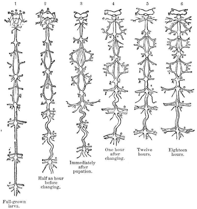
Fig. 603.—Changes in the nervous system of Vanessa urticæ, during and after pupation.—After Newport.
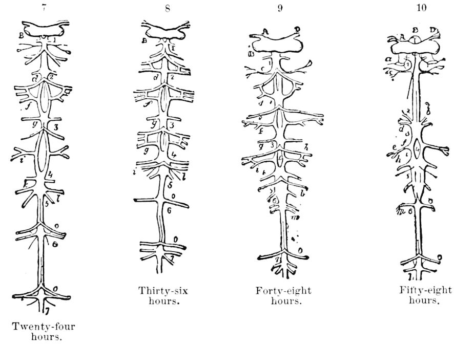
Fig. 604.—Changes in the nervous system of Vanessa urticæ, from 24 to 58 hours after pupation.—After Newport.
650The initial steps and many of the subsequent internal changes escaped the notice of Newport and others of his time, and it was not until the epoch-making work of Weismann on the ultimate processes of transformation of Corethra and of Musca, that we had an adequate knowledge of the subject.
Weismann (1864) was the first to show for the Muscidæ and Corethra that the appendages, wings, and other parts of the imago originate in separate, minute, cellular masses called imaginal disks, buds, or folds (histoblasts of Künckel). These imaginal buds, which arise from the hypodermis, being masses of indifferent cells, are usually present in the very young larva, and even in the later embryonic stages. It has been shown that such imaginal buds exist for each part of the body, not only for the appendages and wings (p. 126), but for the different sections of the digestive canal. During the semipupal stage these buds enlarge, grow, and at the same time there is a corresponding destruction of the larval organs. The process of destruction is due to the activity of the blood corpuscles or leucocytes (phagocytes), the larval organs thus broken up forming a creamy mass, the buds from which the new organs are to arise resisting the attacks of the virulent leucocytes, which attach themselves to the weakened tissue and engulf the pigments (see p. 422). The two processes of destruction of the larval organs (histolysis) and the building up of the imaginal organs (histogenesis) go hand in hand, so that the connection of the organs in question in most cases remains entirely continuous; while the last steps in the destruction of the larval organs only take place after the organs of the imago have assumed their definite shape and size. Other observers have corroborated and confirmed his statements and observations, Gonin extending them to the Lepidoptera and Bugnion to the Hymenoptera.
It is a pity that the observations, such as were set on foot by Weismann, were not first made on the Trichoptera and Lepidoptera, which are much more primitive and unmodified forms than the 651Diptera, but mistakes of this nature have frequently happened in the history of science.
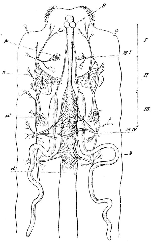
Fig. 605.—Full grown larva of Pieris brassicæ opened along the dorsal line: d, digestive canal; s, silk-gland; g, brain; st I, prothoracic stigma; st IV, 1st abdominal stigma; a, a′, germs (buds) of fore and hind wings; p, bud of thoracic segment;—those of the 3d pair are concealed under the silk-glands; I-III, thoracic rings.—After Gonin.
The latest and most detailed researches are those of J. Gonin on the metamorphoses of Pieris brassicæ, made under the direction of Professor E. Bugnion. They fill an important gap in our knowledge, and show that the Lepidoptera transform in nearly the same manner as described by Weismann in Corethra. We give the following condensed account of Gonin’s observations.
On opening a caterpillar entering on the semipupa state (Fig. 605), 652the relative position of the germs (imaginal buds or folds) of the wings and of the legs are seen.
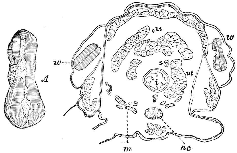
Fig. 606.—Section through thorax of a tineid larva on sycamore, passing through the 1st pair of wings (w): ht, heart; i, œsophagus; s, salivary gland; ut, urinary tube; nc, nervous cord; m, recti muscles; a part of the fat-body overlies the heart. A, right wing-germ enlarged.
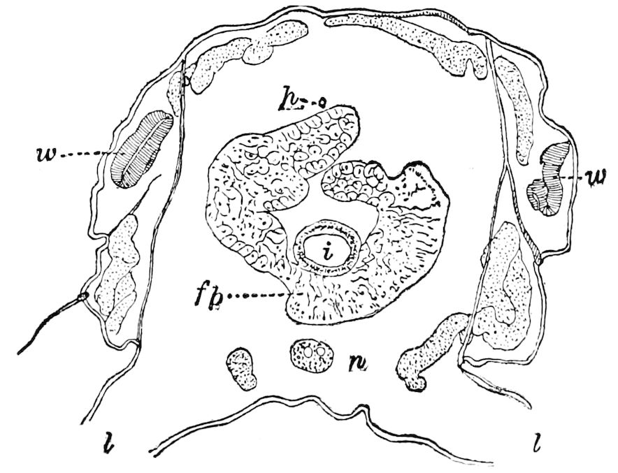
Fig. 607.—Section of the same specimen as in Fig. 606, but cut through the 2d pair of wings (w): i, mid-intestine; h, heart; fb, fat-body; l, leg; n, nervous cord.
These imaginal buds in a more advanced stage are seen in our sections of a tineid larva (Figs. 606, 607).
The number of 12 imaginal buds found by Weismann in the thorax of Muscidæ does not occur in Lepidoptera, since, as in the Hymenoptera (Bugnion), the dorsal buds of the prothoracic segment are wanting. Gonin finds in Pieris that the ventral buds of the 653three thoracic segments are each represented by several distinct folds attached to the femoro-tibial bud and to the tarsal joints.
The imaginal buds serve in some cases for the formation of new organs (wings, legs of insects with apodous larvæ); in others for the growth and the transformation of organs already existing (legs, antennæ, 1st and 2d maxillæ of Lepidoptera).
As to the peripodal sac or hypodermic envelope which contains the imaginal bud, a portion persists and is regenerated, while the other part becomes useless and is detached under the form of débris, as shown by Weismann, Viallanes, and Van Rees in the Muscidæ. On this point Gonin disagrees with Dewitz, who stated that the walls of the wing-sacs are not destroyed, but are only gradually withdrawn at the time of pupation, in order to allow the orifice to distend and let the wing pass out to the exterior.
The portion of the sac which persists (basal portion, peripheral pad of Bugnion, or annular zone of Künckel) serves at first to attach the appendage, while forming, to the hypodermis of the larva, then afterwards to more or less completely regenerate the adjoining portion of the integument. In this way the hypodermis of the thorax is partially, that of the head is almost entirely, replaced by the imaginal epithelium which proliferates at the base of the appendages,[113] while that of the abdominal segments persists, at least in a modified way, and only undergoes (at the end of the pupal period) transformations as regards the appearance of the scales and pigment.
The wings.—The imaginal buds of the wings do not participate in the larval moults. Gonin has observed, contrary to Dewitz, that their surface only forms a cuticle towards the end of the last larval stage.
The network of fine tracheæ of the wing-bud is drawn out at the time of pupation with the internal cuticle of the large tracheæ. The permanent tracheæ of the wing have already appeared at the time of the 3d moult under the form of large rectilinear trunks, the position of which corresponds afterwards to that of the veins, but they are not filled with air until the time of pupation. There are from eight to ten of these tracheæ in each wing (Fig. 159), and they give rise in the pupa to a new system of fine tracheæ (tracheoles) which replaces that of the larva. (For further details the reader is referred to pp. 126–137.)
Development of the feet and the cephalic appendages.[114]—In the apodous larvæ of Diptera and Hymenoptera the rudiments of the legs are, like those of the wings, developed within hypodermal sacs; at times they remain there up to the end of larval life, but sometimes 654they appear early at the surface. This origin of the legs, thanks to Weismann, Künckel, and Van Rees, is well known in the Diptera; in the Hymenoptera it has been proved to be the case with ants by Dewitz, and in Encyrtus by Bugnion. As for the Lepidoptera our knowledge that the legs of the imago arise from the six thoracic legs of the caterpillar, up to the date of Gonin’s paper has not been in advance of that of Malpighi and Swammerdam.
Réaumur, moreover, was supposed to have furnished the proof, having from his experiments concluded that “if the legs of the pupa appear longer and larger than those of the caterpillar wherein they were contained, it is because they were folded and squeezed.” (8e Mém., p. 365.)
This explanation of Réaumur’s has been generally accepted. Graber (Die Insekten, p. 506) accepted it, after examining microscopic sections of the legs, and Künckel averred that “Réaumur, having, in certain caterpillars, completely cut off one of the thoracic legs, had concluded that the butterfly which came from it lacked the corresponding member.” (Rech. sur l’org. et dév. des volucelles, p. 160.)
Newport, it is true, denied this disappearance of the legs, but did not wish to put himself in opposition to received ideas, and supposed that the member cut off was partly reformed in the imago.
Künckel believes that he has found a better solution in his theory of histoblasts or imaginal buds; in his opinion, “Réaumur and Newport are both right,” but “when Réaumur cut off a caterpillar’s leg, he at the same time removed the histoblast, the rudiment of the leg of the butterfly. When Newport repeated this experiment, he simply mutilated the histoblast without completely destroying it: in the first case, the adult insect was born with one leg less; in the second case, it appeared with an atrophied foot.”
“So ingenious an explanation,” says Gonin, “is not necessary.” To prove that the experiments of the two savants are not contradictory, it would have been sufficient to cite, as Künckel did not do, the exact words of Réaumur, for he having cut from a caterpillar “more than half of three of the thoracic legs on the same side,” says he found that the chrysalis had “the three limbs on one side shorter than the corresponding ones on the other side.” The same operation repeated on a somewhat younger caterpillar again showed in the chrysalis three maimed limbs, “so that they could not be said to be entirely absent. These results are like those of Newport; the interpretation only was faulty, as we shall attempt to prove.”
The real relations of the adult legs to the larval legs are thus shown by Gonin.
“If we carefully strip off the skin of a caterpillar near the time of pupation (Fig. 608), we see that the extremity only of the legs of the imago is drawn out of the larval legs; the other parts are pressed against each side of the thorax: near the ventral line a small pad represents the coxa and the trochanter; the femur and the tibia are distinctly recognizable, but soldered to each other and only separated by a slight furrow; they form by their union a very acute knee or bend. The femur is movable on the pad-like coxa, the tibia continues 655without precise limits with the extremity concealed in the larval legs. The three divisions of the latter do not appear to have any relation with the live joints of the perfect state. Under the microscope the rudiment appears very strongly plaited at the level of the tarsus, much less so in the other regions. A large trachea penetrates into the femur with some capillaries; reaching the knee it bends into the tibia at a sharp curve, but does not become truly sinuous in approaching the extremity. It is then the tarsus especially which is susceptible of elongation; it may, on being withdrawn, give rise to the illusion that the whole organ is disengaged from the larval leg.
“This disposition is, we believe, not known. It gives the key to the experiments of Réaumur and of Newport.
“Even when we cut off the limb of the caterpillar at its base, we only remove the tarsus of the imago; the femur and the tibia remain intact. From an evident homology Réaumur has erroneously concluded that there is an identity. His opinion, classical up to this day, that the limb of the butterfly is entirely contained in the leg of the caterpillar, has been found to be inexact and should be abandoned.”
Embryonic cells and the phagocytes.—Up to the last larval stage the legs do not offer, says Gonin, any vestige of an imaginal germ, but they contain a great number of embryonic cells (Fig. 145, ec). They are almost always collected around a nerve or trachea; sometimes they are independent, and sometimes retained in the peritoneal sheath, seeming to arise by proliferation from this sheath. Some thus contribute to the lengthening of the tracheal branches or nerves, and the others, becoming detached, form leucocytes or phagocytes. They are very numerous in the legs, at the beginning of the 4th stage, but are disseminated some days later throughout the whole cavity of the body. At the time of histolysis they attack the larval tissues and increase in volume at their expense; in return they serve for the nutrition of the imaginal parts and exercise no destructive action on them. Van Rees agrees with Kowalevsky in comparing these attacks of the embryonic cells, sometimes victorious and sometimes impotent, to the war which the leucocytes wage against both the attenuated and the virulent bacteria.
Formation of the femur and of the tibia, transformation of the tarsus.—Capillary tracheæ appear in the leg at the same time as in the wing. They arise from the end of a tracheal trunk near the base of the limb on the dorsal and convex side. After the 3d moult the hypodermis thickens near this place; in a few days a pad is formed 656there and then a large imaginal bud with a circular invagination. These buds were noticed by Lyonet, who supposed them to be “les principes des jambes de la phalène.” Nerves and a tracheal branch penetrate into the femoro-tibial bud and form a small bay or constriction which marks the point of junction of the femur with the tibia, and the body-cavity remains in direct communication with the end of the limb.
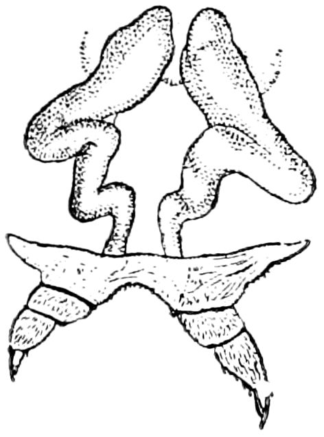
Fig. 608.—Feet of the Pieris butterfly withdrawing from those of the larva.
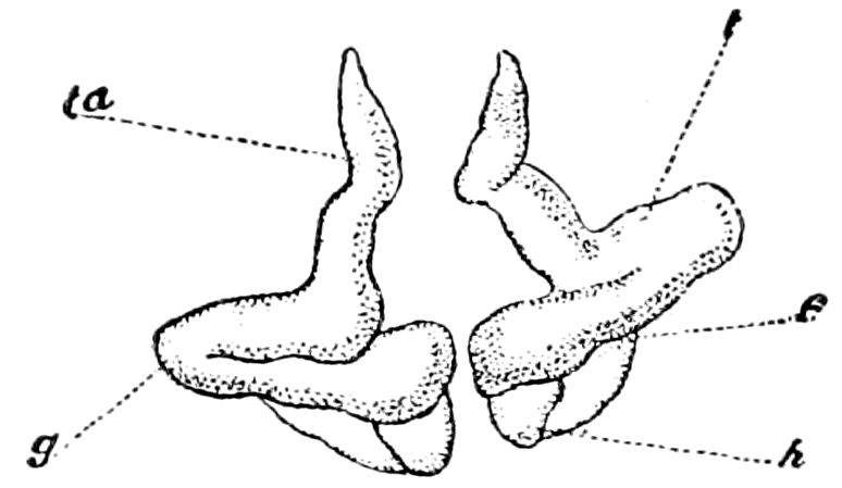
Fig. 609.—Imaginal feet of Pieris uncovered with great care to preserve the position which they had in the larva: ta, tarsus; t, tibia; g, knee; f, femur; h, coxa.
The tarsus undergoes a series of changes; the surface is folded in a very complicated way; at the level of each articulation, but only in the internal and concave region of the leg, is developed a deep fold; on one side there is a hypodermic thickening, on the other a simple leaf of the envelope, which afterwards joins at its base with the parietal hypodermis, and then two leaves are destroyed with the large cells of the setæ. The internal part and end of the tarsus are then reconstituted with the elimination of the débris, while the external and convex region undergoes direct regeneration.
The coxa and trochanter are derived from the base of the larval leg, and only the 1st pair are well separated from the base of the thorax. One or two days before pupation the femoro-tibial bud, after having, until now, preserved its antero-posterior direction, is placed transversely as regards the larva, then becoming directed obliquely forward. This rotatory movement of the coxa may be attributed to the great extension of the fore wings, which push before them the two first pairs of legs. The last pair in their turn are simply covered by the hind wings and are but slightly displaced. This new position of the legs is that of the imago: the knee of the 1st pair is situated in front of the tarsus; that of the 2d a little outward; that of the 3d pair is directed backward. (Gonin.)
The antennæ.—These appendages also have the same relation with those of the caterpillar as in the case of the legs, the larval appendages 657being only the point of departure of the imaginal growth. Weismann has observed in Corethra how at the approach of each moult an invagination like the finger of a glove allows the antenna to elongate from its base. The process, says Gonin, is identical in the caterpillar of Pieris. At the last moult the invagination is so pronounced that it is not effaced with the renewal of the chitinous integument. Several days later it again begins to grow larger. As the imaginal bud gradually sinks into the cavity of the head, it presses back the hypodermic wall and thus forms an envelope around it. Its base, widely opened, gives admission to the nerves, besides capillaries and sometimes a large trachea.
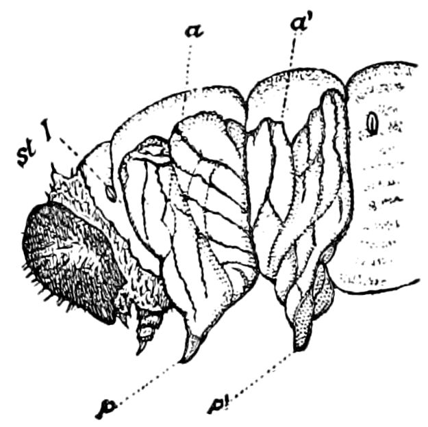
Fig. 610.—Larva in same stage as Fig. 613; side view of head and thorax: a, a′, wings, with the folds on the surface, and the sinuous track of the tracheal bundles; st I, prothoracic stigma; p, p′, ends of the legs.
As soon as it reaches the posterior region of the head, the antenna in lengthening becomes folded and describes the great curves which led Réaumur to compare it to a ram’s horn (Fig. 613). The leaf of the envelope thickens in the interior and all around the base of the organ. Its ultimate rôle is closely like that of the two other hypodermic formations. It is at the outset this layer of cells which in the larva supports the ocelli. This layer, hidden on each side under the parietal region, thickens and regenerates, forming a circular pad which becomes more prominent and finally assumes the form of the compound eye of the imago.
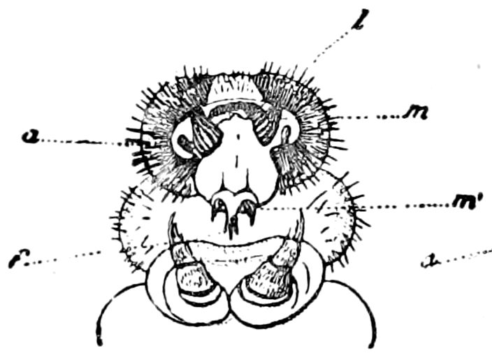
Fig. 611.—Head of the larva just before pupation: between the two mandibles (m) is seen the relief of the tongue or maxillæ (m′); f, spinneret; l, labrum; a, antenna.
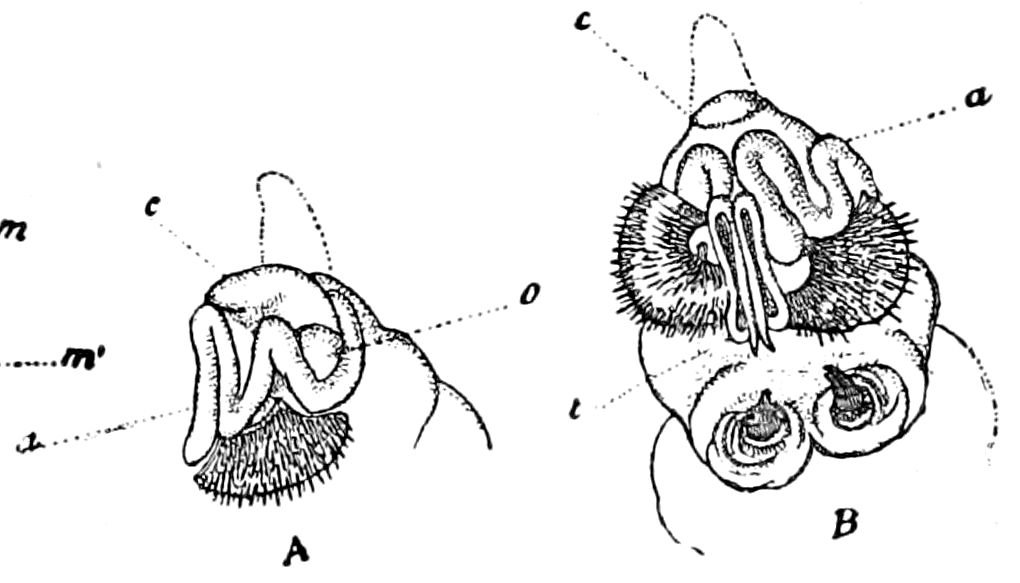
Fig. 612.—Same stage as in Fig. 611, but after the removal of the larval skin, and including the lateral scale: A, side, B, front, view; c, “cimier” (the dotted line shows the position it takes in the pupa); a, antenna; o, eye; t, tongue.—This and Figs. 608–611, after Gonin.
Finally, this layer gives rise to a conical prolongation (Fig. 612, c), which after exuviation appears as a tuft of long hairs, and is called 658by Gonin the crest (cimier, Fig. 612), which is characteristic of the pupæ of Pieridæ. It is only differentiated towards the end of the 4th larval stage in a median depression of the vertex. It is an imaginal bud in the most general sense of the word.
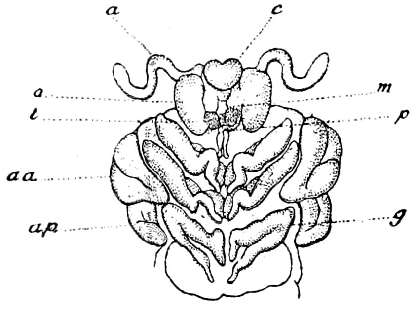
Fig. 613.—Larva of Pieris brassicæ stripped of its skin some minutes before pupation: the antennæ (a) have been displaced, and the tongue cut off, to show the palpi (p); c, cimier: o, eye; m, vestige of a mandible; t, insertion of the tongue (see Fig. 612); aa, fore, ap, hind, wings; g, knee of a foot of the 3d pair.
On each side the base of the antenna comes in contact with the germ of the crest. The envelopes approach each other, and their thickened part constitutes with the ocular disks a new cephalic wall. The head of the butterfly thus marked off is triangular; all the larval parts remaining out of this area then disappear. The muscles and the nerves are resorbed by histolysis, then the external part of the imaginal envelopes and the old parietal hypodermis, reduced very thin and degenerated, is detached in shreds. The antenna becomes external throughout its whole extent. The transformation is in this case, then, almost as complete as in the thorax of Diptera or Hymenoptera. It is necessitated by the change of form and of volume of the head. The region of the ocelli persists unchanged almost alone from the larva to the imago also. The limit is not well marked between the portion which is the replacement or direct renovation of the epithelium.
Maxilla and labial palpi.—The development of the tongue (1st maxillæ) is so like that of the antennæ that it scarcely needs description. Beginning at the last moult, the hypodermic contents of the maxillæ is withdrawn in the cephalic cavity under the form of a hollow bud whose base is turned inward. The invagination remains less distinct than in the antennæ; it does not even reach to the anterior part of the œsophagus. The two symmetrical halves of the tongue approach each other and are thrice folded. When the caterpillar stops feeding, they each curve in in the form of an S, remaining lodged under the floor of the mouth (Fig. 613, t).
Underneath are to be seen two other buds, which by an identical process become the labial palpi (Figs. 614, 615, p).
At the anterior part of the head, where the organs are very close together, the envelopes form several folds without any further use 659(Fig. 615, r). The two leaves then fuse together and decay as at the surface of the tarsus.
Finally, in the mandibles and the labrum, there is only a cellular thickening without any invagination.
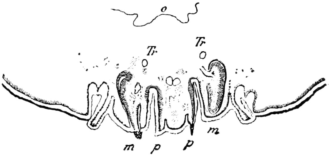
Fig. 614.—Section through the anterior region of the head of Pieris larva, four days after the 3d moult: o, œsophagus; m, m, 1st maxillæ containing the two imaginal buds of the tongue; p, p, labial palpi; Tr, trachea.
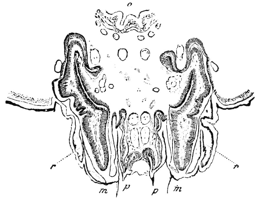
Fig. 615.—Section through the same place as in Fig. 614, 10 days after the 3d moult, the imaginal appendages having grown in size: r, r, caducous folds of the old hypodermis and of the envelopes. Other letters as in Fig. 614.—This and Figs. 613–614, after Gonin.
Process of pupation.—Notwithstanding the great number of persons who have reared Lepidoptera, close and patient observations as to the exact details are still needed. Gonin, who has made the closest observations on Pieris, pertinently asks why the antennæ, which are appendages of the head, are visible in the abdominal region, and why the tongue (maxillæ) is extended between the legs as far as the 3d abdominal segment. To answer these questions he made a series of experiments. Selecting some caterpillars which 660were about to pupate, he produced an artificial metamorphosis by removing the cuticula in small bits. Exposing the appendages in this way, they preserved the position which they are seen to take during growth. Each wing appeared within the limits of the segment from which it grew out (Fig. 610), not extending beyond, as it does in the normal pupa, so that Réaumur was wrong in saying that “the wings are here gathered on each side into a kind of band, which is large enough to lie in the cavity which is between the 1st and 2d segment.” (8e Mém., p. 359.)
All these parts are coated with a viscous fluid secreted by special glands, which hardens after pupation upon exposure to the air. So long as the parts are soft, they can easily be displaced. Gonin drew one of the antennæ like a collar around the head, and one half of the tongue upon the outer side of the wing.
“When pupation is normal, the integument splits open on the back of the thorax, and the pupa draws itself from before backwards. Owing to the feeble adherence which the chitinous secretion gives it, it draws along with it the underlying organs. The legs, antennæ, the two halves of the tongue (maxillæ) retained by their end, each in a small chitinous case, can only disengage themselves from it when in elongating they have acquired a sufficient tension. The curves straighten out and the folds unbend. The chitinous mask of the head in withdrawing from the larval skin follows the ventral line; the tongue and labial palpi free themselves from its median part; the antennæ disengage themselves from the two lateral scales. Between these different appendages a space is left on the surface of the head for the eyes, and on the thorax for the legs. These are not completely extended on account of the lack of freedom of the femoro-tibial articulation; the femur preserves its direction from behind forwards, and the knee in the two first pairs remains at the same height. The wings overlie them and cover the under side of the two basal abdominal segments; their surfaces in becoming united increase much in size.”
As the chitinous frame of each spiracle gradually detaches itself, we see a tuft of tracheæ passing out of the orifice. It is at this moment that the provisional tracheal system is cast off, and it is easy to see that the process is facilitated by the simultaneous elongation of all the appendages. The permanent tracheæ can follow this elongation because they are sinuous, and need only to straighten their curves. It is, however, not the same with the tracheoles, as we have seen in the case of the wings (p. 133), and their extension or stretching is thus explained by a very simple mechanism.
661“The position which the organs assume in the chrysalis is not due to chance, everything is determined in advance, and the microscope shows us that the structure of the hypodermis is specially modified in all the parts which remain external. It is a fact well known to those who rear Lepidoptera that if this normal arrangement is disturbed there are few chances that the perfect insect will survive. A leg lifted up, or an antenna displaced, leaves a surface illy protected against external influences. Almost always this accident causes a drying of the chrysalis.
“Several interesting experiments may be cited as bearing on this subject. If during transformation the chitinous mask of the head is separated from the integument beneath, it is arrested half-way in its development, and the antennæ and tongue are not fully extended. When the case or skin of the caterpillar is drawn, not from before backward, but in the opposite direction, all the appendages of the thorax are placed perpendicularly to the body. Dewitz and Künckel d’Herculais, in stating that the skin of the caterpillar splits open along its whole length, show that they were ignorant of the mechanism; for it is precisely because the chitinous larval skin splits open only in front that it preserves sufficient adherence to the organs beneath to draw them after it in the direction of the abdomen.
“To only read modern authors, one would suppose that the mechanism of pupation had remained hitherto unknown. In reality, it did not escape the notice of Swammerdam or of Réaumur, both of whom have described it with care. The first attached too much importance to the flow of blood, the action of which would be rather to push the organs out than to extend them over the surface of the thorax; the second insists on the movements of the insect. This factor, very admissible in caterpillars, ‘whose under side is situated on a horizontal plane’ (iii, 9e Mémoire, p. 395), cannot be invoked for those which suspend themselves by the tail, as in the genus Vanessa.” (Gonin.)
b. The Hymenoptera
In the Hymenoptera, Ratzeburg was the first to figure and describe the numerous intermediate stages between the larva and pupa, his subjects being the ants, Cynips, and Cryptus, which pass through five stadia before assuming the final pupal shape.
In the bees, as we have observed in the larvæ of Bombus (Proc. Bost. Soc. Nat. Hist., 1866), after hardening a series in alcohol of young in different stages of development, it will be found difficult to draw the line between the different stages since they shade insensibly into each other, those represented in Fig. 616 being selected stages. The head of the incipient semipupa distends the prothoracic segment of the larva whose head is pushed forward and the thoracic segments are much elongated, while the appendages and wings are well developed, and have assumed the shape of those of the pupa. Development both in the head and thorax begins in the most important central parts, and proceeds outwards to the periphery. During this period the “median segment,” or 1st abdominal, has begun to pass forward and to form a part of the thorax.
662
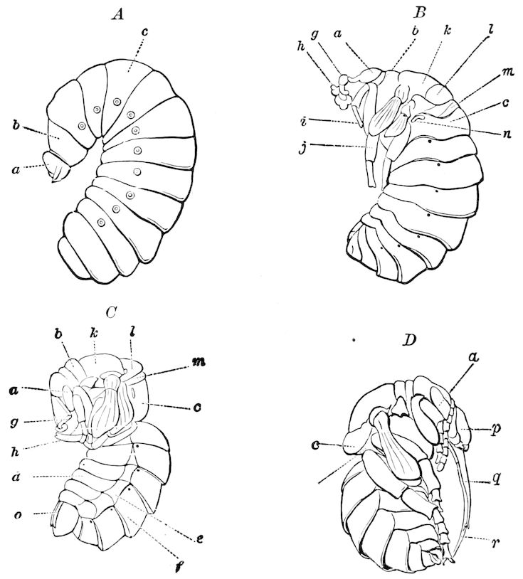
Fig. 616.—Transformation of the bumblebee, Bombus, showing the transfer of the 1st abdominal larval segment (c) to the thorax, forming the propodeum of the pupa (D) and imago: n, spiracle of the propodeum. A, larva; a, head; b, 1st thoracic, c, 1st abdominal, segment. B, semipupa; g, antenna; h, maxillæ; i, 1st, j, 2d leg; k, mesoscutum. l, mesoscutellum; m, metathorax; d, urite (sternite of abdomen); e, pleurite; f, tergite; o, ovipositor; r, lingua; q, maxilla.
In what may be termed the 3d stage (Fig. 616, C), though the distinction is a very arbitrary one, the change is accompanied by a moulting of the skin, and a great advance has been made towards assuming the pupal form. The abdomen is very distinctly separated from the thorax, the propodeum being closely united with the thorax, and the head and thorax taken together are nearly as large as the abdomen, the latter now being shorter and perceptibly changed in form, more like that of the completed pupa, while there are other most important changes in the elaboration of the parts of the thorax, particularly the tergites, and of the head and its appendages. Meanwhile the ovipositor has been completed and nearly withdrawn within the end of the abdomen.
The next to study the transformations of the Hymenoptera was Ganin, who discovered the early remarkable pre-eruciform larvæ, as 663we may call them, of certain egg-parasites (Proctotrypidæ). He discovered the imaginal buds of the wings in the third larva of Polynema (Fig. 185), but his observations, and those of Ayers, need not detain us here, as they have little to do with the subject of the normal metamorphosis of the Hymenoptera, and will be discussed under the subject of Hypermetamorphosis.
To Bugnion we owe the first detailed account of the internal changes in the Hymenoptera, his observations being made on a chalcid parasite, Encyrtus fuscicollis, a parasite of Hyponomeuta. The apodous larva (Fig. 618) moults but once, the next ecdysis being at the time of pupation. It passes through a semipupal stage.
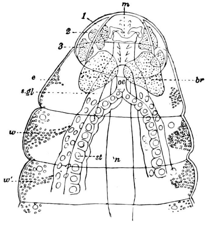
Fig. 617.—Encyrtus larva: 1, 2, 3, ganglia in front of the brain; m, mouth; s. gl, silk-gland; br, brain; n, nervous cord; w, bud of fore, w′, bud of hind, wing.
Bugnion observed in the larva of Encyrtus three pairs of lower thoracic or pedal imaginal buds, two pairs of upper or alary buds, a pair of ocular or oculo-cephalic buds destined to build up all the posterior part of the head, a pair of antennal buds, and three pairs of buds of the genital armature (ovipositor). He also detected the rudiments of the buccal appendages under the form of six small buds (Fig. 619), which do not invaginate, and are not surrounded by a semicircular pad. Also in the abdomen, behind each pair of stigmata, there is a group of hypodermic cells (Fig. 617), which, without doubt, correspond to the wing-buds, but are not differentiated into a central bud and its pad, and does not merit the name of imaginal bud. In fact, except the eye-buds, which are unlike the others, he only observed the imaginal buds of the legs, wings, and ovipositor. The antennal buds are, like those of the buccal appendages, without an annular zone.
The pedal buds were detected in the middle of larval life. They each form a central bud surrounded by a circular thickening. They gradually elongate and become tongue-like and somewhat bent; soon a linear opening or slit appears, forming the mouth of a cavity which communicates with that of the body, allowing the passage into them of the tracheæ, muscles, and nerves, and afterwards of the blood. Finally, the buds grow longer and slenderer, are bent several times, and show traces of the articulations; and soon under the old larval skin, now beginning to rise in anticipation of the moulting, 664we see the coxa, femur, tibia, and tarsus of the perfect insect, the tarsal joints not yet being indicated.
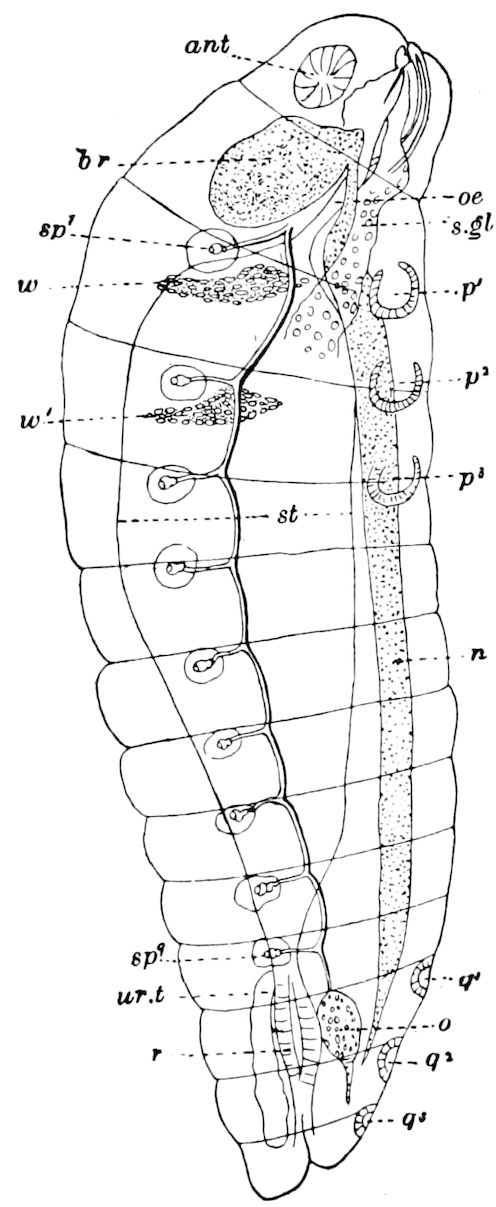
Fig. 618.—Older Encyrtus larva, lateral view, showing the buds of the antennæ (ant), legs, and wings (w, w′): oe, œsophagus; q1, q2, q3, buds of the genital armature; o, rudiment of the sexual gland (ovary or testis); ur.t, urinary tube; st, stomach; i, intestine (rectum); n, ventral nervous cord; r, rectum; sp1-sp9, spiracles.
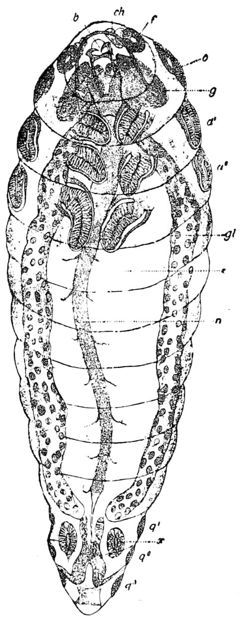
Fig. 619.—A still older larva, ready to transform. The imaginal buds of the antennæ (f), eyes, wings (a1, a2), and legs have become elongated: ch, chitinous arch; b, mouth; o, eye-bud; g, brain; e, stomach; x, rudiment of the sexual glands (either the ovary or testis).—This and Figs. 617 and 618, after Bugnion.
The wing-buds (a1, a2) appear at the same time as those of the legs, as racket-shaped masses of small cells situated directly behind the 1st and 2d pair of stigmata, in contact with the tissue ensheathed by the corresponding tracheal vesicle (Fig. 618). Afterwards they have exactly the form of those of the Lepidoptera (Fig. 619).
The proliferation of the hypodermis is not limited to the thorax, but takes place at corresponding points in the first seven abdominal 665segments. These abdominal agglomerations of cells do not give rise to true buds, but serve simply to reconstitute the hypodermis of the abdominal segments at the time of metamorphosis.
Ocular or oculo-cephalic buds.—The eye of insects, as is well known, is a modification of a portion of the integument, the visual cells being directly derived from the hypodermis, the cornea being a cuticular product of this last, like chitinous formations in general.
The ocular buds appear towards the end of larval life as a simple mass of hypodermic cells, and form a compact layer on the dorsolateral face of the prothoracic segment, and clothe the cephalic ganglion or brain like a skull-cap. The central portion only is destined to form the eye, while the peripheral pad, continuing to thicken, gives rise to a voluminous and rounded mass, which meets on the median line that of the opposite side, and forms the integument of all the posterior part of the head.
Bugnion also observed on the median line a group of small hypodermic cells which he regarded as the rudiment of the anterior ocellus, but he did not detect those of the posterior ocelli.
The antennal buds.—These appear at an early date under the cuticle of the head, as two distinct rounded cellular masses, with a central cavity, but no annular zone (Fig. 619, f). Each one grows longer in a transverse sense, and its summit, extended from the outer side, curves downward. It now forms a hollow tube folded at the end, and terminated by a disk whose centre is perforated (Fig. 619, f). Afterwards, when the larva is ready to transform, it grows longer, becomes folded on itself in its cavity, and, passing beyond on each side the limits of the larval head, encroaches on the prothoracic segment.
The buds of the buccal appendages.—Towards the end of the larval period, the buds of the mouth-parts appear as small digitiform projections, situated on each side and below the mouth. Formed of small epithelial cells pressed against each other, they are all directed anteriorly, and possess no furrow or pad.
The 2d maxillæ (labium) is formed of two separate parts. The imaginal buds of the lower lip appear on each side of the median line, with a fissure indicating the differentiation of the palpus. On each side are to be seen the 1st maxillary buds, bearing each a rudimentary palpus, and, farther in front, the buds of the mandibles.
The buds of the ovipositor.—The six stylets of the ovipositor arise from six small imaginal buds which become visible in the second half of the larval period, on each side of the median line, on the lower face of the three last segments (Fig. 620, q1, q2, q3). 666The bud is differentiated into a central discoidal bud, a furrow, and a marginal, rather thick swelling or pad. Afterwards, these buds elongate and form small papilliform projections directed backwards (Fig. 621); but only during the pupal period do they, as already observed in Bombus, approach each other and assume their definite shape as an ovipositor.
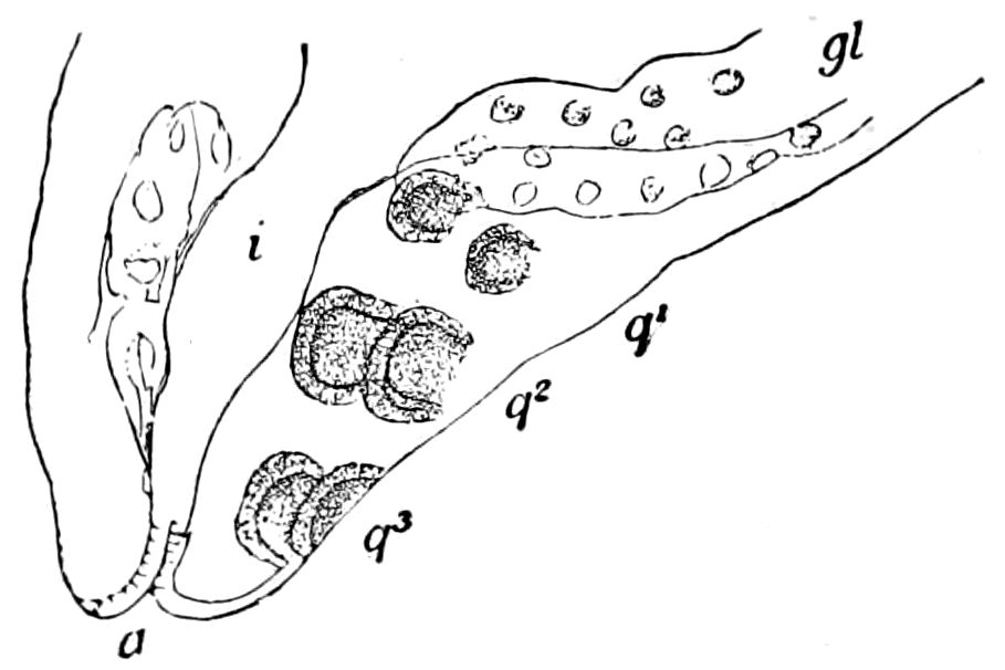
Fig. 620.—End of larva of Encyrtus of 2d stage, showing the three pairs of imaginal buds of the ovipositor q1, q2, q3.
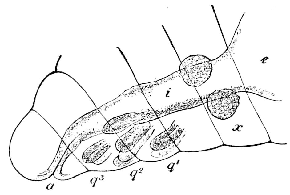
Fig. 621.—The same in an older larva ready to transform: i, intestine; x, genital gland; a, anus.—After Bugnion.
Finally, Bugnion states that while metamorphosis in the Hymenoptera is less highly modified than in the Muscidæ, it is more marked than in the Coleoptera and Lepidoptera. In these orders the pupa moves the abdomen, but in Hymenoptera it is absolutely immovable throughout pupal life, as long as the integument is soft.
DEVELOPMENT OF THE IMAGO IN THE DIPTERA
The flies, particularly the Muscidæ and their allies (Brachycera), are the most highly modified of insects, their larvæ having undergone the greatest amount of reduction and loss of limbs, this atrophy involving even most of the head. The following account has been prepared in part from the works of Weismann, Ganin, Miall, and Pratt, but mostly from the excellent general summarized account given by Korschelt and Heider.
In the holometabolic orders of insects, with their resting pupal stage, during which no food is taken, the entire activity of life 667seems to be turned to deep-seated and complicated internal developmental processes. These inner changes involve an almost complete destruction of many organs of the larva, and their renewal from certain germs (the imaginal buds) already present in the larva, as will be seen in the highly modified Muscidæ. Only a few larval organs become directly transferred into the body of the pupa and imago. Such are the rudiments of the genital system. The heart also, and the central portion of the nervous system, suffer only slight and unimportant, almost trivial, internal changes. On the other hand, most of the other organs of the larva become completely destroyed: the hypodermis, most of the muscles, the entire digestive canal with the salivary glands; while their cells, under the influence of the blood corpuscles (leucocytes), which here act as phagocytes, fall into pieces, which are taken up by them and become digested. Simultaneously with this destructive, histolytic process, the new formation of the organs by the imaginal buds, already indicated in the embryo, is accomplished in such a way that the continuity of the organs in most cases remains unimpaired. This process of transformation can only be understood by considering that of the embryonal germs of the organs, (1) only a part is destined for the use of the larva in growth, and for the performance of certain functions which exhaust themselves during larval life, so that it is no more capable of farther transformation, and finally becomes destroyed; while (2) a second part of the embryonal germs or rudiments persists first in an undeveloped condition, as imaginal buds, in order to undertake during the pupa stage the regeneration of the organs.
Though Swammerdam knew that the rudiments of the wings were already present under the skin of the larvæ, we are indebted, for our present knowledge, to the thorough and profound investigations of Weismann on the metamorphosis of the Diptera, and also to the researches of Ganin and others who have worked on the pupæ of Muscidæ, in which the development is most complicated and modified. In the more generalized and primitive Diptera, such as Corethra, the processes of formation of the pupa and imago are much simpler than in the muscids and Pupipara. These processes are still simpler in the Lepidoptera and Hymenoptera, and for this reason we have given a summary of what has been done on these organs by Newport, Dewitz, and especially by Bugnion.
Our knowledge of this subject is still very imperfect, only the more salient points having been worked out, and, as Korschelt and Heider state, there is still lacking certain proof as to how far the relations of the internal changes known to exist in the Muscidæ also apply to other orders of insects, though it must be considered that in the pupa of Lepidoptera, Hymenoptera, perhaps also the Coleoptera, and we would add in the Neuroptera as well as the male Coccidæ, very similar metamorphic processes take place.
668
a. Development of the outer body-form
The form of the imago is completely marked out in the pupa, so that the transition from the pupa to the imago is comparatively slight and only depends on the modification and development of the parts already present.
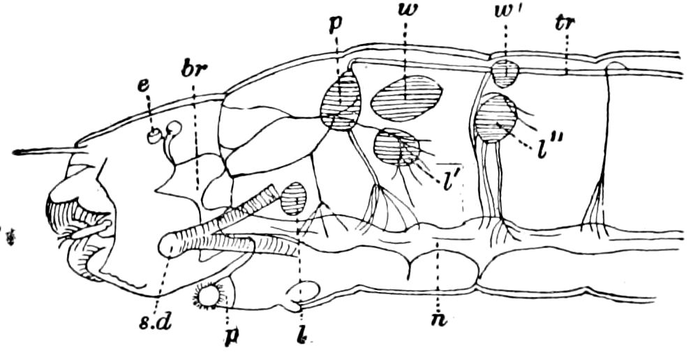
Fig. 622.—Anterior part of young larva of Simulium sericea, showing the thoracic imaginal buds: p, prothoracic bud (only one not embryonic); w, w′, fore and hind wing-buds; l, l′, l″, leg-buds; n, nervous system; br, brain; e, eye; sd, salivary duct; p, prothoracic foot.—After Weismann.
In most cases the modification in question consists of the changes occurring during the passage from the larval form to the imago, the reformation of parts already present being most marked, while the new rudiments only participate in a limited way in the process. Thus, for example, the head of the caterpillar together with the antennæ and mouth-parts, also the thoracic limbs, pass directly and unchanged from the larva into the pupa. The compound eyes and the wings are, however, new formations, the latter arising from imaginal buds. The same is the case with many other Heterometabola, where the passage of the larva into the pupa in general is due to a transformation of parts already present. The changes in the brain, the fusion of certain ganglia of the ventral nervous cord, the changes in the abdomen, involving the reduction in the number of segments and the remodelling of the end of the body, and the formation of the ovipositor or sting, and in the higher Hymenoptera the transfer of the 1st abdominal segment to the thorax, and the origin of the genital armature,—all these should here be taken into account.
It should be observed that in every case where the larvæ are footless, as in Diptera, all the Hymenoptera except the phytophagous ones and certain coleopterous larvæ, the limbs of the imago stage are, in the earliest stages, indicated as new structures in the form of imaginal buds.
Formation of the imago in Corethra.—Corethra may serve as an example of such a relatively simple metamorphosis. Its larva belongs to the group of eucephalous dipterous larvæ. The head of the perfect insect is already indicated in the larva, and its parts, with certain modifications, pass directly into the pupa. The compound 669eyes, and this is a rare exception among the Holometabola, are present in the larva. On the other hand, the thoracic legs, the wings, and halteres are developed out of new rudiments which are present in the last larval stage, before pupation. Each thoracic segment has four of them, two ventral and two dorsal (Fig. 622); the ventral buds becoming the legs. Of the dorsal pairs, that of the mesothorax develops into wings, that of the metathorax into halteres, while from the corresponding rudiments of the prothorax in Corethra arise the stigma-bearing dorsal or respiratory processes of the pupa, and in Simulium a tuft of tracheal gills (Fig. 623, ra; see also Fig. 582).
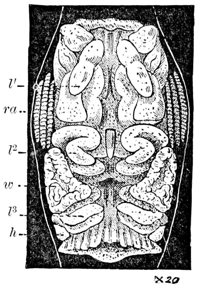
Fig. 623.—Late larva of Simulium, showing the rudiments of the pupal structures within the larval skin: l1, l2, l3, fore, middle, and hind legs of the fly; ra, respiratory appendages of pupa; w, wing of fly; h, halter of fly.—After Miall.
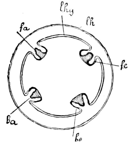
Fig. 624.—Imaginal buds in larva of Corethra (diagrammatic cross-section of thorax): invaginations (fe and be) of the larval hypodermis (lhy) in whose bases the rudiments of wings (fa) and legs (ba) arise; lh, chitinous integument of the larva.—After Lang.
These imaginal buds may be regarded as evaginations of the outer surface of the body. The only difference is that the buds of the appendages as a whole seem sunken below the level of the surface of the body, being situated at the bottom of an evagination, as in the buds of the head and trunk in the Pilidium larva of nemertean worms, and in the rudiments of the lower surface of the body of Echinus present in the pluteus larva.
The lumen of the invagination in which the appendages of Corethra (and other Holometabola) are situated is called by Van Rees the peripodal cavity, and the external sheath bordering it, which is naturally continuous with the hypodermis of the body, the peripodal membrane (Fig. 636, p).
We must adopt the view that the rudiments of the appendages (imaginal buds) are from the first divided into ectodermal and mesodermal portions, which are derived from the corresponding germ-layers of the larva. The ectoderm of the rudiments of the appendages is continuous with the peripodal membrane, and through it with the hypodermis. Weismann was inclined to derive the organs (tracheæ, muscles, etc.) developing within the germs of the appendages from a 670hypertrophy of the neurilemma of a nerve passing down from within into the imaginal bud, and held that nerves and tracheal branches soon after passed into the inner surface of the imaginal bud. (Korschelt and Heider.)
When the imaginal buds of the appendages enlarge, then the peripodal membranes become correspondingly distended, and the limbs within assume a more or less crumpled position, and in Corethra are spirally twisted, while the rudiments of the wings are folded. The completion of the rudimentary limbs is accomplished simply by their passing out of the invagination in which they originated. The limbs thus gradually become free, the peripodal membrane is seen to reach the level of the rest of the hypodermis and become a part of it, and the base of the extremity is no longer situated in a cavity.
The internal organs of Corethra undergo but to a slight degree the destruction (histolysis) which is so thoroughgoing in the Muscidæ. Kowalevsky states that in the mid-intestine of Corethra a histolysis of the larval and reconstruction of the imaginal epithelium goes on in the same way as has been described in Musca. Most of the larval organs pass without histolytic changes directly over into those of the pupal and imaginal stages; the muscles in general are also unchanged, but those of the appendages and wings are made over anew. The last arise, according to Weismann, in the last larval stage from strings of cells which are already present in the embryo.
When we consider how insignificant the internal transformations are during the metamorphosis of the Tipulidæ, of which Corethra serves as an example, we can scarcely doubt that we here have before us conditions which illustrate the passage between an incomplete and a complete metamorphosis. Thus, among other things, should be mentioned the short duration of the pupa stage and the activity of the pupa, as also the early appearance of the germs of the compound eyes, a character which Corethra has in common with the Hemimetabola. (Korschelt and Heider.)
Formation of the imago in Culex.—In respect to the formation of the imaginal head, Culex is still more primitive than Corethra. Miall and Hammond find from Hurst’s partly unpublished descriptions and preparations that there are no deep invaginations for the compound eyes or antennæ of the imago.
“The compound eye forms beneath the larval eye-spots, and is at first relatively simple and composed of few facets. The number increases by the gradual formation of partial and marginal invaginations, each of which forms a new element. The imaginal antenna grows to a much greater length than that of 671the larval antenna, and its base is accordingly telescoped into the head, while the shaft becomes irregularly folded.[115] Culex, though more modified than Chironomus in many respects, e.g. in the mouth-parts, is relatively primitive with respect to the formation of the imaginal head, and shows a mode of development of the eye and antenna which we may suppose to have characterized a remote and comparatively unspecialized progenitor of Chironomus.”
Formation of the imago in Chironomus.—The development of the head of the imago of Chironomus dorsalis has been discussed by Miall and Hammond. The invaginations which give rise to the head of the fly could not be discovered even in a rudimentary state until after the last larval moult.
“Weismann has given reasons for supposing that invaginated imaginal rudiments could not come into existence before the last larval moult in an insect whose life-history resembles that of Corethra or Chironomus. If the epidermis were invaginated in any stage before the ante-pupal one, the new cuticle, moulded closely upon the epidermis, would become invaginated also, and would appear at the next moult with projecting appendages like those of a pupa or imago. This is actually the way in which the wings are developed in some larval insects with incomplete metamorphosis. In Muscidæ the invaginations for the head of the imago have been traced back to the embryo within the egg,[116] but the almost total subsequent separation of the disks from the epidermis renders their development independent of the growth of the larval cuticle and of the moults that probably take place therein.”
The pupal and imaginal cuticles do not follow at all closely the larval skin, but, says Miall, become at particular places folded far into the interior. “The folds which give rise to the head of the fly are two in number and paired. They begin at the larval antenna on each side of the head, and gradually extend further and further backwards. The object of the folds is to provide an extended surface which can be moulded, without pressure from surrounding objects, into the form of the future head. On one part of each fold the facets of the large compound eyes are developed; another part gives rise to the future antenna, a large and elaborate organ, which springs from the bottom of the fold, and whose tip just enters the very short antenna of the larva. The folds for the head ultimately become so large that the larval head cannot contain them, and they extend far into the prothorax. Here a difficulty occurs. If the generating cuticle of the prothorax were also to be folded inwards, the future prothorax would take a corresponding shape. But the prothorax of the fly has a form dictated to it by the limbs which it bears and by the muscles to which it gives attachment. These call for a great reduction in its length, and a peculiar shape, which it is 672not here necessary to describe. It will be enough to realize that the epidermis of the future prothorax cannot be sacrificed to the folds which are to give rise to the head of the fly. All interference between the two developing structures is obviated by the provision of a transverse fold, which pushes into the prothorax from the neck, and forms a sort of internal pocket. The floor of the pocket forms two longitudinal folds, which prolong the folds originating in the larval head. The roof of the pocket shrinks up and forms the connection between the head and thorax of the fly. Ultimately the head-part is drawn out, leaving the prothoracic structures unaffected.”[117]
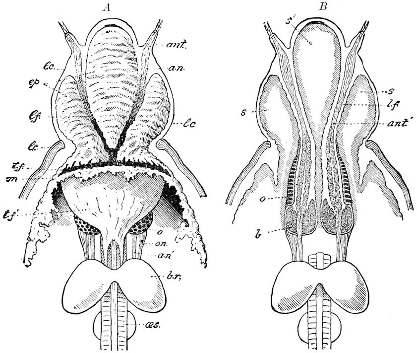
Fig. 625.—Process of formation of the parts of the head of the fly in the larva of Chironomus (male): A, the new epidermis thrown into complicated folds which have been cut away in places to show the parts within. B, the same parts in horizontal section; lc, larval cuticle; tf, transverse fold; tf′, upper wall of the same; m, cut edge of new epidermis; ant, larval antenna; an, nerve to the same; ant′, antenna of fly; lf, longitudinal fold; o, eye of fly; on, optic nerve; an′, root of antennary nerve; br, brain; œs, œsophagus; b, bulb of antenna of fly; s, s, s′, blood-spaces.—After Miall.
The development of the head of the fly of Chironomus appears, as Miall and Hammond state, to be intermediate between the groups Adiscota and Discota of Weismann; i.e. “between the types in 673which the parts of the head of the fly are developed in close relation to those of the larva, and the types in which deep invaginations lead apparently to the formation of similar new parts far within the body, the seeming independence of the new parts being intensified by thoroughgoing histolysis,” and they suggest that possibly types may be discovered intermediate between Chironomus and Muscidæ.
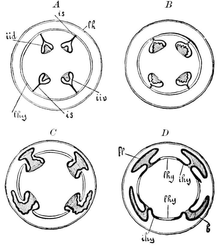
Fig. 626.—A, B, C, D, diagrams of transverse sections showing the development of the wings, legs, and the imaginal hypodermis of muscid flies from the imaginal buds of the larva during metamorphosis: lh, chitinous integument of larva from which the underlying hypodermis (lhy) has withdrawn; iid, imaginal buds of wings, iiv, of legs; is, the cords connecting them with the hypodermis; fl, wing-germs; b, leg-germs; ihy, imaginal hypodermis spreading out in D from the imaginal buds. The imaginal rudiments of the hypodermis are indicated by thick, black outlines, the larval hypodermis by two thin, parallel lines.—After Lang.
We are now prepared to consider the extremely complicated changes, in the Muscidæ, leaving out of consideration the origin of the wings from imaginal buds, which has already been discussed on pp. 126–137.
Formation of the imago in Muscidæ.[118]—In the flesh, and undoubtedly the house, and allied flies the germs or imaginal buds of the legs and wings arise in the same way as in Corethra. But in the Muscidæ, the buds are situated far within the interior of the body, the peripodal cavities appear closed, and the peripodal membrane stands in connection with the hypodermis merely by means of a delicate thread-like stalk. This connecting cord, which was first detected by Dewitz, and whose interpretation was entirely right, shows in its interior, as Van Rees proved, a narrow cavity.
Though the earliest stages in the development of imaginal buds in the embryo of the Muscidæ are still unknown, yet we shall not go far astray if we refer them, like the imaginal buds of Corethra, to hypodermal invaginations. We must, then, regard the stalk-like 674connection just mentioned as the long drawn-out neck of this invagination.
In general, the development of the appendages (Figs. 626, 627) goes on as described in Corethra. The rudiments of the legs enlarge and show at an early date the first traces of the later joints. They are so packed in the peripodal cavities that the single joints of the extremities appear as if pushed in “like the joints of a travelling cup.” (Van Rees.) The evagination of the completely formed buds of the limbs, which occurs on the first day after the beginning of pupation, goes on in such a way that the stalk of the imaginal bud (Figs. 626, B; 627, B) shortens, while its cavity widens so that the limbs finally, as in Corethra, pass out through the widely opened mouth of the peripodal invagination, which at the same time gradually completely disappears. The peripodal membrane is converted into a thickened part of the hypodermis in the region adjoining the base of the leg, and from this thickened hypodermal portion, the formation of the hypodermis of the entire imaginal thorax goes on, as the larval hypodermis is gradually destroyed.
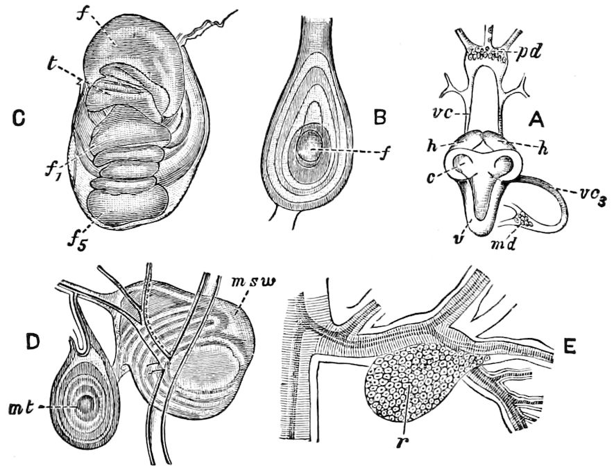
Fig. 627.—Imaginal buds of muscid flies in process of development: A, brain (c) and ventral ganglion (v) of a larva, 7 mm. long, of C. vomitoria; h, head-rudiment; rc, portion of ventral cord; pd, prothoracic rudiment; vc3, third nerve; md, mesothoracic rudiment. B, mesothoracic rudiment more advanced, in a pupa, just formed, of Sarcophaga carnaria, showing the base of the sternum and folds of the forming leg, the central part (f) representing the foot. C, the rudimentary leg of the same more advanced; f, femur; t, tibia; f1, f5, tarsal joints. D, two buds from a larva, 20 mm. long, of Sarcophaga, attached to tracheæ; msw, mesonotal and wing-germ; mt, metathoracic rudiment. E, r, mesothoracic germ of a 7 mm. long larva attached to a tracheal twig.—After Weismann and Graber, from Sharp.
We must here settle the question as to the first origin of the mesodermal portions of the rudiments of the appendages. We can already distinguish in the imaginal buds of the fully grown muscid larva a clear separation between an ectodermal and an inner mesodermal part. Ganin derived the mesodermal 675part through a sort of differentiation and separation of the innermost layer of the ectodermal part, and Van Rees has, in general, confirmed this view. Kowalevsky, on the other hand, is inclined to the view that the mesodermal part of the imaginal bud is derived from the embryonal cells of the mesoderm. He finds scattered throughout the mesoderm, under the hypodermis of the larva, so-called wandering cells (Fig. 632, A, w), which are different in appearance from the leucocytes and from the elements from which the formation of the mesodermal parts of the imaginal rudiments proceed. Kowalevsky is inclined to believe that there are in each segment rudiments of the imaginal mesoderm, but which are so delicate and indifferent that we cannot find them in the first stages of their origin. From these mesodermal imaginal rudiments the above-mentioned wandering cells of the mesoderm are derived, which afterwards come into connection with the ectodermal portion of the imaginal buds.
Still more complicated and difficult to understand is the development of the head-section of muscids. We must remember that in muscid larvæ the head-section exists in its most rudimentary form, being the result of extreme modification and degeneration. The small size of the head is also due to the fact that it is more or less retracted within the thoracic region. Then, as shown by the researches of Weismann, in the last embryonal stages, the forehead, mandibles, and the whole region of the head around the mouth invaginate and form a sunken cavity (Fig. 628, p), in which the chitinous supports of the hooks characteristic of muscid larvæ are soon developed. This sunken part of the head, at whose inner end is the œsophagus, is called by the not entirely appropriate name of “pharynx,” and it must at present be remembered that the hollow space thus named is not a part of the digestive canal. It is an invaginated section of the head, and the formation of the head of the imago mainly depends on the evagination of this region.
The first rudiments of the most important parts of the head (eyes, antennæ, and forehead), occur in the youngest larvæ as paired masses of cells which lie in the thorax next to the two halves of the brain (for this reason called by Weismann “brain-appendages”), which are from their first origin connected with the pharynx, and may be regarded as the imaginal buds of the head. These appear very soon in later stages in the shape of elongated sacs widening at the hinder end (Fig. 628, A and B, h), which from their origin are to be regarded as evaginations of the pharynx. Very soon epithelial thickenings appear in the wall of this sac-shaped brain-appendage, in which the rudiments of the parts of the future head may be recognized.
Disk-shaped thickenings in the hinder widened part of the brain-appendage form the rudiments of the compound eyes, which therefore may be called the eye-buds. On the basal surface of the 676eye-buds is situated a nervous expansion which is connected by a nerve with the supraœsophageal ganglion. This nerve becomes the optic nerve of the perfect animal, while the optic ganglion is clearly separated from the brain.
In the anterior, more cylindrical or tube-like part of the brain-appendage we find the “frontal buds” (ss), on which the antennal rudiments (at) soon bud out, in exactly the same way as the rudiments of the limbs arise from the imaginal buds.
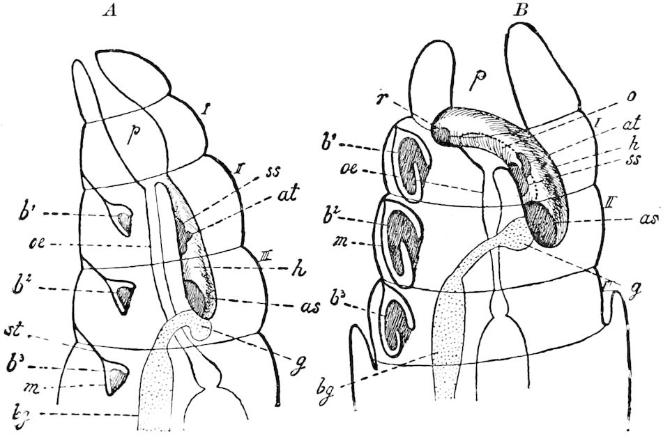
Fig. 628.—Diagrammatic representation of the position of the imaginal buds in the larva (A) and pupa (B) of Musca (the wing rudiments omitted): as, eye-buds; at, antennal germs; b1-b3, germs of the legs; bg, central ganglionic cord: g, brain; h, so-called frontal appendage (Hirnanhang): m, peripodal membrane; o, opening of the frontal appendage into the pharynx; oe, œsophagus; p, so-called “pharynx”; r, rudiment of the proboscis; ss, frontal bud; st, stalk-like connection of the peripodal membrane with the hypodermis; I-III, 1st, 2d, and 3d thoracic segments.—Adapted from Van Rees, by Korschelt and Heider.
Originally (Fig. 628, A) the brain-appendages lie tolerably far behind in the thorax of the larva, so that they connect the hindermost part of the wall of the pharynx with the foremost section of the brain, which they surround in the form of a mushroom. Afterwards, however, subsequent to pupation, they move, together with the central nervous system, farther forward (B), whereby they (if we have correctly understood the descriptions of Weismann and Van Rees) laterally surround the pharynx with their anterior end, which is somewhat ventrally bent. At the same time, there becomes established a gradually widening communication (B, o) between the brain-appendage and the pharynx, which soon extends in the form of a lateral pharyngeal fissure along the entire length of the brain-appendage. As a result, the cavity of the brain-appendage 677and the pharynx so completely unite that the two soon form a single sac, the head-sac or vesicle (Fig. 6–9, k). The walls of this head-vesicle are the later head-wall, the most important parts of which can now be recognized (the antennæ, eyes, rudiments of the beak). It is now necessary that the head-vesicle (Fig. 629, +, +) be, by the eversion of the pharynx, turned outward in order that the head of the pupa may be completed. By this eversion of invaginated parts, the former mouth-opening of the pharynx becomes a neck-section (Fig. 629, +, +) by which head and thorax are now united. (Korschelt and Heider.)
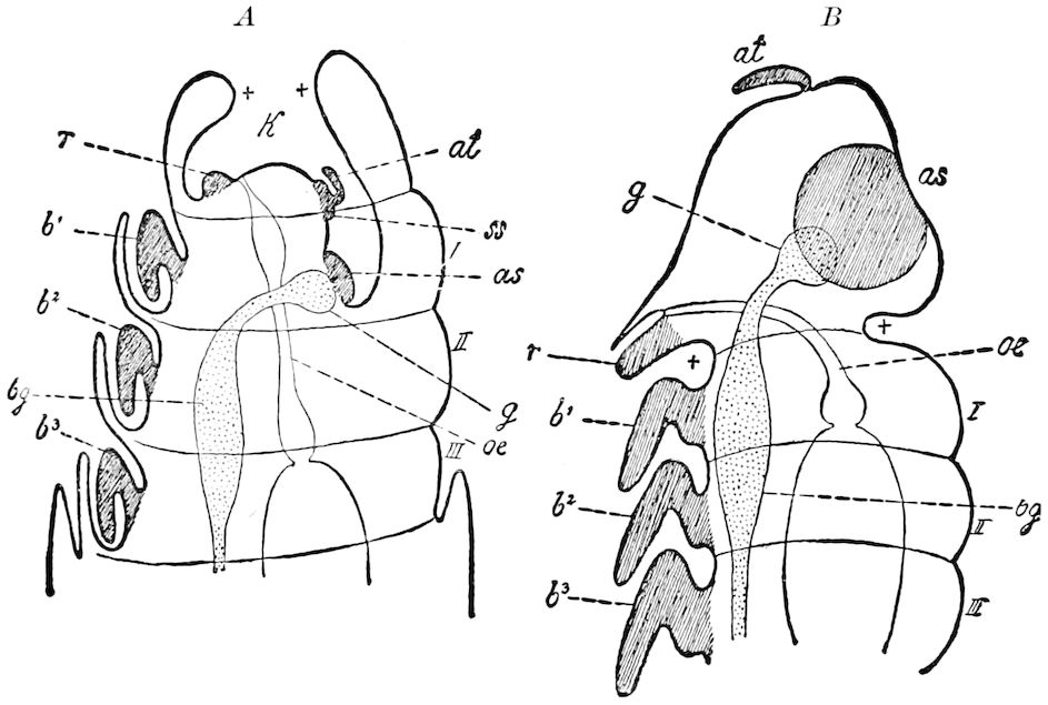
Fig. 629.—Diagram of the changes to pupa of Musca before imago appears; the wing-germs not drawn: as, eye-buds; at, antennal germs; b1-b3, leg-germs; bg, ventral nerve-cord; g, brain; k, head-vesicle (originating from the union of the pharynx with the hypophysis, Hirnanhängen); oe, œsophagus; r, germ of the proboscis; ss, germ of the forehead; I, II, III, 1st, 2d, and 3d thoracic segments.—Based on Kowalevsky and Van Rees, with changes, after Korschelt and Heider.
The cause of the eversion of the head-vesicle, which Weismann directly observed, appears to be due to an increase of the inner pressure through a contraction of the hinder parts of the body. The anterior end of the œsophagus now becomes turned down ventrally corresponding to the conformation of the head of the imago.
It has been shown that the so-called pharynx is only an invaginated part of the outer surface of the larval head. The brain-appendage Korschelt and Heider consider to be the diverticulum of this invagination, in which the single parts of the body lie in an invaginated state. They may throughout be compared to the rudiments of the thoracic limbs. All these imaginal buds have been traced back to the invaginated parts of the outer surface of the body, i.e. the ectoderm.
678It should be borne in mind that the process of development of the head of the highly-modified Muscidæ is much more complex than in the more primitive Diptera.
In their essay on the development of the head of the imago of Chironomus, Miall and Hammond arrange the dipterous types thus far examined, in the order of complexity of the invaginations which give rise to the head of the imago, in the following order:—
1. Culex. Relatively simple. Invaginations of the imaginal buds, shallow. 2. Corethra, Simulium. } Intermediate. 3. Chironomus, Ceratopogon. } 4. Muscidæ. Relatively complex. Invaginations deep, and apparently, but not really, unconnected with the epidermis.
b. Development of the internal organs of the imago
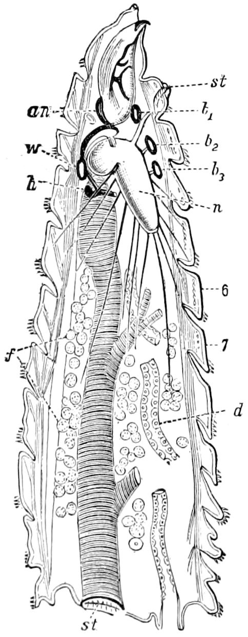
Fig. 630.—Median longitudinal section through larva of blow-fly during the process of histolysis: an, antenna; between an and w, rudiments of eye; w wings; h, halteres; b1-b3, legs; f, fat-body; d, middle of intestine; n, ganglia; st, stigma; 6, 7, 6th and 7th body-segments.—After Graber, from Sharp.
It has already been observed that most of the organs of muscid larvæ (and this applies to most Diptera, Lepidoptera, Coleoptera, and Hymenoptera) are destroyed through the action of leucocytes, and that their reformation is accomplished by definite groups of embryonal cells, the imaginal buds or folds. Destruction and rebuilding occur during the pupa stage in such a way that in many cases while this process is going on the continuity of the organs does not seem to be disturbed. These transformations especially concern the hypodermis, the digestive canal, the muscles, the fat-body, and the salivary glands.
The transformation of the tracheal system is only partial, being in part a simple process of regeneration through cell-division. Slighter changes affect the heart, the central nervous system, and the reproductive system (Fig. 630).