PART I.—MORPHOLOGY AND PHYSIOLOGY (continued 9)
Reading edition
7. There is evidence that in at least one case (Melolontha), the tetranephric is ontogenetically derived from the hexanephric condition by the suppression of one pair of tubules.
3568. It is probable that the insects which never develop more than four Malpighian vessels have lost a pair during their phylogeny.
9. The post-embryonic increase in the number of Malpighian vessels in some orders (Orthoptera, Odonata, Hymenoptera) is secondary and has apparently arisen to supply a demand for greater excreting surface.[56]
LITERATURE ON THE EXCRETORY (URINARY) ORGANS
Malpighi, M. Dissertatio epistolica de Bombyce, Societati regiæ Londini ad scientiam naturalem promovendam institutæ dicata. (Londini, 1669, 12 Pls.)
Herold, M. J. D. Entwicklungeschichte der Schmetterlinge. 1815.
Rengger, J. R. Physiologische Untersuchungen über den tierischen Haushalt der Insekten. Tübingen, 1817, pp. 82.
Wurzer. Chemische Untersuchungen des Stoffes in den Gallgefässen von Bombyx mori. (Meckel’s Archiv f. Physiol., iv, 1818, pp. 213–215.)
Gaede, H. M. Physiologische Bemerkungen über die sogenannten Gallgefässe der Insekten. (Nova Acta Acad. Caes. Leopold.-Carolin., 1821, x, Pars II, pp. 186–196.)
Meckel, J. F. Ueber die Gallen- und Harnorgane der Insekten. (Meckel’s Archiv, i, 1826, pp. 21–36.)
Audouin, J. V. Calculs trouvés dans les canaux biliaires d’un cerf volant. (Ann. sc. nat., 2 Sér., 1836, v, pp. 129–137.)
Frey und Leuckart. Anatomie und Physiologie der Wirbellosen. 1843.
Dufour, L. Mémoire sur les vaisseaux biliaires ou le foie des Insectes. (Ann. sc. nat., 1848, Sér. 2, xix, pp. 145–182, 4 Pls.)
Karsten, H. Harnorgane von Brachinus complanatus. (Müller’s Archiv f. Anat. und Physiol., 1848, pp. 367–374.)
Fabre, J. L. Étude sur l’instinct et les metamorphoses des Sphégiens. (Ann. d. sc. nat., 4 Sér., 1856, vi, pp. 137–189.)
—— Étude sur le rôle du tissu adipeux dans la sécrétion urinaire chez les Insectes. (Ibid., 4 Sér., xix, pp. 351–382.)
Schlossberger, J. E. Untersuchungen über das chemische Verhalten der Krystalle in den Malpighischen Gefässen der Raupen. (Archiv f. Anat. und Physiol., 1857, pp. 61–62.)
Leydig, F. Lehrbuch der Histiologie. 1857.
Sirodot, S. Recherches sur les sécrétions chez les Insectes. (Ann. sc. nat., 4 Sér., Zool., 1858, x, pp. 141–189, 251–334, 12 Pls.)
Kölliker, A. Zur feineren Anatomie der Insekten (Ueber die Harnorgane, u.s.w.) (Verhandl. d. Physikal.-medizin. Gesellsch. in Würzburg, viii, 1858, pp. 225–235.)
Schindler, E. Beitrage zur Kenntnis der Malpighischen Gefässe der Insekten. 3 Taf. (Zeitschr. f. wiss. Zool., xxx, 1878, pp. 587–660.)
Chatin, G. Note sur la structure du noyau dans les cellules marginales des tubes de Malpighi chez les Insectes et les Myriapodes. (Ann. d. sc. nat., 6 Sér., xiv., 1882, pp. 7, 1 Pl.)
Witlaczil, E. Zur Anatomie der Aphiden. (Arbeiten a. d. Zool. Instit. d. Univers. Wien., iv, 1882, pp. 397–441, 3 Taf.)
357Cholodkowsky, N. Sur les vaisseaux de Malpighi chez les Lépidoptères. (Compt. rend. Acad. d. Sc., Paris, xcix, 1884, pp. 631–633.)
—— Sur la morphologie de l’appareil urinaire des Lépidoptères. (Archives de Biologie, 1887, vi, pp. 497–514, 1 Pl.)
Loman, J. C. C. Ueber die morphologische Bedeutung der sogenannten Malpighischen Gefässe der echten Spinnen. (Tijdschr. Nederl. Dierk. Ver. (2) Deel 1, 1887, pp. 109–113, 4 Fig.)
Marchal, P. Contribution à l’étude de la désassimilation de l’azote. L’acide urique et la fonction rénale chez les Invertébrés. (Mém. Soc. Zool. de France, 1889, iii, pp. 42–57.)
Kowalevsky, A. O. Ein Beitrag zur Kenntnis der Exkretionsorgane. (Biol. Centralbl., ix, 1889–90, pp. 33–47, 65–76, 127–128.)
—— Sur les organes excréteurs chez les arthropodes terrestres. (Congrès international de Zool., 2me Session à Moscou, 1892, Pt. I, pp. 186–235, 4 Pls.)
Griffiths, A. B. On the Malpighian tubules of Libellula depressa. (Proc. Roy. Soc., Edinburgh, 1889, xv, pp. 401–403, Figs.)
Grandis, V. Sulle modificazioni degli epitelii ghiandolari durante la secrezione. (Atti Accad. Torino, 1890, xxv, pp. 765–789, 1 Pl.; Archiv Ital. Biol., 1890, xiv, pp. 160–182, 1 Pl.)
Koulaguine, N. Notice pour servire à l’histoire du développement des hyménoptères parasites. (Congrès internat. de Zool., 2me Session à Moscou, 1892, Pt. I, pp. 253–277.)
Sograff, Nicolas. Note sur l’origine et les parentés des Arthropodes, principalement des Arthropodes trachéates. (Congrès internat. de Zool., 2me Session à Moscou, 1892, Pt. I, pp. 278–302.)
Giard, Alfred. (Note on the urinary tubes of larval Cecidomyia. Annals Ent. Soc., France, lxii, 1893, pp. lxxx-lxxxiv, 1 Fig.)
Wheeler, William M. The primitive number of Malpighian vessels in insects. (Psyche, vi, May-December, 1893, Parts 1–6, pp. 457–460, 485–486, 497–498, 509–510, 539–541, 545–547, 561–564.)
Metalnikoff, C. K. Organes excréteurs des insectes. (Bull. Acad. imp. Sci. St. Pétersbourg, 1896, iv, pp. 57–72, in Russian, 1 Pl.)
See also the works of Straus-Dürckheim, Will (Müller’s Archiv. 1848, p. 502), Brugnatelli, Leidy, Dufour, Ramdohr, Basch, Davy, Grassi, Minot, Berlese, Adlerz, Marchal (Bull. Ent. Soc. France, 1896, p. 257); Bordas (Appareil glandulaire des Hyménoptères, 1894), also C. R. Acad. Sc. Paris, 1897.
e. Poison-glands
Poison-glands are mainly confined to the stinging Hymenoptera, i.e. certain ants, and the wasps and bees, but also occur in the mosquito, while many, if not most bugs, seem to instil a drop of poison into the punctured wounds they make.
In the honey and other bees the poison apparatus consists of two poison-glands whose secretion passes by a single more or less convoluted efferential duct into the large poison-sac, and thence by the excretory duct, which is enlarged at the base of the sting (Figs. 194, 195), out through the sting by the same passage as the eggs. According to Carlet, the poison apparatus of bees consists of two kinds of glandular organs, of which one kind secretes a feebly 358alkaline fluid, the other an acid product. The poison is only effective when both fluids are mixed. The resultant venom is always acid. The action of this venom upon some animals, as rabbits, frogs, and certain beetles, is slight; but the domestic fly and the flesh-fly are immediately killed by it. The inoculation of a fly with the secretion of one of the glands does not produce death until after a considerable time, but death follows very quickly if the same fly is subjected to a second inoculation, this time with the secretion of the other gland. The alkaline glands are in bees and all poisonous Hymenoptera strongly developed, but become vestigial in those forms which sting their prey to serve as food for their larvæ. The poison which the solitary sand and wood wasps and Pompilidæ inject into their victims only paralyzes them.
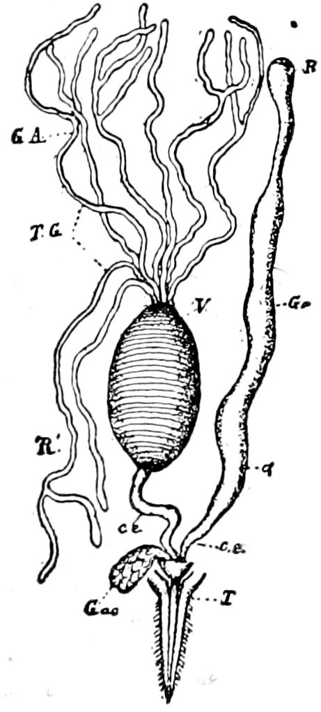
Fig. 348.—The poison apparatus of Ichneumon: T, sting; GA acid gland; TG, R′, its tubes opening into the common poison-sac or reservoir; ce, its efferent canal; Ga, the tubular alkaline gland; R, the glandular end; a, the reservoir; ce, its duct; Gac, the accessory gland—After Bordas.
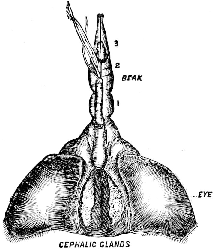
Fig. 349.—Cephalic gland of Belostoma.
Bordas has found both the alkaline gland (gland of Dufour) and the acid gland to occur in a hundred species of Hymenoptera, including not only Aculeata, but also Ichneumonidæ (Fig. 348), Tenthredinidæ, and they may be safely said to be of general occurrence. The acid gland consists of three parts, the glandular portion, the reservoir for the poison, and the secretory canal. The alkaline gland is an irregular tube, with a striated surface and without a reservoir. In most Hymenoptera there is still a third gland, which is unpaired, granular, rectangular or lanceolate, with a short filamentous duct which opens beside the orifice of the alkaline glands.
The poison in ants, wasps, and bees consists of two substances, i.e. formic acid and a whitish, fatty, bitter residue in the secretion 359of the glands; the corroding active formic acid is the essential part of the poison. (Will.)
In Melipona the sting and poison-glands are aborted; in certain ants (Formica, Lasius, etc.) the sting is wanting, but the poison-sac is extraordinarily large.
Bordas finds in various species of Ichneumon three kinds of glands opening into the base of the sting. The first two correspond to the acid (Fig. 348, G.A) and alkaline (G.A) glands of bees and wasps (Vespidæ, etc.), and the third (G.ac) is situated between the two lateral muscular bundles which attach the base of the sting to the last abdominal segment. The poison-reservoir (Fig. 348, V) is recognized by its yellow color and diaphanous and striated appearance. It is situated on the left of the hind-intestine, a little in front of the rectum. The tubular gland (Ga) or alkaline gland of aculeate Hymenoptera is remarkably large; it is situated on the left side of the body. The accessory gland (G.A) is elongated, triangular, flat, its duct opening at the base of the alkaline gland; it is formed of small spherical cells. Bordas has met with well-developed poison-glands in forty species belonging to the Terebrantia, including that of Tenthredo, Emphytus, as well as various genera of Ichneumonidæ, but in all these species the accessory gland was wanting.
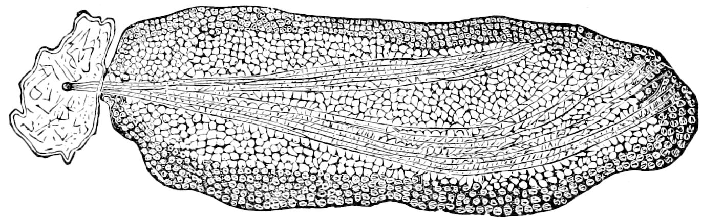
Fig. 350.—View from above of the cephalic gland of Belostoma, × 20.—This and Fig. 349 after Locy.
Under the name of cephalic glands (Fig. 349), Locy describes a pair of glands in the head of Nepidæ. The epithelial or secreting cells are 8–sided (Fig. 350). “When these insects are irritated,” he says, “a secretion is freely thrown out around the base of the beak, which produces death very quickly when introduced on a needle point into the body of an insect.” He infers that the cephalic glands may be the source of this poisonous secretion. The poisonous salivary fluid of the larva of Dyticus is referred to on p. 324.
That the mosquito injects poison into the wound it makes has been proved by Macloskie, who discovering fine droplets of a yellow oily-looking fluid escaping from the end of the hypopharynx, afterwards detected the poison-glands. It appears that the two salivary glands are subdivided, each into three lobes, the middle of which (Fig. 351, pg) differs from the others in having evenly granulated contents and staining more deeply than the others. Having examined the preparations, we agree with the discoverer that these lobes 360secrete the poison. The poison is diluted by the secretion of the salivary lobes, and the two efferent ducts, one from each set of glands, “carry forward and commingle the venomo-salivary products in the main duct; and the stream is then carried by the main duct to the reservoir at the base of the hypopharynx.”
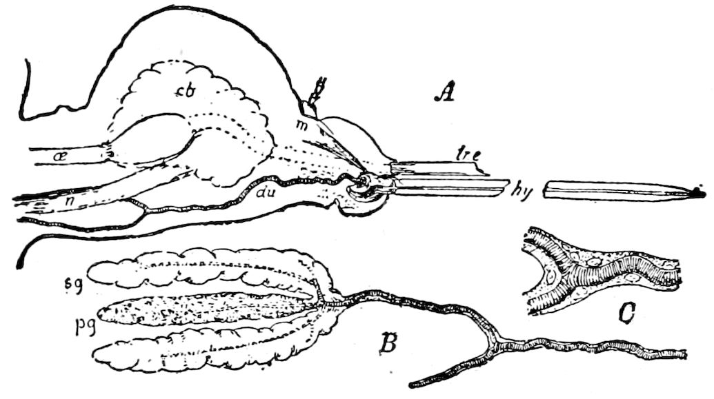
Fig. 351.—A, median section of head, showing (du) the venomo-salivary duct, with its insertion in (hy) the hypopharynx; cb, brain; below is the pharyngeal pump, leading from (œ) the œsophagus; lre, base of labrum-epipharynx; m, muscle; n, commissure (other parts removed). B, the venomo-salivary duct, showing its bifurcation, and the three glands on one of its branches; pg, poison gland; sg, the upper of the two salivary glands. C, the bifurcation of the duct, with its nucleated hypodermis.—After Macloskie.
f. Adhesive or cement-glands
Dewitz has discovered in ants and bees, in close connection with the poison-glands, and like them discharging their secretion through the sting, cement-glands. They arise by budding at the base of the poison-glands.
The two glands in these Hymenoptera correspond to the tubular glands of the Orthoptera, which open at the base of the inner sheath of the ovipositor (Fig. 299, sb), so that the secretion flows out through it as the poison of bees, etc., out of the sting. The use of the secretion of these glands is either to glue the eggs together, or to afford material for the egg-case of cockroaches and Mantidæ and the gummy egg-case of the locusts, etc. The contents of the cement-glands serves for the fixture of the eggs after deposition. In the stinging Hymenoptera one of the cement-glands is an accessory gland; the other becomes the poison-sac. The cement-glands are in the Hemiptera only short blind sacs, in the Lepidoptera and Diptera long convoluted tubes, tubular and branched in the Coleoptera, or richly branched in the Ichneumonidæ and Tenthredinidæ. In the cockroach there are two cement-glands, but the right one is probably of no functional importance. The left one is filled with a milky 361substance, containing many crystals and a coagulable fluid, out of which the egg-capsule (oötheca) is formed. (Miall and Denny.) In the locusts the sebific or cement-gland (Fig. 298, sb) secretes a copious supply of a sticky fluid, which is poured out as the eggs pass out of the oviduct and agglutinates the eggs into a mass, forming a thin coating around each egg, which from the mutual pressure of the eggs causes the tough coating to be pitted hexagonally. In other insects also (Trichoptera, Chrysopidæ, Lepidoptera, etc.) there are similar secretions for the protection or fastening of the eggs when laid.[57] The Trichoptera lay their eggs either in or on the surface of the water in bunches or in strings or in annular gelatinous masses on stones or on plants. This jelly-like substance is secreted by two highly developed paired anal glands. (Weltner, in Kolbe, p. 621.) Also in certain dragon-flies (Libellula, Diplax, and Epitheca) the eggs are laid in jelly-like masses.
With a similar secretion, spun from the end of the abdomen, the Psocidæ cover their little bunches of eggs laid on the under side of leaves; and the silk thread forming the egg-sac of the great water-beetle (Hydrophilus) is secreted from such anal glands.
g. The wax-glands
Besides the honey-bee, which secretes wax in little scales on the under side of the abdomen, the bodies of many other insects, such as the plant and bark lice, as well as the Psyllidæ, Cicadidæ (especially Flata and Lystra), are covered with a waxy powder, or as in Chermes, Schizoneura, Flata, etc., with wool-like filaments of wax.
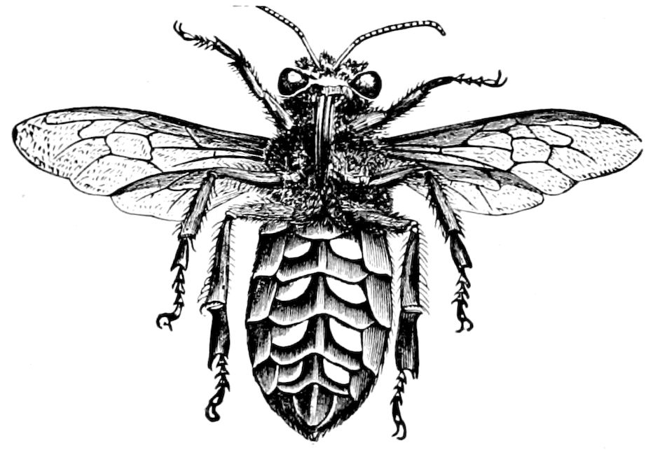
Fig. 352.—Under side of worker honey-bee, carrying wax scales, × 3.—After Cheshire.
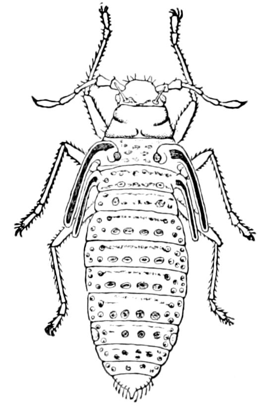
Fig. 353.—Nymph of Lachnus, showing position of wax-glands.—Gissler del.
362The wax is secreted by minute unicellular dermal glands, which in the lower insects (Hemiptera) are distributed nearly all over the body, but in the bees are restricted either to the under (Apis, Fig. 352) or upper side (Trigona) of the end of the abdomen.
The wax-glands of Pemphigus, Chermes, etc., lie under the little warts, seen in Lachnus strobi, the white-pine aphis, to be distributed in transverse lines across the back and sides of the abdominal segments (Fig. 353). These warts are surrounded by a chitinous ring, and divided into delicately marked areas. Through the delicate numerous pits in the chitinous membrane of these areas the little waxen threads project, since under each area ends a duct leading from a large glandular cell, which is a specially modified hypodermis cell (Claus). The wax threads are hollow, and all those arising from a single glued cell form a bundle, whose threads separate from each other and form a white woolly down or bloom covering the body. Witlaczil also shows that gall-forming Aphids secrete a wax-like substance, which, during the movements of the insects in the gall, is rubbed off, becoming a watery layer mixed with the fluid excrement, which forms a spherical impervious layer lining the gall, and thus rendering possible the mode of life of the gall-lice.
In the Psyllidæ Witlaczil has discovered wax-glands which also secrete slender waxen threads. They are situated in groups of two or three at the end of the abdomen near the anus, and arise from hypodermis cells. The wax threads surround the liquid excrement as it passes out of the vent, covering it with a continuous layer of wax. The excrement accordingly is discharged very slowly and gradually, in sausage-shaped masses slightly strung together and rolled into close spirals. The body becomes unavoidably smeared with the sticky excrement, since it is not entirely covered by the waxy layer. Moreover, in the larvæ of many Psyllidæ waxen threads are formed on the upper side of the abdomen; they are for the most part tightly curled or frizzly, like wool, and form, though partly torn, a waxen coat, chiefly on the side and back of the thorax and abdomen. The insects appear therefore as if covered with dust. The mature animals of many species are also covered with a waxen down. The wax threads rapidly dissolve and disappear in alcohol. From a wax-like substance more or less easily dissolved in alcohol arise peculiar hair-like structures which, in the larvæ of Psyllidæ, are situated on the side and end of the body and also on the rudiments of the wings. They are readily distinguished from ordinary hairs, as they arise from glandular cells, and are of very different lengths, more or less like bristles, but hollow, and very brittle. 363They are leaf-like in the first nymphal stages of Trioza rhamni, but in following stages become narrow and form a row around the entire periphery of the body.
The waxen dorsal shield which protects the body of bark-lice (Coccidæ) is a similar product.
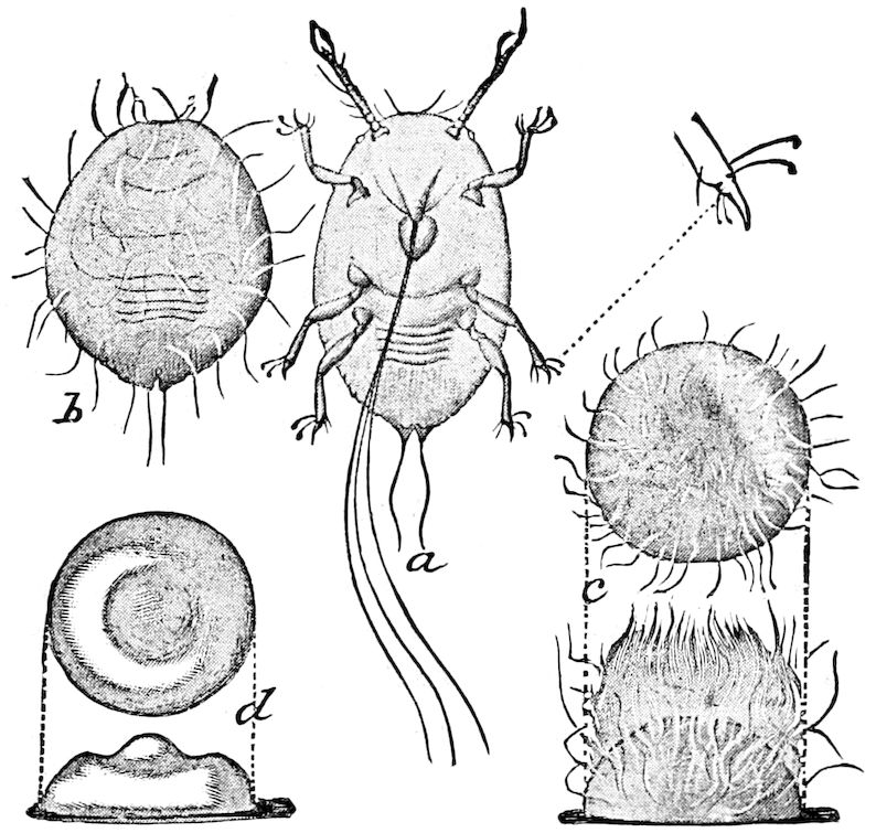
Fig. 354.—Young nymph and developing scale of Aspidiotus perniciosus: a, ventral view of nymph, showing sucking beak with setæ separated, with enlarged tarsal claw at right; b, dorsal view of same, somewhat contracted, with the first waxy filaments appearing; c, dorsal and lateral views of same, still more contracted, illustrating further development of wax secretion; d, later stage of same, dorsal and lateral views, showing matting of wax secretions and first form of young scale; all greatly enlarged.—After Howard and Marlatt, Bull. 3, N. S., Div. Ent., U. S. Dept. of Agr.
Witlaczil has described the way it is formed in Aspidiotus and Leucaspis. The freshly hatched nymph shows no signs of a waxy secretion. But eventually waxen threads arise first on the hinder and anterior end of the body, and then over the whole surface. These threads interlace into a sort of felting and thus form the shield, which is usually much larger than the body and lies closely upon it. The shield is formed after the first moult. It is noteworthy that these threads are matted together to form as thick a tissue as that of the shield itself. The shield is whitish or gray and rather thin. On the thinnest part of the edge the single threads may be drawn out. The growth of the shield advances with the increase in size of the nymph around the entire edge, but is greatest behind. The first two larval skins are retained on the back under the shield. Also a very thin waxen pellicle remains on the resting place of the insect when it is raised. The wax-glands open in the pitted fields, and appear as clear brownish cells which are distinguished from the ordinary hypodermis cells by their greater size. (Witlaczil. Compare also Fig. 354.)
364
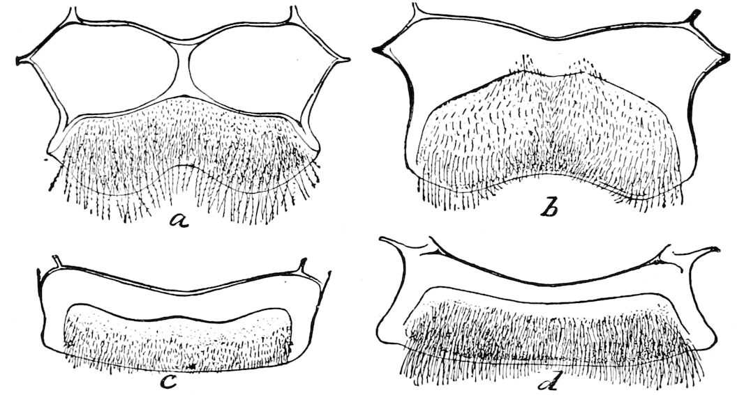
Fig. 355.—Wax disks of social bees: a, Apis mellifica, worker; b, do., queen; c, Melipona, worker; d, Bombus, worker.—From Insect Life, U. S. Dept. Agr.
The wax-glands in the honey-bee are scale-shaped organs situated on the under side of the four last abdominal segments (Fig. 355). These secrete the wax, which appears as whitish scales, and secretion is only possible when the bees have sufficient honey and pollen. The wax is secreted by the hypodermal cells rather than by glands within the abdominal cavity; the wax traverses the cuticular layer, and accumulates on its outer surface (Carlet). According to Fritz Müller, in the stingless bees (Trigona) which he observed, the wax-glands are situated on the back of the abdomen, but Ihering states that in many species of Trigona and Melipona there are also slightly developed wax-organs on the ventral side.
It has been found that certain caterpillars secrete wax. Thus the cells of the Tortrix of the fir (Retinia resinella) formed of resin are lined with wax, as on dissolving away the resin with alcohol, Dr. Knaggs found a slight film of wax; also a secretion of wax has been detected in the larva of a butterfly (Parnassius apollo). The bodies of certain saw-fly larvæ are covered with a white powdery secretion, while the remarkable larva of a Selandria is clothed with snow-white, long, flocculent, waxy masses, nearly concealing the body (Fig. 356).
h. “Honey-dew” or wax-glands of Aphids
The so-called “honey-dew” of Aphids which oozes from two wart-like tubercles or tubes situated near the end of the body, is secreted by hypodermal unicellular glands which open into a modification of a pore-canal, the tube itself being an outgrowth of the cuticula.
Witlaczil states that both in the “honey” tubes and in the body beneath, the sugary matter exists in cells of the connective tissue in the form of granules. “These large ‘sugar-cells’ in contact with the air undergo destruction, while the sugar crystallizes into needles, and thus each cell is transformed into a radiated crystalline mass.”
365“A muscle extends from a horseshoe-shaped place (a valve?) in the middle of the flat terminal plate of the honey tube, through this and down through the abdomen to the ventral surface. By this muscle the honey tube is at times erected, and we then find, as also when we lightly press the body of the insect, lumps of crystallized sugar which have been expressed through the tips of the honey tubes.” (Zool. Anzeiger, 1882, p. 241.)
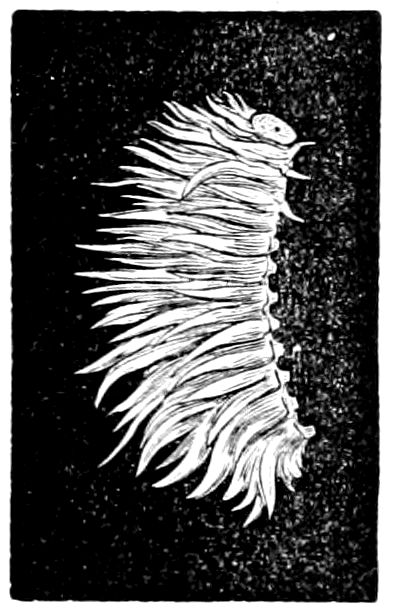
Fig. 356.—Wax-secreting larva of a saw-fly.
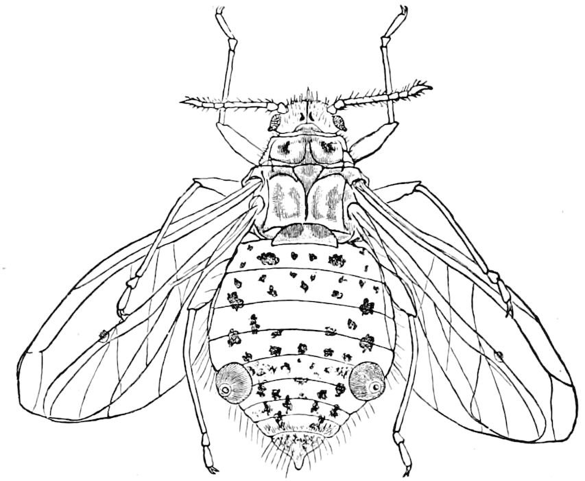
Fig. 357.—Lachnus strobi, and its two “honey” warts.—Gissler del.
Busgen, after careful research, denies that this is a sugar, but claims as the result of chemical analysis, that it is more like wax. He observed that on reaching the air the drops issuing from the “nectary” or “honey” tube stiffened almost instantly into a wax-like mass, which was easily crushed between the teeth, and had no taste at all. No sugar-like substance or urea could be detected. He therefore concludes that the secretion in question should be regarded as a wax-like mass, which agrees well with Witlaczil’s anatomical observations, and confirms the statements of previous observers. Thus, as early as 1815, Kyber stated that the Aphides expelled an excrementitious substance through the “sap tubes.” Burmeister states that the tubes give out a fluid which “dries gumlike, but, so far as I have observed, has no peculiar taste.” Réaumur, and also Kaltenbach, state that the “honey” does not issue from the tubes, but from the anus. Lastly, Forel emphatically states that “the two dorsal tubes of Aphides do not secrete a sweet fluid, but a gluey wax, which is not sought by the ants. Moreover the shield-lice and many leaf-lice have no such tubes, but yet are often sought by ants. The drops of sugar which the ants lick up are rather the excrement of the insects in question.” Hence the opinion first stated by Linné, that a sweet fluid is secreted by Aphides, must be abandoned.
On the other hand, Busgen, after careful observations, finds that the use of the sticky, waxen secretion is in reality a protective one, as he observed that when a larval Chrysopa rudely attacks the Aphides, they smear its face with the sticky wax, causing at least a momentary interruption in its attacks. He also observed that Aphides when invaded by coccinellid larvæ set their tubes in motion and besmear their heads and front part of the body. He thus seems to establish the fact that these tubes secrete a protective, sticky fluid.
i. Dermal glands in general
We have seen that certain of the hypodermal cells may be modified or specialized to form secretory unicellular glands. Such are 366those (trichogens) which secrete chitinous setæ, hairs, and spines, certain setæ in some insects being hollow and containing a poison (p. 187); others secrete wax, certain ones in Aphids “honey-dew”; in some cases dermal glands may excrete protective, sticky, or otherwise offensive matters, or may be depuratory, or facilitate the process of moulting.
There are other minute, unicellular, or compound dermal glands whose function is unknown.
Dermal glands may be segmentally or serially arranged. Thus Verson has detected a series of one or two pairs of unicellular glands near the stigmata in each thoracic, and the first eight abdominal segments of the silkworm (B. mori). In the earliest stages of growth of the caterpillar they give out oxalate of lime, and in later stages uric acid. They thus appear to act interchangeably with the urinary tubes, as excretory organs. They do not, however, carry their products directly outwards, but leave them between the hypodermis and cuticula, in order to facilitate the sloughing off of the latter in the process of moulting.
LITERATURE ON THE SECRETORY GLANDS
a. General
Sirodot, S. Recherches sur les sécrétions chez les Insectes. (Ann. sc. nat., 4 Sér., Zool., 1858, x, pp. 141–189, 251–328, 12 Pls.)
Gazagnaire, G. Des glandes chez les Insectes. (Compt. rend. Acad. Sc., Paris, 1886, cii, pp. 1501–1503; Annal. Soc. Ent. France, 1886, Bull., pp. 104–106.)
Leydig, F. Beitrage zur Anatomie und Histiologie der Insekten. 1887.
Hanow, Karl. Ueber Kerfabsonderungen und ihre Benutzung im eigenen Haushalte. (Programm des Realprogymnasiums zu Delitzsch für das Schuljahr 1889, xc; Delitzsch, 1890, pp. 3–22.)
Verson, E. Di una serie di nuovi organi escretori scoperti nel filugello. (Publ. R. Stazione Bacologica di Padova, v, 1890, pp. 30. 4 Pls.)
—— Altre cellule glandulari di origine postlarvale. (Ibid., vii, 1892, pp. 16, 1 Pl.)
—— ed E. Bisson. Cellule glandulari ipostigmatiche nel Bombyx mori. (Ibid., vi, 1891.)
Borgert, H. Die Hautdrüsen der Tracheaten. Jena, 1891, pp. 80.
Batelli, Andrea. Di una particolarità nell’ integumento dell’ Aphrophora spumaria. (Monitore Zool. Ital., 1891, Anno ii., pp. 30–32, dermal gland in last segment.)
Koschewnikow, G. A. On a new compound dermal gland found in the sting of the bee. (Journal of the Zoological Section of the Society of the Friends of Natural Science. Moscow, ii, Nos. 1, 2, 1892, p. 36. Preliminary notice. In Russian.)
Willem, V., et H. Salbe. Le tube ventral et les glandes céphaliques des Sminthurus. (Ann. Soc. Ent. Belg., 1897, xli, pp. 130–132.)
Henseval, Maurice. Les glandes à essence der Cossus ligniperda. (La Cellule, 1897, xii, pp. 19–26, 27, 29.)
367—— Recherches sur l’essence der Cossus ligniperda. (La Cellule, 1897, xii, pp. 169–181, 183.)
b. Poison-glands
Macloskie, George. The poison-apparatus of the mosquito. (Amer. Nat., xxii, 1888, pp. 884–888. 1 Fig. Also in Science, 1887, p. 106.)
Beyer, Otto W. Der Giftapparat von Formica rufa, ein reduziertes Organ. (Jena. Zeitschr. f. Wissens, xxv, 1891, pp. 26–112, 2 Taf.)
Bordas, L. Sur l’appareil venimeux des Hyménoptères. (Comtes rend., cxviii, 1894, pp. 296–299 and 873–874; also Zool. Anzeiger, xvii Jahrg., 1894, pp. 385–387, Figs.)
—— Appareil glandulaire des Hyménoptères. (Glandes salivaires; Tubes de Malpighi et glandes venimeuses.) Paris, 1894, pp. 362, 11 Pls.
—— Description anatomique et étude histologique des glandes à venin des insectes Hyménoptères. Paris, 1897, pp. 53, 2 Pls.
See Forel (p. 186); also the standard authors, Kolbe, etc.; also Locy (Amer. Nat., xviii, 1884, p. 355), Nagel (p. 324), Fenger.
c. Wax-glands
Brandt und Ratzeburg. Medicinische Zoologie, ii, 1830, p. 179. Taf. xxv, Fig. 18.
Treviranus, George R. Ueber die Bereitung des Wachses durch die Bienen. (Zeitschrift für Physiologie, etc., iii, 1832, pp. 62, 225.)
Dufour, L. Note anatomique sur la question de la production de la cire des abeilles. (Comptes rend. Acad. Sc., Paris, 1843, xvii, pp. 809–813, 1248–1253; Revue Zool. 1843, l’Institut, 1843, xi.)
Dujardin, F. Mémoire sur l’étude microscopique de la cire, etc. (Ann. Sc. Nat., xii, 1849, pp. 250–259.)
Tarzione-Tozzetti, H. Studii sulle cocciniglie. Milano, 1867, 7 Pls.
—— Sur la cire qu’on peut obtenir de la cochenille du figuier (Coccus caricæ). (Comptes rend. Acad. Sc., Paris, lxv, 1867, pp. 246–247.)
Claus, C. Ueber die wachsbereitenden Hautdrüsen der Insekten. (Sitzungsber. Gesells. z. Beförd. d. Gesammt. Naturw. zu Marburg, June, 1867, No. 8, pp. 65–72.)
Witlaczil, E. Die Anatomie der Psylliden. (Zeitschr. wissens. Zool., xlii, 1885, pp. 582–586.)
—— Zur Morphologie und Anatomie der Cocciden. (Zeitschr. wissens. Zool., xliii, 1886, pp. 149–174, 1 Taf.)
Ihering, H. von. Der Stachel der Meliponen. (Ent. Nachrichten, xii Jahrg., 1886, p. 185.)
Carlet, G. Sur les organes sécréteurs et la sécrétion de la cire chez l’Abeille. (Comptes rendus, cx, pp. 361–363, 1890.)
—— La cire et ses organes sécréteurs. (Le Naturaliste, 1890, pp. 149–151, 2 Figs.)
Also the works of Siebold, Cheshire, Kolbe, Howard and Marlatt (Bull., 3, N. S., Div. Ent. U. S. Dept. Agr., 1896, p. 40), Knaggs, Berlese.
d. Wax-like glands of Aphides
Huber et Forel, A. Études myrmécologiques, 1875.
Witlaczil, E. Zur Anatomie der Aphiden. (Zool. Anzeiger, v Jahrg., 1882, pp. 239–241; Arbeiten a. d. Zool. Institut der Univ. Wien., iv, 1882, pp. 397–441, 3 Taf.)
Busgen, M. J. Der Honigtau. Biol. Studien an Pflanzen u. Pflanzenlause. (Jena. Zeitschrift, xxv, 1891, pp. 339–428.)
368
DEFENSIVE OR REPUGNATORIAL SCENT-GLANDS
While these eversible glands are not found in marine or aquatic arthropods such as Crustacea or Merostomata (Limulus), they are often present in the air-breathing forms, especially insects. In the winged insects they are of frequent occurrence, existing under great variety of form, varying greatly in position, and appearing usually to be in immediate relation with their active volant habits. Their presence is in direct adaptation to the needs and habits of their possessors, and being repellent, warning, or defensive structures, the odors they secrete being often exceedingly nauseous, they appear to have been called into existence in direct response to their biological environment. The fact that these singular organs do not exist in marine or aquatic Crustacea suggests that the air-breathing, aërial, or volant insects by these eversible glands, usually in the form of simple evaginable hypodermic pouches, are enabled to protect themselves by emitting an infinitesimal amount of an offensively odorous fluid or ether-like spray which charges the air throughout an extent of territory which may be practically illimitable to the senses of their enemies. The principle is the same as in the mephitic sulphuretted oil ejected by the skunks, the slight quantity these creatures give out readily mixing with and charging the atmosphere within a radius of many miles of what we may call the centre of distribution.
As is now well known, the very delicate, attenuated highly volatile odors exhaled are perceived by insects with extreme ease and rapidity, the degree of sensitiveness to such scents being enormously greater than in vertebrates, their organs of sense being developed in a corresponding degree. Professors Fischer and Penzoldt, of Erlangen, have recently established the fact that the sense of smell is by far the most delicate of the senses. They find that the olfactory nerve is able to detect the presence of 1 2,760,000,000 of a grain of mercaptan.[58] The smallest particle of matter that can be detected by the eye is sodium, when observed by the spectroscope, and this particle is 250 times coarser than the particle of mercaptan which can be detected by the human nose.
In those Arachnida which are provided with poison-glands, these scent-glands are absent, but in certain Acarina and Linguatulidæ, which have no poison-glands, there are various oil-glands, stigmatic glands, as well as scent-glands, 369and in seizing a Thelyphonus with the forceps we have observed it to send out from each side of the body a jet of offensive spray.
We not infrequently find in myriopods (Polydesmidæ, Julidæ, and Glomeris) repugnatorial or the so-called cyanogenic glands, which are either paired, opening on the sides of the body, or form a single row along the median line of the under side of the body. Leidy describes and figures the spherical glands of Julus marginatus, of which there are 50 pairs. These glands have been regarded as modified nephridia, but are more probably coxal glands, and the homologues of the parapodial glands of annelid worms.
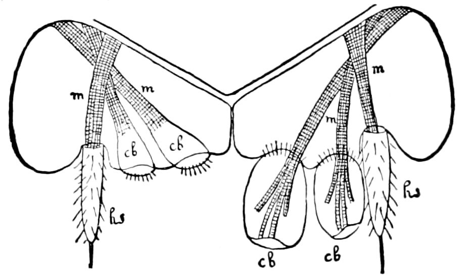
Fig. 358.—Sternite of Machilis maritima, with the pair of coxal sacs (cb) on the right side everted; hs, coxal appendages; m, retractor muscles.—After Oudemans, from Lang.
Eversible coxal glands.—True coxal glands occur in Scolopendrella immaculata on the 2d to 11th segment, on the inner side of the base of the legs (Fig. 15, c.g.). Homologous glands also occur in the same position in Campodea staphylinus (also in C. cookei and C. mexicana) on the 1st to 8th abdominal segments, and Oudemans has described a pair of eversible sacs on each side of segments one to seven of Machilis. These eversible sacs in the synapterous insects are evidently modified coxal glands, and are probably repugnatorial as well as respiratory in function.
The apparatus consists of an eversible gland, composed of hypodermic cells, usually retracted by a slender muscle and with an efferent passage, but the glands vary greatly in shape and structure in different insects. In some cases these fœtid glands appear not to be the homologues of the coxal glands, but simply dermal glands.
These repugnatorial glands are of not infrequent occurrence in the lower or more generalized winged insects, and in situation and appearance are evidently the homologues of the coxal glands of the Symphyla and Synaptera.
Fœtid glands of Orthoptera.—In the ear-wigs (Forficula and Chelidura) Meinert has detected a pair of what he calls fœtid glands at the posterior margin of the dorsal plates of the 2d and 3d abdominal segments.
Vosseler also describes the same glands as consisting of a retort-shaped sac, in whose walls are numerous small hypodermal cells and large single glandular cells provided with an efferent passage, the fluid being forced out by the pressure of the dermal muscles, one acting specially to retract the gland. The creature can squirt to a 370distance of 5 and even 10 cm. (4 inches) a yellowish-brown liquid or emulsion with the odor of a mixture of carbolic acid and creosote.
The large eversible dorsal glands of the Blattidæ, since they contain numerous hairs, which, when everted, are fan-like or like tufts, serve, as in the spraying or scent apparatus, to disseminate the odor, and might be classified with the alluring unicellular scent-glands or duftapparat of other insects, as they are by some authors; but as the glands are large and compound they may prove to be the homologues of the coxal glands rather than of the dermal glands.
Evaginable organs in the Blattids were first observed by Gerstæcker in both sexes of Corydia; they are yellowish white, covered with hairs, and are thrust out from between the dorsal and ventral plates of the 1st and 2d abdominal segments.
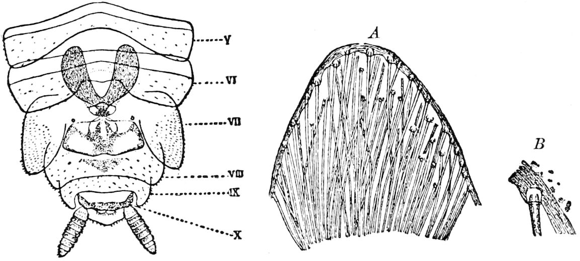
Fig. 359.—Under side of end of Aphlebia, showing the two eversible sacs; V-X, five last abdominal segments; A, portion showing the hairs; B, showing origin of a hair in its follicle.—After Krauss.
In the cockroach (P. orientalis) Minchin detected two pouch-like invaginations of the cuticle, lying close on each side of the middle line of the body between the 5th and 6th tergites of the abdomen. They are lined by a continuation of the cuticle, which forms, within the pouches, numerous stiff, branched, finely pointed bristles, beneath which are a number of glandular epithelial cells. In the male nymph of P. decorata he also found beside these glandular pouches “an additional gland, opening by a tubular duct under the intersegmental membrane between the 5th and 6th terga above the glandular pouch of each side, and extending forward into the body cavity. The gland and its duct are proliferations of the hypodermis, and there is no invagination of the cuticle.” These eversible glands are most complicated in Phyllodromia germanica. While it is absent in the female, in the male it is relatively of enormous size, extending over the 6th and 7th somites, as well as projecting far into the body cavity (Minchin). 371Haase states that these glands become everted by blood-pressure and give out the well-known disagreeable smell of these insects. He states that in the male of P. germanica the dorsal glands in the 6th and 7th abdominal segments are without hairs and produce an oily secretion; they function as odoriferous organs in sexual union.
In the male of another Blattid (Aphlebia bivittata) of the Canary Islands, Krauss has detected two yellowish dorsal sacs 1.5 mm. in length, opening out on the 7th abdominal segment, and filled full of long yellowish hairs, the ends directed towards the opening, where they form a thick tuft. These eversible glands lined with hairs appear to be closely similar to the long slender eversible hairy appendages or scent organs of certain Arctian and Syntomid moths. (Fig. 359.)
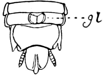
Fig. 360.—External flaps (gl) of glands of Platyzosteria.
We have found the external median wart with lateral lids or flaps in between the 5th and 6th tergites of Platyzosteria ingens Scudder, a large wingless Blattid living under the leaf scars of the cocoanut tree in Southern Florida (Fig. 360), but were unable to detect them in Polyzosteria or in the common Blabera of Cuba, or in another genus from Cordova, Mexico.
In another group of Orthoptera, the Phasmidæ, occur a pair of dorsal prothoracic glands, each opening by a pore and present in both sexes. In the walking-stick, Anisomorpha buprestoides, ♂ and ♀, these openings are situated on each side of the prothorax at its upper anterior extremity, situated at the bottom of a large deep pit. When seized it discharged a “milky white fluid from the pores of the thorax, diffusing a strong odor, in a great measure like that of the common Gnaphalium or life everlasting” (Peale in Say’s American Entomology, i, p. 84). Boll states that the females when captured “spurt from the prothorax, somewhat after the manner of bombardier beetles, a strong vapor, which slightly burnt the skin; when the females were seized by the males a thick fluid oozed from the same spot.” Scudder describes these glands in another Phasmid (Autolyca pallidicornis) as two straight, flattened, ribbon-like bodies, with thick walls, broadly rounded at the end, lying side by side and extending to the hinder end of the mesothorax. In Anisomorpha buprestoides the glands are of the same size and shape (Scudder). In Diapheromera femorata the repugnatorial foramina are very minute, and the apparatus within consists of a pair of small obovate or subfusiform sacs, one on each side of the prothorax, about 1 mm. 372in length, with a short and very slender duct opening externally at the bottom of the pit (Scudder).
In the Mantidæ these seem to be genuine coxal glands, as there is a pair situated between the coxæ of the first pair of legs. An evaginable organ like a wart, with a glandular appearance, occurs on the hind femora of the Acrydiidæ in a furrow on the under side, into which the tibia fits, about one-fourth from the base (Psyche, iii, p. 32).
In the male cricket, the anal odoriferous glands are small lobes opening into a reservoir on each side of the rectum (Dufour). Homologous glands also occur in the Coleoptera (Fig. 302, l and 317, s).
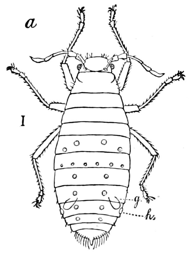
Fig. 361.—Glands (g) of Lachnus; h, “honey” wart.—Gissler del.
Most Hemiptera or bugs send out a fœtid or nauseous odor due to a fluid secreted by a single or double yellow or red pear-shaped gland, situated in the middle of the mesothoracic segment, and opening between the hinder or third pair of coxæ. In Belostoma Leidy describes these glands as consisting of two rather long cœcal tubes situated in the metathorax, beneath the other viscera, extending backwards into the abdomen, and opening between the coxæ of the third pair of legs. Locy states that the smell arising from these glands is pleasant, resembling that of well ripened pears or bananas. Other bugs, moreover, emit an agreeable odor, that of Syromastes resembling that of a fine bergamot pear. (Siebold.) The fluid given out by the European fire-bug (Pyrrhocoris apterus) has a sweetish smell, like ether. In the nymph there are three pairs of dorsal glands, on abdominal segments 2–5, which are atrophied in the mature insect. In the bed-bug, the nymph has three odoriferous glands each with paired openings in the three basal abdominal segments respectively, and situated on the median dorsal line, being arranged transversely at the edge of the tergites; but after the last moult these are aborted, and replaced by the sternal metathoracic glands (Künckel). Gissler has detected a pair of glands in Lachnus strobi (Fig. 361).
Anal glands of beetles.—Certain beetles are endowed with eversible repugnatorial glands. Eleodes gigantea and E. dentipes of both sexes are said by Gissler to possess these glands. When teased “they stand on their anterior and middle legs, holding the abdomen 373high up and spurting the contents of the glands right and left.” The glands (Fig. 366, 1) are two reddish brown, somewhat bilobed sacs, and extend from the base of the last up to the middle of the 2d abdominal segment, with an average length of 6.5 mm. The liquid stains the human skin, has an acid reaction, with a peculiar, “intensely penetrant odor, causing the eye to lachrymate. It is soluble in water, alcohol, and ether. Boiled with concentrated sulphuric acid and alcohol an ethereal aromatic vapor is produced, indicating the presence of one or more organic acids, though neither formic or acetic acid could be detected.” Williston has observed the same habits in seven other species of Eleodes, all ejecting a pungent vile-smelling liquid, one species (E. longicollis) ejecting a stream of fluid from the anal gland, backwards sometimes to the distance of 10 cm. or more, and he regards these beetles as “the veritable skunks of their order.” Leidy briefly describes the odoriferous glands of Upis pennsylvanica.
The anal glands consist, according to Meckel and also Dufour, of two long, simple, flexuous cœca with reservoirs having two short excretory ducts situated near the anus (Siebold).
Glands like those of Eleodes found in Blaps mortisaga are described in detail by Gilson (Fig. 366, 2). They form two pouches or cuticular invaginations situated in the end of the abdomen on the sides of the end of the intestine and unite on the median line underneath the genital organs, forming a very short tube with a chitinous wall, continuous with the cuticle of the last abdominal segment. Into each pouch open a large number of fine slender lobules varying in shape, giving a villous aspect to the surface. These lobules are composed of as many as fifty unicellular glands, each of which is composed of four parts: (1) A radiated vesicle, (2) a central sac, giving rise (3) to a fine excretory tube, and (4) a sheath near the origin of the excretory tube. These are all modifications of the cytoplasm of the cell with its reticulum; the nucleus with its chromosomes is also present, but situated on one side of the central sac. The fine excretory tubules form a bundle passing down into the mouth of each lobule.
Similar glands, though usually smaller, which have not been carefully examined, occur in Carabus (Fig. 300, 3) and Cychrus, which eject from the vent a disagreeable fluid containing butyric acid (Pelouse). The bombardier beetle Brachinus, with its anal glands, ejects a jet of bluish vapor accompanied with a considerable explosion, which colors the human skin rust red; it is caustic, smells like nitrous acid, and turns blue paper red. Westwood states that individuals of a large South American Brachinus on being seized “immediately began to play off their artillery, burning and staining the flesh to such a degree 374that only a few specimens could be captured with the naked hand, leaving a mark which remained for a considerable time.” The fluid ejected by another species, in Tripoli, blackened the fingers of the collector. “It is neither alkaline nor acid, and it is soluble in water and in alcohol.” (Kirby and Spence, iv, p. 149.)
Species of other genera (Agonum, Pheropsophus, Galerita, Helluo, Paussus, Ozæna) are also bombardiers, though less decidedly so than Brachinus. A Paussid beetle (Cerapterus) ejects explosively a fluid containing free iodine (Loman), while Staphylinus, Stenus, Ocypus olens, Lacon, etc., have similar anal fœtid glands, the liquid being more or less corrosive. The secretion of Mormolyce phyllodes is so corrosive that it is said to paralyze the fingers for 24 hours after. (Cuénot.)
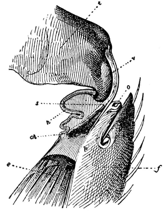
Fig. 362.—Median section through the femoro-tibial joint of leg of Coccinella, showing at o the opening through which the blood oozes out; f, femur; t, tibia; e, extensor muscle of the tibia: s, sinew of the same; at ch, chitinized; h, articular membrane; v, tibial process.—After Lutz.
The two pairs of remarkably large, soft, eversible, forked, orange-yellow glands of the European genus Malachius, are thrust out from the side of the 1st and the 3d thoracic segments. They are everted by blood-pressure, and retracted by muscles. The larva of Hydrophilus piceus ejects by the anus a black, fœtid fluid.
Claus has shown that the larva of Lina populi and other Chrysomelidæ possess numerous minute, eversible glands in each of the warts on the upper surface of the body, each gland containing a whitish, repellent fluid smelling like the oil of bitter almonds and containing salicylic acid derived from its food-plant, which issues as pearl-like drops. Candèze thinks the fluid may contain prussic acid. The fluid is secreted by a variable number of glandular cells, each provided with an efferent duct. The larvæ of saw-flies, notably Cimbex americana, also eject droplets of a clear fluid from non-evaginable glands situated near each stigma (Chlolodkovsky).
The blood as a repellent fluid.—In this connection it may be mentioned that though there are no special glands present, many beetles emit drops of blood from the femoro-tibial joints of their legs as a means of defence. Such are the oil-beetles (Meloë), Cantharis, Lytta. The cantharadine secreted by these beetles, according to Beauregard, is an efficient means of defence, as birds, reptiles, and carnivorous insects will not usually attack them. This substance 375is formed in the blood and also in the genital organs, and is so extremely caustic that scavenger insects which feed upon their dead bodies will leave untouched the parts containing cantharadine, and if May-beetles or mole-crickets are washed with the blood of Meloë or with cantharidate of potassa, it will for several days render them safe from the attacks of the carabids which usually prey upon them. The eggs even after deposition are strongly vesicant, and are thus free from the attacks of egg-eating insects (Cuénot). The Coccinellidæ are also protected by a yellow, mucilaginous, disagreeable fluid oozing out of the ends of the femora; in our common, two-spotted lady-bird (C. bipunctata) the yellow fluid is disagreeable, smelling like opium. Lutz has found that the blood in Coccinellidæ passes out through a minute opening situated at the end of each femur (Fig. 362). The blood is very repellent to insectivorous animals.
The Dyticidæ eject from the anus a colorless, disagreeable fluid, while these beetles, and especially the Gyrinidæ, when captured send out a milky fluid which appears to issue from the joints of the body. The Silphidæ throw out both from the mouth and vent a fœtid liquid with an ammoniacal odor. They possess but a single anal gland, the reservoir opening on one side of the rectum (Dufour).
Other malodorous insects have not yet been investigated; such are the very persistent odors of lace-winged flies (Chrysopa).
More agreeable secretions, but probably formed by similar glands, is the odor of rose or hyacinth given out by Cicindelæ, or the rose fragrance exhaled by the European Aromia moschata.
Eversible glands of caddis-worms and caterpillars.—Gilson, while investigating the segmentally disposed thoracic glands of larval Trichoptera, has found in the larva of Limnophilus flavicornis that the sternal prothoracic tubercle gives exit to an underlying tubular gland. In Phryganea grandis each thoracic sternum affords an exit to an eversible gland. Many caterpillars, as our subjoined list will show, are very well protected by eversible repugnatorial glands situated either in the under or upper side of the body. Since the time of De Geer (1750) the fork-tailed larva of Cerura has been known to throw out a secretion, which was described by Bonnet in 1755 as a true acid, sharp, sour, and biting. This spraying apparatus in Cerura (Harpyia) vinula has been well described by Klemensiewicz (Fig. 366, 4), though Rengger in 1817 noticed the general form of the secretory sac, and that it opens out in two muscular eversible tubes, out of which the secretion is ejected.
The fork-tailed larva of Macrurocampa marthesia, which is much like that of Cerura, when teased sends out a jet of spray to the 376distance of nearly an inch from each side of the neck. While examining the very gayly-colored and heavily-spined caterpillars of Schizura concinna we observed that when a fully-grown one was roughly seized with the forceps or fingers it sent out a shower of spray from each side of the prothoracic segment, exactly like that of Cerura and Macrurocampa.
In the European Cerura vinula the apparatus consists of a single sac, which opens by a narrow transverse slit on the under side of the neck, out of which is rapidly everted four lateral tubes, two on each side (Fig. 366, 4, t), which are withdrawn within the opening by the contraction of several fine muscles. The apparatus in the American C. multiscripta is as in the European C. vinula. In a living specimen the large secretory sac was seen to be of the same size and shape as in Macrurocampa, and of the color of raw silk. The sac when distended extends back to a little behind the middle pair of legs, and is nearly two-thirds as wide as the body. The caterpillar sent out the fluid when handled, but we could not make it spray.
In the larva of Macrurocampa marthesia the cervical or secretory gland (Fig. 366, 5) is situated in the 1st and 2d thoracic segments, extending to the hinder edge of the latter and lying between the nervous cord and the œsophagus and proventriculus, and when empty the bulk of it lies a little to one side of the median line of the body. It is partly held in place by small tracheæ, one quite large branch being sent to it from near the prothoracic spiracle. The short, large duct, leading from it to the transverse opening in the membrane between the head and prothoracic segment, is a little narrower than this opening, and is kept distended by tænidia, or a series of short, spiral threads which are pale, not honey-yellowish, in color. This duct lies on one side of the prothoracic ganglion, resting just under the commissures passing up to the brain; it is also situated between the two silk ducts.
The very distensible sac (Fig. 366, 5) is rendered elastic by a curious arrangement of the cuticle, the tænidia of the duct itself being represented by very thickly-scattered, irregular, separate, sinuous, chitinous ridges, which stand up from the cuticular lining of the wall of the sac (Fig. 366, 6). The secretory cells of the walls of this sac in Cerura vinula are said by Klemensiewicz to be large hexagonal cells, resembling those of silk-glands, having like them large branched nuclei.
The fluid thrown out is said by Poulton to be formic acid; it causes violent effervescence when allowed to fall upon sodium-bicarbonate, 377and colors blue litmus paper red. It also appears from the researches of Latter that these creatures in the imago state secrete free potassium hydroxid, a substance for the first time known to exist in the animal kingdom.
In the caterpillar of Astyanax archippus (Limenitis disippus) a dark, bladder-like sac is everted, but the lateral tubes appear to be wanting, and no spray is sent out; it occurs in the larvæ of many Nymphalidæ and other butterflies and moths.
These glands are functionally active in Perophora, but obsolete (at least the external openings) in Lacosoma.
The osmeterium in Papilio larvæ.—The caterpillars of the swallowtailed butterflies (Papilio, Doritis, and Thais), as is well known, when irritated thrust out from a transverse slit on the upper part of the prothoracic segment a large orange-yellow V-shaped fleshy tubular process (the osmeterium), from which is diffused a more or less melonlike but disagreeable, in some cases insufferable, odor; the secretion is acid and reddens litmus paper. The mechanism has been described and figured by Klemensiewicz.
When at rest, or retracted, the osmeterium lies in the upper part of the body in the three thoracic segments, and is crossed obliquely by several muscular bundles attached to the walls of the body, and by the action of these muscles the evagination of the osmeterium is strongly promoted. After eversion the tubes are slowly retracted by two slender muscles inserted at the end of each fork or tube, and arising from the sides of the 3d segment behind the head, crossing each other in the median line (Fig. 366, 7 r.m.). The secretion is formed by an oval mass of glandular cells at the base of the forks; in the glandular mass is a furrow-like depression about which the secretory cells are grouped. The secretion collects in very fine drops on the side of each furrow opposite the glandular cells.
According to C. D. Ash the larva of an Australian Notodontid (Danima banksii Lewin) protrudes from the under side of the prothoracic segment a Y-shaped red organ like that of Papilio; no fluid or odor is given out.
Dorsal and lateral eversible metameric sacs in other larvæ.—The showy caterpillars of Orgyia and its allies have a conspicuous coral-red tubercle on the back of the 6th and also the 7th abdominal segment, which on irritation are elongated, the end of the tubercles being eversible. When at rest the summit is crateriform, but on eversion the end becomes rounded and conical. These osmeteria are everted by blood pressure, and retracted by a muscle. Fig. 366, 9, represents a section of an osmeterium of Orgyia leucostigma when 378retracted by the muscle (m); at the bottom of the crater are the secreting or glandular cells (gc), being modified hypodermal cells. These doubtless serve as terrifying organs to ichneumons and other insect enemies, and though we have been unable to detect any odor emanating from the tubercles, yet possibly they give out a scent perceived by and disagreeable to their insect assailants.
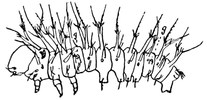
Fig. 363.—Freshly hatched larva of Hyperchiria io, with its two pairs of eversible glands (g).
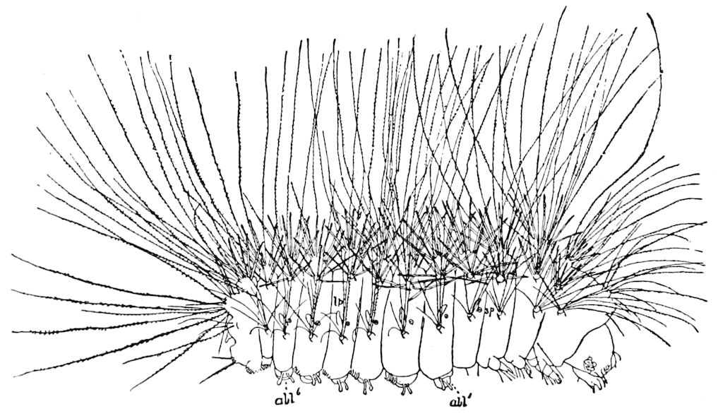
Fig. 364.—Young larva of Megalopyge crispata, enlarged, showing the seven pairs of lateral processes (lp): sp, spiracle; abl′, abl6, six pairs of abdominal legs besides the anal pair.
In the Hemileucidæ there is a pair of lateral osmeteria, on the 1st and on the 7th abdominal segments, which however, are not highly colored (Figs. 363, 366, 10). In Megalopyge (Lagoa, Fig. 364) there is a lateral row of singular pale permanently everted processes which appear to be the homologues of the osmeteria of larvæ of other lepidopterous families. As these are repeated on seven segments, their metameric arrangement is obvious. The relation of these curious glands to the viscera is seen in Fig. 297, lgp, and their minute structure in Fig. 365.
At A, the lumen (l) is a deep narrow cavity, with the secretion (secr.), collected at the mouth of the cavity, composed of a thin, mucus-like, coagulated fluid, containing granules of varying degrees of fineness, which take the stain readily. Outside of these are collected fine nuclei (bc), stained dark, and enveloped in a slight, transparent, pale, protoplasmic envelope, which may be blood corpuscles. The glandular cells themselves are simply modified hypodermal 379cells, as seen at C. In some of the nuclei, indistinct nucleoli are seen, and deeply stained granules, especially around the periphery of the nuclei. At B is represented a section on one side of the middle, but still showing the spacious lumen. In the section represented by C, the knife passed through the process still nearer the outer edge, and near the base; at C1, three of the glandular cells, with their large, deeply stained nuclei, are drawn. A transverse section at D shows the large lumen or cavity (l).
As to the function and homologies of these structures, it is difficult to decide. We have never noticed that they give off any odor, though they may prove to be repugnatorial; they are not visible in the fully grown, living insect, being concealed by the long, dense hairs clothing the body; they are not spraying organs, as they are imperforate at the end, not ending as the lateral, eversible glands of Hyperchiria io, etc., in a crateriform orifice.
They may be permanently everted glands, or osmeteria, which have, by disuse, lost their power of retraction and their crateriform opening, as well as the power of secreting a malodorous fluid.
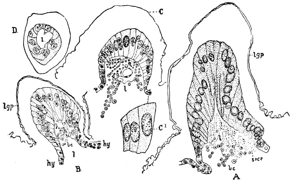
Fig. 365.—Section of lateral processes of larva of Megalopyge.
In certain of the butterflies, the Heliconidæ (Colænis, Heliconius, Euides, and Dione), there is thrust from the end of the abdomen a pair of large, irregular, rounded, eversible glands, which give out a disagreeable odor, and are consequently repellent, and which seem to be the homologues of the odoriferous glands of other butterflies.
The large, soft, rounded, eversible glands, looking like puff-balls or a rounded pudding (Fig. 366, 12), are everted, when the butterflies are roughly seized, from the dorsal side of the penultimate segment of the abdomen. The males possess two smaller tubercles on the inside of the anal claspers or lobes. Müller also detected, in the females of various species of the Heliconidæ enumerated above, a pair of club-shaped processes like the balancers of flies, which are thrust out on each side of and under the odoriferous puff-balls of the hinder edge of the penultimate segment (Fig. 366, 13). The club or head is armed with hairs or bristles, which, in Heliconius, are like the scales of a butterfly.
381
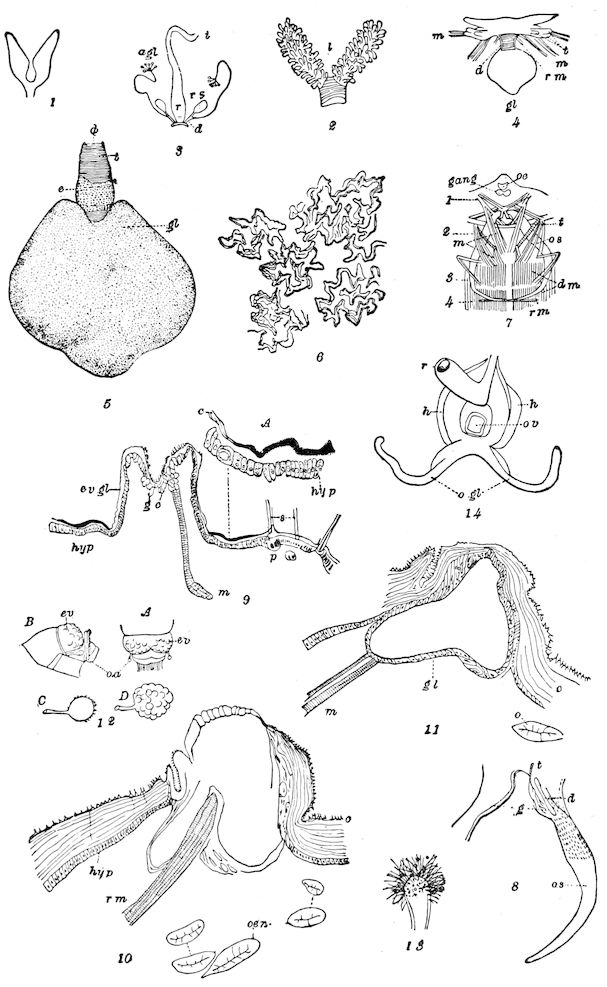
Fig. 366.—Scent-glands of insects: 1. Anal eversible glands of Eleodes.—After Gissler. 2. Anal eversible glands of Blaps.—After Gilson. 3. Anal glands (agl) of Carabus hortensis: rs, reservoir; d, excretory duct; i, intestine; r, rectum.—After Kolbe. 4. Prothoracic spraying apparatus of Cerura vinula: gl, the gland; d, its duct, with tænidia; t, the spraying tubes; m, muscles; rm, retractor muscles.—After Klemensiewicz. 5. The thoracic glandular sac of Macrurocampa marthesia: gl, the glandular sac; d, its duct; e, peritracheal epithelium; t, the spiral threads or tænidia. 6. Irregular separate masses of chitinous ridges on the cuticular lining of the wall of the sacs of Macrurocampa marthesia. 7. Osmeterium (os) of the larva of Papilio machaon at rest: rm, the retractor muscles at the ends; m, the numerous oblique muscles; dm, dorsal longitudinal muscles; t, trachea; oe, œsophagus; gang, brain; 1, head; 2, 3, 4. thoracic segments. 8. Osmeterium (os) of one side, enlarged: g, glandular portion at the base; d, depressions in the cuticula of the glandular portion; t, trachea.—This and Fig. 7 after Klemensiewicz. 9. Eversible dorsal glands (ev. gl) of larva of Orgyia leucostigma in Stage II: gc, glandular cells at bottom of the crater-like depression; m, retractor muscle; p, poison gland-cells of the root of the seta (s); c, cuticula; hyp, hypodermis; A, portion of the cuticle and hypodermis enlarged. 10. Lateral eversible gland of Hyperchiria io, Stage II: rm, retractor muscle; oen, œnocytes. 11. The same as Fig. 10, but representing a section through one side of the eversible gland. 12. A, end of body of Colænis julia; ev, eversible anal gland; oa, odoriferous appendages; B, the same in Heliconius apseudes, side view; C, odoriferous appendages of Colænis dido in fresh condition; D, tested with alcohol and benzine. 13. Odoriferous appendages of Heliconius eucrate, head cleansed.—Figs. 12, 13, after F. Müller. 14. Odoriferous glands (ogl) in the pupa of Vanessa io: r, rectum; h, the folds of hypodermis which forms the terminal papilla of the abdomen; ov, oviduct.—After Jackson.
In the caterpillars of certain blue butterflies (Lycænidæ) is an internal osmeterium, being a very minute sac which is everted from a transverse slit on the top of the 7th abdominal segment. Its function is quite the opposite of those of the caterpillars of other families, since the sac exudes a sweet fluid very attractive to ants, which may be diffused more widely by the delicate spinulose bristles crowning the summit. W. H. Edwards states that in several species of Lycæna, besides that on the 7th abdominal segment, there is on the 8th segment a pair of minute dorsal evaginable tubercles.
A pair of small ramose odoriferous glands are said by Siebold, who regarded them as alluring glands, to occur in Argynnis, Melitæa, and Zygæna, to be situated near the orifice of the oviduct, and Scudder has detected them near the anus of the female pupa of Danais archippus. The appearance of the odoriferous glands in the pupa of Vanessa io is well shown by Jackson (Fig. 366, 14). They develop as two tubular ingrowths of the hypodermis, perfectly distinct one from the other, each having its own separate aperture to the exterior. In Fig. 366, 14 the condition of parts is nearly as in the imago, the glands being situated below the rectum and opening of the oviduct. In both sexes of another Brazilian butterfly (Didonis biblis) on the median line of the abdomen between the 4th and 5th segments are two roundish vesicles covered with short gray hairs, which emit a disagreeable smell.
It is possible that the dark-green fluid in Parnassius, secreted by an evaginable gland, and which is moulded into shape by the scimetar-shaped peraplast (Scudder), is formed by the homologues of the anal glands of other butterflies.
382
Distribution of repugnatorial or alluring scent-glands in insects[59]
A. Larval Insects
a. Each thoracic segment; sternal. Phryganea grandis.
b. Prothoracic, sternal, discharging a lateral jet of spray; with a single large internal sack.
LEPIDOPTERA
Family Tineidæ
Hyponomeuta evonymella.
Family Noctuidæ
Bryophila, Cucullia formosa, C. scrophulariæ, Habrostola, Cleophana linariæ, Catocala (sp.), Aporia cratægæ, Aplecta nebulosa, Leucania staminea, L. hispanica, L. nonagrioides, Plusia gamma.
Family Notodontidæ
Pheosia rimosa, Schizura concinna, Danima Banksii (Australia), Macrurocampa marthesia, Heterocampa pulverea, Cerura vinula, C. furcula, C. borealis, C. multiscripta.
Family Nymphalidæ
Probably all the species.
c. Prothoracic, dorsal; sending out a V-shaped odoriferous organ (osmeterium).
Family Papilionidæ
All the species as a rule.
d. Thoracic sternal, evaginable glands.
Family Perophoridæ
Lacosoma chirodota, Perophora melsheimerii.
Family Nolidæ
In three, and probably in all the species of Nola.
e. Lateral, abdominal, non-eversible glands, one near each spiracle, emitting a clear fluid.
Family Tenthredinidæ
Cræsus septentrionalis, C. varus, Cimbex americana, C. betulæ, Trichiosoma.
f. Lateral, abdominal, partly eversible glands emitting neither moisture nor odor, but flesh-colored.
Family Tineidæ
Phyllocnistis? (eight pairs.)
383
Family Hemileucidæ
Hyperchiria io (two pairs, viz. on 1st and 7th segments), H. sp. (Mexico), Hemileuca yavapai, pamina, H. maia, H. artemis, Pseudohazis eglanterina.
g. Lateral, abdominal, permanently everted, metameric glands, not known to secrete a fluid, nor to be odoriferous.
Family Megalopygidæ
Megalopyge crispata.
h. Medio-dorsal, partly eversible glands, emitting a spray of liquid but no odor(?), and colored coral-red or orange-yellow (P. auriflua), but usually in the European species yellowish.
Family Liparidæ
All the species except those of Demas.
i. A single, median, abdominal, dorsal gland, emitting a fluid attractive to ants, on 7th segment; with a pair of minute, index glands on the 8th segment.
Family Lycænidæ
All the species.
j. Protrusile organs near the anus.
Myrmeleon larva (Hagen? Dimmock).
B. Nymph of Heterometabolous Insects
a. Paired, dorsal glands, on abdominal segments 1, 2, and 3.
Cimex lectularius (Künckel).
b. The same on abdominal segment 5.
Lachnus strobi.
C. Pupa of Certain Bombyces
At anterior end of certain pupæ, internal glands to moisten threads of the cocoon for exit of moth.
D. Adult Insects
a. Occurring on the prothorax only; strongly repugnatorial, best developed in ♂.
Anisomorpha buprestoides, Autolyca pallidicornis, Phasma putidum, Phyllium (sp.), Heteropteryx (sp.), Diapheromera femoratum (probably in all the species of the family), Mantis carolina.
b. Occurring on the pro- and mesothorax, and on the middle of the abdomen, orange-yellow, fleshy tubercles or evaginations.
Malachius bipustulatus, Anthocomus equestris, Evæus thoracicus.
c. Segmental, eversible glands, homologues of the coxal glands of other Arthropods, occurring on all, or nearly all, the abdominal segments.
Scolopendrella immaculata (coxal glands on 3d to 11th pair of legs), Campodea staphylinus (a pair of coxal glands on 1st to 8th abdominal segments), Machilis maritima (eversible, coxal glands on segments 1–7).
384d. Occurring in the abdomen.
d1. In the two first abdominal segments.
Corydia carunculigera ♂ and ♀.
d2. Alluring (?) organs situated on the dorsal side of the abdomen, in the 6th, or 6th and 7th, abdominal segment.
Periplaneta americana ♂, P. orientalis (nymph), P. decorata ♂ (nymph), Ectoblatta germanica ♂, Ectobia lapponica ♂, Phyllodromia ♂, Aphlebia bivittata ♂, Platyzosteria ingens (on seventh segment).
e. At the end of the body.
Colænis julia ♀ (F. Müller), Heliconius apseudes (F. Müller).
LITERATURE ON DEFENSIVE OR REPUGNATORIAL GLANDS
Aldrovandus, U. De animalibus insectis libri septem cum singulorum iconibus ad vivum expressis. (Denuo impress: Bonon. apud Clementem Ferronium, 1638, p. 273. The first edition was in 1602.)
Moufet, T. Insectorum sive minimorum animalium theatrum, etc. London, 1634, pp. 185, 186.
Gœdart, J. Metamorphosis naturalis sive insectorum historia, etc. Amstelodami, 1700, Pars ii, p. 136. (French ed. of 1700, ii, p. 162; Lister’s Latin ed., London, 1685, p. 60.)
Réaumur, R. A. F. Mémoires pour servir à l’histoire des insectes, etc. Paris, 1736, ii, pp. 266–269, Pls. 21, 22. [ii, partie ii, pp. 21–23, of the Amsterdam ed. of 1737–1748.]
De Geer, C. Observation sur la propriété singulière qu’ont les grandes chenilles à quatorze jambes et à double queue, du saule, de seringuer de la liqueur. (Mém. sav. étrang, Paris, 1750, i, pp. 530, 531, Pl.; Goetze und Bonnet, etc., Auserlesene Abhandlungen, 1774, p. 220.)
Schaeffer, J. C. Neuendeckte Theile an Raupen und Zweyfaltern, etc. Regensburg, 1754.
Sulzer, J. H. Die Kennzeichen der Insekten, etc. Zürich, 1761, pp. 65–67, Taf. 5, Fig. 34.
Müller, O. F. Pile-larven med dobbelt Hale, og dens Phalæne, etc. Kjöbenhavn, 1772, pp. 53–56, Pl. 2, Figs. 3–5.
Bonnet, C. Mémoire sur une nouvelle partie commune à plusieurs espèces de chenilles. (Mém. math. d. savants étrang., Paris, 1755, ii, pp. 44–52; Collection complète des œuvres de C. Bonnet, 1779, ii, pp. 3–16.)
—— Mémoire sur la grande chenille à queue fourchue du saule, dans lequel on prouve, que la liqueur que cette chenille fait jaillir, est un véritable acide, et un acide très-actif. (Mém. math, de savants étrang., Paris, 1755, ii, pp. 267–282; Collection complète des œuvres de C. Bonnet, 1779, ii, pp. 17–24.)
Amoreux, P. J. Notice des insectes de la France, réputés venimeux, etc. Paris, 1789, pp. 282–285.
Schwarz, C. Neuer Raupenkalender. Nürenberg, 1791, Abth. i, p. 59.
Petzhold, C. P. Lepidopterologische Beyträge. (L. G. Scriba’s Beiträge zu der Insekten-geschichte, Frankfurt am Main, 1793, Heft 3, pp. 230–251.)
Nouvelle Dictionnaire d’Hist. Nat., xv, p. 487. (Larva of Hydrophilus ejects with a slight noise a fœtid and blackish fluid.)
385Rengger, Johann Rudolph. Physiologische Untersuchungen über die thierische Haushaltung der Insecten. Tübingen, 1817. (In the chapter entitled Abgesonderte Säfte bei den Raupen, he speaks of the glandular apparatus of the larva of B. vinula, noticing the general form of the secretory sac, that it opens out in two muscular evertible points, out of which the secretion is ejected.)
Dufour, L. Mémoire anatomique sur une nouvelle espèce d’insecte du genre Brachinus. (Ann. de mus. d’histoire nat., xviii, 1811, pp. 70–81.)
—— Recherches anatomiques sur les carabiques et sur plusieurs autres coléoptères. (Ann. d. Sci. Nat., 1826, viii, pp. 5–54.)
—— Mémoire sur les métamorphosis et l’anatomie de la Pyrochroa coccinea. Glande odorifique. (Ibid., ii sér. Zoologie, xiii, 1840, pp. 340, 341.)
—— Recherches anatomiques sur les Diptères. 1851, pp. 195, 313. (Alimentary canal of Sepsis contains the seat of a “glande odorifique.”)
Kirby and Spence. Introduction to entomology, etc. (2d ed., i, 1815; London, 1818, ii, pp. 238, 239.)
Lyonet, P. Recherches sur l’anatomie et les metamorphoses de différentes espèces d’insectes. Ouvrage posthume. Paris, 1832.
Morren, C. Mémoire sur l’émigration du puceron du pêcher (Aphis persicæ), et sur les caractères et l’anatomie de cette espèce. (Ann. Sci. Natur. Zool., 1836, vi, pp. 65–93, Pls. 6, 7.)
Ratzeburg, J. T. C. Die Forstinsekten, etc. (Theil i, Die Käfer, etc., 1837, p. 246.)
Aubé, C. Note sur une sécrétion fétide d’Eumolpus pretiosus. (Ann. Soc. Ent. Fr., 1837, i, vi; Bull., p. 58.)
Lacordaire, J. S. Introduction à l’entomologie. 1838, ii, p. 45.
Meckel, von Hemsbach, Johann Friedrich. Mikrographie einiger Drüsenapparate der niederen Thiere. (Anat. Phys. u. wiss. Med., 1846, pp. 1–73, Taf. 1–3; p. 46, Müller’s Archiv.)
Stein, Friedrich. Vergleichende Anatomie und Physiologie der Insekten. Berlin, 1847.
Leidy, Joseph. History and anatomy of the hemipterous genus Belostoma. (Journ. Acad. Nat. Sci. Philadelphia, Ser. 2, 1847, i, Part i, pp. 57–67, Pl. 1.)
—— Odoriferous glands of invertebrata. (Proc. Acad. Philadelphia, 1849, iv, 234–236, 1 Pl.; Ann. and Mag. N. H., Ser. 2, 1850, v, pp. 154–156.)
Chapuis et Candèze. Catalogue des larves des coléoptères, etc. (Mém. Soc. Sci. de Liège, 1853, viii, pp. 351–653, Pls. 1–9, pp. 611, 612.)
Siebold, Carl Theodor. Lehrbuch der vergleichenden Anatomie der wirbellosen Thiere, 1848. (Burnett’s transl., Boston, 1854.)
Burnett, Waldo Irving. Translation of Siebold’s Anatomy of the Invertebrates, 1854. (Note on the osmeteria of Papilio asterias, which he regards as an odoriferous and defensive, rather than tactile, organ, p. 415.)
Karsten, H. Bemerkungen über einige shaarfe und brennende Absonderungen verschiedener Raupen. (Müller’s Archiv für Anat. Phys. u. wiss. Med., 1848, pp. 375–382, Taf. 11, 12.) Describes the poison-glands at the base of the spines of Saturnia larvæ.
—— Harnorgane des Brachinus complanatus. (Müller’s Archiv, 1848, pp. 367–376, Taf.)
Laboulbéne, Alexandre. Note sur les caroncules thoraciques du Malachius. (Annales de la Soc. Ent. de France, 3e Sér., vi, 1858, pp. 521–528.)
Saussure, Henri de. Recherches zoologiques de l’Amerique centrale et du Mexique. (6e Partie, Études sur les Myriopodes et les Insectes, Paris, 1860.)
386Gerstaecker, C. E. A. Ueber das vorkommen von ausstülpbaren Hautanhängen am Hinterleibe an Schaben. (Archiv f. Naturgesch., 1861, xxi, pp. 107–115.)
Liegel, Hermann. Ueber den Ausstülpungsapparat von Malachius und verwandten Formen. Inaug. Diss., Göttingen, pp. 31, 1 Taf. (n. d., since 1858 and before 1878.)
Leydig, F. Zur Anatomie der Insecten. (Archiv f. Anat. Phys. u. wiss. Med., 1859, pp. 33–89, 149–183, Taf. 2–4, pp. 35 and 38.)
—— Ueber bombardier Käfer. (Biolog. Centralbl., x, 1890, pp. 395, 396.)
Claus, C. Ueber die Seitendrüsen der Larve von Chrysomela populi. (Zeits. f. wissens. Zool., xi, 1861, pp. 309–314, Taf. xxv.)
—— Ueber Schutzwassen der Raupen des Gabelschwanzes. (Würzburger Naturw. Zeitschrift, 1862, iii, xiv; Sitz. am., 28 Juni, 1862.)
Rogenhofer, Alois. Drei Schmetterlingsmetamorphosen. (Verhandlungen der k. k. zoolog.-bot. Gesellschaft, Wien, xiii, 1862, pp. 1224–1230.)
Fitch, Asa. Eighth report on the noxious and other insects of ... New York. (Trans. N. Y. State Agric. Soc., 1862, xxii, pp. 657–684), p. 677. (Separate.)
Guenée, Achille. D’une organe particulier que présente une chenille de Lycæna. (Annales Soc. Ent. de France, Sér. 4, 1867, pp. 665–668, Pl. 13.)
Landois, L. Anatomie der Bettwanze, Cimex lectularius, mit berücksichtigung verwandter Hemipterengeschlechter. (Zeitsch. f. wissens. Zool., 1868, xvii, pp. 206–224, 218–223, Taf. 11, 12.)
Studer, Theodor. Mittheilungen der naturforsch. Gesellschaft in Bern, 1872–1873, No. 792–811, p. 101.
Candèze, E. Les moyens d’attaque et de défense chez les insectes. (Bull. Acad. royale de Belgique, 2 Sér., xxxviii, 1874, pp. 787–816.)
Mayer, Paul. Anatomie von Pyrrhocoris apterus. (Reichert und du Bois-Reymond’s Archiv f. Anat. Phys., etc., 1874, pp. 313–347, 3 Taf.)
Scudder, Samuel Hubbard. Odoriferous glands in Phasmidæ. (Psyche, i, pp. 137–140, Jan. 14, 1876; Amer. Nat., x, p. 256, April, 1876.)
—— Prothoracic tubercles in butterfly caterpillars. (Psyche, i, pp. 64, 168, 1876.)
—— Organs found near the anus of the ♀ pupa of Danais, which recall the odoriferous organs mentioned by Burnett, transl. Siebold’s Comp. Anat. as occurring in Argynnis and other genera. (Psyche, iii, p. 278, 1882, p. 453, note 22.)
—— Glands and extensile organs of larvæ of blue butterflies. (Proc. Bos. Soc. Nat. Hist., xxxiii, pp. 357, 358, 1888.)
—— Butterflies of Eastern United States. i-iii, 1889.
—— New light on the formation of the abdominal pouch in Parnassius. (Trans. Ent. Soc. London for 1892, January, 1893, pp. 249–253.)
Müller, Fritz. Die Stinkkölbchen der weiblichen Maracujáfalter. (Zeitschr. f. wissens. Zool., 1877, xxx, pp. 167–170, Taf. 9.)
Plateau, Félix. Note sur une sécrétion propre aux coléoptères dytiscides. (Ann. Soc. Ent. Belg., 1876, v, xix, pp. 1–10.)
Edwards, William H. Notes on Lycæna pseudargiolus and its larval history. (Can. Ent., x, Jan., 1878, pp. 1–14. Fig.)
—— On the larvæ of Lycæna pseudargiolus and attendant ants. (Can. Ent., x, July, 1878, pp. 131–136.)
—— Butterflies of North America, i-iv. Many Pls. Phil., 1868—.
Voges, Ernst. Beiträge zur Kenntniss der Juliden. (Zeitsch. f. wissens. Zoologie, xxxi, p. 127, 1878. The scent-glands are retort-shaped bodies, the necks of which open into foramina repugnatoria.)
387Rye, E. C. Secretion of water-beetles. (Ent. Month. Mag., xiv, 1877–1878, pp. 232, 233.)
Forel, A. Der Giftapparat und die anal Drüsen der Ameisen. (Zeits. f. wissens. Zool., 1878, xxx, Suppl., pp. 28–68, Taf. 3, 4.)
Saunders, William. Notes on the larva of Lycæna scudderi. (Can. Ent., x, Jan., 1878, p. 14.)
Weismann, A. Ueber Duftschuppen. (Zool. Anzeiger, 26th Aug., 1878, Jahrg., i, pp. 98, 99.)
Gissler, Carl Friedrich. On the repugnatorial glands in Eleodes. (Psyche, ii, Feb., 1879, pp. 209, 210.)
—— Odoriferous glands on the 5th abdominal segment in nymph of Lachnus strobi. (Fig. 273, p. 804, of Packard’s Report on Forest and Shade Tree Insects, 1890.)
Brunner von Wattenwyl, K. Ueber ein neues Organ bei den Acridiodeen. (Verhandl. k. k. Zool. Bot. Gesells. Wien., 1879, xxix; Sitzungsber., pp. 26, 27.)
—— Verhandl. k. k. Zool. Bot. Gesells. Wien. (A peculiar organ on hind femora of Acridiidæ.)
Rougement, P. Observations sur l’organe détonnant du Brachinus crepitans Oliv. (Bull. Soc. Sci. Nat. Neuchâtel, 1879, xi, pp. 471–478, Pl.)
Goossens, Th. Sur une organe entre la tête et la première paire de pattes de quelques chenilles. (Ann. Soc. Ent. France, ix, p. 4, 1809; Bull., pp. 60, 61.)
—— Des chenilles vésicantes. (Ann. Ent. Soc. France, vi, pp. 461–464, 1887.)
Coquillett, D. W. On the early stages of some moths. (Can. Ent., March, 1880, xii, pp. 43–46.)
Chambers, Victor Tousey. Notes upon some Tineid larvæ. (Psyche, iii, July, 1880, p. 67. Certain retractile processes “from the sides of certain segments of the larva.”)
—— Further notes on some Tineid larvæ. (Psyche, iii, p. 135, Feb. 12, 1881. Larva of Phyllocnistis has eight pairs of lateral pseudopodia on first eight abdominal segments.)
French, G. H. Larvæ of Cerura occidentalis Lint, and C. borealis Bd. (Can. Ent., July, 1881, xiii, pp. 144, 145.)
Passerini, N. Sopra i due tubercoli abdominali della larva della Porthesia chrysorrhœa. (Bull. Soc. Ent. Ital., 1881, xiii, pp. 293–296.)
Klemensiewicz, Stanislaus. Zur näheren Kenntniss der Hautdrüsen bei den Raupen und bei Malachius. (Verhandlungen d. Zool. Bot. Gesellsch. Wien., xxxii, pp. 459–474, 1882, 2 Taf.)
Weber, Max. Ueber eine Cyanwasserstoffsäure bereitende Drüse. (Archiv für Mikr. Anat., xxi, pp. 468–475, xxiv, 1882.)
Bertkau, Philip. Ueber den Stinkapparat von Lacon murinus L. (Archiv f. Naturg., 1882, Jahrg., xlviii, pp. 371–373.)
Dimmock, George. Organs, probably defensive in function, in the larva of Hyperchiria varia Walk. (Saturnia io Harris). (Psyche, iii, pp. 352, 353, Aug. 19, 1882. Account of lateral eversible glands on 1st and 7th abdominal segments; they emit neither moisture nor odor.)
—— On some glands which open externally on insects. (Psyche, iii, pp. 387–399, Jan. 15, 1883. Treats of poison-glands, glandular hairs, eversible glands of Cerura, etc.)
Coleman, N. Notes on Orgyia leucostigma. (Papilio, November-December, 1882, Jan., 1883, ii, pp. 164–166.)
Müller, F. Der Anhang am Hinterleibe der Acræa-weibchen. (Zool. Anzeiger, 6th Aug., 1883, Jahrg., vi, pp. 415, 416.)
388Dewitz, H. Ueber das durch die Foramina repugnatoria entleerte Secret bei Glomeris. (Biol. Centralblatt, iv, pp. 202, 203, 1884.)
Williston, S. A. Protective secretion of Eleodes ejected from anal gland. (Psyche, iv, p. 168, May, 1884.)
Poulton, Edward Bagnall. Notes in 1885 upon lepidopterous larvæ and pupæ, including an account of the loss of weight in the freshly-formed lepidopterous pupæ. (Trans. Ent. Soc., London, June, 1886, pp. 156, 157, 159.)
—— Notes in 1886 upon lepidopterous larvæ, etc. (Trans. Ent. Soc., London, Sept., 1887, pp. 295–301.)
—— Notes in 1887 upon lepidopterous larvæ, etc. (Trans. Ent. Soc., London, 1888, p. 597.)
Künckel-d’Herculais, J. La punaise de lit et ses appareils odoriférants. (Comptes rendus, ciii, 1886, pp. 81–83; Annals & Mag. Nat. Hist., 5th Ser., xviii, 1886, pp. 167, 168.)
—— Étude comparée des appareils odorifiques dans les differents groupes d’Hemiptères hétéroptères. (Compt. rend. Acad. Sc., Paris, cxx, pp. 1002–1004.)
Packard, A. S. The fluid ejected by notodontian caterpillars. (Amer. Nat., 1886, xx, pp. 811, 812.)
—— An eversible “gland” in the larva of Orgyia. (Amer. Nat., 1886, xx, p. 814.)
—— Fifth Rep. U. S. Ent. Comm. Insects injurious to forest and shade trees, p. 136, 1890.
—— Hints on the evolution of the bristles, spines, and tubercles of certain caterpillars. (Proc. Boston Soc. Nat. Hist., xxiv, 1890, p. 551.)
—— Notes on some points in the external structure and phylogeny of lepidopterous larvæ. (Proc. Bost. Soc. Nat. Hist., xxv, 1890, pp. 83–114.)
—— A study of the transformations and anatomy of Lagoa crispata, a bombycine moth. (Proc. Amer. Phil. Soc., Philadelphia, xxxii, 1893, pp. 275–292, 7 Pls.)
—— The eversible repugnatorial scent glands of insects. (Journ., N. Y. Ent. Soc. iii, 1895, pp. 110–127; iv, p. 896; pp. 26–32, 1 Pl.)
Loman, J. C. C. Freies Jod als Drüsensecret. (Tijdschr. Neder. Dierk. Ver. Deel 1, 1887, pp. 106–108.)
Riley, Charles Valentine. Proc. Ent. Soc., Washington, March 13, 1888, i, pp. 87–89.
—— Notes on the eversible glands of larvæ of Orgyia and Parorgyia leucopæa and P. clintonii (achatina). (See 5th Rep. U. S. Ent. Comm., p. 137.)
Denham, Ch. S. The acid secretion of Notodonta concinna. (Insect Life, i, p. 147, 1888; hydrochloric acid.)
Michin, Edward A. Note on a new organ, and on the structure of the hypodermis, in Periplaneta orientalis. (Quart. Journ. Micros. Sc., Dec., 1888, xxiv, 1 Pl.)
—— Further observations on the dorsal gland in the abdomen of Periplaneta and its allies. (Zool. Anz., 27 Jan., 1890, pp. 41–44.)
Maynard, C. L. The defensive glands of a species of Phasma, Anisomorpha buprestoides. (Contributions to Science, i, April, 1889.)
Schaeffer, Cæsar. Beiträge zur Histologie der Insekten. (Zool. Jahrb. Morph. Abth. iii, pp. 611–652, Taf. xxix, xxx, 1889; treats of the ventral glands in prothorax of caterpillars; scales and hairs are secretions from the very greatly enlarged hypodermic cells.)
Gilson, G. Les glandes odorifères der Blaps mortisaga et de quelques autres espèces. (La Cellule, v. pp. 1–21, 1 Pl., 1889.)
—— The odoriferous apparatus of Blaps mortisaga. (Rep. 58th Meeting Brit. Assoc. Adv. Sc., 1889, pp. 727, 728.)
389Haase, Erich. Ueber die Stinkdrüsen der Orthoptera. (Sitzgsber. Ges. Naturf. Freunde, Berlin, pp. 57, 58, 1889.)
—— Zur Anatomie der Blattiden. (Zool. Anz., xii Jahrg., pp. 169–172, 1889.)
Herbst, Curt. Anatomische Untersuchungen an Scutigera coleoptrata. Ein Beitrag zur vergleichenden Anatomie der Articulaten. Dissert., Jena, pp. 36 (Hautdrüsen, Coxal-Organ.); p. 1, 1889.
Wheeler, William M. Hydrocyanic acid secreted by Polydesmus virginiensis Drury. (Psyche, v, p. 422.)
—— New glands in the hemipterous embryo. (Amer. Nat., Feb. 1890, p. 187; odorous(?) glands.)
Jackson, W. Hatchett. Studies in the morphology of the Lepidoptera, Pt. i. (Trans. Linn. Soc., London, 2 Ser., Zoöl., v, May, 1890.)
Krauss, Hermann. Die Duftdrüse der Aphlebia bivittata Brullé (Blattidæ) von Teneriffa. (Zool. Anz., xiii Jahrg., 1890, pp. 584–587, 3 Figs.)
Fernald, H. T. Rectal glands in Coleoptera. (Amer. Nat., xxiv, pp. 100, 101, Pls. 4, 5, 1890.)
Verson, E. Hautdrüsen system bei Bombyciden (Seidenspinner). (Zool. Anzeiger, 1890, pp. 118–120.)
Vosseler, Julius. Die Stinkdrüsen der Forficuliden. (Arch. Mikr. Anat., xxxvi, 1890, pp. 565–578, Taf. 29.)
Carrière, J. Die Drüsen am ersten Hinterleibsringe der Insektenembryonen. (Biol. Centralblatt, xi, pp. 110–127, 1891.)
Borgert, Henry. Die Hautdrüsen der Tracheaten. Inaugural Diss., Jena, 1891, pp. 1–80.
Lang, Arnold. Lehrbuch der vergleichende Anatomie, English Trans. by Henry M. and Matilda Bernard, 1891, pp. 458, 459.
Kennel, J. von. Die Verwandtschaftverhältnisse der Arthropoden. (Schriften herausgegeben von der Naturforscher Gesellschaft bei der Universität Dorpat, vi, Dorpat, 1891.)
Patton, W. H. Scent-glands in the larva of Limacodes. (Can. Ent., xxiii, Feb. 1891, pp. 42, 43; eight pairs of glands with pores along the edges of the back.)
Batelli, Andrea. Di una particolarita nell integumento dell’ Aphrophora spumaria. (Monitore Zoöl. Ital. Anno 2, pp. 30–32, 1891. Dermal glands in the hindermost segment.)
Ash, C. D. Notes on the larva of Danima banksii Lewin. (Ent. Month. Mag., Sept. 1892, p. 232, Fig.) notodontian larva protrudes from under side of prothoracic segment a Y-shaped, red organ like that of Papilio; no odor or fluid given out.
Bernard, Henry M. An endeavor to show that the tracheæ of the Arthropoda arose from setiparous sacs. (Spengel’s Zool., Jahrbuch, 1892, pp. 511–524, 3 Figs.)
Latter, Oswald. The secretion of potassium hydroxide by Dicranura vinula, and the emergence of the imago from the cocoon. (Trans. Ent. Soc. London, 1892, 287, also xxxii; Prof. Meldola adds that the larva of D. vinula secretes strong, formic acid, and is the only animal known to secrete a strong, caustic alkali.)
—— Further notes on the secretion of potassium hydroxide by Dicranura vinula (imago), and similar phenomena in other Lepidoptera. (Trans. Ent. Soc. London; Nature, 1895, p. 551, March 20, 1895.)
Zograff, Nicolas. Note sur l’origine et les parentes des Arthropodes, principalement des Arthropodes trachèates. (Congrès Internationale de Zoologie, 2e Session à Moscow, Aug. 1892; Part i, Moscow, 1892, pp. 278–302, 1892; cyanogenic glands in Myriopods, p. 287.)
390Swale, H. Odor of Olophrum piceum. (Ent. Month. Mag., v, Jan. 1896, pp. 1, 2.)
Cuénot, L. Moyens de défense dans la série animale, Paris, n. d. (1892); the ejection of blood as a means of defence by some Coleoptera. (Comptes rendus, Acad. Sc. France, April 16; Nature, April 26, 1894.)
—— Sur la saignée réflexe et les moyens de défense de quelques insectes. (Arch. Zool. expér. (3), 1897, iv, pp. 655, 679, 680.)
Holmgren, Emil. Studier öfverhudens och de körtelartade hudorganens morfologi hos skandinaviska macrolepidopterlarver. (K. Svenska Vetenskaps-Akademiens Handlingar, xxvii, No. 4, Stockholm, 4º 1895, pp. 82, 9 Pls.)
Lutz, K. G. Das Blut der Coccinelliden. (Zool. Anzeiger, 1895, pp. 244–255, 1 Fig.)
Gilson, Gustav. Studies in insect morphology. (Proc. Linn. Soc. London, March 5, 1896; Nature, p. 500.)
—— On segmentally disposed thoracic glands in the larvæ of the Trichoptera. (Journ. Linn. Soc., London, xxv. 1897.)
Cholodkowsky, N. Entomotomische Miscellen, v, Ueber die Spritzapparate der Cimbiciden Larven, pp. 135–143, 2 Taf. Ibid., vi. Ueber das Bluten der Cimbiciden Larven, pp. 352–357, 1 Fig. (Horæ Soc. Ent. Rossicæ, xxx, 1897.)
Also the writings of Darwin, Wallace, Poulton, Weir, Beddard, Butler, Busgen, (pp. 365–367), Girard, Kolbe, Locy.
391
THE ALLURING OR SCENT-GLANDS
It is difficult to draw the line between repelling and alluring glands. Attention was first definitely called to the alluring odors of Lepidoptera by Fritz Müller, who showed that the males of certain butterflies are rendered attractive to the other sex by secreting odorous oils of the ether series. He pointed out that the seat of the odor is the androconia (see p. 199), while either repellent or pleasant odors are exhaled from abdominal glands.
Those of Pieris napi yield a scent like that of citrons, Didonis biblis gives off three different odors from different parts of the body, besides having a distinctly odorous spot on the hind wings. Both sexes have a sac between the fourth and fifth abdominal segments which exhales a very unpleasant (protective) odor, while the males have on the succeeding segment a pair of glands from which proceeds an agreeable odor like that of the heliotrope. Callidryas argante throws off a musky odor. In Prepona laertes the odor is like that of a bat, in Dircenna xantho it is vanilla-like, the androconia being situated on the front edge of the hind wings. In Papilio grayi the odor is said to be as agreeable and intense as in flowers. Certain sphingids are known to exhale a distinct odor, which Müller has traced to a tuft of hair-like scales at the base of the abdomen, and which fits into a groove in the first segment, so as to be ordinarily invisible.
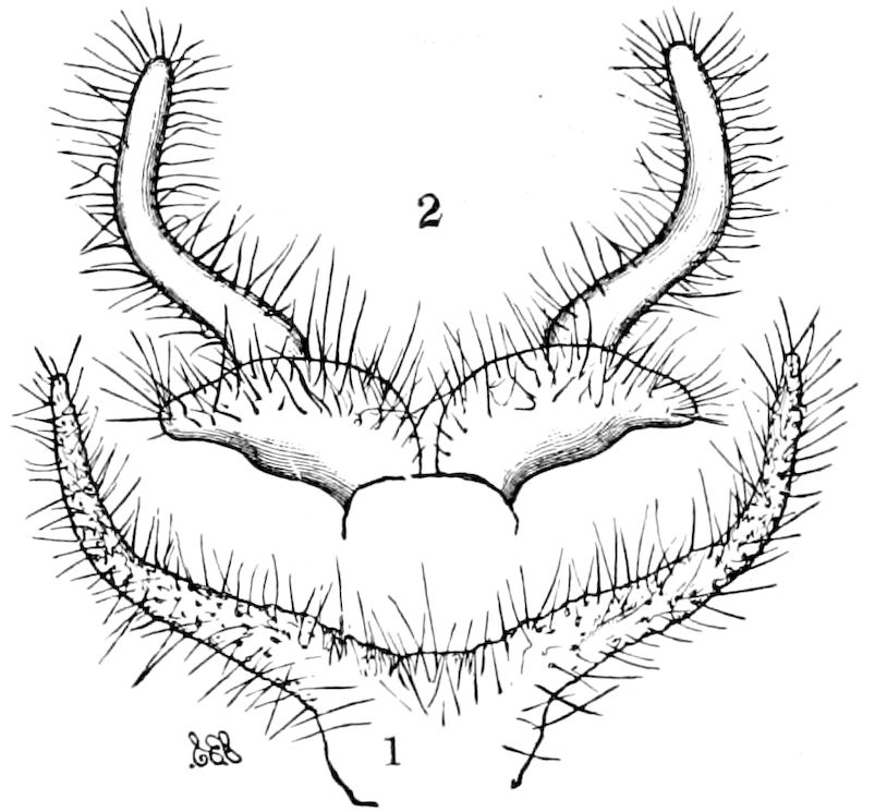
Fig. 367.—Scent-tufts: 1, of Leucarctia acræa; 2, of Pyrrarctia isabella.—After Smith.
In the noctuid genus, Patula, the costal half of the hind wing is modified to form a large scent-gland, and in consequence the venation has been modified. The still greater distortion of the veins in the allied genus, Argida, was attributed by the author to its once having possessed a similar scent-gland, now become rudimentary by disuse. (Hampson.)
Peculiar white or orange-colored, hairy, thread-like processes have been found protruding from narrow openings near the tip of the abdomen of Arctian moths (Fig. 367), which throw off, according to J. B. Smith, “an intense odor, somewhat like the smell of laudanum.” We have perceived the same unpleasant odor emanating from the males of Spilosoma virginica and Arctia virgo, as well as Leucarctia acræa.
We are informed by C. Dury that similar but longer hairy appendages are thrust out by the male of Haploa clymene. Many glaucopid moths protrude similar glandular processes. Thus Müller tells us that on seizing a glaucopid female by the wings, nearly the whole body became enveloped in a large cloud of snow-white wool which came out of a sort of pouch on the ventral side of the abdomen.
The male of a glaucopid was seen to dart out a pair of long hollow hairy retractile filaments which in some species exceed the whole body in length. The 392apparatus secretes a peculiar odor, probably serving to allure the female (Nature), and certain Zygænidæ have on the inner side of the paranal lobes (Afterklappen) glands filled with a sweetly scented fluid. Smith has detected a peculiar brush of hair-like scales in a groove between the dorsal and ventral parts of the basal two segments of the abdomen of Schinia marginata (family Noctuidæ), and when removed it exhaled a laudanum-like smell.
The pupa of Citheronia regalis gives out from the end of the abdomen a scent reminding us of laudanum.
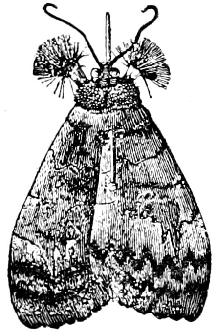
Fig. 368.—Scent-tufts on middle legs of Catocala concumbens.—After Bailey.
Another mode of disseminating pleasant, alluring odors is that of the males of certain moths, which bear pencils and tufts on their fore or hind legs, and in the case of an Indian butterfly on the greatly elongated palpi. Those on the legs are ordinarily concealed in cavities or furrows in the leg, and may be thrust out and expanded so as to widely diffuse their odor. Such are those of the males of Catocala (Fig. 368), which resemble an artist’s fitch brush. In Hepialus hecta, where the arrangements for protecting the tufts are quite abnormal, Bertkau has detected the cells which secrete the odorous fluid. In the male of another Hepialus (H. humuli) a peculiar scent proceeds from the curiously aborted and altered hind tibiæ. (Barrett.) In one case, that of a geometrid moth (Bapata dichroa of New Guinea), these pencils occur on all the legs. (Haase). In many species a distinct odor is perceptible when the leg bearing the pencil or tuft is crushed.
These eversible scent-glands have been supposed to be mostly restricted to the Lepidoptera, and to a single known case in the Trichoptera, but similar alluring male glands also occur in the Orthoptera (Locustidæ). H. Garman has described and figured in the cave cricket (Hadenœcus subterraneus) “a pair of white fleshy appendages protruding from slits between the terga of the 9th and 10th abdominal somites, the nature of which is not clear,” adding, “the slits through which the organs appear are situated one on each side anterior to and a little within the cerci. When fully protruded, the glands are white, cylindrical, a little tapering, and are about one-eighth of an inch long.” He believes that they are protruded during the period of sexual excitement, and suggests that “the sense of smell is certainly the one best calculated to bring the sexes together in the darkness of caves.” We had previously noticed these organs in alcoholic specimens, but supposed that they were fungous growths. 393On dissecting and making microscopic sections of them, the gland is, when extended (Fig. 369), seen to be a long, ensiform, sharp, band-like process, with numerous retractor muscular fibres. When at rest each gland is folded about five times, forming a bundle lying on each side of the end of the intestine. The walls are formed of a single layer of epithelium, as seen in Fig. 369, B.
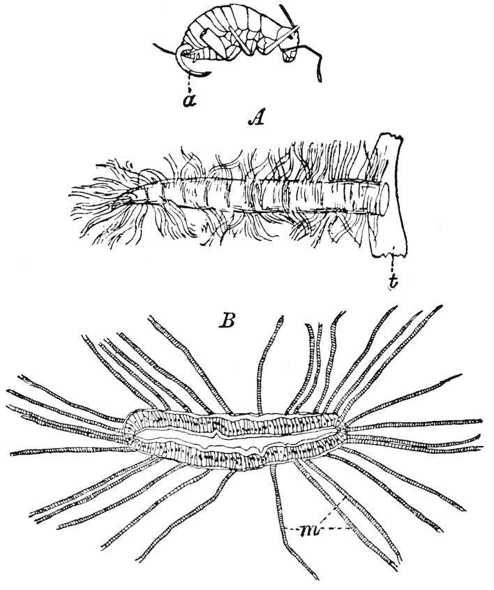
Fig. 369.—Eversible scent-glands (a) of Hadenœcus, nat. size: Kingsley, del.; A, a gland outstretched, with the retractor muscular fibres; t, part of the tergite. B, section of the gland, showing the single layer of epithelial cells, and the muscular fibres (m).—Author del.
In the male of the common wingless cricket, Ceuthophilus maculatus, we have discovered what appears to be a pair of scent-glands lying directly over the last abdominal ganglion. They form two large white sacs situated close together, with a short common duct which passes back and opens externally upwards by a transverse slit on the under side of the last segment of the body.
394
LITERATURE ON ALLURING GLANDS
Watson, J. On the microscopical examination of plumules, etc. (Ent. Month. Mag., ii, 1865, p. 1.)
—— On certain scales of some diurnal Lepidoptera. (Mem. Lit. and Phil. Soc. Manchester, Ser. 3, ii, 1868, p. 63.)
—— On the plumules or battledore scales of Lycænidæ. (Mem. Lit. and Phil. Soc. Manchester, Ser. 3, iii, 1869, p. 128.) Further remarks, etc. (Ibid., p. 259.)
Anthony, J. Structure of battledore scales. (Month. Microsc. Journ., vii, 1872, p. 250; see also p. 200.)
Morrison, Herbert Knowles. On an appendage of the male Leucarctia acræa. (Psyche, i, pp. 21–22, October, 1874.)
Müller, Fritz. The habits of various insects. (Nature, June 11, 1874, pp. 102–103.)
—— Ueber Haarpinsel, Fitzflecke und ähnliche Gebilde auf den Flügeln männlicher Schmetterlinge. (Jena. Zeitschr. f. Naturw., 1877, xi, pp. 99–114.)
—— Beobachtungen an brasilianischen Schmetterlingen, ii. I. Die Duftschuppen der männlichen Maracujáfalter. (Kosmos, 1877, i, pp. 391–395, Figs. 5, 6.) II. Die Duftschuppen des männchens von Dione vanillæ. (Kosmos, ii, 1877, pp. 38–42, 7 Taf.)
—— As maculas sexuaes dos individuos masculinos das especies Danais erippus e D. gilippus. (Arch. Mus. Nac. Rio Janeiro, ii, 1877 (1878), pp. 25–29, 1 Pl.)
—— Die Duftschuppen der Schmetterlinge (nach dem “Kosmos” in Ent. Nachr., 1878, pp. 29–32, 109).
—— Wo hat der Moschusduft der Schwärmer seinen Sitz? (Kosmos, ii Jahrg., 1878, pp. 84, 85.)
—— Os orgaos odoriferos dos especias Epicalia acontius, Lin. e de Myscelia orsis, Dru. (Arch. Mus. Nac. Rio Janeiro, ii, 1879, pp. 31–35.)
—— Os orgaos odoriferos nas pernas de certos Lepidopteres. (Arch. Mus. Nac. Rio Janeiro, ii, 1879, pp. 37–46, 3 Pls.)
—— Os orgaos odoriferos da Antirrhœa archœa. (Arch. Mus. Nac. Rio Janeiro, iii, 1878, pp. 1–7, 1 Pl.)
—— A prega costal das Hesperideas. (Arch. Mus. Nac. Rio Janeiro, iii, 1880, pp. 41–50, 2 Pls.)
Weismann, August. Ueber Duftschuppen. (Zool. Anzeiger, i, 1878, pp. 98, 99.)
Arnhart, L. Sexundäre Geschlechtscharaktere von Acherontia atropos. (Verh. d. k. k. zool. bot. Ges. Wien, xxix, 1879, p. 54.)
Bertkau, Philipp. Duftapparat an Schmetterlingsbeinen. (Ent. Nachrichten, 1879, Jahrg., pp. 223, 224.)
—— Ueber den Duftapparat von Hepialus hecta. (Archiv f. naturg., xlviii Jahrg., 1882, pp. 363–370, Figs.; also in Biol. Centralblatt., ii Jahrg., 1882, pp. 500–502.)
—— Ergänzung (Duftvorrichtungen bei Lepidopteren). (Ent. Nachr., 1880, p. 206.)
—— Entomologische Mizellen. 1. Ueber Duftvorrichtungen einiger Schmetterlinge. (Verh. d. naturhist. Ver. d. preuss. Rheinlande und Westf., 1884, pp. 343–350.)
Reichenau, W. von. Der Duftapparat von Sphinx ligustri. (Ent. Nachr., 1880, p. 141; also Kosmos, iv Jahrg., 1880, pp. 387–390.)
395Fügner, R. Duftapparat bei Sphinx ligustri. (Ent. Nachr., 1880, p. 166.)
Lelievre, Ernest. (Note in Le Naturaliste, June 1, 1880. Both sexes of Thais polyxena emit an odorous exhalation. Notes on exhalation from Spilosoma fuliginosa.)
Hall, C. G. Peculiar odor emitted by Acherontia atropos. (Entomologist, London, xvi, p. 14.)
Åurivillius, Christopher. Ueber secundäre Geschlechtscharactere nordischer Tagfalter. (Stockholm, 1880, Bihang till K. Svensk. Vet. Akad. Handl., v, pp. 56, 3 Taf.)
—— Des caractères sexuels secundaires chez les papillons diurnes. (Ent. Tidskrift, 1880, pp. 163–166.)
—— Anteckningar om några skandinaviska fjärilarter. (Ent. Tidskr., iv, Årg., 1884, pp. 33–37; Résumé (French), ibid., pp. 55–57.)
Kirby, W. F. Fans on the fore legs of Catocala fraxini. (Papilio, ii, p. 84, 1882.)
Bailey, James S. Femoral tufts or pencils of hair in certain Catocalæ. (Papilio, ii, 1882, pp. 51, 52, 146; also in Stettin Ent. Zeitung, xliii, p. 392.)
Edwards, Henry. Fans on the feet of Catocaline moths. (Papilio, ii, p. 146, 1882.)
Stretch, R. H. Anal appendages of Leucarctia acræa. (Papilio, iii, pp. 41, 42, 1883, 1 Fig.)
Weed, Clarence M. Appendages of Leucarctia. (Papilio, iii, 1883, p. 84.)
Grote, Aug. R. Appendages of Leucarctia acræa. (Papilio, iii, 1883, p. 84.)
Haase, Erich. Ueber sexuelle Charactere bei Schmetterlingen. (Zeitschr. f. Ent., Breslau, N. F., 1885, pp. 15–19, 36–44; also Ent. Nachr., xi Jahrg., pp. 332, 333.)
—— Duftapparate indo-australischer Schmetterlinge. (Corresp. Blatt. Ent. Ver. Iris, Dresden, 1886, pp. 92–107, 1 Taf.; ibid., 1887, pp. 159–178; ibid., 1888, pp. 281–336.)
—— Ueber Duftapparate bei Schmetterlingen. (Sitzgsber. Nat. Ges. Iris, Dresden, 1886, pp. 9–10; Abstr. in Journ. R. Micr. Soc., vi, pp. 969–970, 1886.)
—— Der Duftapparate von Acherontia. (Zeitschr. f. Ent., Breslau, N. F., 1887, pp. 5–6.)
—— Dufteinrichtung indischer Schmetterlinge. (Zool. Anzeiger, 1888, pp. 475–481.)
Dalla Torre, K. W. von. Die Duftapparate der Schmetterlinge. (Kosmos, 1885, ii, pp. 354–364, 410–423; Abstr. by J. B. Smith in Proc. Ent. Soc., Washington, i, pp. 38, 1888.)
Smith, John B. Cosmosoma omphale. (Entomologica Americana, i, pp. 181–185, 1886. Describes and figures cavities in under side of 2d–4th abdominal segments of male, filled with a silky substance. This may be for display to attract ♀, as the whole mass must be very conspicuous when protruded. No odor noticed.)
—— Scent organs in some Bombycid moths. (Entomologica Americana, ii, No. 4, pp. 79–80, 1886. Describes and figures long, slender, forked hairy, orange or white, eversible glands, everted from between 7th and 8th segments of abdomen of ♂ of Leucarctia acræa, Pyrrharctia isabella, Scepsis fulvicollis, and Cosmosoma omphale.)
—— [Notes on odors and odoriferous structures of various moths and a note by L. O. Howard on odor of Dynastes.] (Proc. Ent. Soc., Washington, i, pp. 40, 55, 56.)
Müller, W. Duftorgane der Phryganiden. (Archiv f. Naturgesch., 1887, Jahrg. liii, pp. 95–97.)
396Pollack, W. Duftapparate der Hadena atriplicis und Litargyria. (xv Jahrb. Westphäl. Prov. Ver. Münster, 1887, p. 16.)
Patton, W. H. Scent-glands in the larva of Limacodes. (Can. Ent., 1891, xxiii, pp. 42, 43.)
Garman, H. On a singular gland possessed by the male Hadenœcus subterraneus. (Psyche, 1891, p. 105, 1 Fig.)
Barrett, C. G. Scent of the male Hepialus humuli. (Ent. Month. Mag., Ser. 2, iii, 1892, p. 217. Arises from the curiously aborted and altered hind tibiæ.)
Also the writings of Baillif, Duponchel, F. Müller, Scudder (Psyche, iii, p. 278, 1881), Burgess, Keferstein, Alpheraky, Plateau, Marshall and Nicéville, Wood-Mason, White, Hampson.
397
THE ORGANS OF CIRCULATION
Although Malpighi was the first to discover the heart in the young silkworm, it was not until 1826 that Carus proved that there was a circulation of blood in insects, which he saw flowing along each side of the body, and coursing through the wings, antennæ, and legs of the transparent larva of Ephemera, though three years earlier Herold demonstrated that the dorsal vessel of an insect is a true heart, pulsating and impelling a current of blood towards the head. This discovery was extended by Straus-Dürckheim, who discovered the contractile and valvular structures of the heart. It is noteworthy that both Cuvier and Dufour denied that any circulation, except of air, existed in insects; and so great an anatomist as Lyonet doubted whether the dorsal vessel was a genuine heart, though he pointed out the fact that there are no arteries and veins connected with this vessel. Another French anatomist, Marcel de Serres, thought that the dorsal vessel was merely the secreting organ of the fat-body.
The so-called peritracheal circulation claimed by Blanchard and by Agassiz has been shown by McLeod to be an anatomical impossibility, the view having first been refuted by Joly in 1849.
Except the aorta-like continuation in the thorax and head which divides into two short branches, there are, with slight exceptions (p. 405), no distinct arteries, such as are to be found in the lobster and other Crustacea, and no great collective veins, such as exist in Crustacea and in Limulus. This is probably the result of a reduction by disuse in the circulatory system, since in myriopods (Julidæ and Scolopendridæ) lateral arteries are said to diverge near the ostia.