PART I.—MORPHOLOGY AND PHYSIOLOGY
Reading edition

POSITION OF INSECTS IN THE ANIMAL KINGDOM
Although the insects form but a single class of the animal kingdom, they are yet so numerous in orders, families, genera, and species, their habits and transformations are so full of instruction to the biologist, and they affect human interests in such a variety of ways, that they have always attracted more attention from students than any other class of animals, the number of entomologists greatly surpassing that of ornithologists, ichthyologists, or the special students of any other class, while the literature has assumed immense proportions.
Insects form about four-fifths of the animal kingdom. There are about 250,000 species already named and contained in our museums, while the number of living and fossil species in all is estimated to amount to between one and two millions.
In their structure insects are perhaps more complicated than any other animals. This is partly due to the serial arrangement of the segments and the consequent segmental repetition of organs, especially of the external appendages, and of the muscles, the tracheæ, and the nerves. The brain is nearly or quite as complicated as that of the higher vertebrates, while the sense-organs, especially those of touch, sight, and smell are, as a rule, far more numerous and only less complex than those of vertebrates. Moreover, in their psychical development, certain insects are equal, or even superior, to any other animals, except birds and mammals.
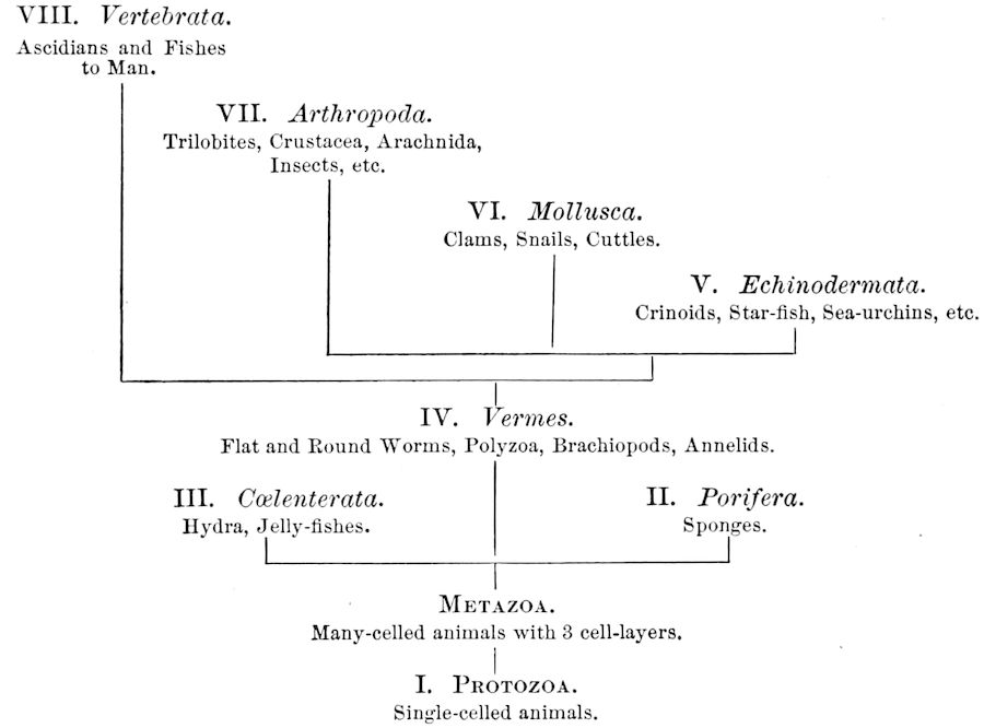
The animal kingdom is primarily divided into two grand divisions, the one-celled (Protozoa) and many-celled animals (Metazoa). In the latter group the cells and tissues forming the body are arranged in three fundamental cell-layers; viz. the ectoderm or outer layer, the mesoderm, and endoderm. The series of branches, or phyla, comprised 2under the term Metazoa are the Porifera, Cœlenterata, Vermes, Echinodermata, Mollusca, Arthropoda, and Vertebrata. Their approximate relationships may be provisionally expressed by the following
Tabular View of the Eight Branches or Phyla of the Animal Kingdom.
RELATIONS OF INSECTS TO OTHER ARTHROPODA
The insects by general consent stand at the head of the Arthropoda. Their bodies are quite as much complicated or specialized, and indeed, when we consider the winged forms, more so, than any other class of the branch, and besides this they have wings, fitting them for an aërial life. It is with little doubt that to their power of flight, and thus of escaping the attacks of their creeping arthropod enemies, insects owe, so to speak, their success in life; i.e. their numerical superiority in individuals, species, and genera. It is also apparently their power of moving or swimming swiftly from one place to another which has led to the numerical superiority in species of fishes to other Vertebrata. Among terrestrial vertebrates, the birds, by virtue of their ability to fly, greatly surpass in number of species the reptiles and mammals.
The Arthropoda are in general characterized by having the body 3composed of segments (somites or arthromeres) bearing jointed appendages. They differ from the worms in having segmented appendages, i.e. antennæ, jaws, and legs, instead of the soft unjointed outgrowths of the annelid worms. Moreover, their bodies are composed of a more or less definite number of segments or rings, grouped either into a head-thorax (cephalothorax) and hind-body, as in Crustacea, or into a head differentiated from the rest of the body (trunk), the latter not being divided into a distinct thorax and abdomen, as in Myriopoda; or into three usually quite distinct regions—the head, thorax, and hind-body or abdomen, as in insects. In certain aberrant, modified forms, as the Tardigrada, or the Pantopoda, and the mites, the body is not differentiated into such definite regions.
In their internal organs arthropods agree in their general relations with the higher worms, hence most zoölogists agree that they have directly originated from the annelid worms.
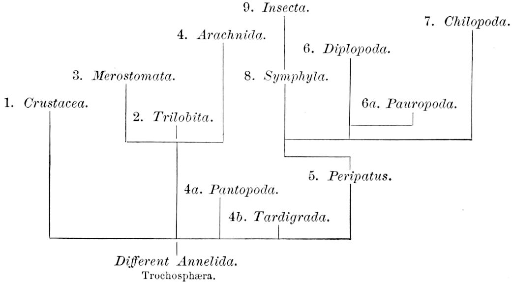
The position and general shape of the digestive canal, of the nervous and circulatory systems, are the same in Arthropoda as in annelid (oligochete) worms, so much so that it is generally thought that the Arthropoda are the direct descendants of the worms. It is becoming evident, however, that there was no common ancestor of the Arthropoda as a whole, and that the group is a polyphyletic one. Hence, though a convenient group, it is a somewhat artificial one, and may eventually be dismembered into at least three or four phyla or branches.
The following diagram may serve to show in a tentative way the relations of the classes of Arthropoda to each other, and also may be regarded as a provisional genealogical tree of the branch.
4We will now rapidly review the leading features of the classes of Arthropoda.
The Crustacea.—These Arthropoda are in many most important characteristics unlike the insects; they have two pairs of antennæ, five pairs of buccal appendages, and they are branchiate Arthropoda. They have evidently originated entirely independently, and by a direct line of descent from some unknown annelid ancestor which was either a many-segmented worm, with parapodia, or the two groups together with the Rotifera may have originated from a common appendigerous Trochosphæra. Their segments in the higher forms are definite in number (23 or 24) and arranged into two regions, a head-thorax (cephalothorax) and hind-body (abdomen). Nearly all the segments, both of the cephalothorax and abdomen, bear a pair of jointed limbs, and to them at their base are, in the higher forms, appended the gills (branchiæ). The limbs are in the more specialized forms (shrimps and crabs) differentiated into eye-stalks, two pairs of antennæ, a pair of palpus-bearing jaws (mandibles), two pairs of maxillæ and three pairs of maxillipeds; these appendages being biramose, and the latter bearing gills attached to their basal joints. The legs are further differentiated into ambulatory thoracic legs and into swimming or abdominal legs, and in the latter the first pair of the male is modified into copulatory organs (gonopoda). The male and female reproductive organs as a rule are in separate individuals, hermaphrodites being very unusual, and the glands may be paired or single. The sexual outlets are generally paired, and, as in the male lobster and other Macrura, open in the basal joint of the last pair of legs, and in the female in the third from the last; while originally in all Crustacea the sexual organs were most probably paired (Fig. 3, B).
They are, except a few land Isopoda, aquatic, mostly marine, and when they have a metamorphosis, pass through a six-legged larval stage, called the Nauplius, the shrimps and crabs passing through an additional stage, the Zoëa. Crustacea also differ much from insects in the highly modified nature of the nephridia, which are usually represented by the green gland of the lobster, or the shell-glands of the Phyllopoda, which open out in one of the head-segments; also in the possession of a pair of large digestive glands, the so-called liver.
Intermediate in some respects between the Crustacea and insects, but more primitive, in respect to what are perhaps the most weighty characters, than the Crustacea, are the Trilobita, the Merostomata (Limulus), and, finally, the Arachnida, these being allied groups. In the Trilobita and Merostomata (Limulus), the head-appendages are 5more like feet than jaws, while they have in most respects a similar mode of embryonic development, the larval forms being also similar.
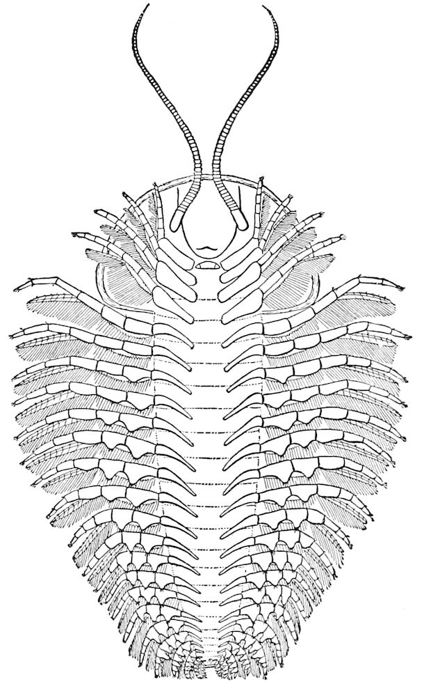
Fig. 1.—Restoration of under side of a trilobite (Triarthrus becki), the trunk limbs bearing small triangular respiratory lobes or gills.—After Beecher.
The Merostomata.—The only living form, Limulus, is undoubtedly a very primitive type, as the genital glands and ducts are double, opening wide apart on the basal pair of abdominal legs (Fig. 3). Moreover, their head-appendages, which are single, with spines on the basal joint, are very primitive and morphologically nearer in shape to those of the worms (Syllidæ, etc.) than even those of the Crustacea. Besides, their four pairs of coxal glands, with an external opening at the base of the fifth pair of head-appendages, and which probably are modified nephridia (Crustacea having but a single pair in any one form, either opening out on the second antennal, green gland, or second maxillary, shell-gland, segment), indicate a closer approximation to the polynephrous worms. Limulus has other archaic features, especially as regards the structure of the simple and compound eyes and the simple nature of the brain.
The Trilobita.—These archaic forms are still more generalized and primitive than the Merostomata and Crustacea, and probably were the first Arthropoda to be evolved from some unknown annelid worm. They had jointed biramose limbs of nearly uniform shape and size on each segment of the body, which were not, as in Crustacea, differentiated into antennæ, jaws (mandibles), maxillæ, maxillipeds, and two kinds of legs (thoracic and 6abdominal), showing that they are a much more primitive type, and nearer to the annelids than any other Arthropoda. Their gills, as shown by the researches of Walcott and of Beecher, were attached to nearly if not every pair of limbs behind the antennæ (Figs. 1, 2). The fact that in Trilobita the first pair of limbs is antenniform does not prove that they are Crustacea, since Eurypterus has a similar pair of appendages.
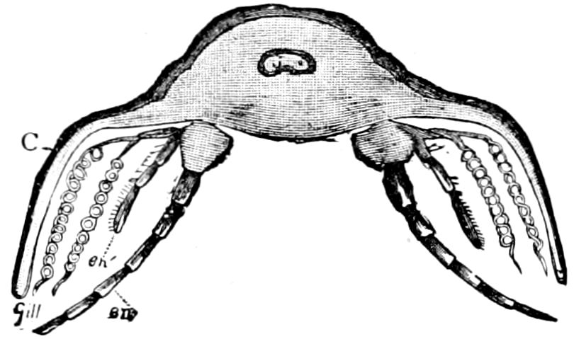
Fig. 2.—Restored section of Calymene: C, carapace; en, endopodite; en′, exopodite; with the gills on the epipodal or respiratory part of the appendage.—After Walcott.
The limbs in trilobites, as well as the abdominal ones of merostomes, and all those of Crustacea, except the first antennæ, are biramose, consisting of an outer (exopodite) and an inner division (endopodite). In this respect the terrestrial air-breathing tracheate forms, Arachnida, Myriopoda, and Insecta, differ from the branchiate forms, as their legs are single or undivided, being adapted for supporting the body during locomotion upon the solid earth. It is to be observed that when, as in Limulus, the body is supported by cephalic ambulatory limbs, they are single, while the abdominal limbs, used as they are in swimming, are biramose, much as in Crustacea.
The Arachnida.—The scorpions and spiders are much less closely allied to the myriopods and insects than formerly supposed. Their embryology shows that they have descended from forms related to Limulus, possibly having had an origin in common with that animal, or having, as some authors claim, directly diverged from some primitive eurypteroid merostome. But they differ in essential respects, and not only in the nature and grouping of their appendages; the first pair instead of antenniform being like mandibles, and the second pair like the maxillæ, with the palps, of insects, the four succeeding segments (thoracic) bearing each a pair of legs. They also have a brain quite unlike that of Limulus, the nervous cord behind the brain, however, being somewhat similar, though that of Limulus differs in being enveloped by an arterial coat. Arachnida respire by tracheæ, besides book-lungs, which, however, are possibly derivatives of the book-gills of Limulus, while they perform the office of excretion by means of the malpighian tubes, and like Limulus possess two large digestive glands (“liver”). Their embryos have, on at least six abdominal segments, rudiments of limbs, three pairs of which form the spinnerets, showing their origin from Limulus-like or eurypteroid forms; their coxal glands are retained from their eurypteroid ancestors. The Arachnida probably descended from marine merostomes, and not from an independent annelid ancestry, hence we have represented them in the diagram on p. 3 as branching off from the merostomatous phylum, rather than from an independent one.
7
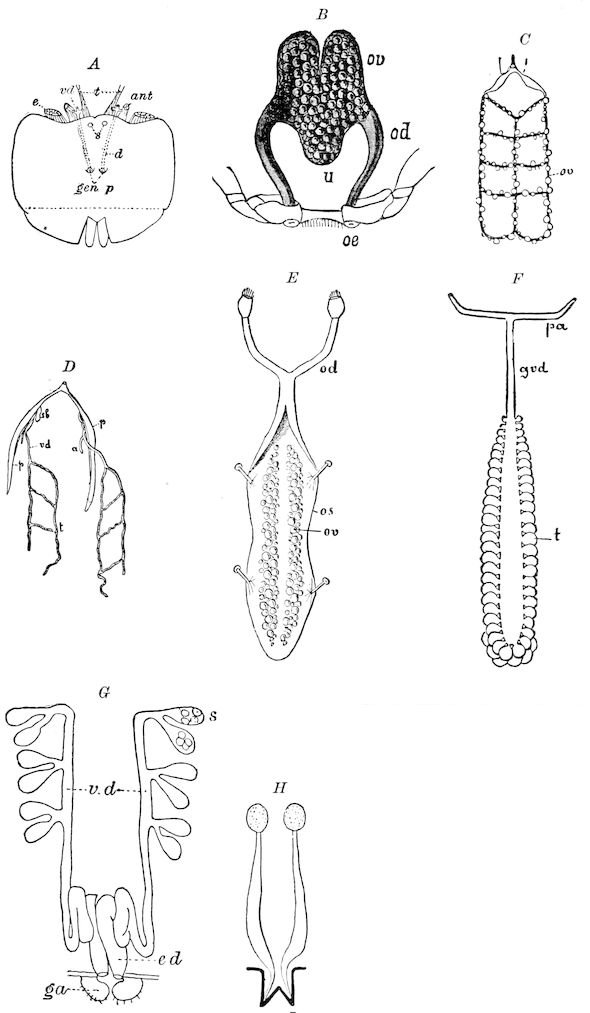
Fig. 3.—Paired genital openings of different classes of arthropods. A, the most primitive, of Limulus polyphemus: gen. p, generative papillæ; d, duct; vd, vas deferens; t, tendinous stigmata; stig, stigmata; e, external branchial muscle; ant, anterior lamellar muscle.—After Benham, with a few changes. B, lobster (Homarus vulgaris), ♀: oe, genital aperture on 3d pair of legs; ov, ovary; u, unpaired portion of the same; od, oviduct. C, ♀, scorpion: ov, ovary, with a single external opening. D, ♂: t, testis; vd, vasa deferentia; sb, seminal vesicle; a, glandular appendage; p, penis.—After Blanchard. E, a myriopod (Glomeris marginata, ♀): os, ovarian sac, laid open; od, paired oviducts. F, ♂: t, testis; gvd, common vas deferens; pa, paired ducts.—After Favre, from Lang. G, Lepisma saccharina, young ♂: vd, vas deferens, ed, ejaculatory duct; ga, external appendages.—After Nassonow. H, Ephemera, ♂, showing the double outlets.—After Palmén.
8The characters in which arachnids approach insects, such as tracheæ and malpighian tubes (none occur, as a rule, in marine or branchiate arthropods), may be comparatively recent structures acquired during a change from a marine to a terrestrial life, and not primitive heirlooms.
Arachnida also show their later origin than merostomes by the fact that their sexual glands are in most cases single, and though with rare exceptions the ducts are paired, these finally unite and open externally by a common single genital aperture in the median line of the body, at the base of the abdomen (Fig. 3, C, D). In this respect Limulus, with its pair of genital male or female openings, situated each at the end of a papilla, placed widely apart at the base of the first abdominal limbs, is decidedly more archaic. Unlike Crustacea and insects, Arachnida do not, except in the mites (Acarina), which is a very much modified group, undergo a metamorphosis.
We see, then, that the insects, with the Myriopoda, are somewhat isolated from the other Arthropoda. The Myriopoda have a single pair of antennæ, and as they have other characters in common with insects, Lang has united the two groups in a single class Antennata; but, as we shall see, this seems somewhat premature and unnecessary. Yet the two groups have perhaps had a common parentage, and may prove to belong to a distinct, common phylum.
Not only by their structure and embryology, as well as their metamorphosis, do the myriopods and insects stand apart from the Arachnida and other arthropods, but it seems probable that they have had a different ancestry, the arthropods being apparently polyphyletic.
There are two animals which appear to connect the insects with the worms, and which indicate a separate line of descent from the worms independent of that of the other classes. These are the singular Peripatus, which serves as a connecting link between arthropods and worms, and Scolopendrella (Symphyla). These two animals are guide-posts, pointing out, though vaguely to be sure, the way probably trod by the forms, now extinct, which led up to the insects.
9Relations of Peripatus to Insects.—We will first recount the characteristics of this monotypic class. Peripatus (Fig. 4) stands alone, with no forms intermediate between itself and the worms on the one hand, and the true Arthropoda on the other. Originally supposed to be a worm, it is now referred to a class by itself, the Malacopoda of Blainville, or Protracheata of Haeckel. It lives in the tropics, in damp places under decaying wood. In general appearance it somewhat resembles a caterpillar, but the head is soft and worm-like, though it bears a pair of antenna-like tentacles. It may be said rather to superficially resemble a leech with clawed legs, the skin and its wrinkles being like those of a leech. There is a pair of horny jaws in the mouth, but these are more like the pharyngeal teeth of worms than the jaws of arthropods. The numerous legs end each in a pair of claws. The ladder-like nervous system is unlike that of annelid worms or arthropods, but rather recalls that of certain molluscs (Chiton, etc.), as well as that of certain flat and nemertine worms. Its annelid features are the large number of segmentally arranged true nephridia, and the nature of the integument. Its arthropodan features, which appear to take it out of the group of worms, are the presence of tracheæ, of true salivary and slime glands, of a pair of coxal glands (Fig. 4, C, cd) as well as the claws at the end of the legs. The tracheæ, which are by no means the only arthropodan features, are evidently modified dermal glands. The heart is arthropodan, being a dorsal tube lying in a pericardial sinus, with many openings. This assemblage of characters is not to be found in any marine or terrestrial worm.
The tracheæ (Fig. 4, D, tr) are unbranched fine tubes, without a “spiral thread,” and are arranged in tufts, in P. edwardsii opening by simple orifices or pores (“stigmata”) scattered irregularly over the surface of the body; but in another species (P. capensis) some of the stigmata are arranged more definitely in longitudinal rows,—on each side two, one dorsally and one ventrally. “The stigmata in a longitudinal row are, however, more numerous than the pairs of legs.” (Lang.)
The salivary glands, opening by a short common duct into the under side of the mouth, in the same general position as in insects, are evidently, as the embryology of the animal proves, transformed nephridia, and being of the arthropodan type explain the origin and morphology of those of insects. It is so with the slime glands; these, with the coxal glands, being transformed and very large dermal glands. Those of insects arose in the same manner, and are evidently their homologues, while those of Peripatus were probably originally derived from the setiparous glands in the appendages (parapodia) of annelid worms.
10
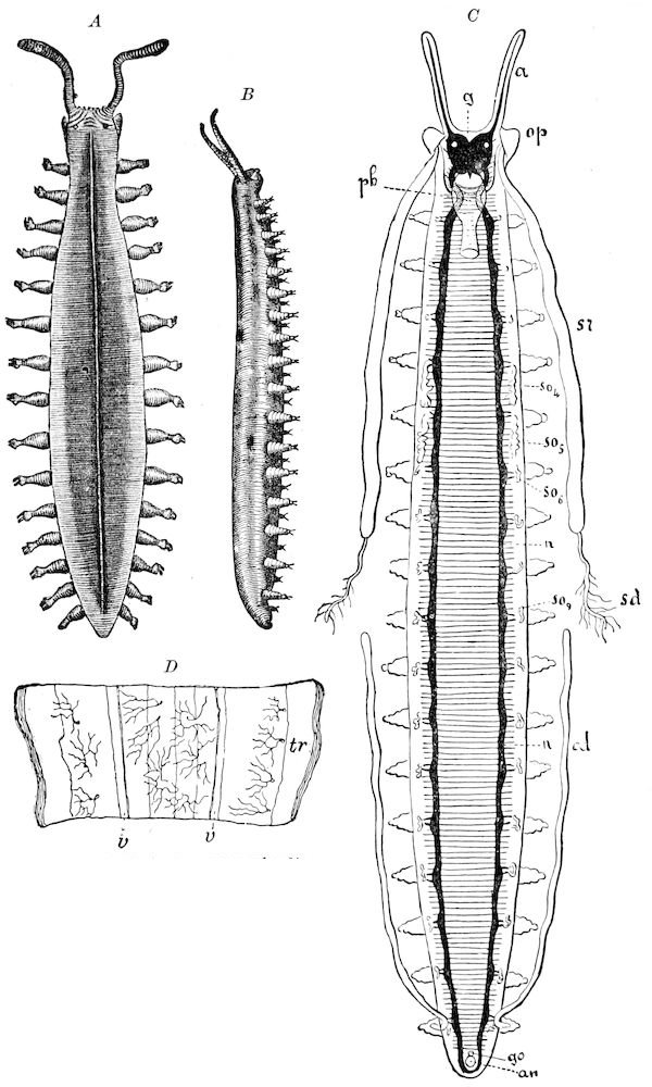
Fig. 4.—A, Peripatus novæ zealandiæ.—After Sedgwick, from Lang. B, Peripatus capensis, side view, enlarged about twice the natural size.—After Moseley, from Balfour. C, Anatomy of Peripatus capensis. The enteric canal behind the pharynx has been removed. g, brain; a, antenna; op, oral or slime papillæ; sd, slime gland; sr, slime reservoir, which at the same time acts as a duct to the gland; so4, so5, so6, so9, nephridia of the 4th, 5th, 6th, and 9th pairs of limbs; cd, elongated coxal gland of the last pair of feet; go, genital aperture; an, anus; ph, pharynx; n, longitudinal trunk of the nervous system.—After Balfour, from Lang. D, Portion of the body of Peripatus capensis opened to show the scattered tufts of tracheæ (tr); v, v, ventral nerve cords.—After Moseley.
11The genital glands and ducts are paired, but it is to be observed that the outlets are single and situated at the end of the body. In the male the ejaculatory duct is single; in its base a spermatophore is formed. It will be seen, then, that Peripatus is not only a composite type, and a connecting link between worms and tracheate arthropods, but that it may reasonably be regarded, if not itself the ancestor, as resembling the probable progenitor of myriopods and insects, though of course there is a very wide gap between Peripatus and the other antennate, air-breathing Arthropoda.
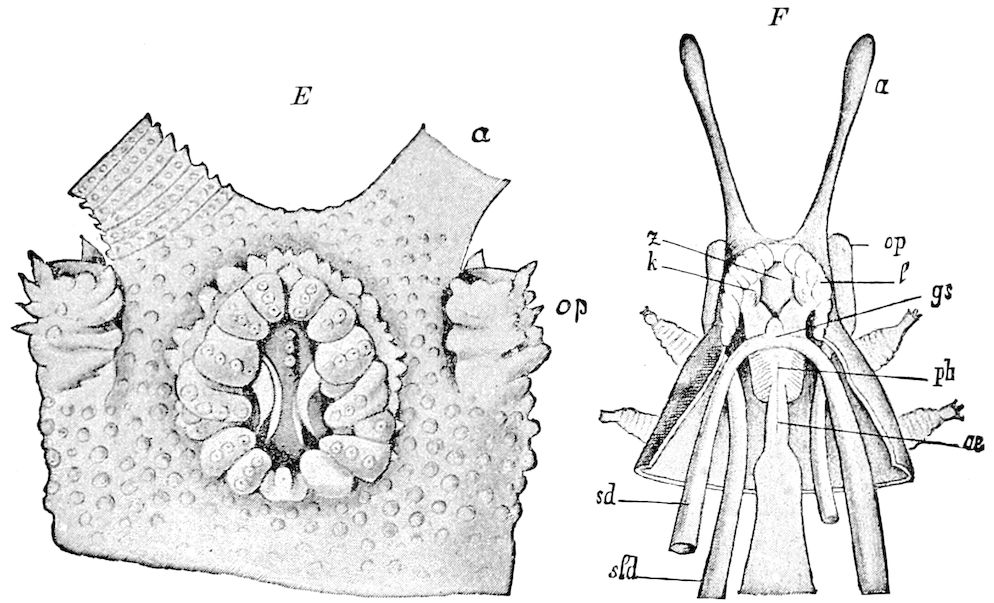
Fig. 4.—E, Peripatus edwardsii, head from the under side: a, base of antenna; op, oral papilla; the figure also shows the papillæ around the mouth, and the four jaws.—After Balfour, from Lang. F, Anterior end of Peripatus capensis, ventral side, laid open: a, antenna; z, tongue; k, jaw; sd, salivary gland; gs, union of the two salivary glands; ph, pharynx; œ, œsophagus; l, lip papillæ around the mouth; op, oral or slime papilla; sld, duct or reservoir of the slime gland.—After Balfour, from Lang.
Relation of Myriopods to Insects.—The Myriopoda are the nearest allies of the insects. They have a distinct head, with one pair of antennæ. The eyes are simple, with the exception of a single genus (Cermatia), in which they are aggregated or compound. The trunk or body behind the head is, as a rule, long and slender, and composed of a large but variable number of segments, of equal size and shape, bearing jointed legs, which invariably end in a single claw.
The mouth-parts of the myriopods are so different in shape and general function from those of insects, that this character, together with the equally segmented nature of the portion of the body behind the head (the trunk), forbids our merging them, as some have been 12inclined to do, with the insects. There are two sub-classes of myriopods, differing in such important respects that by Pocock[1] and by Kingsley they are regarded as independent classes, each equivalent to the insects.
Of these the most primitive are the Diplopoda (Chilognatha), represented by the galley-worms (Julus, etc.).
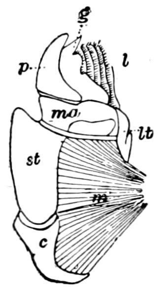
Fig. 5.—Mandible of Julus: l, lacinia; g, galea; p, dens mandibularis; ma, “mala”; lt, lamina tritoria; st, stipes; c, cardo; m, muscle.—After Latzel.
In the typical Diplopoda the head consists of three segments, a preoral or antennal, and two postoral, there being two pairs of jaw-like appendages, which, though in a broad morphological sense homologues of the mandibles and first maxillæ of insects, are quite unlike them in details.
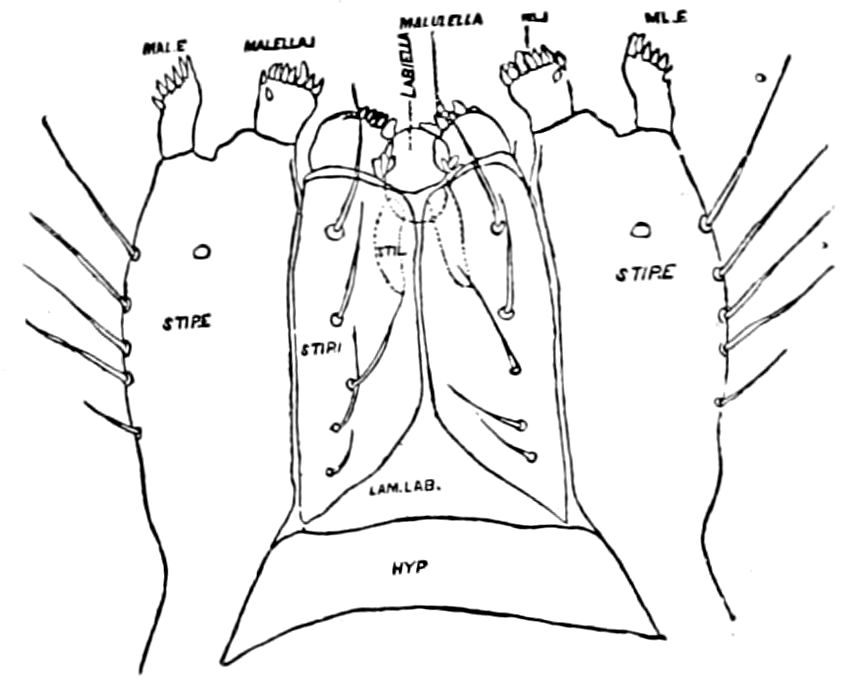
Fig. 6.—Under lip or deutomala of Scoterpes copei: hyp, hypostoma or mentum; lam. lab, lamina labialis; stip. e, stipes exterior; with the malella exterior (mal. e) and malella interior (mal. i); the stipes interior, with the malulella; and the labiella (hypopharynx of Vom Rath) with its stilus (stil.).
As we have previously stated,[2] the so-called “mandibles” of diplopods are entirely different from those of insects, since they appear to be 2– or 3–jointed, the terminal joint being 2–lobed, thus resembling the maxillæ rather than the mandibles of insects, which consist of but a single piece or joint, probably the homologue of the galea or molar joint of the diplopod protomala. The mandible of the Julidæ (Fig. 5, Julus molybdinus), Lysiopetalidæ, and Polydesmidæ consists of three joints; viz. a basal piece or cardo, a stipes, and the mala mandibularis, which supports two lobes analogous to the galea and lacinia of the maxilla of an insect. There is an approach, as we shall see, in the mandible of Copris, to that of the Julidæ, but in insects in general the lacinia is wanting, and the jaw consists of but a single piece.
The deutomalæ (gnathochilarium), or second pair of diplopod jaws, are analogous to the labium or second maxillæ of insects, forming a flattened, plate-like under-lip, constituting the floor of the mouth (Fig. 6). This pair of appendages needs farther study, especially in the late embryo, before it can be fully understood. So far as 13known, judging by Metschnikoff’s work on the embryology of the diplopods, these myriopods seem to have in the embryo but two pairs of post-antennal mouth-parts, which he designated as the “mandibles” and “labium.” Meinert, however, regards as a third pair of mouth-parts or “labium” what in our Fig. 7 is called the internal stipes (stip. i.), behind which is a triangular plate, lamina labialis (lam. lab), which he regards as the sternite of the same segment.
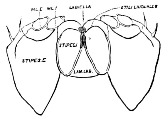
Fig. 7.—Deutomala of Julus, the lettering as in Fig. 6.
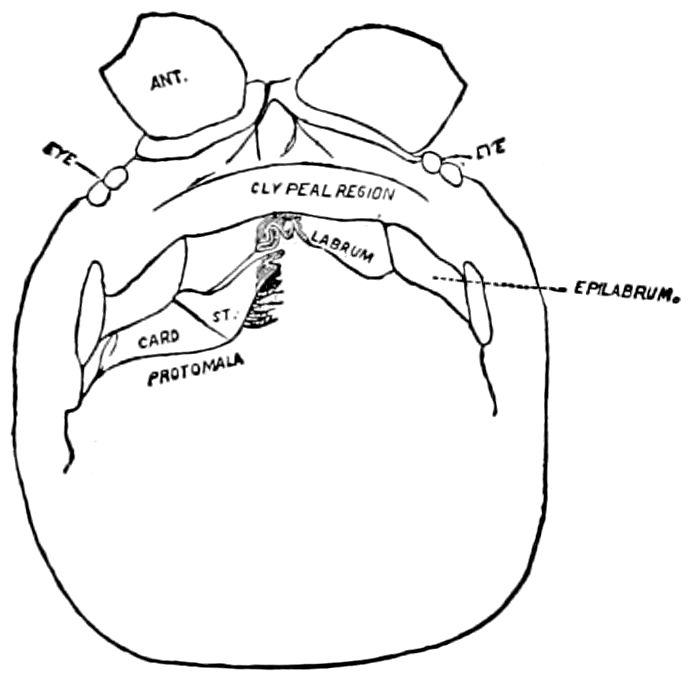
Fig. 8.—Head of Scolopendra, seen from beneath, showing the “mandible” (protomala) with its cardo (card.) and stipes (st.), also the labrum and epilabrum.
The hypopharynx, our “labiella,” (Fig. 6), with the supporting rods or stili linguales (sti. l), of Meinert, are of nearly the same shape as in some insects.
Of the clypeus of insects there is apparently no homologue in myriopods, though in certain diplopods there is an interantennal clypeal region. The labium of insects is represented by a short, broad piece, which, however, unlike that of insects, is immovable, and is flanked by a separate piece called the epilabrum (Fig. 8). Vom Rath has observed an epipharynx, which has the same general relations as in insects.
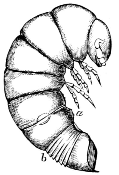
Fig. 9.—Larva of Julus: a, the 3d abdominal segment, with the new limbs just budding out; b, new segments arising between the penultimate and the last segment.—After Newport.
The embryology of myriopods is in many respects like that of insects. The larva of diplopods hatches with but few segments, and with but three pairs of limbs; but these are not, as in insects, appended to consecutive segments, but in one species the third, and in another, Julus multistriatus? (Fig. 10), the second, segment from the head is footless, while Vom Rath represents the first segment of an European Blaniulus as footless, the feet being situated consecutively on segments 2 to 4. The new segments arise at “the growing point” situated between the last and penultimate segment, growing out in groups of sixes (Newport) or in our Julus multistriatus? in fives (Fig. 10). In 14adult life diplopods (Julus) have a single pair of limbs on the three first segments, or those corresponding to the thoracic segments of insects, the succeeding segments having two pairs to each segment.
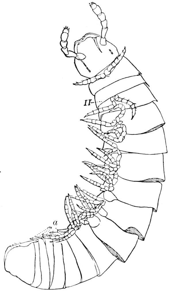
Fig. 10.—Freshly hatched larva of Julus multistriatus? 3 mm. long: a, 5 pairs of rudimentary legs, one pair to a segment.
Sinclair (Heathcote) regards each double segment in the diplopods as not two original segments fused together, nor a single segment bearing two pairs of legs, but as “two complete segments perfect in all particulars, but united by a large dorsal plate which was originally two plates which have been fused together.” (Myriopods, 1895, p. 71.) That the segments were primitively separate is shown, he adds, by the double nature of the circulatory system, the nerve cord, and the first traces of segmentation in the mesoblast. Kenyon believes that from the conditions in pauropods, Lithobius, etc., there are indications of alternate plates (not segments) having disappeared, and of the remaining plates overgrowing the segments behind them, so as to give rise to the anomalous double segments.[3]
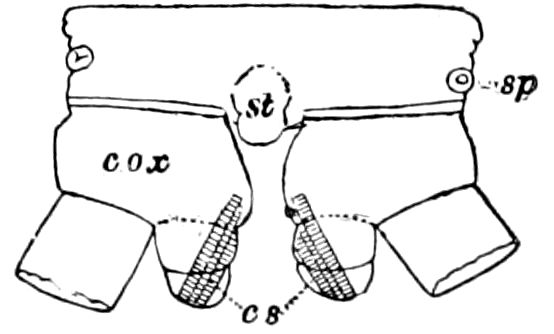
Fig. 11.—Sixth pair of legs of Polyzonium germanicum, ♀: cs, ventral sacs; cox, coxa; st, sternal plate; sp, spiracle.—After Haase.
Diplopods are also provided with eversible coxal sacs, in position like those of Symphyla and Synaptera; Meinert, Latzel, and also Haase having detected them in several species of Chordeumidæ, Lysiopetalidæ, and Polyzonidæ (Fig. 11). In Lysiopetalum anceps these blood-gills occur in both sexes between the coxæ of the third to sixteenth pair of limbs. In the Diplopods the blood-gills appear to be more or less permanently everted, while in Scolopendrella they are usually retracted within the body (Fig. 15, cg).
Diplopods also differ externally from insects in the genital armature, a complicated apparatus of male claspers and hooks apparently arising from the sternum of the sixth segment and being the modified seventh pair of legs. In myriopods 15there are no pleural pieces or “pleurites,” so characteristic of winged insects.
Perhaps the most fundamental difference between diplopods and insects is the fact that the paired genital openings of the former are situated not far behind the head between the second and third pair of legs. Both the oviducts and male ejaculatory ducts are paired, with separate openings. The genital glands lie beneath, while in chilopods they lie above the intestine; this, as Korschelt and Heider state, being a more primitive relation, since in Peripatus they also lie above the digestive canal.
The nervous system of diplopods is not only remarkable for the lack of the tendency towards a fusion of the ganglia observable in insects, but for the fact that the double segments are each provided with two ganglia. The brain also is very small in proportion to the ventral cord, the nervous system being in its general appearance somewhat as in caterpillars.
The arrangement of the tracheæ and stigmata is much as in insects, but in the Diplopoda the tracheary system is more primitive than in chilopods, a pair of stigmata and a pair of tracheal bundles occurring in each segment, while the bundles are not connected by anastomosing branches, branched tracheæ only occurring in the Glomeridæ. The tracheæ themselves are without spiral threads (tænidia). It is noteworthy that the tracheæ arise much later than in insects, not appearing until the animal is hatched; in this respect the myriopods approximate Peripatus.
In the Chilopoda also the parts of the head, except the epicranium, are not homologous with those of insects, neither are the mouth-parts, of which there are five pairs.
The structure of the head of centipedes is shown in part in Fig. 12, compare also Fig. 8. It will be seen that it differs much from that of the diplopods, though the mandibles (protomalæ) are homologous; they are divided into a cardo and stipes, thus being at least two-jointed.
The second pair of postoral appendages is in centipedes very different from the gnathochilarium of diplopods. As seen in Fig. 12 2, they are separate, cylindrical, fleshy, five-jointed appendages, the maxillary appendages of Newport, which are “connected transversely at their base with a pair of soft appendages” (c), the lingua of Newport. The third and fourth pair are foot-jaws, and we have called them malipedes, as they have of course no homology with the maxillipedes of Crustacea. The second pair of these malipedes, forming the last pair of mouth-appendages, is the poison-fangs (4), 16which are intermediate between the malipedes and the feet; Meinert does not allow that these are mouth-appendages.
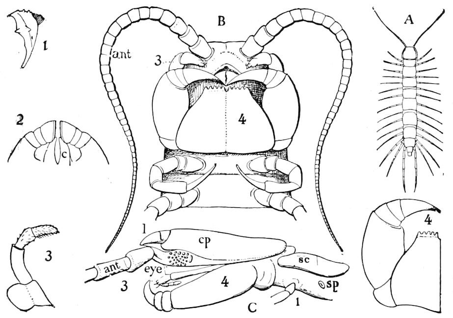
Fig. 12.—Structure of a chilopod. A, Lithobius americanus, natural size. B, under side of head and first two body-segments and legs, enlarged: ant, antenna; 1, jaws; 2, first accessory jaw; c, lingua; 3, second accessory jaw and palpus; 4, poison-jaw. (Kingsley del.) C, side view of head (after Newport): ep, epicranium; l, frontal plate; sc, scute; 1, first leg; sp, spiracle.
The embryology of Geophilus by Metschnikoff shows plainly the four pairs of post-antennal appendages. The embryo Geophilus is hatched in the form of the adult, having, unlike the diplopods, no metamorphosis, its embryological history being condensed or abbreviated. But in examining Metschnikoff’s figures certain primitive diplopod features are revealed. The body of the embryo shortly before hatching is cylindrical; the sternal region is much narrower than in the adult, hence the insertions of the feet are nearer together, while the first six pairs of appendages begin to grow out before the hinder ones. Thus the first six pairs of appendages of the embryo Geophilus correspond to the antennæ, two pairs of jaws, and three pairs of legs of the larval Julus. These features appear to indicate that the chilopods may be an offshoot from the diplopod stem. The acquisition of a second pair of legs to a segment in diplopods, as in the phyllopod Crustacea, is clearly enough a secondary character, as shown by the figures of Newport in his memoir on the development of the Myriopoda (Pl. IV.). Thus the tendency in the Myriopoda, both diplopods and chilopods, is towards the multiplication of segments and the elongation of the body, while in insects the polypodous embryo has the three terminal segments of the 17abdomen well formed, these being, however, before hatching, partly atrophied, so that the body of insects after birth tends to become shortened or condensed. This indicates the descent of insects from ancestors with elongated polypodous hind-bodies like Scolopendrella. Korschelt and Heider suggest that the stem-form of myriopods was a homonomously jointed form like Peripatus, consisting of a rather large number of segments, but we might, with Haase, consider that the great number of segments which we now find indicates a late acquisition of this form.
The genital opening in chilopods is single, and situated in the penultimate segment of the body, as in insects. While recognizing the close relationship of the Myriopoda with the insects, it still seems advisable not to unite them into a single group (as Oudemans, Lang, and others would do), but to regard them as forming an equivalent class. On the other hand, when we take into account the form and structure of the head, antennæ, and especially the shape of the first pair of mouth-appendages, being at least two-jointed in both groups, we think these characters, with the homonomously segmented body behind the head, outweigh the difference in the position of the genital outlet, important as that may seem. It should also be taken into account that while insects are derived from polypodous ancestors, no one supposes, with the exception of one or two authors, that these ancestors are the Myriopoda, the latter having evidently descended from a six-legged ancestor, quite different from that of the Campodea ancestor of insects.[4]
In regard to the sexual openings of worms, though their position is in general in the anterior part of the body, it is still very variable, though, in general, paired. In the oligochete worms the genital zone, with the external openings, is formed by the segments lying between the 9th and 14th rings, though in some the genital organs are situated still nearer the head. The myriopods, which evolved from the worms earlier than insects, appear to have in their most primitive forms (the Diplopoda) retained this vermian position of the genital outlets. In the later forms, the chilopods, the genital openings have been carried back to near the end of the body, as in insects. From observations made by three different observers on the freshly hatched larva of the Julidæ, it appears that the ancestral diplopods were six-footed, or oligopod, the larva of Pauropus 18(Fig. 13) approaching nearest to our idea of the ancestral myriopod, which might provisionally be named Protopauropus.
Relations of the Symphyla to Insects.—Opinions respecting the position of the Symphyla, represented by Scolopendrella (Fig. 14), are very discordant. By most writers since Newport, Scolopendrella has been placed among the myriopods. The first author, however, to examine its internal anatomy was Menge (1851), who discovered among other structures (tracheæ, etc.) the silk-glands situated in the last two segments, and which open at the end of each cercus. He regarded the form as “the type of a genus or family intermediate between the hexapod Lepismidæ and the Scolopendridæ.”
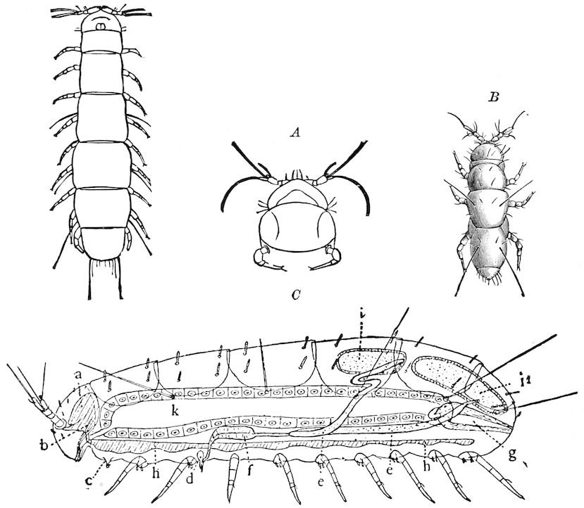
Fig. 13.—Pauropus huxleyi, much enlarged. A, enlarged view of head, antennæ, and first pair of legs (original). B, young.—After Lubbock. C, longitudinal section of Pauropus huxleyi, ♂: a, brain; b, salivary gland; k, mid-intestine; g, rectum; h, ventral nerve-cord; c, bud-like remnants of coxæ; d, penis; e, vesicula seminalis; f, ductus glandularis; i1, divisions of testes.—After Kenyon.
In 1873[5] the writer referred to this form as follows: “It may be regarded as a connecting link between the Thysanura and Myriopoda, 19and shows the intimate relation of the myriopods and the hexapods, perhaps not sufficiently appreciated by many zoölogists.”
In 1880 Ryder regarded it as “the last survival of the form from which insects may be supposed to have descended,” and referred it to “the new ordinal group Symphyla, in reference to the singular combination of myriopodous, insectean, and thysanurous characters which it presents.[6]”
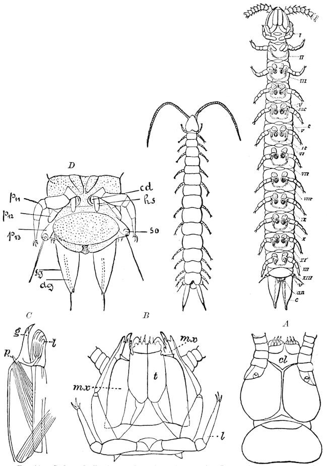
Fig. 14.—Scolopendrella immaculata, from above,—after Lang; also from beneath, the genital opening on the 4th trunk-segment: sac, eversible or coxal sac; an, anus; c, cereopod; v, vestigial leg.—After Haase, from Peytoureau. A B C, head and buccal appendages of Scolopendrella immaculata: A, head seen from above; cl, clypeus. B, head from beneath; l, first pair of legs; mx, 1st maxilla; mx1, 2d maxilla; t, “labial plates” of Latzel, labium of Muhr. C, 1st maxilla; l, lacinia; g, galea; p, rudiment of the palpus.—After Latzel. D, end of the body: p11, eleventh, p12, twelfth undeveloped pair of legs; p13, modified, vestigial legs, bearing tactile organs (so); sg, cercopod, with duct of spinning gland, dg; cd, eversible or coxal gland; h8s, coxal spur of the 11th pair of legs.—After Latzel from Lang.
Wood-Mason considered it to be a myriopod, and “the 20descendant of a group of myriopods from which the Campodeæ, Thysanura, and Collembola may have sprung.” We are indebted to Grassi for the first extended work on the morphology of Scolopendrella (1885). In 1886 he added to our knowledge facts regarding the internal anatomy, and gives a detailed comparison with the Thysanura, besides pointing out the resemblances of Scolopendrella to Pauropus, diplopods, chilopods, as well as Peripatus.
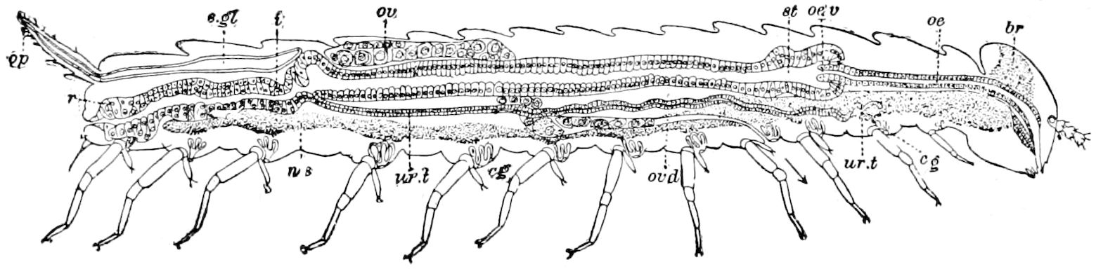
Fig. 15.—Section of Scolopendrella immaculata: œ, œsophagus; oe. v, œsophageal valve entering the mid-intestine (“stomach”); i, intestine; r, rectum; br, brain; ns, abdominal chain of ganglia; ovd, oviduct; ov, ovary; s. gl, silk-gland, and op, its outer opening in cercus, ur. t, urinary tube; cg, coxal glands or blood-gills.—Author del.
In 1888 Grassi expressed his view as to the position of the Symphyla, stating that it should not be included in the Thysanura, since it evidently has myriopod characters; these being the supraspinal vessel, the ventral position of the genital glands; the situation of the genital opening in the fourth segment of the trunk, its ganglionic chain being like that of diplopods, its having limbs on all the segments, etc. On the other hand, Grassi has with much detail indicated the points of resemblance to the Thysanura. The principal ones are the thin integument, the want of sympathetic ganglia, the presence of a pair of cephalic stigmata, like that said to occur in certain Collembola, and in the embryo of Apis; two endoskeletal processes situated near the ventral fascia of the head; the epicranial suture also occurring in Thysanura, Collembola, Orthoptera, and other winged insects, and being absent in diplopods and chilopods. He also adds that the digestive canal both in Symphyla and Thysanura is divided into three portions; the malpighian tubes in Thysanura present very different conditions (there being none in Japyx), among which may be comprised those of Scolopendrella. In both groups there is a single pair of salivary glands. The cellular epithelium of the mid-intestine of Scolopendrella is of a single form as in Campodea and Japyx. The fat-body, dorsal vessel, with its valves and ostia, are alike in the two groups, as are the appendages of the end of the abdomen, the anal cerci (cercopoda) of Scolopendrella being the homologues of the 21multiarticulate appendages of Lepisma, etc., and of the forceps of Japyx. In those of Scolopendrella, we have found the large duct leading from the voluminous silk-gland, a single large sac extending forwards into the third segment from the end of the body (Fig. 15, s. gl). Other points of resemblance, all of which he enumerates, are the slight differences in the number of trunk-segments, the presence in the two groups of the abdominal “false-legs” (parapodia), the dorsal plate, and the mouth-parts. As regards the latter, Grassi affirms that there is a perfect parallelism between those of Scolopendrella and Thysanura. To this point we will return again in treating more especially of those of the Symphyla. Finally, Grassi concludes that there is “a great resemblance between the Thysanura and Scolopendrella.” He, however, believed that the Symphyla are the forerunners of the myriopods, and not of the insects, his genealogical tree representing the symphylan and thysanuran phyla as originating from the same point, this point also being, rather strangely, the point of origin of the arachnidan phylum.
Haase (1889) regarded Scolopendrella as a myriopod, and Pocock (1893) assigned the Symphyla to an independent class, regarding Scolopendrella as “the living form that comes nearest to the hypothetical ancestor of the two great divisions of tracheates.” Schmidt’s work (1895) on the morphology of this genus is more extended and richly illustrated than Grassi’s, his method of research being more modern. He also regards this form as one of the lower myriopods.
In conclusion, it appears to us that, on the whole, if we throw out the single characteristic of the anteriorly situated genital opening, the ovarian tubes being directed toward the end of the body (Fig. 15, ovd, ov), there is not sufficient reason for placing the Symphyla among the Myriopoda, either below or near the diplopods. This is the only valid reason for not regarding Scolopendrella as the representative of a group from which the insects have descended, and which partly fills the wide abyss between Peripatus and insects. With the view of Pocock, that both insects and myriopods have descended from Scolopendrella, we do not agree, because this form has so many insectean features, and a single unpaired genital opening. For the same reason we should not agree with Schmidt in interpolating the Symphyla between the Pauropoda and Diplopoda. In these last two progoneate groups the genital openings are paired, hence they are much more primitive types than Scolopendrella, in which there is but a single opening. It seems most probable that the Symphyla, though progoneate, are more recent forms than the 22progoneate myriopods, which have retained the primitive feature of double sexual outlets. It is more probable that the Symphyla were the descendants of these polypodous forms. Certainly Scolopendrella is the only extant arthropod which, with the sole exception of the anteriorly situated genital opening, fulfils the conditions required of an ancestor of Thysanura, and through them of the winged insects. No one has been so bold as to suggest the derivation of insects from either diplopods or chilopods, while their origin from a form similar to Scolopendrella seems not improbable. Yet Uzel has very recently discovered that Campodea develops in some respects like Geophilus, the primitive band sinking in its middle into the yolk, with other features as in chilopods.[7] The retention of a double sexual opening in the diplopods is paralleled by the case of Limulus with its double or paired sexual outlets, opening in a pair of papillæ, as compared with what are regarded as the generalized or more primitive Crustacea, which have an unpaired sexual opening.
The following summary of the structural features of the Symphyla, as represented by Scolopendrella, is based mainly on the works of Grassi, Haase, and Schmidt, with observations of my own.
Diagnostic or essential characters of Symphyla.—Head shaped as in Thysanura (Cinura), with the Y-shaped tergal suture, which occurs commonly in insects (Thysanura, Collembola, Dermaptera, Orthoptera, Platyptera, Neuroptera, etc.), but is wanting in Myriopoda (Diplopoda and Chilopoda); antennæ[8] unlike those of Myriopoda in being very long, slender, and moniliform. Clypeus distinct. Labrum emarginate, with six converging teeth. Mandibles 2–jointed, consisting of a vestigial stipes and distal or molar joint, the latter with eight teeth. First maxillæ with an outer and inner mala situated on a well-developed stipes; with a minute, 1–jointed palpus. Second pair of maxillæ: each forming two oblong flat pieces, median sutures distinct, with no palpi; these pieces are toothed in front, and appear to be homologous with the two 23median pieces of the gnathochilarium of Diplopoda. Hypopharynx? Epipharynx?
Trunk with from fifteen to sixteen dorsal, more or less free subequal scutes, the first the smallest. Pedigerous segments twelve; also twelve pairs of 5–jointed legs, which are of nearly equal length, the first pair 4–, the others 5–jointed, all ending in two claws, as in Synaptera and winged insects. A pair of 1–jointed anal cerci homologous with those of Thysanura and Orthoptera, into each of which opens a large abdominal silk-gland. Abdominal segments with movable styles or “pseudopods” (“Parapodia” of Latzel and of Schmidt), like those of Campodea and Machilis, and situated on the base of the coxal joint in front of the ventral sac. Within the body near the base of each abdominal style is an eversible coxal sac or blood-gill (Fig. 15, cg). The single genital opening is on the fourth trunk-segment in both sexes (Fig. 15, indicated by the arrow). The malpighian tubes (ur. t) are two in number, opening into the digestive canal at the anterior end of the hind intestine; they extend in front to the third or second segment from the head. They are broad and straight at their origin, becoming towards the end very slender and convoluted.
The three divisions of the digestive tract are as in insects, the epithelium of the mid-gut being histologically as in Campodea and Japyx; rectal glands are present. A pair of very large salivary glands are situated in the first to the fourth trunk-segments, consisting of a glandular portion with its duct, which unite into a common duct opening on the under side of the head, probably in the labium.
But a single pair of stigmata is present, and these are situated in the front of the head, beneath the insertion of the antennæ and within the stipes of the mandibles; the tracheæ are very fine, without spiral threads (tænidia), and mostly contained within the head, two fine branches extending on each side into the second trunk-segment.
After birth the body increases in length by the addition of new segments at the growing point.
In respect to the nervous system, there are no diagnostic characters; there are, however, not as many as two pairs of ganglia to a segment. The brain is well developed, sending a pair of slender nerves to the small eyes. The ganglia of the segment bearing the first pair of legs is fused with the subœsophageal ganglion. Grassi was unable to detect a true sympathetic system, but he suspects the existence of a very small frontal ganglion.
The slender dorsal vessel, provided with ostia and valvules, pulsates along the entire length of the trunk; an aorta passes into the head.
24The internal genital organs of both sexes are paired, and extend along the greater part of the trunk; in either sex they may be compared to two long, slender, straight cords extending from the fourth to the tenth pair of legs. The two oviducts do not unite before reaching the sexual opening (Fig. 15, ovd).
The male sexual organs are more complicated than the feminine. The paired testicular tubes lie in trunk-segments 6 to 12, on each side, and partly under the intestinal canal, communicating with each other by a cross-anastomosis situated under the intestine, and which, like the testes, is filled with sperm. Of the paired seminal ducts (vas deferens) in trunk-segment 4, each unites again into a thick tube, sending a blind tube forward into the third segment. Under the place of union of the two vasa deferentia arise the paired ductus ejaculatorii, which open beneath in the uterus masculinus. The anterior blind ends of the vasa deferentia form a sort of small paired vesiculæ seminales in which a great quantity of ripe sperm is stored. The uterus masculinus is in its structure homologous with the evaginable penis of Pauropus, Polyxenus, and some diplopods, and the sexual opening has without doubt become secondarily unpaired. The sexual opening is rather long and is closed by two longitudinal folds. “In several respects the male sexual organs of Scolopendrella are like those of Pauropus; in the last-named form we have indeed an unpaired testis, but also in Scolopendrella we see the beginning of such a singleness; namely, the presence of an anastomosis uniting the two tubes, their communication by means of a transverse connecting canal and a glandular structure in the epithelium forming them. The male sexual organs of Pauropus differ only through a still greater complication.” (Schmidt.)
Scolopendrella in habits resembles chilopods, being found in company with Geophilus burrowing deep in light sand under leaves, or living at the surface of the ground under sticks or stones. It is very agile in its movements, and is probably carnivorous. It was considered by Haase to be eyeless, but the presence of two ocelli has been demonstrated both by Grassi and by Schmidt. Whether the pigment and corneous facet are present is not certain. The embryology is entirely unknown (although Henshaw reports finding a hexapodous young one), and it need not be said that a knowledge of it is a very great desideratum. It is most probable that the young is hexapodous, since the first pair of limbs are 4–jointed, all the rest 5–jointed; while Newport, and also Ryder, observed specimens with nine, ten, eleven, and twelve pairs, and Wood-Mason confirms their observations, “which prove that a pair of legs 25is added at each moult,” and he concludes that the addition of new segments “therefore takes place in this animal by the intercalation of two at each moult between the antepenultimate and penultimate sterna, as in the Chilognatha, and as also in some of the Chilopoda.”
There is but one family, Scolopendrellidæ, and a single genus, Scolopendrella, which seems to be, like other archaic types, cosmopolitan in its distribution.
Our commonest species is S. immaculata Newport, which occurs from Massachusetts to Cordova, Mexico, and in Europe from England to the Mediterranean and Russia; Mr. O. F. Cook tells me he has found a species in Liberia, West Africa. The other species are S. notacantha Gervais, Europe and Eastern United States; S. nivea Scopoli (S. gratiæ Ryder), Europe and United States; S. latipes Scudder, Massachusetts.
LITERATURE ON SCOLOPENDRELLA
Newport, George. Monograph of the class Myriopoda, order Chilopoda. (Trans. Linn. Soc. xix, pp. 349–439, 1 Pl., 1845.)
Menge, A. Myriapoden der Umgegend von Danzig. (Neuste Schriften der naturforsch. Gesell. Danzig. iv, 1851.)
Ryder, John H. Scolopendrella as the type of a new order of articulates (Symphyla). (Amer. Nat., May, 1880, xiv, pp. 375, 376.)
—— The structure, affinities, and species of Scolopendrella. (Proc. Acad. Nat. Soc. Phil., pp. 79–86, 1881, 2 Figs.)
Packard, A. S. Scolopendrella and its position in nature. (Amer. Nat., 1881, pp. 698–704, Fig.)
Muhr, Jos. Die Mundtheile von Scolopendrella und Polyzonium. Prag, 1882, 1 Pl.
Mason, J. Wood. Morphological notes bearing on the origin of insects. (Trans. Ent. Soc. London, 1879, pp. 145–167, Figs.)
—— Notes on the structure, post-embryonic development, and systematic position of Scolopendrella. (Ann. and Mag. Nat. Hist., July, 1883, pp. 53–63.)
Latzel, Robert. Die Myriapoden der osterreichisch-ungarischen Monarchie, ii, Wien, 1884, pp. 1–39, Pls.
Haase, Erich. Die Abdominalanhänge der Insekten mit Berücksichtigung der Myriapoden. (Morph. Jahrbuch, xv, pp. 331–435, 2 Pls., 1889.)
Schmidt, Peter. Beiträge zur Kenntnis der niederen Myriapoden. (Zeits. f. wissen. Zool., lix, pp. 436–510, 2 Pls., 1895.)
26
INSECTA (HEXAPODA)
We are now prepared to discuss the fundamental or essential characters of the insects, including the wingless subclass (Synaptera), and the winged (Pterygota).
Diagnostic characters of insects.—Body consisting of not more than twenty-one segments, which are usually heteronomous or of unequal size and shape, arranged in three usually well-defined regions; i.e. a head, thorax, and hind-body or abdomen. Head small and flattened or rounded, composed of not less than six segments, and bearing, besides the eyes, at least four pairs of jointed appendages; i.e. one pair of antennæ, and three pairs of masticatory appendages, the distal or molar portion of which is primarily divided into three divisions, supported on a stipes and cardo, and in certain orders modified into piercing or sucking structures. The head is composed of an epicranium, bearing a distinct clypeus and labrum, with the epipharynx. Mandibles 1–jointed, without a palpus and very generally with no, or uncertain, traces of a lacinia and a stipes. Two pairs of maxillæ; the first pair separate, usually 3–lobed, comprising a lacinia, galea, and palpifer, with a palpus which is never more than 6–jointed. The second pair united to form the labium or under lip, composed of two laciniæ fused together; in the generalized forms with a rudimentary galea; bearing a pair of palpi, never more than 4–jointed; with paraglossæ sometimes present.
(A third pair of mouth-appendages situated between the antennæ and mandibles in the embryo of Anurida, and Apis, and adult Campodea.)
The epipharynx forming the roof of the mouth, and bearing gustatory organs. Hypopharynx usually well developed, lying on the under side of the mouth, just above the labium, and receiving the end of the salivary duct.
Eyes of two kinds: a pair of compound, and from two to three simple eyes (ocelli).
The thorax consisting of three segments, the two latter segments in the winged orders highly differentiated into numerous tergal and lateral pieces and a single sternum; in the Synaptera the segments are undivided. (In the higher Hymenoptera the basal abdominal segment coalesced with the thorax.) Three pairs of legs, each foot ending in a pair of claws. Two pairs of wings (except in the Synaptera), a pair to each of the two hinder thoracic segments; the wings occasionally reduced or wanting in certain adaptive forms, which, however, had winged ancestors.
Abdomen consisting at the most of from ten to twelve segments. No 27functional abdominal legs except in the Thysanura, and in the larvæ of Lepidoptera. A pair of 1– or many-jointed cercopods on the tenth segment; and in certain forms a pair of styles on the ninth segment. In certain orders an ovipositor or sting formed of three pairs of styliform processes; in Collembola a single pair of processes forming the elater.
The genital openings opisthogoneate, usually single, but paired in Thysanura (Lepisma), Dermaptera, and Plectoptera (Ephemeridæ).
The digestive canal in the winged orders is highly differentiated, the fore-intestine being divided into an œsophagus and proventriculus, the hind-intestine into an ileum, colon, and rectum, with rectal glands.
The nervous system consists of a well-developed brain, in the more specialized orders highly complicated; no more than thirteen pairs of ganglia, which may be more or less fused in the more specialized orders. Three frontal ganglia, and a well-developed, sympathetic system present.
Stigmata confined (except possibly in Sminthurus) to the thorax and abdomen, not more than ten pairs in all, and usually but nine pairs. Tracheal system as a rule highly differentiated; invariably with tænidia.
Dorsal vessel with ostia and valvules; no arteries except the cephalic aorta; no veins. After birth there is in the more specialized pterygote orders a reduction in the number of terminal segments of the abdomen.
Development either direct (Synaptera), or with an incomplete (with nymph and winged or imaginal stages), or complete metamorphosis; in the latter case with a larval, pupal, and imago stage.
The insects may be divided into two sub-classes,—the Synaptera, and the winged orders, Pterygota, of Gegenbaur (1877), since the differences between the two groups appear on the whole to be of more than ordinal rank.
1. EXTERNAL ANATOMY
a. The regions of the body
The insects differ from other arthropods in that the body is divided into three distinct regions,—the head, thorax, and abdomen, the latter regions in certain generalized forms not always very distinctly differentiated. The body behind the head may also conveniently be called the trunk, and the segments composing it the trunk-segments.
In insects the head is larger in proportion to the trunk than in other classes, notably the Crustacea; the thorax is usually slightly or somewhat larger than the head, while the hind-body or abdomen 28is much the larger region, as it consists of ten to eleven, and perhaps in the Dermaptera and Orthoptera twelve, segments, and contains the mid- and hind-intestine, as well as the reproductive organs.
When we compare the body of an insect with that of a worm, in which the rings are distinctly developed, we see that in insects ring distinctions have given way to regional distinctions. The segments lose their individuality. It is comparatively easy to trace the segments in the hind-body of an insect, as in this region they are least modified; so with the thorax; but in the head of the adult insect it is impossible to discover the primitive segments, as they are fused together into a sort of capsule, and have almost entirely lost their individuality.
In general it may be said that the head contains or bears the organs of sense and of prehension and mastication of the food; the thorax the organs of locomotion; and the abdomen those of reproduction.
When we compare the body of a wasp or bee with that of a worm, we see that there is a decided transfer of parts headward; this process of cephalization so marked in the Crustacea likewise obtains in insects. Also the two hinder regions of the body are, in a much greater degree than in worms, governed by the brain, the principal seat of the intelligence, which, so to speak, dominates and unifies the functions of the body, both digestive, locomotive, and reproductive, as also those of the muscles moving the different segments and regions of the body. To a large extent arthropodan morphology and class distinctions are based on the regional arrangement of the somites themselves. Thus in the process of grouping of the segments into the three regions, some increase in size, while others undergo a greater or less degree of reduction; one segment being developed at the expense of one or more adjoining ones. This principle was first pointed out by Audouin, and is called Audouin’s law. It is owing to the greater development of certain segments and the reduction of others, both of the body-segments and of the segments of the limbs, that we have the wonderful diversity of form in the species and genera, and higher groups of insects, as well as those of other arthropods.
b. The integument (exoskeleton)
The skin or integument of insects consists, primarily, as in worms and all arthropods, of an epithelial layer of cells called the hypodermis. This layer secretes the cuticle, which is of varying thickness 29and flexibility, and is usually very dense, impermeable, and light, compared with the crust of the Crustacea, where the cuticle becomes heavy and solid by the deposition of the carbonate and phosphate of lime. This is due to the presence of a substance called by Odier chitin.[9] The cuticle is thin, delicate, and flexible between the joints; it is likewise so in such diaphanous aquatic larvæ as that of Corethra, and in the gills of aquatic insects, also in the walls of the tracheæ and of the salivary ducts. The cuticle thus forms a more or less solid crust which is broken into joints and pieces (sclerites), forming supports for the attachments of the muscles and serving to protect the soft parts within.
Chitin.—If we allow an insect to soak for a long time in acids, or boil it in liquid potassa or caustic potash, the integument is not affected. The muscles and the other soft parts are dissolved, leaving the cuticle clear and transparent. This insolubility of the cuticle is due to the presence of chitin, the insoluble residue left after such treatment. It also resists boiling in acids, in any alkalies, alcohol or ether. The chemical formula is C15H26N2O10.[10]
“Chitin forms less than one-half by weight of the integument, but it is so coherent and uniformly distributed that when isolated by chemical reagents, and even when cautiously calcined, it retains its original organized form. The color which it frequently exhibits is not due to any essential ingredient; it may be diminished or even destroyed by various bleaching processes.” (Miall and Denny.)
“The chemical stability of chitin is so remarkable that we might expect it to accumulate like the inorganic constituents of animal skeletons, and form permanent deposits. Schlossberger (Ann. d. chem. u. pharm., bd. 98) has, however, shown that it changes slowly under the action of water. Chitin kept for a year under water partially dissolved, turned into a slimy mass, and gave off a peculiar smell. This looks as if it were liable to putrefaction. The minute proportion of nitrogen in its composition may explain the complete disappearance of chitin in nature.” (Miall and Denny, The Cockroach, p. 29.)
Chitin, or a substance closely similar to it, occurs in worms and in their tubes, especially in the pharyngeal teeth of annelids and in their setæ. The shell of Lingula and the pen of cuttle-fish contain true chitin (Krukenberg). The integument of Limulus, of trilobites, and of Arachnida, as well as Myriopoda, appears to consist of chitin.[11]
The chitin is rapidly deposited at the end of embryonic life, also during the larval and pupal stages. As is well known, insects after 30moulting are white, but in a few hours turn dark, and those which live in total darkness are white, showing that light has a direct effect in causing the dark color of the integument.
Moseley analyzed one pound weight of Blatta, and found plenty of iron with a remarkable quantity of manganese.
Schneider regarded chitin as a hardening of the protoplasm rather than a secretion, and the cuticle is looked upon as an exudation. It is structureless, not consisting of cells, and consists of fine irregular laminæ. “A cross-section of the chitinous layer or ‘cuticle’ examined with a high power shows extremely close and fine lines perpendicular to the laminæ.” In the cockroach the free surface of the cuticle is divided into polygonal, raised spaces or areas which correspond each to a chitinous cell of the hypodermis. (Miall and Denny.)
Numerous pore-canals pass through the cuticle of all the external parts of the body. The larger canals nearly always form the way for the passage of secretions from dermal cells, or connect with the cavities of hairs or setæ; when very fine and not connected with hairs or scales, they are either empty or filled with air, and may possibly serve for respiration.
Vosseler distinguishes in the cuticle two layers of different physical and chemical characters. Besides the external chitinous layer there is an inner layer which entirely agrees with cellulose. (Zool. Centralblatt, ii, 1895, p. 117.)
The reparative nature of chitin is seen in the fact that Verhoeff finds that a wound on an adult Carabus, and presumably on other insects, is speedily closed, not merely by a clot of blood, but by a new growth of chitin.
c. Mechanical origin and structure of the segments (somites, arthromeres, metameres, zonites)
The segments are merely thickenings of the skin connected by folds or duplications of the integument, and not actually separate or individual rings or segments. This is shown by longitudinal (sagittal) sections through the body, and also by soaking or boiling the entire insect in caustic potash, when it is seen that the integument is continuous and not actually subdivided into separate somites or arthromeres, since they are seen to be connected by a thin intersegmental membrane (Fig. 16). But this segmentation or metamerism of the integument is, however, the external indication of the segmentation of the arthropodan body most probably inherited 31from the worms, being a disposition of the soft parts which is characteristic of the vermian type. This segmentation of the integument is correlated with the serial repetition of the ganglia of the nervous system, of the ostia of the dorsal vessel, the primitive disposition of the segmental and reproductive organs, of the soft, muscular dissepiments which correspond to the suture between the segments, and with the metameric arrangement of the muscles controlling the movements of the segments on each other, and which internal segmentation or metamerism is indicated very early in embryonic life by the mesoblastic somites.
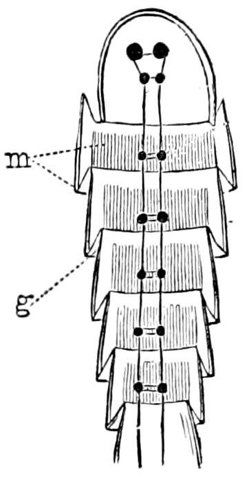
Fig. 16.—Diagram of the anterior part of an insect, showing the membranous intersegmental folds, g.—After Graber.
In the unjointed worms, as Graber states, the body forms a single but flexible lever. In the earthworm the muscular tube or body-wall is enclosed by a stiffer cuticle, divided into segments; hence the worm can move in all required directions, but only by sections, as seen in Fig. 16, which represents the thickened integument divided into segments, and folded inward between each segment, this thin portion of the skin being the intersegmental fold. Each segment corresponds to a special zone of the subdivided muscular tube (m), the fascia extending longitudinally. The figure shows the mode of attachment of the fascia of the muscle-tube to the segment. The anterior edge is inserted on the stiff, unyielding, inner surface of each segment: the hinder edge of the muscle is attached to the thin, flexible, intersegmental fold, which thus acts as a tendon on which the muscle can exert its force. (Graber.)
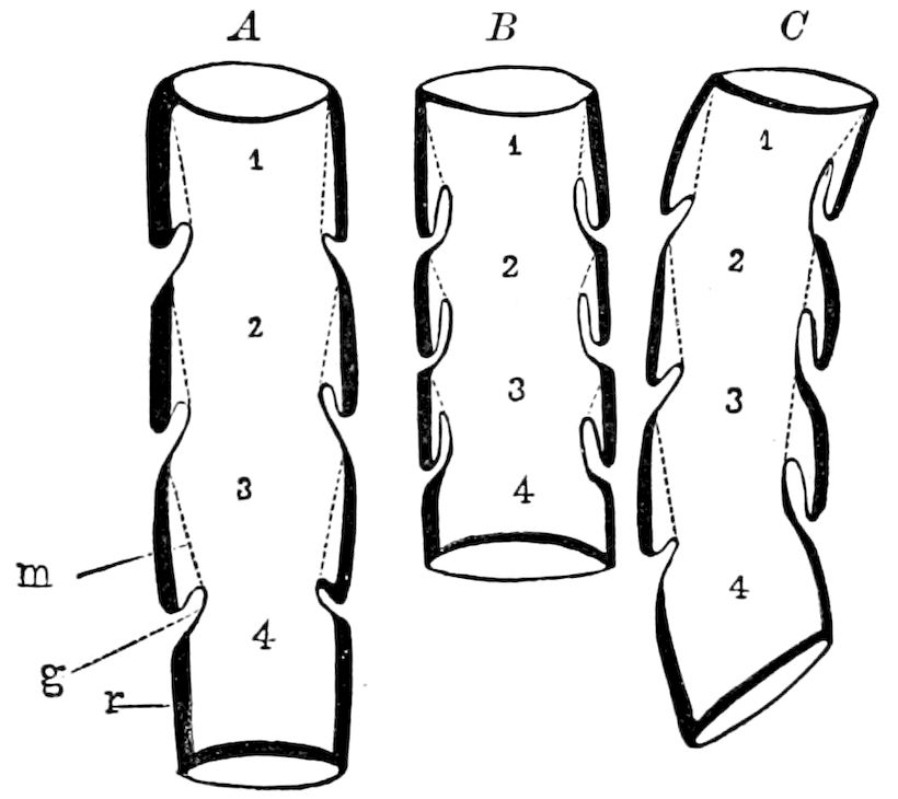
Fig. 17.—Diagram of the integument and arrangement of the segmental muscles: A, relaxed; m, muscle; g, membranous articulation; r, chitinous ring. B, the same contracted on both sides. C, on one side.—After Graber.
“Fig. 17 makes this still clearer. The muscles (m) extend between two segments immediately succeeding each other. Supposing the anterior one (A) to be stationary, what do we then see when the muscle contracts? Does it also become shorter? The intersegmental 32fold is drawn forwards, and hence the entire hinder segment moves forward and is shoved into the front one, and so on with the others, as at B. Afterwards, if the strain of the muscle is relieved by the diminishing action of the tensely stretched, intersegmental membrane, it again returns to a state of rest.” (Graber.)
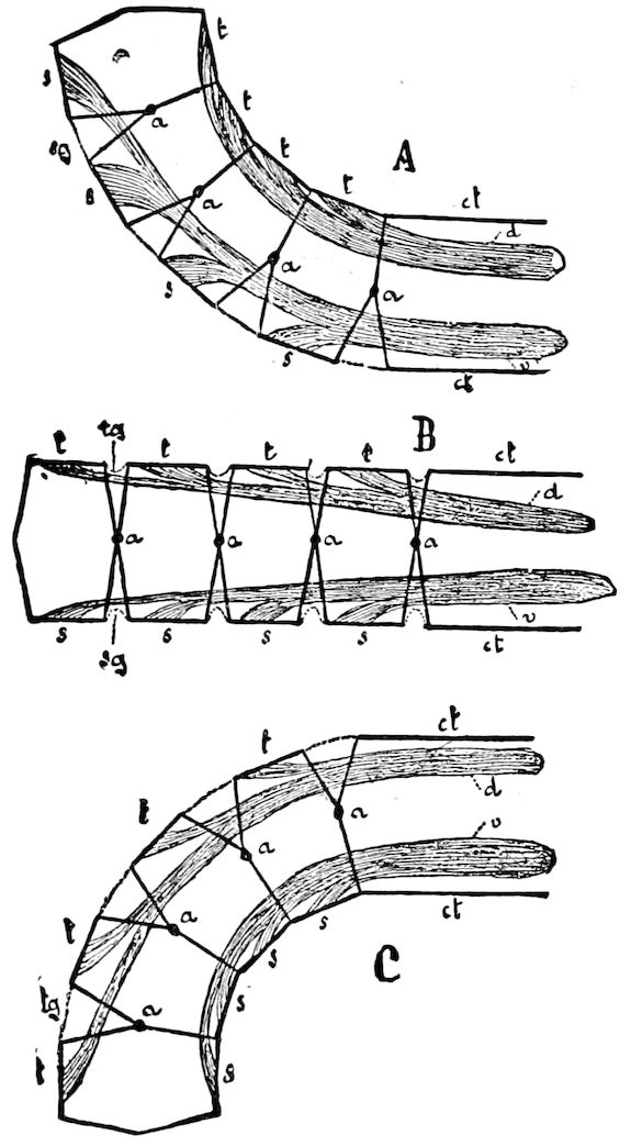
Fig. 18.—Diagrams to demonstrate the mechanism of the motion of the segmented body in the Arthropoda: One larger segment (cf) and 4 smaller. The exoskeleton is indicated by black lines, the interarticular membranes by dotted lines. The hinges between consecutive segments are marked at, tergal (dorsal) skeleton; s, sternal (ventral) skeleton; d, dorsal longitudinal muscles = extensors (and flexors in an upward direction); v, ventral longitudinal muscles = flexors. In B, the row of segments is stretched; in A, by the contraction of the muscles (d) bent upward; in C, downward; tg, tergal; sg, sternal interarticular membranes.—After Lang.
While we look upon the dermal tube of worms as a single but flexible lever, the body of the arthropods, as Graber states, is a linear system of stiff levers. We have here a series of stiff, solid rings, or hooks, united by the intersegmental membrane into a whole. When the muscles, extending from one ring to the next behind contract, and so on through the entire series, the rings approximate each other.
The ectoskeletal segments bend to one side by the contraction of the muscles on one side, the point of the outer segmental fold opposite the fixed point becoming converted into the turning-point (C).
The usual result of the arrangement of the locomotive system is the simple curving of the body (C), and then the alternate bending of the body to right and left, which produces the serpentine movements characteristic of the earthworms, the centipede, and many insect larvæ. The most striking example of the wonderful variety of movements which can be made by an insect are those of the Syrphus larva. When feeding amid a herd of aphides, it is seen to now raise the front part of the 33body erect and stiff, then to bend it down, or rapidly turn it to either side, or move it in a complete circle. (Graber, pp. 23–26.)
The arrangement and mode of working of the muscles, says Lang, is illustrated by Fig. 18, which shows us five segments, one larger (ct) and four smaller, in vertical projection. The thicker portion of the integument is marked by strong outlines, the delicate and flexible interarticular membranes (tg, sg) in dotted lines. The hinges between two consecutive segments are marked a. A dorsal muscle (d) is attached to the larger segment (ct), and runs through the smaller segments, being inserted in the dorsal portion of the crust (t) of each by means of a bundle of fibres. A ventral muscle (v) does the same on the sternal side (s).
“The skeletal segments,” adds Lang, “may be compared to a double-armed lever, whose fulcrum lies in the hinges. If the dorsal muscle contracts, it draws the dorsal arm of the lever (the tergal portion of the skeleton) in the direction of the pull towards the larger segments; the tergal interarticular membranes become folded, the ventral stretched, and the four segments bend upward (Fig. 18, A). If the ventral muscle contracts, while at the same time the dorsal slackens, the row of segments will be bent downwards (Fig. 18, C).”
L. B. Sharp suggests, that in the Crustacea the rings formed by “the regularity and stress of muscular action” would be hardened by the deposition of lime at the most prominent portion, i.e. between what we have called the intersegmental folds. (American Naturalist, 1893, p. 89.) Cope also states that “with the beginning of induration of the integument, segmentation would immediately appear, for the movements of the body and limbs would interrupt the deposit at such points as would experience the greatest flexure. The muscular system would initiate the process, since flexure depends on its contractions, and its presence in animals prior to the induration of the integuments in the order of phylogeny, furnishes the conditions required.” (The Primary Factors of Organic Evolution, p. 268, 1895.)
It is apparent that the jointed or metameric structure of the bodies of insects and other arthropods is an inheritance from the segmented worms. In the worms the body is a continuous dermo-muscular tube, while in arthropods this tube is divided into regions, and the cuticle is thicker and more resistant. To go back to the incipient stages in the process of segmentation of the body, we conceive that the worms probably arose from a creeping gastrula-like form, the gastræa. The act of creeping gradually induced an elongated shape of the body. The movement of such an organism in a 34forward direction would gradually evolve a fore and aft, dorsal and ventral, and bilateral symmetry. As soon as this was attained, as the effect of creeping over rough irregular surfaces there would result mechanical lateral strains intermittently acting during the serpentine movements of the worm. The integument would, we can readily suppose, tend to bend or yield, or become permanently wrinkled, at more or less regular intervals. The arrangement of the muscles would gradually conform to this habit of creeping, and finally the nervous system and other organs more directly connected with the creeping movements of the organism would tend to be correlated in their arrangement with that of the segments. In this way the homonomous segments of the annelid body probably became developed, and their relations and shapes were eventually fixed by inheritance. After this stage was reached, and limbs began to appear, the segments would tend to become heteronomous, and to be grouped into regions.
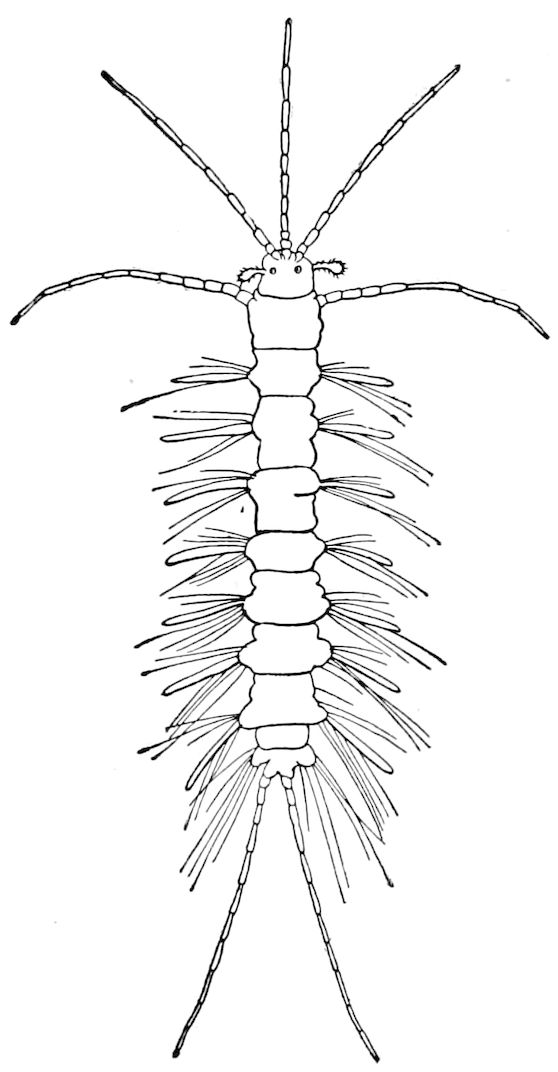
Fig. 19.—Dujardinia rotifera, with jointed tentacles and caudal appendages.—With some changes, after Quatrefages.
The origin of the joints or segments in the limbs of arthropods was probably due to the mechanical strains to which what were at first soft fleshy outgrowths along the sides of the body became subjected. Indeed, certain annelid worms of the family Syllidæ have segmented tentacles and parapodia, as in Dujardinia (Fig. 19). We do not know enough about the habits of these worms to understand how this metamerism may have arisen, but it is possibly due to the act of pushing or repeated efforts to support the body while creeping over the bottom among broken shells, over coarse gravel, or among seaweeds.
It is obvious, however, that the jointed structure of the limbs of arthropods, if we are to attempt any explanation at all of the origin of such structure, was primarily due mainly to lateral strains and 35impacts resulting from the primitive endeavors of the ancestral arthropods to raise and to support the body while thus raised, and then to push or drag it forward by means of the soft, partially jointed, lateral limbs which were armed with bristles, hooks, or finally claws.
On the other hand, by adaptation, or as the result of parasitism and consequent lack of active motion, the original number of segments may by disuse be diminished. Thus in adult wasps and bees, the last three or four abdominal segments may be nearly lost, though the larval number is ten. During metamorphosis the body is made over, and the number, shape, and structure of the segments greatly modified. In the female of the Stylopidæ the thorax loses all traces of segments, and is fused with the head, and the abdominal segments are faintly marked, losing their chitin.
While the maxillæ have several joints, the mandibles are 1–jointed, but there are traces of two joints in Campodea, certain beetles, etc. In the antenna there is a great elasticity in respect to the number of joints, which vary from one or two to a hundred or more. It is likewise so in the thoracic legs, where the number of tarsal joints varies from one to five; also in the cercopoda, the number of joints varying from one or two to twelve or more.
d. Mechanical origin of the limbs and of their jointed structure
We have already hinted at the mode of origin of the limbs of arthropods. Like the body or trunk, the limbs are chitinous dermo-muscular tubes, with a dense solid cuticle, and internal muscles, and were it not for their division at more or less regular intervals into segments, forming distinct sets of levers, set up by the strains in these tubular supports, there would be no power of varied motion.
Even certain worms, as already stated, have their tentacles and parapodia, or certain appendages of their parapodia, more or less jointed, but there are no indications of claws or of any other hard chitinous armature at the extremity, and the skin is thin and soft.
In the most simple though not the most primitive arthropods, such as the Tardigrades, whose body is not segmented, there are four pairs of short unjointed legs, ending each in two claws, which have probably arisen in response to the stimulus of pushing or dragging efforts.
The legs of Peripatus are unjointed, and have a thin cuticle, but end in a pair of claws, which have evidently arisen as a supporting 36armature, the result of the act of moving or pulling the body over the uneven surface of the ground.
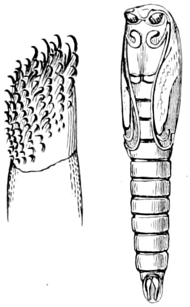
Fig. 20.—A prothoracic leg of Chironomus larva; and pupa.
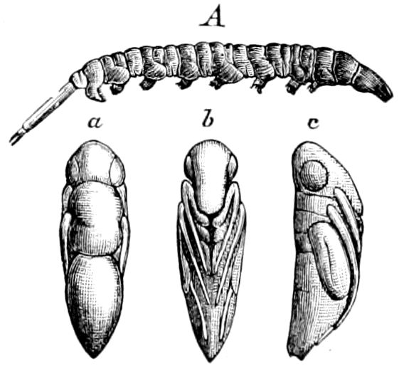
Fig. 21.—A, larva of Ephydra californica: a, b, c, pupa.
There is good reason to suppose that such limbs arose from dynamical causes, similar to those exciting the formation of secondary adaptations such as are to be seen in the prop or supporting legs of certain dipterous larvæ, as the single pair of Chironomus (Fig. 20) and Simulium, or the series of unjointed soft tubercles of Ephydra (Fig. 21), etc., which are armed with hooks and claws, and are thus adapted for dragging the insect through or over vegetation or along the ground.
Now by frequent continuous use of such unjointed structures, the cuticle would tend to become hard, owing to the deposit of a greater amount of chitin between the folds of the skin, until finally the body being elongated and homonomously segmented, the movements of walking or running would be regular and even, and we would have homonomously jointed legs like those of the trilobites, or of the most generalized Crustacea and of Myriopoda.
In the most primitive arthropods,—and such we take it were on the whole the trilobites, rather than the Crustacea,—the limbs were of nearly the same shape, being long and slender and evenly jointed from and including the antennæ, to the last pair of limbs of the abdominal region. In these forms there appear to be, so far as we now know, no differentiation into mandibles, maxillæ, maxillipedes, and thoracic legs, or into gonopoda. The same lack of diversity of structure and function of the head-appendages has survived, with little change, in Limulus. In the trilobites (Fig. 1) none of the limbs have yet been found to end in claws or forceps; being in this respect nearly as primitive as in the worms. Secondary adaptations have arisen in Limulus, the cephalic appendages being forcipated, adapted as supports to the body and for pushing it onward through the sand or mud, while the abdominal legs are broad and flat, adapted for swimming and bearing the broad gill-leaves.
It is thus quite evident that we have three stages in the evolution of the arthropodan limb; i.e. 1, the syllid stage, of simple, jointed, soft, yielding appendages not used as true supports (Fig. 19); 2, the 37trilobite stage, where they are more solid, evenly jointed, but not ending in claws; and by their comparatively great numbers (as in the trilobite, Triarthrus) fully supporting the body on the bottom of the sea. In Limulus they are much fewer in number, thicker, and acting as firm supports, the cephalic limbs of use in creeping, and ending in solid claws. 3, The third stage is the long slender swimming head-appendages of the nauplius stage of Crustacea.
As regards the evolution of limbs of terrestrial arthropods, we have the following stages: 1, the soft unjointed limbs of Tardigrades, ending in two claws, and those of Peripatus, and the pseudo- or prop-legs of certain dipterous larvæ; 2, finally the evolution of the long, solid, jointed limbs of Pauropus and other primitive myriopods, the legs forming solid, firm supports elevating the body, and enabling the insect to drag itself over the ground or to walk or run. When the body is elongated and many-segmented, the legs are necessarily numerous; but when it is short, the legs become few in number, i.e. six, in the hexapodous young of myriopods and in insects, or eight in Arachnida. Whenever the legs are used for walking, i.e. to raise and support the body, they end in a solid point or in a pair of forceps or claws. On the other hand, as in phyllopods, where the legs are used mainly for swimming, they are unarmed and are soft and membranous, or, as in the limbs of the nauplius or zoëa stage of crustaceans, end in a simple soft point, which often bears tactile setæ.
The tarsal joints are more numerous in order to give greater flexibility to the limb in seizing and grasping objects, both to drag the body forwards and to support it.
Unlike those of the Crustacea, the limbs of insects are not primitively biramose, but single, the three-lobed first maxillæ, and secondarily bilobed second maxillæ being the result of adaptation. Embryology on the whole proves the truth of this assumption; the maxillæ of both pairs are at first single buds, afterwards becoming lobed. All the appendages of the body, including the ovipositor or sting, are modified limbs, as shown by their embryological development.
It is noticeable that in the crab, where the body is raised by the limbs above the bottom, it is much shorter and more cephalized than in the shrimps. Also in the simply walking and running spiders, the hind-body is shorter than in scorpions, while in the running and flying insects, such as the Cicindelidæ, and in the swiftly flying flies and bees, there is a tendency to a shortening of the body, especially of the abdomen. The long body of the dragon-fly is an 38impediment to flight, but compensated for by the action of the large wings.
The arthropodan limb is a compound leverage system. It is, says Graber, a lateral outgrowth of the trunk, which repeats in miniature that of the main trunk, its single series of joints or segments forming a jointed dermo-muscular tube. Yet the lateral appendages of an insect differ from the main trunk in two ways: (1) they taper to the end which bears the two claws, and (2) their segments are in the living animal arranged not in a straight line, but at different angles to each other. The basal joint turning on the trunk acts as the first of a whole series of levers. The second joint, however, is connected with the musculature of the first or basal joint, and thus each succeeding joint is moved on the one preceding. Each lever, from the first to the last, is both an active and a passive instrument. (Graber.)
While, however, as Graber states, the limbs possess their own sets of muscles and can move by the turning of the basal joint, the labor is very much facilitated, as is readily seen, by the trunk, though the latter has to a great extent delegated its locomotive function to the appendages, which again divide its labor among the separate joints.
Graber then calls attention to the analogy of the mechanics of locomotion of insects to those of vertebrates. An insect’s and a vertebrate’s legs are constructed on the same general mechanical principles, the limbs of each forming a series of levers.
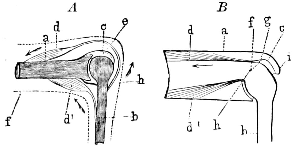
Fig. 22.—Diagram of the knee-joint of a vertebrate (A) and an insect’s limb (B): a, upper; b, lower, shank, united at A by a capsular joint, at B by a folding joint; d, extensor or lifting muscle; d1, flexor or lowering muscle of the lower joint. The dotted line indicates in A the contour of the leg.—After Graber.
Fig. 22, A, represents diagrammatically the knee joint of a vertebrate, and B that of an insect; a, the femur or thigh, and b, the tibia or shank. In the vertebrate the internally situated bones are brought into close union and bend by means of a hinge-joint; so also in the chitinous-skinned insect.
The stiff dermal tube of the insect acts as a lever by means of the thin intersegmental membrane (c) pushed in or telescoped in to the thigh joint, a special joint-capsule being superfluous. The muscles are in general the same in both types; they form a circle. In both the shank is extended by the contraction of the upper muscles (d) and is bent by the contraction of the lower 39(d1). The intersegmental membrane of the insect’s limb is in a degree a two-armed lever, whose pivot (f) lies in the middle. The internal invagination of the intersegmental fold (B, g-h) affords the necessary support to the muscles acting like the tendon in the vertebrate. (Graber.)
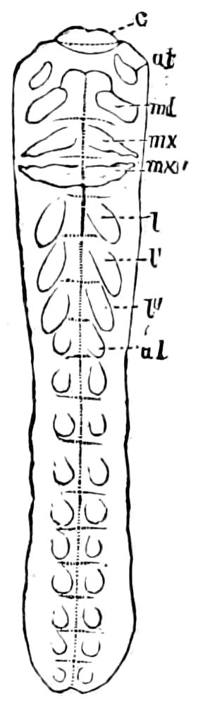
Fig. 23.—Primitive band or germ of a Sphinx moth, with the segments indicated, and their rudimentary appendages: c, upper lip; at, antennæ; md, mandibles; mx, mx′, first and second maxillæ; l, l′, l″, legs; al, abdominal legs.—After Kowalevsky.
Graber also calls attention to the fact that this insect limb differs in one important respect from that of land vertebrates. The leverage system in the last is divided at the end into five parallel divisions or digits. In arthropods, on the contrary, all the joints succeed one another in a linear series.
In insects, as well as in other arthropods, modifications of the limbs usually take the form of a simple reduction in the number of segments. Thus while the normal number of tarsal joints is five, we have trimerous and dimerous Coleoptera, and in certain Scarabæidæ the anterior tarsi are lost.
Savigny was the first, in 1816, in his great work, “Théorie des organes de la bouche des Crustacés et des Insectes,” to demonstrate that not only were the buccal appendages of biting insects homologous with those of bugs, moths, flies, etc., but that they were homologous with the thoracic legs, and that thus a unity of structure prevails throughout the appendages of the body of all arthropods. Oken also observed that “the maxillæ are only repeated feet.”
What was modestly put forth as a theory by the French morphologist has been abundantly proved by the embryology of insects of different orders to be a fact. As shown in Fig. 23 the antennæ and buccal appendages arise as paired tubercles exactly as the thoracic legs. The abdominal region also bears similar embryonic or temporary limbs, all of which in those insects without an ovipositor disappear, except the cercopoda, after birth.
40
LITERATURE ON THE EXTERNAL ANATOMY
General
Swammerdam, Johann. Biblia naturæ. (In Dutch, German, and English.) 1737–1738, fol., London, 1758, Pls.
Réaumur, Réné Antoine Ferchault, de. Mémoires pour servir à l’histoire des insectes. i-iv, 4º, Paris, 1734–1742.
Lyonet, Pieter. Traité anatomique de la chenille, qui ronge le bois de saule, etc. 4º, pp. xxii, 616. À la Haye, 1732. Tab. 18.
—— Recherches sur l’anatomie et les metamorphoses de differentes espèces d’insectes. Ouvrage posthume, publié par M. W. de Haan. pp. 580, tab. 54, 1832.
Latreille, Pierre André. Des rapports généraux de l’organization extérieure des animaux invertébres articulés, et comparaison des Annelides avec les Myriapodes. (Mémoires du Mus. d’Hist. Nat., 1820, vi, pp. 116–144.)
—— De quelques appendices particuliers du thorax de divers insectes. (Mémoires du Mus. d’Hist. Nat., 1821, vii, pp. 1–21).
—— Observations nouvelles sur l’organization extérieure et générale des animaux articulés et à pieds articulés, et application de ces connoissances à la nomenclature des principales parties des mêmes animaux. (Mèmoires du Mus. d’Hist. Nat., viii, 1822, pp. 169–202.)
Cuvier, George Leopold Christian Dagobert. Rapport sur les recherches anatomiques sur le thorax des animaux articulés et celui des insectes en particulier par M. V. Audouin. 4º, pp. 15, tab. 1, Paris, 1823.
Audouin, Jean Victor. Recherches anatomiques sur le thorax des animaux articulés et celui des insectes hexapodes en particulier. (Annales des Sciences naturelles, i, pp. 97–135, 416–432, 1824.)
Kirby, William, and William Spence. Introduction to entomology. i-iv, 1816–1828, London.
Straus-Durckheim, Hercule. Considérations générales sur l’anatomie comparée des animaux articulés. Paris, 1828, atlas of 19 plates.
MacLeay, William Sharp. Explanation of the comparative anatomy of the thorax in winged insects, with a review of the present state of the nomenclature of its parts. (Zoöl. Journal, v, pp. 145–179, 1830, 2 Pls.)
Burmeister, Hermann. A manual of entomology. Trans. by W. E. Shuckard, 8º, pp. 654, London, 1836, 32 Pl.
Westwood, John Obadiah. An introduction to the modern classification of insects, i, ii, 8º, pp. 462, 587, 158, 1 Pl. and 133 blocks of figs., 1839–1840.
Newport, George. Art. Insecta in Todd’s Cyclopædia of Anatomy and Phys. ii, pp. 853–994, 1839, Figs. 329–439.
Erichson, Wilhelm Ferdinand. Entomographien. Berlin, 1840.
Brullé, Auguste. Recherches sur les transformations des appendices dans les articulés. (Annales des Sciences nat. Sér. 3, ii, pp. 271–374, tab. 1, 1844.)
Winslow, A. P.: son. Om byggnaden af thorax hos Insekterna. Helsingborg, 1862, 1 Pl, pp. 24.
Packard, Alpheus Spring. Guide to the study of insects. 1869.
—— Systematic position of the Orthoptera in relation to other insects. (Third report U. S. Ent. Commission, pp. 286–345, 1883, Pls. xxiii-lxi.)
Graber, Vitus. Die Insekten. 12º, pp. 403, 603, München, 1877, many Figs.
Huxley, Thomas Henry. A manual of the anatomy of invertebrated animals. 12º, pp. 397–451, Figs., London, 1877.
41Hammond, Arthur. Thorax of the blow-fly. (Journ. Linn. Soc., London, xv. Zoöl., 1880, pp. 31.)
Brauer, Friedrich. Ueber das Segment médiaire Latreille’s. (Sitzb. d. k. Akad. d. Wissensch. Wien, 1882, pp. 218–241, 3 tab.)
—— Systematisch-zoologische Studien. (Ibid., 1885, pp. 237–413.)
Gosch, C. C. A. On Latreille’s theory of “Le Segment médiaire.” (Nat. Tidsskrift (3), xiii, pp. 475–531, 1883.)
Miall, L. C., and Denny, Alfred. The structure and life-history of the cockroach (Periplaneta orientalis). An introduction to the study of insects. 8º, pp. 224, London, 1886.
Cheshire, Frank R. Bees and bee-keeping. i, Scientific, London, 1886, Pls. and Figs.
Lang, Arnold. Text-book of comparative anatomy. i, pp. 426–508, 1891, many Figs.
Kolbe, H. J. Einführung in die Kenntniss der Insekten. 8º, pp. 709, 324 figs., Berlin, 1893.
Sharp, David. The Cambridge natural history. Insecta, i, 8º, pp. 83–584, 1895, Figs. 47–371.
Also the works of Bos, Chabrier, Cholodkowsky, Comstock, Dewitz, Eaton, Erichson, Gerstaecker, Girard, Grassi, Hagen, Haase, Kellogg, Knoch, Lacordaire, Latreille, Leuckart, Lendenfeld, Lowne, Lubbock, Mayer, Meinert, F. Müller, Osten-Sacken, Pagenstecher, Reinhard, Schaum, Schiödte, Scudder, J. B. Smith, Spinola, Stein, Weismann, Wood-Mason.
42
THE HEAD AND ITS APPENDAGES
a. The head
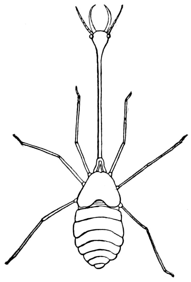
Fig. 24.—Presumed larva of Nemoptera (Necrophilus arenarius), Pyramids of Egypt.—After Roux, from Sharp.
While the head is originally composed of probably not less than six segments, these are in the adult insect fused together into a capsule or hard chitinous box, the epicranium, with no distinct traces of the primitive segments. The head contains the brain and accessory ganglia, the mouth or buccal cavity, also the air-sacs in many winged forms, and gives support to the external organs of sense, the antennæ, and to the buccal appendages, the larger part of the interior being filled with the muscles moving these structures. The solid walls of the head serve as a lever or support for the attachment of these muscles, especially those of the mandibles. Thus there is a correlation between the large size of the mandibles of the soldier white ants and ants, the head being correspondingly large to accommodate the great mandibular muscles. The other extreme is seen in the larva of Necrophilus (Fig. 24), with its long slender neck and diminutive head.
The clypeus.—This is that part of the head situated in front of the epicranium, and anterior to the eyes, forming the roof of the posterior part of the mouth, and is, as embryology shows, probably a tergal sclerite. It varies greatly in shape and size in the different orders of insects. It is often divided into two parts, the clypeus posterior and clypeus anterior, or which may be designated as the post- and ante-clypeus (Figs. 29, B).
The labrum.—The “upper lip” or labrum is an unpaired flap-like piece hinged to the front edge of the clypeus, and may be seen to move up and down when the insect moves its mandibles. It forms the roof of the anterior part of the mouth (Figs. 69, 74), and its inner side is lined with a soft membrane, usually provided with hairs and sense-papillæ or cups, forming the epipharynx.
The labrum is more or less deeply bilobed, especially in caterpillars and in adult Staphylinidæ, and has been thought by some 43writers (Kowalevsky, Carrière, and also Chatin) to represent a pair of appendages, but Heymons (1895) refutes this view, stating as his reason that the labrum arises between the two halves of the nervous system (protocerebrum), while all the true appendages arise on each side of the nervous system. (See also Fig. 34.)
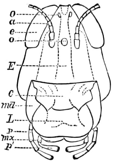
Fig. 25.—Front view of the head of C. spretus: E, epicranium; C, clypeus; L, labrum; O, o, ocelli; e, eye; a, antenna; md, mandible; mx, portion of maxilla uncovered by the labrum; p, maxillary palpus; p′, labial palpus.
In the fleas (Siphonaptera) both the clypeus and labrum are wanting.
While it apparently forms an anterior specialized portion of the procephalic lobes, Viallanes regarded it as belonging to the third, or his tritocerebral, segment, since the labral nerves arise from the tritocerebral ganglia. But since in all the early as well as late stages of embryonic life it appears to be situated in front of the mouth, it would seem to belong to the first segment.
In the embryo of Blatta it first appears as a thick crescentic fold being slightly divided anterior to the mouth, and in Doryphora it appears as a heart-shaped or deeply bilobed prominence situated in front of the mouth (Wheeler).
The epipharynx and labrum-epipharynx.—The epipharynx is the under surface or pharyngeal lining of the clypeus and labrum, forming the membranous roof of the mouth. As it contains the organs of taste and has been generally overlooked by entomologists, we may dwell at some length on its structure in different orders.
Réaumur was, so far as we have been able to ascertain, the first author to describe and figure the epipharynx, which he observed in the honey bee and bumble bee, and called la langue, remarking that it closes the opening into the œsophagus, and that it is applied against the palate. According to Kirby and Spence, De Geer described the epipharynx of the wasp; and Latreille referred to it, calling it the sous labre.
The name epipharynx was bestowed upon this organ by Savigny, who thus speaks of that of the bees: “Ce pharynx est, à la vérité, non seulement caché par la lèvre supérieure, mais encore exactement recouvert par un organe particulier que Réaumur a déjà décrit. C’est une sorte d’appendice membraneux qui est reçu entre les deux branches des mâchoires. Cette partie ayant pour base le bord supérieur du pharynx, peut prendre le nom d’épipharynx ou d’épiglosse.”
44He also describes that of Diptera. What Walter has lately proved to be the epipharynx of Lepidoptera was regarded by Savigny and all subsequent writers as the labrum.
The latest account of the function of this organ is that by Cheshire, who states that the tube made by the maxillæ and labial palpi cannot act as a suction pipe, because it is open above. “This opening is closed by the front extension of the epipharynx, which closes down to the maxillæ, fitting exactly into the space they leave uncovered, and thus the tube is completed from their termination to the œsophagus.”
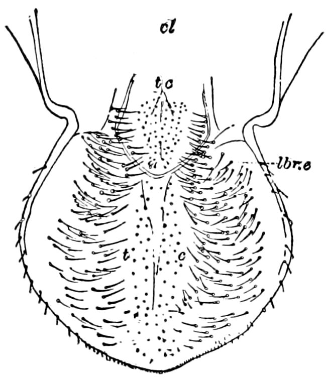
Fig. 26.—Epipharynx of Phaneroptera angustifolia: cl, clypeus; lbr. e, labrum-epipharynx; t c, taste cups, both on the clypeal and on the labral regions.
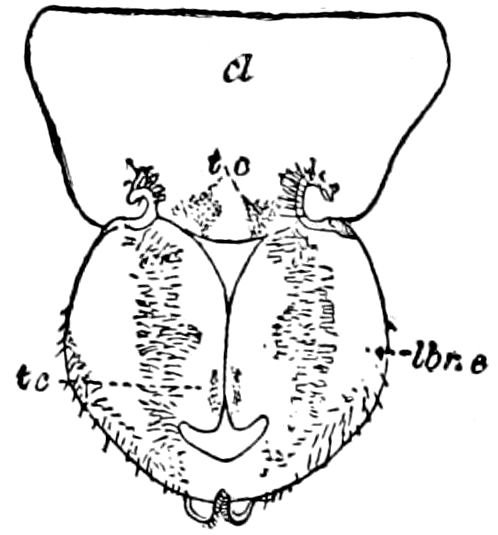
Fig. 27.—Epipharynx of Hadenœcus subterraneus, cave cricket.
It is singular that this organ is not mentioned in Burmeister’s Manual of entomology, in Lacordaire’s Introduction à l’entomologie, or by Newport in his admirable article Insecta in Todd’s Cyclopedia of anatomy. Neither has Straus-Durckheim referred to or figured it in his great work on the anatomy of Melolontha vulgaris.
In their excellent work on the cockroach, Miall and Denny state that “The epipharynx, which is a prominent part in Coleoptera and Diptera, is not recognizable in Orthoptera” (p. 45). We have, however, found it to be always present in this order (Figs. 26, 27).
We are not aware that any modern writers have described or referred to the epipharynx of the mandibulate orders of insects. Although Dr. G. Joseph speaks of finding taste-organs on the palate of almost every order of insects, especially plant-feeding forms, we are unable to find any specific references, his detailed observations being apparently unpublished.
The epipharynx is so intimately associated with the elongated labium of certain Diptera, that, with Dr. Dimmock, we may refer to the double organ as the labrum-epipharynx; and where, as in the lepidopterous Micropteryx semipurpurella, described and figured by Walter, and the Panorpidæ (Panorpa and Boreus), the labrum seems pieced out with a thin, pale membranous fold which appears to be 45an extension of the epipharynx, building up the dorsal end of the labrum, this term is a convenient one to use.
In the lower orders of truly mandibulate insects, from the Thysanura to the Coleoptera, excluding those which suck in liquid food, such as the Diptera, Lepidoptera, and Hymenoptera, and the Mecoptera (Panorpidæ) with their elongated head and feeble, small mandibles, the epipharynx forms a simple membranous palatal lining of the clypeus and labrum. In such insects there is no soft projecting or pendant portion, fitted to close the throat or to complete a partially tubular arrangement of the first and second maxillæ.
In all the mandibulate insects, then, the epipharynx forms simply the under surface or pharyngeal lining of the clypeus and labrum, the surface being uniformly moderately convex, and corresponding in extent to that of the clypeus and labrum, posteriorly merging into the palatal wall of the pharynx; the armature of peculiar gathering-hairs sometimes spreading over its base, being continuous with those lining the mouth and beginning of the œsophagus. The suture separating the labrum from the clypeus does not involve the epipharynx, though since certain gustatory fields lie under the front edge of the clypeus, as well as labrum, one may in describing them refer to certain fields or groups of cups or pits as occupying a labral or clypeal region or position.
The lack of traces of a suture in the epipharynx corresponding to the labral suture above, suggests that the labrum does not represent a pair of coalesced appendages, and that it, with the clypeus, simply forms the solid cuticular roof of the mouth.
The only soft structures seen between the epipharynx and labrum, besides the nerves of special sense, are the elevator muscles of the labrum, and two tracheæ, one on each side.
The structure and armature of the epipharyngeal surface even besides the taste-pits, taste-cups and rods, is very varied, the setæ assuming very different shapes. There seem to be two primary forms of setæ, (1) the normal forms which arise from a definite cell; and (2) soft, flattened, often hooked hairs which are cylindrical towards the end, but arise from a broad triangular base, without any cell-wall. These are like the “gathering hairs” of Cheshire, situated on the bees’ and wasps’ tongue; they also line the walls of the pharynx and extend toward the œsophagus. They are also similar to the “hooked hairs” of Will. The first kind, or normal setæ, are either simply defensive, often guarding the sense-cups or sensory fields on which the sense-cups are situated, or they have a nerve extending to them and are simply tactile in function.
46The surface of the epipharynx, then, appears to be highly sensitive, and to afford the principal seat of the gustatory organs, which are described under the head of organs of taste.
LITERATURE ON THE EPIPHARYNX
Réaumur. Mémoires pour servir à l’histoire des insectes, v, 1740, p. 318, Pl. 28, Figs. 4, 7, 8, 9, 10, 11 l.