PART I.—MORPHOLOGY AND PHYSIOLOGY (continued 4)
Reading edition
Now, speculating on the primary origin of wings, we need not suppose that they originated in any aquatic form, but in some ancestral land insect related to existing cockroaches and Termes. We may imagine that the tergites (or notum) of the two hinder segments of the thorax grew out laterally in some leaping and running insect; that the expansion became of use in aiding to support the body in its longer leaps, somewhat as the lateral expansions of the body aid the flying squirrel or certain lizards in supporting the body during their leaps. By natural selection these structures would be transmitted in an improved condition until they became flexible, i.e. attached by a rude hinge-joint to the tergal plates of the meso- and metathorax. Then by continued use and attempts at flight they would grow larger, until they would become permanent organs, though still rudimentary, as in many existing Orthoptera, such as certain Blattariæ and Pezotettix. By this time a fold or hinge having been established, small chitinous pieces enclosed in membrane would appear, until we should have a hinge flexible enough to allow the wing to be folded on the back, and also to have a flapping motion. A stray tracheal twig would naturally press or grow into the base of the new structure. After the trachea running towards the base of the wing had begun to send off branches into the rudimentary structure, the number and direction of the future veins would become determined on simple mechanical principles. The rudimentary structures beating the air would need to be strengthened on the front or costal edge. Here, then, would be developed the larger number of main veins, two or three close together, and parallel. These would be the costal, subcostal, and median veins. They would throw out branches to strengthen the costal edge, while the branches sent out to the outer and hinder edges of the wings might be less numerous and farther apart. The net-veined wings of Orthoptera and Pseudoneuroptera, as compared with the wings of Hymenoptera, show that the wings of net-veined insects were largely used for respiration as well as for flight, while in beetles and bees the leading function is flight, that of respiration being quite subordinate. The blood would then supply the parts, and thus respiration or aëration of the blood would be demanded. As soon as such expansions would be of even slight use to the insect as breathing organs, the question as to their permanency would be settled. Organs so useful both for flight and aëration of the blood would be still further developed, until they would become permanent structures, genuine wings. They would thus be readily transmitted, and being of more use in adult life during the season of reproduction, they would be still further developed, and thus those insects which could fly the best, i.e. which had the strongest wings, would be most successful in the struggle for existence. Thus also, not being so much needed in larval life before the reproductive organs are developed, they would not be transmitted except in a very rudimentary way, as perhaps masses of internal indifferent cells (imaginal discs), to the larva, being the rather destined to develop late in larval and in pupal life. Thus the development of the wings and of the generative organs would go hand in hand, and become organs of adult life.[33]
The development and structure of the tracheæ and veins of the wing.—The so-called veins (“nervures”) originate from fine tracheal 145twigs which pass into the imaginal discs. A single longitudinal trachea grows down into the wing-germ (Fig. 147), this branch arising through simple budding of the large body-trachea passing under the rudiment of the wing.
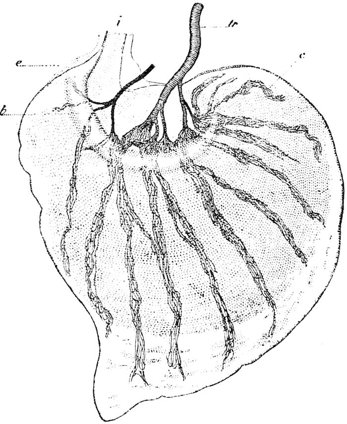
Fig. 159.—Germ of a hind wing detached from its insertion, and examined in glycerine: i, pedicel of insertion to the hypodermis; tr, trachea; b, semicircular pad; e, enveloping membrane; c, bundle of capillary tracheoles; the large tracheæ of the wing not visible; they follow the course of the bundles of tracheoles.—After Gonin.
Gonin states that before the tracheæ reach the wing they divide into a great number of capillary tubes united into bundles and often tangled. This mass of tracheæ does not penetrate into the wing-germ by one of its free ends, but spreading over about a third of the surface of the wing, separates into a dozen bundles which spread out fan-like in the interior of the wing. (Fig. 159). These ramifications, as seen under the microscope, are very irregular; they form here and there knots and anastomoses. They end abruptly in tufts at a little distance from the edge of the wing. A raised semicircular ridge (b) surrounds the base of the wing, and within this the capillaries are formed, while on the other side they are covered by a cellular layer.
Landois, he says, noticed neither the pedicel of the insertion of the wing (i) nor the ridge (b). Herold only states that the tracheæ pass like roots into the wing. Landois believed that they formed an integral part of it. Dewitz and Pancritius used sections to determine their situation.
Fig. 160 will illustrate Landois’ views as to the origin of the tracheæ and veins. A represents the germ of a hind wing attached 146to a trachea; c the elongated cells, in which, as seen at B, c, a fine tangled tracheal thread (t) appears, seen to be magnified at C. The cell walls break down, and the threads become those which pass through the centre of the veins.
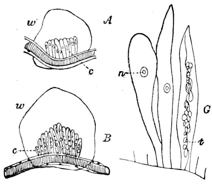
Fig. 160.—Origin of the wings and their veins.—After Landois.
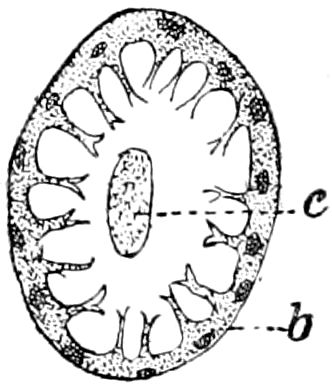
Fig. 161.-Section of the “rib” of a vein: c, cord; b, twig.—After Schaeffer.
The wing-rods.—Semper discovered in transverse sections of the wings, what he called Flügelrippen; one such rib accompanying the trachea in each vein. He did not discover its origin, and his description of it is said to be somewhat erroneous. Schaeffer has recently examined the structure, remarking: “I have surely observed the connection of this cellular tube with the tracheæ. It is found in the base of the wing where the lumen of the tracheæ is much widened. I only describe the fully formed rib (rippe). In a cross-section it forms a usually cylindrical tube which is covered by a very thin chitinous intima which bears delicate twigs (Fig. 161). These twigs are analogous to the thickened ridge of the tracheal intima. I can see no connection between the branches of the different twigs. Through the ribs (rippen) extend a central cord (c) which shows in longitudinal section a clear longitudinal streaking. Semper regarded it as a nerve. But the connection of the tube with the trachea contradicts this view. I can only regard the cord as a separation-product of the cells of the walls.”
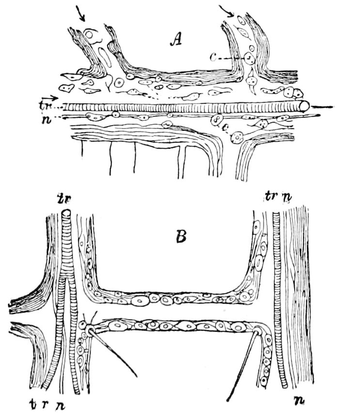
Fig. 162.—Parts of a vein of the cockroach, showing the nerve (n) by the side of the trachea (tr); c, blood-corpuscles.—After Moseley.
Other histological elements.—These are the blood-lymph, corpuscles, blood-building masses, and nerves. Schaeffer states that in the immature pupal wings we find besides the large tracheæ, which are more or less branched, 147and in the wing-veins at a later period, blood-corpuscles which are more or less gorged with nutritive material, and also the “balls of granules” of Weismann, which are perhaps the “single fat-body cells” detected by Semper. Schaeffer also states that into the hypodermal fold of the rudiments of the wings pass peculiar formations of the fat-body and tracheal system, and connected with the fat-body are masses of small cells which by Schaeffer are regarded as blood-building masses.
Fine nerves have also been detected within the veins, Moseley stating that a nerve-fibre accompanies the trachea in all the larger veins in the insects he has examined (Fig. 162), while it is present in Melolontha, where the trachea is absent.
LITERATURE ON THE WINGS
Jurine, L. Nouvelle méthode de classer les Hyménoptères et les Dipterès. Genève, 1807, 4º pp. 319, 14 Pls.
—— Observations sur les ailes des Hyménoptères. (Mém. acad. Turin, 1820, xxiv, pp. 177–214.)
Latreille, P. A. De la formation des ailes des Insectes. (Mém. sur divers sujets de l’histoire naturelle des Insectes, etc. Paris, 1819. Fasc. 8.)
—— De quelques appendices particuliers du thorax de divers Insectes. (Mém. du Mus. d’Hist. nat., 1821, vii, pp. 1–21, 354–363.)
Chabrier, J. Essai sur le vol des insectes. (Mém. du Mus. d’Hist. nat., 1820, vi, pp. 410–476; 1821, vii, pp. 297–372; 1822, viii, pp. 47–99, 349–403.) Separate, pp. 328, 13 Pls.
Burmeister, Hermann. Handbuch der entomologie, i, 1832, pp. 96–106, 263–267, 494–505.
—— Untersuchungen über die Flügeltypen der Coleopteren. (Abhandl. d. naturf. Ges. Halle, 1854, ii, pp. 125–140, 1 Taf.)
Romand, B. E. de. Tableau de l’aile supérieure des Hyménoptères, 1 Pl. Paris, 1839. (Revue Zool., ii, pp. 339; Bericht von Erichson für 1839, pp. 54–56.)
Lefebure, A. Communication verbale sur la ptérologie des Lépidoptères. (Annal. Soc. Ent. France, 1842, i, pp. 5–35, 3 Pls. Also Revue Zool. Paris, 1842, pp. 52–58, 1 Pl.)
Deschamps, B. Recherches microscopiques sur l’organisation des élytres des Coléoptères. (Ann. sc. nat., sér. 3, iii, 1845, pp. 354–363.)
Heer, Oswald. Die Insektenfauna der Tertiärgebilde von Oeningen und Radaboj., 1847, 1. Teil, pp. 75–94.
Newman, E. Memorandum on the wing-rays of insects. (Trans. Ent. Soc. London, ser. 2, iii, 1855, pp. 225–231.)
Westwood, J. O. Notes on the wing-veins of insects. (Trans. Ent. Soc. London, ser. 2, iv, 1857, pp. 60–64.)
Loew, H. Die Schwinger der Dipteren. (Berlin, Entom. Zeitschr., 1858, pp. 225–230.)
Saussure, H. de. Études sur l’aile des Orthoptères. (Ann. scienc. nat., 5 sér. x, p. 161.)
Schiner, J. R. Ueber das Flügelgeäder der Dipteren. (Verhdl. k. k. Zool.-bot., Ges. Wien, 1864, pp. 193–200, 1 Taf.)
148Hagen, H. A. Ueber rationelle Benennung des Geäders in den Flügeln der Insekten. (Stettin. Ent. Zeitung, 1870, xxxi, pp. 316–320, 1 Taf.)
—— Kurze Bemerkungen über das Flügelgeäder der Insekten. (Wiener Entom. Zeit., 1886, v, pp. 311, 312.)
Plateau, F. Qu’est-ce que l’aile d’un insecte? (Stett. Ent. Zeit. Jahrg. 32, 1871, pp. 33–42, 1 Taf. Journal d. Zool., ii, 1873, pp. 126–137.)
Moseley, H. N. On the circulation in the wing of Blatta orientalis and other insects, etc. (Quart. Journ. Micr. Sc. 1871, xi, pp. 389–395, 1 Pl.)
Roger, Otto. Das Flügelgeäder der Kafer. Erlangen, 1875, 90 p.
Rade, E. Die westfalischen Donacien und ihre nachsten Verwandten. 3 Taf. (Vierter Jahresber. d. Westfal Prov.-Vereins f. Wiss. u. Kunst, 1876, pp. 52–87; Flügel, pp. 61–68.)
Katter, F. Ueber Inseckten, speziell Schmetterlingsflügel. (Entom. Nachr., iv, 1878, pp. 279–281, 293–298, 304–309, 321–323.)
Hofmann, Georg v. Ueber die morphologische Deutung der Insektenflügel. (Jahresber. d. akad.-naturwiss. Vereins, Graz, v Jahrg., 1879, pp. 63–68.)
Kolbe, H. J. Das Flügelgeäder der Psociden und seine systematische Bedeutung. (Stettin. Entom. Zeitung, 1880, pp. 179–186, 1 Taf.)
—— Die Zwischenraume zwischen den Punktstreifen der punktiertgestreiften Flügeldecken der Coleoptera als rudimentare Rippen aufgefasst. (Jahresber. zool. Sektion d. Westfal. Prov.-Ver. f. Wiss. u. Kunst. Münster, 1886, pp. 57–59, 1 Taf.)
Lee, A. Bolles. Les balanciers des Diptères, leurs organes sensifères et leurs histologie. (Recueil Zool. Suisse, i, 1885, pp. 363–392, 1 Pl.)
Poppius, Alfred. Ueber das Flügelgeäder der finnischen Dendrometriden. 1 Taf. (Berl. Entom. Zeitschr., 1888, pp. 17–28.)
Comstock, J. H. On the homologies of the wing-veins of insects. (American Naturalist, xxi, 1887, pp. 932–934.)
Brauer, F. Ansichten über die paläozoischen Insekten und deren Deutung. (Annal. d. k. k. naturhist. Mus. Wien, Bd. i, 1886, pp. 86–126, 2 Taf.)
Brauer, F., und J. Redtenbacher. Ein Beitrag zur Entwicklung des Flügelgeäders der Insekten. (Zool. Anz. 1888, pp. 443–447.)
Redtenbacher, J. Vergleichende Studien über das Flügelgeäder der Insekten. 12 Taf. (Annalen d. k. k. naturhist. Hofmuseums zu Wien, 1886, i, pp. 153–231.)
Schoch, G. Miscellanea entomologica. I. Das Geäder des Insektenflügels; II. Prolegomena zur Fauna dipterorum Helvetiae, Wissenschaftl. (Beilage z. Programm d. Kantonsschule Zurich, 1889, 4º, 40 p.)
Bondsdorff, A. von. Ueber die Ableitung der Skulpturverhältnisse bei den Deckflügeln der Coleopteren. (Zool. Anz., 1890, xiii Jahrg., pp. 342–346.)
Spuler, Arnold. Zur Phylogenie und Ontogenie des Flügelgeäders der Schmetterlinge. (Zeitschr. wissens. Zool., liii, 597–646, 2 Taf., 1892.)
Also the writings of Adolph, Bugnion, Calvert, Comstock, Diez, Giraud, Gonin, Graber, Kellogg, Packard, Pratt, Scudder, Walsh.
g. Mechanism of flight
Marey’s views on the flight of insects.—As we owe more to Marey than to any one else for what exact knowledge we have of the theory of flight of insects, the following account is condensed from his work entitled “Movement.” The exceedingly complicated 149movements of the wings would lead us, he says, to suppose that there exists in insects a very complex set of muscles of flight, but in reality, he claims, there are only the two elevator and depressor muscles of each wing.[34] And Marey says that when we examine more closely the mechanical conditions of the flight of insects, we see that an upward and downward motion given by the muscles is sufficient to produce all these successive acts, so well coordinated with each other; the resistance of the air effecting all the other movements. He also refers to the experiments of Giraud which prove that the insect needs for flight a rigid main-rib and a flexible membrane.
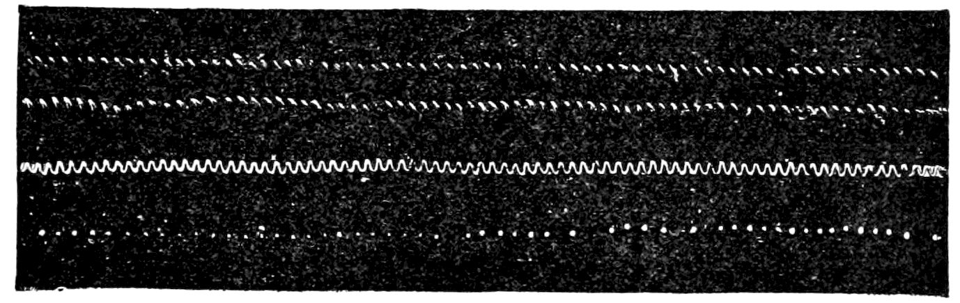
Fig. 163.—The two upper lines are produced by the contacts of a drone’s wing on a smoked cylinder. In the middle are recorded the vibrations of a tuning-fork (250 vibrations per second) for comparison with the frequency of the wing movements. Below are seen the movements of the wing of a bee.—After Marey.
If we take off the wing of an insect, and holding it by the small joint which connects it with the thorax, expose it to a current of air, we see that the plane of the wing is inclined more and more as it is subjected to a more powerful impulse of the wind. The anterior nervure resists, but the membranous portion which is prolonged behind bends on account of its greater pliancy.
The wings of insects may be regarded simply as vibrating wires, and hence the frequency of their movements can be calculated by the note produced. Their movements can be recorded directly on a revolving cylinder, previously blackened with smoke, the slightest touch of the tip of the wing removing the black and exposing the white paper beneath; Fig. 163 was obtained in this way. By this method it was calculated that in the common fly the wings made 330 strokes per second, the bee 190, the Macroglossus 72, the dragon-fly 15028, and the butterfly (Pieris rapæ) 9. Thus the smaller the species, the more rapid are the movements of the wings.
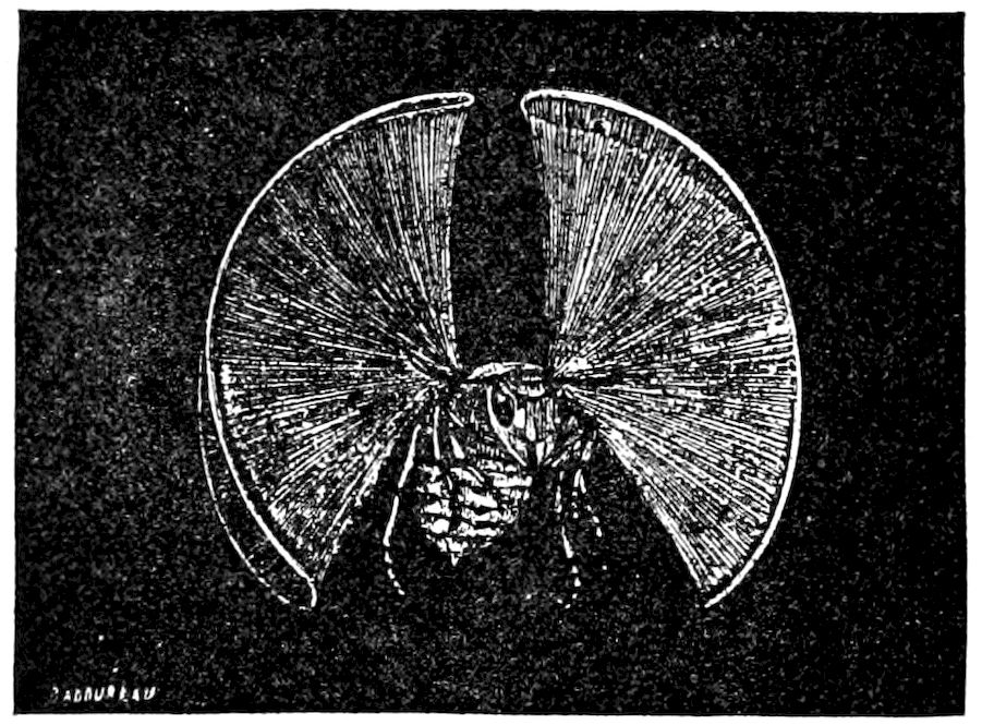
Fig. 164.—Appearance of a wasp flying in the sun: the extremity of the wing is gilded.—After Marey.
The path or trajectory made by the tip of the wing is like a figure 8. Marey obtained this by fastening a spangle of gold-leaf to the extremity of a wasp’s wing. The insect was then seized with a pair of forceps and held in the sun in front of a dark background, the luminous trajectory shaping itself in the form of a lemniscate (Fig. 164).
To determine with accuracy the direction taken by the wing at different stages of the trajectory, a small piece of capillary glass tubing was blackened in the smoke of a candle, so that the slightest touch on the glass was sufficient to remove the black coating and show the direction of movement in each limb of the lemniscate. This experiment was arranged as shown in Fig. 165. Different points on the path of movement were tested by the smoked rod, and from the track along which the black had been removed the direction of movement was deduced. This direction is represented in the figure by means of arrows.
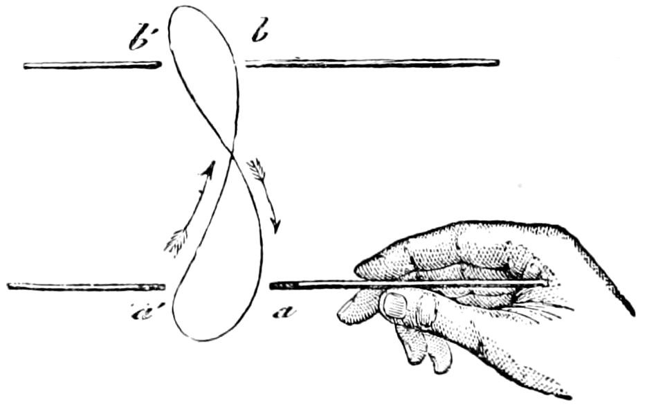
Fig. 165.—Experiment to test the direction of movement of an insect’s wing: a, a′, b, b′, different positions of the smoked rod.
Theory of insect flight.—“The theory of insect flight,” says Marey, “may be completely explained from the preceding experiments. The wing, in its to-and-fro movement, is bent in various directions by the resistance of the air. Its action is always that of an inclined plane striking against a fluid and utilizing that part of the resistance which is favorable to its onward progression.
“This mechanism is the same as that of a waterman’s scull, which as it moves backwards and forwards is obliquely inclined in opposite directions, each time communicating an impulse to the boat.”
The mechanism in the case of the insect’s wing is far simpler, 151however, than in the process of sculling, since “the flexible membrane which constitutes the anterior part of the wing presents a rigid border, which enables the wing to incline itself at the most favorable angle.”
“The muscles only maintain the to-and-fro movement, the resistance of the air does the rest, namely, effects those changes in surface obliquity which determine the formation of an 8–shaped trajectory by the extremity of the wing.”
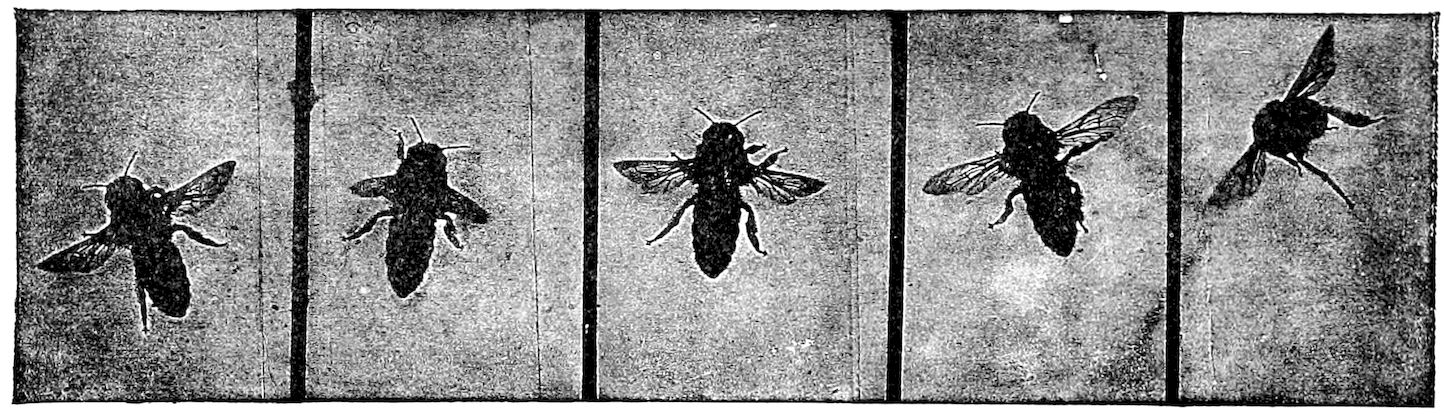
Fig. 166.—Bee flying about in the chamber of the apparatus.—After Marey.
Lendenfeld has applied photography to determine the position of the wings of a dragon-fly, and Marey has carried chronophotography farther to indicate the normal trajectory of the wing, and to show the position in flight. Fig. 166 shows a bee in various phases of flight. “The insect sometimes assumes almost a horizontal position, in which case the lower part of its body is much nearer the object-glass than is its head, and yet both extremities are equally well defined in the photograph. The successive images are separated by an interval of 1 20 of a second (a long time when compared to the total time occupied by a complete wing movement, i.e. 1 190 of a second). And hence it is useless to attempt to gain a knowledge of the successive phases of movement by examining the successive photographs of a consecutive series representing an insect in flight. Nevertheless an examination of isolated images affords information of extreme interest with regard to the mechanism of flight.
“We have seen that owing to the resistance of the air the expanse of wing is distorted in various directions by atmospheric resistance. Now, as the oscillations during flight are executed in a horizontal plane, the obliquity of the wing-surface ought to diminish the apparent breadth of the wing. This appearance can be seen in Fig. 167. There is here a comparison between two Tipulæ: the one in the act of flight, the other perfectly motionless and resting against the glass window.
152
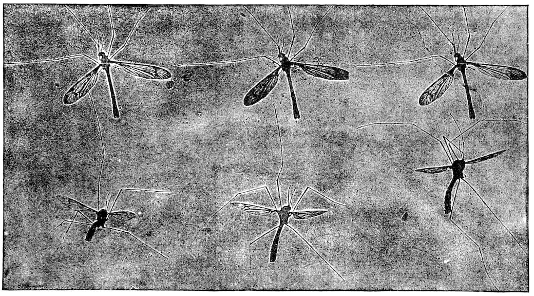
Fig. 167.—Illustration to show two Tipulæ, one of them remaining motionless on the glass, and the other moving its limbs in different directions, and setting its body at various inclinations: the illustration only represents a small part of a long series.—After Marey.
“The motionless insect maintains its wings in a position of vertical extension; the plane is therefore at right angles to the axis of the object-glass. The breadth of the wing can be seen in its entirety; the nervures can be counted, and the rounding off of the extremities of the wings is perfectly obvious. On the other hand, the flying insect moves its wings in a horizontal direction, and owing to the resistance of the air the expanse of the wings is obliquely disposed, and only the projection of its surface can be seen in the photograph. This is why the extremity of the wings appears as if it were pointed, while the other parts look much narrower than normal. The extent of the obliquity can be measured from the apparent alteration in width, for the projection of this plane with the vertical is the sine of the angle. From this it may be gathered that the right wing (Fig. 168, third image) was inclined at an angle of about 50° with the vertical, say 40° with the horizontal. This inclination necessarily varies at different points of the trajectory and must augment with the rapidity 153of movement; the obliquity reaching its maximum in those portions of the wings which move with the greatest velocity, namely, towards the extremities. The result is that the wing becomes twisted at certain periods of the movement.” (See the fourth image in Fig. 168.) The position of the balancers seems to vary according to that of the wings. (Marey’s Movement, pp. 253–257.)
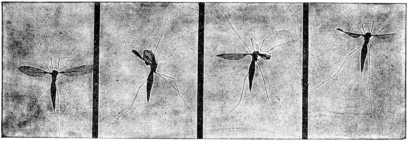
Fig. 168.—Tipula in the act of flying, showing the various attitudes of the wings and the position of the balancers.
Graber’s views as to the mechanism of the wings, flight, etc.—Although in reality insects possess but four wings, nature, says Graber, evidently endeavors to make them dipteral. This end is attained in a twofold manner. In the butterflies, bees, and cicadas, the four wings never act independently of each other, as two individual pairs, but they are always joined to a single flying plate by means of peculiar hooks, rows of claws, grooved clamps, and similar contrivances proceeding from the modified edges of the wings; indeed, this connection is usually carried so far that the hind wings are entirely taken in tow by the front, and consequently possess a relatively weak mechanism of motion. The other mode of wing reduction consists in the fact that one pair is thrown entirely out of employment. We observe this for instance in bugs, beetles, grasshoppers, etc.
In the meantime, then, we may not trust to appearances. As their development indeed teaches us, the wings as well as the additional members must be regarded as actual evaginations of the common sockets of the body, and in order especially to refute the prevalent opinion that these wing-membranes are void of sensation, it should be remembered that Leydig has proved the existence, as well as one can be convinced by experiment, of a nerve-end apparatus in certain basal or radical veins 154of the wing-membrane, which is very extensive and complicated, and therefore indicates the performance of an important function, perhaps of a kind of balancing sense, and also that these same insect wings, with their delicate membrane, are very easily affected by different outside agents, as, for instance, warmth, currents of air, etc.
Usually in their inactive or passive state the wings are held off horizontally from the body during flight, and are laid upon the back again when the insect alights; but an exception occurs in most butterflies and Neuroptera, among which the wing-joint allows only one movement round the oblique and long axis of the wings. From this cause, too, the insects just mentioned can unfold their wings suddenly.
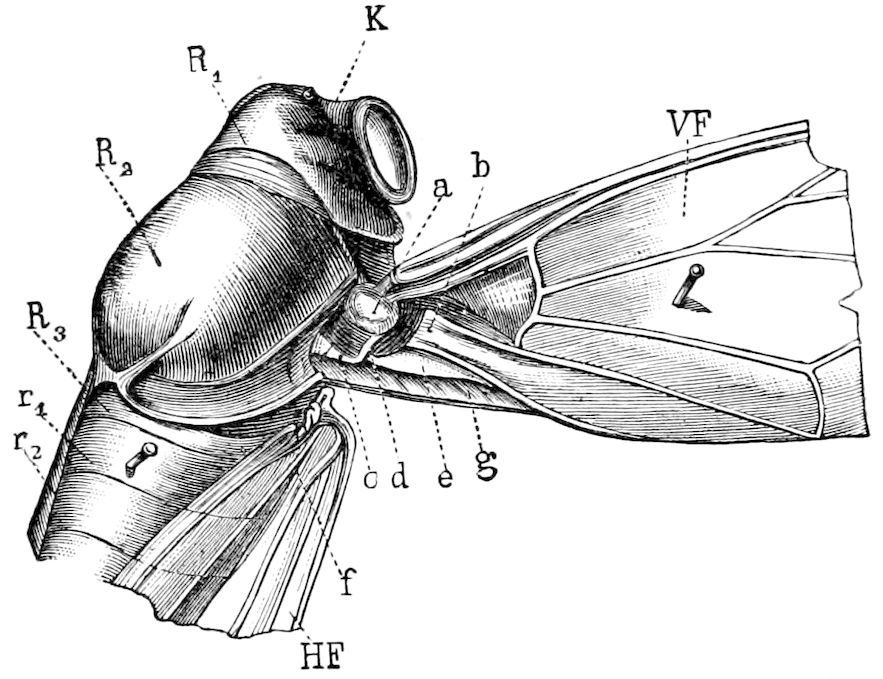
Fig. 169.—Anterior part of a Cicada for demonstrating the mechanism of the articulation of the fore wing: a, articular head; b, articular pan, frog, or cotyla; g, elastic band; c, d, e, system of elastic rods; r1, r2, 1st and 2d abdominal segments. HF, hind wings.—After Graber.
The transition of the wings from the active to the resting condition seems to be by way of a purely passive process, which, therefore, usually gives no trouble to the insect. The wing being extended by the tractive power of the muscles, flies back, when this ceases, to its former or resting posture by means of its natural elasticity, like a spiral spring disturbed from its balance. The structure of this spring joint is very different, however.
It usually consists (Fig. 169) of two parts. The wing can move itself up and down in a vertical plane by means of the forward joint, and at the same time can rotate somewhat round its long axis, because the chitinous part mentioned above is ground off after the fashion of a mandrel.
The hinder joint, at a greater distance from the body, virtually consists of a rounded piece (a) capitate towards the outside, and of a prettily hollowed socket (b) formed by the union of the thick ribs of the hind wings, which slides round 155the head joint when the wings snap back upon the back. The mechanism which causes this turning is, however, of a somewhat complicated nature. The most instrumental part of it is the powerful elastic band (g) which is stretched over from the hinder edge of the mesothorax (R2) towards that of the wings. This membrane is extended by the expansion of the wings, and draws them towards the body as soon as the contraction of the muscles relaxes. This closing band of the wings is assisted by a leverage system consisting of three little chitinous rods (c, d, e), which at its joining presses inwards on the body on one side, and on the hinder edge and head-joint of the wing on the other.
We must, however, lay great stress on a few more kinds of wing support.
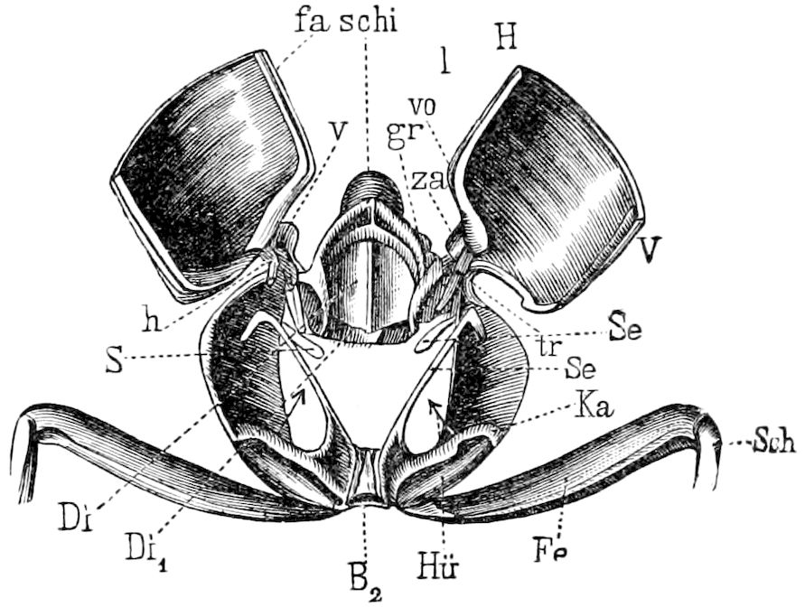
Fig. 170.—Mesothoracic skeleton of a stag beetle: schi, scutellum, on each side of which is the articulation of the fore wing (V), consisting of two small styliform processes (v, h) of the base of the wing; za, tooth which fits into the cavity of the wing-lock (gr); l, edge of the right wing, passing into the corresponding groove (fa) of the left; Di, diaphragm for the attachment of the tergal muscle of the metasternum; Di1 (not explained by author); Ka, acetabulum of the coxa (Hü); Se, chitinous process for the attachment of the coxal muscle; Fe, femur; Sch, tibia; B2, sternum.—After Graber.
The wing-cases of beetles at their return from flight are joined together like the shells of a mussel on the inside as well as to the wedge-shaped plate (Fig. 170, schi) between their bases. There is even a kind of clasp at hand for this purpose. The base of the wing, that is, bears a pair of tooth-like projections (za), which fit into the corresponding hollows of the little plate.
The commissure arising from the joining of the inner edges is characteristic. Usually the wings on both sides interlock by means of a groove, as in stag-beetles, but sometimes even, as in Chlamys, after the manner of two cog-wheels, so that we have here also an imitation of the two most prevalent methods which the cabinet-maker uses in joining boards together.
The act of folding the broad hind wings among beetles is not less significant than the arrangement of the fore wing. If we forcibly spread out the former in a beetle which has just been killed and then leave it to its own resources again, we observe the following result: According to its peculiar mode of joining, the costal vein on the fore edge approaches the mid or discoidal vein of the basal half as well as the distal half of the wing, whence arises a longitudinal fold which curves in underneath. Then the distal half snaps under like the blade of a pocket knife and lies on the plane of the costal edge of the wing, while it also draws after it the neighboring wing-area. The soft hinder-edge portion turns in simultaneously when this wing-area remains fixed to the body while the costal portion is moving towards the middle line of the body.
The wing-membranes of almost all insects have, moreover, the capability of folding themselves somewhat, and this power of extending or contracting the wing-membrane at will is of great importance in flight.
Yes, but how is the folded wing spread out again? The fact may be shown more simply and easily than one might suppose, and may be most plainly demonstrated even to a larger public by making an artificial wing exactly after the pattern of the natural one, in which bits of whalebone may take the place 156of veins and a piece of india rubber the membrane spread out between them. The reader will be patient while we just explain to him the act of unfolding of the membranous wing of the beetle. The actual impulse for this unfolding is due to the flexor muscles which pull on, and at the same time somewhat raise the vein on the costal edge. By this means the membranous fold lying directly behind the costal vein is first spread out. But since this fold is connected with the longitudinal fold of the distal end of the wing which closes like a blade, the wing-area last mentioned which is attached to the middle fold of the wing by the elastic spring-like diagonal vein becomes stretched out. The hinder rayed portion adjacent to the body is, on the other hand, simply drawn along when the wing stands off from the body.
In order to properly grasp the mechanism of the insect wing we must again examine its mode of articulation to the body somewhat more accurately.
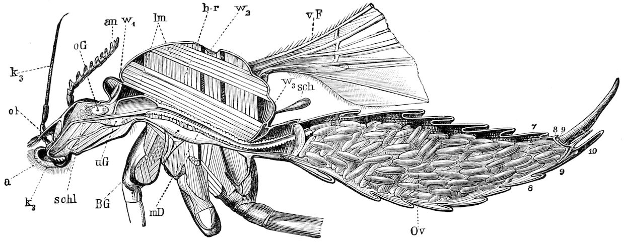
Fig. 171.—Longitudinal section through a Tipula: a, mouth; an, antenna; k3, maxillary palpus; ol, labrum; oG, brain; uG, subœsophageal ganglion; BG, thoracic ganglion; schl, œsophagus; mD, digestive canal; Ov, ovary; vF, fore wing; sch, halter; lm, longitudinal—b-r, lateral muscles.—After Graber.
If we select the halteres of a garden gnat (Tipula) at the moment of extension, we shall find them to be formed almost exactly after the pattern of our oars, since the oblong oar-blade passes into a longitudinal handle. The pedicel of the balancer is formed by the thick longitudinal primary veins of the wing-membrane. This pedicel (Fig. 171) is implanted in the side of the thorax in such a manner that the wing may be compared to the top of a ninepin. One may think, and on the whole it is actually the fact, that the stiff pedicel of the wing is inserted in the thoracic wall, and that a short portion of it (Fig. 172), projects into the cavity of the thorax. It is true there is no actual hole to be found in the thoracic wall, as the intermediate 157space between the base or pedicel of the wing and the aperture in the thorax is lined with a thin yielding membrane, on which the wing is suspended as on an axle-tree. According to this, therefore, the insect wing, as well as any other appendage of arthropods, acts as a lever with two arms. The reader can then conjecture what may be the further mechanism of the wing machine. We only need now two muscles diametrically opposed to each other and seizing on the power arm of the wing, one of which pulls down the short wing arm, thereby raising the oar, while the other pulls up the power arm. And indeed the raising of the wing follows in the manner indicated, since a muscle (hi) is attached to the end of the wing-handle (a) which projects freely into the breast cavity by the contraction of which the power arm is drawn down.
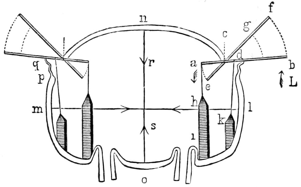
Fig. 172.—Scheme of the flying apparatus of an insect: mnl, thoracic walls; ab, wings; c, pivot; d, point of insertion of the depressor muscle of the wing (kd);—a, that of the elevator of the wing (ai); rs, muscle for expanding,—ml, for contracting, the walls of the thorax.—After Graber.
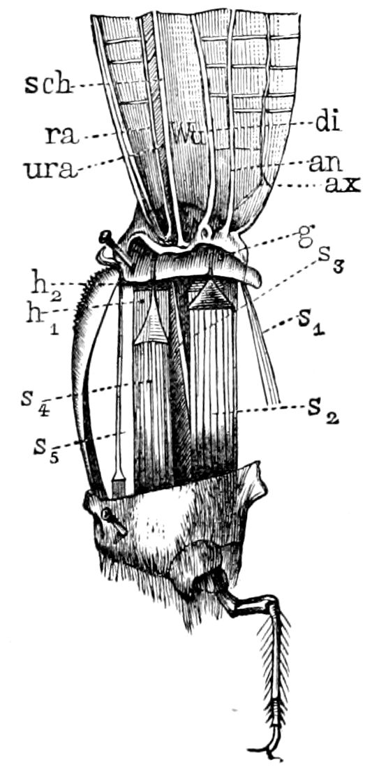
Fig. 173.—Muscles of the fore wing of a dragon-fly (an, ax), exposed by removing the thoracic walls: h1, h2, elevators,—s1-s5, depressors, of the wings (s1, s2, rotators).—After Graber.
On the other hand, we have been entirely mistaken in reference to the mechanism which lowers the wings. The muscle concerned, that is kd, is not at all the antagonist of the elevator muscle of the wing, since it is placed close by this latter, but nearer to the thoracic wall. But then, how does it come to be the counterpart of its neighbor? In fact, the lever of the wing is situated in the projecting piece alone. The extensor muscle of the wing does not pull on the power arm, but on the resistant arm on the other side of the fulcrum (c). The illustration shows, however, how such a case is possible. The membrane of the joint fastening the wing-stalk to the thorax is turned up outwards below the stalk like a pouch. The tendon of the flexor of the wing passes through this pouch to its point of attachment (c) lying on the other side of the fulcrum (d). Thus it is very simply explained how two muscles which act in the same direction can nevertheless have an entirely contrary working power.
This is in a way the bare physical scheme of the flying machine by the help of which we shall more easily become acquainted with its further details.
Dragon-flies are unquestionably the most suitable objects for the study of the muscles pulling directly on the wing itself. If the lateral thoracic wall (Fig. 173) be removed or the thorax opened lengthwise there appears a whole storehouse of muscular cords which are spread out in an oblique direction between the base of the wing and the side of the thoracic plate. There is first to be ascertained, by the experiment of pulling the individual muscles in the line with a pincers, which ones serve for the lifting and which for the lowering 158of the wings. In dragon-flies the muscles are arranged in two rows and in such a way that the flexors or depressors (s, 1 bis) cling directly to the thoracic wall (compare also the muscle dk in Fig. 172 and se in Fig. 174), while the raiser or extensor (h 1, to h 2, Fig. 172, hi and Fig. 174 he) lie farther in. The form of the wing-muscles is sometimes cylindrical, sometimes like a prism, or even ribbon-like. However, the contracted bundles of fibres do not come directly upon the joint-process we have described, but pass over often indeed at a very considerable distance from them, into peculiar chitinous tendons. These have the form of a cap-like plate, often serrate on the edge, which is prolonged into a thread, which should be considered as the direct continuation of the base of the wings. The wings, therefore, sink down into the thoracic cavity as if they were a row of cords ending in handles where the strain of the muscles is applied.
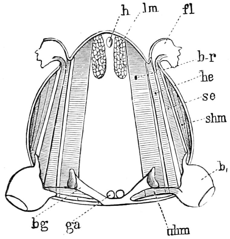
Fig. 174.—Transverse section through the thorax of a locust (Stenobothrus): b1, leg; h, heart; ga, ventral cord; se, depressor,—he, elevator, of the wing (fl); b-r, lateral muscles which expand the thoracic walls;—lm, longitudinal muscles which contract them; shm, uhm, muscles to the legs; bg, apodemes.—After Graber.
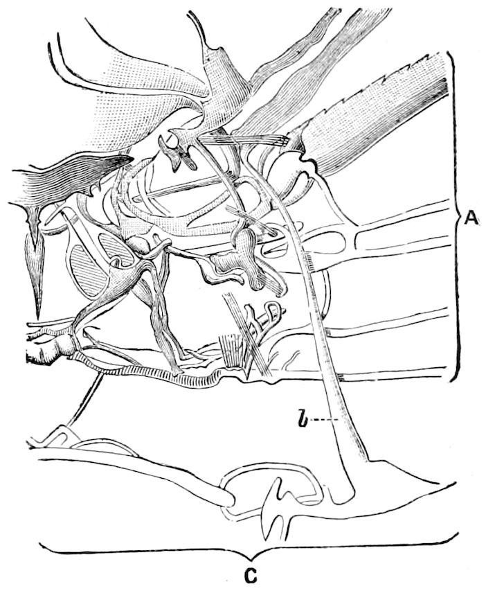
Fig. 175.—Inner view of a portion of the left side of body of Libellula depressa, showing a part of the mechanism of flight, viz., some of the chitinous ridges at base of the upper wing, and some of the insertions of the tendons of muscles: A, line of section through the base of the upper wing, the wing being supposed to be directed backwards. C, upper portion of mechanism of the lower wing; b, lever extending between the pieces connected with the two wings.—After von Lendenfeld, from Sharp.
As may be seen in Fig. 173, the contractile section of several of the muscles of the wing (s5) is extraordinarily reduced, while its thread-like tendon is proportionately longer. This gradation being almost like that of the pipes of an organ in the length of the wing-muscles, as may so easily be observed in the large dragon-flies, plainly indicates that the strain of the individual muscles is quite different in strength, since, as the phenomenon of flight demands it, the different parts of the base of the wing become respectively relaxed in very dissimilar measure.
We have thus far discussed only the elevator and depressor muscles. Other groups (s1s3) are yet to be added, however, crossing under the first at acute 159angles, which when pulling the wing sidewise, bring about in union with the other muscles a screw-like turning of the wings.
While in dragon-flies all the muscles which are principally influential in moving the wing are directly attached to it, and thus evidently assert their strength most advantageously, the case is essentially different with all other insects. Here, as has already been superficially mentioned above, the entire set of muscles affecting the wing is analyzed into two parts of which the smaller only is usually directly joined to the wings, while the movement is indirectly influenced by the remainder (Graber).
In the dragon-fly the two wings are “brought into correlative action by means of a lever of unusual length existing amongst the chitinous pieces in the body wall at the base of the wings (Fig. 175, b). The wing-muscles are large; according to von Lendenfeld there are three elevator, five depressor, and one abductor muscles to each wing. He describes the wing-movements as the results of the correlative action of numerous muscles and ligaments, and of a great number of chitinous pieces connected in a jointed manner” (Sharp).
If again we take the longitudinal section of the thoracic cavity of gnats in Fig. 171, we shall perceive a compactly closed system of muscular bars intersecting each other almost at right angles and interlaced with a tangled mass of tracheæ, some of which muscles extend (lm) longitudinally, that is from the front to the back, while others (b-r) stretch out in a vertical direction, that is between the plates of the abdomen and back.
In order that we may more easily comprehend this important muscular apparatus we will illustrate the thoracic cavity of insects by an elastic steel ring (Fig. 172), to which we may affix artificial wings. If this ring be pressed together from above downward, along the line rs, thus imitating the pulling of the vertical or lateral thoracic muscles, then the wings on both sides spring up. This is to be explained by the fact that through this manipulation a pressure is exerted on the lifting power arm of the wings. If, on the other hand, the ring be compressed on the sides (ml), which is the same thing as if the longitudinal muscles contracted the thorax from before backward, and thus arched it more, then the wings are lowered.
Agrioninæ, according to Kolbe, can fly with the fore pair of wings or with the hind pair almost as well as with both pairs together. Also the wings of these insects can be cut off before the middle of their length without injuring their power of flight. Butterflies, Catocalæ, and Bombycidæ fly after the removal of the hind wings. Also the balancers of the Diptera must be useful in flying, since their removal lessens the power of flight.
Chabrier regarded the under sides of the shell-like extended wing-covers of the beetles as wind-catchers, which, seized by wind currents, carry the insect through the air. We may also consider the wing-covers as regulators of the centre of gravity of flight.
The observations of insects made by Poujade (Ann. Soc. Ent., France, 1887, p. 197) during flight teaches us, says Kolbe, that in respect to the movement during flight of both pairs of wings, they may be divided into two categories:—
1. Into those where both pairs of wings (together), either united, and also when separated from each other, perform flight. Such are the Libellulidæ, Perlidæ, Sialidæ, Hemerobiidæ, Mymeleonidæ, Acridiidæ, Locustidæ, Blattidæ, Termitidæ, etc.
2. Into those whose fore and hind wings act together like one wing, since they are connected by hooks (hamuli), as in certain Hymenoptera, or are attached in other ways. Here belong Hymenoptera, Lepidoptera, Trichoptera, Cicadidæ, Psocidæ, etc.
160The musculature of the mesothorax and metathorax is similar in those insects both of whose pairs of wings are like each other, and act independently during flight, viz. in the Libellulidæ. On the other hand, in the second category, where the fore and hind wings act as a single pair and the fore wings are mostly larger than the hinder (except in most of the Trichoptera), the musculature of the mesothorax is more developed than that of the metathorax.
To neither category belong the beetles, whose wing-covers are peculiar organs of flight, and not for direct use, and the Diptera, which possess but a single pair of wings. In the beetles the hind wings, in the Diptera the fore wings, serve especially as organs of flight. It may be observed that the Diptera are the best fliers, and that those insects which use both pairs of wings as a single pair fly better than those insects whose two pairs of wings work independently of each other. An exception are the swift-flying Libellulidæ, whose specially formed muscles of flight explain their unusual capabilities for flying (Kolbe).
LITERATURE ON FLIGHT
Marey, E. J. La machine animale. Locomotion terrestre et aërienne. Paris, 1874.
—— Mémoire sur le vol des insectes et des oiseaux. (Annal. Scienc. natur., 5 sér., Zool. xii, 1869, pp. 49–150; 5 sér., Zool. xv, 1872, 42 Figs.)
—— Note sur le vol des insectes. (Compt. rend. et Mém. Soc. d. Biol. Paris, 4 sér., v, 1869, C. R. pp. 136–139.)
—— Recherches sur le mécanisme du vol des insectes. (Journal de l’Anatomie et de la Physiologie, 6 Année, 1869, pp. 19–36, 337–348.)
—— Animal mechanism. New York, 1879, pp. 180–209.
—— Movement. New York, 1895, pp. 239–274.
Hartings. Ueber den Flug. (Niederland. Archiv f. Zoologie, iv, Leiden, 1877–78.)
Lucy. Le vol des oiseaux, chauvesouris et insectes. Paris.
Tatin, V. Expériences physiologiques et synthétiques sur le mécanisme du vol. (Ecole prat. d. haut. étud. Physiol. expérim. Trav. du laborat. de Marey, 1877, pp. 293–302.)
—— Expériences sur le vol mécanique. (Ibid., 1876, pp. 87–108.)
Bellesme, Jousset de. Recherches expérimentales sur les fonctions du balancier chez les insectes Diptères, Paris, 1878, 96 pp., Figs.
—— Sur une fonction de direction dans le vol des insectes. (Compt. rend., lxxxix, 1879, pp. 980–983.)
Pettigrew, J. Bell. On the mechanical appliances by which flight is attained in the animal kingdom. (Trans. Linn. Soc., 1868, xxvi, Pt. I, pp. 197–277, 4 Pl.)
—— On the physiology of wings. (Trans. Roy. Soc. Edinburgh, 1871, xxvi, pp. 321–446.)
Krarup-hansen, C. J. L. Beitrag zu einer Theorie des Fluges der Vogel, Insekten und Fledermause. (Copenhagen u. Leipzig, Fritsch, 1869, 48 pp.)
Lendenfeld, R. V. Der Flug der Libellen. (Sitzungsber. d. kais. Akad. d. Wiss. Wien, lxxxiii, 1881, pp. 289–376, 7 Taf.; Zool. Anz., 1880, p. 82.)
Girard, M. Note sur diverses expériences relatives à la fonction des ailes chez les insectes. (Ann. Soc. Ent. France, 4 sér., ii, 1862, pp. 154–162.)
Mühlhäuser, F. A. Ueber das Fliegen der Insekten. (22. bis 24., Jahresb. d. Pollichia, Dürkheim, 1866, pp. 37–42.)
Plateau, Félix. Recherches expérimentales sur la position du centre de gravité chez les insectes. (Archiv d. Scienc. phys. et natur. d. Genève, Nouv. période, xliii, 1872, pp. 5–37.)
161Plateau, Félix. Ueber die Lage des Schwerpunktes bei den Insekten. Auszug. (Naturforscher v. Sklarek, v. Jahrg., 1872, pp. 112–113.)
—— Recherches physico-chimiques sur les articulés aquatiques. (Bull. d. l’Acad. Roy. Belg., xxxiv, 1872, pp. 1–50, Fig.)
—— Qu’est-ce que l’aile d’un insecte? (Stett. Ent. Zeit., 1871, pp. 33–42, Pl.)
—— L’aile des insectes. (Journ. d. Zool., ii, 1873, pp. 126–137.)
Perez, J. Sur les causes de bourdonnement chez les insectes. (Comptes rend., lxxxvii, p. 535, Paris, 1878.)
Strasser, Hans. Mechanik des Fluges. (Archiv f. Anat. u. Phys., 1878, p. 310–350, 1 Taf.)
—— Ueber die Grundbedingungen der aktiven Locomotion. (Abhandl. d. naturf. Gesellsch., Halle, 1880, xv, pp. 121–196, Figs.)
Moleyre, L. Recherches sur les organes du vol chez les insectes de l’ordre des Hemiptères. (Compt. rend. de l’Acad. d. Scienc. de Paris, 1882, xcv, pp. 349–352.)
Amans, P. Essai sur le vol des insectes. (Revue d. Sc. Nat. Montpellier, 3 sér., ii, 1883, pp. 469–490, 2 Pl.; iii, 1884, pp. 121–139, 3 Pl.)
—— Étude de l’organe du vol chez les Hyménoptères. (Ibid., iii, pp. 485–522, 2 Pl.)
—— Comparaisons des organes du vol dans la série animale. Des organes du vol chez les insectes. (Annal. d. Scienc. nat. Zool., 6 sér., xix, pp. 1–222, 8 Pl.)
Mullenhoff, K. Die Grosse der Flugflächen. (Pflüger’s Archiv f. d. ges. Physiologie, 1884, xxxv, pp. 407–453.)
—— Die Ortsbewegungen der Tiere. (Wissensch. Beil. z. Programm d. Andreas-Realgymnas. Berlin, 1885, 19 pp.)
Poujade, G. A. Note sur les attitudes des insectes pendant le vol. (Ann. Soc. Ent. France, 1884, 6 sér., iv, pp. 197–200, 1 Pl.)
Krancher, O. Die Töne der Flügelschwingungen unserer Honigbiene. (Deutscher Bienenfreund, 1882, 18. Jahrg., pp. 197–204.)
Landois, H. Ueber das Flugvermögen der Insekten. (Natur. und Offenbarung, vi, 1860, pp. 529–540.)
Ungern-Sternberg, von. Betrachtungen über die Gesetze des Fluges. (Zeitschr. d. Deutschen Vereins z. Förderung d. Luftschiffahrt-Naturwissensch. Wochenschrift v. Potonie, iv, 1889, p. 158.)
Baudelot, E. Du mecanisme suivant lequel s’effectue chez les Coléoptères le retract des ailes inférieures sous les élytres au moment du passage a l’état de repos. (Bull. Soc. d. Scienc. nat., Strasbourg, 1 Année, 1868, pp. 137–138.)
Ris, Fr. Die schweizerischen Libellen. Schaffhausen, 1885. (Beiheft der Mitteil. d. Schweiz. Ent. Ges., vii, pp. 35–84.)
162
THE ABDOMEN AND ITS APPENDAGES
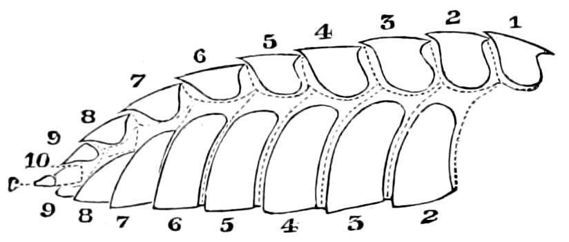
Fig. 176.—Abdomen of Termes flavipes: 1–10, the ten tergites; 1–9, the nine urites; c, cercopod.
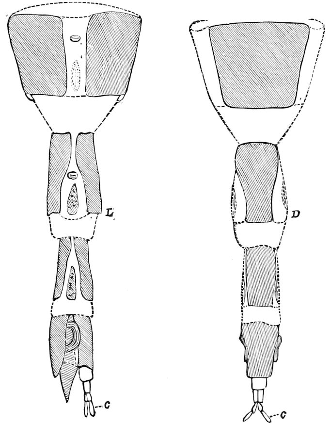
Fig. 177.—End of abdomen of Panorpa debilis drawn out, the chitinous pieces shaded: L, lateral, D, dorsal view; c, jointed cercopoda.—Gissler del.
In the abdomen the segments are more equally developed than elsewhere, retaining the simple annular shape of embryonic life, and from their generalized nature their number can be readily distinguished (Fig. 176). The tergal and sternal pieces of each segment are of nearly the same size, the tergal often overlapping the sternal (though in the Coleoptera the sternites are larger than the tergites), while there are no pleural pieces, the lateral region being membranous when visible and bearing the stigmata (Fig. 177, L). In the terminal segments beyond the genital outlet, however, there is a reduction in and loss of segments, especially in the adults of the metabolous orders, notably the Panorpidæ (Fig. 177), Diptera, and aculeate Hymenoptera; in the Chrysididæ only three or four being usually visible, the distal segments being reduced and telescoped inward.
The typical number of abdominal segments (uromeres), i.e. that occurring in each order of insects, is ten; and in certain families of Orthoptera, eleven. In the embryos, however, of the most generalized winged orders, Orthoptera (Fig. 199), Dermaptera, and Odonata, eleven can be seen, while Heymons has recently detected twelve in blattid and Forficula embryos, and he claims that in the nymphs of certain Odonata there are twelve segments, the twelfth being 163represented by the anal or lateral plates. It thus appears that even in the embryo condition of the more generalized winged insects, the number of uromeres is slightly variable.
We have designated the abdomen as the urosome; the abdominal segments of insects and other Arthropods as uromeres, and the sternal sclerites as urosternites, farther condensed into urites. (See Third Report U. S. Entomological Commission, 1883, pp. 307, 324, 435, etc.)
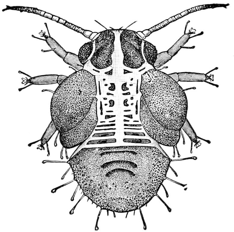
Fig. 178.—Nymph of the pear tree Psylla, with its glandular hairs.—After Slingerland. Bull. Div. Ent. U. S. Dep. Agr.
The reduction takes place at the end of the abdomen, and is usually correlated with the presence or absence of the ovipositor. In the more generalized insects, as the cockroaches, the tenth segment is, in the female, completely aborted, the ventral plate being atrophied, while the dorsal plate is fused during embryonic life, as Cholodkowsky has shown, with the ninth tergite, thus forming the suranal plate.
In the advanced nymph of Psylla the hinder segments of the abdomen appear to be fused together, the traces of segmentation being obliterated, though the segments are free in the first stage and in the imago (Fig. 178). It thus recalls the abdomen of spiders, of Limulus, and the pygidium of trilobites.
The median segment.—There has been in the past much discussion as to the nature of the first abdominal segment, which, in those Hymenoptera exclusive of the phytophagous families, forms a part of the thorax, so that the latter in reality consists of four segments, what appearing to be the first abdominal segment being in reality the second.
Latreille and also Audouin considered it as the basal segment of the abdomen, the former calling it the “segment médiaire,” while Newman termed it the “propodeum.” This view was afterward held by Newport, Schiödte, Reinhard, and by the writer, as well as Osten-Sacken, Brauer, and others. The first author to attempt to prove this by a study of the transformations was Newport in 1839 (article “Insecta”). He states that while the body of the larva is in general composed of thirteen distinct segments, counting the head as the first, “the second, third, fourth, and, as we shall hereafter see, in part also the fifth, together form the thorax of the future imago” (p. 870). Although at first inclined to Audouin’s opinion, he does not appear to fully accept it, yet farther 164on (p. 921) he concludes that in the Hymenoptera the “fifth” segment (first abdominal) is not in reality a part of the true thorax, “but is sometimes connected more or less with that region, or with the abdomen, being intermediate between the two. Hence we have ventured to designate it the thoracico-abdominal segment.” Had he considered the higher Hymenoptera alone, he would undoubtedly have adopted Latreille’s view, but he saw that in the saw-flies and Lepidoptera the first abdominal segment is not entirely united with the thorax, being still connected with the abdomen as well as the thorax. Reinhard in 1865 reaffirmed Latreille’s view. In 1866 we stated from observations on the larvæ made three years earlier, that during the semipupa stage of Bombus the entire first abdominal segment is “transferred from the abdomen to the thorax with which it is intimately united in the Hymenoptera,” and we added that we deemed this to be “the most essential zoölogical character separating the Hymenoptera from all other insects.” (See Fig. 93, showing the gradual transfer and fusion of this segment with the thorax.) In the saw-flies the fusion is incomplete, as also in the Lepidoptera, while in the Diptera and all other orders the thorax consists of but three segments. (See also pp. 90–92.)
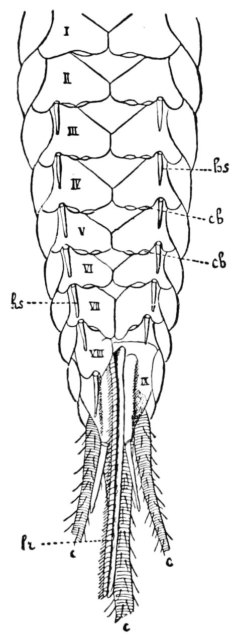
Fig. 179.—Abdomen of Machilis maritima, ♀, seen from beneath: the left half of the 8th ventral plate removed; I-IX, abdominal segments; c, cercopoda; cb, coxal glands; hs, coxal stylets; lr, ovipositor.—After Oudemans, from Lang.
The cercopoda.—We have applied this name to the pair of anal cerci appended to the tenth abdominal segment, and which are generally regarded as true abdominal legs. As is now well known, the embryos of insects of different orders have numerous temporary pairs of abdominal appendages which arise in the same manner, have the same embryonic structure, and are placed in a position homologous with those of the thorax. In the embryo of Œcanthus rudimentary legs appear, as shown by Ayers, on the first to tenth abdominal segment, the last or tenth pair becoming the cercopoda; and similar rudimentary appendages have been detected in the embryos of Coleoptera, Lepidoptera, and Hymenoptera (Apidæ). Cholodkowsky has observed eleven pairs of abdominal appendages in Phyllodromia.
They are very long and multiarticulate in the Thysanura (Fig. 179). In the Dermaptera they are not jointed and are forcep-like. It should also be observed that in the larva or Sisyra (Fig. 181) there are seven pairs of 5–jointed abdominal appendages, though these may be secondary structures or tracheal gills. In the Perlidæ 165and the Plectoptera (Ephemeridæ), they are very long, sometimes over twice as long as the body, and composed of upward of 55 joints; they also occur in the Panorpidæ (Fig. 177). In the dragon-flies the cerci are large, but not articulated, and serve as claspers or are leaf-like[35] (Fig. 180). In a few Coleoptera, as the palm-weevil (Rhynchophorus phœnicis), Cerambyx, Drilus, etc., the so-called ovipositor ends in a hairy, 1–jointed, palpiform cercus. Short 25–jointed cercopoda are present in Termitidæ, and 2–jointed ones in Embiidæ.
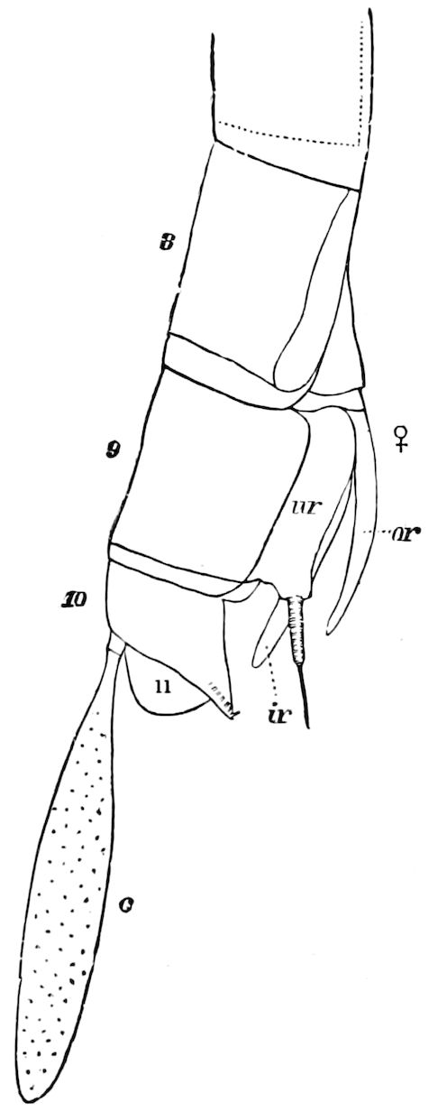
Fig. 180.—End of abdomen of Æschna heros, ♀: ur, urosternite; or, outer, ir, inner styles of the ovipositor; 11, 11th abdominal segment; c, cercopod.
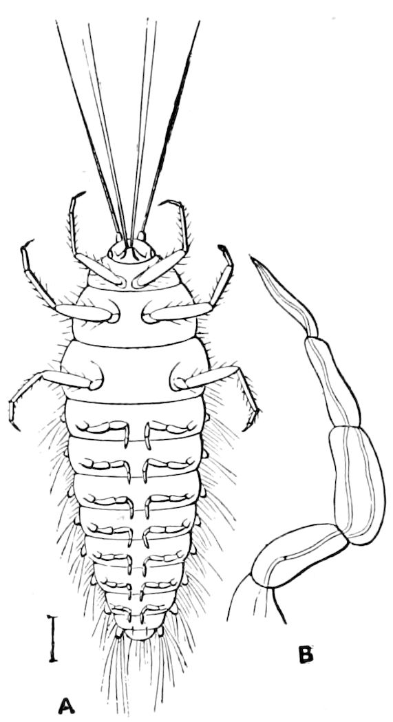
Fig. 181.—Larva of Sisyra, from beneath. B, an abdominal appendage.—After Westwood, from Sharp.
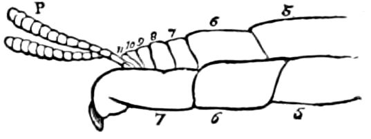
Fig. 182.—Cercopoda (P) of Mantis.—After Lacaze-Duthiers.
The anal cerci are present in the Orthoptera and, when multiarticulate, function as abdominal antennæ. They are longest in the Mantidæ (Fig. 182); they also occur in the larva of the saw-fly, Lyda (Fig. 183). Dr. A. Dohrn has stated that the cerci of Gryllotalpa are true sensory organs, and we have called those of the cockroach abdominal antennæ, having detected about ninety sacs on the upper side of each joint of the stylets, which are supposed to be olfactory in nature, and which are larger and more numerous than similar sacs or pits in the antennæ 166of the same insect.[36] From his experiments upon decapitated cockroaches, Graber concluded that these cerci were organs of smell.
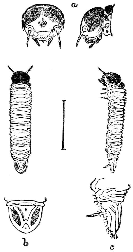
Fig. 183.—Lyda larva: a, head; b, end of body seen from above; c, from side, with cercopod.
Haase regarded these appendages, from their late development and frequent reduction, as old inherited appendages which are approaching atrophy through disuse.
Cholodkowsky states that Tridactylus, a form allied to Gryllotalpa, bears on the tenth abdominal segment two pairs of cerci (ventral and dorsal), and that the ventral pair may correspond to the atrophied appendages of the tenth embryonic segment of Phyllodromia, with which afterward the eleventh segment becomes fused.
The cercopods are not necessarily confined to the eleventh or to the tenth segment, for when there are only nine segments, with the vestige of a tenth, as in Xiphidium, they arise from the ninth uromere, and in the more modern cockroaches, as Panesthia, in which there are but seven entire segments, they are appended to the last or eighth uromere.
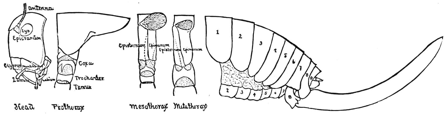
Fig. 184.—Anabrus, ♀, side-view, dissected; showing the relative size of the ovipositor: c, the minute cercopod.—Kingsley del.
167As to the homology and continuity of these cercopods with the ventral outgrowths of the embryo, several embryologists, notably Wheeler, are emphatic in regarding them as such. It thus appears that either the embryonic appendages of the seventh or eighth, ninth or tenth uromere may persist, and form the cercopoda of the adult.
The ovipositor.—The end of the oviduct is guarded by three pairs of chitinous, unjointed styles closely fitted together, forming a strong, powerful apparatus for boring into the ground or into leaves, stems of plants, the bodies of insects, or even into solid wood, so that the eggs may be deposited in a place of safety. In the ants, wasps, and bees the ovipositor also functions as a sting, which is further provided with a poison-sac.
Morphologically, the ovipositor is composed of three pairs of unjointed styles (rhabdites of Lacaze-Duthiers, gonapophyses of Huxley), which are closely appressed to or sheathed within each other, the eggs passing out from the end of the oviduct, which lies, as Dewitz states, between the two styles of the lowest or innermost pair, and under the cross-bars or at the base of the stylets mentioned; the styles or blades spreading apart to allow of the passage of the egg.
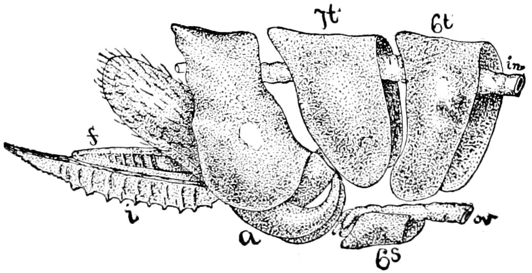
Fig. 185.—Saw of Hylotoma: a, lateral scale; i, saw; f, gorget; 7t, 7th tergite; 6s, 6th sternite; ov, oviduct; in, intestine.—After Lacaze-Duthiers.
The ovipositor is best developed in the Thysanura (Fig. 179, Campodea excepted), in Orthoptera (Fig. 184), in the Odonata, Hemiptera, certain Physapoda, Rhaphiidæ, and in the phytophagous Hymenoptera, where it is curiously modified to form a rather complicated saw for cutting slits in wood or leaves (Fig. 185). It is wanting or quite imperfect in Coleoptera, Diptera, and Lepidoptera.
Morphologically, the ovipositor appears to be formed out of the abdominal appendages of the seventh, eighth, and ninth segments of the female, which, instead of disappearing in the orders first mentioned, persist as permanent styles.
Wheeler asserts from his study of the embryonic development of Xiphidium “there can be no doubt concerning the direct continuity of the embryonic appendages with the gonapophyses.” He goes on to say:—
168“One embryo, which had just completed katatrepis, still showed traces of all the abdominal appendages. The pairs on the eighth, ninth, and tenth segments were somewhat enlarged. In immediately succeeding stages the appendages of the second to sixth segments disappear; the pair on the seventh disappear somewhat later. Up to the time of hatching the gonapophyses could be continuously traced, since in Xiphidium there is no flexure of the abdomen, as in other forms, to obscure the ventral view of the terminal segments. From the time of hatching Dewitz has traced the development of the ovipositor in another locustid (Locusta viridissima), so that now we have the complete history of the organ.”
Heymons, however, is inclined to believe that they are simply hypodermal outgrowths.
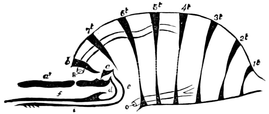
Fig. 186.—Ideal plan of the structure of the ovipositor to illustrate Lacaze-Duthiers’ view: b, 8th tergite; c, epimerum; a′, a, two pieces forming the outer pair of rhabdites; i, the 2d pair, or stylets; and f, the inner pair, or sting; d, support of sting; e, piece supporting the stylet; R, anus; o, outlet of oviduct. The 7th, 8th, and 9th sternites are aborted.—After Lacaze-Duthiers.
The first to study the morphology of the ovipositor was Lacaze-Duthiers, who referred their origin to the partially atrophied dorsal or ventral sclerites of one of the last abdominal segments; a view accepted by Gerstaecker[37] (Figs. 186, 187). The present writer (1866), however, showed that the sting of Bombus was not formed of the reduced pieces of the segments themselves, but arose from special outgrowths on the ventral side of the eighth and ninth abdominal segments. These appendages he did not at first regard as the homologues of the limbs, until in 1871, after studying the origin of the spring of the Podurans (Isotoma), he found that it was a true jointed appendage and therefore a homologue of a pair of the styles forming the ovipositor of the winged insects, and that the three pairs of styles of the latter were homologues of the thoracic legs and cephalic appendages. The view was stated in the Guide to the Study of Insects. (See also Amer. Nat., March, 1871, p. 6.) Kraepelin also affirms that the styles of the ovipositor are segmental appendages and homologues of the antennæ, wings (sic), and legs.
169
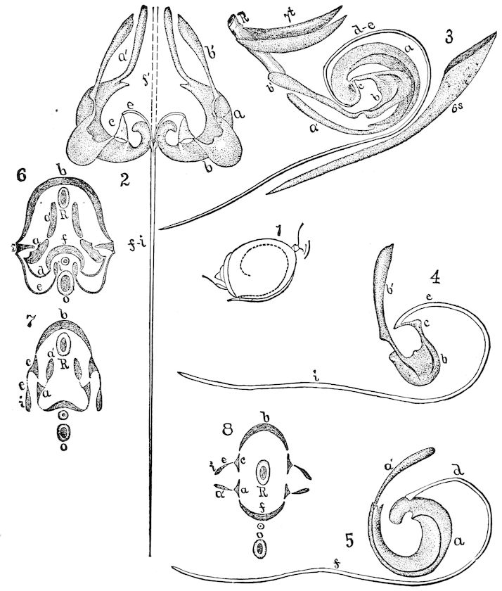
Fig. 187.—1, abdomen of Cynips, showing the great dorsal segment, the peduncle, and the position of the ovipositor within; 2, the entire ovipositor; a, lateral scale; a′, its valve; b, anal scale; b′, stylet; c, support of the stylet; e, base or support of sting (fi); 3, profile showing the relation of the genital armature to the rest of the abdomen, the 6th sternite having been drawn to show its full size; 4, anal scale (b) and stylet; e, i, supports and body of the stylet; c, piece uniting the two scales; 5, lateral scale (a), and a′ sheath; d, support of the sting (f); 6, transverse section of the body through the sting (diagrammatic); R, internal armature; o, oviduct; a, lateral scale; a′ its valve; e, support of the stylets (i); b, anal scale; c, piece uniting two scales; f, sting; d, its support; 7, a second section simpler and more theoretical than the first; 8, diagrammatic, all the elements of the sting have been reduced to pieces of the same form.—After Lacaze-Duthiers.
170An objection to this view is the fact that the posterior pairs of styles appear to arise both from one and the same segment,—the ninth. Dewitz questions whether the four appendages of the ninth segment represent two pairs of limbs, or one pair split into two branches, and prefers the latter view, but leaves it as a point to be settled by future investigations. As will be seen below, both Kraepelin and Bugnion observed a pair of rudiments to each of the three penultimate segments, those of the middle pair splitting in two. Wheeler maintains, erroneously we think, that the inner of the two pairs on the ninth segment represents the tenth pair of abdominal appendages; but in reality this latter pair become the cercopods. That there are probably originally in insects of all the orders provided with an ovipositor three distinct pairs of appendages, one to each segment, is proved, or at least strongly suggested, by Ganin’s researches on the three pairs of abdominal imaginal discs of the third larva of Platygaster and Polynema (Fig. 188), which are transformed into the ovipositor. He remarks that these imaginal discs have the same origin and pass through the same changes as those in front, i.e. those destined to form the thoracic legs. Dewitz has shown that the germs of the ovipositor of the honey-bee arise as buds on the two segments before the last (Fig. 189).
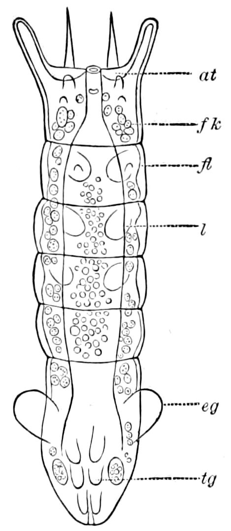
Fig. 188.—Third larva of Polynema: at, antenna; fl, imaginal buds of the wing; l, of the legs; tg, buds of the middle pair of stylets of the ovipositor; fk, fat-body; eg, ear-like process.—After Ganin.
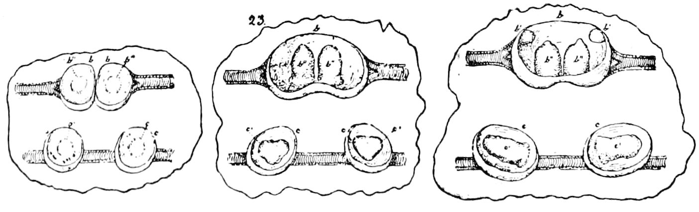
Fig. 189.—Imaginal buds and papillæ of the ovipositor of the honey-bee attached to tracheæ; at different stages: b′, 1st; b″, 2d or middle; and c′, 3d pair of papillæ.—After Dewitz.
Kraepelin also detected in the larva of the honey-bee a pair of what he regarded as genuine imaginal buds on abdominal segments eight, nine, and ten; the buds on the tenth segment are divided each in two; of these four appendages the two median ones form the barbed sting (gorgeret or stachelrinne), and the two lateral stylets, the valves (stachelscheiden). The two buds of the ninth segment give rise to the vagina and to the oviducts, and these unite secondarily with the posterior end of the ovaries. The genital appendages 171of the male correspond to those of the female, and arise from four imaginal buds situated on the under side of the tenth abdominal segment.
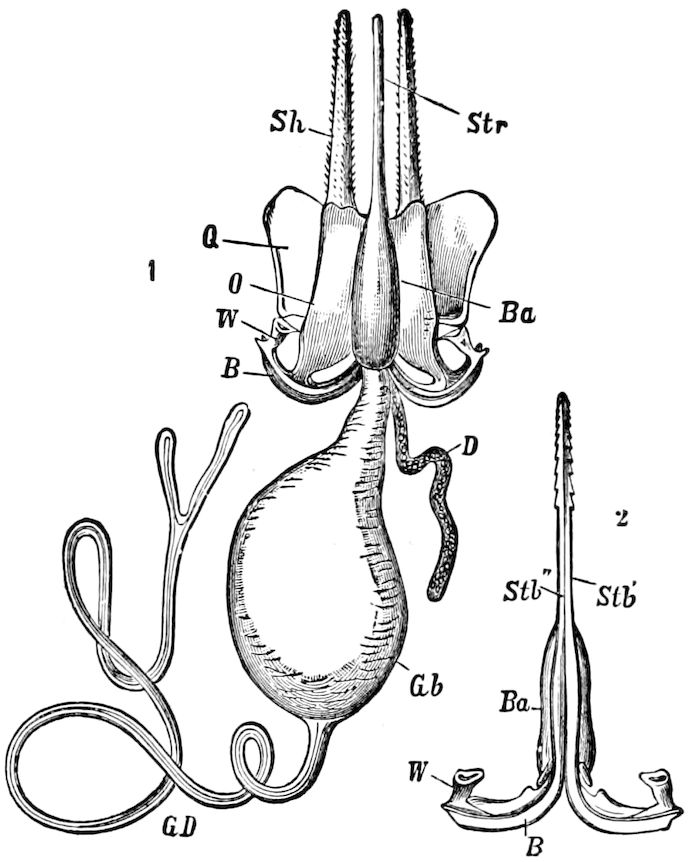
Fig. 190.—1, sting and poison sac of the honey-bee: GD, poison gland; Gb, poison reservoir; D, accessory gland; sh, sheathing style or sting-“feeler”; Str sting; Ba, sheath; Q, quadrate plate; O, oblong piece; W, angular piece; B, base of the sting and stylets; Stb′, Stb″, the two barbed stylets or darts. 2, sting seen from the ventral face: lettering as in the other figure.—After Kraepelin, from Perrier.
In the ants, according to Dewitz, the genital armature is derived from imaginal buds situated on the under side of the seventh, eighth, and ninth abdominal segments. Bugnion has observed the formation of six imaginal buds of the genital armature in the larva of a chalcid (Encyrtus, Figs. 41, 42, 191, q1, q2, q3), the transformation of the central part of these structures into small digitiform pads, then the division of the two intermediate buds into four (?) (Fig. 191, B, q2), but was unable to trace their farther development.
The subject still needs farther investigation, since certain observers, as Haase, and, more recently, Heymons, do not believe that they are homologues of the legs, but integumental structures, though of somewhat higher value than the style of the base of the legs of Scolopendrella and Thysanura; but it is to be observed that as yet we know but little of the embryological history of these styles.
Those authors who have examined the elements of the ovipositor, and regard them as homologues (homodynamous) of the limbs, are Weismann (1866), Ganin (1869), Packard (1871), Ouljanin (1872), Kraepelin, Kowalevsky (1873), Dewitz (1875), Huxley (1877), Cholodkowsky, Bugnion (1891), and Wheeler (1892).
As shown, then, by our observations and those of Dewitz (Figs. 189 and 192), the rudiments of the ovipositor consist of three pairs of tubercles, arising, as Kraepelin and also Bugnion (Fig. 191) have shown, from three pairs of imaginal discs, situated respectively on the seventh, eighth, and ninth uromeres, or at least on the three penultimate segments of the abdomen. With the growth of the 172semipupa, the end of the abdomen decreases in size, and is gradually incurved toward the base (Fig. 193), and the three pairs of appendages approach each other so closely that the two outer ones completely ensheath the inner pair, until a complete extensible tube is formed, which, by the changes in form of the muscles within, is gradually withdrawn entirely within the body.
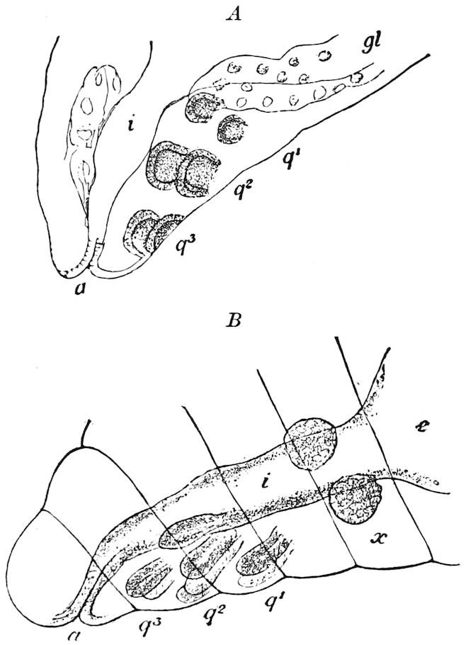
Fig. 191.—A, end of larva of Encyrtus of 2d stage, showing the three pairs of imaginal buds of the ovipositor q1, q2, q3. B, the same in an older larva ready to transform; i, intestine; x, genital gland; a, anus.—After Bugnion.
174An excellent account of the honey-bee’s sting is given by Cheshire (Figs. 194, 195). The outermost of the three pairs of stylets forming the apparatus is the two thick, hairy “palpi” or feelers (P), these being freer from the sting proper than in the ovipositor of Orthoptera. The sting itself is composed of the two inner pairs of stylets; one of these pairs is united to form the sheath (sh), while the other pair form the two barbed darts. The sheath has three uses: first, to open the wound; second, to act as an intermediate conduit for the poison; and third, to hold in accurate position the long barbed darts. The sheath does not enclose the darts as a scabbard, but is cleft down the side presented in Fig. 194, which is below when the sting points backward. But, says Cheshire, as the darts move up and down, they would immediately slip from their position, unless prevented by a mechanical device, exhibited by B and C, giving in cross-section sheath and darts near the end, and at the middle of the former. “The darts (d) are each grooved through their entire length, while upon the sheath (sh) are fixed two guide rails, each like a prolonged dovetail, which, fitted into the groove, permits of no other movement than that directly up and down.” The darts are terminated by ten barbs of ugly form (D, Fig. 194), and much larger than those of the sheath, and as soon as the latter has established a hold, first one dart and then the other is driven forward by successive blows. These in turn are followed by the sheath, when the darts again more deeply plunge, until the murderous little tool is buried to the hilt. But these movements are the result of a muscular apparatus yet to be examined, and which has been dissected away to bring the rigid pieces into view. The dovetail guides of the sheath are continued far above its bulbous portion, as we see by E, Fig. 195; and along with these the darts are also prolonged upward, still held to the guides by the grooved arrangement before explained; but both guides and darts, in the upper part of their length, curve from each other somewhat like the arms of a Y, to the points c, c′ (A, Fig. 194), where the darts 175make attachment to two levers (i, i′). The levers (k, l and k′, l′) are provided with broad muscles, which terminate by attachment to the lower segments of the abdomen. These, by contraction, revolve the levers aforesaid round the points f, f′, so that, without relative movement of rod and groove, the points 176c, c′ approach each other. The arms of the Y straighten and shorten, so that the sheath and darts are driven from their hiding-place together and the thrust is made by which the sheath produces its incision and fixture. The sides being symmetrical, we may, for simplicity’s sake, concentrate our attention on one, say the left in the figure. A muscular contraction of a broad strap joining k and d (the dart protractor) now revolves k on l, so that a is raised, by which clearly c is made to approach d; i.e. the dart is sent forward, so that the barbs extend beyond the sheath and deepen the puncture. The other dart, and then the sheath, follow, in a sequence already explained, and which G, Fig. 195, is intended to make intelligible, a representing the entrance of the sheath, b the advance of the barbs, and c the sheath in its second position. The barb retractor muscle is attached to the outer side of i, and by it a is depressed and the barbs lifted. These movements, following one another with remarkable rapidity, are entirely reflex, and may be continued long after the sting has been torn, as is usual, from the insect. By taking a piece of wash-leather, placing it over the end of the finger, and applying it to a bee held by the wings, we may get the fullest opportunity of observing the sting movements, which the microscope will show to be kept up by continued impulses from the fifth abdominal ganglion and its multitudinous nerves (n, Fig. 194, A), which penetrate every part of the sting mechanism, and may be traced even into the darts. These facts, together with the explanation at page 49, will show why an abdomen separated many hours may be able to sting severely, as I have more than once experienced.
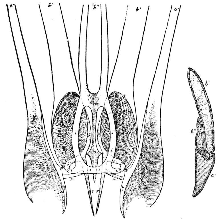
Fig. 192.—Base of the ovipositor of Locusta viridissima seen from beneath: c′, sheath, or outer and lower pair of stylets turned to one side to show the others; b′, upper and inner pair; b″, third or innermost, smallest pair of stylets. A, the same on one side, in section. The shaded parts show the muscular attachments. The muscles which extend the apparatus and are attached to ν, δ, and η, as also the membranes which unite the pieces from η; to γ with each other and the body, are removed, so that only the chitinous parts remain.—After Dewitz.
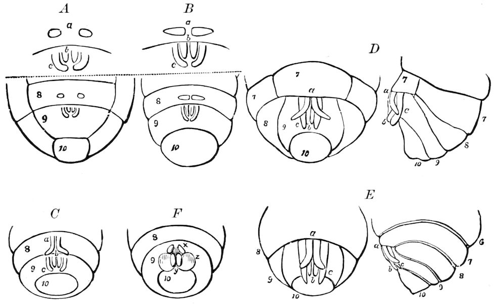
Fig. 193.—Development of the sting in Bombus: A, a, 1st pair on 8th sternite; b, 2d inner pair forming the darts; c, outer pair. B-E, more advanced stages. F, x, y, z, three pairs of tubercles, the germs of the male organs.
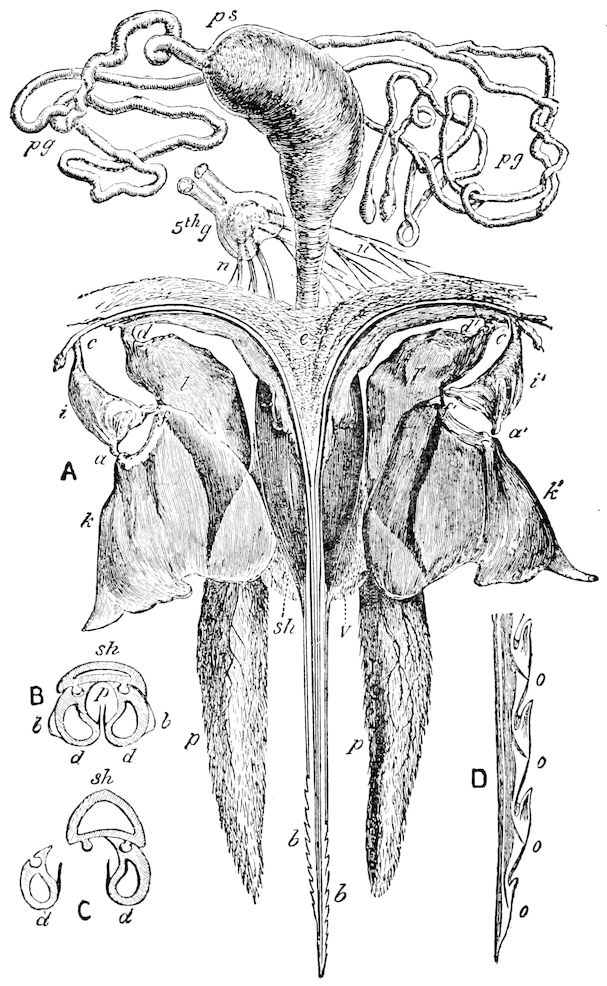
Fig. 194.—Sting of bee × 30 times: A, sting separated from its muscles; ps, poison sac; pg, poison gland; 5th g, 5th abdominal ganglion; n, n, nerves; e, external thin membrane joining sting to last abdominal segment; i, k, l, and i′, k′, l′, levers to move the darts; sh, sheath; v, vulva; p, sting-palpus or feeler, with tactile hairs and nerves. B and C, sections through the darts and sheath, × 300 times: sh, sheath; d, darts; b, barbs; p, poison-channel. D, end of a dart, × 200: o, o, openings for poison to escape into the wound.—After Cheshire.
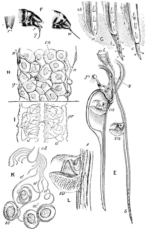
Fig. 195.—Details of sting of bee: E, darts, sheath, and valves; pb, poison-bag duct; fo, fork; s, slide piece; va, valve; b, barbs. F, terminal abdominal segments; w, worker’s sting; q, queen’s sting; r, r′, anal plate; G, sting entering skin; sh, sheath; a, b, c, positions in first, second, and third thrusts with the sting. H, portion of poison gland, × 300; cn, cell nucleus; n, nerve; g, ganglionic cell. I, portion of the poison gland, cells removed; cd, central duct; d, individual small ducts; pr, tunica propria. K, gland of Formica rufa; cd, central duct; d, small ducts; sc, secreting cells. L, valve and support; t, trachea; va, valve; tr, truss or valve-prop.—After Cheshire.
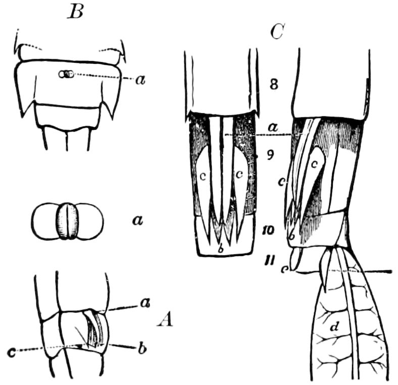
Fig. 196.—A, rudimentary ovipositor of nymph of Æschna. B, the corresponding ♂ structures; a, enlarged. C, ovipositor of nymph of Agrion; d, gill.
The male genital armature in the bees is originally composed of three pairs of tubercles, homologous with those of the female, all originally arising from three abdominal segments, two afterward being anterior, and the third pair nearer the base of the abdomen.
The ovipositor of the dragon-flies (Odonata) is essentially like that of the Orthoptera and Hymenoptera. Thus in Æschna (Fig. 196), Agrion (Fig. 196, C), and also in Cicada it consists of a pair of closely appressed ensiform processes which grow out from under the posterior edge of the eighth uromere and are embraced between two pairs of thin lamelliform pieces of similar form and structure.
The styles and genital claspers (Rhabdopoda).—Other appendages of the end of the abdomen of pterygote insects, and generally, if not always, arising from the ninth segment, are the clasping organs, or rhabdopoda as we may call them, of Ephemeridæ (Fig. 197), Neuroptera (Corydalus [Fig. 198], Myrmeleon, Rhaphidia), Trichoptera, 177Lepidoptera, Diptera, and certain phytophagous Hymenoptera. They do not appear to occur in insects which are provided with an ovipositor. In Thysanura the styles are present on segments 1–9 (Fig. 179). Those of the male Ephemeridæ, of which there are two pairs arising from the ninth segment, are remarkable, since they are jointed, and they serve to represent or may be the homologues of two of the pairs of stylets composing the ovipositor of insects of other orders. The lower pair (Fig. 197, rh) are either 2–, 3–, or 4–jointed (in Oniscigaster 5–jointed), while those of the upper pair are 2–jointed (rh′). These rhabdopods in the ephemerids are evidently very primitive structures, since they approach nearest in shape and in being jointed to the abdominal legs of Scolopendrella and the Myriopoda. The styles of the Orthoptera are survivals of the embryonic appendages of the ninth segment (Wheeler, etc.). In Mantis they are seen to have the same relations as the cerci, as shown by Heymons (Fig. 200).
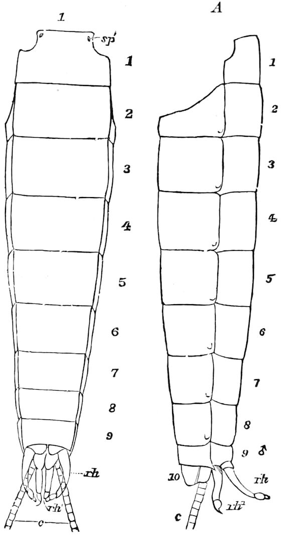
Fig. 197.—Abdomen of Ephemera (Leptophlebia) cupida, ♂: c, base of cercopoda; rh, outer 3–jointed claspers or rhabdopods; rh′, inner pair. A, side view.
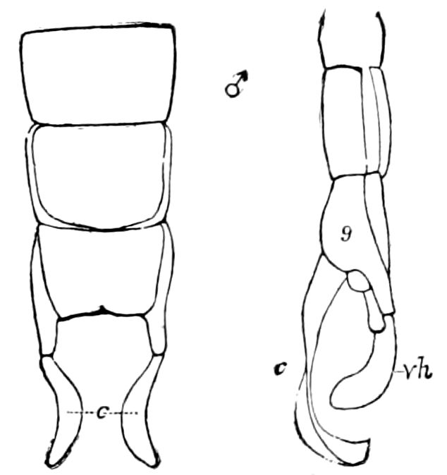
Fig. 198.—End of abdomen of Corydalus cornutus, ♂: vh, rhabdopod; c, cercopod.
In the Phasmidæ, in Anabrus, and in the Odonata the cercopods, which are not jointed, are converted into claspers, and in the Odonata the claspers are spiny within, so as to give a firmer hold. The suranal plate is apparently so modified as to aid in grasping the female. In nearly all the Trichoptera there are, besides the suranal plate, which is sometimes forked (Nosopus), a pair of superior and of inferior claspers, and in certain genera 178(Ascalaphomerus, Macronema, Rhyacophila, Hydropsyche, Amphipsyche, Smicridea, and Ganonema) the lower pair are 2–jointed like those of Ephemeridæ. The number of abdominal segments in the adult Trichoptera is nine, and McLachlan states that the genital armature consists of three pairs of appendages, i.e. a superior, inferior, and intermediate pair, besides the suranal plate (vestige of a tenth segment) and the penis. Judging by his figures, these three pairs of appendages arise from the last or ninth uromere, and the upper pair seem to be the homologue of the cercopoda of ephemerids. It needs still to be ascertained whether the intermediate pair is a separate set, or merely subdivisions of the upper or lower, and whether one of the latter may not arise from the penultimate segment, because we should not expect that the last segment should bear more than one pair of appendages, as we find to be the case in arthropods in general, and in the Neuroptera, from which the Trichoptera may have originated.
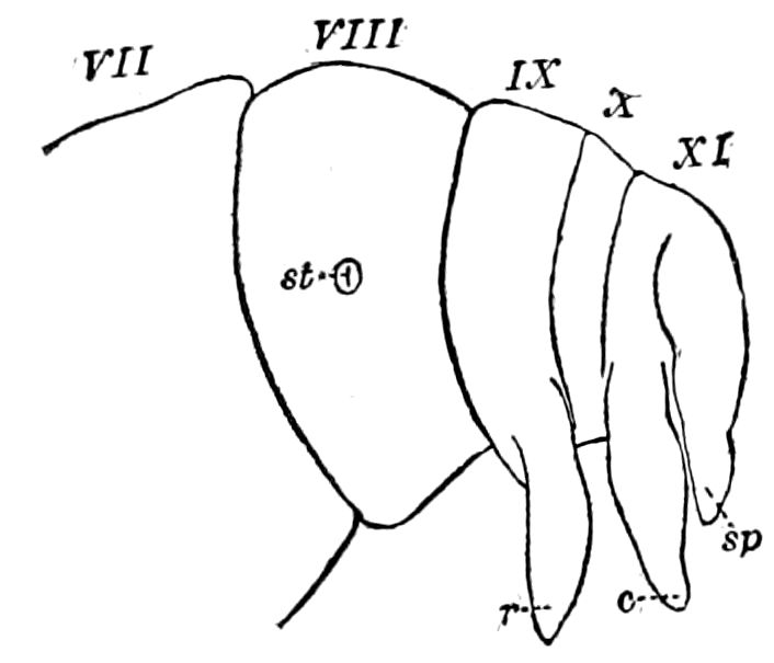
Fig. 199.—End of abdomen of embryo of Mantis: r, rhabdopod; c, cercopod; sp, suranal plate; st, stigma on 8th segment.—After Heymons.
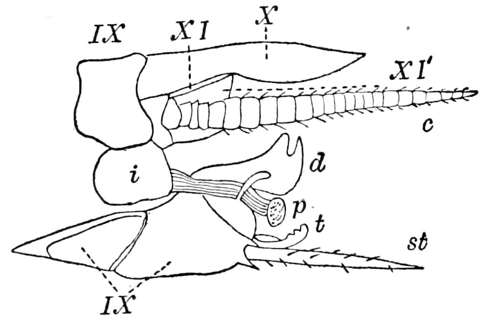
Fig. 200.—End of abdomen of Periplaneta americana, ♂, side view: c, cercopod; st, stilus; p, penis; t, titillator; d, “bird’s head” (clasper?); i, “oblong plate”; IX-XI, terminal segments; X, suranal plate; XI′, 11th sternite.—After Peytoureau.
In most larvæ of the Trichoptera, especially the Rhyacophilidæ and Hydropsychidæ, the last abdominal segment bears a pair of 2–jointed legs (cercopoda), ending in either one or two claws, which under various forms, sometimes forming long processes, persist in the pupa; and there appears to be a suranal plate, the vestige of the tenth uromere. In the pupa, judging by Klapálek’s figure of Leptocerus (248,9, 25), a pair of lateral spines arise which may in the imago form one of the pairs of appendages or styles. In the pupa of Œcetis furva his figure 289 shows two pairs of 1–jointed appendages arising from the last segment; whether the long dorsal or upper styles arise from the vestige of a more distal 179segment is not distinctly shown in Klapálek’s sketch. The origin of these elements of the genital armature evidently needs further study.
Whether the abdominal legs or so-called false or prop-legs of lepidopterous larvæ are genuine legs, homologous with those of the thorax and with the cephalic appendages, or whether they are secondary adaptive structures, is a matter still under discussion. That, however, they are true legs is shown by the embryology of the Lepidoptera, where there is a pair to each abdominal segment. It may also be asked whether the anal legs of lepidopterous larvæ are not the homologues of the 2–jointed anal appendages of caddis-worms.
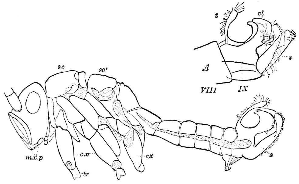
Fig. 201.—Eriocephala calthella, ♂, side view: t, palpiform suranal plate; cl, claspers; s, inferior claspers; mxp, maxillary palpi; cx. coxa; tr, trochanter; sc, scutum; sc′, scutellum.
In Lepidoptera, notably the male of the very generalized Eriocephala calthella (Fig. 201), besides the broad unjointed claspers, which are curved upward and provided with a brush of stiff hooked setæ (this upper pair being perhaps modified cercopods), there is an accessory lower slenderer pair, while the suranal plate (t) is palpiform or clavate and also adapted to aid in the action of the claspers. The examination of the cercopods and rhabdopods in the Trichoptera and in a generalized lepidopterous form like this enables one to understand the morphology of the genital armature, since it consists, besides the suranal plate, which is often deeply forked (in Sphingidæ, Smith), of a pair of modified hook-like cercopoda, and in some cases (Eriocephala) of an additional pair of claspers which 180may be the homologues of the ephemerid rhabdopods. A pair of hooks, often strong and claw-like (harpes), are situated, one near the base on the inside of each clasper; they are especially developed in the Noctuidæ (Smith), and appear to be present in certain Trichoptera, but this remains to be proved. This complicated apparatus of claspers and hooks is utilized by those insects which pair while on the wing, and is wanting in such forms as Coleoptera and Hemiptera. Besides the forceps of Panorpa, there are two pairs of slender filiform appendages which need farther examination. In the Diptera, especially Tipulidæ, there is a pair of 2–jointed appendages or forceps, as in Limnophila (Osten Sacken). The male genital armature of Diptera appears to be on the same general plan as in Lepidoptera, but more complicated.
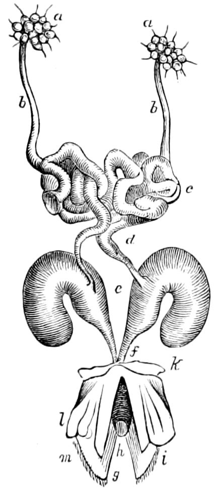
Fig. 202.—Male organs of generation of Athalia.—After Newport.
Notice should also be taken of the paired uncinate hooks which are modifications of the penis-sheath of the male of cockroaches (Phyllodromia), which Haase states appear to originate on the tenth ventral plate, and which probably “serve to open and dilate the vagina of the female, especially as a perforated penis, which is highly developed in Machilis, seems to be wanting in the Blattidæ.” (Haase.)
The penis.—This is a single or double median style-like structure either hollow and perforated, or solid, very variable in shape, receiving the end of the ejaculatory duct. It is usually enclosed between two lateral plates, the homologues perhaps of the inner pair of sheaths of the ovipositor. In the Coleoptera, as in Carabidæ and Melolonthidæ, the penis is a long chitinous tube, “retractile within the abdomen on the under surface as far as the anterior segments.” (Newport.) In the Hymenoptera, of which that of the saw-flies is a type, Newport states that it “consists of a short valvular projectile organ, covered externally by two pointed horny plates (i) clothed with soft hairs.” Above these are two other irregular double-jointed plates (Fig. 202, l) surrounded at their base by a chitinous ring (k); they are edged with prehensile hooked spines (i). Between these in the middle line are two elongated muscular parts (m) which enclose the penis (h), and which, like those in beetles, perhaps aid in dilating the vulva of the female.
An examination of Figs. 203–207 will aid in understanding the various modifications in beetles, etc., of this organ.
181A general study of the anatomy and homologies of the male genital armature, from a developmental point of view, together with a comparison of them with the corresponding female organs, is still needed.
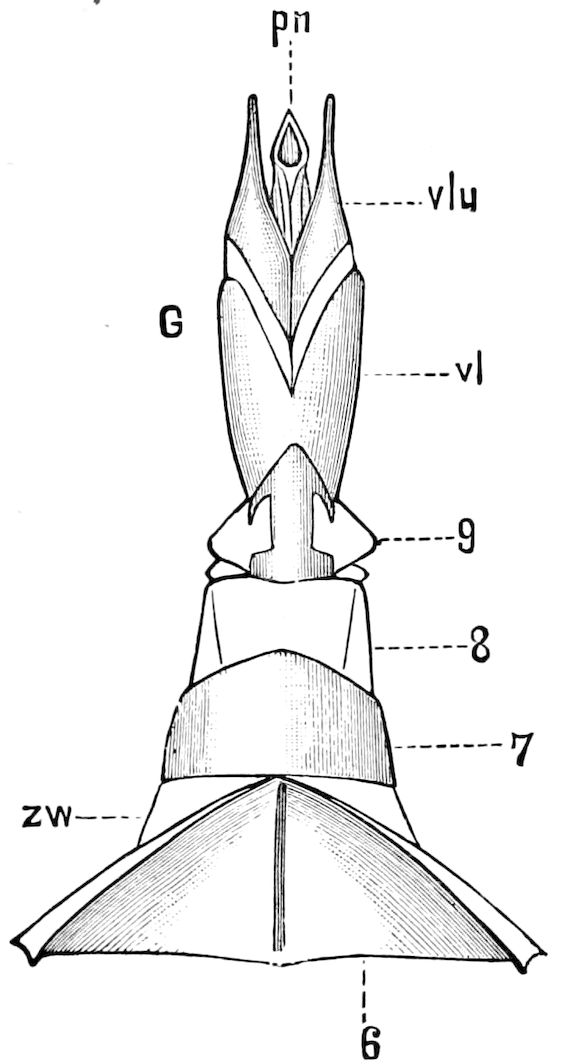
Fig. 203.—End of abdomen situated under the anal lobes of Hydrophilus piceus, drawn out, seen from the ventral side: 6, sternal region of 6th segment; 7, 8, 9, segments telescoped, when retracted, in 6th segment; zw, membrane connecting 6th and 7th segments; G, intromittent apparatus; vl, external lobes; vlu, inner lobes; pn, penis.
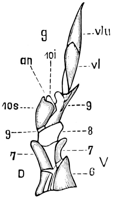
Fig. 204.—The same as in Fig. 203, seen from the side: 6, the free 6th segment; 7–10, the four last, when at rest, retracted and telescoped within the 6th segment, with the copulatory apparatus (g); vl, outer, vlu, inner lobe; 10s, tergite of 10th segment; 10i, sternite of the same; an, anal opening.
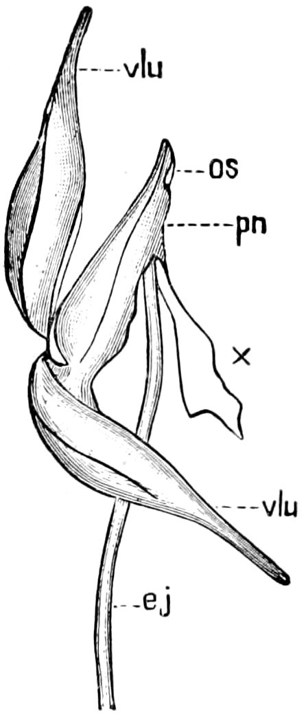
Fig. 205.—Terminal parts of the male copulatory apparatus of Hydrophilus piceus, torn apart: vlu, the two inner lodes; pn, penis; x, membrane torn from under side of penis; ej, ejaculatory duct; os, its opening on the under side of the penis, directly under its tip. The muscles, tracheæ, and nerves are not drawn.
Velum penis.—In the locusts (Acrydiidæ) the penis is concealed by a convex plate, flap, or hood, free anteriorly and attached posteriorly and on the sides to the ridge forming the upper edge of the tenth sternite. When about to unite sexually, the tip of the abdomen is depressed, the hood is drawn backward, uncovering the chitinous penis.
The suranal plate.—This is a triangular, often thick, solid plate or area, the remnant of the tergum of the last, usually tenth, segment of the abdomen, the supra-anal or suranal plate, or anal operculum (lamina supra-analis) of Haase. In most lepidopterous larvæ this plate is well marked; in those of the Platypteridæ it is remarkably elongated, forming an approach to a flagellum-like terrifying 182appendage, and in that of Aglia tau it forms a long, prominent, sharp spine. In the cockroach, both Cholodkowsky and Haase maintain that the tenth abdominal segment is suppressed in the female, the tergal portion being fused with the suranal plate (the latter in this case, as we understand it, being the remnant of the eleventh segment of the embryo). As to the nature of the middle jointed caudal appendage in Thysanura and May-flies Heymons has satisfactorily shown that it is a hypertrophied portion of the suranal plate, being in Lepisma but a filamental elongation of the small eleventh abdominal tergite.
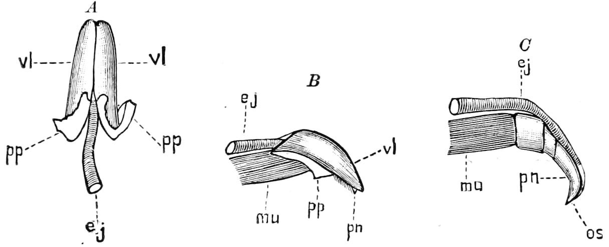
Fig. 206.—Copulatory organ of a weevil, Rhychophorus phœnicis, seen from above. A, vl, the lobes united into a capsule; pp, torn membrane which connects the capsule with the 9th abdominal segment; ej, ejaculatory duct. B, the same seen from the side; mu, end of the muscle of the penis. C, the same as B, without the capsule; os, opening of the ejaculatory duct (ej). Other letters as in A.
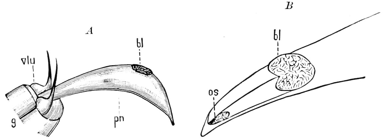
Fig. 207.—A, penis (pn) of Carabus hortensis: bl, wrinkled membranous vesicle; vlu, the valves; g, part of 9th segment. B, end of penis of the same, enlarged; os, cleft-like opening; also a wrinkled vesicle, as at bl.—This and Figs. 203–205 after Kolbe.
At the base of the suranal plate of locusts (Acrydiidæ) is the suranal fork or suranal furcula (furcula supra-analis, as we have called it) (Fig. 88, 89, f).
The podical plates or paranal lobes.—In the cockroach and other insects, also in the nymphs of Odonata, the anus is bounded on each side by a more or less triangular plate, the two valves being 183noticeable in lepidopterous larvæ. They are the valvulæ of Burmeister, and podical plates of Huxley, who also regarded them as the tergites of an eleventh abdominal segment;[38] and the subanal laminæ of Heymons. They are wanting in Ephemeridæ.
The infra-anal lobe.—Our attention was first called to this lobe or flap, while examining some geometrid larvæ. It is a thick, conical, fleshy lobe, often ending in a hard, chitinous point, and situated directly beneath the vent. Its use is evidently to aid in tossing the pellets of excrement away so as to prevent their contact with the body. The end may be sharp and hard or bear a bristle. Whether this lobe is the modified ventral plate of the ninth urite, we will not undertake at present to say.
The egg-guide.—In the Acrydiidæ the external opening of the oviduct is bounded on the ventral side by a movable, triangular, acute flap, the egg-guide (Fig. 88, B, eg). Whether this occurs in other orders needs to be ascertained.
LITERATURE ON THE ABDOMEN AND ITS APPENDAGES
a. General (including the cerci, stili, etc.)
Cornelius, C. Beiträge zur naheren Kenntnis von Palingenia longicauda. (Programm d. Real- u. Gewerbeschule zu Elberfeld, 1848, pp. 1–38, 4 Taf.)
Schiödte, J. G. Bemerkungen über Myrmecophilen. Ueber den Bau des hinterleibes bei einigen Käfergattungen. (Germar’s Zeits. f. Ent., 1844.)
—— De metamorphosi Eleutheratorum observationes. (Naturhist. Tidsskr. i-xiii, 1861–1883.)
Meinert, F. Anatomia Forficularum. Anatomisk undersogelse af de Danske Orentviste, i. (Naturhist. Tidsskr. 3 raekke, ii, 1863, pp. 427–482, 1 Pl.)
—— Om dobbelte Saedgange hos insecter. (Naturhist. Tidsskrift, 3 raekke, v, 1868.)
Tullberg, Tycho. Sveriges Podurider. (Svenska Vetensk.-Akad. Handl., 1872, x, Nr. 10, 12 Pls.)
Davis, H. Notes on the pygidia and cerci of insects. (Journ. R. Micr. Soc., 1879, ii.)
Westhoff, F. Ueber den Bau des Hypopygiums der Gattung Tipula Meig. (Münster, 1882, pp. 1–62, 6 Taf.)
Saunders, Edward. Further notes on the terminal segments of aculeate Hymenoptera. (Trans. Entom. Soc. London, 1884, pp. 251–267, 1 Pl.)
Palmén, J. A. Ueber paarige Ausführungsgange der Geschlechtsorgane bei Insekten. (Helsingfors, 1884, 5 Taf.)
Haase, E. Die Abdominalanhänge der Insekten mit Berücksichtigung der Myriopoden. (Morpholog. Jahrbuch, 1889, xv, pp. 331–435, 2 Taf.)
—— Abdominalanhänge bei Hexapoden. (Sitzungsber. d. Gesellsch. naturforsch. Freunde, 1889, pp. 19–29.)
184Wheeler, William M. On the appendages of the first abdominal segment of embryo insects. (Trans. Wis. Acad. Sc., viii, 1890, pp. 87–140, 3 Pls.)
Janet, Charles. Études sur les fourmis, 5e note, sur la morphologie du squelette des segments post-thoraciques chez les Myrmicides. (Mém. Soc. Acad, de l’Oise, xv, pp. 591–611, Figs. 1–5, 1894.)
Heymons, Richard. Die Segmentirung des Insektenkörpers. (Anh. Abh. Akad. Berlin, Phys. Abh., pp. 39, Taf., 1895. See also Die Embryonalent, von Dermapteren und Orthopteren, under Embryology.)
—— Grundzüge der Entwicklung und des Körperbaues von Odonaten und Ephemeriden. (Abhandl. k. Preuss. Akad. d. Wissens. Berlin, 1896, 2 Taf., pp. 1–66.)
—— Zur Morphologie der Abdominalanhänge bei den Insecten. (Morphol. Jahrb., iv, 1896, pp. 178–203, 2 Taf.)
Peytoureau, S. A. Contribution à l’étude de la morphologie de l’armure génital des insectes. (Bordeaux, 1895, pp. 248, 22 Pls., 43 Figs. in text.)
Verhoeff, C. Cerci und styli der Tracheaten. (Ent. Nachr., xxi, pp. 166–168, 1895.)
Also the writings of Oudemans, Packard, etc.
b. The ovipositor
Lacaze-Duthiers, Henri. Recherches sur l’armure génitale femelle des insectes. (Ann. d. sc. natur., 1849, xii, pp. 353–374, 1 Pl.; 1850, xiv, pp. 17–52, 1 Pl. (Hyménoptères); 1852, xvii, pp. 206–251, 1 Pl. (Orthoptères); 1853, xix, pp. 25–88, 4 Pl. (Neuroptères, Thysanures, Coléoptères, Diptères); pp. 203–237 (Lépidoptères, Aphaniptères en general. Also separate.))
Sollmann, A. Der Bienenstachel. (Zeitschr. f. Wissens. Zool., xiii, 1863, pp. 528–540, 1 Taf.)
Fengger, H. Anatomie und Physiologie des Giftapparates bei den Hymenopteren. (Archiv f. Naturgesch., 1863, pp. 139–178, 1 Taf.)
Eaton, A. E. Remarks upon the homologies of the ovipositor. (Trans. Ent. Soc. London, 1868, pp. 141–144.)
Packard, A. S. On the structure of the ovipositor and homologous parts in the male insects. (Proc. Boston Soc. Nat. Hist., xi, 1868, pp. 393–399, Figs. 1–11.)
Lambrecht, A. Samtliche Teile des Stechapparates in Bienenkörper und ihre Verwendung zu technischen und vitalen Zwecken. (Bienemwirtsch. Centralbl., 7 Jahrg., 1871, pp. 5–11.)
Kraepelin, K. Untersuchungen über den Bau, Mechanismus und die Entwicklung des Stachels der bienenartigen Tiere. (Zeitschr. f. wiss. Zoologie, xxiii, 1873.)
Dewitz, H. Vergleichende Untersuchungen über Bau und Entwicklung des Stachels der Honigbiene und der Legescheide der grünen Heuschrecke. (Königsberg, 1874.)
—— Ueber Bau und Entwicklung des Stachels und der Legescheide einiger Hymenopteren und der grünen Heuschrecke. (Zeitschr. f. wissen. Zoologie, xxv, 1874, pp. 174–200, 2 Taf.)
—— Ueber Bau und Entwicklung des Stachels der Ameisen. (Zeitschr. f. wiss. Zoologie, 1877, xxviii, pp. 527–556, 1 Taf.)
Adler, H. Legeapparat und Eierlegen der Gallwespen. (Deutsche Entom. Zeitschr., 1877, xxi Jahrg., pp. 305–332, 1 Taf.)
Cholodkowsky, N. Ueber den Hummelstachel und seine Bedeutung für die Systematik. (Zool. Anzeiger, vii, 1884, pp. 312–316.)
185Ihering, H. von. Der Stachel der Meliponen. (Ent. Nachr., 1886, xii, Jahrg., pp. 177–188, 1 Taf.)
Meinert, F. Bidrag til de danske Myrers Naturhistorie. (Kjöbenhavn, 1890, 68 s.u. Danske Vidensk. Selsk. Skrifter, 5 Raek, v, 3 Pls.)
Beyer, O. W. Der Giftapparat von Formica rufa ein reduziertes Organ. (Jena. Zeitschr. Naturw., 1890, xxv, pp. 26–112, 2 Taf.)
Carlet, G. Mémoire sur le venin et l’aiguillon de l’abeille. (Ann. d. sc. nat. Zool., 7 sér., ix, 1890, pp. 1–17, 1 Pl.)
Künckel d’Herculais, J. Méchanisme physiologique de la ponte chez les insectes orthoptères de la famille des Acridides.—Rôle de l’air comme agent mécanique et fonctions multiples de l’armure génitale. (Compt. Rend., cxix, pp. 244–247, 1894.)
Also the writings of Verhoeff, Heymons.
c. The external genital armature
Klug, Johann C. F. Versuch einer Berichtigung der Fabriciusschen Gattungen Scolia u. Tiphia. (Ueber u. Mohr Beiträge zur Naturkunde, i, pp. 8–40, 1805, 1 Taf.)
Audouin, J. V., and Lachat. Observations sur les organes copulateurs males des Bourdons. (Annal. général, d. sc. phys., 1821, viii, pp. 285–289.)
Audouin, J. V. Lettre sur la génération des insectes. (Ann. des sc. nat., sér. 1, ii, 1824.)
Suckow, F. W. L. Geschlechtsorgane der Insekten. (Heusinger, Zeitschr. organ. Physik., 1828, ii, pp. 231–264, 1 Taf.)
Rathke, H. De Libellularum partibus genitalibus. Regiomonti, 1832, pp. 6 + 38, 3 Pls.
Siebold, C. Th. E. von. Ueber die Fortpflanzungsweise der Libellulinen. (Germar’s Zeitschr. f. Ent., 1840, ii, pp. 421–438.)
Selys-Longchamps, E. de. Monographie des Libellulidees d’Europe. 4 Pls., 1840.
Bassi, C. A. Studi sulle funzioni degli organi genitali degl’ insetti da lui osservati piu specialmente nella Bombyx mori. (Atti della 5 Riun. d. Scienz. Ital. Lucca, 1844, pp. 39–94.)
Stein, F. Vergleichende Anatomie und Physiologie der Insekten, i. Die weiblich. Geschlechtsorgane der Käfer. Berlin, 1847, 9 Taf.
Ormancey, P. Recherches sur l’étui penial considéré comme limite de l’espèce dans les coléoptères. (Ann. sc. nat., 1849, 3 sér., Zool., xii, pp. 227–242.)
Roussel, C. Recherches sur les organes génitaux des insectes coléoptères de la famille des Scarabéides. (Compt. rend. Acad. d. sc. Paris, 1860, l, pp. 158–161.)
MacLachlan, R. A monographic revision and synopsis of the Trichoptera of the European fauna. London, 1874–80, 59 Pls.
—— On the sexual apparatus of the male Acentropus. (Trans. Ent. Soc. London, 1872, pp. 157–162.)
Thomson, C. G. Nagra anmarkningar ofver arterna af slagtet Carabus. (Thomson’s Opuscula Entomologica, vii, 1857, pp. 615–729, 1 Pl.)
Dufour, L. Sur l’appareil génital male du Coræbus bifasciatus. (Thomson, Archiv ent., 1857, i, pp. 378–381.)
White, F. Buchanan. On the male genital armature in the Rhopalocera. (Trans. Linn. Soc., ser. 1, Zool., i, pp. 357–369, 1876, 3 Pls.)
Graber, V. Die Aehnlichkeit im Baue der ausseren weiblichen Geschlechtsorgane bei den Lokustiden und Akridiern auf Grund ihrer Entwicklungsgeschichte. (Sitzber. k. Akad. d. Wissensch. Wien., 1870, lxi, pp. 1–20, 1 Taf.)
186Scudder, Samuel H., and Edward Burgess. On asymmetry in the appendages of hexapod insects, especially as illustrated in the lepidopterous genus Nisoniades. (Proc. Boston Soc. Nat. Hist., 1871, xiii, pp. 282–306.)
Chadima, J. Ueber die Homologie zwischen den männlichen und weiblichen ausseren Sexualorganen der Orthoptera Saltatoria Latr. (Mitteil. d. naturwiss. Vereins f. Steiermark, 1872, pp. 25–33, 1 Taf.)
Hagens, von. Ueber die Genitalien der männlichen Bienen, besonders der Gattung Sphecodes. (Berlin Ent. Zeitschr., 1874, pp. 25–43.)
—— Ueber die männlichen Genitalien der Bienengattung Sphecodes. (Deutschen Entom. Zeitschr., 1882, pp. 209–228, 2 Taf.)
Lindenman, C. Vergleichend-anatomische Untersuchung ueber das männliche Begattungsglied der Borkenkäfer. (Bull. Soc. Imp. d. Natural. Moscou, 1875–77.)
Forel, A. Der Giftapparat und die Analdrüsen der Ameisen. (Zeitschr. f. wiss. Zoologie, xxx, suppl., 1878.)
Kraatz, G. Ueber die Wichtigkeit der Untersuchung des männlichen Begattungsgliedes der Käfer für die Systematik und Artunterscheidung. (Deutschen Entom. Zeitschr., 1881, xxv, pp. 113–126.)
—— Ueber das männliche Begattungsglied der europaischen Cetoniiden und seine Verwendbarkeit für deren scharfe spezifische Unterscheidung. (Ibid., pp. 129–149.)
Gosse, Ph. H. On the clasping-organs ancillary to generation in certain groups of the Lepidoptera. (Trans. Linn. Soc., 1882, Ser. 2, Zool., ii, pp. 265–345, 8 Pls.)
—— The prehensors of male butterflies of the genera Ornithoptera and Papilio. (Proc. Roy. Soc. London, 1881, xxxiii, pp. 23–27.)
Radoszkowski, O. Revision des armures copulatrices des males du genre Bombus. (Bull. Soc. Natur. Moscou, 1884, xlix, pp. 51–92, 4 Pls.)
—— Revision des armures copulatrices des males de la tribu Philérémides. (Ibid., 1885, lxi, pp. 359–370, 2 Pls.)
—— Revision des armures copulatrices des males de la famille des Mutillidæ. (Horæ Soc. Ent. Ross., 1885, xix, pp. 3–49, 9 Pls.)
—— Revision des armures copulatrices des males de la tribu des Chrysides. (Horæ Soc. Ent. Ross, xxiii, 1890, pp. 3–40, 6 Pls.)
Hofmann, O. Beiträge zur Kenntnis der Butaliden. (Stett. Ent. Zeit., 1888, pp. 335–347, 1 Taf.)
Driedzichi, H. Revue des espèces européennes du genre Phronia Winn. (Horæ Soc. Ent. Ross., 1889, xxiii, pp. 404–532, 10 Pls.)
Sharp, David. On the structure of the terminal segment in some male Hemiptera. (Trans. Ent. Soc. London, 1890, pp. 399–427, 3 Pls.)
Escherich, K. Die biologische bedeutung der Genitalanhänge der Insekten. (Verhandl. d. zool. bot. Ges. Wien., 1892.)
—— Anatomische Studien über das männliche Genitalsystem der Coleopteren. (Zeits. f. wissens. Zool., lvii, pp. 620–641, 1 Taf., 3 figs.)
Verhoeff, C. Zur vergleichenden Morphologie der “Abdominalanhänge” der Coleopteren. (Ent. Nachr., xx, pp. 93–96, 1894. Compare also O. Schwarz and J. Weise’s criticisms in D. Ent. Zeit., pp. 153–157; also pp. 101–109, 155–157. Zool. Anzeiger, pp. 100–106, 1894.)
—— Vergleichende—morphologische Untersuchungen ueber das Abdomen der Endomychiden, etc., und über die Musculature des Copulationsapparates von Triplax. (Archiv f. Naturg., lxi, pp. 213–287, 2 Taf., 1895.)
—— Vergleichende Untersuchungen über die abdominal Segmente der weiblichen Hemiptera-Heteroptera und Homoptera. (Verh. Nat. Ver. Bonn, l, pp. 307–374, 1894.)
187Verhoeff, C. Beiträge zur vergleichenden Morphologie des Abdomens der Coccinelliden, etc. (Archiv f. Naturg., lxi, pp. 1–80, 6 figs., 1895.)
Boas, J. E. V. Organe copulateur et accouplement der hanneton. (Oversigt over det K. Danske Vidensk. Selskab Forhand, 1892, Copenhagen, 1893, 1 Pl., pp. 239–261.)
Pérez, J. De l’organe copulateur mâle des Hyménoptères et de sa valeur taxonomique. (Ann. Soc. Ent. France, lxiii, pp. 74–81, Figs., 1894.)
Goddard, Martha Freeman. On the second abdominal segment in a few Libellulidæ. (Proc. Amer. Philos. Soc., xxxv, pp. 205–212, January 11, 1897, 2 Pls.)
Also the writings of Eaton, Emery, Fischer, Forel, Géhin, Godart, Hagen, Joly, Koletani, Loew, Meinert, Mik, Nicolet, Osten Sacken, Pictet, Roussel, Schaeffer (1754), Schaum, Schenk, J. B. Smith, Thompson, Buchanan-White, Brunner von Wattle-Wyll, Weise, Wyenbergh.
The subject of copulation has been treated by Hoffer, Hartig, Schiedeknecht, Verhoeff, etc.
THE ARMATURE OF INSECTS: SETÆ, HAIRS, SCALES, TUBERCLES, ETC.
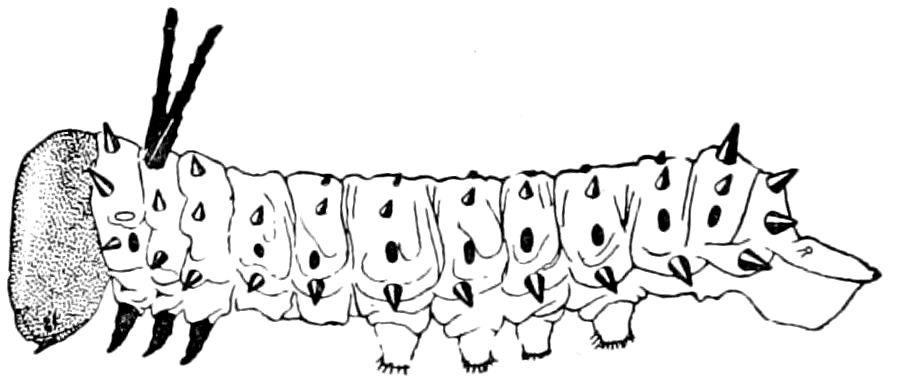
Fig. 208.—Larva of Dryocampa rubicunda, stage II.—Bridgham del.
The cuticula.—The integument is externally either smooth and shining or variously punctured, granulated, tuberculated, striated, or hairy. In certain orders the skin is clothed with flattened setæ or scales, while many forms, as some caterpillars (Figs. 208, 209), beetles (Fig. 210), etc., are protected by spines, horns, etc., these in adult insects often forming secondary sexual characters, usually being more developed in the males than in the females.
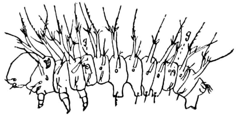
Fig. 209.—Larva of Hyperchiria io, on hatching.
The cuticula is not always smooth, but is often finely granulated or even minutely spinulated. On the abdominal segments of Anabrus, as observed by Minot, the cuticula is armed with microscopic conical nodules scattered irregularly over it. They do not correspond, he says, in any way to hairs; for they do not rest over pores, nor did he see any specially modified cells underlying them. “As far as I have observed, they are mere local irregularities, each nodule being apparently supported by some four or six unmodified epidermal cells.” Minot adds that the whole of the cuticula, except the cones just described and the hairs, is divided into numerous minute fields, each of which corresponds 188to a single cell of the underlying hypodermis. Each field is bounded by a distinct polygonal outline, and its surface is either covered by a large number of extremely minute projecting points, as on the dorsal arch of the segment, or is smooth, as upon the articular membrane and ventral arch. Upon the sides of the dorsal arch and upon the spiracular membrane each field has a projecting spine or sometimes two or even three. (See also pp. 28, 30.)
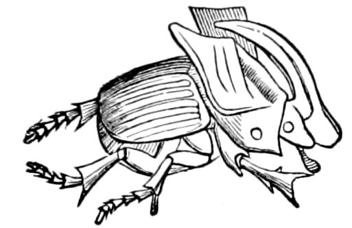
Fig. 210.—Phanæus pegasus, ♂, from Mexico.—After Graber.
Fig. 211.—Section of integument of Datana ministra: c, cuticula; hyp, hypodermis; p, outer pigmented nodulated layer.
The cuticle of lepidopterous larvæ has also been described and figured by Minot. In the caterpillars of different groups investigated by him, the cuticle was found to be rough with microscopic teeth or spinules, erect or flattened and scale-like, and either densely crowded or scattered, and affording excellent generic and specific characters. In the slug-worms (Limacodids) we have observed that the cuticula is unusually rough, especially on the spiniferous tubercle of Empretia, Parasa, etc. (Fig. 213, c). The skin of the body between the tubercles is seen to be finely shagreened, due to the presence of fine teeth, which are more or less curved and bent, these teeth arising from a very finely granulated surface (d). The cuticle of neuropterous, trichopterous, and tenthredinid larvæ will probably afford similar cases. The integument of the larva of Datana is, on the black bands, rough and nodulated, the irregular nodules being filled with a black pigment, and forming a layer (p) external to the true cuticula (Fig. 211).