PART I.—MORPHOLOGY AND PHYSIOLOGY (continued 10)
Reading edition
a. The heart
The heart or “dorsal vessel” is a delicate, pulsating tube, situated just under the integument of the back, in the median line of the body, and above the digestive canal. It can be partially seen without dissection in caterpillars. It is covered externally and lined within by membranes which are probably elastic; and between these two membranes extends a system of delicate muscular fibres, which generally have a circular course, but sometimes cross each other. The heart is divided by constrictions into chambers, separated by valvular folds. The internal lining membrane referred to forms the valvular folds separating the chambers. Each of these chambers has, at the anterior end, on each side, a valvular orifice (Fig. 370, ostium, i) which can be inwardly closed.
398Miall and Denny thus describe the different layers of the wall of the heart of the cockroach:
“There are: (1) a transparent, structureless intima, only visible when thrown into folds; (2) a partial endocardium, of scattered, nucleated cells, which passes into the interventricular valves; (3) a muscular layer, consisting of close-set, annular, and distant, longitudinal fibres. The annular muscles are slightly interrupted at regular and frequent intervals, and are imperfectly joined along the middle line above and below, so as to indicate (what has been independently proved) that the heart arises as two half-tubes, which afterwards join along the middle. Elongate nuclei are to be seen here and there among the muscles. The adventitia (4), or connective tissue layer, is but slightly developed in the adult cockroach.”
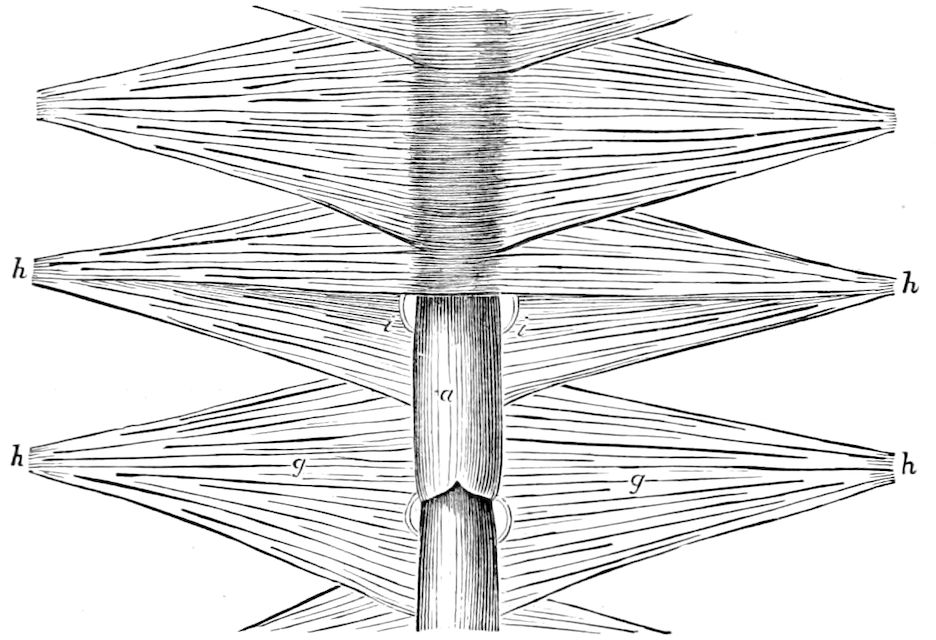
Fig. 370.—Part of the heart of Lucanus cervus: a, the posterior chambers (the anterior ones are covered by a part of the ligaments which hold the heart in place); i, auriculo-ventricular openings; g, g, the lateral muscles fixed by the prolongations h, h, to the upper side of the abdomen.—After Straus-Dürckheim.
Graber says that the heart of insects may be regarded not as an organ de novo, but only as the somewhat modified contractile dorsal vessel of the annelids, in which, however, the transverse arteries arising on each side became, with the gradual development of the tracheæ, superfluous and finally abortive. He describes it as a muscular tube composed of very delicate annular fibres, which within and without is covered by a relatively homogeneous, strong, elastic membrane.
The division into separate chambers is effected by means of a folding inwards and forwards of the entire muscular wall. “A portion of each side of the heart is first extended inwards so as very nearly to meet a corresponding portion from the opposite side, and then, being reflected backwards, forms, according to Straus (Consid., etc., p. 356), the interventricular valve which separates each chamber from that which follows it. Posteriorly to this valve, at the anterior part of each chamber, is a transverse opening or slit (Fig. 371, b), the auriculo-ventricular orifice, through which the blood passes into each chamber, and immediately behind it is a second, but much smaller, semilunar valve (c), which, 399like the first, is directed forwards into the chamber. It is between these two valves on each side that the blood passes into the heart, and is prevented from returning by the closing of the semilunar valve. When the blood is passing into the chamber, the interventricular valve is thrown back against the side of the cavity, but is closed when, by the contraction of the transverse fibres, the diameter of each chamber is narrowed, and the blood is forced along into the next chamber.” (Newport.)
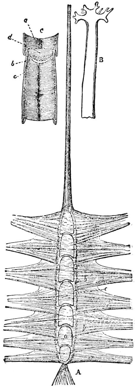
Fig. 371.—A, heart of Lucanus cervus: a, valves or chambers; bb, alary muscles; c, supposed auricular space around the heart. B, division into arteries of the end of the aorta in larva of Vanessa urticæ. C, interior of the chamber, showing the transverse fibres; b, auriculo-ventricular opening and valve into the chambers; c, semilunar valve; d, interventricular valve.—After Straus-Dürckheim, from Newport.
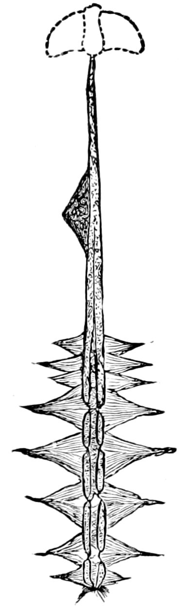
Fig. 372.—Heart of Belostoma.—After Locy.
According to Müller, there is but a single pair of ostia in Phasma, and, in the larva of Corethra, the heart is a simple, unjointed tube, not divided into chambers, and Viallanes states that, in the very young larva of Musca, there are no ostia (Kolbe). In the larva of Ptychoptera, Grobben found a short oval heart, with one pair of ostia situated in the 6th abdominal segment; a long aorta proceeds from it, the thoracic portion of which pulsates; from behind the heart arises a pulsating pouch, which connects with the hinder aorta, which does not pulsate, and ends at the base of two tracheal gills. Burmeister was able to find only four pairs of openings in the larva of Calosoma. Newport states that, while Straus figures nine chambers in Melolontha, and, consequently, eight pairs of openings, he has not been able to observe more than seven pairs of openings in Lucanus cervus. He has invariably found eight pairs of openings both in the larva and imago of Sphinx ligustri, as well as in other Lepidoptera. According to Béla-Dezso, the number of pairs of ostia corresponds to that of the pairs of stigmata.
There also occur, on each side of each chamber, two so-called pear-shaped bodies which are separated from the tubular portion of the heart itself, but, by 400means of muscular fibres, are united with the chamber and with their valves. These pyriform bodies appear as vesicles or cells with granular contents, besides some nuclei with nucleoli. They are of very small size. According to the measurements of Dogiel, in the larva of Corethra plumicornis, they are 0.02 to 0.1 mm. long, and 0.06 to 0.08 mm. broad. He regards these peculiar bodies as apolar nerve-cells of the heart. (Kolbe.)
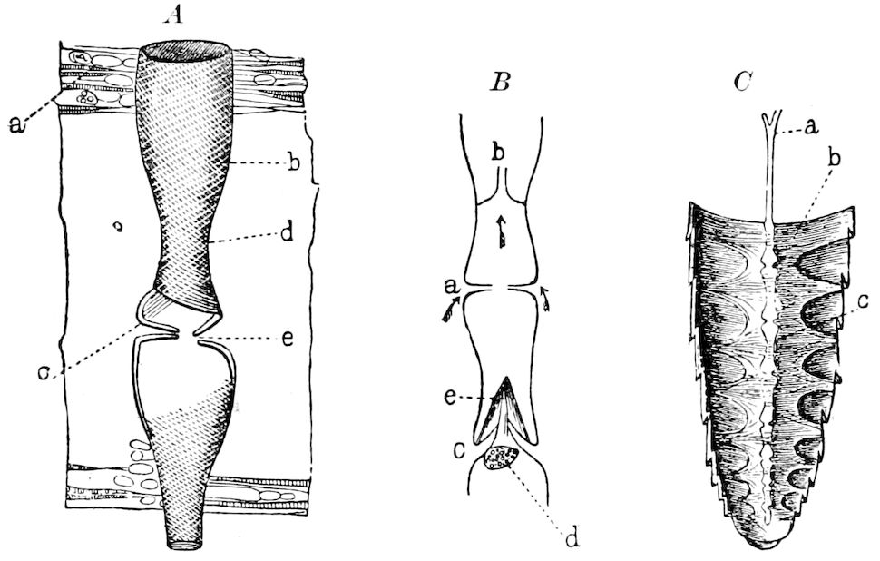
Fig. 373.—A, part of the heart of Dyticus marginalis, showing the spiral arrangement of the muscular fibres; c, closed, e, open, valve; a, dorsal diaphragm with interwoven muscular fibres; b, arrangement of fibres, recalling the screw-like features of the fibres of the human heart; d, narrow end. B, diagrammatic figure of the valvular openings, with the terminal flap (e), and the cellular valve, of a May beetle; a, valvular opening of a dipterous larva, with the interventricular valve (b). C, abdomen of a mole-cricket, ventral view; c, the segmented heart; a, aorta; b, segmented diaphragm under it.—After Graber.
Besides the venous openings of the heart which open into the pericardial region, Kowalevsky has discovered, in the heart of some Orthoptera (Caloptenus, Locusta, etc.), five pairs of openings by which the cardiac chambers receive the blood of the peri-intestinal region. Graber had divided the cœlom of insects into three regions (pericardial, peri-intestinal, and perineural regions), and hitherto only a union of the heart with the pericardial region by slit-like openings was known. These openings are symmetrically distributed on five abdominal segments; each section of the heart in this region has, therefore, four openings, which are all of a truly venous nature. These openings, called cardio-cœlomic apertures, are visible to the naked eye, being situated on conical papillæ of the walls of the heart. These papillæ pass through the outer diaphragm, and open into the peri-intestinal part of the cœlom, in the Acrydiidæ directly, in the Locustidæ through special canals. The cells of the papillæ are spongy, possessing large nuclei, and similar, as a whole, to glandular cells. (Comptes rendus, cxix, 1894.)
The mechanism by which the ostia are closed consists, according to Graber, of an ∞-shaped muscle passing around the two openings, and which, being interlaced, is sufficient to close the openings. But this is not all. The fore and hinder edge of the ostia project, leaf-like, into the cavity of the heart, and thus form, with the outer walls, two valves which, during the systole, filled with the blood rushing in, not only hermetically close the lateral openings, but also, by the simultaneous closure of the entire chamber by the circular muscles in the middle of the same, the two valves, simultaneously approaching each other, so 401nearly touch that they form a transverse partition wall in the chamber. But, for the last purpose, i.e. for the separation of the chambers from one another, there is a very special contrivance. In the May beetle, we find, besides a valve (Fig. 373, B, e), opening into the middle of the chambers, a large, stalked cell (d), which, in the diastole, i.e. in the expansion of the heart, hangs down free on the walls of the heart; but, in the systole or contraction, like a cork, closes the middle of the valve, but does not wholly close the cavity. He has observed, in the larva of Corethra, formal, interventricular valves, which also are not in the middle, but are separated from one another in the interlaced ends. They consist of two longitudinally membranous flaps which move against each other like two valves (Fig. 373, B, b).
“But what is the necessity for such a complicated mechanism? All the blood from behind passes into the heart, and, for its propulsion a simple muscular tube, whose circular fibres would draw together and contract it, would be thought to be sufficient. But the heart, except in some larvæ, ends posteriorly in a blind sac, and the blood can only pass into it by a series of pairs of lateral openings. Now, as regards the reception and the propulsion of the blood forwards, two modes are conceivable. The simplest way would be that the tubular heart should, along its whole length, contract or expand; that, moreover, the blood should be simultaneously sucked in through all the openings, and that then, also, the contraction, or systole, should take place in every part of the heart at the same moment. But this would, plainly, in so long and thin-walled a vessel, be highly impracticable, since, through such a manipulation, the mass of blood enclosed in the heart would be crowded together rather than really impelled forwards. Only the second case could be admissible, and that is this, that each chamber pulsates, one after another, from behind forwards. But, then, each segmental heart must be separated from the others by a valve. To make the matter wholly clear, we may observe an insect heart pulsating, and this is best seen in one of its middle chambers. This chamber expands (simply by the relaxation of its circular muscles), the ostia, also, consequently open, and a given quantity of blood is drawn in from the pericardial cavity. What now would happen after the succeeding contraction if there were no valves between? The blood would not flow forwards, but seek a way out backwards.
“But, in fact, the valve of the hinder chamber, at this time, closes itself, while, by the simultaneous expansion of the anterior ones, their door opens, and this section of the heart, at the same time, causes a sucking in of the contents of the posterior chamber. This phenomenon is repeated, in the same way, from chamber to chamber, which also acts alternately as ventricle and auricle, or by a sucking and pumping action. One is involuntarily reminded of the ingenious manipulation by which, by the alternate opening and shutting of the flood-gates, a vessel is carried along a canal.
“This wave-like motion of an insect’s heart also has the advantage that, just before a pulse-wave has reached the chambers farthest in front, the hinder ones are already prepared for the production of a second, for, as a matter of fact, often 60, and even 100, and, in very agile insects, 150, waves pass, in a single minute, through the series of chambers, which make it very difficult to follow the flowing of their waves.” (Graber.)
The propulsatory apparatus.—But the heart itself is only a part of the entire propulsatorial apparatus to which belongs the following contrivance, the nature of which has been worked out by Graber.
Under the dorsal vessel is stretched a sort of roof-like diaphragm, i.e. a membrane, arched like the dorsal wall of the hind-body which is attached, in a peculiar way, to the sides of the body. The best idea can be gained by a cross-section 402through the entire body (Fig. 374): H is the true dorsal vessel; S, the diaphragm. A surface view is seen at 373, C, b, where it appears as a plate with the edge regularly curved outwards on each side. Its precise mode of working is thus: from each dorsal band of the sides of the abdomen arises a pair of muscles spreading out fan-like, and extending to the heart, so that the fibres of one side pass directly over to those of the other, often splitting apart, or, between the two, extends outwards a perforated, thin web, like an elastic, fibrous sheet (Fig. 373, A, a), with numerous perforations, forming a diaphragm.
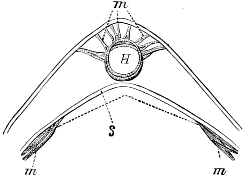
Fig. 374.—Diagram of transverse section of pericardial sinus of Ædipoda cœrulescens: H, heart; s, septum; m, muscles,—the upper suspensory, the lower alary.—After Graber, from Sharp. (See also Fig. 377.)
Graber has thus explained the action of the pericardial diaphragm and chamber, as freely translated by Miall and Denny: “When the alary muscles contract, they depress the diaphragm, which is arched upwards when at rest. A rush of blood towards the heart is thereby set up, and the blood streams through the perforated diaphragm into the pericardial chamber. Here it bathes a spongy or cavernous tissue (the fat-cells), which is largely supplied with air-tubes, and having been thus aerated, passes immediately forwards to the heart, entering it at the moment of diastole, which is simultaneous with the sinking of the diaphragm.”
In the cockroach, however, Miall and Denny think that the facts of structure do not altogether justify this explanation: “The fenestræ of the diaphragm are mere openings without valves. The descent of a perforated non-valvular plate can bring no pressure to bear upon the blood, for it is not contended that the alary muscles are powerful enough to change the figure of the abdominal rings.... The diaphragm appears to give mechanical support to the heart, resisting pressure from a distended alimentary canal, while the sheets of fat-cells, in addition to their proper physiological office, may equalize small local pressures, and prevent displacement. The movement of the blood towards the heart must (we think) depend, not upon the alary muscles, but upon the far more powerful muscles of the abdominal wall, and upon the pumping action of the heart itself.”
“The peculiar office,” says Graber, “performed by the heart has already been stated. It is nothing more than a regulator; than an organ for directing the blood in a determinate course in order that this may not wholly stagnate, or only be the plaything of a force acting in another way, as, for example, through that afforded by the body-cavity and the inner digestive canal. At regular intervals a portion of the blood is sucked through the same, and then by means of the anterior supply tube it is pushed onward into the head, whence it passes into the cavities of the tissues. The different conditions of tension under which the mass of blood stands in the different regions of the body then causes a farther circulation. Besides this, the blood passes through separate smaller pumping apparatuses, and through vessel-like modifications of cavities, also through hollow spaces between the muscles, as, for example, in the appendages where a regular backward and forward flow of the blood, especially in the limbs, wings, antennæ, and certain abdominal appendages takes place. Here and there may occasionally occur a narrow place where the flow of blood is obstructed by the accumulation of the blood corpuscles, causing a considerable stagnation.” (Graber.)
403
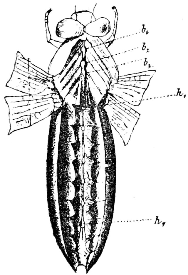
Fig. 375.—Libellula depressa, opened from the back, showing the nervous cord (b1-b3, thoracic, h1-h7, abdominal, ganglia), also the furrow-like ventral sinus closed by a muscular diaphragm.
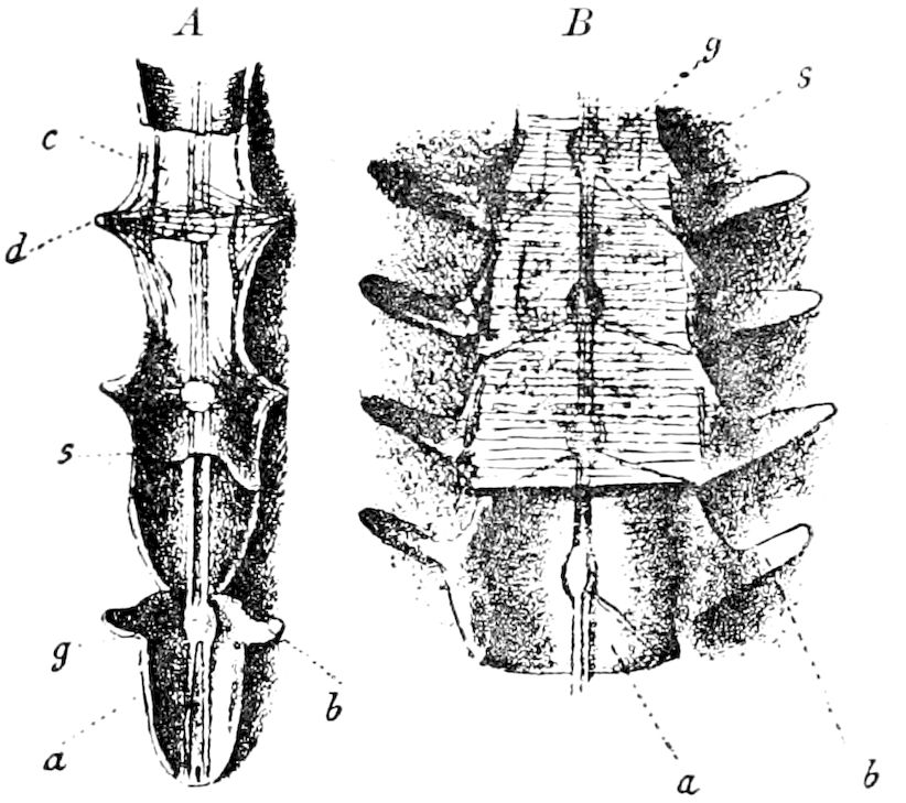
Fig. 376.—A, part of the ventral furrow of Libellula depressa more highly magnified: a, a sternal plate (urite); c, the septum stretched over it, at s in a relaxed or collapsed state; b and d, the wing-like, sternal processes from which the muscular bundles of the diaphragm arise. B, same in Acridium.
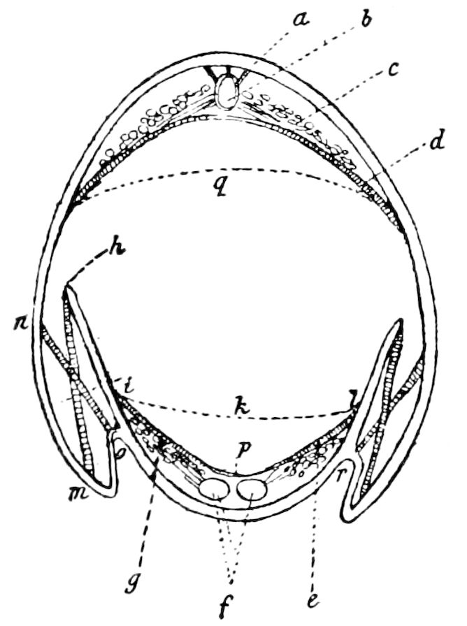
Fig. 377.—Diagrammatic section of the abdomen of Acridium tartaricum, showing the ventral septum (i, p, l) contracted, and (i, k, l) stretched out; oh, rib-like lateral processes of the urite; f, ganglia; b, heart, with its suspensorium (a); c, fat tissue in the pericardial tissue sinus; d, dorsal septum or diaphragm contracted. q, extended; g, fat-body; e, muscular part of diaphragm; no, expiration, hm, inspiration, muscle.—This and Figs. 375, 376, after Graber.
The supraspinal vessel.—In many insects there is a ventral heart acting on the heart’s blood as an aspirator, or more correctly a ventral sinus lying on the nervous cord, and closed by a pulsating diaphragm. This was discovered by Réaumur in the larva of a fly, and by Graber in the dragon-fly and locusts (Acrydiidæ). A glance at Figs. 375 and 376 will save a long description. The ventral wall forms a furrow, and between its borders (Fig. 377, e) extends the diaphragm. During the contraction of the muscles—and this, here, acts from before backwards—the membrane rises up and makes a cavity for the blood, which passes backwards over the nervous cord. The dorsal and ventral sinuses together thus bring about a closed circulation.
It thus appears that the insects are well provided with the means of distribution of their nutritive fluid, and that the blood is kept continually fresh and rich in oxygen. (Graber.)
404The aorta.—While the heart is mostly situated within the abdomen, it is continued into the thorax and the head as a simple, non-pulsating tube, called the aorta. In Sphinx the aorta, as described by Newport, begins at the anterior part of the 1st abdominal segment, where it bends downwards to pass under the metaphragma and enter the thorax; it then ascends again between the great longitudinal dorsal muscles of the wings, and passes onwards until it arrives at the posterior margin of the pronotum; it then again descends and continues its course along the upper surface of the œsophagus, with which it passes beneath the brain, in front of which and immediately above the pharynx, it divides into two branches, each of which subdivides. Newport, however, overlooked a thoracic enlargement of the aorta called by Burgess the “aortal chamber” (Fig. 310, a, c).
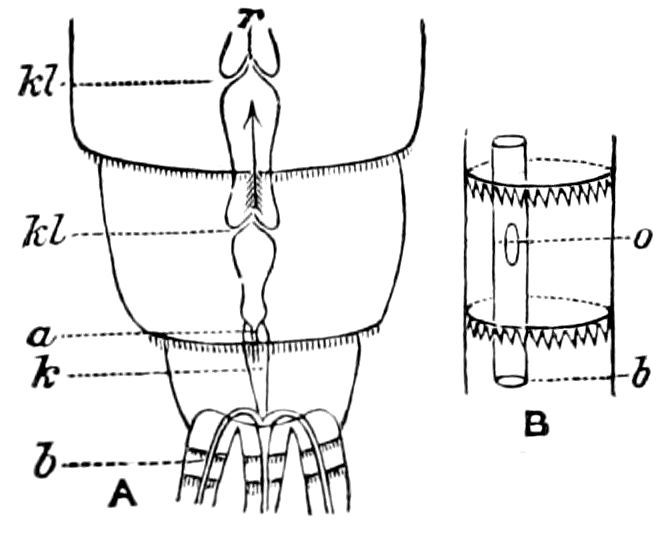
Fig. 378.—A, last three abdominal segments and bases of the three caudal processes of Cloëon dipterum: r, dorsal vessel; kl, ostia; k, special terminal chamber of the dorsal vessel with its entrance a; b, blood-vessel of the left caudal process. B, 26th joint of the left caudal appendage from below: b, a portion of the blood-vessel; o, orifice in the latter.—After Zimmermann, from Sharp.
“In Sphinx and Vanessa urticæ, immediately after the aorta has passed beneath the cerebrum, it gives off laterally two large trunks, which are each equal in capacity to about one-third of the main vessel. These pass one on each side of the head, and are divided into three branches which are directed backwards, but have not been traced farther in consequence of their extreme delicacy. Anterior to these trunks are two smaller ones which appear to be given to the parts of the mouth and antennæ, and nearer the median line are two others which are the continuations of the aorta. These pass upwards, and are lost in the integument. The whole of these parts are so exceedingly delicate that we have not, as yet, been able to follow them beyond their origin at the termination of the aorta, but believe them to be continuous, with very delicate, circulatory passages along the course of the tracheal vessels. It is in the head alone that the aorta is divided into branches, since, throughout its whole course from the abdomen, it is one continuous vessel, neither giving off branches, nor possessing lateral muscles, auricular orifices, or separate chambers.” (Newport, art. Insecta, p. 978.)
Dogiel observed in the transparent larva of Corethra plumicornis that the aorta extends only to the hinder border of the brain. Here it divides into two lamellæ, each of which independently extends farther on. One lamella is seen under the brain and under the eye, the other reaches near the eye. The lamellæ are tied to the integument by threads. At the point of division of the aorta is an opening. (Kolbe.)
True blood-vessels appear to exist in the caudal appendages of the May-flies, as the heart appears to divide and pass directly into them (Fig. 378). The last 405chamber of the heart diminishes in size at the end of the body, and then divides into three delicate tubular vessels which pass into the three caudal appendages, and extend to the end of each one, along the upper side. While the valves of the heart, in all insects, are directed anteriorly because the blood flows from behind, in the larva of the Ephemeridæ the valves of the last chamber of the heart are directed backwards, because from this chamber the blood flows in the opposite direction, i.e. into the caudal appendages. During the contraction of the heart, the elongated section of the same in the last abdominal segment receives a part of the mass of blood contained in the last chamber, which is driven by independent contractions into the caudal appendages. These vessels have openings before the end through which the blood enters into the cavity of the appendages, and can also pass back, in order to be taken up by the body cavity. It is possible that these blood-vessels stand in direct relation to respiration. (Zimmermann, Creutzburg, in Kolbe, p. 544.)
The pericardial cells.—Along the heart, on both sides, occur the so-called pericardial cells, which differ from the fat-cells, and also the peritracheal cells of Frenzel, and are mostly arranged in linear series, which have a close relation to the circulation of the blood. In the larva of Chironomus, they lie in groups; in that of Culex, they are arranged segmentally. In caterpillars, these pericardial cells are not situated in the region of the heart, but are arranged linearly on the side, and form a network of granulated cells situated between the fat-bodies. Other rows of these cells are situated near the stigmata and the main lateral tracheæ. (Kolbe.)
According to Kowalevsky, the pericardial cells, and the garland-shaped, cellular cord consist of cells, whose function it is to purify the blood, and to remove the foreign or injurious matters mingled with the blood.
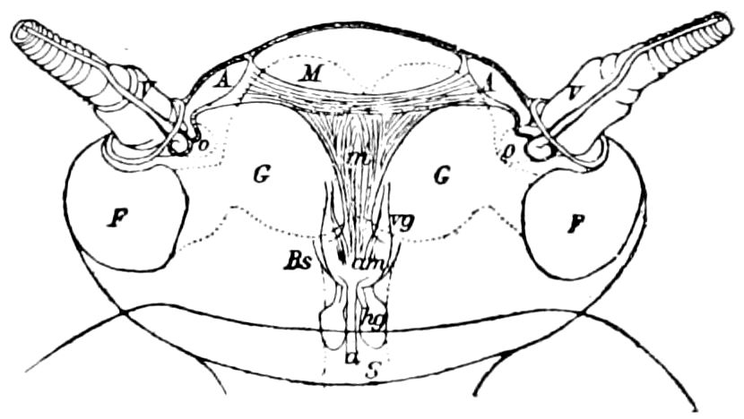
Fig. 379.—Diagram of the circulatory organs in the head of the cockroach, seen from above: A, ampulla; V, antennal vessel; M, chief muscular cord; m, muscular band; Bs, wall of the blood sinus; am, opening of the aorta (a); rg, anterior sympathetic or visceral ganglion; hg, hinder visceral ganglion; F, F, facetted eyes; o, vestigial ocellus; G, G, brain; S, œsophagus.—After Pawlowa.
Ampulla-like blood circulation in the head.—In the head of the cockroach occurs, according to Pawlowa, a contractile vascular sac at the base of each antenna. The cavity has a valvular communication with the blood space below and in front of the brain, and muscle-fibres effect systole and diastole. Each sac is beyond doubt an independently active part of the circulatory system. These organs also occur in Locusta and other Acrydiidæ, and Selvatico has described similar structures in Bombyx mori and certain other Lepidoptera.
Pulsatile organs of the legs.—Accessory to the circulation is a special system of pulsatile organs in the three pairs of legs of Nepidæ, generally situated in the tibia just below its articulation with the femur, but in the fore legs of Ranatra, in the clasp-joint or tarsus, just below its articulation with the tibia. First observed by Behn (1835), Locy has studied the organ (Fig. 380) in Corixa, Notonecta, Gerris, besides the Nepidæ. It is a whip-like structure attached at both ends, with fibres extending upward and backward to the integument of the leg, separate from the muscular fibres and does not involve them in its motions, and is not affected by the muscles themselves. “As the blood-corpuscles flow near the pulsating body they move faster, and around the organ itself there is a whirlpool of motion.” The beating of these organs aids the circulation in both directions, and when the motion ceases, the blood-currents in the legs stop; the rate of the pulsating organ is always faster than that of the heart, and the action is automatic.
406
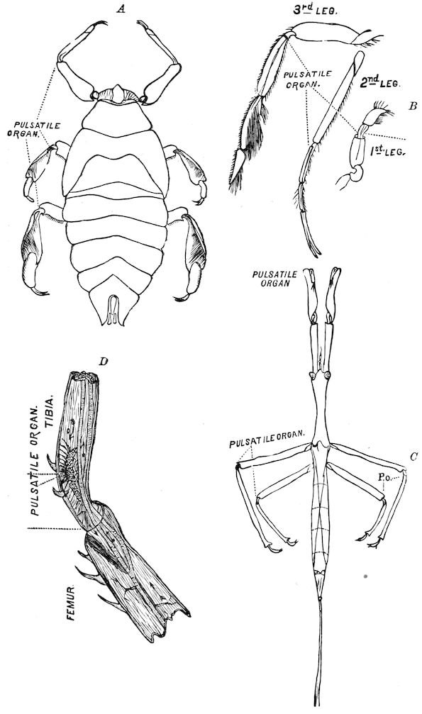
Fig. 380.—Pulsating organs in Hemiptera: A, Belostoma nymph, B, legs of Corixa. C, Ranatra, adult, to show the exceptional position of the pulsating organ in the fore legs. D, pulsatile organ in tibia of Ranatra.—After Locy.
407
b. The blood
The blood of insects, as in other invertebrates, differs from that of the higher animals in having no red corpuscles. It is a thin fluid, a mixture of blood (serum) and chyle, usually colorless, but sometimes yellowish or reddish, which contains pale amœboid corpuscles corresponding to the white corpuscles (leucocytes) of the vertebrates, though they are relatively less numerous in the blood of insects. The yellow fluid expelled from the joints of certain beetles (Coccinella, Timarcha, and the Meloidæ) is, according to Leydig, only the serum of the blood. In phytophagous insects the blood is colored greenish by the chlorophyll set free during digestion. The blood of Deilephila euphorbia is colored an intense olive-green, and that of Cossus ligniperda is pale yellow. (Urech.) The blood of case-worms (Trichoptera) is greenish. In some insects it is brownish or violet. The serum is the principal bearer of the coloring material, yet Graber has shown that in certain insects the corpuscles are more or less beset with bright yellow or red fat-globules, so as to give the same hue to the blood.
The leucocytes.—The corpuscles are usually elongated, oval, or flattened oat-shaped, with a rounded nucleus, or are often amœbiform; and they are occasionally seen undergoing self-division. When about to die the corpuscles become amœbiform or star-shaped. (Cattaneo.) Their number varies with the developmental stage of the insect, and in larvæ increases as they grow, becoming most abundant shortly before pupation. The blood diminishes in quantity in the pupal stage, and becomes still less abundant in the imago. (Landois.) The quantity also varies with the nutrition of the insect, and after a few days’ starvation nearly all the blood is absorbed. Crystals may be obtained by evaporating a drop of the blood without pressure; they form radiating clusters of pointed needles. The freshly drawn blood is slightly alkaline. (Miall and Denny.)
The size of the corpuscles has been ascertained by Graber, who found that the diameter of the circular blood-disks of the leaf-beetle, Lina populi, is 0.006 mm.; of Cetonia aurata and Zabrus gibbus, 0.008 to 0.01 mm.; and those of certain Orthoptera (Decticus verrucivorus, Ephippiger vitium and Œdipoda cœrulescens), 0.011 to 0.014 mm. The longest diameter of the elongated corpuscles of Carabus cancellatus is 0.008 mm.; of Gryllus campestris, Locusta viridissima, Cossus ligniperda, Sphinx ligustri (pupa), and others, 0.008 to 0.01 mm.; of Caloptenus 408italicus, Saturnia pyri, Anax formosus, and others, 0.011 to 0.014 mm.; of Ephippiger vitium, Œdipoda cœrulescens, Pezotettix mendax, Zabrus gibbus, Phryganea, and others, 0.012 to 0.022 mm.; in Stenobothrus donatus and variabilis, 0.012 to 0.035 mm. The largest known are those of Melolontha vulgaris, which measure from 0.027 to 0.03 mm.
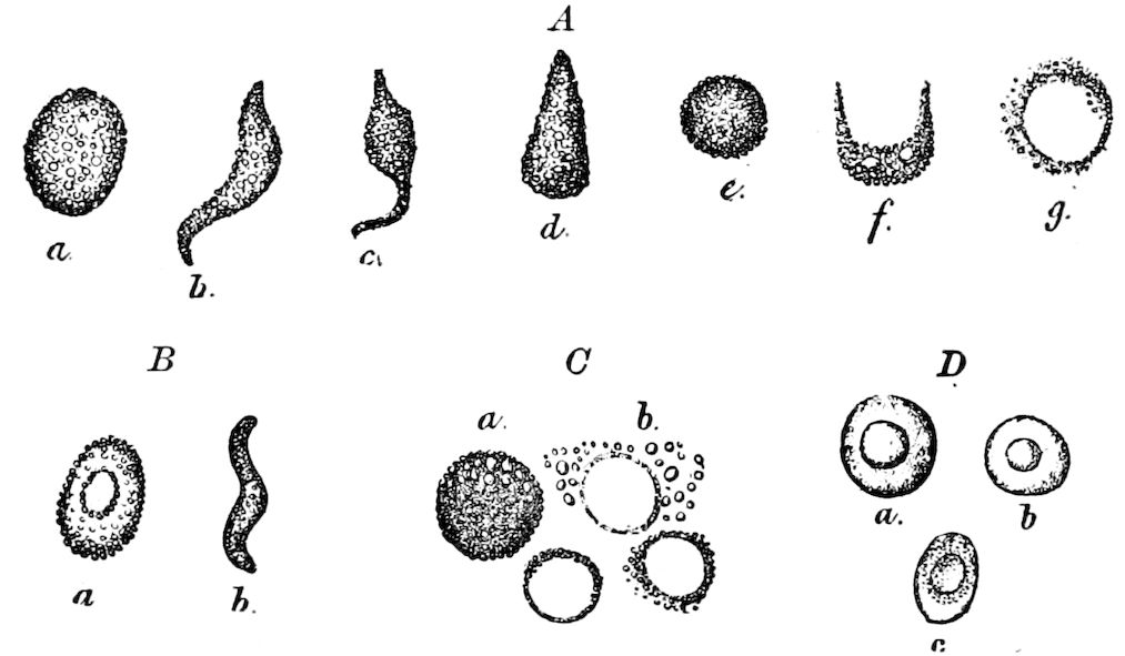
Fig. 381.—Blood corpuscles, or leucocytes, of insects: A, a-g, of Stenobothrus dorsatus (the same forms occur in most Orthoptera and in other insects). B, a, leucocyte of the same insect with the nucleus brought out by ether; b, another of serpentine shape. C, leucocytes of the same insect after a longer stay in ether. D, leucocytes of the same after being in glycerine 14 days.—After Graber.
As regards the nature of the corpuscles, Graber concludes that they are more like the cells of the fat-bodies than genuine cells. That they are not true cells is shown by the fact that after remaining in their normal condition a long time they finally coalesce and form cords. After shrivelling, or after the blood has been subjected to different kinds of treatment, the nucleus is clearly brought out (Fig. 381).
Besides the blood corpuscles there have been detected in the blood round bodies which are regarded as fat-cells. They are circular, and for the most part larger than the blood corpuscles, have a sharp, even, dark outline, and an invariably circular nucleus. (Kolbe.)
The blood of Meloe, besides the amœboid corpuscles, according to Cuénot, contains abundant fibrinogen, which forms a clot; a pigment (uranidine), which is oxidized and precipitated when exposed to the air; a dissolved albuminoid (hæmoxanthine), which has both a respiratory and nutritive function; and, finally, dissolved cantharidine.
The corpuscles arise from tissues which are very similar to the fat-bodies, and which, at given times, separate into cells. The position of these tissues is not always the same in different insects. In caterpillars, they occur in the thorax, near the germs of the wings; in the saw-flies (Lyda), in all parts of the thorax and abdomen; in larval flies (Musca), in the end of the abdomen, just in front of the large terminal stigmata. The place where the blood corpuscles are formed is usually near, or in relation with, the fat-bodies. But while the fat-bodies 409mostly serve as the material for the formation of the blood-building tissues, in caterpillars the tracheal matrix also, and, in dipterous larvæ, the hypodermis serve this purpose. (Cæsar Schaeffer in Kolbe. See also Wielowiejski, Ueber das Blutgewebe.)
Other substances occur in the blood of insects. Landois (1864) demonstrated the existence of egg albumen, globulin, fibrin, and iron in the blood of caterpillars. Poulton found that the blood of caterpillars often contained chlorophyll and xanthophyll derived from their food plants. A. G. Mayer has recently found that the blood (hæmolymph) of the pupæ of Saturniidæ (Callosamia promethea) contains egg albumin, globulin, fibrin, xanthophyll, and orthophosphoric acid, and Oenslager has determined that iron, potassium, and sodium are also present. (Mayer.)
c. The circulation of the blood
Every part of the body and its appendages is bathed by the blood, which circulates in the wings of insects freshly emerged from the nymph or pupal state, and even courses through the scales of Lepidoptera, as discovered by Jaeger (Isis, 1837).
In describing the mechanism of the heart we have already considered in a general way the mode of circulation of the blood.
The heart pumping the blood into the aorta, the nutritive fluid passes out and returns along each side of the body; distinct, smaller streams passing into the antennæ, the legs, wings (of certain insects), and into the abdominal appendages when they are present. All this may readily be observed in transparent aquatic insects, such as larval Ephemeræ, dragon-flies, etc., kept alive for the purpose under the microscope in the animalcule box.
Carus, in 1827, first discovered the fact of a complete circulation of the blood, in the larva of Ephemera. He saw the blood issuing in several streams from the end of the aorta in the head and returning in currents which entered the base of the antennæ and limbs in which it formed loops, and then flowing into the abdomen, entered the posterior end of the heart. Wagner (Isis, 1832) confirmed these observations, adding one of his own, that the blood flows backward in two venous currents, one at the sides of the body and intestine, and the other alongside of the heart itself, and that the blood not only entered at the end of the heart, but also at the sides of each segment, at the position of the valves discovered by Straus-Dürckheim.
Newport maintains that the course of the blood is in any part of the body, as well as in the wings, almost invariably in immediate connection with the course of the tracheæ, for the reason that “the currents of blood in the body of an insect are often in the vicinity of the great tracheal vessels, both in their longitudinal and transverse direction across the segments.”
410The circulation of the blood in the wings directly after the exuviation of the nymph or pupa skin, and before they become dry, has been proved by several observers. As stated by Newport, the so-called “veins” or “nervures” of the wings consist of tracheæ lying in a hollow cavity, the peritracheal space being situated chiefly under and on each side of the trachea.
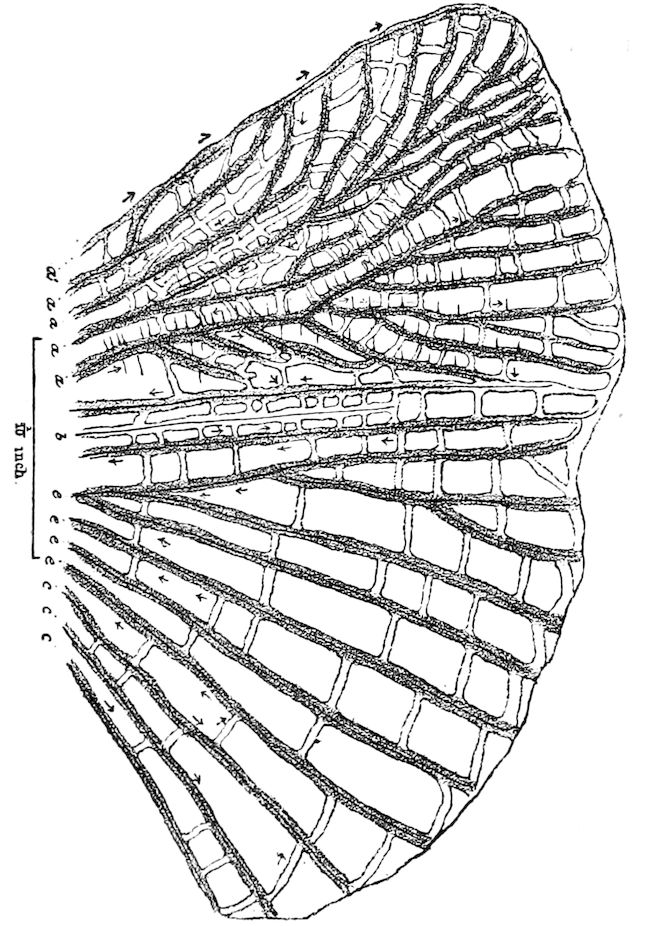
Fig. 382.—Circulation of the blood in hind wing of Periplaneta orientalis: the arrows indicate the usual direction of the blood currents.—After Moseley.
Newport gives the following summary of the observations of the early observers, to which we add the observations of Moseley. “A motion of the fluids has been seen by Carus in wings of recently developed Libellulidæ, Ephemera lutea and E. marginata, and Chrysopa perla; among the Coleoptera, in the elytra and wings of Lampyris italica and L. splendidula, Melolontha solstitialis and Dytiscus.” Ehrenberg saw it in Mantis, and Wagner in the young of Nepa cinerea and Cimex lectularius. Carus detected a circulation in the pupal wings of some Lepidoptera, and Bowerbank witnessed it in a Noctuid (Phlogophora meticulosa); Burmeister observed it in Eristalis tenax and E. nemorum, and Mr. Tyrrel in Musca domestica, but it has not been observed in the wings of Hymenoptera.
Bowerbank observed that in the lower wing of Chrysopa perla the blood passes from the base of the wing along the costal, post-costal, and externo-medial veins, outwards to the apex of the wing, giving off smaller currents in its course, and that it returns along the anal vein to the thorax. He found that the larger veins, 1 408 in. in diameter, contained tracheæ which only measured 1 2222 of an inch in diameter; but in others the tracheæ measured 1 1340, while the cavity measured only 1 500 of an inch. He states, also, that the tracheæ very rarely give off branches while passing along the main veins, and that they lie along the canals in a tortuous course. (Newport, art. Insecta, p. 980.)
Bowerbank, also, in his observations on the circulation in the wings of Chrysopa, 411“used every endeavor to discover, if possible, whether the blood has proper vessels, or only occupied the internal cavities of the canals; and that he is convinced that the latter is the case, as he could frequently perceive the particles not only surrounding all parts of the tracheæ, and occupying the whole of the internal diameter of the canals, but that it frequently happens that globules experienced a momentary stoppage in their progress, occasioned by their friction against the curved surface of the tracheæ, which sometimes gave them a rotatory motion.”
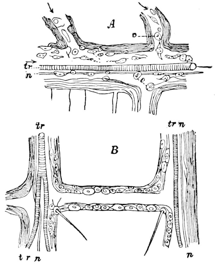
Fig. 383.—Parts of a vein of the cockroach, showing the nerve (n) by the side of the trachea (tr); c, blood corpuscles.—After Moseley.
Moseley found, owing to the large size and number of the corpuscles, that the circulation of the blood in the wings of insects is most easily observed in the cockroach, especially the hind wings. As seen in Moseley’s figure, the blood flows outward from the body through the larger veins (I and II) of the front edge of the wings, which he calls the main arteries of the wings, and more generally returns to the body through the veins in the middle of the wing; the blood also flows out from the body through the inner longitudinal veins (those behind vein IV), and the blood is also seen to flow through some of the small cross-veins. Fig. 383 shows one of the main trunks during active circulation. The corpuscles change their form readily, “the spindle-shaped ones doubling up in order to pass crossways through a narrow aperture.... In the irregularly formed corpuscles, which seem to represent leucocytes amœboid movements were observed.... Corpuscles pass freely above and under the tracheæ, showing that these latter lie free in the vessels.” The hypodermis lining the vessels is best seen in the small transverse veins.
The pulse or heart-beat of insects varies in rapidity in different insects, rising at times of excitement, as Newport noticed in Anthophora retusa, to 142 beats in a minute.
When an insect, as, for example, a tineid caterpillar, has been enclosed in a tight box for a day or more, the pulsations of the heart are very languid and slow, but soon, on giving it air, the pulsations will, as we have observed, rise in frequency to about 60 a minute, Herold observed 30 to 40 in a minute in a fully-grown silkworm, and from 46 to 48 in a much younger one. Suckow observed but 30 a minute in a full-grown caterpillar of Gastropacha pini, and 18 only in its pupa.
In a series of observations made by Newport on Sphinx ligustri from the fourth day after hatching from the egg until the perfect insect was developed, he found that before the larva cast its first skin the mean number of pulsations, 412in a state of moderate activity and quietude, was about 82 or 83 a minute; before the second moult 89, while before the third casting it had sunk down to 63; and before its fourth to 45, while, before leaving its fourth stage, and before it had ceased to feed, preparatory to pupating, the pulse was not more than 39. “Thus the number gradually decreases during the growing larva state, but the force of the circulation is very much augmented. Now when the insect is in a state of perfect rest, previously to changing its skin, the number is pretty nearly equal at each period, being about 30. When the insect has passed into the pupa state it sinks down to 22, and subsequently to 10 or 12, and after that, during the period of hibernation, it almost entirely ceases. But when the same insect which we had watched from its earliest condition was developed into the perfect state in May of the following spring, the number of pulsations, after the insect had been for some time excited in flight around the room, amounted to from 110 to 139; and when the same insect was in a state of repose, to from 41 to 50. When, however, the great business of life, the continuation of the species, has been accomplished, or when the insect is exhausted, and perishing through want of food or other causes, the number of pulsations gradually diminishes, until the motions of the heart are almost imperceptible.” Insects, then, he remarks, do not deviate from other animals in regard to their vital phenomena, though it has been wrongly imagined that the nutrient and circulatory functions are less active in the perfect than in the larval condition.
The heart of a larval Gastrus equi taken the day previous from a horse’s stomach beat from 40 to 44 times a minute (Scheiber); while Schröder van der Kolk observed only 30 beats in the same kind of maggot.
In the larva of Corethra, while at rest, the heart contracts from 12 to 16 or 18 times a minute, but when active the number rises to 22. The systole and diastole last from 5 to 6 minutes. (Dogiel.)
Temperature also affects the pulsations, as they increase in frequency with a rise and decrease with a fall in temperature.
Influence of electricity.—The influence of electricity on the action of the insect’s heart, from Dogiel’s experiments, is such as to cause an acceleration in the frequency of the beats, while an increase in the strength of the electric currents either diminishes the frequency of the beats or entirely stops the heart’s action. A violent excitation with the induction current causes a systole when the heart’s action has stopped for a long time; and if the excitation lasts uninterruptedly, then the contractions after a while become noticeable, according to the strength of the current. In such a case there are, however, interruptions in the regularity, strength, and order of the contractions. (Kolbe.)
Effects of poisons on the pulsations.—Dogiel has also experimented on the influence of poisons in the form of vapor or as liquid solutions on the pulsations of insects, which is much as in vertebrates. The application of carbonic oxide to the larva of Corethra, whose heart one minute previous to the poisoning beat 15 times a minute, accelerated the heart-beats in about 55 minutes to 25 pulsations in a minute. Afterwards there was a retardation in the pulse to the normal beat. Carbonic acid had a similar effect.
The following results obtained by Dogiel are somewhat as tabulated by Kolbe:—
I. Substances which cause the pulsations of the heart to accelerate.
a. An induction current of electricity, acting feebly.
b. Ammonia, acting feebly.
c. Ethyl ether, acting feebly.
d. Oxalic acid, acting feebly.
e. Carbolic acid, acting feebly.
f. Potassium nitrate, acting feebly.
g. Aconite, acting feebly.
413
II. Substances retarding the heart’s action.
a. An induction current of electricity, acting energetically.
b. Ammonia, acting energetically.
c. Ethyl ether, acting energetically.
d. Oxalic acid, acting energetically.
e. Carbolic acid, acting energetically.
f. Veratrine, acting energetically.
g. Atropine, acting energetically.
h. Aconitine, acting energetically.
i. Potassium nitrate, acting energetically.
g. Ethyl alcohol.
h. Chloroform.
i. Carbonic oxide.
j. Carbonic acid.
k. Sulphuretted hydrogen.
III. Substances whose action is indifferent.
1. Muscarine.
2. Curare.
3. Atropine, acting slowly.
4. Strychnine.
The above-named substances comprise those which in the vertebrates effect a change in the activity of the motor nerve-ganglia of the heart and the muscular fibres. Hence it follows that the heart of the larval Corethra consists of muscular fibres provided with ganglia, and that the contractions of the muscular fibres are provoked through the agency of the ganglia. But since muscarine, atropine, and curare, whose influence in stopping the heart’s action of vertebrates is known, in insects either have no action or only make the pulsations slower; it seems to follow that the heart of the larval Corethra possesses no similar apparatus for lessening the heart’s action, and this is also confirmed by anatomical studies. On the contrary, aconite acts, as we must from observations conclude, exclusively on the motor centres and the muscles, but not on the apparatus for lessening the heart’s action, which, as has been remarked, is not present in the larval Corethra. (Kolbe ex Dogiel.)
Dewitz has discovered an onward movement of the blood corpuscles, somewhat independent of the general circulation. This independent motion of the blood corpuscles is not only a creeping one like the amœboid motion of the white corpuscles of vertebrates, but they have besides a peculiar swimming movement. Dewitz noticed this in the hind wings of a recently emerged meal-worm beetle (Tenebrio molitor), still white and soft, after they had been cut off. The tissues forming the matrix within the wings constitute a network filled with blood. The current of blood within the wing thus cut off may be stopped flowing by a tap on the firmly clamped object-bearer on which the wing is placed, or by drawing it by an apparatus described by the same author, to incite in one way or another the blood corpuscles to swim forwards. When a corpuscle is disposed to move, we see it first stirring restlessly, or wabbling about, in this way changing its form; then it moves forwards, and does not come to a standstill. If it remains still there, after a while, by tapping, it begins again its movements.
“Should one yet doubt the fact of this spontaneous movement of the blood corpuscles, he will surely be convinced of its correctness by observing the so-to-speak reluctantly springing motion of a blood corpuscle in the wing of Tenebrio molitor with the simultaneous change of appearance and shape of the corpuscle.”
This spontaneous or independent motion of the blood corpuscles is also produced by the heating apparatus. As soon as the corpuscles lie still in the severed wing and they are warmed, the corpuscles begin to pass through the meshes of the tissue. When cooled, the motion ceases, but as soon as the temperature rises to a certain grade, the corpuscles again move onwards.
To explain this independent motion Dewitz thinks that they take up and then expel the blood-fluid, and in this way cause their motion. This independent motion is necessitated, in order that the stream of blood may become so regulated, 414that the blood corpuscles shall not be arrested in their course, but even turn back again out of the farther end of the antennæ and limbs. The chief mechanical power for the blood circulation must go on independently of the propulsatorial apparatus and of the heart. (Kolbe.)
LITERATURE ON THE HEART AND ON THE CIRCULATION OF THE BLOOD
a. Anatomy of the organs
Meckel, J. F. Ueber das Rückengefäss der Insekten. (Meckel’s Archiv, i, 1815, pp. 469–476.)
Müller, J. G. De vasi dorsali Insectorum. Berolini, 1816, pp. 22.
Serres, P. Marcel de. Observations sur les usages du vaisseau dorsal ou sur l’influence que le cœur exerce dans l’organisation des animaux articulés, etc. (Ann. du Mus. d’hist. nat., 1818, iv, pp. 149–192, 313–380, 2 Pls.; v, 1819, pp. 59–147, 1 Pl.)
Herold. Physiologische Untersuchungen über das Rückengefäss der Insekten. (Schriften d. Gesellsch. z. Beförderung d. Naturk. in Marburg, 1823, i, pp. 41–107.)
Carus, C. G. Entdeckung eines einfachen vom Herzen aus beschleunigten Blutkreislaufes in den Larven netzilügliger Insekten. Leipzig, 1827, pp. 40, 3 Taf.
—— Fernere Untersuchungen über Blutlauf in Kerfen. (Acta Acad. Leopold. Carol., 1831, xv, pp. 1–18, 1 Taf.)
Stadelmayr, L. Ansichten vom Blutlauf nebst Beobachtungen über das Rückengefäss der Insekten. Diss. München, 1829, pp. 24.
Berthold. Beitrage zur Anatomie, Zoologie und Physiologie. Göttingen, 1831.
Treviranus, G. R. Ueber das Herz der Insekten, dessen Verbindung mit den Eierstocken und ein Bauchgefäss der Lepidopteren. (Zeitschr. f. d. Physiologie, von F. Tiedemann. G. R. u. L. C. Treviranus, 1832, iv, pp. 181–184, 1 Taf.)
—— Beobachtungen aus der Zootomie und Physiologie. Bremen, 1839.
Wagner, R. Beobachtungen über Kreislauf des Blutes und den Bau des Rückengefässes bei den Insekten. (Isis, 1832, iii, p. 30; vii, pp. 320–331, 778–783, Fig.)
Bowerbank, J. S. Observations on the circulation of the blood in insects. (Ent. Mag., 1833, i, pp. 239–244, 1 Pl; also in Müller’s Archiv f. Physiolog., 1834, i, pp. 119–120.)
—— Observations on the circulation of the blood and the distribution of the tracheæ in the wing of Chrysopa perla. (Ent. Mag., 1837, iv, pp. 179–185.)
Jaeger. Ueber die Entdeckung von einer Bewegung in den Schuppen des Schmetterlingsflügel. (Isis, 1837, v, p. 512.)
Behn, W. Découverte d’une circulation de fluide nutritif dans les pattes de plusieurs insectes hémipteres. (Ann. Sc. nat., 1835, Sér. 2, iv, pp. 1–12.)
Newport, G. Insecta, in Todd’s Cyclopædia of Anatomy and Physiology, 1839. London, pp. 853–994. On the circulation of the blood, p. 976, Figs.
Duvernoy, G. L. Résumé sur le fluide nourricier, ses réservoirs et son mouvement dans tout règne animal. (Ann. Sc. nat., 1839, Sér. 2, xii, pp. 300–346.)
Dufour, L. Études anatomiques et physiologiques sur une mouche dans le but d’eclairer l’histoire des metamorphoses et de la prétendue circulation dans les insectes. (Ann. Sc. nat. Zool., Sér. 2, 1841, xvi, pp. 5–14.)
415—— Note sur la prétendus circulation dans les insectes. (Compt. rend. Acad., Paris, 1844, xix, pp. 188–189.)
—— Études anatomiques et physiologiques sur une mouche, dans le but d’éclaircir l’histoire des metamorphoses et de prétendue circulation des insectes. (Mém. mathémat. des Savants étrangers, Paris, 1846, ix, pp. 545–628, 1 Pl.)
—— Sur la circulation dans les insectes. Bordeaux, 1849, 8º, pp. 40. (Compt. rend. Acad. Sci., Paris, 1849, xxviii, pp. 28–33, 101–104, 163–170.)
—— De la circulation du sang et de la nutrition chez les insectes. Bordeaux, 1851. (Act. Soc. Linn., Bordeaux, 1851, xvii, p. 9.)
—— Études anatomiques et physiologiques et observations sur les larves des Libellules, Appareil circulatoire. (Annal. Sci. nat., Sér. 3, Zool., xvii, 1852, pp. 98–101, 1 Pl.)
Schröder van der Kolk, J. S. C. Mémoire sur l’anatomie et physiologie du Gastrus equi. (N. Verhandl. Kl. Nederl. Instit., 11, 1845, pp. 1–155, 13 Pl.)
Nicolet, H. Note sur la circulation du sang chez les Coléoptères. (Ann. Sc. nat., 1847, Sér. 3, vii, pp. 60–64.)
Verloren, C. Mémoire en réponse à la question suivante: éclaircir par des observations nouvelles le phénomène de la circulation dans les insectes, en recherchant si on peut la reconnaître dans les larves de différents ordres de ces animaux. (Mém. couronn. et Mém. d. savants étrang. de l’Acad. Roy. Belgique, xix, 1847.)
Blanchard, E. De la circulation dans les insectes. (Ann. Sc. nat., 1848, Sér. 3, ix, pp. 359–398, 5 Pls.)
—— Sur la circulation du sang chez les insectes et sur la nutrition. (Compt. rend. Acad. Sc., Paris, 1849, xxviii, pp. 76–78; 1851, xxxiii, pp. 367–370.)
—— Nouvelles observations sur la circulation du sang et la nutrition chez les insectes. (Ibid., pp. 371–376.)
Joly, N. Mémoire sur l’existence supposée d’une circulation péritrachéenne chez les insectes. (Ann. Sc. nat. Zool., Sér. 3, 1849, xii, pp. 306–316.)
Bassi, C. A. Rapporto alla sezione di zoologia, anatomia comparata e fisiologia del congresso di Venezia, sul passagio delle materie ingerite nel sistema tracheale degli insetti. (Gazette di Milano, 1847, vi; also Ann. Sc. nat. Zool., Sér. 3, 1851, xv., 362–371.)
Agassiz, Louis. On the circulation of the fluids in insects. (Proceed. Amer. Assoc. Adv. Sc., 1849, pp. 140–143; Ann. Sc. nat. Zool., Sér. 3, xv, 1851, pp. 358–362.)
Leydig, F. Anatomisches und Histiologisches über die Larve von Corethra plumicornis. (Zeitschr. f. wissen. Zool., iii, 1851, pp. 435–451.)
Wedl, C. Ueber das Herz von Menopon pallidum. (Sitzungsber. der k. Akad. d. Wissensch. Wien., 1855, xvii, pp. 173–180.)
Scheiber, S. H. Vergleichende Anatomie und Physiologie der Oestriden Larven. (Sitzungsber. d. k. Akad. d. Wiss. Wien. Math.-naturwiss. Cl., xli, 1860, pp. 409–496, 2 Taf.; The circulatory system, pp. 463–490.)
Brauer, Fr. Beitrag zur Kenntnis des Baues und der Funktion der Stigmenplatten der Gastrus-Larven. (Verhdl. d. k. k. zool.-bot. Gesellsch. Wien., xiii, 1863, pp. 133–136.)
Moseley, H. N. On the circulation in the wings of Blatta orientalis and other insects, and on a new method of injecting the vessels of insects. (Quart. Jour. of Micr. Science, xi, n. s., pp. 389–395, 1871, 1 P1.)
Graber, V. Ueber die Blatkörperchen der Insekten. (Sitzber. Akad. Wien. Math.-naturw. Classe, lxiv, 1871, pp. 9–44, 1 Taf.)
416—— Vorläufiger Bericht über den propulsatorischen Apparat der Insekten. (Sitzber. d. k. Ak. d. Wiss. Wien., lxv, 1872, pp. 16, 1 Taf.)
—— Ueber den propulsatorischen Apparat der Insekten. (Archiv f. mikroskop. Anatomie, ix, 1873, pp. 129–196, 3 Taf.)
—— Ueber den pulsierenden Bauchsinus der Insekten. (Archiv f. mikroskop. Anat., xii, 1876, pp. 575–582, 1 Taf.)
Grobben, Carl. Über bläschenförmige Sinnesorgane und eine eigenthümliche Herzbildung der Larve von Ptychoptera contaminata L. (Sitzb. k. Akad. Wissensch. Wien., 1875, lxxii, p. 22, 1 Taf.)
Liebe, Otto. Ueber die Respiration der Tracheaten, besonders über den Mechanismus derselben und über die Menge der ausgeatmeten Kohlensäure. Inaug.-Diss. Chemnitz, 1872, pp. 28.
Dogiel, John. Anatomie und Physiologie des Herzens der Larve von Corethra plumicornis. (Mém. Acad. imp. St. Petersbourg, 7 Sér., xxiv, 1877, Nr. 10, pp. 37, 2 Pls.) Separate, Leipzig, Voss.
Bütschli, O. Ein Beitrag zur Kenntnis des Stoffwechsels, insbesondere der Respiration bei den Insekten. (Reichert’s und Du Bois-Reymond’s Archiv f. Anatomie u. Physiologie, 1874, pp. 348–361.)
Béla-Dezso. Ueber den Zusammenhang des Kreislaufs und der respiratorischen Organe bei den Arthropoden. (Zool. Anzeiger, i Jahrg., 1878, p. 274.)
Plateau, F. Communication préliminaire sur les mouvements et l’innervation de l’organe central de la circulation chez les animaux articulés. (Bull. Acad. roy. de Belgique, Sér. 2, xlvi, 1878, pp. 203–212.)
Jaworovski, Ant. Ueber die Entwicklung des Rückengefässes und speziell der Muskulatur bei Chironomus und einigen anderen Insekten. (Sitzgsber. d. k. Akad. d. Wissensch. Wien. Math.-naturwiss. Cl., lxxx, 1879, pp. 238–258.)
Zimmermann, O. Ueber eine eigentümliche Bildung des Rückengefässes bei einigen Ephemeridenlarven. (Zeitschr. f. wissens. Zool., 1880, xxxiv, pp. 404–406.)
Burgess, E. Note on the aorta in lepidopterous insects. (Proc. Bost. Soc. Nat. Hist., xxi, 1881, pp. 153–156, Figs.)
—— Contributions to the anatomy of the milk-weed butterfly, Danais Archippus F. (Anniversary Memoirs Boston Soc. Nat. Hist., 1880, pp. 16, 2 Pls.)
Vayssière, A. Recherches sur l’organisation des larves des Éphémérines. (Ann. Sc. nat. Zool., Sér. 6, xiii, 1882, pp. 1–137, 11 Pls.)
Viallanes, H. Recherches sur l’histologie des insectes et sur les phénomènes histologiques qui accompagnent le développement post-embryonnaire de ces animaux. (Ann. Sc. nat., Sér. 6, xiv, 1882, pp. 1–348, 18 Pls.)
Schimkewitsch, W. Ueber die Identität der Herzbildung bei den wirbel und wirbellosen Tieren. (Zool. Anzeiger, 1885, viii Jahrg., pp. 37–40, Fig.)
—— Noch etwas über die Identität der Herzbildung bei den Metazoen. (Zool. Anzeiger, 1885, pp. 384–386.)
Creutzburg, N. Ueber den Kreislauf der Ephemerenlarven. (Zool. Anzeiger, 1885, pp. 246–248.)
Poletajewa, Olga. Du cœur des insectes. (Zool. Anzeiger, 1886, ix Jahrg., pp. 13–15.)
Selvatico, S. L’aorta nel corsaletto e nel capo della farfalla del bombice del gelso. (Padova, 1887, p. 19, 2 Pls.)
—— Die Aorta im Brustkasten und im Kopfe des Schmetterlings von Bombyx mori. (Zool. Anzeiger, 1887, x Jahrg., pp. 562–563.)
Kowalevsky, A. Ein Beitrag zur Kenntnis der Excretionsorgane. (Biol. Centralbl., 1889, ix, pp. 33–47, 65–76, 127–128.)
417Tosi, Alessandro. Osservazioni sulla valvola del cardias in varii generi della famiglia delle Apidi. (Ricerche Lab. Anat. R. Univ. Roma, v, 1895, pp. 5–26, 16 Figs., 3 Pls.)
Pawlowa, Mary. Ueber ampullenartige Blutcirculationsorgane im Kopfe verschiedener Orthopteren. (Zool. Anzeiger, xviii Jahrg., 1895, pp. 7–13, 1 Fig.)
Also the writings of Kolbe.
b. The blood, blood corpuscles, leucocytes, and blood tissue
Wagner, R. Ueber Blutkörperchen bei Regenwürmern, Blutegeln und Dipterenlarven. (Müller’s Archiv f. Anatomie u. Physiologie, 1835, pp. 311–313.)
—— Nachtrage zur vergleichenden Physiologie des Blutes. (Archiv f. Anat. u. Physiologie, 1838.)
Newport, G. On the structure and development of the blood. First series. The development of the blood corpuscle in insects and other invertebrata, and its comparison with that of man and the vertebrata. (Abstr. of the paper in Roy. Soc., 1845, v, pp. 544–546: also in Ann. Mag. Nat. Hist., Ser. 3, 1845, iii, pp. 364–367.)
Landois, H. Beobachtungen über das Blut der Insekten. (Zeitschr. f. wissens. Zool., xiv, 1864, pp. 55–70, 3 Pl.)
——, and L. Landois. Ueber die numerische Entwicklung der histiologischen Elemente d. Insektenkörpers. (Ibid., xv, 1865, pp. 307–327.)
Rollett, A. Zur Kenntnis der Verbreitung des Hämatins. (Sitzgsber. d. k. Akad. d. Wiss. Wien., lxiv, 1871.)
Wielowiejski, H. v. Ueber das Blutgewebe der Insekten. Eine vorläufige Mitteilung. (Zeitschr. f. wissens. Zool., 1886, xliii, pp. 512–536.)
MacMunn, C. A. Researches on myohæmatin and the histohæmatins. (Proc. Roy. Soc. London, 1886, xxxix, pp. 248–252.)
Peyron, J. Sur l’atmosphere interne des insectes comparée à celle des feuilles. (Compt. rend. Acad. Sc., Paris, 1886, cii, pp. 1339–1341.)
Cuénot, L. Études sur le sang, son rôle et sa formation dans la série animale. Part 2, invertébrés; note préliminaire. (Arch. Zool. Expériment., 1888, Sér. 2, v, pp. xliii-xlvii. See also ibid., Sér. 3, 1897, pp. 655, 679–680.)
Dewitz, H. Die selbstandige Fortbewegung der Blutkörperchen der Gliedertiere. (Naturwiss. Rundschau. Braunschweig, 1889, iv Jahrg., pp. 221–222.)
—— Eigenthätige Schwimmbewegung der Blutkörperchen der Gliedertiere. (Zool. Anzeiger, 1889, xii Jahrg., pp. 457–464, Fig.)
Schäffer, C. Beitrage zur Histiologie der Insekten. II, Ueber Blutbildungsherde bei Insektenlarven. (Spengel’s Zool. Jahrbücher Abt. f. Anat. u. Ontogenie, iii, 1889, pp. 626–636, 1 Taf.)
Cattaneo, G. Sulla morfologia delle cellule ameboidi dei Molluschi e Artropodi. (Boll. Sc. Pavia. Anno 11, 1889, p. 59, 2 Pls.)
Wagner, W. A. Ueber die Form der körperlichen Elemente des Blutes bei Arthropoden, Würmern und Echinodermen. (Biolog. Centralblatt, 1890, x, p. 428.)
Preyer, W. Zur Physiologie des Protoplasma. II, Die Funktionen des Stoffwechsels. Die Saftströmung. (Patanie’s Naturwiss. Wochenschr., 1891, vi, pp. 1–5.)
Cholodkowsky, N. Ueber das Bluten der Cimbiciden-Larven. (Entomologische Miscellen, vi, Horæ Soc. Ent. St. Petersburg, 1897, pp. 352–357, 1 Fig. The fluid thrown out through pores or fissures in the skin is the blood.)
With the writings of Korotaiev, Tichomeroff, Pékarsky, Balbiani, Korotneff, Cuénot, and others.
418
c. The fat-bodies
Dufour, L. Recherches anatomiques sur les Carabiques et sur plusieurs autres insectes Coléoptères. Du tissu adipeux splanchnique. (Ann. Sc. nat., viii, 1826, pp. 29–35.)
—— Histoire comparative des métamorphoses et de l’anatomie des Cetonia aurata et Dorcus parallelepipedus. Tissu adipeux splanchnique. (Ann. Sc. nat., Zoologie, Sér. 2, 1842, xviii, pp. 178–179.)
Meyer, H. Ueber die Entwicklung des Fettkörpers, der Tracheen und der keimbereitenden Geschlechtsteile bei den Lepidopteren. (Zeitschr. f. wissens. Zool., 1849, i, pp. 175–179, 4 Taf.)
Fabre, J. H. Étude sur le rôle du tissu adipeux dans la sécretion urinaire chez les insectes. (Ann. Sc. nat., Sér. 4, xix, 1862, pp. 351–382.)
Leydig, Fr. Einige Worte über Fettkörper der Arthropoden. (Reichert, u. du Bois-Reymond’s Archiv f. Anat., 1863, pp. 192–203.)
Lindemann, K. Zoologische Skizzen. 1. Struktur des Fettkörpers. (Bull. Soc. Imp. d. Natural. Moscou, 1864, pp. 521–526, 1 Pl.)
Landois, Leonh. Ueber die Funktion des Fettkörpers. (Zeitschr. f. wissens. Zoologie, xv, 1865, pp. 371–372.)
Wielowiejski, H. v. Ueber den Fettkörper von Corethra plumicornis und seine Entwicklung. (Zool. Anzeiger, 1883, vi Jahrg., pp. 318–322.)
—— Ueber das Blutgewebe der Insekten. (Zeitschr. f. wissens. Zool., 1886, xliii, pp. 512–536.)
Kowalevsky, A. O. Sur les organes excréteurs chez les arthropodes terrestres. (Congrès internat. Zool., 2me Sess., pp. 196–205, Moscou, 1892.)
419
THE BLOOD TISSUE
Under this name Wielowiejski has included several important tissues or cellular bodies intimately concerned with the nutrition of the insect. These are:—
1. The blood corpuscles. (See p. 407, leucocytes and phagocytes.)
2. The fat-body proper (Corpus adiposum).
3. The pericardial fat-body (pericardial cells).
4. The œnocytes.
5. The garland-shaped cord of muscid larvæ.
6. The subœsophageal body, a peculiar organ found by Wheeler in the embryos and young larvæ of Blatta and Xiphidium.
7. The phosphorescent organs.
a. The fat-body
In the body cavity of winged insects and of their larvæ occur yellowish masses of large cells filled with small drops of fat, and forming the “fat-body.” It is of various shapes, more or less lobulated or net-like, and covers or envelops parts of the viscera, also forming a layer under the integument (Fig. 143). The tracheal endings are usually enveloped by the fat-body. It is larger in the larvæ than in the adults, especially in Lepidoptera, in them forming a reserve of nutrition, used during metamorphosis and during the formation and ripening of the eggs and male cells.
Wielowiejski has shown that there is a regular arrangement of the fat-body in the general cavity of the body. For example, in the larva of Chironomus occur the following forms of this tissue. Around the periphery, on each side of the body cavity, is a loose network of lobes with large meshes constituting the peripheral layer or external lobular fat-body; these lobular masses are segmentally arranged.
Within these segmental lobes, on each side of and along the digestive tract, extending along through almost the entire body, is an unbroken strand of this tissue, forming the internal fat-body cords. From the first larval stage, and even before hatching, its cells are so unusually large, being filled with large, clear, mostly colorless fat-drops, that their limits cannot be defined, and their nuclei can only with great difficulty be detected. Only in some large larvæ of Chironomus has Wielowiejski found clearly defined cells; the protoplasm of these cells contain almost no fat-drops.
The fat-body is of mesodermal origin, and as Wheeler insists, is not derived from the œnocytes, as supposed by Graber. Formed from the mesoderm, it is a differentiation of portions of the cœlomic walls, and therefore metameric in origin. That the fat-body gives origin to the blood corpuscles Wheeler is doubtful.
420The fat-cells are distinct, spherical, and as a rule possess only one nucleus, though in those of Apis and Melophagus there are two nuclei, and in Musca several. Sometimes the cells contain a substance like the white of an egg, and concretions of uric acid, or these take the place of the fat-drops. The presence of uric acid shows that a very active metabolism goes on in the fat-body. “In some cases it has been proved that the fat-body in the larva is rich in fat and poor in concretions of uric acid, while in the imago it is poor in fat and rich in concretions of uric acid” (Lang).
Leydig, in 1857 (Lehrbuch der Histiologie), spoke of the presence of dark concretions in the fat-body, and afterwards (1864) showed that there was a wide distribution of uric acid salts and concretions. Witlaczil, also, has detected concretions in the fat-body of the Psyllidæ, in larval Cecidomyiidæ, in the larvæ and pupæ of ants, and in the pupa of Musca.
The physiological processes which take place in the fat-bodies are obscure. Graber regarded the whole system of the fat-bodies as “a single, many-lobed lung,” while before him Landois, taking into account the intimate relation existing between the finer tracheal branches and the fat-body, considered that the latter was concerned in respiration. Marchal thinks that the fat-body is a urinary organ, as the urates are formed within the cells of this body.
Moreover, Schäffer maintains that a special kind of fat-body cell has the important function of taking up and giving out nutritious matters during the internal processes of metamorphosis, while he also believes that there is a genetic connection between the fat-body and the blood corpuscles—a view combated by Wheeler.
Kowalevsky finds that the fat-body remains absolutely insensible to the action of the substances which stained the Malpighian tubes (p. 352). So long as the cells are healthy and living they are not stained and do not absorb the colors in question; and this insensibility persists, even when the cells are of a different nature, as those of the fly (adipose and “intercalary” cells).
b. The pericardial fat-body or pericardial cells
We have already, on p. 405, called attention to these organs, but they also have an intimate relation to the fat-body.
Kowalevsky (1892) remarks that the disposition of these cells varies much in different insects and even in the same animal. Thus, in the Diptera and the ordinary flies there are found around the lower part of the dorsal vessel 13 pairs of large pericardial cells which lie next to a crowded bed of small cells forming a compact mass around the anterior part of the dorsal vessel. In caterpillars, notably silkworms, from the compact layer of pericardial cells which surround the heart, pass off trunks which are directed towards the lateral walls of the body, also forming close networks around the tracheæ and then passing down into the abdominal cavity of the body of the larva.
In the larvæ of certain Hymenoptera, the trunks which pass off from the pericardial region form a loose cord, a sort of fatty tissue covering the entire body cavity.
421This tissue, adds Kowalevsky, entirely differs from œnocytes, or from the so-called glandular body whose formation in Gryllotalpa has been described by Korotaiev, and in Bombyx mori by Tichomiroff. In a recent work wherein has been collected everything known regarding these last-named cells, Pékarsky proves that they are unique in nature and cannot be regarded either as fat-cells, or as pericardial cells, or even as formative leucocytes.
As to the structure of the pericardial cells, Kowalevsky adds that they are always attached to muscular fibres passing off from the heart, and that they lie, so to speak, upon them. In the locusts the muscular fibres supporting the pericardial cells appear distinctly like little staves or sticks. The attachment of the pericardial cells to the muscular fibres has been observed by Cuénot and reproduced by him in his work, but his description somewhat differs from that observed by Kowalevsky in the locust (Acrydium migratorium).
As to the nature of the acid excretions which are formed in the pericardial cells, in spite of his attempts to solve the problem, Kowalevsky has been unsuccessful. The only observations in this direction are those of Letellier on the pericardial glands of lamellibranch molluscs, which he found to contain hypouric acid, and it is probable, says Kowalevsky, that the acidity of the pericardial cells in insects is due to the presence of the same acid.
Leucocytes or phagocytes in connection with the pericardial cells.—It is thought by Schäffer that the leucocytes or phagocytes may be free or wandering fat-body cells. They play an important part in metamorphosis, while they absorb or feed upon the remains of the larval organs, and thus prove of use in the building up of the organs of the adult insects.
While the faculty of phagocytosis is wanting in the urinary tubes, Balbiani and more recently Cuénot have expressed the opinion that the pericardial cells of insects may have the power of absorbing hard bodies, “acting as a phagocytic gland.” This, however, is called in question by Kowalevsky, from studies made on different insects. On introducing powdered carmine into the body of an insect it has not been absorbed by the pericardial cells, as they have not been colored red. It is the leucocytes which absorb the grains of carmine, and which, after having dissolved them, transmit them to the pericardial cells. Hence, then, the pericardial cells have not the phagocytic power of which Cuénot speaks.
Returning to his own observations on hard bodies introduced into insects, or large globules introduced under the form of a milk emulsion, Kowalevsky has found that these bodies were absorbed in the first place by the free-swimming leucocytes, and in the second place by whole groups or nests of leucocytes situated in different parts 422of the body, principally on the threads of the adipose body. In the Orthoptera the absorption is immediately effected by means of the cells of the membrane which separates the pericardium from the cavity of the body underneath the heart. The regions where the hard bodies are absorbed in great number coincide with the regions of formation of the blood corpuscles. In his researches on the larvæ of Hyponomeuta and other Lepidoptera, Schäffer describes these regions as forming a sort of island. The nests where the blood globules are formed are the most active centres of phagocytosis.
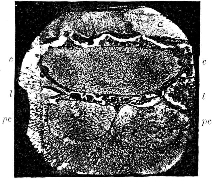
Fig. 384.-Section of the heart (c) and pericardial cells (pc, pc) from the posterior part of the heart of a fly: l, l, nests of leucocytes situated between the heart and pericardial cells.—From a microphotograph, after Kowalevsky.
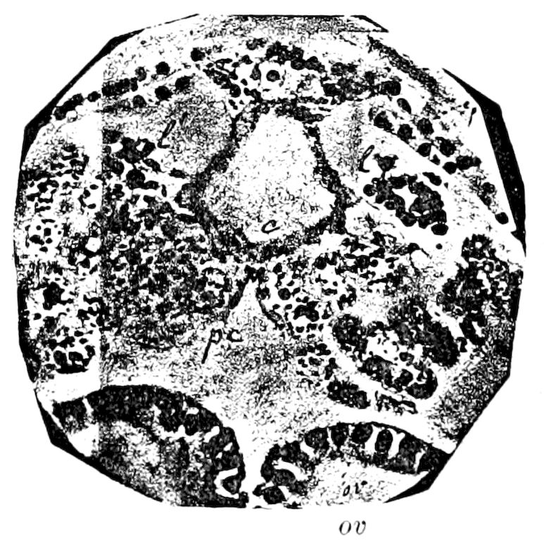
Fig. 385.—Cross-section of the heart of Truxalis nasata and of the structures around it: c, heart: ep, epithelium under the cuticula (hvpodermis); or, ovarian tubes; pc, pericardial cells, with one or two nuclei containing a deposit of carmine; l and l′, group of leucocytes, which have absorbed granules of India ink.—After Kowalevsky.
Balbiani, and also Cuénot, have supposed that the formation of the blood corpuscles takes place in the pericardial cells, but Kowalevsky insists that these cells cannot form the leucocytes, which “are probably formed in different parts of the body, notably in the special nests [Herde of Jäger] situated near the heart, but outside of the pericardial cells.”
In Fig. 384, where the nests of leucocytes (l) are shown, it is evident that they are formed where observed, and “could not have come from the pericardial cells, which have their own structure and their special function,” these cells being very large and characteristic.
In Kowalevsky’s preparations of Truxalis, the pericardial cells with deposits of carmine and the groups of leucocytes (Fig. 385, l and l′) stained with India ink, we have to deal with elements absolutely different. If the formation of leucocytes was caused by the pericardial cells, these last would be obliged to free themselves from their contents and to modify their essential nature.
423
c. The œnocytes
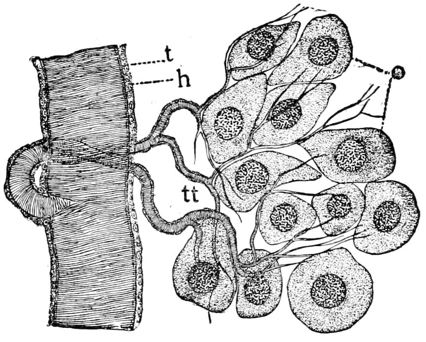
Fig. 386.—Cluster of œnocytes from a nearly mature Phryganeid larva: o, œnocytes; t, large tracheal branch; tt, smaller tracheal ramifications; h, tracheal hypodermis.
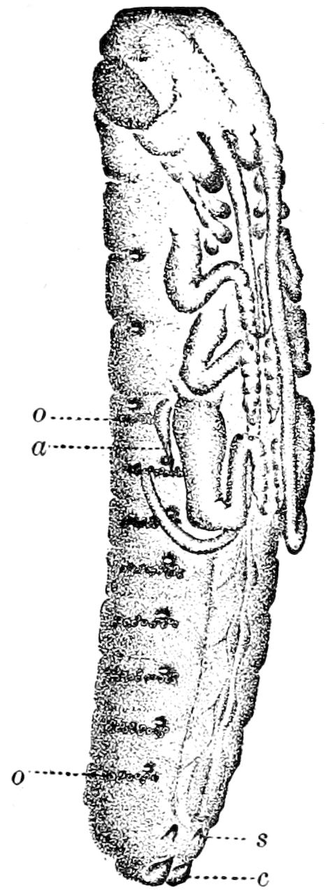
Fig. 387.—A nearly mature embryo of Xiphidium ensiferum: o, o, œnocyte clusters seen from the surface through the integument; a, pleuropodium of the right side (appendage of the first abdominal segment); s, styli; c, cercopods.—This and Fig. 386 after Wheeler.
These cells (Fig. 386), with the exception of the eggs, are the largest in the body, and occur in most if not all winged insects. They were called œnocytes (oinos, wine; kustis, cyst), by Wielowiejski in allusion to their wine-yellow color. These cells are arranged segmentally (Fig. 387) in clusters, held in place by tracheæ, and are situated mostly on each side of the abdomen, rarely being found in the adjoining parts of the thorax. They are more or less intimately associated with the blood and fat-body. Unlike the fat-body, however, they arise in embryonic life from the ectoderm, either by delamination or by immigration, just behind the tracheal involutions.
The separate cells of each cluster are usually separate, but in rare cases may fuse in pairs or form smaller clusters. In shape they are round or oval, often sending out pseudopodia-like processes, by which they are attached to the tracheal twigs or to each other. “The cytoplasm, which is very abundant, is full of yellowish granules and is sometimes radially situated towards its periphery. The large spherical or oval nucleus contains a densely wound and delicate chromatic filament.” (Wheeler.)
Graber first pointed out the identity of these clusters of cells with certain metameric cell-masses in insect embryos, observed by Tichomiroff in those of the silkworm, and by Korotneff in the embryo mole-cricket.
Although they resemble the blood corpuscles in some insects, they are always much larger, and do not seem to be amœboid, while they are never seen to 424undergo self-division, or to exhibit any appearance of giving rise to the blood-cells (Wheeler). They have not yet been detected in Thysanura (Synaptera) or in Myriopoda.
d. The phosphorescent organs
Phosphorescence is not infrequent in the Protozoa, cœlenterates, worms, and has been observed in the bivalve Pholas, in a few abyssal Crustacea, in myriopods (Geophilus), in an ascidian, Pyrosoma, and in certain deep-sea fishes.
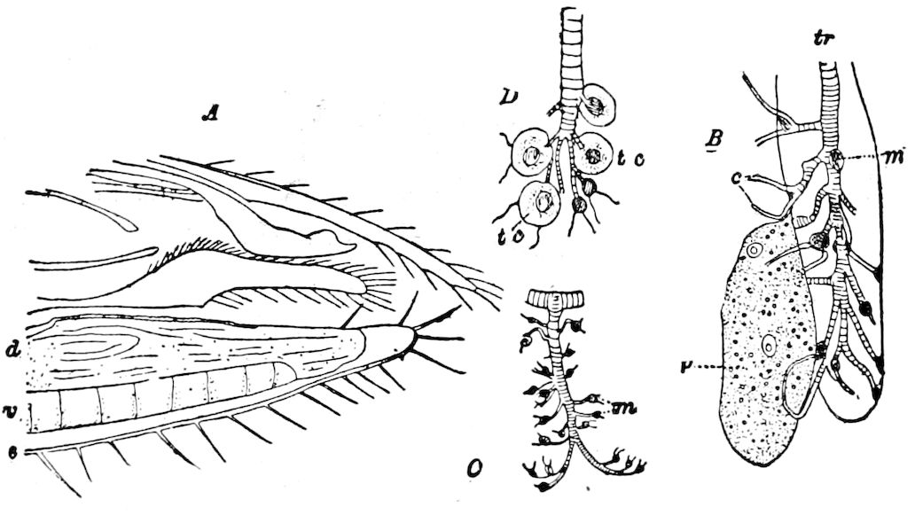
Fig. 388.—A, sagittal section through the hinder end of a male Luciola, the organs above the phosphorescent plate only drawn in outline: s, integument of the last segment, somewhat removed by the section-knife from the phosphorescent tissues; d, dorsal layer of the phosphorescent plate penetrated by irregular tracheal branches, and rendered opaque by numerous urate concretions imbedded in it; v, ventral phosphorescent layer of the plate, with perpendicular tracheal stems whose branches, where they pass into capillaries, bear lumps which stain brown with osmic acid; n, structureless substance (coagulum?) filling the end of the last ventral segment. B, isolated portion of the ventral layer of the phosphorescent plate; tr, tracheal stem surrounded by a cylindrical lobe: p, parenchym cell attached to the cylinder; c, capillary, without the spiral threads; m, coagulum stained brown. C, a tracheal stem of the ventral layer: at the fork of the brown-stained capillaries are lumps stained brown with osmic acid. D, a part of C, more highly magnified, showing the remains of the tracheal end-cells (tc) enveloping the brown lumps (m).—After Emery.
In insects luminosity is mostly confined to a few Coleoptera, and besides the well-known fireflies, an Indian Buprestid (Buprestis ocelata) is said to be phosphorescent; also a telephorid larva. Other luminous insects are the Poduran Anurophorus, Fulgora, certain Diptera (Culex, Chironomus[60] and Tyreophora), and an ant (Orya).
The seat of the light is the intensely luminous areas situated either in the head (Fulgora), in the abdomen (Lampyridæ), or in the thorax (in a few Elateridæ of the genus Pyrophorus). The luminous or photogenic organ is regarded by Wielowiejski and also by Emery as morphologically a specialized portion of the fat-body, being a plate consisting of polygonal cells, situated directly under the integument, and supplied with nerves and fine tracheal branches.
In Luciola as well as in other fireflies, including Pyrophorus, the 425phosphorescent organ or plate consists, as first stated by Kölliker, of two layers lying one over the other, a dorsal one (Fig. 388, d) which is opaque, chalky white, and non-photogenic, and a lower one (v), the active photogenic layer, which is transparent. Through the upper or opaque layer and on its dorsal surface extend large tracheæ and their horizontal branches, from which arise numerous very fine branches which pass down perpendicularly into the transparent or photogenic layer of the organ. Each tracheal stem, together with its short branches, is enveloped by a cylindrical mass of transparent tissue, so that only the short terminal branches or very fine tracheal capillaries project on the upper part of the cylinder. These finest tracheal capillaries are not in Luciola filled with air, but with a colorless fluid, as was also found by Wielowiejski and others in Lampyris.
These transparent cylinders, with the tracheæ within, forming longitudinal axes, resemble lobules. These lobules are so distributed that they appear on a surface section of this plate as numerous round areas in which circular periphery the tracheal capillaries are arranged with the axially disposed tracheal end-cells. These “tracheal end-cells” are only membranous enlargements at the base of the tracheal capillaries (Wielowiejski). The cylindrical lobules are separated from each other by a substance consisting of abundant large granular cells (parenchym cells) among which project the tracheal capillaries. The cylindrical lobules extend to the hypodermis and come in contact only by their lateral faces with the parenchym.
The structure of the upper opaque chalky white layer of the phosphorescent organ is, compared with that of the photogenic lower portion, very simple. In its loose, pappose, mass are no cellular elements, but when treated with different reagents it is seen to be filled with countless urate granules (guanine) swimming in the fluid it contains, the cell plasma appearing to be dissolved, the cells having lost their cohesion.
In comparing the phosphorescent plate or organ of Luciola with that of Lampyris, the general structure, including the clear cell elements of the cylindrical lobules, which envelop the perpendicular tracheal twigs and their branches, and also the granular parenchymatous cells are alike in both, though the arrangement and distribution of the elements in Luciola is more regular, in Lampyris the tracheal stems being irregularly scattered through the parenchym.
Wielowiejski found in the larval and female Lampyris a higher degree of differentiation than in the male, and Luciola has a more differentiated photogenic organ than Lampyris, as seen in the more regular structure of the lobules.
426As regards the light-apparatus of Pyrophorus, or the cucujo, Heinemann shows that, as in the Lampyridæ, it consists of distinct cells, and may be regarded as a glandular structure. It is rich in tracheæ and the other parts already described. In still later researches on a Brazilian Pyrophorus Wielowiejski shows that the phosphorescent plate consists of two layers, the upper usually being filled with crystalline urate concretions, and entirely like those of the Lampyridæ, consisting of distinct polygonal cells, among which are numerous tracheal stems, with tænidia, coursing in different directions, when freshly filled with air, and sending capillaries into the underlying photogenic layer. The latter shows in its structure a striking difference in the cellular arrangement from that of Lampyrids. In the upper or non-photogenic layer are tracheal capillaries which pass down into the underlying cellular plate and which are in the closest possible relations with the single cells—a point overlooked by Heinemann.
Physiology of the phosphorescence.—As is well known, the phosphorescence of animals is a scintillating or glowing light emitted by various forms, the greenish light or luminous appearance thus produced being photogenic, i.e. without sensible heat.
Langley rates the light of the firefly at an efficiency of 100 per cent, all its radiations lying within the limits of the visible spectrum. “Langley has shown that while only 2.4 per cent of luminous waves are contained in the radiation of a gas-flame, only 10 per cent in that of the electric arc, and only 35 per cent in that of the sun, the radiation of the firefly (Pyrophorus noctilucus) consists wholly of visible wave-frequencies.” (Barker’s Physics, p. 385.)
The spectrum of the light of the cucujo was found by Pasteur to be continuous. (C. R. French Acad. Sc. 1864, ii, p. 509.) A later examination by Aubert and Dubois showed that the spectrum of the light examined by the spectroscope is very beautiful, but destitute of dark bands. When, however, the intensity diminishes, the red and orange disappear, and the green and yellow only remain.
Heinemann studied the cucujo at Vera Cruz, Mexico. At night in a dark room it radiates a pale green light which shows a blue tone to the exclusion of any other light. The more gas or lamp light there is present, the more apparent becomes the yellowish green hue, which in clear daylight changes to an almost pure very light yellow with a very slight mixture of green. “In the morning and evening twilight, more constantly and clearly in the former, the cucujo light, at least to my eyes, is an intensely brilliant yellow with a slight mixture of red. In a dark room lighted with a sodium light the yellow 427tone entirely disappears; on the other hand, the blue strikingly increases.” As regards the spectrum he found that almost exactly half of the blue end is wanting and that the red part is also a little narrower than in the spectrum of the petroleum flame.
Professor C. A. Young states that the spectrum given by our common firefly (Photinus?) is perfectly continuous, without trace of lines either bright or dark. “It extends from a little above Fraunhofer’s line C, in the scarlet, to about F in the blue, gradually fading out at the extremities. It is noticeable that precisely this portion of the spectrum is composed of rays which, while they more powerfully than any others affect the organs of vision, produce hardly any thermal or actinic effect. In other words, very little of the energy expended in the flash of the fire is wasted. It is quite different with our artificial methods of illumination. In the case of an ordinary gaslight the best experiments show that not more than one or two per cent of the radiant energy consists of visible rays; the rest is either invisible heat or actinism; that is to say, over 98 per cent of the gas is wasted in producing rays that do not help in making objects visible.”
Panceri also remarks that while in the spectroscope the light of some Chætopteri, Beroë, and Pyrosoma exhibit one broad band like that given by monochromatic light, that of Lampyris and Luciola is polychromatic. (Amer. Nat., vii, 1873, p. 314.)
The filtered rays of Lampyris pass (like Röntgen and uranium rays) through aluminium (Muraoka).
The physiology of insect phosphorescence is thus briefly stated by Lang: “The cells of this luminous organ secrete, under the control of the nervous system, a substance which is burnt during the appearance of the light; this combustion takes place by means of the oxygen conveyed to the cells of the luminous body by the tracheæ, which branch profusely in it and break up into capillaries.”
Emery states that the males of Luciola display their light in two ways. When at night time they are active or flying, the light is given out at short and regular intervals, causing the well-known sparkling or scintillating light. If we catch a flying Luciola or pull apart one resting in the day time, or cut off its hind body, it gives out a tolerably strong light, though not nearly reaching the intensity of the light waves of the sparkling light. In this case the light is constant, yet we notice, especially in the wounded insect, that the phosphorescent plate in its whole extent is not luminous, but glows at different places as if phosphorescent clouds passed over it.
It is self-evident that a microscopic observation of the light of the glow-worm or firefly is not possible, but an animal while giving out 428its light, or a separated abdomen, may readily be placed under the microscope and observed under tolerably high powers. By making the experiment in a rather dark room Emery saw clear shining rings on a dark background. “All the rings are not equally lighted. Comparing this with the results of anatomical investigation, it is seen that the rings of light correspond with the previously described circular tracheal capillaries, i.e. the limits between the tracheal-cell cylinder and the parenchym-cells. The parenchym-cells are never stained of a deep brown; this proves that its plasma may be the seat of the light-producing oxidation. Hence this process of oxidation takes place in the upper surface of the parenchym-cells, but outside of their own substance. The parenchym-cells in reality secrete the luminous matter; this is taken up by the tracheal end-cells and burnt or oxidized by means of the oxygen present in the tracheal capillaries. Such a combustion can only take place when the chitinous membrane of the tracheæ is extraordinarily fine and easily penetrable, as is the case in the capillaries of the photogenic plate; therefore the plasma of the tracheal cells only oxidizes at the forking of the terminal tracheal twigs and in the capillaries.” (Emery.)
The color of the light of Luciola is identical in the two sexes, and the intensity is much the same, though that of the female is more restricted. The rhythm of the flashes of light given out by the male is more rapid, and the flashes briefer, while those of the female are longer, more tremulous, and appear at longer intervals.
Emery then asks: What is the use of this luminosity? Is it only to allure the females of Luciola, which are so much rarer than the males? Contrary to the general view that it is an alluring act, he thinks that phosphorescence is a means of defence, or a warning or danger-signal against insectivorous nocturnal animals. If we dissect or crush a Luciola, it gives out a disagreeable cabbage-like smell, and perhaps this is sufficient to render it inedible to bats or other nocturnal animals. An acrid taste they certainly do not possess.
It has long been known that the eggs of fireflies, both Lampyridæ and Pyrophorus, are luminous. Both Newport and more recently Wielowiejski attributes the luminosity not to the contents of the egg, but to the portions of the fat-body cells or fluid covering on the outside of the eggs, due to ruptures of the parts within the body of the female during oviposition. The larvæ at different ages are also luminous.
The position of the luminous organs changes with age. In the larvæ of Pyrophorus before moulting, according to Dubois, the luminous 429organs are situated only on the ventral side of the head and prothoracic segment. In larvæ of the second stage there are added three shining spots on each of the first eight abdominal segments, and a single luminous spot on the last segment. These spots are arranged in a linear series and thus form three luminous cords. In the adult beetles there is a luminous spot in the middle of the first abdominal sternite, but the greatest amount of light is produced by the two vesicles on the hinder part of the prothorax, the position of which varies according to the species.
LITERATURE ON PHOSPHORESCENCE
Peters, W. Ueber das Leuchten der Lampyris italica. (Müller’s Archiv f. Anatomie, 1841, pp. 229–233.)
Kölliker, A. Die Leuchtorgane von Lampyris, eine vorläufige Mittheilung. (Verhandl. d. phys. medizin. Gesellsch. Würzburg, 1857, viii, pp. 217–224.)
Schultze, Max. Ueber den Bau der Leuchtorgane der Männchen von Lampyris splendidula. (Sitzber. d. niederrhein. Gesellsch. f. Natur. u. Heilkunde zu Bonn, 1864, Sep. p. 7.)
—— Zur Kenntniss der Leuchtorgane von Lampyris splendidula. (Archiv f. mikroskop. Anat., 1865, i, pp. 124–137, 2 Taf.)
Wielowiejski, H. Ritter von. Studien über die Lampyriden. (Zeits. f. wissens. Zool., 1882, xxxvii, pp. 354–428, 2 Taf.)
Emery, Carlo. Untersuchungen über Luciola italica L. (Zeits. f. wissens. Zool., 1884, xl, pp. 338–355, 1 Taf.)
—— La luce della Luciola italica osservata col microscopio. (Bull. Soc. Ent. Ital. Anno xvii, 1885, pp. 351–355, 1 Taf.)
Wheeler, William Morton. Concerning the blood tissue of the Insecta, III; Psyche, vi, 1892, p. 255. (Structure of the light organ of Photuris pennsylvanica.)
Heinemann, C. Zur Anatomie und Physiologie der Leuchtorgane Mexikanischer Cucujos, Pyrophorus. (Archiv f. mikroskop. Anat., 1886, xxvii, pp. 296–383.)
Dubois, R. Contribution à l’étude de la production de la lumière par les êtres vivants. Les Elaterides lumineux. (Bull. Soc. Zool. France, 1886, Année ii, pp. 1–275, 9 Pls.)
Wielowiejski, H. Ritter von. Beiträge zur Kenntniss der Leuchtorgane der Insekten. (Zool. Anzeiger, 1889, Jahrg. xii, pp. 594–600.)
Hudson, G. V. The habits and life-history of the New Zealand glowworms. (Trans. N. Zealand Inst., xviii, 1890, pp. 43–49, 1 P1.)
Dubois, R. Sur le mechanisme de la production de la lumière chez l’Orya barbarica d’Algérie. (Comptes rend. Acad. Sc. France, 1892, cxvii, pp. 184–186. Also in Ann. and Mag. Nat. Hist., 1893 (6), xii, pp. 415–416.)
Schmidt, P. Ueber das Leuchten der Zuckmücken (Chironomidæ). (Zool. Jahrb. Morph., Abth. viii, 1894, pp. 191–216, 2 Taf. Also Ann. and Mag. Nat. Hist., xv, pp. 133–141, 1895. The light is due to bacteria. Nature, 1897.)
Chun, C. Leuchtorgane und Facettenaugen. (Biol. Centralbl., 1896, pp. 315–320.)
Muraoka, H. (On the filtered rays of Lampyris, Wiedemann’s Annalen, Dec., 1896.)
See also the writings of Audouin, Carus, D. Turner, Thompson.
430
THE RESPIRATORY SYSTEM
While land vertebrates breathe by inhaling the air through the mouth into the lungs, insects respire by internal air-tubes (tracheæ), which ramify throughout every part of the body and its appendages. The air enters these tubes through a few openings, called spiracles or stigmata, arranged segmentally in the sides of the body. These tracheæ are everywhere bathed by the blood, and thus the latter is constantly aërated or kept fresh; the blood not, as in vertebrates or as in molluscs, seeking the lungs or gills, or any specialized respiratory portion of the body where the oxygen combines with the hæmoglobin, but the respiratory tubes, so to speak, themselves seek out the blood and the blood-tissue in every part of the insect body, penetrating to the tips of the antennæ and of the legs, entering the most delicate tissues, even perhaps passing through the walls of epithelial cells. As Lang remarks, the want of an arterial vascular system is compensated for as well as conditioned by the extremely profuse branching of the tracheæ.
Fig. 389.—Rat-tailed larva of Eristalis.
The aquatic larvæ of certain dragon-flies (Agrionidæ), may-flies, case-worms, etc., respire by means of tracheal gills or branchiæ, which are either filamental or leaf-like appendages containing tracheæ. Somewhat similar structures appended to the thorax of pupal aquatic Diptera, as in the mosquito and its allies, enable them to breathe while stationed a little beneath the surface of the water. Other larvæ, as the rat-tail larva of Eristalis, etc., lying at the bottom of shallow pools or in ditches, etc., can breathe by raising slightly above the surface a long appendage with two spiracles at the end, through which the air enters the tracheal system. (See p. 461.)
Although Aristotle, as well as the natural philosophers of the Middle Ages, supposed that insects did not breathe, one can easily see that they do by holding a grasshopper or dragon-fly in one’s hand and observing the rhythmical rise and fall of the upper and lower walls of the abdomen, during which the air enters and passes out of the air-openings or spiracles on each side of the body.
It is plain that insects consume very little air, since caterpillars may be confined in very small, almost air-tight tin boxes, and continue to eat and undergo their transformations without suffering from the confinement. According to H. Müller an insect placed in 431a small, confined space absorbs all the oxygen. Insects can survive for many hours when placed in an exhausted receiver, or in certain irrespirable gases. “Cockroaches in carbonic acid speedily become insensible, but after twelve hours’ exposure to the pure gas they survive and appear none the worse.” (Miall and Denny, p. 165.) Insects of the swiftest flight breathe most rapidly, their great muscular activity requiring the absorption of an abundance of oxygen.
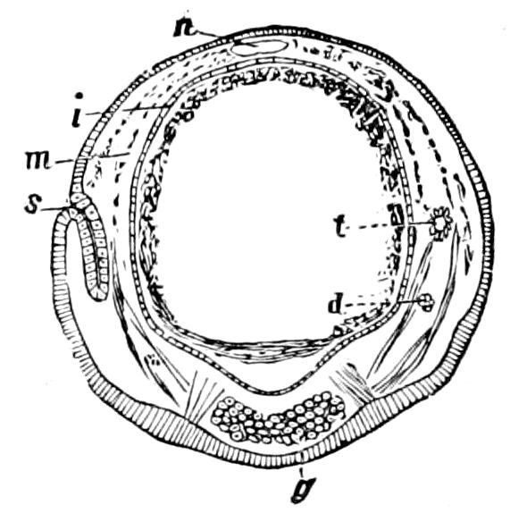
Fig. 390.—Section of Sphinx embryo, showing at s the ectoderm invaginated, and forming the germ of a stigma and trachea (t).—After Kowalevsky.
Warmth, plenty of food, besides muscular activity, increases the demand for oxygen and the quantity of carbonic acid exhaled.
a. The tracheæ
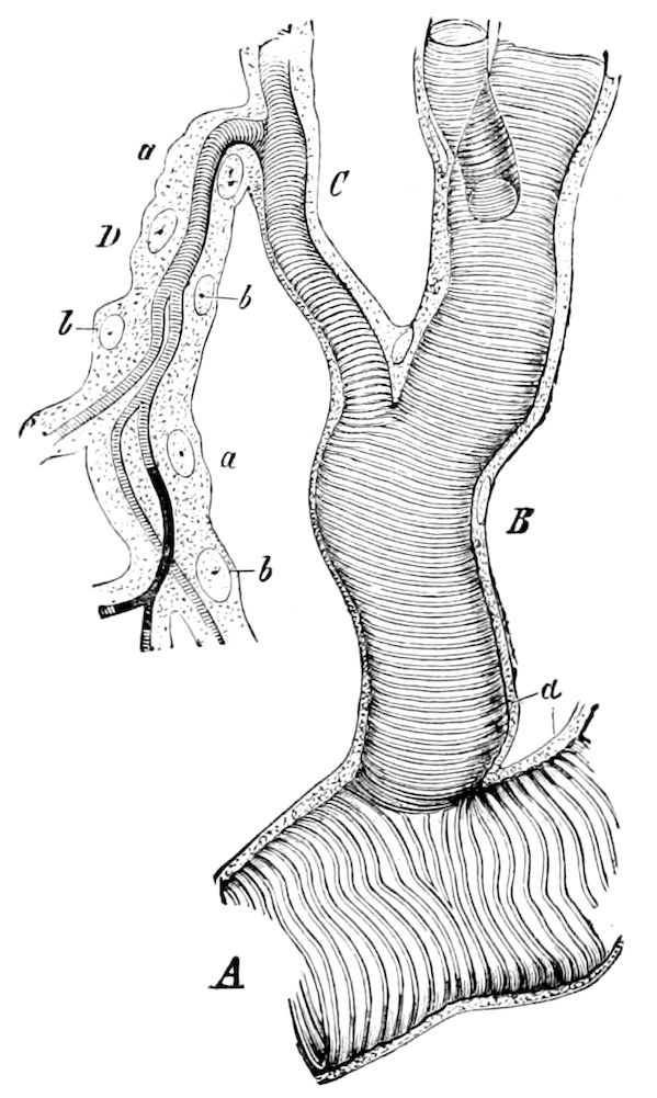
Fig. 391.—Portion of a trachea of a caterpillar, with its branches B, C, D: a, peritracheal membrane; b, nucleus.—After Leydig, from Gegenbaur.
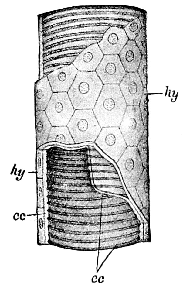
Fig. 392.—Structure of a trachea, diagrammatic: portions of the peritracheal membrane (hy) and chitinous intima (cc) removed to show the structure; in the chitinous intima or endotrachea (cc) can be seen the spiral thickenings or tænidia.—After Lang.
It will much simplify our conception of the nature of the air-tubes when we learn that they originate in the embryo as tubular ingrowths of the integument (ectoderm), these branching and finally reaching every part of the interior of the body. They are elastic tubes, and being filled with air are silvery in color, though at their origin near the spiracles they are reddish or violet bluish; or, in 432the larva of Æschna, reddish brown, this tint being due to a finely granular pigment situated in the peritoneal membrane.
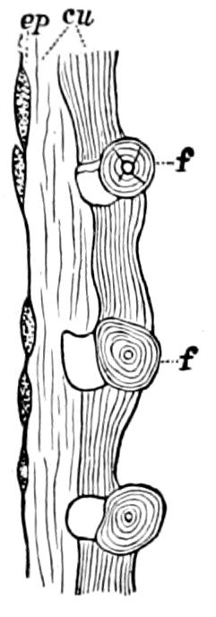
Fig. 393.—Longitudinal section of the trachea of Hydrophilus piceus: ep, epithelium; cu, cuticula; f, spiral threads.—After Minot.
In their essential structure the tracheæ consist of the chitinous intima, which is a continuation of the cuticle of the integument, and of a cellular membrane or outer layer of cells (a continuation of the hypodermis) called the peritoneal membrane, or ectotrachea (Figs. 392, 393).
Leydig discovered that the spiral filaments are not distinct and separate, but intimately connected with the inner membrane (intima), and he detected the outer or peritoneal membrane, which Chun afterwards found to be epithelial in its nature, Minot stating that it is a true pavement epithelium.
Figure 393 represents a longitudinal section of a large trachea of Hydrophilus, showing the peritoneal membrane (ectotrachea, ep) and the intima or endotrachea, divided into the cuticula (cu), with the darker colored inner layer, in which are embedded the dark-colored tænidia (f).
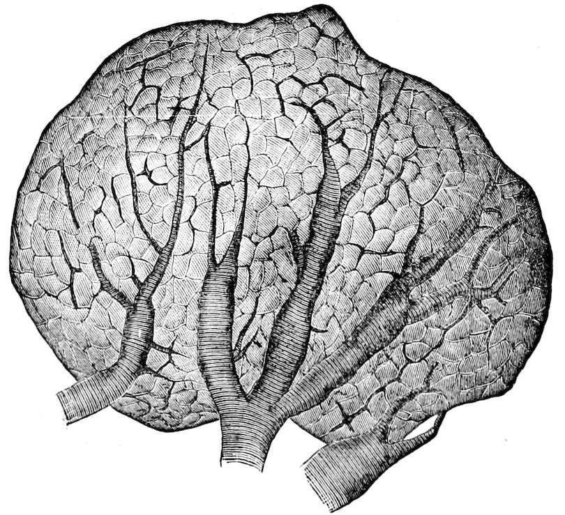
Fig. 394.—Testis of Anabrus, showing the ramifications of the tracheæ.—After Minot.
Distribution of the tracheæ.—The distribution of the air-tubes, as Lubbock and also Minot state, depends first upon the shape of the 433organs, and upon the size of those whose size is variable. Around the large, hollow organs (digestive canal, sexual organs) the tracheæ ramify in all directions, forking so that the branches diverge at a wide angle. In the organs which have muscular walls, like the oviduct, the tracheæ run straight when the walls are distended, but have a sinuous course when the walls are contracted. (Minot.)
“Around the organs of more elongated form the branches of the tracheæ run more longitudinally, as is shown by the air-tubes of the muscles, which also present some peculiarities worthy of especial notice.
“A short, thick trunk arrives at the muscular bundle, and dividing very rapidly, breaks up into a large number of delicate tubes, which penetrate between the muscular fibres, then terminating in tubes of exceeding fineness, which at first sight seem to form a network that might well be called a rete mirabile. A closer examination, however, reveals that it is not a real network, but rather an interlacing confusing to the eye. The longitudinal direction of the tracheæ of the muscles presents a striking contrast to the system of divarication represented in Figs. 13 and 14. The course of the tracheæ of the Malpighian tubes is also very curious. There is one large trachea which winds around the tube in a long spiral, giving off numerous small branches which run to the surface of the tube, upon which they form delicate ramifications. Each tube has but a single main trachea, and I think the trachea continues the whole length of the tube, but of this last point I am not quite sure.” (Minot.)
While in the nymphs of Orthoptera the tracheæ very closely resemble those of the adult, in larvæ of insects with a complete metamorphosis the tracheæ differ very much in distribution from those of the adult. The larval tracheæ are also more generalized and more like those of the original type than the tracheæ of perfect insects. (Lubbock.)
In general there are two main tracheæ, one passing along each side of the body, near the digestive canal, connected with its mate by a few transverse anastomosing branches, and sending off a branch to each spiracle, this arrangement being most simple and apparent in the maggots of Diptera. From these two main branches smaller twigs branch off into every part of the body with its appendages, passing among the different organs, often serving as cables to hold them loosely in place; they also penetrate into the component parts of the organ themselves, passing into the fat-bodies, and among the fibres of muscles, where they become finely attenuated and refined like the capillaries of the vascular system of vertebrates. (Figs. 395, 396.)
435
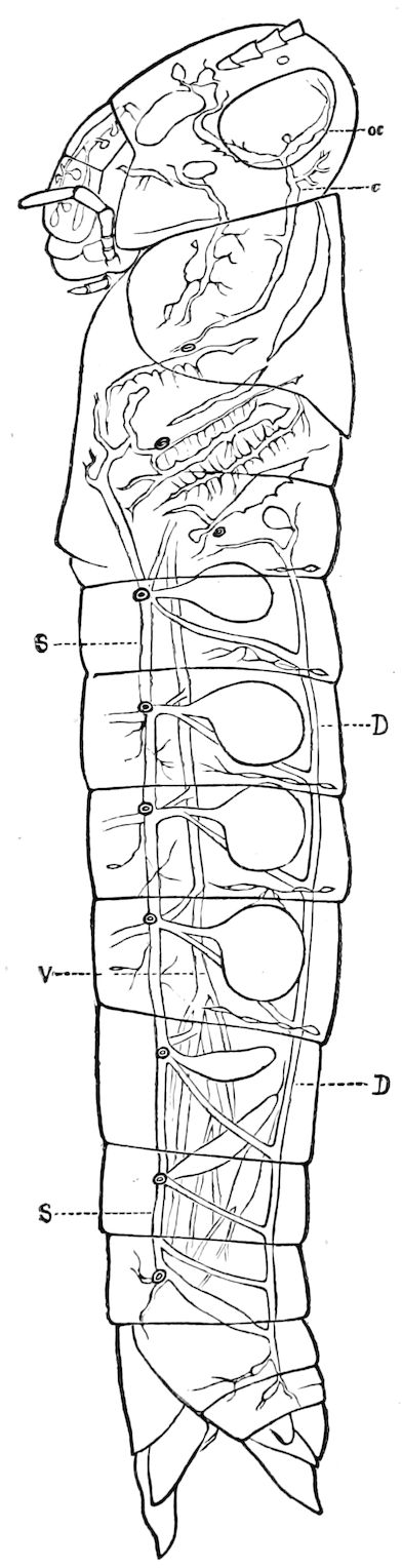
Fig. 395.—Melanoplus femur-rubrum, showing distribution of air-tubes (tracheæ) and air-sacs; V, main ventral trachea (only one of the two shown); S, left stigmatal trachea, connecting by vertical branches with D, the left main dorsal trachea; c, left cephalic trachea; oc, ocular dilated trachea. From the first, second, third, and fourth spiracles arise the first four abdominal air-sacs, which are succeeded by the plexus of three pairs of dilated tracheæ, I, II, III, in Fig. 396. Numerous air-sacs and tracheæ are represented in the head and thorax. The two thoracic spiracles are represented, but not lettered.
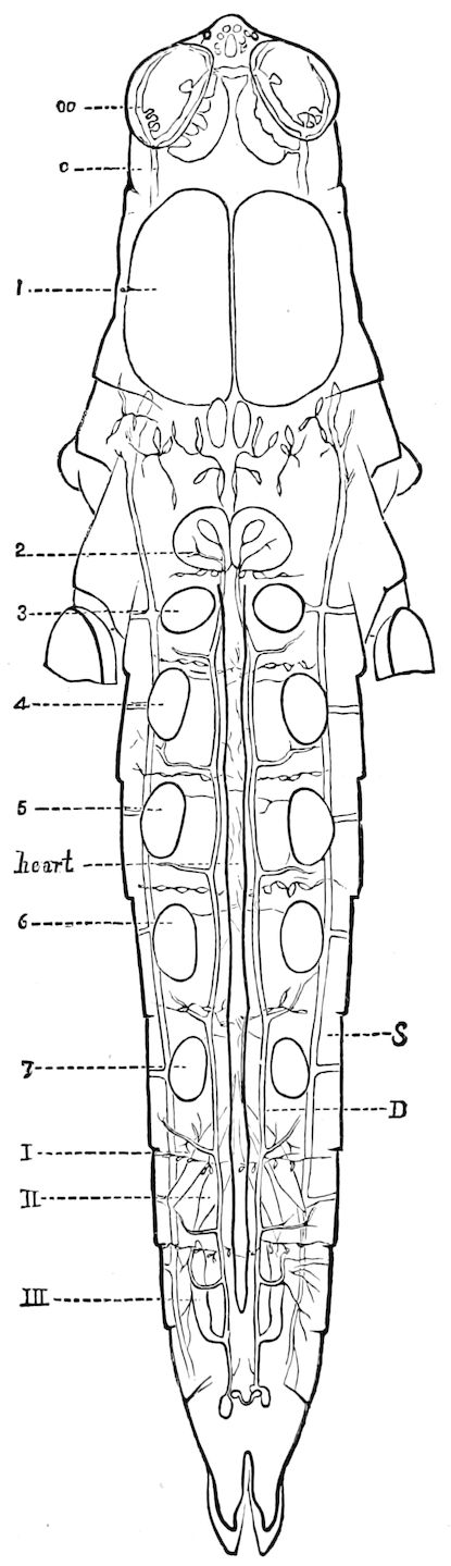
Fig. 396.—D, left dorsal trachea; S, left stigmatal trachea; I, II, III, first, second, and third pairs of abdominal dilated tracheæ, forming a plexus behind the ovaries; 1, pair of enormous thoracic air-sacs; 2, pair of smaller air-sacs; 3–7, abdominal air-sacs; oc, ocular dilated trachea and air-sacs; c, cephalic trachea. The relations of the heart to the dorsal tracheæ are indicated.—Drawn by Emerton from dissections by the author.
In the youngest larva of Corethra plumicornis Weismann ascertained the thickness of the longitudinal stem to be 0.0017 mm. That of the finest tracheal endings in the silk-glands of the silkworm was found by Von Wistinghausen to be 0.0016 mm. (Zeits. f. Wiss. Zool. xlix, 1890, p. 575.) Weismann states that in the larvæ of Corethra and Chironomus the tracheal system is only incompletely developed; the tracheæ are not united with each other, and in the youngest larvæ they do not contain air.
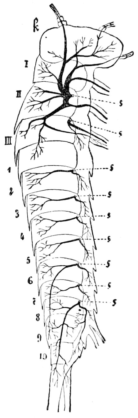
Fig. 397.—Tracheal system of the right side of Machilis maritima: k, head; I, II, III, thoracic segments; 1–10, abdominal segments; s, stigma.—After Oudemans, from Lang.
Each of the two main tracheæ, as Kolbe states, sends off into each segment of the body three branches.
1. An upper or dorsal branch, which supplies the muscles of the dorsal region.
2. A middle (visceral) branch, whose twigs pass to the digestive canal and back to the organs of reproduction.
3. A lower (ventral) branch, whose twigs are distributed to the ganglia and to the muscles of the ventral region.
In certain Thysanura, as a species of Machilis (Fig. 397), we probably have the primitive condition of the tracheal system, the longitudinal and transverse anastomoses being absent, but in other Thysanura (Japyx, Nicoletia, Lepisma, and a few species of Machilis) they are present.
As Kolbe remarks, whether the fine ends of the tracheæ are closed or open, whether after the analogy of the blood capillaries of vertebrates they anastomose with each other, whether the ends of the air-tubes pass between the cells or penetrate into them, these questions are not fully settled. According to Leydig’s[61] latest views the tracheæ penetrate into the cells and unite with the hyaloplasma. Hence the process of respiration in the last instance takes place in the hyaloplasma. This assumption accords with the fact that in the tracheate Arthropods the terminations of the tracheæ carry the atmospheric air into the space bounded by the cellular network, also to the hyaloplasma filling the spaces. Leydig[62] also thinks that the finest tracheal endings penetrate into the muscular tissue and unite with the primitive muscular fibres.
Kupffer is likewise of the opinion that the fine tracheæ penetrate into the cells, and Lidth de Jeude asserts that they enter the epithelial cells, “each cell containing several branches.” Kölliker, Emery, etc., maintain, however, 436that the tracheal endings lie between the cells. Wielowiejski,[63] in describing the line tracheæ of the phosphorescent organs, thinks that the tracheal endings (tracheal capillaries) rarely end blindly, but anastomose with one another, forming an irregular network. The latest observer, Gilson (1893), asserts that tracheal twigs penetrate deeply into the epithelial cells of the silk glands of larval Trichoptera as well as of caterpillars, passing through their protoplasm.