PART I.—MORPHOLOGY AND PHYSIOLOGY (continued 7)
Reading edition
The allurement of the substance acts for a longer time on those deprived of their antennæ, then they become restless, then they wander away from the glass tube held before them; still all their movements are but slightly energetic, and the entire reaction is indeterminate and enfeebled.
Experiments with the Hemiptera gave still more unfavorable results; after the loss of their antennæ they reacted to smells as eagerly as those did which were uninjured.
Experiments on the use of the antennæ in seeking for food.—Under this head experiments were made with Silpha, Sarcophaga, Calliphora, and Cynomyia.
Silpha and its larva were treated in the following manner: they were placed in large boxes whose bottoms were covered with moss, etc.; in a corner of the box was placed a bottle with a small opening, in which was placed strong-smelling meat. So long as the beetles were in possession of their antennæ they invariably after a while discovered the meat exposed in the bottle, while after the loss of their antennæ they did not come in contact with it.
In a similar way acted the species of Sarcophaga, Calliphora, and Cynomyia. Hauser, in experimenting with these, placed a dish with a large piece of decayed flesh on his writing-table. In a short time specimens of the flies referred to entered through the open window of the room. The oftener he drove them away from the meat would they swarm thickly upon it. Then closing the window and catching all the flies, he deprived them of their antennæ and again set them free. They flew about the room, but none settled upon the flesh nor tried to approach it. Where a fly had alighted on a curtain or other object, the decayed flesh was placed under it so that the full force of the effluvium should pass over it, but even then no fly would settle upon it.
Experiments testing the influence of the antennæ of the males in seeking the females.—For this purpose Hauser chose those kinds in which the male antennæ differ in secondary sexual characters from those of the female, and in which it is known that they readily couple in confinement, as Saturnia pavonia, Ocneria dispar, and Melolontha vulgaris. The two first-named insects did not couple after the extirpation of their antennæ. Of Melolontha vulgaris 271twenty pairs were placed in a moderately sized box. On the next morning twelve pairs of them were found coupling. Hauser then, after removing the first lot, placed a new set of thirty pairs in the same box, cut off all the antennæ of the males and those of a number of females. On the following morning only four pairs were found coupling, and at the end of three days five others were observed sexually united.
From these experiments Hauser inferred that those insects deprived of their antennæ were placed in the most favorable situation, such as they would not find in freedom; for the space in which the insects moved about was so limited that the males and females must of necessity meet. But at the same time the results of the experiments cannot absolutely be regarded as proving that the males, after the loss of their antennæ, were then not in condition to find the females, because in the case of the above-mentioned moths, under similar conditions, after the extirpation of the antennæ no sexual union took place. If, however, the experiments made do not all lead to the results desired, Hauser thinks that the results agree with those of his histological researches, that in the greater number of insects the sense of smell has its seat in the antennæ. His results also agree with those of Perris.
Structure of the organs of smell in insects.—The olfactory organs consist, in insects,—i.e., all Orthoptera, Termitidæ, Psocidæ, Diptera, and Hymenoptera, also in most Lepidoptera, Neuroptera, and Coleoptera,—
1. Of a thick nerve arising from the brain, which passes into the antennæ.
2. Of a sensitive apparatus at the end, which consists of staff-like cells, which are modified hypodermis cells, with which the fibres of the nerves connect.
3. Of a supporting and accessory apparatus, consisting of pits, or peg- or tooth-like projections filled with a serous fluid, and which may be regarded as invaginations and outgrowths of the epidermis.
Hauser adds a remark on the distribution of the pits and teeth in the larvæ of insects, saying that his observations are incomplete, but that it appears that in the larvæ the teeth are most generally distributed, and that they occur not on the antennæ alone, but on the palpi; but in very many larvæ neither pits nor teeth[47] occurred. In the Myriopoda teeth-like projections occur on the ends of the antennæ. In Lithobius they form very small, almost cylindrical, pale organs.
272
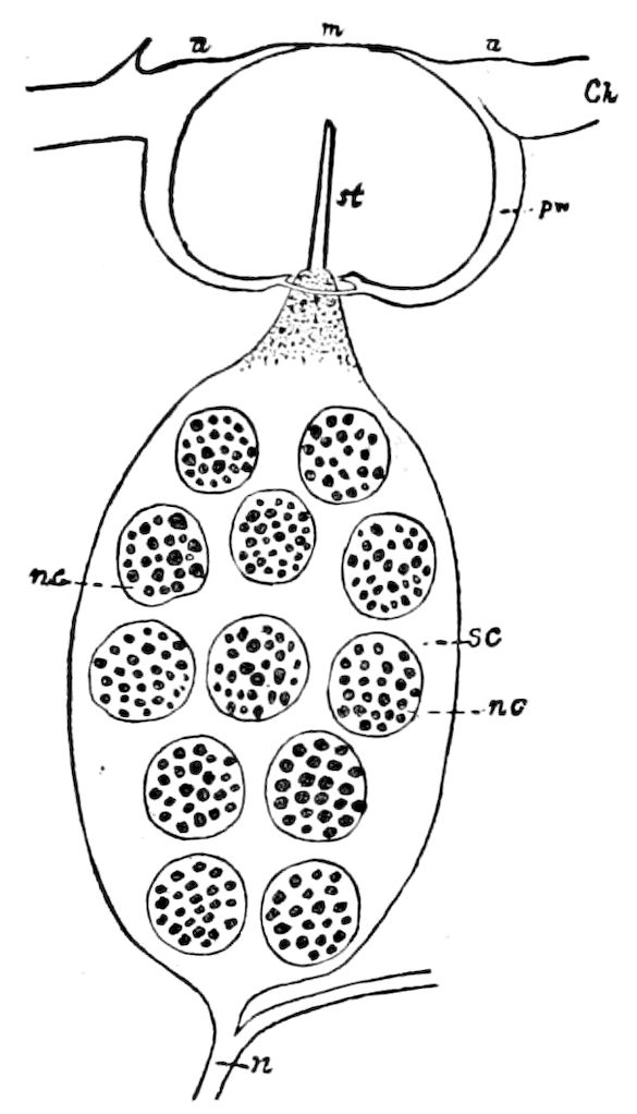
Fig. 268.—Olfactory organ of Caloptenus.
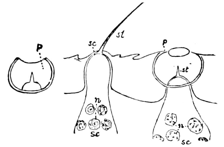
Fig. 269.—Olfactory pits of the antenna of Stenobothrus. This and Fig. 268 after Hauser.
Lettering for Figs. 268, 269, 273, 275, 276, 278–281.—a, a, circular thickening of the skin surrounding the opening of the olfactory pit; ax, thread-like continuation of the nerve-cell; b, vesicle-like bottom of the olfactory pit, through which the olfactory style passes; br bristle in Fig. 283, stout, and protecting the olfactory pit; bs, bent bristle or seta; ch, chitinous integument of the antennæ; d, seen in section; f, invaginated pit; Fv, Forel’s flask-shaped organ; Fvo, its opening seen from the surface; gl, gland-like mass of cells; hyc, hypodermic cells; i, entrance into the canal belonging to the pit; m, olfactory membrane; m′, m″, mc, membrane-forming cell; n, nerve of special sense; nc, nucleus of the sense- or ganglion-cell; o, opening into the olfactory pit; p, olfactory pit; cp, compound pits; pw, wall of the pit; s, a large seta; sc, sense- or ganglion-cell; st, olfactory or sense-style, sometimes peg-shaped; tb, tactile bristle.
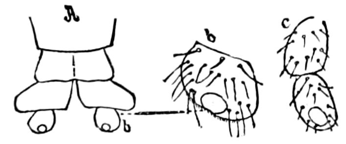
Fig. 270.—A, b, sense-organ on the abdominal appendages of a fly (Chrysopila); c, sense-organ on the terminal joint of palpus of Perla.
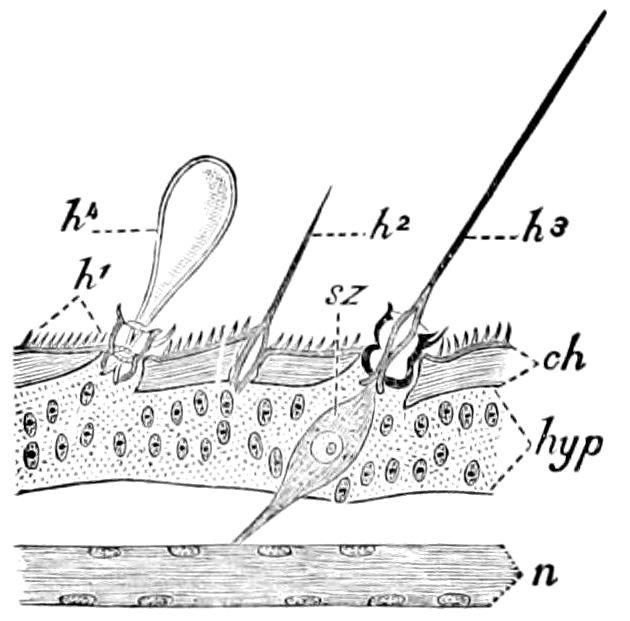
Fig. 271.—Longitudinal section of part of cercus of Acheta domestica: ch, cuticula; hyp, hypodermis; n. nerve; h′1, integumental hairs, not sensory; h2, ordinary hair; h3, sensory hair; h4, bladder-like hair; sz, sense-cell.—After Vom Rath, from Sharp.
In the course of a special description of these sense-organs in the Orthoptera, Hauser describes at length those of Œdipoda cœrulescens and Caloptenus italicus. On one antennal joint of Caloptenus (Fig. 268) was often counted 50 pits; on the anterior joints the number diminishes to about 30. Hauser thinks that in all Orthoptera whose antennæ are like those of Caloptenus occur similar pits, as he found them in Stenobothrus (Fig. 269) as well as in Œdipoda. Gryllotalpa possesses similar pits,—four to six on each antennal joint, making between 300 and 400 pits on each antenna.[48] In Mantis 273religiosa the pits were not detected, but on each joint, except the eighth basal, there are about 200 small, hollow, curved teeth with a fine opening in front.
In the Neuroptera (Chrysopa) there occur on the antennæ, besides numerous very long tactile bristles, small pale, transparent teeth. No pits could be detected.
In the Hemiptera (two species of Pyrrhocoris only were examined) only two kinds of tactile bristles occurred, but Hauser detected no pits, though Lespès states that they are present.
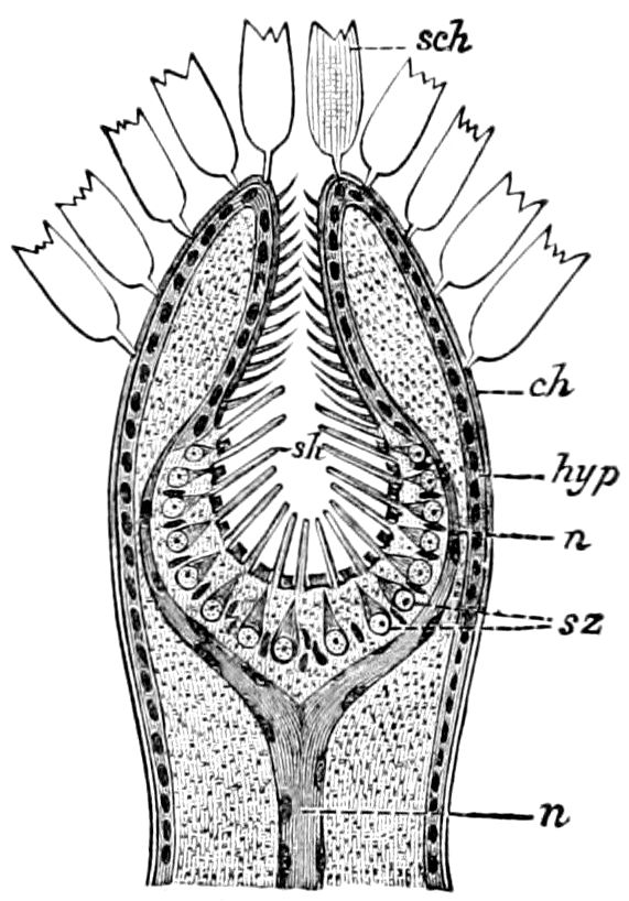
Fig. 272.—Longitudinal section of apex of palpus of Pieris brassicæ: sch, scales; ch, cuticula; hyp, hypodermis; n, nerve; sz, sense-cells; sh, sense-hairs.—After Vom Rath, from Sharp.
Of the Diptera, Hauser examined more than 60 species. The pits in the Diptera brachycera (Muscidæ, etc.) are unexceptionally confined to the third antennal joint. Their number varies extraordinarily in the different species. Helophilus florens has on each antennal disk only a single pit, while Echinomyia grossa possesses 200 of them. In flies of certain families the pits are compound, and contain 10, 20, and often 100 olfactory hairs, partly arising from the coalescence of several pits. Such pits are usually divided by lateral walls into several chambers, whose connection is only indicated by their common outlet. Simple olfactory pits with a single olfactory style were observed only in the Tabanidæ, Asilidæ, Bombylidæ, Leptidæ, Dolichopidæ, Stratiomyidæ, and Tipulidæ. In the last the compound forms do not occur at all, but in the other families mentioned also occur compound pits, receiving from two to ten nerve-terminations.
The antennal pits of flies are always sac-like invaginations of the external chitinous integument, of manifold shapes, opening externally and never closed by a membrane. The pits differ but slightly in the different species, and that of Cyrtoneura stabulans (Fig. 273) is described at length as typical of those of brachycerous flies in general.
The olfactory pits of the Tipulidæ seem to have a somewhat different structure, since the external passage is closed. It is circular, surrounded with a slight chitinous wall, and not covered with bristles. Such pits in their external appearance are like 274those of the locust (Caloptenus) and many Hymenoptera. They are situated usually on the third antennal joint. Pachyrhina pratensis L. has about 60 of them, as have Tipula oleracea L. and Ctenophora.
In the Lepidoptera, olfactory pits are much like those of flies. Hauser describes in detail those of Vanessa io. Those of the moths were not examined, but they can be readily and satisfactorily proved to be the site of the olfactory sense.
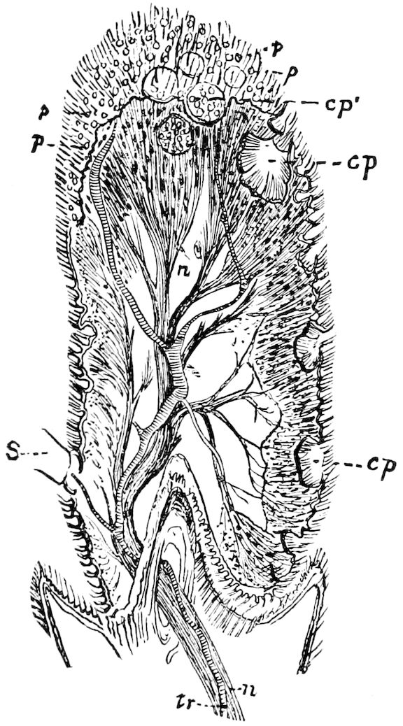
Fig. 273.—Longitudinal section through the third antennal joint of a fly (Cyrtoneura stabulans), showing the compound pits from above and in section.—After Hauser.
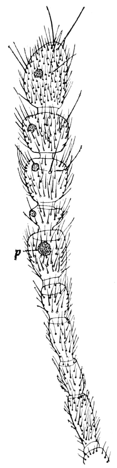
Fig. 274.—Antenna of Adelops, showing the olfactory organs (p) in the five last joints.
Historical researches in respect to the Coleoptera generally gave a very unfavorable result, contrary to Lespès’s views. That author states that in the Carabidæ the pits are found on the four first joints, but Hauser could discover them in none which he examined. Usually only tactile bristles occur, so also in the Cerambycidæ, Curculionidæ, Chrysomelidæ, and Cantharidæ. In a blind silphid beetle 275(Adelops hirtus) of Mammoth Cave we have found well-marked olfactory organs (Fig. 274). Similar organs occur in the antennæ of the Panorpidæ.
Olfactory pits, however, without doubt occur in Silpha, Necrophorus, Staphylinus, Philonthus, and Tenebrio. The openings of the pits are small and surrounded with a small chitinous ring; in Silpha, Necrophorus, and Tenebrio they cannot easily be distinguished from the insertion-cavities of the bristles, but in Philonthus and Staphylinus they are less like them, being distinguished by their somewhat larger size and their often more oval form. In Philonthus æneus about 100 such small pits occur irregularly on the terminal joints; besides, in this species on each side of the terminal joint is an apparatus which is like the compound pit generally occurring in the Diptera.
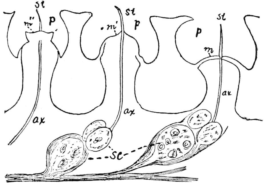
Fig. 275.—Olfactory pits of the antenna of Melolontha vulgaris.—After Kraepelin.
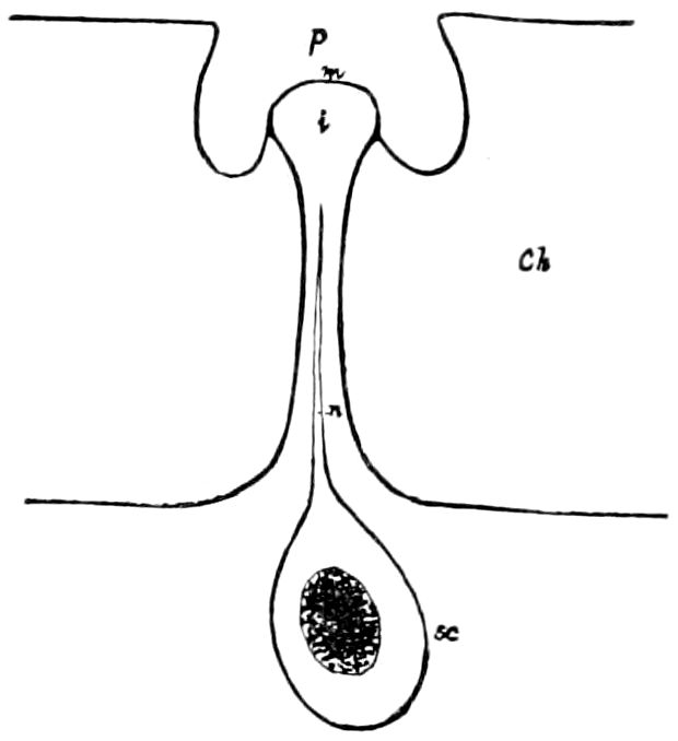
Fig. 276.—Antennal pit of Melolontha vulgaris, seen in vertical section.—After Hauser.
Very remarkable pits occur in the antennal lamellæ of Melolontha vulgaris (Fig. 275) and other lamellicorns. On the outer surface of the first and seventh (in the female the sixth) antennal leaf, as also on the edges of the other leaves, only arise scattered bristles; on the inner surface of the first and seventh leaves, as also on both surfaces of the second to sixth leaves, are close rows of rather shallow depressions of irregular form, some circular, others regularly hexagonal. Their number is enormous: in the males 39,000, in the females about 35,000, occur on each antenna.
276
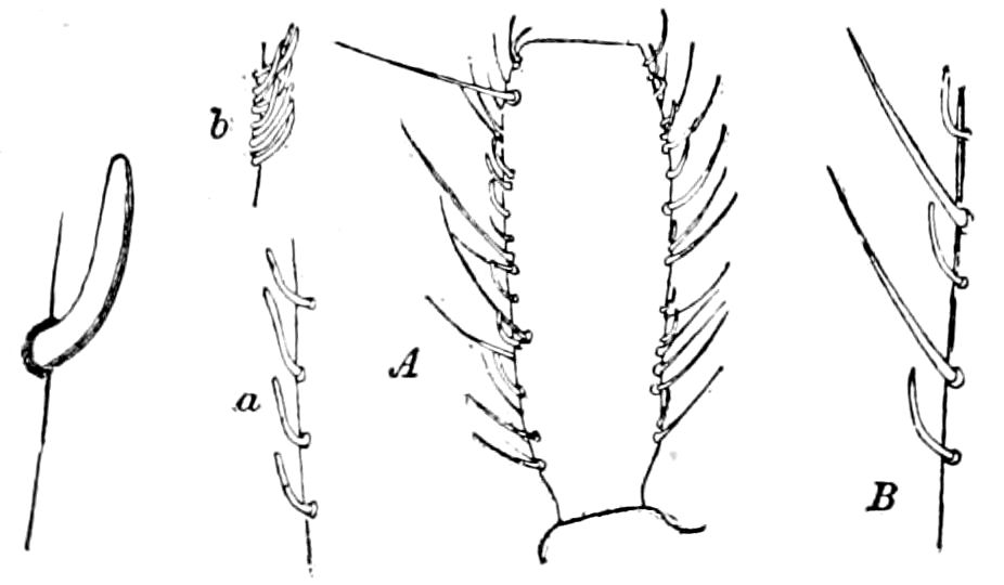
Fig. 277.—Organ of smell of Anophthalmus.—After Hauser. A, a, b, the same in A. tenuis, B in A. tellkampfii.
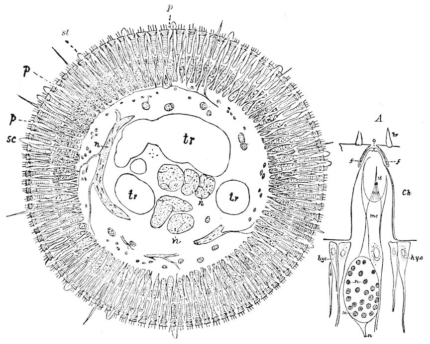
Fig. 278.—Section through antennal joint of Vespa crabro, showing the great number of olfactory pits, olfactory and tactile bristles. A, section through an olfactory pit of Vespa crabro.—After Hauser.
The antennal pits and teeth of Dyticus marginalis are morphologically and physiologically identical with those of bees and wasps. In Anophthalmus bilimekii, Hauser found on the last antennal joints about 60 teeth, which essentially differ in form from those previously described; they are very pale, transparent, cylindrical, elongated, and bent elbow-shaped on the first third, so that the last two-thirds run parallel with the antenna. The length of these remarkable teeth is 0.035 mm., their breadth 0.005 mm. He only found them in Anophthalmus, and in no other species of Carabidæ; they must resemble the teeth described in Chrysopa. Our species possesses similar processes (Fig. 277). Similar teeth occur on the maxillary and labial palpi of beetles. Dyticus marginalis possesses at the end of each terminal palpal joint a group of very small teeth, which were also detected in Anophthalmus bilimekii, Melolontha vulgaris, etc. In 277Carabus violascens were detected on the maxillary palpi large, plainly microscopical, white disks, which are surrounded with a great number of extremely small teeth.
Whether the above-described organs on the palpi of beetles should be considered as olfactory or gustatory in their nature can only be determined by means of physiological experiments; they probably receive taste-nerve terminations.
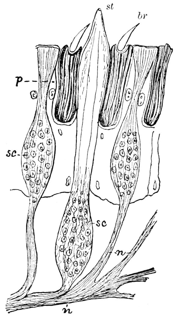
Fig. 279.—Olfactory pits of the antenna of Vespa vulgaris.—After Kraepelin.
The Hymenoptera furnished very good material for histological purposes, so that Hauser could not only study the terminal apparatus of the olfactory nerves in the perfect insect, but also in three different stages of the pupa. These are described at length, as regards the distribution of the pits and teeth, in Vespa crabro; each joint of the antenna (flagellum) possesses between 1300 and 1400 pits, nearly 60 teeth, and about 70 tactile hairs; on the terminal joint there are more than 200 teeth, so that each antenna has between 13,000 and 14,000 olfactory pits and about 700 teeth (Kegeln). Fig. 278 represents a cross-section through the penultimate antennal joint of Vespa crabro; we can see how thick are the series of openings on the surface of the antennæ, and how regular is the distribution of the teeth.
The distribution of the olfactory pits and olfactory teeth is thus seen to be very general; the deviations are so insignificant that there is no reason for the establishment of more than one type.
Antennal pits with a small crevice-like opening occur in genera nearly allied to Vespa and also in most Ichneumonidæ, Braconidæ, and Cynipidæ. But the crevice-like openings in these families are considerably longer and often are of a somewhat twisted shape. In all the species with translucent antennæ we can recognize the inner mouth of the pit as a round or nearly round disk situated usually under the middle of the opening. The antennal pits of Apis mellifica, as well as those of Bombus (Fig. 280) and allied genera, differ 278from those of the Ichneumonidæ in being not like crevices, but circular openings.
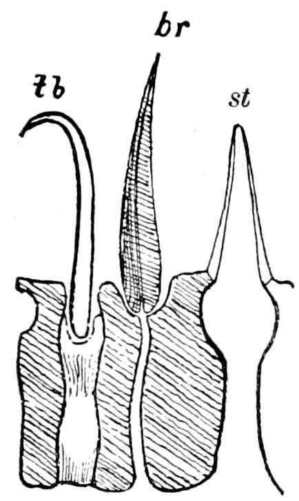
Fig. 280.—Olfactory pits of the antenna of Bombus.—After Kraepelin.
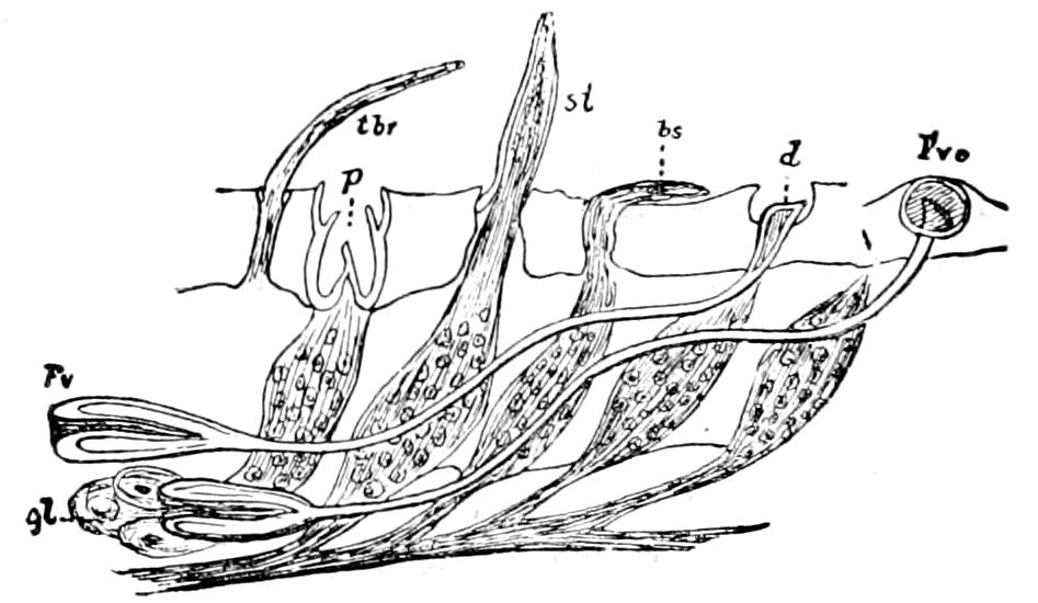
Fig. 281.—Olfactory pits of the antenna of Formica: Fv, Hicks’ “bottle,” Forel’s flask-shaped organ, Fvo, its opening.—After Kraepelin.
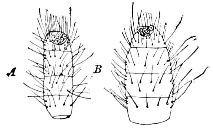
Fig. 282.—Supposed olfactory organs at end of antenna of Campodea: A, C. staphylinus. B, C. cookei, from Mammoth Cave.
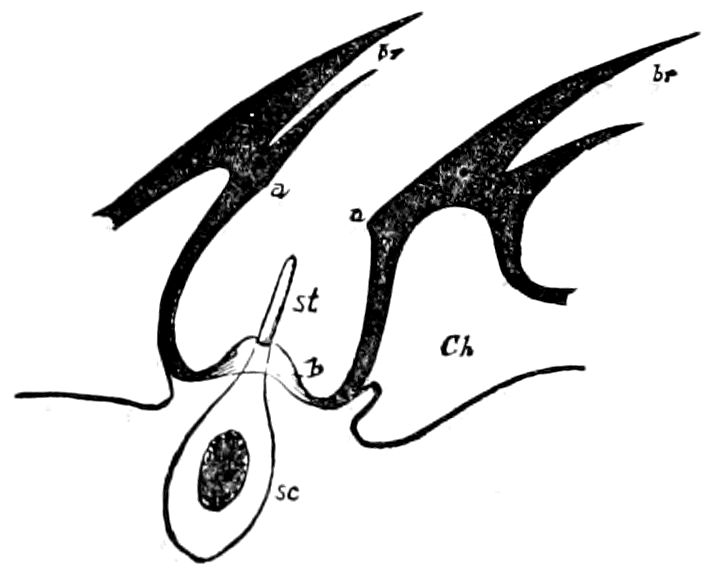
Fig. 283.—Vertical section through a single olfactory pit in the antenna of the horse-fly (Tabanus bovinus). For lettering see p. 272.—After Hauser.
The distribution of the olfactory peg or tooth-like projections seems to be much more limited than that of the pits in the Ichneumonidæ. Hauser could not find any. Apis mellifica possesses on each antennal joint only about twenty slender pale teeth, scarcely a third as many as in Vespa crabro; on the other hand, Formica, of which genus several species were examined, seems to have far more teeth than pits; they are relatively long, pale, transparent, and somewhat clavate; they are not unlike those of Chrysopa; on the terminal joint only occur the round openings (Fvo), which lead into a bottle-shaped invagination of the integument (Fv) and contain an olfactory style (Fig. 281). In the Tenthredinidæ only teeth and no pits were to be detected. Sirex has on the under side of the nine last joints of each antenna a group of from 200 to 300 small teeth, which resemble those of Vespa crabro; Lyda has on the terminal joints about 100 279teeth. We may add that supposed organs of smell occur on the antennæ of Campodea (Fig. 282).
Kraepelin also thus briefly summarizes Hauser’s statements as to the forms of the different organs of smell.
The manifold nature of the antennal organs has, by Hauser, from thorough studies of the nerve-elements belonging to them, been not simplified but rendered more complicated. According to this naturalist we may distinguish the following forms which the olfactory organs may assume: 1. “Pale, tooth-like chitinous hairs on the outer surface of the antennæ, which are perforated at the end; nothing is known as to the relation of the nerve passing into it (Chrysopa, Anophthalmus). 2. In pit-like depressions of the antennæ arise nerve-rods (without a chitinous case) which stand in direct relation with a ganglion-cell lying under it. These pits are either simple, viz. with only an ‘olfactory rod’ (Tabanus, Fig. 283, and other Diptera, Vanessa), or compound (Muscidæ, and most other Diptera, and Philonthus). It is important that these pits are partly open (in the above-named groups of insects), and partly closed and covered with a thin membrane, under whose concavity the olfactory rods end (Orthoptera, Melolontha, and other lamellicorns). 3. Short, thick pits sunken slightly into the surface of the antennæ, and over this a chitinous peg perforated at the end, in whose base, from the interior, projects a very singular nerve-peg, which is situated over an olfactory ganglion-cell, and provided with a slender crown of little rods, and flanked on each side by a flagellum-cell (Hymenoptera). 4. Round or crevice-like pits covered over by a perforated chitinous membrane with nerve-rods like those in 3, but in place of the flagellum-cell with ‘membrane-forming’ cells spread before it. Hauser finally mentions further differences in the ganglion-cells sent out into the nerve-end apparatus. These exhibit in Diptera and Melolontha only one nucleus, in Hymenoptera a single very large one (with many nucleoli) and three small ones, in Vanessa six, in Orthoptera a very large number of nuclei, etc.”
LITERATURE OF THE ORGANS OF SMELL
Réaumur, Réné Ant. de. Mémoires pour servir à l’histoire des insectes. Paris, 1734–42. (i, 283; ii, 224).
Lesser, F. C. Insecto-theologia, 1740, p. 262.
Roesel, A. J. Insektenbelustigungen, 1746, ii, p. 51.
Reimarus, H. S. Allgemeine Betrachtung ueber die Triebe der Thiere hauptsächlich ihre Kunsttriebe (Instinct). Hamburg, 1760, p. 355.
Sulzer, J. H. Die Kenntzeichen der Insecten. Zürich, 1761.
Lyonet, P. Traité anatomique de la chenille, 1762, pp. 42, 96, 195.
Comparetti, A. De aure interna comparata. Patavii, 1769.
Bonnet, C. Œuvres complètes, 1779–1783, ii, p. 36.
—— Contemplation de la nature, Ch. iii, p. 18.
Scarpa, Ant. Anatomicæ disquisitiones de auditu et olfactu. Ticini, 1789.
Huber, F. Nouvelles observations sur les abeilles, 1792, ii, p. 475.
Lehrmann, M. C. G. De antennis insectorum. Londini, Hamburgi, 1799, p. 48, Diss. posterior; Hamburg and London, 1800, p. 80 (especial sense, ærocepsis).
Latreille, P. A. Histoire naturelle des crustacés et des insectes, 1806–1809, ii, 50.
Blainville, M. H. D. Principes d’anatomie comparée, 1822, i, p. 339.
Dugès, A. L. Traité de physiologie comparée, 1838, i, p. 161.
280Newport, G. On the use of the antennæ of insects. (Trans. Ent. Soc., London, ii, 1840, pp. 229–248.)
Robineau-Desvoidy, A. J. B. Sur l’usage réel des antennes chez les insectes. (Ann. Soc. Ent. France, 1842, xi Bull., pp. 23–27.)
Erichson, W. F. De fabrica et usu antennarum in insectis. Berlin, 1847, 1 Tab., p. 13.
Perris, E. Mémoire sur le siège de l’odorat dans les articulés. (Ann. sc. nat., Sér. 3, 1850, xiv, pp. 159–178.)
Dufour, L. Quelques mots sur l’organe de l’odorat et sur celui de l’ouie dans les insectes. (Actes d. l. Soc. Linn., Bordeaux, 1850, xvii, Ann. sc. nat., Sér. 3, Zool., xiv, 1850, pp. 179–184.)
Leydig, F. Zum feineren Bau der Arthropoden. (Müller’s Archiv, 1855, pp. 376–480); Lehrbuch der Histologie, 1857, p. 220; Zur Anatomie der Insekten (Archiv für Anatomie, 1859, pp. 35–89 and 149–183).
—— Ueber Geruchs- und Gehörorgane der Krebse und Insekten. (Archiv f. Anat. u. Phys., 1860.)
—— Die Hautsinnesorgane der Arthropoden. (Zool. Anzeiger, 1886, pp. 284–291, 308–314, 265–314.)
Landois, H. Das Gehörorgan des Hirschkäfers. (Archiv f. mikrosp. Anat., 1868, iv, pp. 88–95.)
Troschel, H. Ueber das Geruchsorgan der Gliedertiere. (Verhandl. d. naturhist. Vereins d. preuss. Rheinlande u. Westfal., xxvii Jahrg., 1870, pp. 160–161.)
Packard, A. S. The caudal styles of insects sense-organs, i.e., abdominal antennæ. (Amer. Naturalist, 1870, pp. 620, 621. Also Proc. Bost. Soc. Nat. Hist., 1868, xi, p. 398.)
Paasch, A. Von den Sinnesorganen der Insekten im Allgemeinen, von Gehörund Geruchsorganen im Besondern. (Archiv für Naturgesch., xxxix Jahrg., i, 1873, pp. 248–275.)
Chadima, Jos. Ueber die von Leydig als Geruchsorgane bezeichneten Bildungen bei den Arthropoden. (Mitteil. d. naturwiss. Ver. f. Steiermark, 1873, pp. 36–44.)
Forel, A. Les Fourmis de la Suisse. (Neue Denkschr. Allg. Schweiz. Gesellsch. f. d. ges. Naturw., xxvi, 1874, pp. 118, 144.)
—— Études myrmécologiques en 1884, avec une description des organes sensoriels des antennes. (Bull. Soc. Vaud. sc. nat., 1885, Sér. 2, xx, pp. 316–380.)
Graber, V. Die Insekten. München, 1877.
—— Neue Versuche über die Functionen der Insektenfühler. (Biol. Centralb., vii, 1887, pp. 13–19.)
Trouvelot, L. The use of the antennæ in insects. (Amer. Naturalist, xi, 1877, pp. 193–196.)
Mayer, P. Sopra certi organi di senso nelle antenne dei Ditteri. (Atti R. Accad. d. Lincei, Roma, Ser. 3, Mem. Cl. sc. fis., mat. e natur., iii, 1879, p. 11.)
Hauser, Gustav. Physiologische und histiologische Untersuchungen über das Geruchsorgan der Insekten. (Zeitschr. f. wissens. Zool., xxxiv, 1880, pp. 367–403, 3 Taf.)
Kraepelin, Karl. Ueber die Geruchsorgane der Gliedertiere (Oster-Programm der Realschule des Johanneums. Hamburg, 1883, pp. 48, 3 Taf.)
Schiemenz, Paulus. Ueber das Herkommen des Futtersaftes und die Speicheldrüsen der Bienen nebst einem Anhange über das Riechorgan. (Zeitschr. f. wissens. Zool., xxxviii, pp. 71–135, 3 Taf.)
Plateau, F. Expériences sur le rôle des palpes chez les arthropodes maxillés, I., Palpes des insectes broyeurs. (Bull. Soc. Zool. France, x, 1885, pp. 67–90.)
281Plateau, F. Une expérience sur la fonction des antennes chez la Blatte (Periplaneta orientalis). (Comptes rend. Soc. Ent. de Belgique, 1886, pp. 118–122, 1 Fig.)
Lubbock, J. On some points in the anatomy of ants. (Monthly Micr. Journ., 1887, pp. 121–142.)
Ruland, Franz. Beiträge zur Kenntniss der antennalen Sinnesorgane der Insekten. Diss. Marburg, 1888, p. 31, 1 Taf.
Wasmann, E. Die Fühler der Insekten. (Stimmen aus Maria-Laach. Freiburg i. B., 1891, p. 37.)
Sergi, G. Ricerche su alcuni organi di senso nelle antenne delle formiche. (Riv. Filos. Sci. Milano, 1891, p. 10, 3 Figs.)
Nagel, Wilibald. Die niederen Sinne der Insekten, 19 Figs., Tübingen, 1892, pp. 67.
With the writings of Baster, Lamarck, Cuvier, Treviranus, Oken, Lefebure, Duméril, Schelver, Bousdorf, Rosenthal, Burmeister, Slater, Balbiani, Marcel de Serres, Garnier, Berté, Porter, Sazepin, Reuter, Pierret, Duponchel, Driesch, Küster, Peckham, Lubbock, A. Dohrn, Lespès.
c. The organs of taste
The gustatory organs of insects are microscopic pits or setae, either hair-like or resembling short pegs, which form the ends of ganglionated nerves. They are difficult to distinguish morphologically from certain olfactory structures, and it is owing to their position at or very near the mouth that they are supposed to be gustatory in nature.
Meinert was the first (1860) to suggest that organs of taste occurred in ants. He observed in the maxillæ, and tongue of these insects a series of canals in the cuticula of these organs connected with ganglion-cells, and through them with the nerves, and queried whether they were not organs of taste. Forel afterwards (1874) confirmed these observations. Wolff in an elaborate work (1875) described a group of minute pits (Fig. 284) at the base of the tongue of the honey bee, which he supposed to possess the sense of smell, but Forel and also Lubbock attributed to these sensory pits the function of taste. Ten years afterward Will showed conclusively, both by anatomical studies and by experiments, that Diptera and Hymenoptera possess gustatory organs. He, however, denied that the organs of Wolff were gustatory, and maintained that organs of smell were confined to the maxillæ, paraglossæ, and tongue. As we shall see, however, what appear to be with little doubt taste-pits, with hairs or pegs arising from them, are most numerous on the epipharynx of nearly all insects, and situated at a point where they 282necessarily must come in contact with the food as it enters the mouth and passes down the throat.
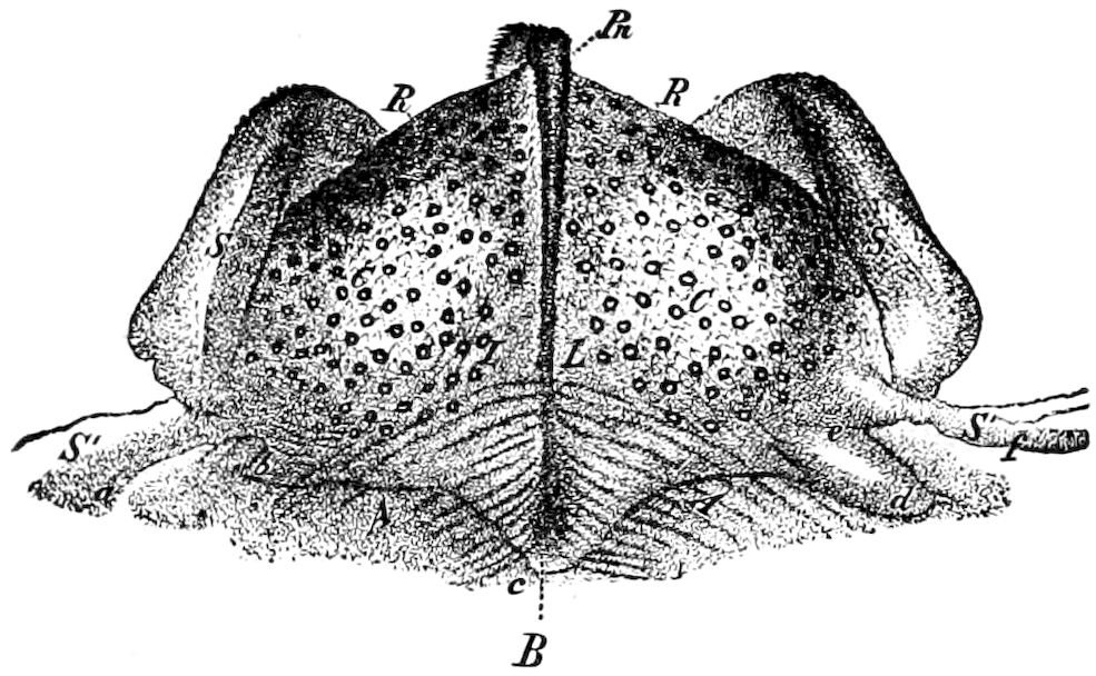
Fig. 284.—Taste-pits on the epipharynx (C) of the honey-bee: B, horny ridge; R, R, taste-pits; L, A, A, muscular fibres; S, S′, a b c d e f, section of skin of œsophagus.—After Wolff.
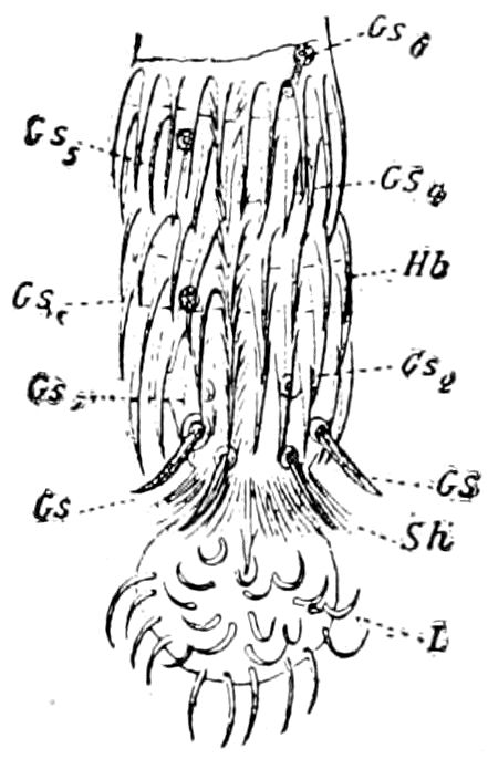
Fig. 285.—Tip of the proboscis of the honey bee, × 140: L, terminal button or ladle: Gs taste-hairs; Sh, guard-hairs; Hb, hooked hairs.—After Will.
Kraepelin (1883) discovered taste-organs on the proboscis of the fly, and taste-hairs at the end of the tongue of the humble-bee (Fig. 285), and afterwards Lubbock critically discussed the subject, and concluded that the organs of taste in insects are situated “either in the mouth itself, or on the organs immediately surrounding it.”
Structure of the taste-organs.—The organs have been best studied by Will, who, besides describing and figuring the chitinous structures, such as the pits or cups, hairs and the pegs, showed that they were the terminations of ganglionated nerves.
Figure 286 represents the taste-cups on the maxilla of a wasp, and Fig. 287 the taste-cone or peg projecting from the cup or pit. The cell out of which the pit and projecting hair or peg are formed is a modified hypodermis cell; and the seta is apparently a modification of a tactile hair, situated at the end of a nerve, which just beneath the chitinous structures passes into a ganglion-cell, which sends off a nerve-fibre to the main nerve.
Will detected on the tongue of the yellow ant (Lasius flavus) from 20 to 24, and in Atta from 40 to 52, of these structures. The number of pits on the maxillæ vary much, not always being the same on the two sides of the same insect. We have observed these taste-cups in the honey and humble bee, not only at the base of the second maxillæ (Fig. 288, g), but also on the paraglossæ (pg).
Distribution in other orders of insects.—The writer has detected these taste-cups in other orders than Diptera and Hymenoptera. They very generally occur in mandibulate insects on the more exposed surface of the epipharynx 283(compare pp. 43–46). We have not observed them in the Synaptera (Lepisma and Machilis).
In the Dermaptera the taste-cups appear to be undeveloped in the nymph, while in the adult they are fewer in number than in any other pterygote order yet investigated.
In a species of Forficula from Cordova, Mexico, the taste-pits are few in number, there being only about a dozen on each side in all; most of them being situated on the anterior half, and a few near the base. The taste-pits are provided each with a short fine seta, as usual arising from the centre.
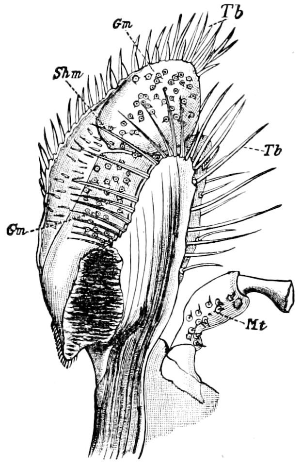
Fig. 286.—Under side of left maxilla of Vespa: Gm, taste-cups; Shm, protecting hairs; Tb, tactile hairs; Mt, base of maxillary palpus.—After Will.
In the order Platyptera (including Perla, Pteronarcys, Psocus, Termes, Eutermes, and Termopsis) we have been unable to detect any organs of taste.
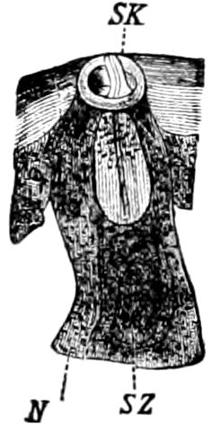
Fig. 287.—Section through a taste-cup: SK, supporting cone; N, nerve; SZ, sense-cell.—After Will. This and Figs. 284–286 from Lubbock.
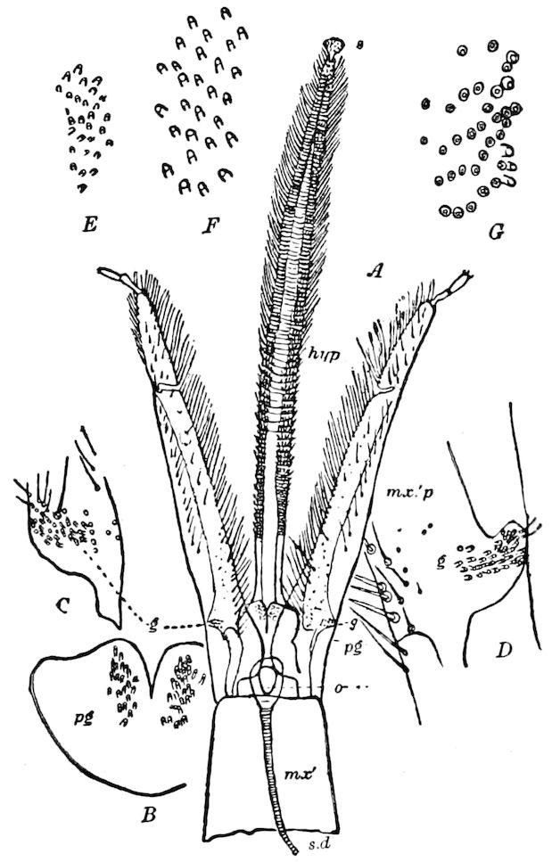
Fig. 288.—Tongue of worker honey-bee: pg, paraglossæ; B, the same enlarged, showing the taste-papillæ; C, D, base of a labial palpus (mx.′p) with the taste-papillæ; E, taste-cups on paraglossæ of Bombus; F, group of same on left, G, on right side, at base of labial palpus.
In the Odonata, however, they are fairly well developed; in Calopteryx, about 50 taste-cups were discovered; in a species of Diplax about 28, there being a group of 14 at the base of the epipharynx on each side of the median line, while in Æschna heros there are two groups of from 25 to 30 taste-cups, situated as in the two aforenamed genera.
In the Orthoptera the gustatory cups are numerous, well developed, and present in all the families except the Phasmidæ, where, however, they may yet be found to occur.
284In a large cockroach (Blabera) from Cuba they are well developed. On each side of the middle of the epipharynx is a curved row of stiff, defensive spines, and at the distal end of each row is a sensory field, containing 20 taste-cups on one side and 23 on the other. Near the front edge of the clypeal region are two more sensory fields, situated on each side of the median line, there being 35 taste-cups in each field. The taste-cups in this form are rather smaller than usual in the order.
In the Acrydiidæ they are more numerous than in the Blattidæ. For example, in Camnula pellucida, near what corresponds to the front edge of the clypeus are two gustatory fields, each bearing about 35 taste-pits. Just in front, under the clypeo-labral suture, are two similar fields, each containing from 40 to 42 taste-pits. There are none in front of these. There are thus about 140–150 sense-cups in all.
The members of the Locustidæ (Fig. 26) appear to be better provided with the organs of taste than any other Orthoptera, those of the katydid numbering from 170 to 180. There are from 50 to 60 taste-cups in the front region; behind the middle a group of 25 on each side, and over an area corresponding to the base of the labrum and front edge of the clypeus is a sensory field with about 70 taste-cups on each side. They are true cups or beaker-like papillæ, some with a fine, others with a short, stout, conical seta.
The gustatory organs in the cave cricket (Hadenœcus subterraneus, Fig. 27), from Mammoth Cave, are highly developed, being rounded papillæ with the nucleus at the top or end. They are grouped on each side of the middle near the front edge, there being 25 on each side. An irregular row of these beaker-like organs extends along each side; some occur under the base of the labrum, but they are most numerous in a field corresponding to the front edge of the clypeus, there being 50 on each side, or 100 in all, where in Ceuthophilus there are only 9 or 10. It would thus appear as if the sense of taste were much more acute in the cave-dweller than in the out-of-doors form.
In the Coleoptera taste-cups and setæ are very generally distributed, though we were unable to detect them in Dendroctonus or in Lucanus dama. As seen in Fig. 57, we have observed numerous taste-pegs along the maxilla of Nemognatha lurida, but otherwise taste-organs have only been detected in the epipharynx. They not only occur in the adult beetles, but we have found them in the larvæ of cerambycid, scarabæid, and other beetles. In the adults taste-cups appear to be about as well developed in the carnivorous forms (Carabidæ) as in the phytophagous or lignivorous groups.
In Chlænius tomentosus there are about half as many of these organs as in Harpalus, while in Calosoma there are 90 taste-cups, 45 on each side, under the base of the labrum. The cups are papilliform, being rather high, with a seta arising from each.
In the Cicindelidæ, the epipharynx bears a sensory field quite different from that of the Carabidæ. There are no normal taste-cups, except a few situated on two large, round, raised areas which are guarded in front by a few very long setæ. On the surface of each area are numerous very long setæ which may, if not tactile, have some other sense, as they arise from cup-like bases or cells. Those on the outside are like true taste-cups, with a bristle but little larger than normal in taste-cups generally. We are disposed to regard this sensory field as a highly specialized gustatory apparatus.
In the Dyticidæ the taste-cups are nearly as described in the Carabidæ.
The Staphylinidæ are not well provided with taste-organs. Under the clypeus of Staphylinus violaceus, on each side near the middle, is a bare rounded area, in which are situated 4 or 5 papilliform taste-cups, and at the base behind them 285is another linear group of about 7 slenderer, somewhat curved, taste-cups. In the Elateridæ these organs are scantily developed.
In the Buprestidæ (Buprestis maculiventris alone examined) no true taste-cups were detected. On the other hand, the Lampyridæ are well supplied with them. Under the clypeus is situated a sensory field bearing 26 taste-cups, which are rather smaller than usual. Over the epipharyngeal surface are scattered a few taste-cups, but they are small and perhaps not gustatory. Under the clypeus of Lucidota punctata Lec. is a group of 12 taste-cups, and in the middle region of the epipharynx, situated in a field extending from near the base to near the front edge, are about 40 taste-cups, which, however, are not, as is usual, arranged on each side of the median line, but are scattered among the hairs of the pilose surface of the epipharynx. In the Cleridæ the taste-cups are few in number.
In the great family of Scarabæidæ, the presence of gustatory organs is variable. None occur in Lucanus dama, though in the June beetle (Lachnosterna fusca Fröhl.) they are abundantly developed. The epipharynx bears on each side outside of a spiny area a group of about 50 taste-cups, each bearing a long seta, those on the outside of the area passing into a few high, rather slender, papillæ, without a seta. On the under side of the clypeus is a median group of 10 taste-cups of singular form, the cups being large, with broad bases, which posteriorly bear three spines, of which the median one is the largest.
Taste-cups occur without any known exception in the longicorn beetles. In Leptura canadensis they are numerous; in Euryptera lateralis they are abundant along and near the middle of the anterior half of the labral region, and in Cyllene robiniæ Forst. (or pictus Drury) they are more numerous than usual, extending in an unbroken sensory field from near the front margin of the clypeal region to near the front edge of the epipharynx. The cups vary much in size, some being one-half as large as others; and those on the sides of the sensory field bear short, and a few others rather long, bristles, showing that the taste-cups are modified tactile bristles.
The Tenebrionidæ are fairly well endowed with taste-cups, their number in Eleodes obsoleta Say amounting to 30 or 40.
Those of the Meloidæ especially are unusual in size and number.
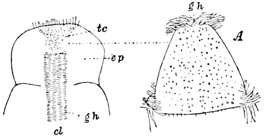
Fig. 289.—Epipharynx (ep) of Nemognatha: cl, clypeus; gh, gathering hairs; tc, triangular sensory field dotted with taste cups; A, the field enlarged.
In Nemognatha lurida Lec. (Fig. 289) the front edge of the epipharynx contains about 80 remarkably small taste-cups, arranged irregularly in a triangular sensory space, and not more than ¼ to ⅙, as large as those on the maxillæ of the same beetle. Unless the former structures are gustatory it is difficult to account for their presence here, and it will be observed that the taste-cups in Epicauta are unusually abundant. Thus in the middle and near the front of the epipharynx of the blister-beetle over 100 gustatory cups were counted. They are conical, papilliform, and truncated at the end as if open, the edge of the opening being ragged, though bearing no bristle, except in a few cases. Around the edge of the sinus, on the under side of the labrum, is a regular marginal row of large, longer, more distinctly chitinized taste-cups, whose walls are streaked up and down by chitinous thickenings. In E. callosa Lec. there are about 55 taste-cups under 286the labrum, besides about 10 cells, which may be gustatory structures, situated on either side of a median setose ridge which passes back under the clypeal region.
The taste-cups of the leaf-beetles are fairly numerous, judging from an examination of Diabrotica vittata. The surface of the epipharynx is pilose, but the median region is naked, and on the anterior half bears from 11 to 12 taste-cups, arranged each side of the median line in a rude Y. On each side, at the base of the labial region, are two sensitive fields, each bearing about 25 to 26 taste-cups. More were seen under the clypeus.
In the Neuroptera unmistakable taste-cups are not always present. In Sialis infumata along the median line of the epipharynx and near the front are about 20 scattered gustatory pegs, which are minute, but longer and more acute than usual. In Chauliodes maculatus there are one or two taste-cups under the front edge of the clypeus; others are scattered along the middle from the base of the labrum to the front, but are not arranged in definite order. In Corydalis cornutus no sense-cups, pits, or rods are present. In Chrysopa there are scattered cups armed with a short acute bristle, which are possibly gustatory in function. In Myrmeleon diversum also the presence of sense-pits or of taste-cups is doubtful, though a group of about 12 pits on each side of the clypeal region of the epipharynx, and a few situated at the base of the labral region, may be endowed with the sense of taste. In Mantispa brunnea, however, along the middle of the epipharynx are scattered about 30 unmistakable taste-cups, each bearing a short, fine hair.
In the Mecoptera (Panorpa debilis?) taste-cups, giving rise to a minute hair, occur on the labium in two regions, and also on the maxillæ situated on the stipes near the base of the palpi, and on the lacinia and galea. They are also to be found on the maxillæ of Boreus californicus, but were not detected on the labium.
They were first detected by Reuter in various microlepidoptera, and occur on the “basal spot” of the palpi of many butterflies. In a Tineid moth (Coleophora coruscipennella) we have detected what we suppose to be a group of four taste-pits on the inner side of the basal joint of the labial palpi.
Experimental proof.—No one, says Lubbock, who has ever watched a bee or wasp can entertain the slightest doubt as to their possession of the sense of taste. “Forel mixed morphine and strychnine with some honey, which he offered to his ants. Their antennæ gave them no warning. The smell of the honey attracted them, and they began to feed; but the moment the honey touched their lips they perceived the fraud.”
Will at first fed wasps with sugar, so that they frequently visited it; afterwards he substituted alum for the sugar. Eagerly flying to it, they had scarcely touched it when they drew back from the distasteful substance with the most comical gestures, and cleaned their tongues by frequently running them in and out, repeatedly stroking them with their fore feet. He noticed a great repugnance to quinine in nearly all the insects experimented on. Bees and wasps were observed to have a more delicate gustatory sense than flies, etc., which are more omnivorous in their tastes.
287
LITERATURE ON THE ORGANS OF TASTE
Meinert, F. Bidrag til de danske myrers naturhistorie. (Kgl. Dansk Vidensk. selsk. skrifter. Kjoebenhavn. Raekke 5 Naturvid. og math. Afd., v, 1861, pp. 273–340.)
Wolff, O. J. B. Das Riechorgan der Biene. (Nova acta d. K. Leop.-Carol. Akad., xxxviii, 1875, pp. 1–251, 8 Taf.)
Joseph, G. Zur Morphologie des Geschmacksorganes bei Insekten. (Amtlicher Bericht der 50. Versammlung deutscher Naturforscher und Aerzte in München, 1877, pp. 227–228.)
Künckel et Gazagnaire. Du siège de la gustation chez les insectes Diptères. Constitution anatomique et physiologique de l’epipharynx et l’hypopharynx. (Comptes-rend. Acad. Sc., Paris, 1881, xcv, pp. 347–350.)
—— Recherches sur l’organisation et le développement des Diptères et en particulaire des Volucelles, i, 1875, ii, 1881, 26 Pls.
Kraepelin, K. Zur Kenntniss der Anatomie und Physiologie des Rüssels von Musca. (Zeitschr. f. wissens. Zool., xxxix, 1883, pp. 683–719, 2 Taf.)
Kirbach, P. Ueber die Mundwerkzeuge der Schmetterlinge. (Zool. Anzeiger, 1883, pp. 553–558, 2 Figs.)
Will, F. Das Geschmacksorgan der Insekten. (Zeitschr. f. wissens. Zool., 1885, xlii, pp. 674–707, 1 Taf.)
Gazagnaire, J. Du siège de la gustation chez les insectes Coléoptères. (Comptes-rend. Acad. Sc., Paris, 1886, cii, pp. 629–632; Ann. Soc. Ent. France, Sér. 6, Bull., pp. 79–80.)
Forel, A. Expériences et remarques critiques sur les sensations des insectes. 2me Part. (Recueil Zool. Suisse, iv, 1887, pp. 161–240.)
Reuter, Enzio. Ueber den “Basalfleck” auf den Palpen der Schmetterlinge. (Zool. Anzeiger, 1888, pp. 500–503.)
—— Ueber die Palpen der Rhopaloceren, etc., 6 Taf. Helsingfors, 1896, pp. 1–578. (Acta Soc. Sc. Fennicæ, xxii, 1896.)
Packard, A. S. On the occurrence of organs, probably of taste, in the epipharynx of the Mecoptera (Panorpa and Boreus). (Psyche, 1889, v, pp. 159–164.)
—— Notes on the epipharynx, and the epipharyngeal organs of taste in mandibulate insects. (Psyche, 1889, v, pp. 193–199, 222–228.)
Also Lubbock’s Senses, etc., of animals, and the writings of Briant, Breithaupt (titles on p. 85).
d. The organs of hearing
Although it has been denied by Forel that insects have the sense of hearing, yet the majority of writers and experimenters agree that insects are not deaf. On general grounds if, as we know, many insects produce sounds, it must follow that they have ears to hear, for there is every reason to suppose that the sounds thus made are, as in other animals, either for attracting the sexes, for a means of communication, or to express the emotions. We will begin by briefly describing the structures now generally supposed to be auditory in 288function, and about which there can be no reasonable doubt, and then consider the more problematical organs, closing with an account of the extremely various means of producing sounds and cries.
The ears or tympanal and chordotonal sense-organs of Orthoptera and other insects.—The ears or tympana of locusts (Acrydiidæ) are situated one on each side, on the basal joint of the abdomen, just behind the first abdominal spiracle. That this is a true ear was first suggested by J. Müller, and his opinion was confirmed by Siebold, Leydig, Hensen, Graber, Schmidt, Lubbock, etc.[49]
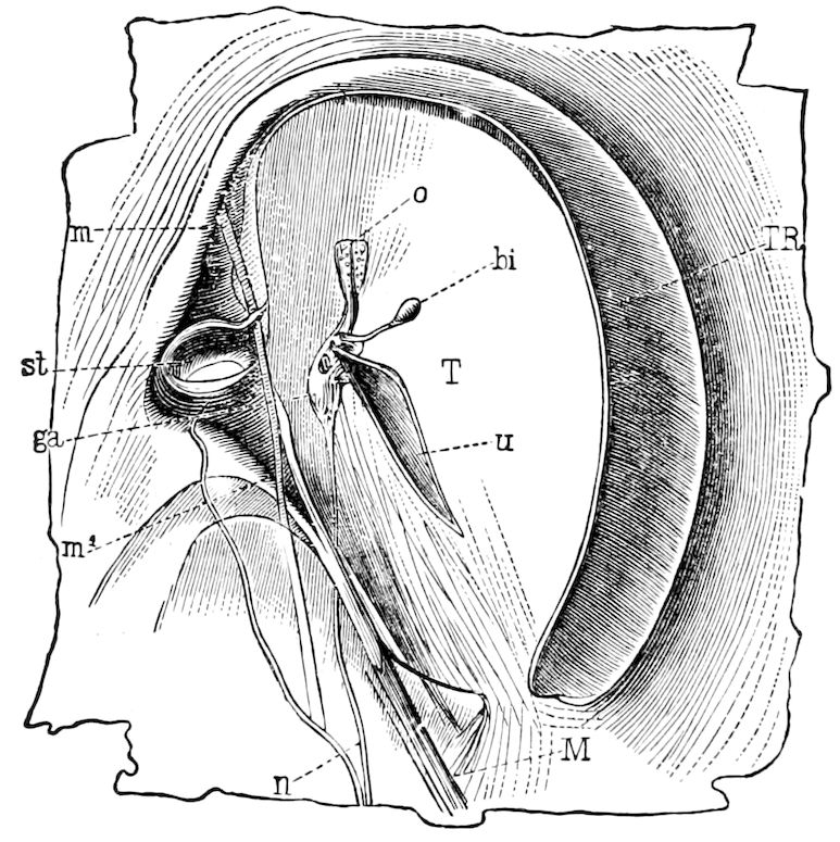
Fig. 290.—Ear of a locust (Caloptenus italicus), seen from the inner side: T, tympanum; TR, its border; o, u, two horn-like processes; bi, pear-shaped vesicle; n, auditory nerve; ga, terminal ganglion; st, stigma; m, opening, and m′, closing, muscle of the same; M, tensor muscle of the tympanum membrane.—After Graber.
The apparatus consists of a tense membrane, the tympanum, surrounded by a horny ring (Fig. 290). “On the internal surface of this membrane are two horn-like processes (o, u), to which is attached an extremely delicate vesicle (bi) filled with a transparent fluid, and representing a membranous labyrinth. This vesicle is in connection with an auditory nerve (n) which arises from the third thoracic 289ganglion, forms a ganglion (ga) upon the tympanum, and terminates in the immediate neighborhood of the labyrinth by a collection of cuneiform, staff-like bodies, with very finely pointed extremities (primitive nerve-fibres?), which are surrounded by loosely aggregated ganglionic globules” (Siebold’s Anatomy of the Invertebrates).
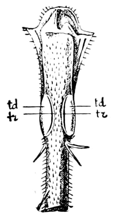
Fig. 291.—Fore tibia of Locusta viridissima. td, cover of the drum; tr, fissure between the drum and its cover.—After Graber, from Lang.
In the green grasshoppers, katydids, and their allies, the ears are situated on the fore tibiæ, where these organs can be found after a careful search (Figs. 291, 292).
The presence of the structure is indicated by the oval disc, the drum, which is a thin tense membrane covering the auditory apparatus of nerves, ganglion cells, and auditory rods beneath.
The tympana, or drums, are not present in all Locustidæ and Gryllidæ, and, as Lubbock states, it is an additional reason for regarding them as auditory organs, that in those species which possess no stridulating organs the tympana are also wanting. In many of the Locustidæ the tympana are covered or protected by a fold of the skin projecting over them. These covered ones are, Graber thinks, derived from the open ones.
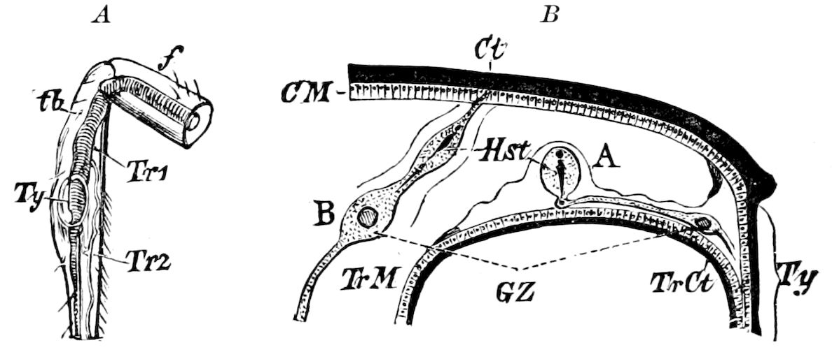
Fig. 292.—A, fore tibia of a European grasshopper (Meconema), containing the ear: Ty, tympanum or outer membrane; Tr 1, Tr 2, tracheæ. B, diagrammatic cross-section through the tibia and ear of the same; Ty, tympanum; Ct, cuticula; CM, hypodermis: A, the auditory organ connecting with the tympanum; B, supra-tympanal auditory organ; GZ, the ganglion-cell belonging to them; Hst, the auditory rod connecting with the ganglion-cells.—After Graber, from Judeich and Nitsche.
On examining the apparatus within the leg under the drum, it is seen to consist of the trachea, the auditory vesicles and rods, ganglion cells, and acoustic nerve. The trachea is greatly modified (Fig. 292, Tr 1). On passing into the tibia the trachea enlarges and divides into two branches, which reunite lower down. The spiracles supplying the air to this enlarged trachea are considerably enlarged, while in the dumb species it is of the normal size. The enlarged 290trachea passes close to the tympanum, which thus has air on both sides of it: the open air on the outer, the air of the trachea on its inner surface. In fact, as Lubbock states, “the trachea acts like the Eustachian tube in our own ear; it maintains an equilibrium of pressure on each side of the tympanum, and enables it freely to transmit the atmospheric vibrations.”
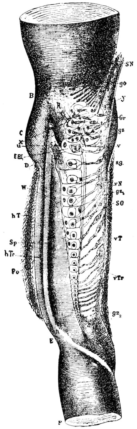
Fig. 293.—The auditory apparatus in the tibia of a grasshopper, showing the tympanal nerve-endings in situ: EBI, terminal vesicles of Siebold’s organ; SN, nerve of the organ of Siebold; Gr, group of vesicles of same; SO, nerve-endings of the same; vT, front tympanum; vTr, front branch of the trachea; hT, hinder tympanum; hTr, hinder branch of the trachea; Sp, space between the tracheæ; go, supra tympanal ganglion; rN, connecting nerve-fibrils between the ganglion cells and the terminal vesicles; R, upper, n-S, lower, root of the transparent covering membrane. (Other lettering not explained by author.)—After Graber.
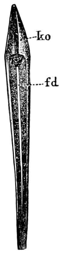
Fig. 294.—Auditory rod of Gryllus viridissimus: fd, auditory rod; ko, terminal piece.—After Graber, from Lubbock.
These tracheæ, says Graber, though formed on a similar plan, present many variations, corresponding to those of the tympana, and showing that the tympana and the tracheæ stand in intimate connection with one another. For instance, in those species where the tympana are equal, the tracheæ are so likewise; in Gryllotalpa, where the front tympanum only is developed, though both tracheal branches are present, the front one is much larger than the other; and where there is no tympanum, the trachea remains comparatively small, and even in some cases undivided (Lubbock, ex Graber).
The acoustic nerve, which next to the optic is the thickest in the body, divides soon after entering the tibia into two branches, one almost immediately forming a ganglion, the supra-tympanal ganglion, the other passing down to the tympanum, where it expands into an elongated flat ganglion, the organ of Siebold (Fig. 293), and closely applied to the anterior tracheæ.
At the upper part of the ganglion is a group terminating below in a single row of vesicles, the first few of which are approximately equal, but which subsequently diminish regularly in size. Each 291of these vesicles is connected with the nerve by a fibril (Fig. 293, vN), and contains an auditory rod (Fig. 294). They are said by Graber to be brightly refractive, hollow (thus differing from the retinal rods, which are solid), and terminate in a separate end-piece (ko). The rods were first discovered by Siebold, and, as Lubbock remarks, may be regarded as specially characteristic of the acoustic organs of insects.
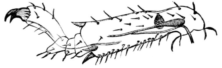
Fig. 295.—Chordotonal organ in nymph of a white ant.—After Müller, from Sharp.
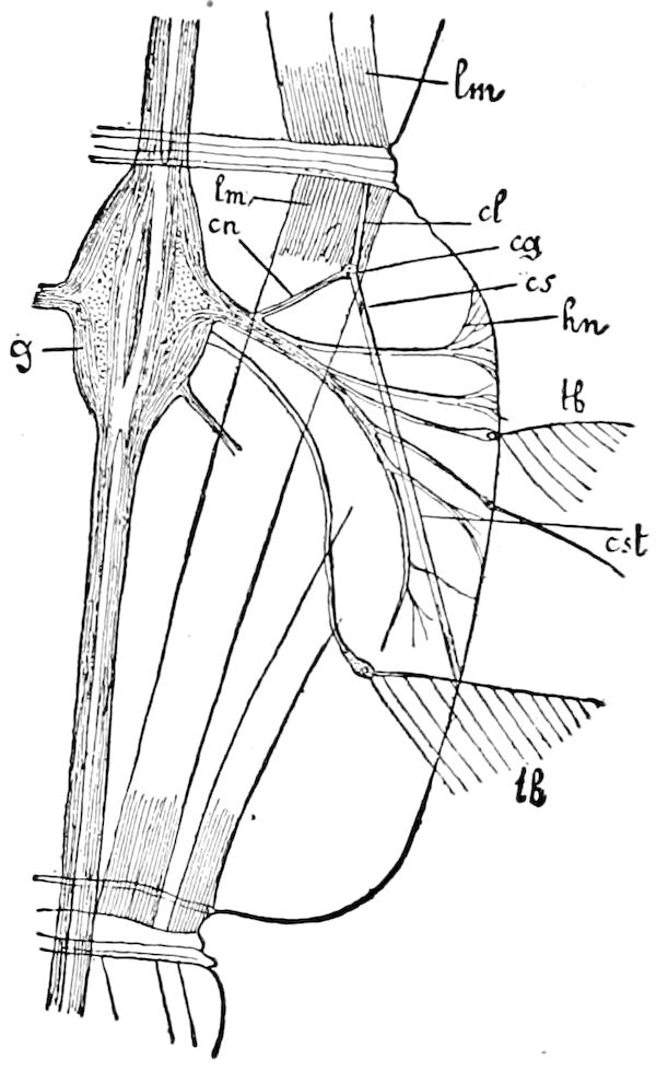
Fig. 296.—Right half of 8th body-segment of Corethra plumicornis: g, ganglion of ventral cord; lm, longitudinal muscle; cn, chordotonal nerve; cl, chordotonal ligament; cg, chordotonal ganglion; cs, rod of chordotonal organ; cst, terminal cord; tb, tactile setæ; hn, out-going fibres of the integumental nerves.—After Graber, from Lang.
As will be seen in Fig. 293, at the upper part of the tibial organ of Ephippigera there is a group of cells, and below them a single row of cells gradually diminishing in size from above downwards. “One cannot but ask oneself,” says Lubbock, “whether the gradually diminishing size of the cells in the organ of Siebold may not have reference to the perception of different notes, as is the case with the series of diminishing arches in the organ of Corti of our own ears.”
These organs were supposed to be restricted to the Orthoptera, but in 1877 Lubbock discovered what seems to resemble the supra-tympanal auditory organ of Orthoptera in the tibia of the yellow ant (Lasius flavus). Graber confirmed Lubbock’s account, and also discovered these organs in the tibia of a Perlid (Isopteryx apicalis), and Fritz Müller has detected them in the fore tibiæ of the nymph of Calotermes rugosus (Fig. 295). To these structures Graber gave the name of chordotonal organs.
He has also detected these organs in all the legs of other insects (Trichoptera, Pediculidæ), and auditory rods have been discovered in the antennæ of Dyticus and of Telephorus by Hicks, Leydig, and Graber. Graber classifies the chordotonal organs into truncal and membral. In Coleoptera and Trichoptera they may occur on several joints of the leg; others are more localized,—thus he distinguishes femoral (Pediculidæ), tibial (Orthoptera, Perlidæ, Formicidæ), and tarsal organs (Coleoptera).
A type of chordotonal organ, observed in the body-segments of the larvæ of several insects by Leydig, Weismann, Graber, Grobben, and Bolles Lee, is to be seen in the transparent larva of Corethra (Fig. 296), where the auditory organ extends to the skin. It contains at the point cs two or three auditory 292rods. In the opposite direction a fine ligament (cl) passes from cg to the skin; in this way the auditory organ is suspended in a certain state of tension, and is favorably situated to receive even very fine vibrations. A similar apparatus has been detected in the larva of Ptychoptera.
Antennal auditory hairs.—It is not at all improbable that the antennæ of different insects contain auditory as well as olfactory structures. Lubbock has suggested that the singular organs which have only been found in the antennæ of ants and certain bees, and to which he gives the name of “Hicks’ bottles” (Fig. 281), may act as microscopic stethoscopes, while Leydig also regards them as chordotonal organs.
That, however, some of the antennal hairs of the mosquito, as first suggested by Johnson and afterwards proved experimentally by Mayer, are auditory, seems well established. Fastening a male mosquito down on a glass slide, Mayer then sounded a series of tuning-forks. With an Ut4 fork of 512 vibrations per second, some of the hairs were seen to vibrate vigorously, while others remained comparatively at rest. The lower (Ut3) and higher (Ut5) harmonics of Ut4 also caused more vibration than any intermediate notes. These hairs, then, are specially tuned so as to respond to vibrations numbering 512 per second. Other hairs vibrated to other notes, extending through the middle and next higher octave of the piano.
Mayer then made large wooden models of these hairs, the one corresponding to the Ut3 hair being about a metre in length, and on counting the number of vibrations they made when they were clamped at one end and then drawn on one side, he found that it “coincided with the ratio existing between the numbers of vibrations of the forks to which covibrated the fibrils,” or hairs. It should be observed that the song of the female mosquito corresponds nearly to this note, and would consequently set the hairs in vibration. Mayer observed that the song of the female vibrates the hairs of one of the antennæ more forcibly than those of the other. Those auditory hairs are most affected which are at right angles to the direction from which the sound comes. Hence from the position of the antennæ and the hairs a sound will be loudest or most intense if it is directly in front of the head. If, then, the song of the female affects one antenna more than another, the male turns his head until the two antennæ are equally affected, and is thus able to fly straight towards the female. From his experiments Mayer found that the male can thus guide himself to within 5° of the direction of the female. Hence he concludes that “these insects must have the faculty 293of the perception of the direction of sound more highly developed than in any other class of animals.” (Also see Child’s work.)
Special sense-organs in the wings and halteres.—Organs of a special sense, which Hicks supposed to be those of smell, were found by him near or at the base of the wings of Diptera, Coleoptera, and less perfect ones in Lepidoptera, Neuroptera, and Orthoptera, with a trace of them in Hemiptera; but these were considered by Leydig to be auditory organs, since he found the nerves to end in club-shaped rods, like those of Orthoptera.
Hicks found, as to the halteres and their sense-organs, that the nerve in the halter is the largest in the insect, except the optic nerve; and that at the base of the halteres is a number of vesicles arranged in four groups, to each of which the nerve sends a branch. Afterwards Bolles Lee discovered that the vesicles, undoubtedly perforated, contain a minute hair, those of the upper groups being protected by hoods of chitin. He regarded them as olfactory organs, while Lubbock seems inclined to consider them as auditory structures. Graber also regards the vesicles of Hicks as chordotonal organs.
In his elaborate account of the balancers, Weinland concludes that the organs of sense of varying structure occurring at the base of these appendages allow the perception of movements which the halteres perform and which enable the fly to steer or direct its course. The halteres can thus cause differences in the direction of the flight of a fly in the vertical plane. If the balancers act unequally, there is a change in direction.
e. The sounds of insects
Insects have no true voice; but sounds of different intensity, shrill cries, and other noises are produced mechanically by insects, either being love-songs to attract the sexes, to give signals, to communicate intelligence, or perhaps to express the emotions. The loud, shrill cry of the Cicada, or chirp of the cricket, is evidently a love-call, and results in the mating of individuals of separate broods more or less widely scattered, thus preventing too close interbreeding.
The simplest means of making a noise is that of the death-watch (Anobium), which strikes or taps on the wall with its head or abdomen. Longicorn beetles make a sharp sound by the friction of the mesoscutellum against the edge of the prothoracic cavity, the head being alternately raised and lowered, Burying-beetles (Necrophorus) rub the abdomen against the hinder edges of the elytra. Weevils make a loud noise by rapidly rubbing the tips of the abdomen on the ends of the elytra.
Landois offers the following summary of the kinds of noises produced by beetles:
1.
Tapping sounds (Bostrycinæ, Anobium).
2.
Grating sounds (Elateridæ). 294
3.
Friction without special rasping organs (Euchirus longimanus).
4.
Rasping sounds produced by friction:
a. Rubbing of the pronotum on the mesonotum (Cerambycidæ except Spondyli and Prionus).
b. Friction of the prosternum on the mesosternum (Omaloplia brunnea).
c. Elytra with a rasp at the end (Curculionidæ, Dyticidæ, Pelobius).
d. With a coxal rasp (Geotrupes, Ceratophyus). The male of Ateuchus stridulates to encourage the female in her work, and from distress when she is removed. (Darwin.)
e. Friction of the edge of the elytra against the femur (Chiasognathus grantii).
f. Pygidium with two rasps in the middle (Crioceris, Lema, Copris, Oryctes, Necrophorus, Tenebrionidæ).
g. Abdomen with a grating ridge and four grating plates (Trox).
h. Abdomen with two toothed ridges rubbing on a rasp on edge of wing-cover (Elaphus, Blethisa, Cychrus).
i. Rubbing the elytra on a rasp on the hind wings (Pelobius hermanni).
j. Friction of the wing against the abdominal segments (Melolontha fullo).
Mutilla makes a rather sharp noise by rubbing one abdominal segment against another. Ants (Ponera) have a stridulating apparatus, and other genera numerous (20) ridges between the segments.
Even certain moths and butterflies emit a rasping or crackling noise. The death’s-head moth and other sphinges cause it by rubbing the palpi against the base of the proboscis. These and certain butterflies are provided with parallel ridges forming a rasp on the “basal spot” of the inner side of the basal joint of each palpus (Reuter). A South American butterfly (Ageronia feronia) can be heard for several yards as it flies with a crackling sound. Hampson finds that the cause of the clicking sound is due to a pair of strong chitinous hooks attached to the thorax, against which play the spatulate ends of a pair of hooks attached to the fore wings. An Australian moth (Hecatesia) flies with a whizzing sound; Vanessa is said to be sonorous.
The males of Orthoptera produce their shrill cries or chirping noises, 1, by rubbing the thighs against the sides of the body (Acrydiidæ); 2, by the friction of the base of the fore wings on each other (Locustidæ); 3, by rubbing the base of the upper on the base of the hinder or under pair (Gryllidæ), in the two last there being a shrilling apparatus consisting of a file on the hind wings, which rubs on a resonant surface on the fore wings. The females are not invariably dumb, both sexes of the European Ephippigera being able to faintly stridulate. Corixa also produces shrill chirping notes. (Carpenter.)
Certain insects also hum, and have what may perhaps be called a voice. The cockchafer, besides humming with the wings, produces a sound almost like a voice. In the large trachea, just behind each spiracle, is a chitinous process, which is thrown into vibrations by the air during respiration, and thus produces a humming noise. (Lubbock.) Such is also the case with flies, the mosquito, dragon-flies, and bees. In flies and dragon-flies the “voice” is caused by the air issuing from the thoracic spiracles; while in the humble-bee the abdominal spiracles are also musical. The sound made by the spiracles bears no relation to that caused by the wings. Landois 295tells us that the wing-tone of the honey-bee is A′; its voice, however, is an octave higher, and often goes to B″ and C″.
The sounds produced by the wings are constant in each species, except where, as in Bombus, there are individuals of different sizes; in these the larger ones generally give a higher note. Thus the comparatively small male of Bombus terrestris hums on A′, while the large female hums an entire octave higher.
From the note produced the rapidity of the vibrations can be calculated. For example, the house-fly, which produces the sound of F, vibrates its wings 21,120 times in a minute, or 335 times in a second; and the bee, which makes a sound of A′, as many as 26,400 times, or 440 times in a second. On the contrary, a tired bee hums on E′, and therefore, according to theory, vibrates its wings only 330 times in a second. Marey has confirmed these numbers graphically, and found by experiment that the fly actually makes 330 strokes in a second. (Lubbock.)
A different kind of musical apparatus is that of the cicada, which has been elaborately described by Graber. The shrill, piercing notes issue from a pair of organs on the under side of the base of the abdomen of the male, these acting somewhat as two kettle-drums, the membrane covering the depressions being rapidly vibrated.
LITERATURE ON THE ORGANS OF HEARING
a. The auditory organs
Siebold, C. Th. E. von. Ueber das Stimm- und Gehörorgan der Orthopteren. (Archiv f. Naturgesch., 1844, x, pp. 52–81.)
Johnston, Christopher. Auditory apparatus of the culex mosquito. (Quart. Journ. Micr. Soc., 1855, iii, pp. 97–102, 1 Fig.)
Hicks, Braxton. On a new organ in insects. (Journ. Linn. Soc. Zool., London, 1857, pp. 130–140, 1 Pl.)
—— Further remarks on the organ found on the bases of the halteres and wings of insects. (Trans. Linn. Soc., London, 1857, xxii, pp. 141–145, 2 Pls.)
Hensen, V. Ueber das Gehörorgan von Locusta. (Zeitschr. f. wissens. Zool., xvi, 1866, pp. 190–207.)
Graber, V. Bemerkungen über die Gehör- und Stimmorgane der Heuschrecken und Cicaden (Wiener Sitzungsber. Math.-natur-wiss. Cl., lxvi, 1 Abt., 1872, pp. 205–213, 2 Figs.)
—— Die tympanalen Sinnesapparate, der Orthopteren. (Denkschr. d. k. Akad. d. wissens. Wien, xxxvi, 1876, 2 Abt., pp. 1–140, 10 Taf.)
—— Die abdominalen Tympanalorgane der Cicaden und Gryllodeen. (Ibid., 1870, xxxvi, pp. 273–290, 2 Taf.)
—— Ueber neue, otocystenartige Sinnesorgane der Insekten. (Archiv f. mikroskop. Anat., 1878, pp. 35–57, 2 Taf.)
—— Die chordotonalen Sinnesorgane und das Gehör der Insekten. (Archiv f. mikroskop. Anat., 1882, xx, pp. 506–640; 1883, xxi, pp. 65–145, Taf.)
296Mayer, Alfred Marshall. Researches in Acoustics No. 5. 3. Experiments on the supposed auditory apparatus of the culex mosquito. (Amer. Jour. Sc. and Arts, Ser. 3, viii, 1874, pp. 81–103; also Amer. Naturalist, viii, pp. 577–592.)
Schmidt, Oscar. Die Gehörorgane der Heuschrecken. (Archiv f. mikroskop. Anat., xi, 1875, pp. 195–215, 3 Taf.)
Ranke, J. Beiträge zu der Lehre von den Uebergangssinnesorganen, das Gehörorgan der Acridier und das Sehorgan der Hirudineen. (Zeitschr. f. wissens. Zool., xxv, 1875, pp. 143–164, 1 Taf.)
Lee, A. Bolles. Les balanciers des Diptères, leurs organes sensifères et leur histologie. (Recueil Zool. Suisse, ii, 1885, pp. 363–392, 1 Pl.)
—— Bemerkungen über der feineren Bau der Chordotonalorgane. (Archiv f. mikroskop. Anat., 1883, xxiii, pp. 133–140, 1 Taf.)
—— Les organes chordotonaux des Diptères et la méthode du chlorure d’or. (Observations critiques.) (Recueil Zool. Suisse, 1884, ii, pp. 685–689, 1 Pl.)
Weinland, E. Ueber die Schwinger (Halteren) der Dipteren (Zeitschr. f. wissens. Zool., 1890, li, pp. 55–166, 5 Taf.)
Adelung, N. v. Beiträge zur Kenntnis des tibialen Gehörapparates der Locustiden. Inaug. Diss., Leipzig, 1892, 2 Taf.
Child, Ch. M. Ein bisher wenig beachtetes antennales Sinnesorgan der Insekten, mit besonderer Berücksichtigung der Culiciden und Chironomiden. (Zeitschr. f. wissens. Zool., lviii, 1894, pp. 475–528, 2 Taf.; also, Zool. Anzeiger, xvii Jahrg., pp. 35–38, and in Annals and Mag. Nat. Hist., 1894 (6), xiii, pp. 372–374).
Also the writings of J. Müller, Kirby and Spence, Burmeister, Gilbert White, Westwood, Guilding, Meinert, Paasch, Leydig, Viallanes, Minot, Forel, Mayer, Darwin (Descent of Man, i, ch. x.), F. Müller, Lubbock (Senses of animals), Westring, Köppen, Bates, Vom Rath, Peckham, Jourdan, Nagel, etc.
b. The sounds made by insects
Scudder, S. H. Notes on the stridulation of grasshoppers. (Proc. Bost. Soc. Nat. Hist., xi, 1868, pp. 306–313 and 316.)
—— The songs of the grasshoppers. (Amer. Naturalist, ii, 1868, pp. 113–120, 5 Figs.)
Riley, C. V. The song notes of the periodical Cicada. (Proc. Amer. Assoc. Adv. Science, xxxiv, 1885, pp. 330–332; also in Kansas City Rev., October, 1885, pp. 173–175.)
Swinton, A. H. (Ent. Month. Mag., 1877.) Sound produced in Ageronia by a modification of the hook and bristle of the wings.
Hampson, G. F. On stridulation in certain Lepidoptera, etc. (Proc. Zool. Soc. London, 1892, ii, pp. 188–193, Fig; also Psyche, vi, p. 491, 1 Fig.)
With the writings of Landois, Lubbock, Graber, Kolbe, Carpenter (Nat. Science), Bruyant, and others.
297
THE DIGESTIVE CANAL AND ITS APPENDAGES
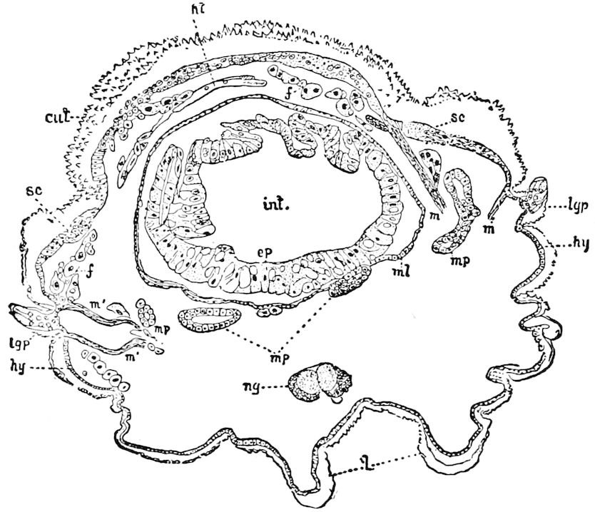
Fig. 297.—Transverse section through an abdominal segment of larva of Megalopyge crispata, showing the relations of the digestive canal to the other organs: int, hind-intestine, with its mucous or epithelial layer (ep), and ml its outer or muscular layer; ng, ventral ganglion; ht, heart; mp, urinary tubes; f, fat-body; sc, thickened portion of the hypodermis (hy) containing the setigenous cells; m, muscles; m′, a pair of retractor muscles inserted near the base of the lateral glandular process (lgp); cut, cuticula; l, legs. Also compare Figs. 142–144 and 234.
298
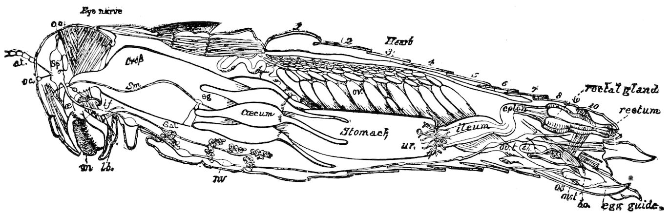
Fig. 298.299 The alimentary or digestive canal of insects is a more or less straight tube, which connects the mouth and anus, the latter invariably situated in the last segment of the body, under the last tergite or suranal plate. It lies directly over the ventral nervous cord and under the dorsal vessel, passing through the middle of the body (Fig. 297). It is loosely held in place by delicate retractor muscles (retractores ventriculi, found by Lyonet in the larvæ of Lepidoptera, and occurring in those of Diptera), but is principally supported by exceedingly numerous branches of the main tracheæ. Fig. 298.—Internal anatomy of Melanoplus femur-rubrum: at, antenna and nerve leading to it from the “brain” or supraœsophageal ganglion (sp); oc, ocelli, anterior and vertical ones, with ocellar nerves leading to them from the brain; œ, œsophagus; m, mouth; lb, labium or under lip; if, infraœsophageal ganglion, sending three pairs of nerves to the mandibles, maxillæ, and labium respectively (not clearly shown in the engraving); sm, sympathetic or vagus nerve, starting from a ganglion resting above the œsophagus, and connecting with another ganglion (sg) near the hinder end of the crop; sal, salivary glands (the termination of the salivary duct not clearly shown by the engraver); nv, nervous cord and ganglia; ov, ovary; ur, origin of urinary tubes; ovt, oviduct; sb, sebaceous gland; bc, bursa copulatrix; ovt, site of opening of the oviduct (the left oviduct cut away); 1–10, abdominal segments. The other organs labelled in full.—Drawn from his original dissections by Mr. Edward Burgess.
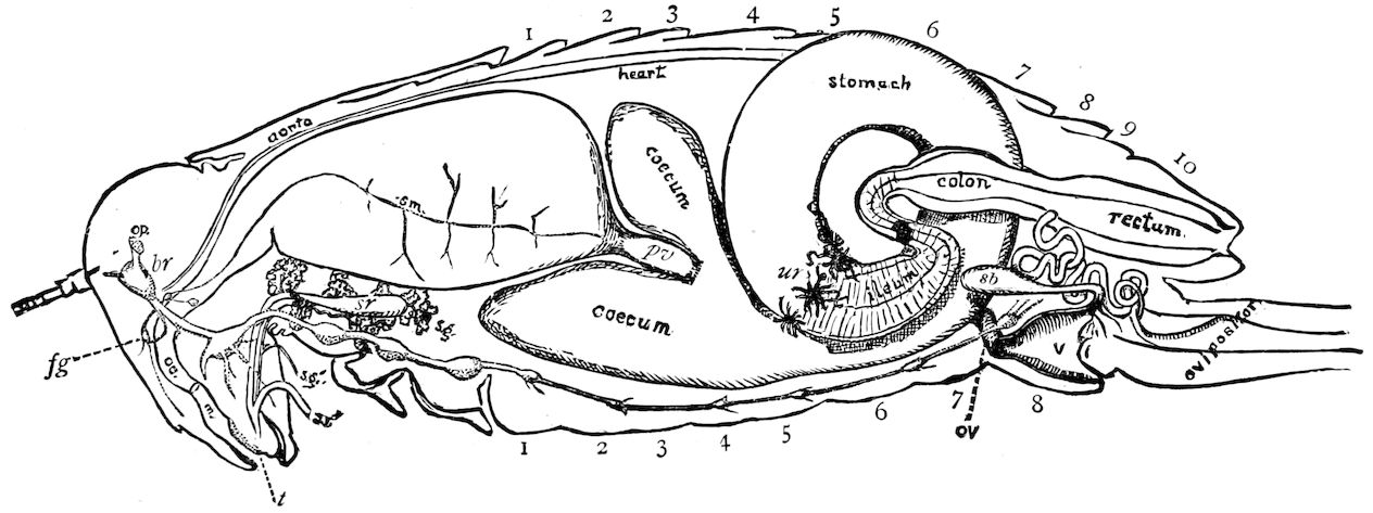
Fig. 299.—Digestive canal of Anabrus: m, mouth: œ, œsophagus; sm, the sympathetic nerve passing along the crop; t, tongue; fg, frontal ganglion; br, brain, the nervous cord passing backward from it; sr, salivary reservoir; sg, salivary gland; pv, proventriculus; ur, origin of urinary tubes; sb, sebaceous gland; 1–10, the ten abdominal segments.—Burgess del.
It is in the higher adult insects differentiated into the mouth and pharynx, the œsophagus or gullet, supplementary to which is the crop (ingluvies) or “sucking stomach” of Lepidoptera, Diptera, and Hymenoptera; the proventriculus or gizzard; the ventriculus, “chyle-stomach,” or, more properly, mid-intestine, and the hind-intestine, which is divided into the ileum, or short intestine, the long intestine, often slender and coiled, with the colon and the rectum. Morphologically, however, the digestive or enteric canal is divided into three primary divisions, which are indicated in the embryo insect; i.e., the fore-intestine (stomodæum of the embryo), mid-intestine or “chyle-stomach,” and hind-intestine or proctodæum (Fig. 300). The three primary regions, with their differentiations, may be tabulated thus:—
Fore-intestine (Stomodæum). Mouth and pharynx.
Pumping apparatus of Hemiptera, Lepidoptera, and Diptera.
Œsophagus.
Crop or ingluvies, food reservoir, or “sucking stomach.”
Proventriculus.
Mid-intestine (Mesenteron). Mid-intestine, “chylific stomach,” or ventriculus (with cœcal glands).
Hind-intestine (Proctodæum). Ileum, or short intestine (with the urinary tubes).
Long intestine.
Colon.
Rectum (with rectal glands).
Anus (with anal glands).
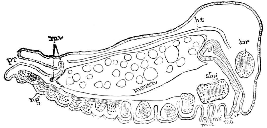
Fig. 300.—The three primary divisions of the alimentary canal of an embryonic orthopterous insect: br, brain; sbg, subœsophageal ganglion; ng, nervous cord; st, stomodæum; pr, proctodæum; mv, malphigian tubes; mesen, mid-intestine; ht, heart; md, mandibles; mx, mx′, 1st and 2d maxillæ.—After Ayers, with some changes.
The appendages of the alimentary canal are: (1) the salivary and poison glands, which arise from the stomodæum in embryonic life; 300(2) while to the chylific stomach a single pair of cœcal appendages (Orthoptera and larval Diptera, e.g. Sciara), or many cœca may be appended; (3) the urinary tubes, also the rectal glands and the paired anal glands. In a Hemipter (Pyrrhocoris apterus) appendages arise from the intestine in front of the origin of the urinary tubes. In certain insects a single cœcal appendage (Nepa, Dyticus, Silpha, Necrophorus, and the Lepidoptera) arises from the proctodæum.
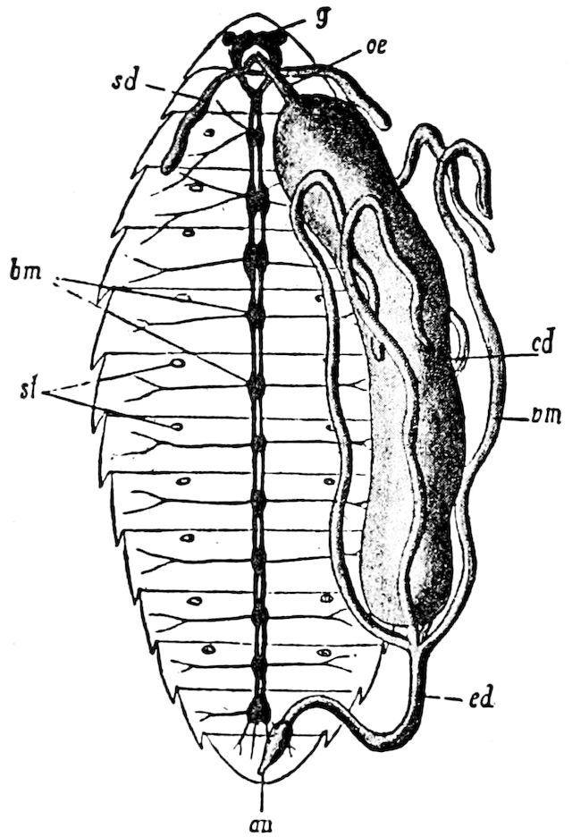
Fig. 301—Larva of honey-bee: g, brain; bm, ventral nervous cord; œ, œsophagus; sd, spinning-gland; cd, mid-intestine or chyle-stomach; ed, hind-intestine, not yet connected with the mid-intestine; vm, urinary tube; an, anus; st, stigmata.—After Leuckart, from Lang.
In certain larval insects, as those of the Proctotrypidæ (first larval stage), the higher Hymenoptera (ichneumons, ants, wasps, and bees, Fig. 301), in the Campodea-like larvæ of the Meloidæ and Stylopidæ, the larva of the ant-lion (Myrmecoleo), and those of Diptera pupipara (Melophagus), the embryonic condition of the separation of the proctodæum and mid-gut (mesenteron) persists, the stomach ending in a blind sac; in such cases the intestine, together with the urinary tubes, is entirely secretory.
The anus is wanting in the larva of the ant-lion, as also in the wasps (in which there is a rudimentary colon) and in freshly hatched bees, though it becomes perfectly formed in the fully grown larvæ (Newport, art. Insecta, p. 967, and H. Müller).
In the larvæ of lamellicorn Coleoptera (Melolontha vulgaris) the digestive tube is nearly as simple as in bees, though there is a large colon, which at its beginning forms an immense cœcum, and has also one anal aperture (Newport).
The length and shape of the digestive canal is dependent on the nature of the food and also on the mode of life, especially the ease or difficulty with which the food is digested.
301Newport, while stating that the length of the alimentary canal in larvæ is not in general indicatory of the habits of the species, makes this qualification after describing the digestive canal of Calandra as compared with that of Calosoma: “The length and complication of the intestines, therefore, appear to have some reference to the quality of the food to be digested, since it is well known that the food of these latter insects (weevils) is of difficult assimilation, being as it is chiefly the hard ligneous fibres of vegetable matter; but they cannot be received as always indicatory of a carnivorous [or] vegetable feeder, since, as above remarked, the length of the canal is considerable in one entirely carnivorous larva, while it is much shorter in some herbivorous, and particularly in pollenivorous larvæ, as in the Melolontha and the apodal Hymenoptera.”
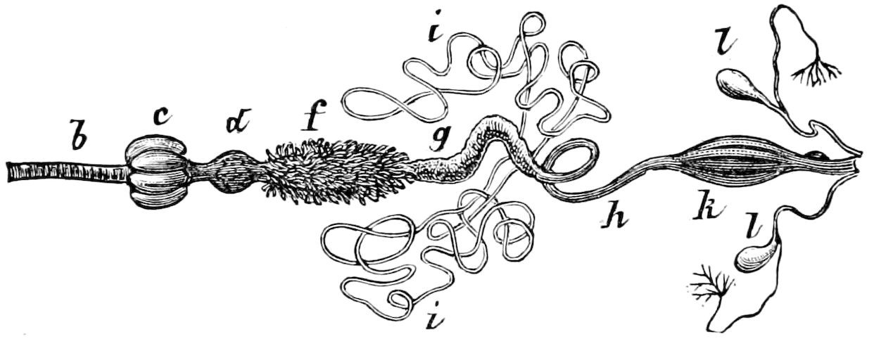
Fig. 302.—Digestive canal of a carabid beetle: b, œsophagus; c, crop; d, proventriculus; f, mid-intestine, or “chyle-stomach,” with its cœeca; g, posterior division of the stomach; i, the two pairs of urinary tubes; h, intestine; k, rectum; l, anal glands.—After Dufour, from Judeich and Nitsche.
Newport also contends that the length of the alimentary canal is not more indicative in the perfect insect of the carnivorous or phytophagous habits of the species than in the larva. It is nearly as long (being from two to three times the length of the whole body), and is more complicated, in the rapacious Carabidæ (Fig. 302) than in the honey-sipping Lepidoptera, whose food is entirely liquid. Referring to the digestive canal of Cicindelidæ, which is scarcely longer than the body, he claims that “we cannot admit that the length of the digestive organs, and the existence of a gizzard and gastric vessels, are indicatory of predacity of habits in the insect, because a similar conformation of parts exists often in strictly vegetable feeders. The existence and length of these parts seem rather to refer to the comparative digestibility of the food than to its animal or vegetable nature.” Newport then refers to the digestive canal of Forficulidæ (in which the gizzard is present, the canal, however, passing in an almost direct line through the body, making but one slight convolution), “a farther proof that the length of the canal must not be taken as a criterion whereby to judge of the habits of a species.” He adds this will apply equally well to the omnivorous Gryllidæ, in which there exists a short alimentary canal, but a gizzard of more complicated structure than that of the Dytiscidæ.
In larval insects and others (Synaptera, Orthoptera, etc.), in which the digestive canal is simplest, it is scarcely longer than the body, and passes through it as a straight tube.
In the caterpillar, which is a voracious and constant feeder, the digestive canal is a large straight tube, not clearly differentiated into fore-stomach, stomach, and intestine; but in the imago, which only 302takes a little liquid food, it is slender, delicate, and highly differentiated. In the larva the mid-gut forms the largest part of the canal; in the imago, the intestine becomes very long and coiled into numerous turns; at the same time the food-reservoir (the “sucking stomach”) develops, and the excretory tubes are longer.
a. The digestive canal
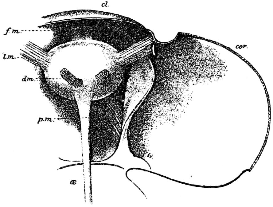
Fig. 303.—Interior view of the bottom of the head of Danais archippus, the top having been cut away, showing, in the middle, the pharyngeal sac with its five muscles: the frontal (f.m), dorsal pair (d.m), and the lateral pair (l.m); cl, clypeus; cor, cornea; œ, œsophagus; p.m, one of the large muscles which move the labial palp.—After Burgess.
It will greatly simplify our conception of the anatomy of the digestive canal if we take into account its mode of origin in the embryo, bearing in mind the fact that during the gastrula condition the ectoderm is invaginated at each pole to form the primitive mouth and fore-gut (stomodæum) and hind-gut (proctodæum). The cells of the ectoderm secrete a chitinous lining (intima), which forms the continuation of the outer chitinous crust, and thus the lining of each end of the digestive canal is cast whenever the insect molts; while the mid-intestine (mesenteron), arising independently of the rest of the canal much later in embryonic life from the mesoderm, is not the result of any invagination, being directly derived from the mesoderm, and is not lined with chitin.
The mouth, or oral cavity, and pharynx.—This is the beginning of the alimentary bounded above by the clypeus, and labrum, with the epipharynx, and below by the hypopharynx, or tongue, as well as the labium. Into it pour the secretion of the salivary glands, which passes out through an opening at the base of the tongue or hypopharynx. On each side of the mouth are the mandibles and first maxillæ.
The sucking or pharyngeal pump.—This organ has been observed by Graber in flies and Hemiptera, but the fullest account is that by Burgess, who was the first to discover it in Lepidoptera. In the milk-weed butterfly (Danais archippus) the canal traversing the proboscis 303opens into a pharynx enclosed in a muscular sac (Figs. 303, 304, and 310).
The pharyngeal sac, says Burgess, serves as a pumping organ to suck the liquid food through the proboscis and to force it backwards into the digestive canal.
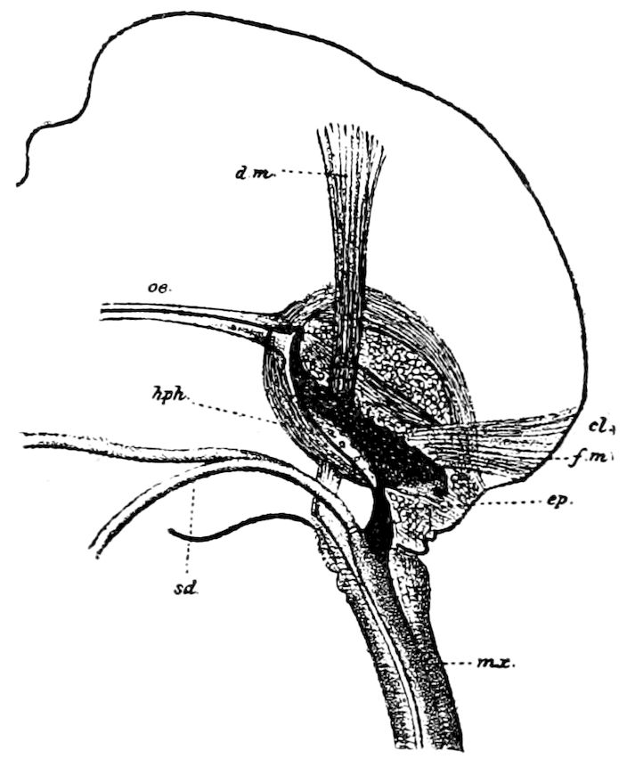
Fig. 304.—Longitudinal section through the head of Danais, showing the interior of the left half: mx, left maxilla, whose canal leads into the pharynx; hph, floor of the latter, showing some of the taste-papillæ; oe, œsophagus; ep, epipharyngeal valve; sd, salivary duct; d.m, f.m, and cl, as in Fig. 302.—After Burgess.
Meinert (“Trophi Dipterorum”) has made elaborate dissections of the mouth and its armature, including the pharynx of several types of Diptera, with its musculature. He describes the pharynx as the principal, and in most Diptera, as the only part of the pump (antlia), and says: “By the muscles of the pump (musculis antliæ) the superior lamina of the pharynx is varied that the space between the two laminæ may be increased, and the liquid is thus led through the siphon formed by the mouth-parts into the mouth” (Fig. 81).
The œsophagus.—This is a simple tube, largest in those insects feeding on solid, usually vegetable, food, and smallest in those living on liquid food. It usually curves upwards and backwards, passing directly under the brain, and merges into the crop or proventriculus either at the back part of the head or in the thorax, its length being very variable. Its inner walls longitudinally are folded and lined with chitin.
According to Newport, in the œsophagus of the Gryllidæ, of the two layers of the mucous lining the second is distinctly glandular and secretory, and in it there are many thousands of very minute granular glandular bodies, which probably secrete the “molasses” or repellent fluid often ejected by these and other insects when captured.
The crop or ingluvies.—This, when present, is an enlargement of the end of the œsophagus, and lined internally with a muscular coat. It is very large in locusts (Fig. 298), Anabrus (Fig. 299), and other Orthoptera (the Phasmidæ excepted), in the Dermaptera, and most 304adult Coleoptera. A crop-like dilatation in front of a spherical gizzard is also present in the Synaptera (Poduridæ and Lepismidæ), as well as in the Mallophaga (Nirmidæ).
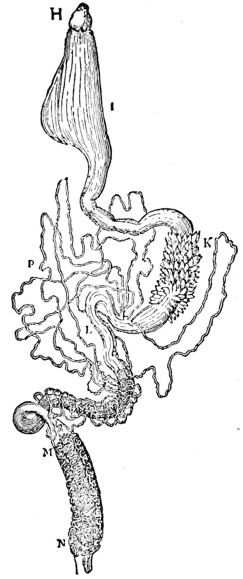
Fig. 305.—Digestive canal of Calandra: H, pear-shaped œsophagus; I, crop; K, gastric cœca L, ilium; MN, colon; P, urinary tubes.—After Newport.
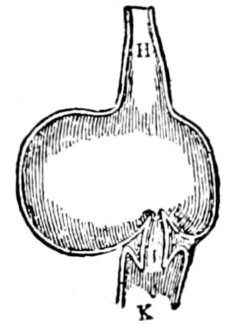
Fig. 306.—Section of the crop (H), gizzard (I), and stomach (K) of Athalia.-After Newport.
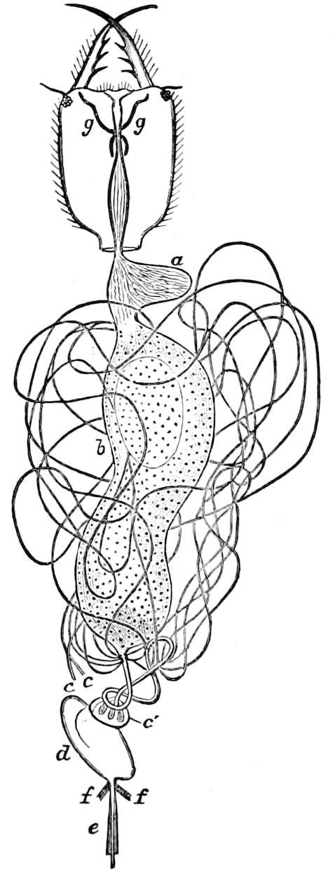
Fig. 307.—Upper side of head and digestive canal of Myrmeleon larva: a, crop; b, “stomach”; c, free ends of two urinary tubes; c′, common origin of other six tubes; d, cœcum; e, spinneret; ff, muscles for protruding its sheath; gg, maxillary glands.—After Meinert, from Sharp.
In the larvæ of weevils (Calandra sommeri) there is a crop (Fig. 305), but not in the larva of Calosoma; also, according to Beauregard, in the pollen-eating beetles Zonitis, Sitaris, and Malabris it is wanting, while in Meloe it is highly developed (Kolbe).
The crop forms a lateral dilatation of the end of the œsophagus in the larvæ of weevils and of saw-flies (Athalia centifoliæ, Fig. 306).
305The “sucking stomach” or food-reservoir.—This is a thin muscular pouch connected by a slender neck with the end of the œsophagus or the crop, when the latter is present. There is no such organ in Orthoptera, except in Gryllotalpa. It is wanting in the Odonata and in the Plectoptera (Ephemeridæ); in Platyptera (Perlidæ and Termitidæ), in Trichoptera, and in Mecoptera (Panorpidæ). In most adult Neuroptera (Myrmeleonidæ, Hemerobiidæ, and Sialidæ), but not in Rhaphidiidæ, the long œsophagus is dilated posteriorly into a kind of pouch or crop, and besides there is often a long “food-reservoir” arising on one of its sides, that of Myrmeleon (Fig. 307) and Hemerobius being on the right side.
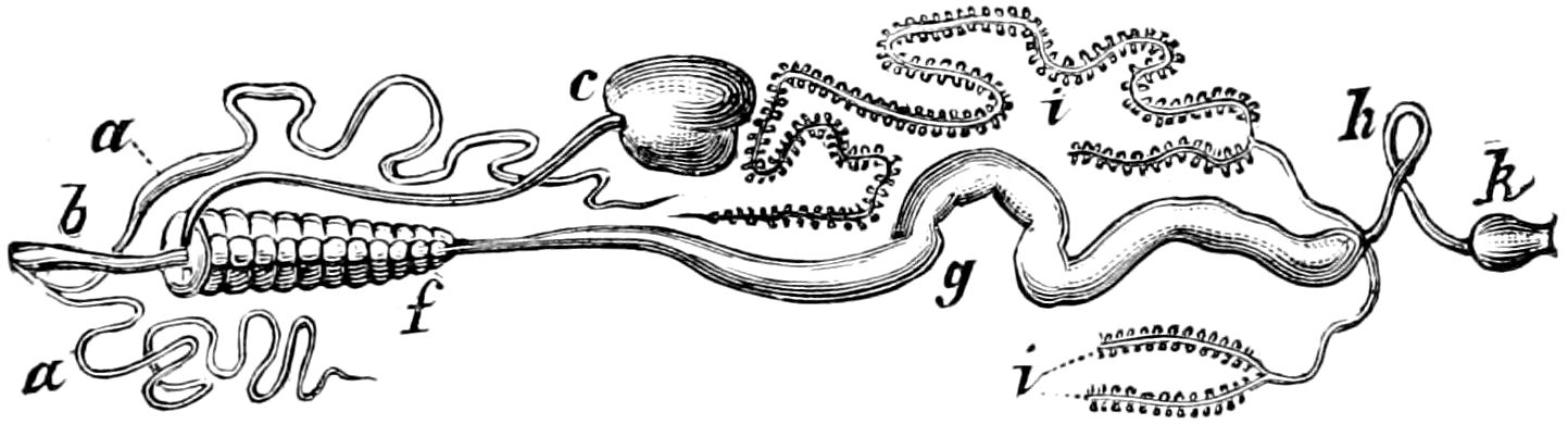
Fig. 308.—Digestive canal of Sarcophaga carnaria: a, salivary gland; b, œsophagus; c, food reservoir; f-g, stomach; h, intestine; i, urinary tubes; k, rectum.—From Judeich and Nitsche.
A true food-reservoir is present in most Diptera (Fig. 308) as well as in the larvæ of the Muscidæ, but according to Dufour it is wanting in some Asilidæ and in Diptera pupipara, and according to Brauer in the Œstridæ. The food-reservoir in Diptera is always situated on the left side of the digestive canal; there is usually a long neck or canal, while the reservoir is either oval or more usually bilobed, and often each lobe is itself curiously lobed.
In Lepidoptera (Figs. 309, 310) the so-called “sucking stomach” is, as Graber has proved, simply a reservoir for the temporary reception of food; though generally found to contain nothing but air, Newport has observed that in flies it is filled with food after feeding. He has found this to be the case in the flesh fly, and in Eristalis he has found it “partially filled with yellow pollen from the flowers of the ragwort upon which the insect was captured,” the pollen grains also occurring in the canal leading to the bag, in the gullet, and in the stomach itself. Graber has further proved by feeding flies with a colored sweet fluid that this sac is only a food-receptacle. As he says: “It can be seen filling itself fuller and fuller with the colored fluid, the sac gradually distending until it occupies half the hind-body.”
The food-reservoir of the Hymenoptera is a lateral pouch at the end of the long, slender œsophagus, and has been seen in the bee to be filled with honey.
306
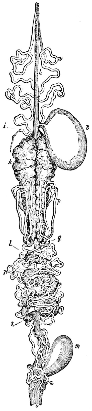
Fig. 309.—Digestive canal of Sphinx ligustri: h, œsophagus; i, rudiment of the gizzard; k, “stomach”; q, its pyloric end; t, food reservoir; p, urinary tubes; l, ilium; m, cœcum of colon; n, rectum; v, vent.—After Newport.
In the mole-cricket the hinder part of the crop is armed within with hook-like bristles directed backwards so as not to prevent the energetic pressure of the food backwards into the proventriculus, and to obviate the possibility of a regurgitation. (Eberli.)
The fore-stomach or proventriculus.—This is especially well developed in the Dermaptera, in the Orthopterous families Locustidæ, Gryllidæ, and Mantidæ, while in the Thysanura (Lepisma) there is a spherical gizzard provided with six teeth. It also occurs in many wood-boring insects, and in most carnivorous insects, notably the Carabidæ, Dyticidæ, Scolytidæ, in the Mecoptera (scorpion-flies), in the fleas, and in many kinds of ants, as well as Cynips, Leucospis, and Xyphidria. It is very muscular, lined within with chitin, which is usually provided with numerous teeth arising from the folds. These folds begin in the œsophagus or crop, and suddenly end where the mesenteron (“chylific stomach”) begins. It has been compared with the gizzard of birds, and is usually called by German authors the chewing or masticating stomach. (Kaumagen.)
The proventriculus is best developed in the Gryllidæ (Acrida viridissima), where the six folds at the end of the crop close together to form a valve between the crop and proventriculus. “They are each armed with five very minute hooked teeth; and, continued into the gizzard, develop many more in their course through that organ. These first teeth are arranged around the entrance to the gizzard, and seem designed to retain the insufficiently comminuted food and to pass it on to that organ.
307
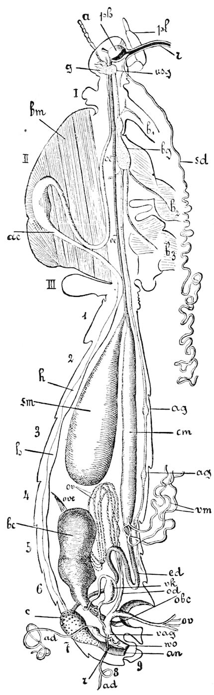
Fig. 310.—Anatomy of Danais archippus after removal of right half of the body. Lettering of the head: a, antenna; ph, pharynx; pl, labial palpi; r, proboscis; g, brain; usg, subœsophageal ganglion. Lettering of the thorax: I. II. III. thoracic segments; b1, b2, b3, the coxal joints of the three pairs of legs; bm, muscles of the wings; ac cephalic aorta with its swelling; œ, œsophagus; bg, thoracic ganglia of the ventral cord; sd, salivary glands of one side, those of the other side cut off near their entrance into the common salivary duct. Lettering of the abdomen: 1–9. abdominal segments; h, heart; sm, so-called sucking-stomach (food-reservoir); cm, chyle-stomach; ag, abdominal ganglia: ed, hind intestine with colon (c) and rectum (r); rm, urinary vessels; ov, ovarial tubes, those of the right side cut off; ove, terminal filaments of the ovaries; bc, bursa copulatrix; obc, its outer aperture; od, oviduct; vag, vagina; wo, its outer aperture; ad, glandular appendages of the vagina partly cut away; vk, connective canal between the vagina and bursa copulatrix with swelling (receptaculum seminis); an, anus.—After Burgess, from Lang.
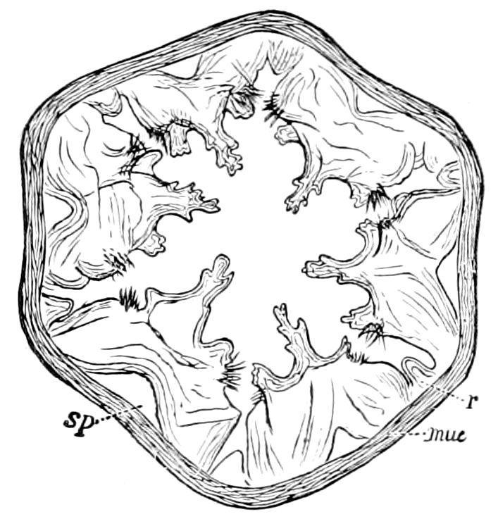
Fig. 311.—Transverse section of the proventriculus of Gryllus cinereus: muc, muscular walls; r, horny ridge between the large teeth (sp).—After Minot.
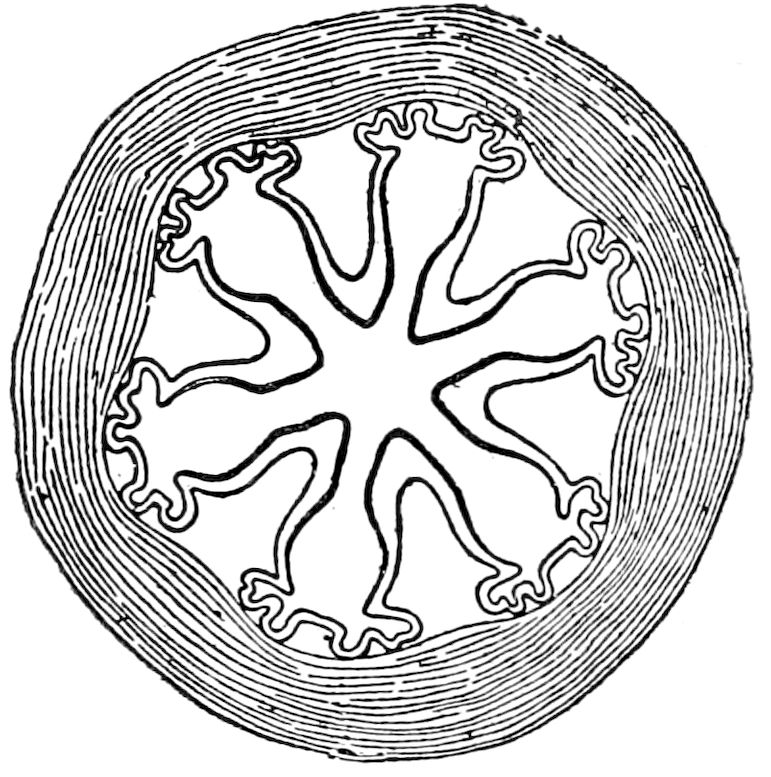
Fig. 312.—Transverse section of the proventriculus of the cockroach.—After Miall and Denny.
308
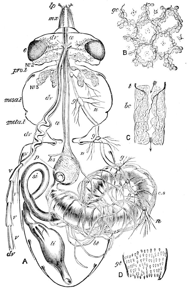
Fig. 313.—Digestive canal of the honey-bee: A, horizontal section of the body; lp, labial palpus; mx, maxilla; e, eye; pro. t, prothorax; mesa. t, mesothorax; meta. t, metathorax; dv, dorsal vessel; v, v, ventricles of the same; No. 1, No. 2, No. 3, salivary gland systems; œ œsophagus; g, g, ganglia of chief nerve-chain; n, nerves; hs, honey-sac; p, petaloid stopper or calyx of honey-sac or stomach-mouth; c. s, chyle stomach; bt, urinary tubes; si, small intestine (ilium); l, “lamellæ or gland-plates of colon,” rectal glands; li, rectum. B, cellular layer of stomach; gc, gastric cells, × 200. C, urinary tube; bc, cells; t, trachea. D, inner layer, with gastric teeth (gt).—After Cheshire.
“Next to these in succession on each of the longitudinal ridges are four flat, broad, somewhat quadrate teeth, each of which is very finely denticulated along its free margin. These extend about half-way through the gizzard. They appear to be alternately elevated and depressed during the action of the gizzard, and to serve to carry on the food to the twelve cutting teeth, with which each ridge is also armed, and which occupy the posterior part of the organ. These 309teeth are triangular, sharp-pointed, and directed posteriorly, and gradually decrease in size in succession from before backward. Each tooth is very strong, sharp-pointed, and of the color and consistence of tortoise shell, and is armed on each side by a smaller pointed tooth. These form the six longitudinal ridges of the gizzard, between each two of which there are two other rows of very minute teeth of a triangular form, somewhat resembling the larger one in structure, occupying the channels between the ridges. The muscular portion of the gizzard is equally interesting. It is not merely formed of transverse and longitudinal fibres, but sends from its inner surface into the cavity of each of the large teeth other minute but powerful muscles, a pair of which are inserted into each tooth. The number of teeth in the gizzard amounts to 270, which is the same number in these Gryllidæ as found formerly by Dr. Kidd in the mole-cricket. Of the different kinds of teeth there are as follows: 72 large treble teeth, 24 flat quadrate teeth, 30 small single-hooked teeth, and 12 rows of small triangular teeth, each row being formed of 12 teeth. This is the complicated gizzard of the higher Orthoptera.” (Newport.)
In the more generalized cockroach, there are six principal folds, the so-called teeth, which project so far inwards as to nearly meet (Fig. 312). The entire apparatus of muscles and teeth is, as Miall and Denny state, “an elaborate machine for squeezing and straining the food, and recalls the gastric mill and pyloric strainer of the crayfish. The powerful annular muscles approximate the teeth and folds, closing the passage, while small longitudinal muscles, which can be traced from the chitinous teeth to the cushions, appear to retract these last, and open a passage for the food.”
As in the fore-stomach or proventriculus of the lobster, the solid, rounded teeth do not appear to triturate the solid fragments found in the organ, but act rather as a pyloric strainer to keep such bodies out of the chylific stomach. We accept the view of Plateau that this section of the digestive canal in insects, which he compares to the psalterium of a ruminant, is a strainer rather than a masticatory stomach, and both Forel and Emery, as well as Cheshire, take this view.
The proventriculus of the honey-bee (Fig. 313, hs) is called by apiarians the “honey-sac” or “honey-stomach.” Cheshire states that if it be carefully removed from a freshly killed bee, its calyx-like “stomach-mouth” may be seen to gape open and shut with a rapid snapping movement. The entrance to the stomach is guarded by four valves, each of which is strongly chitinous within, and fringed along its edge with downward-pointing fine stiff bristles. By the contraction of the longitudinal muscles (lm), the valves open to allow the passage of food from the honey-sac to the “chyle-stomach.” It is closed at will by circular muscles (tm). Then the bee can carry food for a week’s necessities, either using it rapidly in the production of wax, or eking it out if the weather is unfavorable for the gathering of a new store.
310
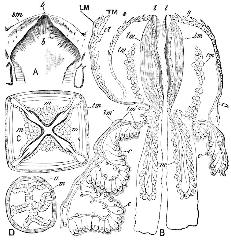
Fig. 314.—“Honey-sac stopper,” “stomach-mouth,” or calyx-bell of honey-bee, × 50. A, front view of one of the lobes of the calyx-bell; l, lip-like point, covered by down-turned bristles (b); sm, side membrane. B, longitudinal section of the stomach-mouth, with continuations into entrance of chyle-stomach; l, l, lip-like ends of leaflets; s, setæ; lm, longitudinal muscles; tm, transverse muscles in cross-section; cl, cell-layer of honey-sac; LM, TM, longitudinal and transverse muscles of same; nc, nucleated cells of tubular extension of stomach-mouth into chyle-stomach; lm′, tm′, longitudinal and transverse muscles of chyle stomach; c, c, cells covered within by an intima. C, cross-section of stomach-mouth; m, cross-section of muscles seen at lm in B; tm, transverse muscles surrounding stomach-mouth. D, cross-section through small intestine; a and m, longitudinal and surrounding muscles.—After Cheshire.
Cheshire also shows that when bees suck up from composite and other flowers nectar together with much pollen, the outside wrinkled membrane (sm, A, Fig. 314) “is seen to continually run up in folds, and gather itself over the top of the stomach-mouth, bringing with it, by the aid of its setæ, the large pollen-grains the nectar contains.” The lips (l, l, B, Fig. 314), now opening, take in this pollen, which is driven forwards into the cavity made between the separating lips by an inflow of the fluid surrounding the granules. The lips in turn close, but the down-pointing bristles are thrown outwards from the face of the leaflet, in this way revealing their special function, as the pollen is prevented from receding while the nectar passes back into the honey-sac, strained through between the bristles aforesaid, the last parts escaping by the loop-like openings seen in the corners of C, Fig. 314. The whole process is immediately and very rapidly repeated, so that the pollen collects and the honey is cleared. “Three purposes, in addition to those previously enumerated, are thus subserved by this wondrous mechanism. First, the bee can either eat or drink from the mixed diet she carries, gulping down the pollen in pellets, or swallowing the nectar as her necessities demand. Second, when the collected pollen is driven 311forwards into the chyle-stomach, the tube extension, whose necessity now becomes apparent, prevents the pellets forming into plug-like masses just below p, Fig. 313, for, by the action of the tube, these pellets are delivered into the midst of the fluids of the stomach, to be at once broken up and subjected to the digestive process. And third, while the little gatherer is flying from flower to flower, her stomach-mouth is busy in separating pollen from nectar, so that the latter may be less liable to fermentation and better suited to winter consumption. She, in fact, carries with her, and at once puts into operation, the most ancient, and yet the most perfect and beautiful, of all ‘honey-strainers.’”
Forel’s experiments on the proventriculus of ants prove that through its valvular contrivance it closes the passage from the crop to the mid-intestine (“chylific stomach”), and allows the contents of the former to pass slowly and very gradually into the latter. Emery confirms this view, and concludes that the organ in the Camponotidæ and in the Dolichoderidæ provided with a calyx-bell, usually regarded as a triturating stomach (Kaumagen), but more correctly as a pumping stomach, consists of parts which perform two different functions. Under the operation of the muscles of the crop the entrance to the pumping stomach becomes closed, in order by such spasmodic contraction to prevent the flow of the contents of the crop into the proventriculus. By the pressure of the transverse muscles of the proventriculus its contents are emptied into the mid-intestine, while simultaneously a regurgitation into the crop is prevented. In the Dolichoderidæ and Plagiolepidinæ the closure in both cases is effected by the valves. In the true Camponotidæ there are two separate contrivances for closing; the calyx belonging to the crop-musculature, while the valves essentially belong to the proventricular pumping apparatus.
Opinions vary as to the use of this portion of the digestive canal. Graber compares it to the gizzard of birds, and likens the action of the rosette of teeth to the finer radiating teeth of the sea-urchin, and styles it a chopping machine, which works automatically, and allows no solid bits of food to pass in to injure the delicate walls of the stomach (mid-gut).
He also states that the food when taken from the proventriculus is very finely divided, while that found in the œsophagus contains large bits.
Kolbe says that this view has recently been completely abandoned, and that the teeth are used to pass the food backwards into the chylific stomach. “But Goldfuss had denied the triturating action of the proventriculus of the Orthoptera (Symbolæ ad Orthopterorum quorundam Œconomiam, 1843), stating that the contents of the same are already fluid in the gullet, so that the fore-stomach (Kaumagen) does not need to comminute the food” (Kolbe). In the Gryllidæ and Locustidæ, just before the posterior opening of the proventriculus into the stomach the chitinous lining swells into a ring and projects straight back as the inner wall of the cylindrical chylific stomach. The muscular layer forms two sac-like outgrowths or folds, which separate on the circular fold from the chitinous membrane. This apparatus only allows very finely comminuted food to pass into the stomach.
In the Acrydiidæ (Eremobia muricata) at the end of the proventriculus, where it passes into the stomach, is a small circular fold which hangs down like a curtain in the stomach.
The œsophageal valve.—Weismann[50] states that the origin of the proventriculus in the embryo of flies (Muscidæ) shows that it should 312be regarded as an intussusception of the œsophagus. While in the embryo the invaginated portion of the œsophagus is short, after the hatching of the larva it projects backwards into the mid-intestine. Kowalevsky also observed in a young muscid larva, 2.2 mm. in length, that the œsophagus, shaped like a tube, extends back into the expanded portion (proventriculus) and opens into the stomach (Fig. 315, A). In a larva 10 mm. long the funnel is shorter, the end being situated in the proventriculus (Fig. 315, B, pr). In the cavity between the outer (o) and inner wall (i) no food enters, and the use of this whole apparatus seems to be to prevent the larger bits of food from passing into the chylific stomach (Kowalevsky).
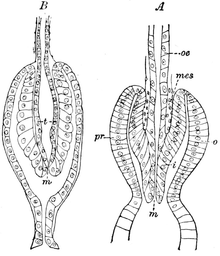
Fig. 315.—Œsophageal valve of young muscid larva: m, its opening: t, thickening of the cells; mes, mesoderm.—After Kowalevsky.
Beauregard has found a similar structure in the Meloidæ, and calls it the “cardiac valvule” (Fig. 318, Kl). It was observed by Mingazzini in the larvæ of phytophagic lamellicorn beetles, and Balbiani described it in a myriopod (Cryptops) under the name of the “œsophageal valvule.”
Gehuchten describes a homologous but more complicated structure in a tipulid larva (Ptychoptera contaminata), but differing in containing blood-cavities, as a tubular prolongation of the posterior end of the œsophagus which passes through the proventriculus and opens at various positions in the anterior part of the chylific stomach (Fig. 316).
The three layers composing this funnel are distant from each other and separated by blood-cavities, the whole forming “an immense blood-cavity extended between the epithelial proventricular lining and the muscular coat.”
According to Schneider the longitudinal muscular fibres of the fore and hind gut in insects pass into the stomach (mid-gut). The anterior part of the fore-gut has generally only circular fibres. When, however, the longitudinal fibres arise behind the middle, then they separate from the digestive canal and are inserted a little behind the beginning of the chylific stomach. Hence there is formed an invagination of the proventriculus, which projects into the cavity of the stomach.
Schneider describes this process, which he calls the “beak,” as an invagination of the fore-stomach which projects into the cavity of the stomach. The two layers of the invagination in growing together form a beak varying in shape, being either simple or lobed and armed with bristles or teeth. This beak is tolerably large in Lepisma, Dermaptera (Forficula), Orthoptera, and in the larvæ and adults of Diptera, but smaller in the Neuroptera and Coleoptera, while in other insects it is wanting.
313Proventricular valvule.—Gehuchten also describes in Ptychoptera what he calls “the proventricular valvule,” stating that it is “a circular fold of the intestinal wall” (Fig. 310, vpr). He claims that it has not before been found, the “proventricular beak” of Schneider being regarded by him as the œsophageal valvule.
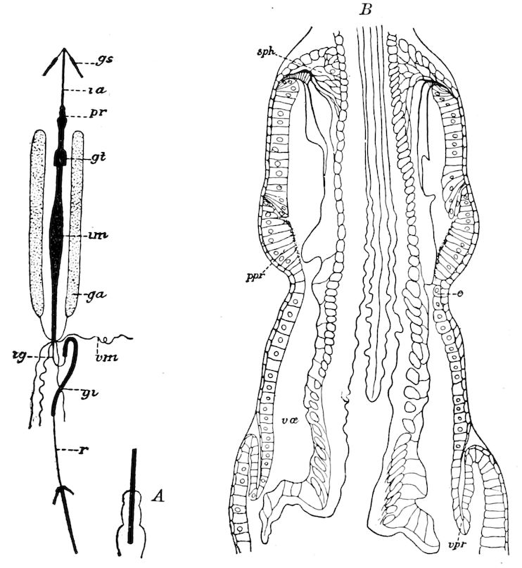
Fig. 316.—Digestive canal of Ptychoptera contaminata: gs, salivary glands; ra, œsophagus; pr, proventriculus; gt, crown of eight small tubular glands; im, mid-intestine; ga, two accessory white glands; vm, urinary vessels; ig, small intestine; gi, large intestine; r, rectum; A, the proventriculus in which the hinder end of the œsophagus extends as far as the chyle-stomach. B, longitudinal section of the proventricular region; sph, muscular ring or œsophageal sphincter; ppr, wall of the proventriculus; e, circular constriction dividing the cavity of the proventriculus in two; vpr, circular fold of the wall of the mid-intestine forming the proventricular valve; vœ, œsophageal valve.—After Gehuchten.
The peritrophic membrane.—This membrane appears first to have been noticed by Ramdohr in 1811 in Hemerobius perla. It has been found by Schneider, who calls it the “funnel.” On the hinder end of the fore-stomach, he says, the cuticula forms a fold enclosing the outlet of the fore-stomach, and extending back like a tube to the anus. This “funnel,” he adds, occurs in a great number of insects. It has been found in Thysanura, but is wanting in Hemiptera. In the Coleoptera it is absent in Carabidæ and Dyticidæ. It is generally present in Diptera and in the larvæ of Lepidoptera, but not in 314the adults. In Hymenoptera it has been found in ants and wasps, but is absent in Cynipidæ, Ichneumonidæ, and Tenthredinidæ. All those insects (including their larvæ) possessing this funnel eat solid, indigestible food, while those which do not possess it take fluid nourishment. It is elastic, and firmly encloses the contents of the digestive tract. Until Schneider’s discovery of its general occurrence, it had only been known to exist in the viviparous Cecidomyia larvæ (Miastor). Wagner, its discoverer, noticed in the stomach of this insect a second tube which contained food. Pagenstecher was inclined to regard the tube as a secretion of the salivary glands. Metschnikoff, however, more correctly stated that the tube consisted of chitin, but he regarded it as adapted for the removal of the secretions. (Schneider.) Plateau, however, as well as Balbiani, the latter calling it the “peritropic membrane,” considers this membrane as a secretion of the chylific stomach, and that it is formed at the surface of the epithelial cells. It surrounds the food along the entire digestive tract, forming an envelope around the fæcal masses. On the other hand, Gehuchten states that in the larva of Ptychoptera its mode of origin differs from that described by Plateau and by Schneider, and that it is a product of secretion of special cells in the proventriculus.