PART I.—MORPHOLOGY AND PHYSIOLOGY (continued 3)
Reading edition
101Loss of limbs by disuse.—Not only are one or both claws of a single pair, or those of all the feet atrophied by disuse, but this process of reduction may extend to the entire limb.
In a few insects one of the claws of each foot is atrophied, as in the feet of the Pediculidæ, of many Mallophaga, all of the Coccidæ, in Bittacus, Hybusa (Orthoptera), several beetles of the family Pselaphidæ, and a weevil (Brachybamus). Hoplia, etc., bear but a single claw on the hind feet, while the allied Gymnoloma has only a single claw on all the feet. Cybister has in general a single immovable claw on the hind feet, but Cybister scutellaris has, according to Sharp, on the same feet an outer small and movable claw. In the water bugs, Belostoma, etc., the fore feet end in a single claw, while in others (Corisa) both claws are wanting on the fore feet. Corisa also has no claws on the hind feet; Notonecta has two claws on the anterior four feet, but none on the hind pair. In Diplonychus, however, there are two small claws present. (Kolbe.)
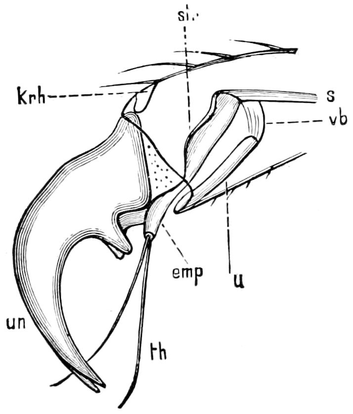
Fig. 110.—Last tarsal joint of Melolontha vulgaris, drawn as if transparent to show the inner mechanism: un, claws; str, extensor plate; s, tendon of the flexor muscle; vb, elastic membrane between the extensor plate and the sliding surface u; krh, process of the ungual joint; emp, extensor spine, and th, its two tactile hairs.—After Ockler, from Kolbe.
Among the Scarabæidæ, the individuals of both sexes of the fossorial genus Ateuchus (A. sacer) and eight other genera, among them Deltochilum gibbosum of the United States, have no tarsi on the anterior feet in either sex. The American genera Phanæus (Fig. 111), Gromphas, and Streblopus have no tarsal joints in the male, but they are present in the female, though much reduced in size, and also wanting, Kolbe states, in many species of Phanæus. The peculiar genus Stenosternus not only lacks the anterior feet, but also those of the second and third pair of legs are each reduced to a vestige in the shape of a simple, spur-like, clawless joint. The ungual joint is wanting in the weevil Anoplus, and becomes small and not easily seen in four other genera.
Ryder states that the evidence that the absence of fore tarsi in Ateuchus is due to the inheritance of their loss by mutilation is uncertain. Dr. Horn suggests that cases like Ateuchus and Deltochilum, etc., “might be used as an evidence of the persistence of a character gradually acquired through repeated mutilation, that is, a loss of the tarsus by the digging which these insects perform.” On the other hand, the numerous species of Phanæus do quite as much digging, and the anterior tarsi of the male only are wanting. “It is true,” he adds, “that many females are seen which have lost their anterior tarsi by digging; have, in fact, worn them off; but in recently developed specimens the 102front tarsi are always absent in the males and present in the females. If repeated mutilation has resulted in the entire disappearance of the tarsi in one fossorial insect, it is reasonable to infer that the same results should follow in a related insect in both sexes, if at all, and not in the male only. It is evident that some other cause than inherited mutilation must be sought for to explain the loss of the tarsi in these insects.” (Proc. Amer. Phil. Soc., Philadelphia, 1889, pp. 529, 542.)
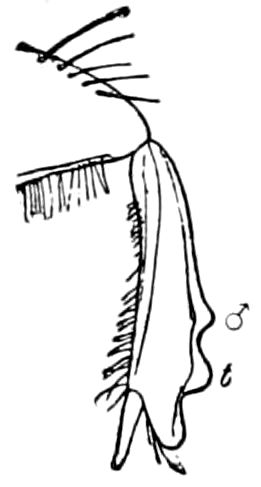
Fig. 111.—Fore tibia of Phanæus carnifex, ♂, showing no trace of the tarsus.
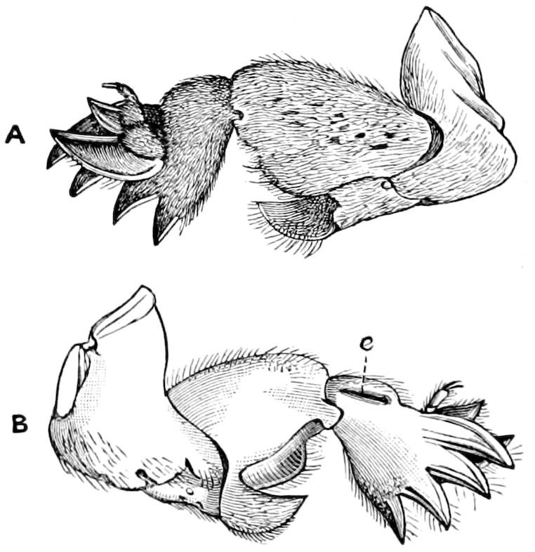
Fig. 112.—Fore leg of the mole-cricket: A, outer, B, inner, aspect; e, ear-slit.—After Sharp.
The loss of tarsi may be due to disuse rather than to the inheritance of mutilations. Judging by the enlarged fore tibiæ, which seem admirably adapted for digging, it would appear as if tarsi, even more or less reduced, would be in the way, and thus would be useless to the beetles in digging. Careful observations on the habits of these beetles might throw light on this point. It may be added that the fore tarsi in the more fossorial Carabidæ, such as Clivina and Scarites, as well as those of the larva of Cicada and those of the mole crickets (Fig. 112), are more or less reduced; there is a hypertrophy of the tibiæ and their spines. The shape of the tibia in these insects, which are flattened with several broad triangular spines, bears a strong resemblance to the nails or claws of the fossorial limbs of those mammals which dig in hard soil, such as the armadillo, manis, aardvark, and Echidna. The principle of modification by disuse is well illustrated in the following cases.
In many butterflies the fore legs are small and shortened, and of little use, and held pressed against the breast. In the Lycænidæ the fore tarsi are without claws; in Erycinidæ and Libytheidæ the fore legs of the males are shortened, but completely developed in the females, while in the Nymphalidæ the fore legs in both sexes are shortened, consisting in the males of one or two joints, the claws being absent in the females. Among moths loss of the fore tarsi is less frequent. J. B. Smith[21] notices the lack of the fore tarsi in the male of a deltoid, Litognatha nubilifasciata (Fig. 113), while the hind feet of 103Hepialus hectus are shortened. In an aphid (Mastopoda pteridis, Esl.) all the tarsi are reduced to a single vestigial joint (Fig. 114).
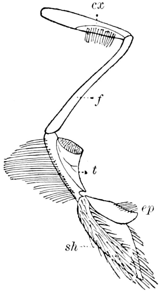
Fig. 113.—Leg of Litognatha: cx, coxa; f, femur; t, tibia; ep, its epiphysis, and sh, its shield-like process. The tarsus entirely wanting.—After Smith.
Entirely legless adult insects are rare, and the loss is clearly seen to be an adaptation due to disuse; such are the females of the Psychidæ, the females of several genera of Coccidæ (Mytilaspis, etc.), and the females of the Stylopidæ.
Apodous larval insects are common, and the loss of legs is plainly seen to be a secondary adaptive feature, since there are annectant forms with one or two pairs of thoracic legs. All dipterous and siphonapterous larvæ, those of all the Hymenoptera except the saw-flies, a few lepidopterous larvæ, some coleopterous, as those of the Rhyncophora, Buprestidæ, Eucnemidæ, and other families, and many Cerambycidæ are without any legs. In Eupsalis minuta, belonging to the Brenthidæ, the thoracic legs are minute.
The legs of larvæ end in a single claw, upon the tips of which the insect stands in walking.
c. Locomotion (walking, climbing, and swimming)
Mechanics of walking.—To Graber we owe the best exposition of the mechanics of walking in insects.
“The first segment of the insect leg,” he says, “upon which the weight of the body rests first of all, is the coxa. Its method of articulation is very different from that of the other joints. The enarthrosis affords the most extensive play, particularly in the Hymenoptera and Diptera.”
In the former the development of their social conditions is very closely connected with the freest possible use of the legs, which serve as hands. In the beetles, however, which are very compactly built, there exists a solid articulation whereby the entire hip rests in a tent-like excavation of the thorax, and can only be turned round a single axis, as may be seen in Fig. 115, where c represents the imaginary revolving axis and d the coxa. In the case we are supposing, therefore, only a backward and forward movement of the coxa is possible, the extent of the play of which depends on the size of the coxal pan, as well as certain groin or bar-like structures which limit further rotation. In the very dissimilar arrangement which draws in the fore, middle, and hind legs toward the body it is self-evident that their extent of action is also different. This arrangement seems to be most yielding on the fore legs, where the hips, to confine ourselves to the stag-beetles, can be turned backward and forward 60° from 104the middle or normal position, and therefore describe on the whole a curve of 120°. The angle of turning on the middle leg hardly exceeds a legitimate limit, yet a forward as well as a backward rotation takes place. The former is entirely wanting in the hind hips; they can only be moved backward.
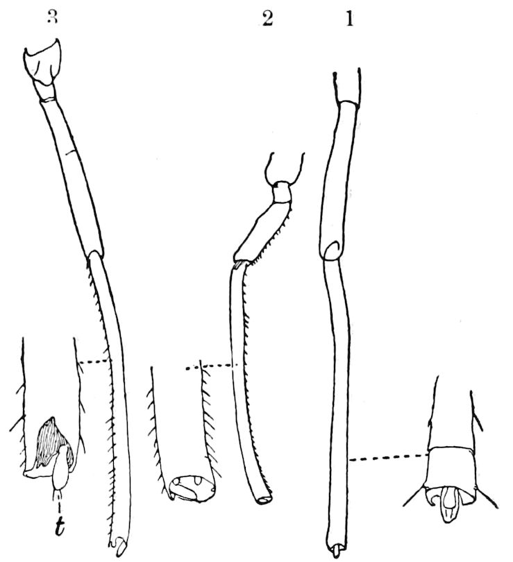
Fig. 114.—Leg of an Aphid, with the tarsus (t) much reduced: 1, 2, 3, legs of 1st, 2d, and 3d pairs.
The number and strength of the muscles on which the rotation of the hips depends, correspond with these varying movements of the individual legs. Thus, according to Straus Durckheim, the fore coxa of many beetles possesses five separate muscles and four forward and one backward roll; the middle coxa a like number of muscles but only two forward rolls, while the hind hips succeed in accomplishing each of the motions named with a single muscle.
One can best see how these muscles undertake their work, and above all how they are situated, if he lays bare the prothorax of the stag beetle (Fig. 116). Here may be seen first the thick muscle which turns to the front the rotating axis in its cylindrical pan, and thus helps to extend the leg, while two other tendons, which take the opposite direction, are fitted for reflex movements.
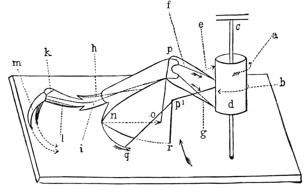
Fig. 115.—Mechanics of an insect’s leg: d, coxa,—c, axis of revolution; a and b, the coxal muscles; e, trochanter muscle (elevator of the femur); f, extensor,—g, flexor, of the tibia (pn); n, tibial spine; h, flexor.—i, extensor, of the foot; k, extensor,—l, flexor, of the claw; po, place of flexure of the tibia; p1q, leg after being turned back by the coxa.—p1r, by the simultaneous flexure of the tibia. The resulting motion of the end of the tibia, through the simultaneous movement (no) and revolution (nq), indicates the curve nr.—After Graber.
In Fig. 115 the muscles mentioned above, and their modes of working, may be distinguished by the arrows a and b.
In order to simplify matters, we will imagine the second component part 105of the normal insect leg, i.e. the trochanter (Figs. 116, 117, r), as grown together with the third lever, i.e. the femur, as the movement of both parts mostly takes place uniformly.
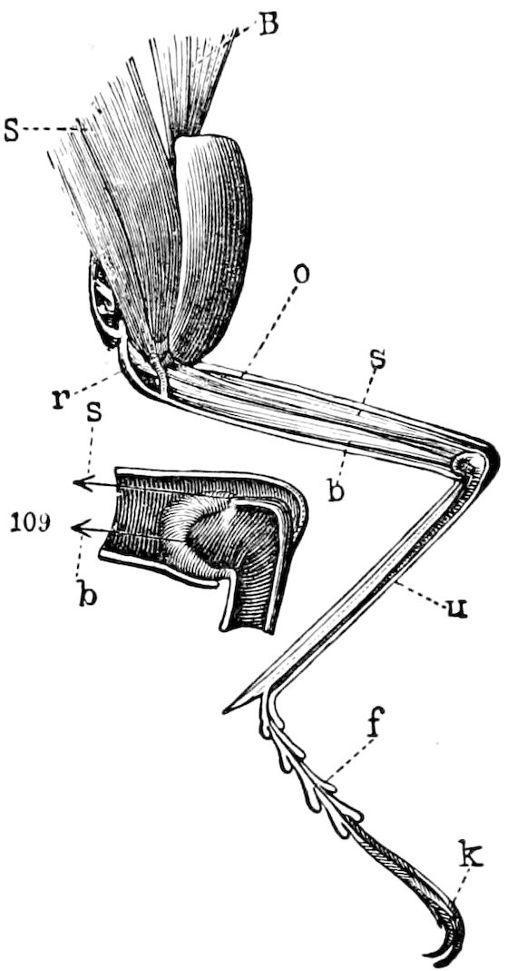
Fig. 116.—Section of the fore leg of a stag-beetle, showing the muscles: S, extensor,—B, flexor, of the leg; s, extensor,—b, flexor, of the femur; o, femur; u, tibia; f, tarsus; k, claw; 109, s, extensor,—b, flexor, of the femoro-tibial joint, both enlarged.—After Graber.
The pulling of the small trochanter muscle works against the weight of the body when this is carried over on to the trochanter by means of the coxa, as seen at the arrow e in Fig. 115. It may be designated as the femoral lever.
The plane of direction in which the femur, as seen by the rotation just mentioned, is moved, exactly coincides in insects with that of the tibia and the foot, while all can be simultaneously raised or dropped, or, as the case may be, stretched out or retracted. Therein, therefore, lies an essential difference from the fully developed extremities of vertebrates among which, even on the lever arms which are stationary at the end, an extensive turning is possible.
The muscles which move the tibia, and indirectly the femur, also consist of an extensor muscle which is situated in the upper side of the femur (Fig. 116, s, Fig. 115, f), and of a flexor (Fig. 116, b, Fig. 115, g), which lies under the former.
The stilt-like spines on the point (Figs. 115 and 118, L3n) on which this segment is directly supported are important parts of the tibia. (Graber.)
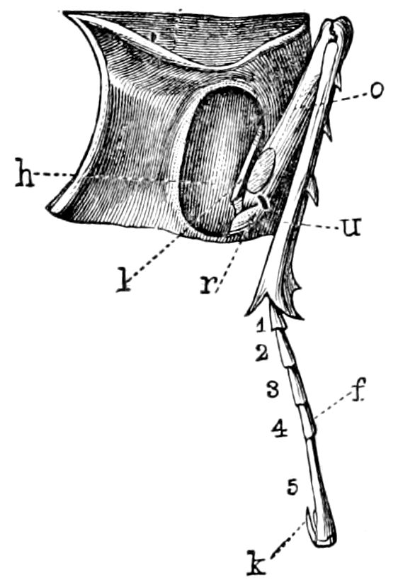
Fig. 117.—Left fore leg of a cerambycid beetle: h, coxa; r, trochanter; o, femur; u, tibia; f, tarsus; k, claw.—After Graber.
Considering the respective positions of the individual levers of the leg and the nature of the materials of which they are made, the legs of insects may be likened, as Graber states, to elastic bows, which, when pressed down together from above, their own indwelling elasticity is able to raise again and thus keep the body upright.
This is very plainly shown in certain stilt-legged bark-beetles, in which, as in a rubber doll, as soon as the body is pressed down on the ground, the organs of motion extend again without the intervention of muscles; indeed this experiment succeeds even with dead, but not yet wholly stiff, insects.
Graber then turns to the analysis of the movements of insect legs when in motion, 106and the mode of walking of these insects in general. This subject had been but slightly investigated until Graber made a series of observations and experiments, of which we can give only the most important results.
The locomotion of insects is an extremely complicated subject.
Let us consider, Graber says, first, a running or carabid beetle, when walking merely with the fore and hind legs. The former will be bent forward and the latter backward.
“Let us begin with the left fore leg (Fig. 118, L1). Let the same be extended and fixed on the ground by means of its sharp claws and its pointed heel. Now what happens when the tibial flexors draw together? As the foot, and therefore the tibia also, have a firm position, then the contraction of the muscles named must cause the femur to approach the tibia, whereby the whole body is drawn along with it. This individual act of motion may be well studied in grasshoppers when they are climbing on a twig by stretching out their long fore leg directly forward, and then drawing up the body through the shortening of the tibial flexors until the middle leg also reaches the branch.
“But while the fore legs advance the body by drawing the free lever to the fixed leg-segment, the hind legs do this in exactly the opposite way. The hind leg, namely, seeks to stretch out the tibia, and thus to increase the angle of the knee (R3), thereby giving a push on the ground, by means of which the body is shoved forward a bit.
“Though it might be supposed that the feet would remain stationary during the extension or retraction of the limbs, this never occurs in actual walking. Not merely the upper, but also the lower, thigh is either drawn in or stretched out, as the case may be. The latter then describes a straight line with its point during this scraping or scratching motion (Fig. 115, no), which is obviously the chord to that quadrant which would be drawn by the tibia or foot in a yielding medium, as water, for instance. But even this motion results extremely rarely, and never in actual walking. If we fix our eye anew upon the fore leg at the very moment when it is again retracted, after the resultant ‘fixing,’ we shall then observe that the hip also is simultaneously turned backward in a definite angle. The tibia would describe the arc nq (Fig. 115) by means of the latter alone.
“This plane, in conjunction with the rectilinear ‘movement’ (no) obtained by the retraction of the tibia, produces a path (nr), and this is what is actually described by a painted foot upon a properly prepared surface, as a sheet of paper;[22] supposing, however, that the body in the meantime is not moved forward by other forces. In the last case, and this indeed always takes place in running, the trunk is moved a bit forward, together with the leg which is just describing its curve with a rapidity corresponding to the momentum obtained; the result of this is that the curve of the foot from its beginning (n) to its end (a) bends round close to itself, just as a man who, when on board a ship in motion, walks across it diagonally, and yet on the whole moves forward, because his line of march, uniting with that of the ship, results in a change of position in space.
107“The case is the same in the middle and hind legs, which must make a double course also, yet in such a way that the straight line is drawn, not during the retraction, but during the extension; during which, however, quite as in the fore leg, the members mentioned (R3) gradually approach the body.
“When the legs have reached the maximum of their retraction, or of their extension, as the case may be, and therefore the end of their active course for that time, then begins the opposite or backward movement; that is, the fore legs are again extended, while their levers draw the remaining legs together again.
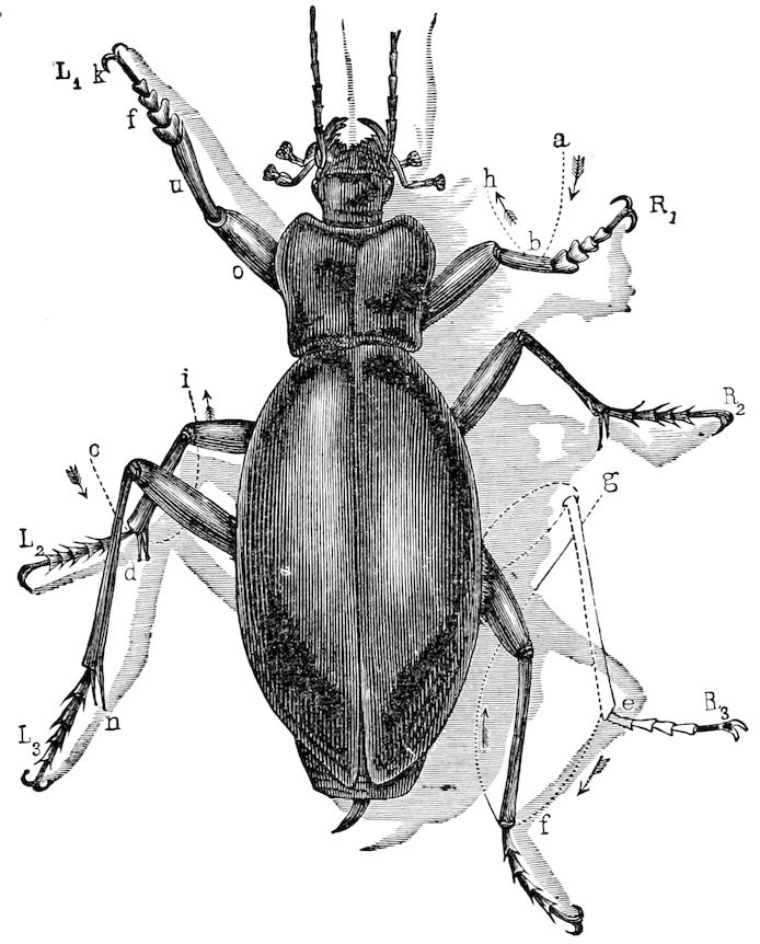
Fig. 118.—A Carabus beetle in the act of walking or running: three legs (L1, R2, L3) are directed forward, while the others (R1, L2, R3), which are directed backward toward the tail, have ended their activity; ab, cd, and ef are curves described by the end of the tibiæ, and passing back to the end of the body; bh, di, and fg are curves described by the same legs during their passive change of position.—After Graber.
“At the same time, as we may see by the uniting leg, the limb is either a little raised, that there may be no unnecessary friction, or it remains during the passive step also, with its means of locomotion in slight contact with the ground.
“The curve of two steps, as inscribed by the end of the tibia of the left fore leg of a stag-beetle, affords an instructive summary of the conditions of which we have been speaking (Fig. 121, B). We see two curves. The thick one (ab), directed toward the axis of the body, corresponds to the effective act of a single 108walking function, which brings the body a bit forward; the thinner, on the other hand, or we might say the hair line (bc), which, however, is but rarely made quite clearly, is produced by the ineffectual backward movement, by which the insect again approaches its working posture (c). It is at first placed at some distance from the body, in order that (like c also) it may draw near to the body again; but in such a way, naturally, that it coincides with the starting-point of the following active curve (cd). It is evident that even the passive curve is not the imprint of the movement accomplished exclusively by the leg, for this latter, while struggling to reach its resting-place, is really involuntarily carried forward with the rest of the body.
“The scroll-like lines drawn by the swimming beetle (Dyticus), with the large, sharp points of its hind tibia, are also very instructive (Fig. 119, A).
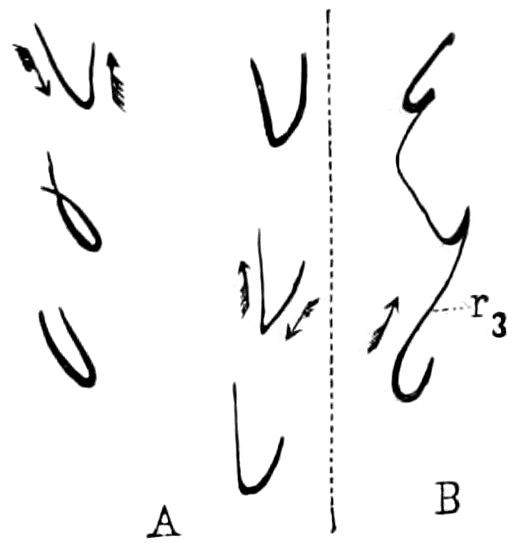
Fig. 119.—A, trail curves described by the tibial spines of the right and left hind limb of Dyticus. B, the same made by the right hind leg (r3) alone. Natural size.—After Graber.
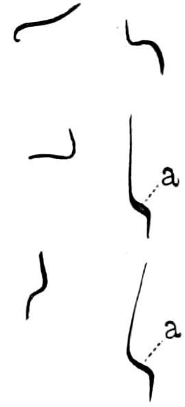
Fig. 120.—The same by the two hind legs of Melolontha: a, the active and thickened section of the curve. Natural size.
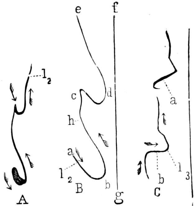
Fig. 121.—A, track curves of two of the tibial spines of the left, middle legs of a stag-beetle. Natural size. B, the same enlarged; fg, the longitudinal axis of the trunk; cd and ab, the active curve passing inward,—bc and de, the passive going outward. C, two curves described by the left hind legs; in this case, the curves are not inwards or backwards, but partly directly inward (b), and in part obliquely forwards (a).
“The diversions and modifications in the course of the active step, as furnished by the moving factor of the remaining legs, are already clearly illustrated by the curves shown by the joints of the hind tibia of a May-beetle (Fig. 120) and a stag-beetle (Fig. 121, c). The actual faint line in this case does not run from the front toward the back, as would correspond to the active leg-motion, but either directly inward (Fig. 121, cb), or even somewhat to the front. In the May-beetles, and even more in the running garden-beetle, the curves of the hind legs present themselves as screw-like lines (Fig. 122, l3), while the scrawling of the remaining members (l1, l2) is much simpler.
“Inasmuch as we now have a cursory knowledge of the movements made by each individual leg for itself,—movements, however, which plainly occur very differently according to the structure of these appendages,—the question now is of the combined play, the total effect of all the legs taken together, and therefore of the walk and measure of the united work of the foot.
“In opposition to the caterpillars and many other crawling animals which extend their legs in pairs and really swing them by the worm-like mode of contraction of the dermo-muscular tube, the legs of fully grown insects are moved in the contrary direction and in no sense in pairs, but alternately—or, more strictly speaking, in a diagonal direction.
109“For an examination of the gait of insects, we choose, for obvious reasons, those which have very long legs and which at the same time are slow walkers.
“Insects may be called ‘double-three-footed,’ from the manner in which they alternately place their legs. There are always three legs set in motion at the same time, or nearly so, while in the meantime the remaining legs support the body, after which they change places.
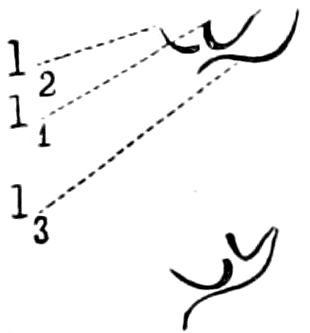
Fig. 122.—The same by the left fore (l1), middle (l2), and hind, leg (l3) of a Carabus. Natural size.
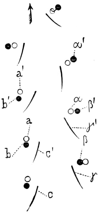
Fig. 123.-Tracks of a Blaps mortisaga marked by the differently painted tibial points: ●, tracks of fore, —○, middle, —/, hind leg. Natural size.
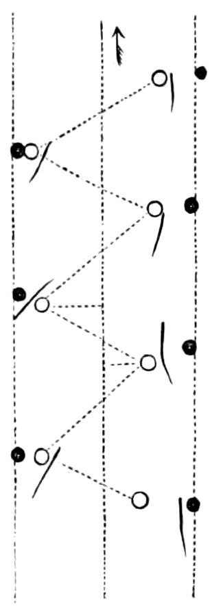
Fig. 124.—Tracks of Necrophorus vespilio. Natural size.
“To be more exact, it is usually thus: At first (Fig. 118) the left fore leg (L1) steps out, then follows the right middle leg (R2), and the left hind leg (L3). Then while the left fore leg begins to retract and thus make the backward movement, the right fore leg is extended, whereupon the left middle leg and the right hind leg are raised in the same order as the first three feet.”
Graber[23] painted the feet of beetles and let them run over paper, and goes on to say:
“Let us first pursue the tracks of the Blaps, for example (Fig. 123). Let the insect begin its motion. The left fore leg stands at a, the right middle leg at β, and the left hind leg at c. The corresponding number of the other set of three feet at α, b, γ. At the first step the three feet first mentioned advance to a′β′c′, the second set on the other hand to α′b′γ′. Thereby the tracks made by the successive steps fall quite, or almost quite, on each other, as appear also in the tracks of a burying beetle (Fig. 124).
“As the fore legs are directed forward and the hind legs backward, while the middle legs are placed obliquely, the reason of the more marked impressions of the latter is evident.
“The highest testimony to the precise exactitude and accuracy of the walking mechanism of insects is furnished by the fact that in most insects, and particularly in those most fleet of foot, which, whether they are running away or chasing their prey, must be able to rely entirely upon their means of locomotion;—the fact, we say, that whether they desire to move slowly or more 110quickly, the distances of the steps, measured by the length as well as by the cross-direction, hardly differ a hair’s breadth from one another, and this is also the case when the tarsi are cut off and the insects are obliged to run on the points of their heels (tibiæ).
“Thence, inasmuch as the trunk of insects is carried by two legs and by one on each side alternately, it may surely be concluded a priori that when walking it is inclined now to the right and now to the left, and that the track, too, which is left behind by a precise point of the leg, can in no wise be a straight line; and in reality this is not the case.
“A plainly marked regular curve, which approaches a sinuous line, as seen in Fig. 125, is often obtained by painting many insects, for example Trichodes, Meloë, etc., which, when running, either bring the end of their hind body near to the ground or into contact with it.
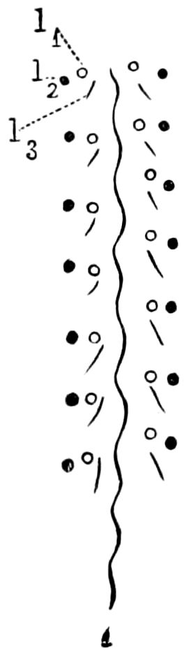
Fig. 125.—Tracks of Trichodes; the middle sinuous line is made by the tip of the abdomen. Natural size.
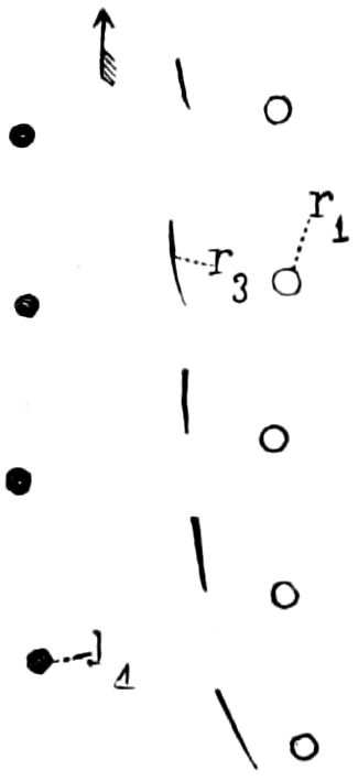
Fig. 126.—Tracks of another insect which, in running, can only use three legs (r1, l4, r3) which become indicated differently from normal conditions. Natural size.
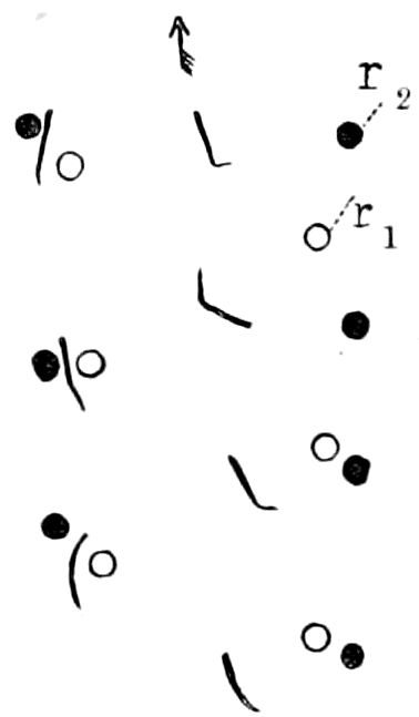
Fig. 127.—The same of an insect crossing over a surface inclined 30° from the horizon, whereby the placing of the feet becomes changed. Natural size.—This and Figs. 120–126 after Graber.
“The locomotive machine of insects may be called, to a certain extent, a double set of three feet each, as most insects, and particularly those provided with a broad trunk, are able to balance themselves with one of these two sets of feet, and indeed when walking, as well as when standing still, can move about even better with one set of these feet than with four legs. In the latter case, that is, if one cuts off a pair of legs from an insect, the trunk can balance itself only with extreme difficulty, and there is therefore little prospect that insects will ever become four-footed.
“But if one compels insects to run on three legs, he will thus make the interesting discovery that to make up the deficiency they place the remaining feet and bring them to the ground somewhat differently than when the second set of feet is active. Figs. 124 and 126 may be compared for this purpose. The former shows the footprints of a burying beetle running with all six legs, the latter the track of the same insect, which, however, has at its disposal only the right fore leg, the left middle leg, and the right hind leg. One may plainly see here that the track of the hind leg on the right side (r3) approaches the track of the middle leg on the left side, and then further, that the right fore leg (r1) steps out more to the right to make up for the deficiency of the middle leg.
111“A similar adaptation of the position of the legs, which is entirely dependent on the choice of the insect, may also be observed there, if one compels insects which are not provided with corresponding adhesive lobes to run away over crooked surfaces. Fig. 123 shows the footprints of a Blaps when running upon a horizontal plane. Fig. 127, on the contrary, shows the tracks of the legs when going diagonally over a gradually inclined surface. Here, also, the insect holds on with his fore and middle legs (r1, r2) stretched upward, whereby also the impressions on both sides come to lie farther apart than in the normal mode of walking.
“It will not surprise the reader who is familiar with the gait of crabs, to hear that many insects also understand the laudable art of going backward, wherein the hind legs simply change places with the fore legs.
“The jumping motion of insects may be best studied in grasshoppers. When these insects are preparing for a jump, they stretch out the upper thigh horizontally, clap the tibiæ together, and also retract the foot-segment. After a slight pause for rest, during which they are getting ready for the jump, they then jerk the tibiæ suddenly backward and against the ground with all their strength by means of the extensor muscles.”
The correctness of Graber’s views has been confirmed by Marey by instantaneous photographs (Figs. 128, 129).
Locomotion on smooth surfaces.—How flies and other insects are able to walk up, or run with the body inverted, on hard surfaces has been lately discovered by Dewitz, Dahl, and others. All authors are agreed that this power is due to the presence of the specialized empodium of each tarsus.
Dewitz confirmed the opinion of Blackwell, that a glutinous liquid is exuded from the apices of the tenent hairs which fringe the empodium. By fastening insects feet uppermost on the under side of a covering glass which projects from a glass slide, the hairs which clothe the empodia of the foot of a fly (Musca erythrocephala) may be seen to be tipped with drops of transparent liquid. On the leg being drawn back from the glass, a transparent thread is drawn out, and drops are found to be left on the glass. In cases where these hairs are wanting, as in the Hemiptera, the adhesive fluid exudes directly from pores in the foot. In the beetles (Telephorus dispar) and other insects the tenent hairs on the foot end in sharp points, below which are placed the openings of the canals. The glands, Dewitz states, are chiefly flask-shaped and unicellular, situated in the hypodermis of the chitinous coat; each gland opening into one of the hairs (Fig. 108); they are each invested by a structureless tunica propria, and contain granular protoplasm, a nucleus placed at the inner side, and a vesicle, prolonged into a tube which, traversing the neck of the gland, is attached to the root of the hair; the vesicle receiving the secretion. Each gland is connected with a fine nerve-twig, and secretion is probably voluntary. Among the tenent 112hairs of the empodium are others which must be supplied with a nerve, forming tactile hairs, as they each proceed from a unicellular ganglion (Fig. 108, n″). The secretion is forced out of the gland by the contraction of the protoplasm, Dewitz having seen the secretion driven out from the internal vesicle into its neck.
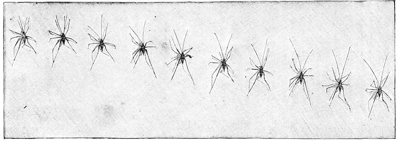
Fig. 128.—The walk of an orthopterous insect: series to be followed from right to left.—After Marey.
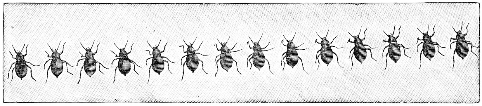
Fig. 129.—Beetle walking: series to be followed from left to right.—After Marey.
113
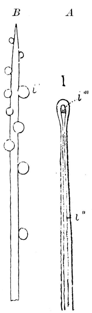
Fig. 130.—A, end of an adhesive hair of a weevil (Eupolus): i′, canal: i‴, its external opening at the end of the hair. B, end of a similar hair of Telephorus with drops of the secretion.—After Dewitz.
In the spherical last tarsal joint of Orthoptera (Fig. 109), which is without these tenent hairs, nearly all the cells of the hypodermis are converted into unicellular glands, each of which sends out a long, fine, chitinous tubule, which is connected with its fellows by very fine hairs and is continuous with the chitinous coat of the foot and opens through it. The sole of the foot is elastic and adapts itself to minute inequalities of surfaces, while the anterior of each tarsal joint is almost entirely occupied by an enlargement of the trachea, which acts on the elastic sole like an air chamber, rendering it tense and at the same time pliant. Dewitz adds that the apparatus situated on the front legs of the male of Stenobothrus sibiricus (Fig. 131) must have the function of causing the legs to adhere closely to the female by the excretion of an adhesive material. The hairs of the anterior tarsi of male Carabi also appear to possess the power of adhesion. In the house-fly the empodia seem to be only called into action when the insect has to walk on vertical smooth surfaces, as at other times they hang loosely down.
Burmeister observed the use of a glutinous secretion for walking in dipterous larvæ, and Dewitz found that the larva of a Musca used for this purpose a liquid ejected from the mouth. The larvæ of another fly (Leucopis puncticornis) perform their loop-like walk by emitting a fluid from both mouth and anus. A Cecidomyia larva is able to leap by fixing its anterior end by means of an adhesive fluid. The larva of the leaf-beetle, Galeruca, moves by drawing up its hinder end, fixing it thus, and carrying the anterior part of the body forward with its feet until fully extended, when it breaks the glutinous adhesion. The abdominal legs of some saw-fly larvæ have the same power.
Dahl could not detect in the foot of the hornet (Vespa crabro) any space which could be considered as a vacuum.
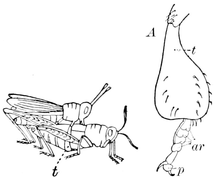
Fig. 131.—Stenobothrus sibiricus pairing: A. the ♂, fore tarsus (t) greatly enlarged; ar, arolia; p, pulvillus.—After Pagenstecher.
Simmermacher states that in most cases of climbing beetles the tubular tenent hairs pour out a secretion (Figs. 133, 134), “and it is probable that we have here to do with the phenomena not of actual attachment by, as it were, gluing, but of adhesion; the orifice of the tubes is divided obliquely, and the tubes are, at this point, extremely delicate and flexible, so as to adhere by their lower surface; 114in this adhesion they are aided by the secreted fluid.” In the case of the Diptera he does not accept the theory by which the movement of the fly along smooth surfaces is ascribed to an alternate fixation and separation, but believes in a process of adhesion, aided by a secretion, as in many Coleoptera. (In the Cerambycidæ there is no secretion, and the tubules are merely sucking organs, like those observed in the male Silphidæ.) “The attaching lobes, closely beset with chitinous hairs, are enabled, in consequence of the pressure of the foot, to completely lie along any smooth surface; this expels the air beneath the lobes, which are then acted on by the pressure of the outer air.” (Journ. Roy. Micr. Soc., 1884, p. 736.) Another writer (Rombouts) thinks this power is due to capillary adhesion.
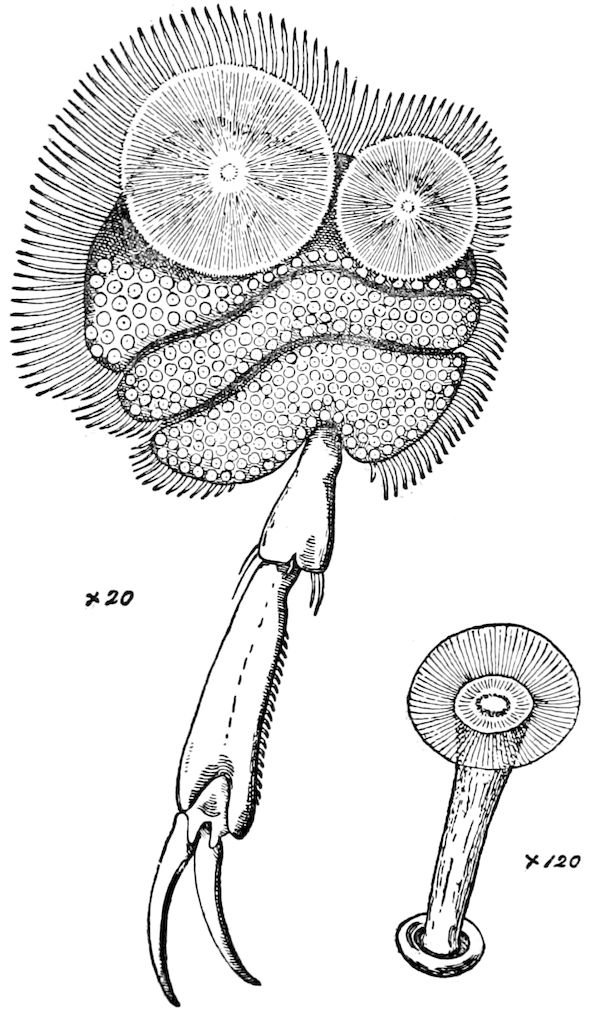
Fig. 132.—Fore leg of ♂ Dyticus, under side, with sucker, formed of 3 enlarged tarsal joints: with a small cupule highly magnified. × 120.—After Miall.
The action of the pulvillus and claws when at rest or in use by the honey-bee is well shown by Cheshire (Fig. 135, B). In ascending a rough surface, “the points of the claws catch (as at B) and the pulvillus is saved from any contact, but if the surface be smooth, so that the claws get no grip, they slide back and are drawn beneath the foot (as at A), which change of position applies the pulvillus, so that it immediately clings. It is the character of the surface, then, and not the will of the bee, that determines whether claw or pulvillus shall be used in sustaining it. But another contrivance, equally beautiful, remains to be noticed. The pulvillus is carried folded in the middle (as at C, Fig. 105), but opens out when applied to a surface; for it has at its upper part an elastic and curved rod (cr, Figs. 105 and 135), which straightens 115as the pulvillus is pressed down; C and D, Fig. 135, making this clear. The flattened-out pulvillus thus holds strongly while pulled, by the weight of the bee, along the surface, to which it adheres, but comes up at once if lifted and rolled off from its opposite sides, just as we should pull a wet postage stamp from an envelope. The bee, then, is held securely till it attempts to lift the leg, when it is freed at once; and, by this exquisite yet simple plan, it can fix and release each foot at least twenty times per second.” (Bees and Bee-keeping, p. 127.)
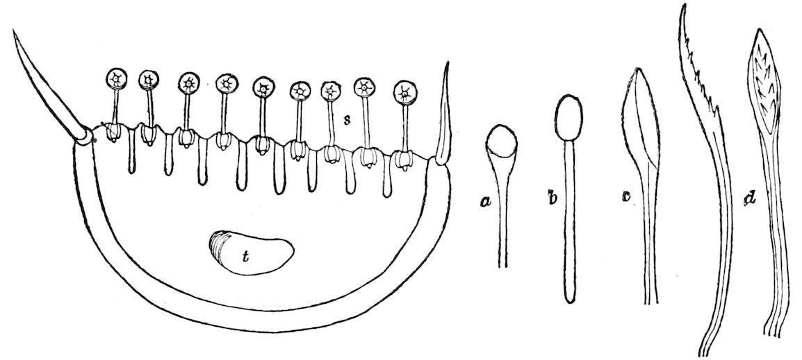
Fig. 133.—Cross-section through a tarsal joint of fore leg of Dyticus, ♂, showing the stalked chitinous suckers (s), with a marginal bristle on each side: t, trachea; a, an isolated tubule or sucker of Loricera,—b, of Chlænius,—c, of Cicindela; d, two views of one of Necrophorus germanicus, ♂.
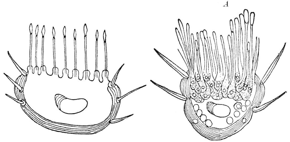
Fig. 134.—Section through the tarsus of a Staphylinid beetle; the glandular or tenent hairs arising from chitinous processes. A, section through the tarsal joint of the pine weevil, Hylobius abietis, showing the crowded, bulbous, glandular, or tenent hairs arising from unicellular glands.—This and Fig. 133 after Simmermacher.
Ockler divides the normal two-clawed foot into three subtypes: 116(1) with an unpaired median empodium; (2) with two outer lateral adhesive lobes; (3) with two adhesive lobes below the claws; the latter is the chief type and forms either a climbing or a clasping foot. The amount of movement possessed by the claws is limited, and what there is, is effected by means of an elastic membrane and the extensor plate (Fig. 110). The “extensor sole” which is always present in insects with an unpaired median fixing or adhesive organ (empodium) is to be regarded as a modification of the extensor seta. The extensor plate is peculiar to an insect’s foot. Ockler states that the so-called “pressure plate” of Dahl is only a movably articulated, skeletal, supporting plate for the median fixing lobule.
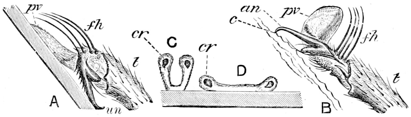
Fig. 135.—Honey-bee’s foot in the act of climbing, showing the automatic action of the pulvillus, × 30: A, position of foot in climbing on a slippery surface, or glass; pv, pulvillus; fh, tactile hairs; un, unguis; t, last tarsal joint. B, position of foot in climbing rough surface. C, section of pulvillus just touching flat surface; cr, curved rod. D, the same applied to the surface.—After Cheshire.
Climbing.—In certain respects the power of climbing supplies the want of wings, and even exists often in house-flies among which there is shown a many-sided motion that is quite unheard of in other groups of insects.
The best climbers are obviously those insects which live on trees and bushes, as, for example, longicorn beetles and grasshoppers. These may be accurately called the monkeys of the insect kind, even if their movements take place less gracefully, and indeed rather stiffly and woodenly. We already know what are the proper climbing organs; that is, the sharp easily movable claws on the foot. With the help of these claws certain insects, May-beetles for example, can hang upon one another like a chain; indeed, bees and ants in this manner bind themselves together into living garlands and bridges. There are still added to the chitinous hooks flaps and balls of a sticky nature, by help of which likewise the insects glue themselves together. To facilitate the spanning of still thicker twigs, the climbing foot of insects has a greater movability even than when it only serves as a sole. (Graber.)
The mode of swimming of insects.—To study the swimming movements of insects, let us examine a Dyticus. It will appear, as Graber states, to be wonderfully adapted to its element.
117“The body resembles a boat. There is nowhere a projecting point or a sharp corner which would offer unnecessary resistance to motion; bulging out in the middle and pointed at the end, it cuts through the resistance of the water like a wedge. The movable parts, the oars, seem to be as well fitted for their purpose as the burden to be moved by them. That the hind legs must bear the brunt of this follows from their position exactly in the middle of the body, where it is widest. In other insects also these legs are used for the same purpose as soon as the insects are put in the water. But the swimming legs of water-beetles are oars of quite peculiar construction. They are not turned about in the coxæ, as are other legs, but at the foot-joint. The coxa, namely, has grown entirely together with the thoracic partition. The muscles we have mentioned, exceeding in strength all the soft parts taken together, take hold directly of the large wing-shaped tendons of the upper thigh, and extend and retract the leg in one of the planes lying close to the abdominal partition. The foot forms the oar, however. It is very much lengthened and still more widened, and can be turned and bent in by separate muscles in such a way that in the passive movement, that is, the retraction, the narrow edge is turned to the fore, and therefore to the medium to be dislodged; however, as soon as the active push is to be performed and the leg is extended with greater force, it cuts down through the water with its whole width. These effective oar-blades are still considerably enlarged by the hairs arising on the side of the foot, which spread out at the decisive moment.
“Every one knows that the oar-blades of swimming beetles always go up and down simultaneously and in regular time. On the other hand, as soon as one puts a Dyticus on the dry land, i.e. on an unyielding medium, it uses its hind legs entirely after the manner of other land insects; that is, they are drawn in and extended again alternately, as takes place clearly enough from the footsteps in Fig. 119, A. We learn from this that water insects have not yet, from want of practice, forgotten the mode of walking of land insects.
“The forcing up of the water as a propelling power is added to the repulsion produced by the strong strokes of the oars. If the beetle stood up horizontally in the water, he would be lifted up.
“As the trunk, however, assumes an oblique position when the insect wishes to swim, one can then imagine the driving up of the water as being divided into two forces, one of which drives the body forward in a horizontal direction, while the other, that is, the vertical component, is supplied by the moving of the oars. The swimming insect is thus, as it were, a snake flying in the water.
“The long streamer-like hind legs of many water-bugs, for example Notonecta, approach more nearly our artificial oars. These legs are turned out from the bottom.
“There is no doubt but that the legs of insects, as regards the many-sidedness and exactitude of their locomotive actions, place the similar contrivances of other animals far in the shade. We shall be forced to admire these ingenious levers still more, however, when we take into consideration their energy and strength. That the force with which the locomotive muscles of insects is drawn together is enormous compared with that of vertebrates, we may learn if we try to subdue the rhythmical movements of the thorax of a large butterfly by the pressure of our finger or to open against the insect’s will the closed jumping leg of a grasshopper, or the fossorial shovel of a mole-cricket.”
118
LITERATURE ON LEGS AND FEET
MacLeay, W. S. On the structure of the tarsus in the tetramerous Coleoptera of the French entomologists. (Trans. Linn. Soc. London, xv, 1825, pp. 63–73.)
Speyer, O. Untersuchung der Beine der Schmetterlinge. (Isis, 1843, pp. 161–207, 243–264.)
Pokorsky Joravko, A. von. Quelques remarques sur le dernier article du tarse des Hyménoptères. (Bull. Soc. imp. Natur. Moscou, 1844, xvii, pp. 140–159. Ref. in Isis, 1848, v, p. 347.)
Rossmassler, E. A. Das Bein der Insekten. (Aus der Heimath, 1860, 3 kap., pp. 327–334, Fig.)
West, Tuffen. The foot of the fly; its structure and action; elucidated by comparison with the feet of other insects, etc. Part I. (Trans. Linn. Soc. London, xxiii, 1861, pp. 393–421, 1 Pl.)
Sundevall, C. On insektenas extremiteter samt deras hufoud och munddelar. (Kongl. Vetenskaps Akad. Handlingar. iii, Nr. 9, 1861.)
Lindemann, C. Notizen zur Lehre vom ausseren Skelete der Insekten (Gelenke und Muskeln der Füsse). 1 Taf. (Bull. Soc. imp. d. Natur. Moscou, xxxvii, 1864, pp. 426–432.)
Liebe, O. Die Gelenke der Insekten. Chemnitz, 1873. 4º. 1 Taf.
Canestrini, J. Ueber ein sonderbares Organ der Hymenopteren. (Zool. Anzeiger, 1880, pp. 421, 422.)
Dahl, F. Beiträge zur Kenntnis des Baues und der Funktionen der Insektenbeine. (Archiv f. Naturgesch. 1 Jahrg., 1884, pp. 146–193, 3 Taf. Sep., 48 pp. Vorlauf. Mitteil, in Zool. Anz., 1884, pp. 38–41.)
Langer, K. Ueber den Gelenkbau bei den Arthrozoen. Vierter Beitrag zur vergleichenden Anatomie und Mechanik der Gelenke. (Denkschriften der Akad. d. Wissensch. Wien, xviii, Bd. Physikal.-mathem. Classe, pp. 99–140. 3 Taf.)
Graber, Vitus. Ueber die Mechanik des Insektenkörpers. (Biolog. Centralbl., iv, 1884, pp. 560–570.)
—— Die ausseren mechanischen Werkzeuge der Tiere, ii Teil. Wirbellose Tiere, 1886, pp. 175–182, 208–210.
Dewitz, H. Ueber die Fortbewegung der Tiere an senkrechten glatten Flächen vermittelst eines Sekretes. 3 Taf. (Pflüger’s Archiv f. d. ges. Physiologie, xxxiii, 1884, pp. 440–481.)
Ockler, A. Das Krallenglied am Insektenfuss. (Archiv f. Naturgesch., 1890, pp. 221–262, 2 Taf.)
LITERATURE OF LOCOMOTION (WALKING, ETC.)
Carlet, G. Sur le mode de locomotion des chenilles. (Compt, rend. Acad. Paris, 1888, cvii, pp. 131–134. Naturwiss. Rundschau, iii Jahrg., 1888, No. 42, p. 543.)
—— De la marche d’un insecte rendu tetrapode par la suppression d’une paire de pattes. (Ibid., pp. 565, 566.)
—— Sur la locomotion des insectes et des arachnides. (Ibid., 1879, T. 89, pp. 1124, 1125.)
—— Ueber den Gang eines vierfüssig gemachten Insekts. (Naturwiss. Rundschau, viii Jahrg., 1888, pp. 666–667; Compt. rend. 1888, cvii.)
119Demoor, J. Recherches sur la marche des insectes et des arachnides. Étude experimentale d’Anatomie et de Physiologie comparées. (Archiv de Biologie, Liège, 1880, 42 pp. 3 Pls.)
—— Ueber das Gehen der Arthropoden mit Berücksichtigung der Schwankungen des Körpers. (Compt. rend. Acad. d. Sc. Paris, 1890, cxi, pp. 839–840.)
Osten-Sacken, C. R. von. Ueber das Betragen des kalifornischen flügellosen Bittacus (apterus McLachl.). (Wiener Ent. Zeit., 1882, pp. 123.)
Dixon, H. H. Preliminary note on the walking of some of the Arthropoda. (Proc. R. Dublin Soc. vii, pp. 574–578, 1892. Also Nature, 1897.)
Also the works of Graber, Marey, Cheshire, etc.
LITERATURE OF WALKING ON SMOOTH SURFACES
Blackwell, J. Remarks on the pulvilli of insects. (Trans. Linn. Soc. London, xvi, 1831, pp. 487–492, 767–770.)
Lowne, B. T. On the so-called suckers of Dytiscus and the pulvilli of insects. (Trans. Roy. Micr. Soc., pp. 267–271, 1871, 1 Pl.)
West, Tuffen. On certain appendages to the feet of insects subservient to holding or climbing. (Journ. of the Proceed. Linn. Soc. London, Zoölogy, vi, 1862, pp. 26–88.)
Dewitz, H. Ueber die Fortbewegung der Tiere an senkrechten, glatten Flächen vermittelst eines Sekrets. (Pflüger’s Archiv f. d. ges. Physiologie, xxxiii, 1884, pp. 440–481. 3 Taf. Also Zool. Anzeiger, 1884, pp. 400–405.)
—— Wie ist es den Stubenfliegen und anderen Insekten möglich, an senkrechten Glaswanden emporzulaufen. (Sitzungsb. Ges. naturf. Freunde zu Berlin, 1882, pp. 5–7.)
—— Weitere Mitteilungen über den Klettern der Insekten (Ibid., 1882, pp. 109–113).
—— Die Befestigung durch einen klebenden schleim beim springen gegen senkrechte Flächen. (Zool. Anzeiger, 1883, pp. 273, 274.)
—— Ueber die Wirkung der Haftlappchen toter Fliegen. (Ent. Nachr., x Jahrg., 1884, pp. 286, 287.)
—— Weitere Mitteilungen über das Klettern der Insekten an glatten senkrechten Flächen. (Zoolog. Anzeiger, 1885. viii Jahrg., pp. 157–159.)
—— Richtigstellung der behauptungen des Herrn F. Dahl. (Archiv f. mikroskop. Anat., 1885, xxvi, pp. 125–128.)
Rombouts, J. E. Ueber die Fortbewegung der Fliegen an glatten Flächen. (Zool. Anzeiger, 1884, pp. 619–623.)
—— De la faculté qu’out les mouches de se mouvoir sur le verre et sur les autres corps polis. (Archiv Museum Teyler (2), 4 Part, pp. 16. Fig.)
Simmermacher, G. Untersuchungen über Haftapparate an Tarsalgliedern von Insekten. (Zeitschr. f. wissensch. Zool. xl, 1884, pp. 481–556. 3 Taf., 2 Figs. Also Zoolog. Anzeiger, vii Jahrg., 1884, pp. 225–228.)
—— Antwort an Herrn Dr. H. Dewitz. (Ibid., pp. 513–517.)
Dahl, F. Die Fussdrüsen der Insekten. (Archiv f. mikroskop. Anat., 1885, xxv, pp. 236–263. 2 Taf. See also p. 118.)
Emery, C. Fortbewegung von Tieren an senkrechten und überhangenden glatten Flächen. (Biolog. Centralbl., 1884, 4 Bd., pp. 438–443.)
Léon, N. Disposition anatomique des organes de succion chez les Hydrocores et les Géocores. (Bull. Soc. des Medec. et Natur, de Jassy., 1888.)
120
d. The wings and their structure
The insects differ from all other animals except birds in possessing wings, and as we at the outset have claimed, it is evidently owing to them that insects are numerically so superior to any other class of animals, since their power of flight enables them to live in the air out of reach of many of their enemies, the greatest destruction to insect life occurring in the wingless larval and pupal stages.
The presence of wings has exerted a profound influence on the shape and structure of the body, and it is apparently due to their existence that the body is so distinctly triregional, since this feature is least marked in the synapterous insects. The wings are thin, broad leaf-like folds of the integument, attached to the thorax and moved by powerful muscles which occupy the greater part of the thoracic cavity. The two pairs of wings are outgrowths of the middle and hinder part of the thorax, the anterior pair being attached to the mesothoracic and the hinder pair to the metathoracic segment. The larger pair is developed from the middle segment of the thorax. The differentiation of the tergites into scutum, scutellum, etc., is the result of the appearance of wings, because these sclerites are more or less reduced or effaced in wingless insects, such as apterous Orthoptera and moths, ants, etc.
The size of the hinder thoracic segments is closely related to that of the wings they bear. In those Orthoptera which have hind wings larger than those of the fore pair, the metathorax is larger than the mesothorax. In such Neuroptera as have the hind wings nearly or quite as large as the anterior pair, or in the Trichoptera and in the Hepialidæ, the metathorax is nearly as large as the mesothorax, while in Coleoptera the metathorax is as large and often much larger. In the Ephemeridæ, Diptera, and Hymenoptera, which have either only rudimentary (halteres) or small hind wings, the metathorax is correspondingly reduced in size.
The wings morphologically, as their development shows, are simple sac-like outgrowths of the integument, i.e. of the free hinder edge of the tergal plates, their place of origin being apparently above the upper edge of the epimera or pleural sclerites. Calvert[24] however, regards the upper lamina of the wing as tergal, and the lower, pleural.
The wings in most insects are attached to the thorax by a membrane containing several little plates of chitin called by Audouin articulatory epidemes.
121The wings, then, are simple, very thin chitinous lamellate expansions of the integument, which are supported and strengthened by an internal framework of hollow chitinous tubes.
The veins.—The so-called “veins” or “nervures,” which are situated between the upper and under layers of the wing are so disposed as to give the greatest lightness and strength to the wings. Hagen has shown that in the freshly formed wings these two layers can be separated, when it can be seen that the veins pass through each layer.
These veins are in reality quite complex, consisting of a minute central trachea enclosed within a larger tube which at the instant the insect emerges from the nymph, or pupa, as the case may be, is filled with blood (Fig. 136). Since these tubes at first contain blood, which has been observed to circulate through them, and since the heart can be most easily injected through them, they may more properly be called veins than nervures. The shape and venation of the wings afford excellent ordinal as well as family and generic characters, while they also enable the systematist to exactly locate the spots and other markings of the wings. The spaces enclosed by the veins and their cross-branches are called cells, and their shape often affords valuable generic and specific characters.
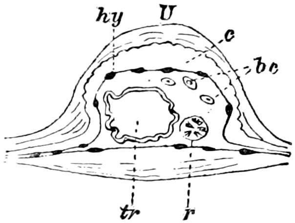
Fig. 136.—Cross-section of wing of Pronuba.—After Spuler.
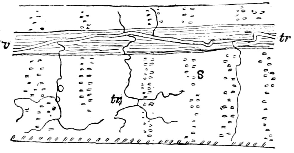
Fig. 137.—Cross-section of wing of Pieris: s, insertions of scales.-After Spuler.
The structure of a complete vein is described by Spuler. In a cross-section of a noctuid moth (Triphæna pronuba, Fig. 136) the chitinous walls are seen to consist of two layers, an outer (U) and inner (c), the latter of which takes a stain and lies next to the hypodermis (hy). In the cavity of the vein is the trachea (tr), which shows more or less distinctly the so-called spiral thread; within the cavity are also Semper’s “rib” (r) and blood-corpuscles (bc), which proves that the blood circulates in the veins of the completely formed wing, though this does not apply to all Lepidoptera with hard mature wings. We have been able to observe the same structure in sections of the wing of Zygæna.
122A cross-section of a vein of Pieris brassicæ shows that the large trachea is first formed, and that it extends along the track between the protoplasmic threads connecting the two hypodermal layers.
The main tracheæ throw off on both sides a number of secondary branches showing at their end a cell with an intracellular tracheal structure; these accessory tracheæ afterwards branch out. The accessory or transverse tracheæ often disappear, though in some moths they remain permanently. Fig. 137 tr2 represents these secondary veins in the edge of the fore wing of Laverna vanella, arising from a main trachea (tr) passing through vein I (v), two of the twigs extending to the centre, showing that the latter has no homology with a true vein. Only rarely and in strongly developed thick folds are the transverse tracheæ provided with a chitinous thickening, as for example in Cossus ligniperda. Since from such accessory tracheæ the transverse veins in lepidopterous wings are developed, we can recognize in them the homologies of the net-veins in reticulated venations. There is no sharply defined difference between reticulated and non-reticulated venations; no genetic difference exists between the two kinds of venation, since there occur true Blattidæ both with and without a reticulated venation (Spuler).
In the fore wings of Odonata, Psocina, Mantispidæ, and most Hymenoptera is an usually opaque colored area between the costal edge and the median vein, called the pterostigma.
In shape the wings are either triangular or linear oval, and at the front edge the main veins are closer together than elsewhere, thus strengthening the wings and affording the greatest resistance to the air in making the downward stroke during flight. It is noticeable that when the veins are in part aborted from partial disuse of the wings, they disappear first from the hinder and middle edge, those on the costal region persisting. This is seen in the wings of Embiidæ (Oligotoma), Cynipidæ, Proctotrupidæ, Chalcids, ants, etc.
The front edge of the wing is called the costal, its termination in the outer angle of the wing is called the apex; the outer edge (termen) is situated between the apex and the inner or anal angle, between which and the base of the wing is the inner or internal edge.
While in Orthoptera, dragon-flies, Termitidæ, and Neuroptera the wings are not attached to each other, in many Lepidoptera they are loosely connected by the loop and frenulum, or in Hymenoptera by a series of strong hooks. These hooks are arranged, says Newport, “in a slightly twisted or spiral direction along the margin of the wing, so as to resemble a screw, and when the wings are expanded attach themselves to a little fold on the posterior margin 123of the anterior wing, along which they play very freely when the wings are in motion, slipping to and fro like the rings on the rod of a window curtain.”
At the base of the hind wings of Trichoptera and in the lepidopterous Micropteryx there is an angular fold (jugum) at the base of each wing (Fig. 138); that of the anterior wings is retained in Eriocephala and Hepialidæ.
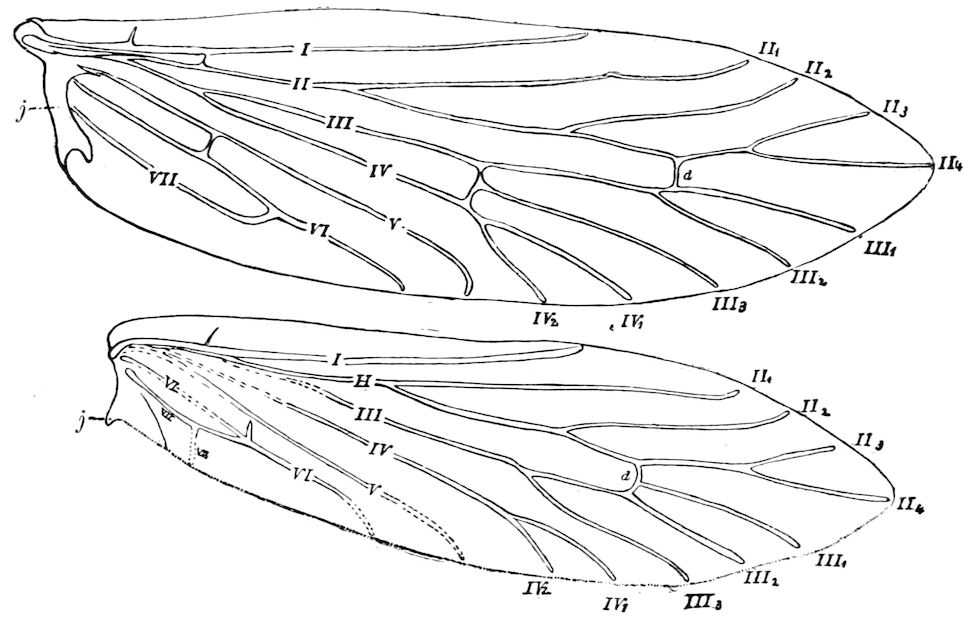
Fig. 138.—Venation of fore and hind wings of Micropteryx purpurella: j, jugum, on each wing; d, discal vein; the Roman numerals indicate veins I.-VIII. and their branches.
In the wings of Orthoptera as well as other insects, the fore wings, especially, are divided into three well-marked areas, the costal, median, and internal; of these the median area is the largest, and in grasshoppers and crickets is more or less modified to form the musical apparatus, consisting of the drum-like resonant area, with the file or bow.
The squamæ.—In the calyptrate Muscidæ, a large scale-like membranous broad orbicular whitish process is situated beneath the base of the wing, above the halter; (Fig. 94, 10 sq.) it is either small or wanting in the acalyptrate muscids. Kirby and Spence state that when the insect is at rest the two divisions of this double lobe are folded over each other, but are extended during flight. Their exact use is unknown. Kolbe, following other German authors, considers the term squama as applicable to the whole structure, restricting the term alula to the other lobe-like division.
124More recently (1890 and 1897) Osten-Sacken recommends “squamæ; in the plural, as a designation for both of these organs taken together; squama, in the singular, would mean the posterior squama alone, and antisquama the anterior squama alone;” the strip of membrane running in some cases between them, or connecting the squama with the scutellum, should be called the post-alar membrane. By a mistake Loew, and others following him, used the word tegula for squama, but this term should be restricted to the sclerite of the mesothorax previously so designated (Fig. 90, A, t). The squama or its two subdivisions has also by various authors been termed alula, calypta, squamula, lobulus, axillary lobe, aileron, cuilleron, schuppen, and scale. (Berlin Ent. Zeitschrift, xli, 1896, pp. 285–288, 328, 338.)
The halteres.—In the Diptera the hind wings are modified to form the halteres or balancers, which are present in all the species, even in Nycteribia, but are absent in Braula.
Meinert finds structures in the Lepidoptera which he considers as the homologues of the halteres of Diptera. “In the Noctuidæ,” he remarks, “I find arising from the fourth thoracic segment (segment médiaire), but covered by hair, an organ like the halter of Diptera.” (Ent. Tidskrift., i, 1880, p. 168.) He gives no details.
In the Stylopidæ, on the contrary, the fore wings are reduced to little narrow pads, while the hind wings are of great size.
The thyridium is a whitish spot marking a break in the cubital vein of the fore wing of Trichoptera; these minute thyridia occur in the fore wings of the saw-flies; there is also an intercostal thyridium on the costal part of the wings of Dermaptera.
The fore wings of Orthoptera are thicker than the hinder ones, and serve to protect the hind-body when the wings are folded; they are sometimes called tegmina. It is noteworthy, that, according to Scudder, in all the paleozoic cockroaches the fore wings (tegmina) were as distinctly veined as the hinder pair, “and could not in any sense be called coriaceous.” (Pretertiary Insects of N. A., p. 39.) Scudder also observes that in the paleozoic insects as a rule the fore and hind wings were similar in shape and venation, “heterogeneity making its appearance in mesozoic times.” In the heteropterous Hemiptera, also, the basal half of the fore wings is thick and coriaceous or parchment-like, and also protects the body when they are folded; these wings are called hemelytra. In the Dermaptera the small short fore wings are thickened and elytriform.
The elytra.—This thickening of the fore wings is carried out to its fullest extent in the fore wings of beetles, where they form the sheaths, shards, or elytra, under which the hind wings are folded. The indexed costal edge is called the epipleurum, being wide in the Tenebrionidæ. During flight “the elytra are opened so as to form an 125angle with the body and admit of the free play of the wings” (Kirby and Spence). In the running beetles (Carabidæ), also in the weevils and in many Ptinidae, the hind wings are wanting, through disuse, and often the elytra are firmly united, forming a single hard shell or case. The firmness of the elytra is due both to the thickness of the chitinous deposit and to the presence of minute chitinous rods or pillars connecting the upper and lower chitinous surfaces.
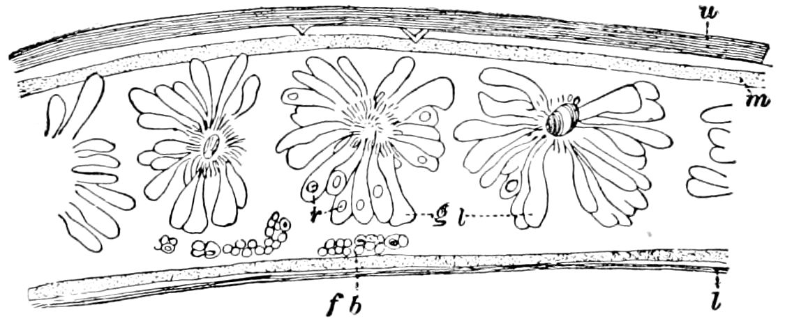
Fig. 139.—Longitudinal section through the edge of the elytrum of Lina ænea: gl, glands; r, reservoir; fb, fat-body; m, matrix; u, upper,—l, lower, lamella.—After Hoffbauer.
Hoffbauer finds that in the elytra of beetles of different families the venation characteristic of the hind wings is wanting, the main tracheæ being irregular or arranged in closely parallel longitudinal lines, and nerve-fibres pass along near them, sense-organs being also present. The fat-bodies in the cavity of the elytra, which is lined with a matrix layer, besides nerves, tracheæ, and blood, contain secretory vesicles filled with uric-acid concretions such as occur in the fat-body of Lampyris. There are also a great many glands varying much in structure and position, such occurring also in the pronotum (Fig. 139).
Meinert considers the elytra of Coleoptera to be the homologues of the tegulæ of Lepidoptera and of Hymenoptera. He also calls attention to the alula observed in Dyticus, situated at the base of the elytra, but which is totally covered by the latter. The alulæ of these beetles he regards as the homologues of the anterior wings of Hymenoptera and Diptera. No details are given in support of these views. (Ent. Tidskrift, i, 1880, p. 168.)
Hoffbauer (1892) also has suggested that the elytra are not the homologues of the fore wings of other insects, but of the tegulæ.
Kolbe describes the alula of Dyticus as a delicate, membranous lobe at the base of the elytra, but not visible when they are closed: its fringed edge in Dyticus is bordered by a thickening forming a tube which contains a fluid. The alula is united with the inner basal portion and articulation of the wing-cover, forming a continuation of them. Dufour considered that the humming noise made by these beetles is produced by the alulets.
Hoffbauer finds no structural resemblances in the alulæ of Dyticus to the elytra. He does not find “the least trace of veins.” They are more like appendages of the elytra. Lacordaire considered that their function is to prevent 126the disarticulation of the elytra, but Hoffbauer thinks that they serve as contrivances to retain the air which the beetle carries down with it under the surface, since he almost always found a bubble of air concealed under it; besides, their folded and fringed edge seems especially fitted for taking in and retaining air. Hoffbauer then describes the tegulæ of the hornet and finds them to be, not as Cholodkowsky states, hard, solid, chitinous plates, but hollow. They are inserted immediately over the base or insertion of the fore wings, being articulated by a hinge-joint, the upper lamella extending into a cavity of the side of the mesothorax, and connected by a hinge-like, articulating membrane with the lower projection of the bag or cavity. The lower lamella becomes thinner towards the place of insertion, is slightly folded, and merges without any articulation into the thin, thoracic wall at a point situated over the insertion of the fore wing. The tegulæ also differ from the wings in having no muscles to move them, the actual movements being of a passive nature, and due to the upward and downward strokes of the wings.
Comstock adopts Meinert’s view that the elytra are not true fore wings, but gives no reasons. (Manual, p. 495.)
Dr. Sharp,[25] however, after examining Dyticus and Cybister, affirms that this structure is only a part of the elytron, to which it is extensively attached, and that it corresponds with the angle at the base of the wing seen in so many insects that fold their front wings against the body. He does not think that the alula affords any support to the view that the elytra of beetles correspond with the tegulæ of Hymenoptera rather than with the fore wings.
That the elytra are modified paraptera (tegulæ) is negatived by the fact that the latter have no muscles, and that the elytra contain tracheæ whose irregular arrangement may be part of the modified degenerate structure of the elytra. Kolbe finds evidences of veins. The question may also be settled by an examination of the structure of the pupal wings. A study of a series of sections of both pairs of wings of the pupa of Doryphora and of a Clytus convinces us that the elytra are the homologues of the fore wings of other insects.
e. Development and mode of origin of the wings
Embryonic development of the wings.—The wings of insects are essentially simple dorsal outgrowths of the integument, being evaginations of the hypodermis. They begin to form in the embryo before hatching, first appearing as folds, buds, or evaginations, of the hypodermis, which lie in pouches, called peripodal cavities. They are not visible externally until rather late in larval life, after the insect, such as a grasshopper, has moulted twice or more times; while in holometabolous insects they are not seen externally until the pupa state is attained.
The subject of their origin is in a less satisfactory state than desirable from the fact that at the outset the development of the wings of the most generalized insects, such as Orthoptera, Termes, etc., was not first examined, that of the most highly modified of any insects, i.e. the Muscidæ, having actually been first studied.
127In the course of his embryological studies on the Muscidæ (Musca comitoria and Sarcophaga carnaria) Weismann (1864) in examining the larvæ of these flies just before pupation, found that the wings, as well as the legs and mouth-appendages, developed from microscopic masses of indifferent cells, which he called “imaginal discs.” From the six imaginal discs or buds in the lower part of the thorax arise the legs, while from four dorsal discs, two in the meso- and two in the metathoracic segment, arise the fore and hind wings (Fig. 141.) These imaginal buds, as we prefer to call these germs, usually appear at the close of embryonic life, being found in freshly hatched larvæ.
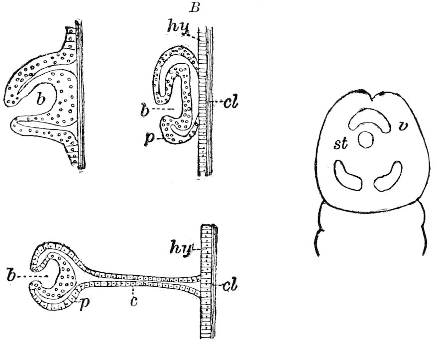
Fig. 140.—Imaginal buds in Musca,—A, in Corethra,—B, in Melophagus,—C, in embryo of Melophagus; dorsal view of the head; b, bud; p, peripodal membrane; c, cord; hy, hypodermis; cl, cuticula; st, stomodæum; v, ventral cephalic, behind are the two dorsal cephalic buds.—After Pratt.
As first observed by Weismann, the buds are, like those of the appendages, simply attached to tracheæ and sometimes to nerves, in the former case appearing as minute folds or swellings of the peritoneal membrane of certain of the tracheæ. In Volucella the imaginal buds were, however, found by Künckel d’Herculais to be in union with the hypodermis. Dewitz detected a delicate thread-like stalk connecting the peripodal membrane with the hypodermis, and Van Rees has since proved in Musca, and Pratt in Melophagus, the connection of the imaginal buds with the hypodermis (Fig. 140). These tracheal enlargements increase in size, and become differentiated into a solid mass which corresponds to the upper part of the mesothorax, while a tongue-shaped continuation becomes the rudiment of the wing. During larval life the rudiments of the wings 128crumple, thus forming a cavity. While the larva is transforming into the pupa, the sheath or peripodal membranes of the rudimentary wings are drawn back, the blood presses in, and thus the wings are everted out of the peripodal cavities.
Due credit, however, should be given to Herold, as the pioneer in these studies, who first described in his excellent work on the development of Pieris brassicæ (1815) the wing-germs in the caterpillar after the third moult. This discovery has been overlooked by recent writers, with the exception of Gonin, whose statement of Herold’s views we have verified. Herold states that the germs of the wings appear on the inside of the second and third thoracic segments, and are recognized by their attachment to the “protoplasmic network” (schleimnetz), which we take to be the hypodermis, the net-like appearance of this structure being due to the cell-walls of the elements of the hypodermal membrane. These germs are, says Herold, also distinguished from the flakes of the fat-body by their regular symmetrical form. Fine tracheæ are attached to the wing-germs, in the same way as to the flakes of the fat-body. It thus appears that Herold in a vague way attributes the origin of these wing-germs, and also the germs of the leg, to the hypodermis, since his schleimnetz is the membrane which builds up the new skin. Herold also studied the later development of the wings, and discovered the mode of origin of the veins, and in a vague way traced the origin of the scales and hairs of the body, as well as that of the colors of the butterfly.
Herold also says that as the caterpillar grows larger, and also the wing-germs, “the larval skin in the region under which they lie hidden is spotted and swollen,” and he adds in a footnote: “This is the case with all smooth caterpillars marked with bright colors. In dark and hairy caterpillars the swelling of the skin through the growth of the underlying wing-germs is less distinct or not visible at all” (pp. 29, 30).
It should be added that Malpighi, Swammerdam, and also Réaumur had detected the rudiments of the wings in the caterpillar just before pupation under the old larval skin. Lyonet (1760) also describes and figures the four wing-germs situated in the second and third thoracic segments, but was uncertain as to their nature. Each of these masses, he says, is “situated in the fatty body without being united to it, and is attached to the skin in a deep fold which it makes there.” He could throw no certain light on their nature, but says: “their number and situation leads to the supposition that they may be the rudiments of the wings of the moth” (pp. 449, 450).
During the transformation into the pupa the imaginal buds unite and grow out or extend along their edges, while the enveloping membrane disappears. The rudimentary wings are now like little sacs, and soon show a fusion of the two wing-membranes or laminæ with the veins, while the tracheæ disappear, the places occupied by the tracheæ becoming the veins. “Very early, as soon as the scales are indicated, begin in a very peculiar way the fusion of the wing-laminæ. There occur openings in the hypodermis into which the cells extend longitudinally and then laterally give way to each other. Hence no complete opening is found, but the epithelium appears by sections through a straight line sharply bordered along the wingcavity. 129It is a continuous membrane formed of plasma which I will call the ground membrane of the epithelium. Through this ground membrane pass blood-corpuscles as well as blood-lymph.” (Schaeffer.)
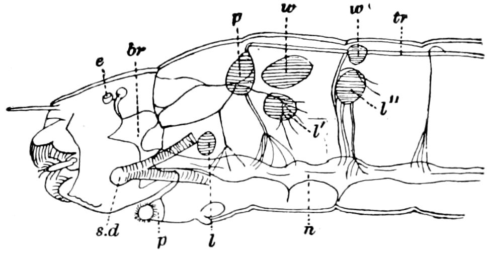
Fig. 141.—Anterior part of young larva of Simulium sericea, showing the thoracic imaginal buds: p, prothoracic bud (only one not embryonic); w, w′, fore and hind wing-buds; l, l′, l″, leg-buds; n, nervous system; br, brain; e, eye; sd, salivary duct; p, prothoracic foot.—After Weismann.
Afterwards (1866) Weismann studied the development of the wings in Corethra plumicornis, which is a much more primitive and generalized form than Musca, and in which the process of development of the wings is much simpler, and, as since discovered, more as in other holometabolous insects. He also examined those of Simulium (Fig. 141).
In Corethra, after the fourth and last larval moulting, there arises at first by evagination and afterwards by invagination a cup-shaped depression on each side in the upper part of the mesothoracic segment within which the rudiment of the wings lies like a plug. The wings without other change simply increase in size until, in the transformation into the pupa by the withdrawal of the hypodermis, the wings project out and become filled with blood, the tracheæ now being wholly wanting, and other tissues being sparingly present.
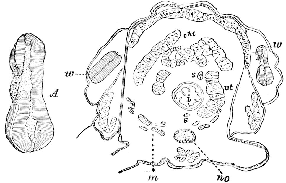
Fig. 142.—Section through thorax of a Tineid larva on sycamore, passing through the 1st pair of wings (w): ht, heart; i, œsophagus; s, salivary gland: ut, urinary tube; nc, nervous cord; m, recti muscles; a part of the fat body overlies the heart. A, right wing-germ enlarged.
130These observations on two widely separate groups of Diptera were confirmed by Landois, and afterwards by Pancritius, for the Lepidoptera, by Ganin for the Hymenoptera, by Dewitz for Hymenoptera (ants) and Trichoptera; also for the Neuroptera by Pancritius. In the ant-lion (Myrmeleon formicarius) Pancritius found no rudiments of the wings in larvæ a year old, but they were detected in the second year of larval life, and do not differ much histologically or in shape from those of Lepidoptera. In the Coleoptera and Hymenoptera the imaginal buds appear rather late in larval life, yet their structure is like that of Lepidoptera. In Cimbex the rudiments of the wings are not found in the young larva, but are seen in the semipupa, which stage lasts over six weeks.
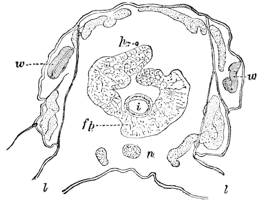
Fig. 143.—Section of the same specimen as in Fig. 142, but cut through the second pair of wings (w): i, mid-intestine; h, heart; fb, fat-body; l, leg; n, nervous cord.
The general relation of the rudiments (imaginal buds) of the wings of a tineid moth to the rest of the body near the end of larval life may be seen in Figs. 142, 143 (Tinea?), the sections not, however, showing their connection with the hypodermis, which has been torn away during the process of cutting. That the wing is but a fold of the hypodermis is well seen in Fig. 144, of Datana, which represents a much later stage of development than in Figs. 142 and 143, the larva just entering on the semipupa stage.
In caterpillars of stage I, 3 to 4 mm. in length, Gonin found the wing-germs as in Fig. 145, A being a thickening of the hypodermis, with the embryonic cells, i.e. of Verson, on the convex border. The two leaves, or sides of the wing, begin to differentiate in stage II (C, D), and in stage III the envelope is formed (E), while the tracheæ begin to proliferate, and the capillary tracheæ or tracheoles at this time arise (Fig. 145, tc). The wall of the principal trachea 131appears to be resolved into filaments, and all the secondary branches assume the appearance of bundles of twine. Landois regarded them as the product of a transformation of the nuclei, but Gonin thinks they arise from the entire cells, stating that from each cell arises a ball (peloton) of small twisted tubes.
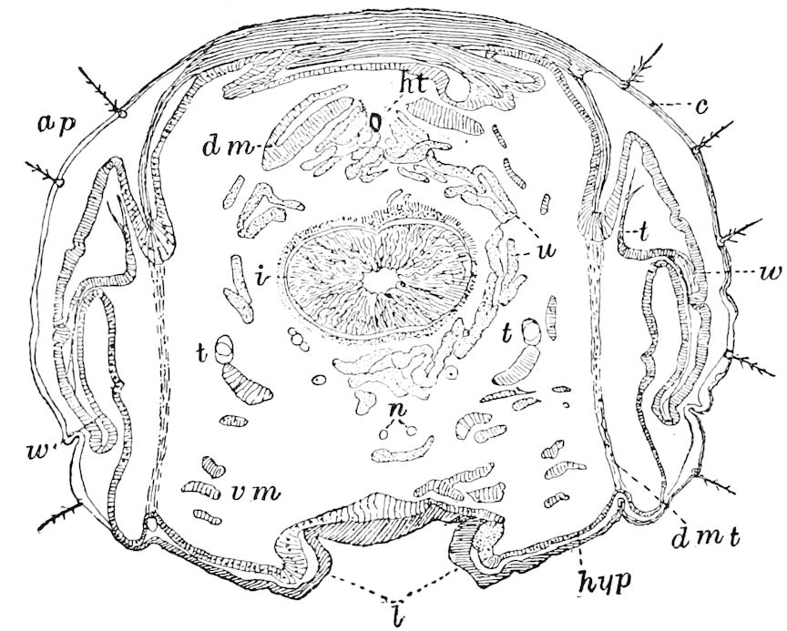
Fig. 144.—Section through mesothoracic segment of Datana ministra, passing through the wings (w): c, cuticula; hyp, hypodermis: ap, apodeme; dm, dorsal longitudinal.—vm, ventral longitudinal. muscles; dmt, depressor muscle of tergum; t, trachea; n, nerve cords; i, intestine; u, urinary tubes; l, insertion of legs.
As the large branches penetrate into the wing, the balls (pelotons) of fine tracheal threads tend to unroll, and each of the new ramifications of the secondary tracheal system is accompanied in its course by a bundle of capillary tubes. This secondary system of wing-tracheæ, then, arises from the mother trachea at the end of the third stage, when we find already formed the chitinous tunic, which will persist through the fourth stage up to pupation. It differs from the tracheoles in not communicating with the air-passage; it possesses no spiral membrane at the origin, and takes no part in respiration.
Gonin thus sums up the nature of the two tracheal systems in the rudimentary wing, which he calls the provisional and permanent systems. “The first, appearing in the second stage of the larva, comprises all the capillary tubes, and arising from numerous branches passes off from the lateral trunk of the thorax before reaching the wing; the second is formed a little later by the direct ramification of the principal branch.
132“These two systems are absolutely independent of each other within the wing. Their existence is simultaneous but not conjoint. One is functionally active after the third moult; the other waits the final transformation before becoming active.”
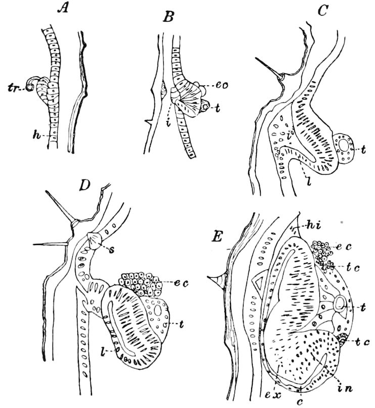
Fig. 145.—A, section of wing-bud of larva of Pieris brassicæ of stage I, in front of the invagination pit. B, section passing through the invagination pit. C, section of same in stage II, through the invagination pit;—D, behind it, making the bud appear independent of the thoracic wall. E, wing-bud at the beginning of the 3d larval stage, section passing almost through the pedicel or hypodermic insertion, the traces of which appear at hi; h, hypodermis; t or tr, trachea; i, opening of invagination; ec, embryonic cells; l, external layer or envelope; in, internal wall of the wing; ex, external wall; s, cell of a tactile hair; tc, capillary tubes; c, cavity of invagination.—After Gonin.
Evagination of the wing outside of the body.—We have seen that the alary germs arise as invaginations of the hypodermis; we will now, with the aid of Gonin’s account, briefly describe, so far as is known, the mode of evagination of the wings. During the fourth and last stage of the caterpillar of Pieris, the wings grow very rapidly, and undergo important changes.
Six or seven days after the last larval moult the chitinous wall is formed, the wing remaining transparent. It grows rapidly and its lower edge extends near the legs. It is now much crumpled on the edge, owing to its rapid growth within the limits of its own segment. Partly from being somewhat retracted, and partly owing to the irregularity of its surface, the wing gradually separates from its envelope, and the cavity of invagination (Fig. 145, c) becomes more like a distinct or real space. The outer opening of the alary sac enlarges quite plainly, though without reaching the level of the edge of the wing.
This condition of things does not still exactly explain how the wing passes to the outside of the body. Gonin compares these conditions to those exhibited by a series of sections of the larva, made forty-eight hours later, on a caterpillar which had just spun its girdle of silk. At this time the wings have become entirely external, but, 133says Gonin, we do not see the why or the how. The partition of the sac has disappeared, and with it the cavity and the leaf of the envelope.
It appears probable that the partition has been destroyed, because the space between the two teguments is strewn with numerous bits, many of which adhere to the chitinous integument, while others are scattered along the edges of the wings, in their folds, or between the wings and the wall of the thorax.
Another series of sections showed that the exit of the fore wings had been accomplished, while the hinder pair was undergoing the process of eversion. In this case the partition showed signs of degeneration: deformation of the nuclei, indistinct cellular limits, pigmentation, granular leucocytes, and fatty globules.
After the destruction of the partition, what remains of the layer of the envelope is destined to make a part of the thoracic wall and undergoes for this purpose a superficial desquamation. The layer of flattened cells is removed and replaced by a firmer epithelium like that covering the other regions. It is this renewed hypodermis which conceals the wing within, serves to separate it from the cavity of the body, and gives the illusion of a complete change in its situation. Other changes occur, all forming a complete regeneration, but which does not accord with the description of Van Rees for the Muscidæ. Finally, Gonin concludes that the débris scattered about the wing comes from the two layers of the partition of the sac, from the flattened hypodermis of the renewed envelope, from the chitinous cuticle of the wing, and from the inner surface of the chitinous integument.
He thinks that the metamorphosis of Pieris is intermediate between the two types of Corethra and of Musca, established by Weismann, as follows:
Corethra.—The wing is formed in a simple depression of the hypodermic wall. No destruction.
Pieris.—The rudiment is concealed in a sac attached to the hypodermis by a short pedicel. Destruction of the partition and its replacement by a part of the thoracic wall by means of the imaginal epithelium.
Musca.—The pedicel is represented by a cord of variable length, whose cavity may be obliterated (Van Rees). The imaginal hypodermis is substituted for the larval hypodermis, which has completely disappeared, either by desquamation (Viallanes), or by histolytic resorption (Van Rees).
Extension of the wing; drawing out of the tracheoles.—When it is disengaged from the cavity, the wing greatly elongates and the creases on its surface are smoothed out; the blood penetrates between the two walls, and the cellular fibres, before relaxed and sinuous, are now firmly extended.
Of the two tracheal systems, the large branches are sinuous, and they are rendered more distinct by the presence of a spiral membrane; but the two tunics are not separated as in the other tracheæ of the thorax; moreover, the mouth choked up with débris does not yet communicate with that of the principal trunk. The bundles of tracheoles on their part form straight lines, as if the folds of the organ had had no influence on them. As they have remained bound together, apart from the chitinous membrane of the tracheal trunk, 134they become drawn out with this membrane, at the time of exuviation, i.e. of pupation, and are drawn out of the neighboring spiracle.
Fig. 146.—Full-grown larva of Pieris brassicæ, opened along the dorsal line: d, digestive canal; s, silk-gland; g, brain; st I, prothoracic stigma; st IV, 1st abdominal stigma; a, a′, germs (buds) of fore and hind wings; p, bud of prothoracic segment;—those of the third pair are concealed under the silk-glands; I–III, thoracic rings.—After Gonin.
“This is a very curious phenomenon, which can be verified experimentally: if we cut off the wing, while sparing the larval integument around the thoracic spiracles, we preserve the two tracheal systems; the same operation performed after complete removal of the larval skin does not give the secondary tracheal system.” (Gonin.) Deceived by the appearance of the tracheoles while still undeveloped, Landois and Pancritius, who have not mentioned the drawing out of the capillaries of the larva, affirm that they are destroyed by resorption in the chrysalis.
135“The study of the tracheæ is closely connected with that of the veins (nervures). It is well to guard against the error of Verson, who mistakes for these last the large tracheal branches of the wing. This confusion is easily explained; it proves that Verson had, with us, recognized that the secondary system is, in the larva, exempt from all respiratory function. Landois thought that the pupal period was the time of formation of the veins. It seems to me probable that they are derived from the sheath of the peritracheal spaces.” (Gonin, pp. 30–33.)
Fig. 147.—Left anterior wing of a larva 3 days before pupation. The posterior part is rolled up: st, prothoracic stigma; tr. i., internal tracheal trunk; tr. e., tr. e.′, external tracheal trunk; p, cavity of a thoracic leg, with the imaginal bud b.—After Gonin.
The appearance of the wing-germs in the fully grown caterpillar, as revealed by simple dissection, is shown at Fig. 146; Fig. 147 136represents a wing of a larva three days before pupation, with the germ of a thoracic leg.
Fig. 148.—Graber’s diagrams for explaining the origin and primary invagination of the hypodermis to form the germs of the leg (b), and wings (f, A-C), and afterwards their evagination D, so that they lie on the outside of the body. E, stage B, showing the hypodermal cavities (f) and stalks connecting the germs with the hypodermis (z).—After Graber.
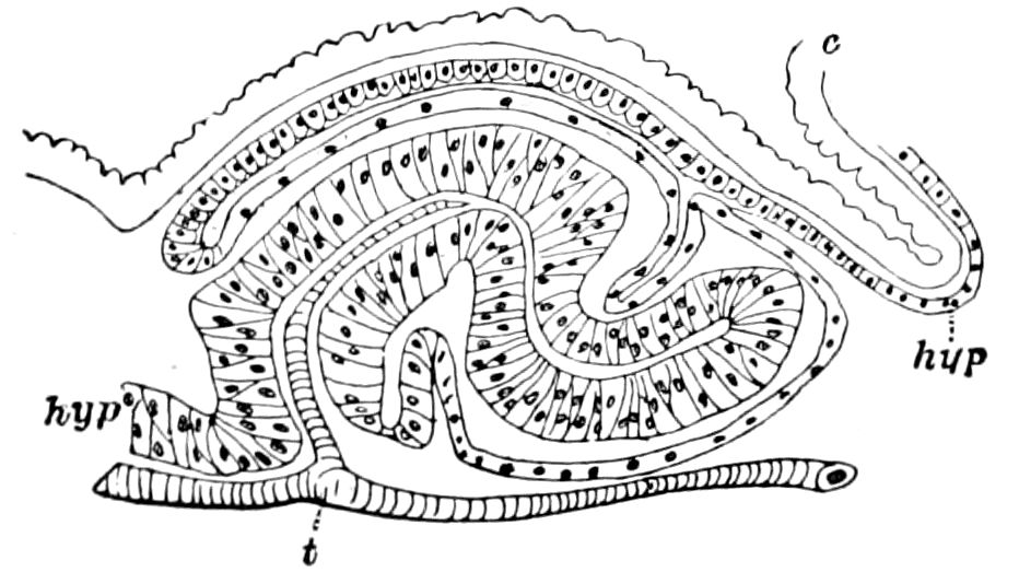
Fig. 149.—Section lengthwise through the left wing of mature larva in Pieris rapæ: t, trachea; hyp, hypodermis; c, cuticula.—After Mayer.
A. G. Mayer has examined the late development of the wings in Pieris rapæ. Fig. 149 represents a frontal section through the left wing of a mature larva and shows the rudiment of the wing, lying in its hypodermal pocket or peripodal cavity. How the trachea passes into the rudimentary wing, and eventually becomes divided into the branches, around which the main veins afterwards form, is seen in Figs. 144, 147, 159.
The histological condition of the wing at this time is represented by Fig. 151, the spindle-like hypodermal cells forming the two walls being separated by the ground-membrane of Semper.
“While in the pupa state,” says Mayer, “the wing-membrane is thrown into a very regular series of closely compressed folds, a single scale being inserted upon the crest of each fold. When the butterfly 137issues from the chrysalis, these folds in the pupal wings flatten out, and it is this flattening which causes the expansion of the wings.... It is evident that the wings after emergence undergo a great stretching and flattening. The mechanics of the operation appears to be as follows. The hæmolymph, or blood, within the wings is under considerable pressure, and this pressure would naturally tend to enlarge the freshly emerged wing into a balloon-shaped bag; but the hypodermal fibres (h) hold the upper and lower walls of the wing-membrane closely together, and so, instead of becoming a swollen bag, the wing becomes a thin flat one. And thus it is that the little thick corrugated sac-like wings of the freshly emerged insect become the large, thin, flat wings of the imago.... The area of the wing of the imago of Danais plexippus is 8.6 times that of the pupa. Now, as the wing of the young pupa has about 60 times the area of the wing in the mature larva, it is evident that in passing from the larval state to maturity the area of the wings increases more than 500 times.”
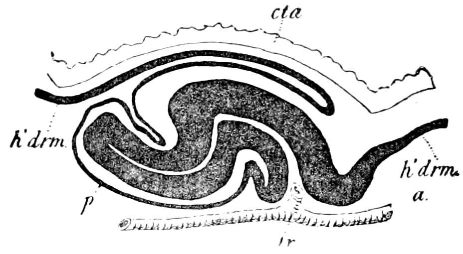
Fig. 150.—Diagrammatic reproduction of Fig. 149 showing the wing-germ in its peripodal cavity (p): h’drm, hypodermis; tr, trachea; cta, cuticula; a, anterior end.—After Mayer.
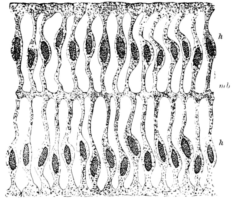
Fig. 151.—Section of the wing-germ, the upper and lower sides connected by spindle-like hypodermic cells (h), forming the rods of the adult wing; mbr, ground-membrane of Semper.—After Mayer.
f. The primitive origin of the wings
Farther observations are needed to connect the mode of formation of the wings in the holometabolous insects with the more primitive mode of origin seen in the hemimetabolous orders, but the former mode is evidently inherited from the latter. Pancritius remarks that the development of the rudiments of the wing in a hypodermal cavity is in the holometabolic insects to be regarded as a later inherited character, the external conditions causing it being unknown.
138Fritz Müller was the first to investigate the mode of development of the wings of the hemimetabolic insects, examining the young nymphs of Termites. He regards the wings as evaginations of the hypodermis, which externally appear as thoracic scale-like projections, into which enter rather late in nymphal life tracheæ which correspond to the veins which afterward arise.
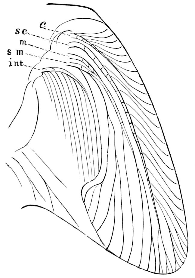
Fig. 152.—Rudimentary wing of young nymph of Blatta, with the five principal veins developed.
The primitive mode of origin of the wings may, therefore, be best understood by observing the early stages of those insects, such as the Orthoptera and Hemiptera, which have an incomplete metamorphosis. If the student will examine the nymphs of any locust in their successive stages, he will see that the wings arise as simple expansions downward and backward of the lateral edges of the meso- and metanotum. In the second nymphal stage this change begins to take place, but it does not become marked until the succeeding stage, when the indications of veins begin to appear, and the lobe-like expansion of the notum is plainly enough a rudimentary wing.
Graber[26] thus describes the mode of development of the wings in the nymph of the cockroach:
“If one is looking only at the exterior of the process, he will perceive sooner or later on the sides of the meso- and metathorax pouch-like sacs, which increase in extent with the dorsal integument and at the same time are more and more separated from the body. These wing-covers either keep the same position as in the flat-bodied Blattidæ, or in insects with bodies more compressed the first rudiments hang down over the sides of the thorax. As soon as they have exceeded a certain length, these wing-covers are laid over on the back. However, if we study the process of development of the wings with a microscope, by means of sections made obliquely through the thorax, the process appears still more simple. The chief force of all evolution is and remains the power of growth in a definite direction. In regard to the skin this growth is possible in insects only in this way; namely, that the outer layer of cells is increased by the folds which are forced into the superficial chitinous skin. These folds naturally grow from one moult to another in proportion to the multiplication of the cells, and are not smoothed out until after the moulting, when the outer resistance is overcome.
139
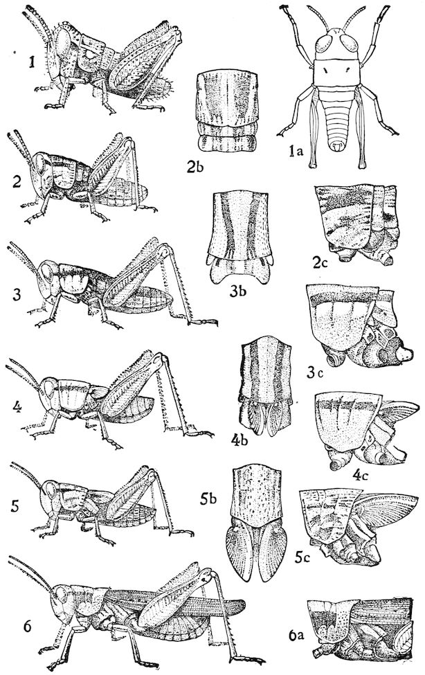
Fig. 153.—Partial metamorphosis of Melanoplus femur-rubrum, showing the five nymph stages, and the gradual growth of the wings, which are first visible externally in 3, 3b, 3c.—Emerton del.
“As, however, the first wing-layers depend upon the wrinkling of the general integument of the body through the increase in the upper layer, the further growth of the wings depends in the later stages upon the wrinkling of the epidermis of the wing-membrane even, which fact we also observe under the microscope when the new wings drawn forth from the old covers appear at 140first to be quite creased together. These wing-like wrinkles in the skin are not empty pouches, but contain tissues and organs within, which are connected with the skin, as the fat of the body, the network of tracheæ, muscles, etc. Alongside the tracheæ, running through the former wing-pouches and accompanied by the nerves, there are canals through which the blood flows in and out.
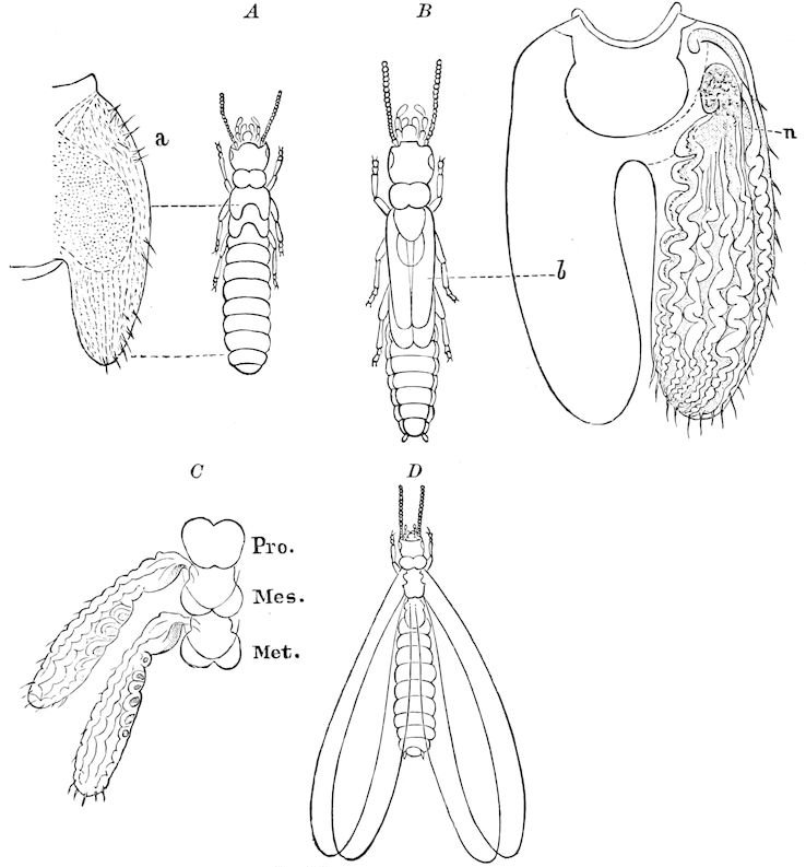
Fig. 154.—Stages in the growth of the wings of the nymph of Termes flavipes: A, young; a, a wing enlarged. B, older nymph; b, fore wing; n, a vein. C, wings more advanced;—D, mature.
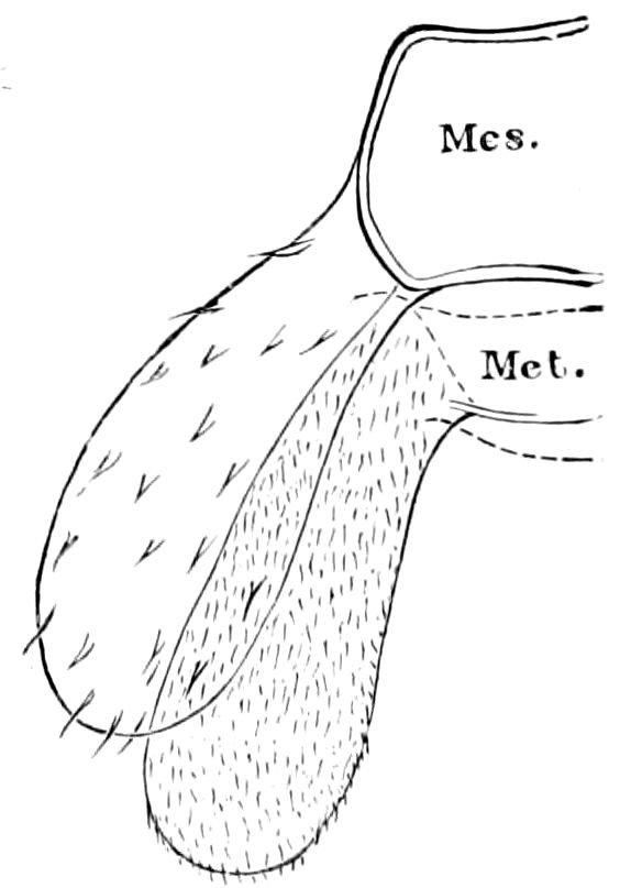
Fig. 155.—Wings of nymph of Psocus.
“After the last moult, however, when the supply of moisture is very much reduced in the wing-pouches, which are contracted at the bottom, their two layers become closely united, and afterward grow into one single, solid wing-membrane.
“These thick-walled blood-tubes arising above and beneath the upper and lower membrane of the wing are the veins of the wings; the development of 141the creased wings in the pupa of butterflies is exactly like that of cockroaches and bugs. The difference is only that the folds of integument furnishing the wings with an ample store of material for their construction reach in a relatively shorter time, that is the space of time between two moults, the same extent that they would otherwise attain only in the course of several periods of growth in the ametabolous insects.”
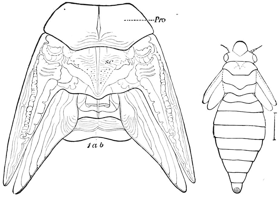
Fig. 156.—Nymph of Aphrophora permutata, with enlarged view of the wings and the veins: pro, pronotum; sc, mesoscutum; 1ab, 1st abdominal segment.
Ignorant of Graber’s paper, we had arrived at the same result, after an examination of the early nymph-stages of the cockroach, as well as the locusts, Termites, and various Hemiptera. In all these forms it is plainly to be seen that the wings are simply expansions, either horizontal or partly vertical (where, as in locusts, etc., the body is compressed, and the meso- and metanota are rounded downwards), of the hinder and outer edge of the meso- and metanotum. As will be seen by reference to the accompanying figures, the wings are notal (tergal) outgrowths from the dorsal arch of the two hinder segments of the thorax. At first, as seen in the young pupal cockroach (Fig. 152) and locust (Fig. 153, also Figs. 154 and 156) the rudiments of the wings are continuous with the notum. Late in nymphal life a suture and a hinge-joint appear at the base of the wing, and thus there is some movement of the wing upon the notum; finally, the tracheæ are well developed in the wings, and numerous small sclerites are differentiated at the base of the wing, to which the 142special muscles of flight are attached, and thus the wings, after the last nymphal moult, have the power of flapping, and of sustaining the insect in the air; they thus become true organs of flight.
It is to be observed, then, that the wings in all hemimetabolous insects are outgrowths from the notum, and not from the flanks or pleurum of the thorax. There is, then, no structure in any other part of the body with which they are homologous.
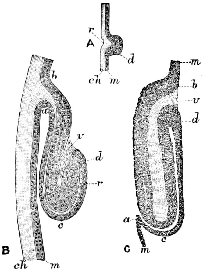
Fig. 157.—Development of wings of Trichoptera: A, portion of body-wall of young larva of Trichostegia; ch, cuticula, forming at r a projection into the hypodermis, m; r, and d, forming thus the first rudiment of the wing. B, the parts in a larva of nearly full size; a, c, d, b, the well-developed hypodermis of the wing-germ separated into two parts by r, the penetrating extension of the cuticula; v, mesoderm, C, wing-pad of another Phryganeid freed from its case at its change to the pupa: b, d, outer layer of the hypodermis (m) of the body-wall; v, inner layer within nuclei.—After Dewitz, from Sharp.
The same may be said of the true Neuroptera, Trichoptera (Fig. 157), the Coleoptera, and the Diptera, Lepidoptera, and Hymenoptera. As we have observed in the house fly,[27] the wings are evidently outgrowths of the meso- and metanotum; we have also observed this to be most probably the case in the Lepidoptera, from observations on a Tortrix in different stages of metamorphosis. It is also the case with the Hymenoptera, as we have observed in bees and wasps;[28] and in these forms, and probably all Hymenoptera, the wings are outgrowths of the scutal region of the notum.
With these facts before us we may speculate as to the probable origin of the wings of insects. The views held by some are those of Gegenbaur, also adopted by Lubbock, and originally by myself.[29] According to Gegenbaur:
“The wings must be regarded as homologous with the lamellar tracheal gills, for they do not only agree with them in origin, but also in their connection with the body, and in structure. In being limited to the second and third thoracic segments they point to a reduction in the number of the tracheal gills. It is quite clear that we must suppose that the wings did not arise as such, but were developed from organs which had another function, such as the tracheal gills; I mean to say that such a supposition is necessary, for we cannot imagine that the wings functioned as such in the lower stages of their development, and that they could have been developed by having such a function.”
143
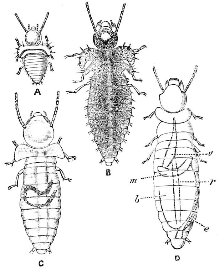
Fig. 158.—Changes in external form of the young larva of Calotermes rugosus, showing, in A and B, the mode of origin of the wing-pads: A, newly hatched, with 9 antennal joints, × 8. B, older larva, with 10 joints, × 8. C, next stage, with 11 joints, × 8. D, larva, with twelve joints; the position of the parts of the alimentary canal are shown: v, crop; m, stomach; b, “paunch”; e, intestine; r, heart, × 16 3.—After Fritz Müller, from Sharp.
If we examine the tracheal gills of the smaller dragon-fly (Agrion), or the May-flies, or Sialidæ, or Perlidæ, or Phryganeidæ, we see that they are developed in a very arbitrary way, either at the end of the abdomen, or on the sternum, or from the pleurum; moreover, in structure they invariably have but a single trachea, from which minute twigs branch out;[30] in the wings there are five or six main tracheæ, which give rise to the veins. Thus, in themselves, irrespective of their position, they are not the homologues of the gills. The latter are only developed in the aquatic representatives of the Neuroptera and Pseudoneuroptera, and are evidently adaptive, secondary, temporary organs, and are in no sense ancestral, primitive structures from which the wings were developed. There is no good reason to suppose that the aquatic Odonata or Ephemerids or Neuroptera were not descendants of terrestrial forms.
To these results we had arrived by a review of the above-mentioned facts, before meeting with Fritz Müller’s opinions, derived from a study of the development of the wings of Calotermes (Fig. 158). Müller[31] states that “(1) The wings of insects have not originated from ‘tracheal gills.’ The wing-shaped continuations of the youngest larvæ are in fact the only parts in which air tubes are completely wanting, while tracheæ are richly developed in all other parts of the body.[32] (2) The wings of insects have arisen from 144lateral continuations of the dorsal plates of the body-segments with which they are connected.”