PART I.—MORPHOLOGY AND PHYSIOLOGY (continued 2)
Reading edition
Kirby and Spence. Intr. to entomology, iii, 1828, p. 457.
De Geer. ii, 1778; v, 26, Fig. 11, M.
Kirby and Spence. Pl. xii, Fig. 2 K.
Latreille. Organisation extérieure des insectes, p. 184. (Quoted from Kirby and Spence.)
Savigny. Mémoires sur les animaux sans vertèbres. Partie Ire, 1816, p. 12.
Walter, Alfred. Beiträge zur Morphologie der Schmetterlinge. Erster Theil. Zur Morphologie des Schmetterlingsmundtheile. (Jena. Zeits., xviii, 1885, p. 752.)
Cheshire, F. R. Bees and bee-keeping, i, London, 1886, p. 93.
Joseph, Gustav. Zur Morphologie des Geschmacksorganes bei Inseckten. (Amtlicher Bericht der 50 Versammlung deutscher Naturforscher u. Artzte in München. 1877, pp. 227, 228.)
Dimmock, George. The anatomy of the mouth-parts and of the sucking apparatus of some Diptera, 1881. (Also in Psyche, iii, pp. 231–241, Pl. 1, 1882.)
Packard, A. S. On the epipharynx of the Panorpidæ. (Psyche, 1889, v, pp. 159–164.)
—— Notes on the epipharynx and the epipharyngeal organs of taste in mandibulate insects. (Psyche, v, pp. 193–199, 222–228, 1889.)
Attachment of the head to the trunk.—The head is either firmly supported by the broad prothoracic segment in Orthoptera, many beetles, etc., into which it is more or less retracted, or it is free and attached by a slender neck, easily turning on the trunk, as in dragon-flies, flies, etc. In some insects there are several chitinous plates, situated on an island in the membrane on the under side of the neck; these are the “cervical sclerites” of Sharp, occurring “in Hymenoptera, in many Coleoptera, and in Blattidæ.”
The basal or gular region of the head.—At the hinder part of the head is the opening (occipital foramen) into the trunk. The cheek (gena) is the side of the head, and to its inner wall is attached the mandibular muscle; it thus forms the region behind the eye and over the base of the mandibles. In the Termitidæ, where the head is broad and flat, it forms a distinct piece on the under side of the head bounding the gulo-mental region (Fig. 28). In the Neuroptera (Corydalus, Fig. 29, and Mantispa, Fig. 30) it is less definitely outlined.
47
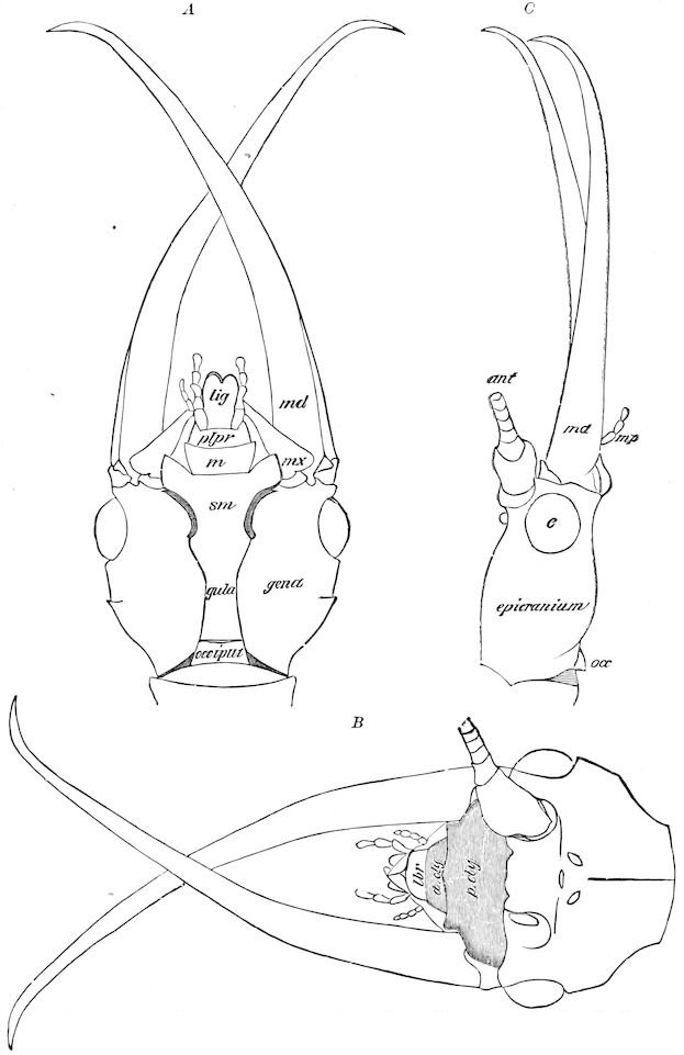
Fig. 29.—Head of Corydalus cornutus, ♂: A, from above. B, from beneath. C, from the side. a. cly, clypeus anterior; p. cly, clypeus posterior; lbr, labrum; md, mandible; mx, base of first maxilla; mp, its palpus; m, mentum; sm, submentum; plpr, palpifer; lig, fused second maxillæ; ant, antenna; occ, occiput.
48
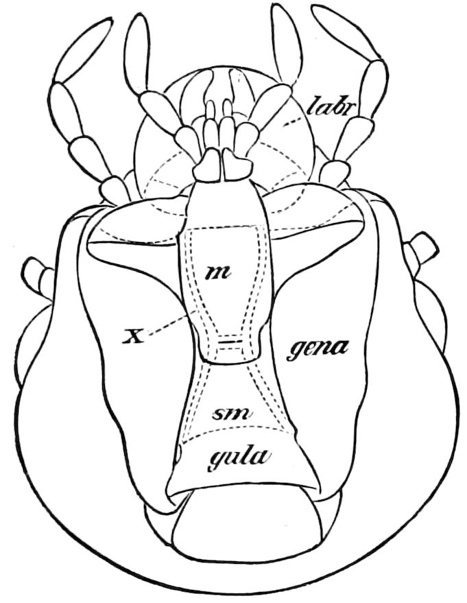
Fig. 28.—Head of Termopsis angusticollis, seen from beneath, showing the gena and gula: m, mentum; sm, submentum; labr, under side of the labrum; x, hypopharyngeal chitinous support.
All the gulo-mental region of the head appears to represent the base of the second maxillæ, and the question hence arises whether the submentum is not the homologue of the cardines of the first maxillæ fused, and the mentum that of the stipites of the latter also fused together. If this should prove to be the case, the homologies between the two pairs of maxillæ will be still closer than before supposed. Where the gula is differentiated, this represents the basal piece of the second maxillæ. In Figs. 28, 29, 30, and 31, these three pieces are clearly shown to belong to the second maxillary segment. It is evident that these pieces or sclerites belong to the second maxillary or labial segment of the head, as does the occiput, which may represent the tergo-pleural portion of the segment. Miall and Denny also regarded the submentum as the basal piece of the second maxillæ.
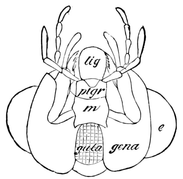
Fig. 30.—Head of Mantispa brunnea, under side: e, eye; other lettering as in Fig. 29.
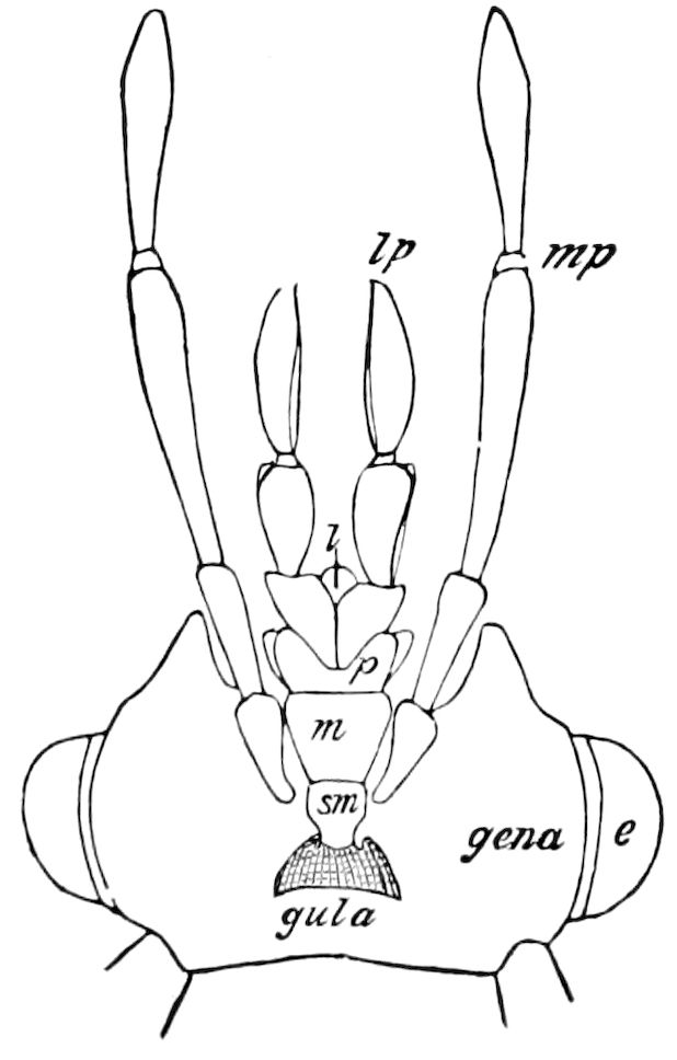
Fig. 31.—Head of Limnephilus pudicus, under side: e, eye; l, ligula; p, palpifer; lp, labial palpi.
The occiput (Fig. 29, B, C), as stated beyond, is very rarely present as a separate piece; in the adult insect we have only observed it in Corydalus. The occipital region may be designated as that part of the head adjoining and containing the occipital foramen. Newport considers the occiput as that portion of the base of the head “which is articulated with the anterior margin of the prothorax. It is perforated by a large foramen, through which the organs of the head are connected 49with those of the trunk. It is very distinct in Hydroüs and most Coleoptera, and in some, the Staphylinidæ, Carabidæ, and Silphidæ is constricted and extended backwards so as to form a complete neck.” (See also p. 51.)
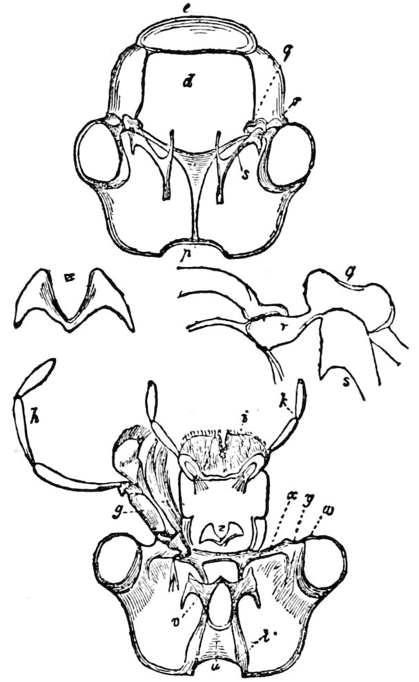
Fig. 32.—Interior and upper and under surface of the head of Hydroüs piceus: d, clypeus; e, labrum; g, maxilla; h, its palpus; i, labium; k, labial palpus; p, sutura epicranii; q, cotyloid cavity; r, torulus; s, v, laminæ squamosa; t, laminaæ posteriores; u, tentorium; w, laminæ orbitales; x, os transversum; y, articulating cavity for the mandible; z, os hypopharyngeum.—After Newport.
The tentorium.—The walls of the head are supported or braced within by two beams or endosternites passing inwards, and forming a solid chitinous process or loop which extends in the cockroach downwards and forwards from the lower edge of the occipital foramen. “In front it gives off two long crura or props, which pass to the ginglymus, and are reflected thence upon the inner surface of the clypeus, ascending as high as the antennary socket, round which they form a kind of rim.” (Miall and Denny.) The œsophagus passes upwards between its anterior crura, the long flexor of the mandible lies on each side of the central plate; the supraœsophageal ganglion rests on the plate above, and the subœsophageal ganglion lies below it, the nerve cords which unite the two passing through the circular aperture. (Miall and Denny.) In Coleoptera (Hydroüs) it protects the nervous cord which passes under it. (Newport, Fig. 32, u.)
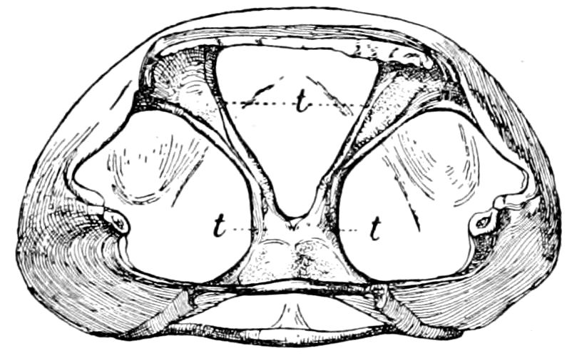
Fig. 33.—Posterior view of head of Anabrus; t, tentorium. Joutel del.
In Anabrus the tentorium is V-shaped, the two arms originating on each side of the base of the clypeus next to the base of each mandible the origin being indicated by two small foramina partly concealed externally and passing inwards and backwards and uniting just before reaching the posterior edge of the large occipital foramen (Fig. 33).
50Palmén regards the tentorium as representing a pair of tracheæ (with the cephalic spiracles) which have become modified for supports or for muscular attachment, since he finds that in Ephemera the tentorium breaks across the middle during exuviation, each half being drawn out of the head like the chitinous lining of a tracheal tube. This view is supported by Wheeler, who has shown that the tentorium of Doryphora originates from five pairs of invaginations of the longitudinal commissures, and which are anterior to those of the second maxillary segment. “These invaginations grow inwards as slender tubes, which anastomose in some places. Their lumina are ultimately filled with chitin.” (Jour. Morph., iii, p. 368.)
This view has also been held by Carrière and Cholodkowsky, but Heymons concludes from his embryological studies on Forficula and Blattidæ (1895) that it is unfounded. That this is probably the case is proved by the fact that the apodemes of the thoracic region are evidently not modified tracheæ, since the stigmata and tracheæ are present.
Number of segments in the head.—While it is taken for granted by many entomologists that the head of insects represents a single segment, despite the circumstance that it bears four pairs of appendages, the more careful, philosophical observers have recognized the fact that it is composed of more than a single segment. Burmeister recognized only two segments in the head; Carus and Audouin recognized three; Macleay and Newman four; Straus-Durckheim even so many as seven. Huxley supposed that there are five segments bearing appendages, remarking, “if the eyes be taken to represent the appendages of another somite, the insect head will contain six somites.” (Manual of Anat. Invert. Animals, p. 398.)
These discordant views were based on the examination of the head in adult insects; but if we confine ourselves to the imago alone, it is impossible to arrive at a solution of the problem.
Newport took a step in the right direction when he wrote: “It is only by comparing the distinctly indicated parts of the head in the perfect insect with similar ones in the larva that we can hope to ascertain the exact number of segments of which it is composed.” He then states that in the head of Hydroüs piceus are the remains of four segments, though still in the next paragraph, when speaking of the head as a whole, he considers it as the first segment, “while,” he adds, “the aggregation of segments of which it is composed we shall designate individually subsegments.”
That the head of insects is composed of four segments was shown on embryological grounds by the writer (1871) and afterwards by Graber (1879). The antennæ and mouth-parts are outgrowths budding out from the four primitive segments of the head; the antennæ grow out from the under side of the procephalic lobes, and these should therefore receive the name of antennal lobes. In like manner 51the mandibles and first and second maxillæ arise respectively from the three succeeding segments.
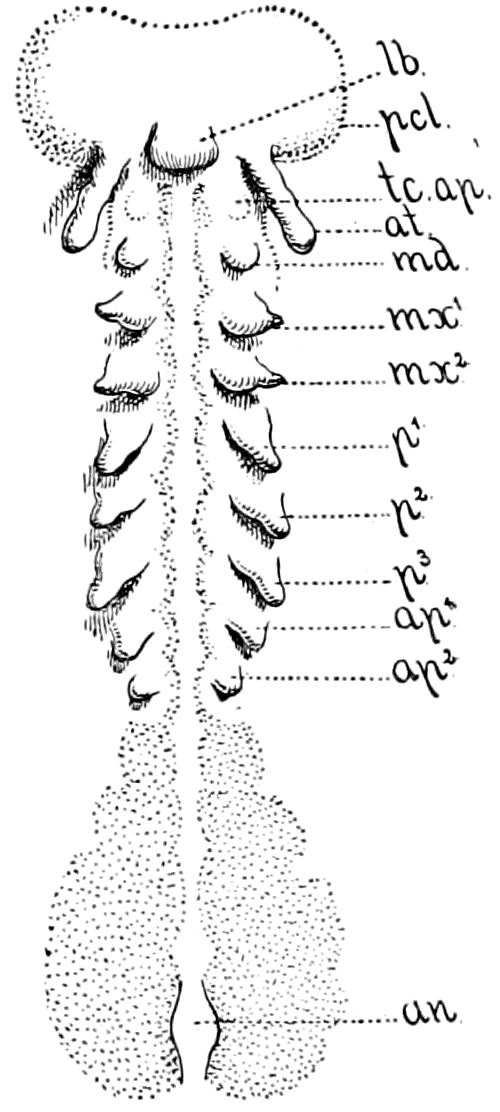
Fig. 34.—Embryo of Anurida maritima: tc. ap, minute temporary appendage of the tritocerebral segment, the premandibular appendage; at, antenna; md, mandible; mx1, first maxilla; mx2, second maxilla; p1–p3, thoracic; ap1, ap2, abdominal appendages; an, anus—After Wheeler.
While the postoral segments and their appendages are readily seen to be four in number, the question arises as to whether the eyes represent the appendages of one or more preoral segments. In this case embryology thus far has not afforded clear, indubitable evidence. We are therefore obliged to rely on the number of neuromeres, or primitive ganglia. In the postoral region of the head, as also in the trunk, a pair of neuromeres correspond to each segment. (See also under Nervous System, and under Embryology.) We therefore turn to the primitive number of neuromeres constituting the procephalic lobes or brain.
From the researches of Patten, Viallanes, and of Wheeler, especially of Viallanes, it appears that the brain or supraœsophageal ganglion is divided into three primitive segments. (See Nervous System, Brain.) The antennæ are innervated from the middle division or deutocerebrum. Hence the ocular segment, i.e. that bearing the compound and simple eyes, is supposed to represent the first segment of the head. This, however, does not involve the conclusion that the eyes are the homologues of the limbs, however it may be in the Crustacea.
The second head-segment is the antennal, the antennæ being the first pair of true jointed appendages.
The third segment of the head is very obscurely indicated, and the facts in proof of its existence are scanty and need farther elucidation.
Viallanes’ tritocerebral lobes or division of the brain is situated in a segment found by Wheeler to be intercalated between the antennal and mandibular segments. He also detected in Anurida maritima, the rudiments of a pair of appendages, smaller than those next to it, and which soon disappear (Fig. 34, tc. ap). He calls this segment the intercalary.[12] Heymons (1895) designates it as the 52“Vorkiefersegment,” and it may thus be termed the premandibular segment.
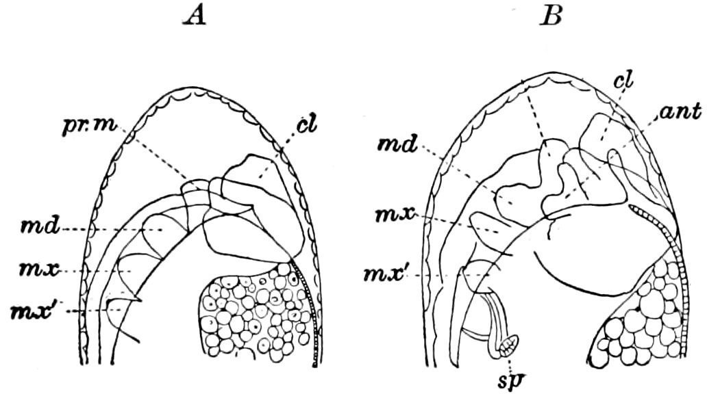
Fig. 35.—Head of embryo of honey bee: B, a little later stage than A. pr.m, premandibular segment; cl, clypeus; ant, antenna; md, mandible; mx, first maxilla; mx′, second maxilla; sp, spiracle.—After Bütschli.
As early as 1870 Bütschli observed in the embryo of the honey bee the rudiments of what appeared to be a pair of appendages between the antennæ and mandibles, but, judging by his figures, nearer to and more like the mandibles than the rudimentary antennæ (Fig. 35); they seemed to him “almost like a pair of inner antennæ.”
“I find,” he says, “in no other insects any indication of this peculiar appendage, which at the time of its greatest development attains a larger size than the antennæ, and which, afterwards becoming less distinct, forms by fusion with that on the other side a sort of larval lower lip. That this appendage does not belong to the category of segmental appendages is indicated by the site of its origin on the upper side of the primitive band.” (Zeitschr. wissen. Zool., xx, p. 538.)
Grassi has also observed it in Apis, and regards it as the germ of a first, but deciduous, pair of jaws. In the embryo of Hylotoma Graber (Figs. 134, 135) found what he calls three pairs of “preantennal projections,” one of which he thinks corresponds to the “inner antennæ” of Bütschli. This subject needs further investigation.
It thus appears that the procephalic lobes of the embryo of insects, with the rudiments of the antennæ, constitute the primitive head, and perhaps correspond to the annelidan head, while gradually the antennal appendages were in the phylogenetic development of the class fused with the two segments of the primary head. That the second maxillary segment, the occiput, was the last to be added, and at first somewhat corresponded in position to the poison-fangs 53of centipedes (Chilopods), is shown by our observations on the embryology of Æschna (Fig. 36).
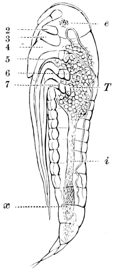
Fig. 36.—Æschna nearly ready to hatch: 4, labium, between T and e the occipital tergite; 5–7, legs.
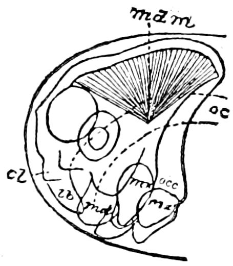
Fig. 37.—Head of embryo Nematus, showing the labial segment: occ, forming the occiput; cl, clypeus; lb, labrum; md, mandible; mdm, muscle of same; mx, maxilla; mx′, second maxilla (labium); oe, œsophagus.
The mandibular segment appears to form a large part of the post-antennal region of the epicranium on account of the great mandibular muscle which arises from so large an area of the anterior region of the head (Fig. 37).
Judging from the embryo of Nematus (Fig. 37), the first maxillary segment is tergally aborted, there being no tergo-pleural portion left.[13]
The second maxillary segment tergally appears to be represented by the occipital region of the head.
All the gular region, including the submentum and mentum, probably represents the base of the labium or second maxillæ.[14] The so-called “occiput” forms the base of the head of Corydalus, a neuropterous insect, which, however, is more distinct in the larva. In most other adult insects the occiput is either obsolete or fused with the hinder part of the epicranium. We have traced the history of this piece (sclerite) in the embryo of Æschna, a dragon-fly, and have found that it represents the tergal portion of the sixth or labial segment. In our memoir on the development of this dragon-fly, Pl. 2, Fig. 9, the head of the embryo is seen to be divided into two regions, the anterior, formed of the antennal, mandibular, and first maxillary segments, and the posterior, formed of the sixth or labial segment. 54This postoral segment at first appears to be one of the thoracic segments, but is afterwards added to the head, though not until after birth, as it is still separate in the freshly hatched nymph (Fig. 4; see also Kolbe, p. 132, Fig. 59, sq. 5). A. Brandt’s figure of Calopteryx virgo (Pl. 2, Fig. 19) represents an embryo of a stage similar to ours, in which the postoral or sixth (labial) segment is quite separate from the rest of the head. The accompanying figure, copied from our memoir, also shows in a saw-fly larva (Nematus ventricosus) the relations of the labial or sixth segment to the rest of the head. The suture between the labial segment and the preoral part of the head disappears in adult life. From this sketch it would seem that the back part of the head, i.e. of the epicranium, may be made up in part of the tergite or pleurites of the mandibular segment, since the mandibular muscles are inserted on the roof of the head behind the eyes. It is this labial segment which in Corydalus evidently forms the occiput, and of which in most other insects there is no trace in larval or adult life, unless we except certain Orthoptera (Locusta), and the larva of the Dyticidæ.
The following table is designed to show the number and succession of the segments of the head, with their respective segments.
Tabular View of the Segments, Pieces (Sclerites), and Appendages of the Head
Name of Segment Pieces or Regions of the Head-capsule Appendages, etc.
Preoral, in early embryo. 1. Ocellar (Protocerebral). Epicranium, anterior region with the clypeus labrum, and epipharynx. Compound and simple eyes (Ocelli).
Postoral, in early embryo. 2. Antennal (Deutocerebral). Epicranium, including the antennal sockets. Antennæ.
3. Premandibular, or intercalary (Tritocerebral). Wanting in postembryonic life, except in Campodea. Premandibular appendages (in Campodea).
4. Mandibular. Epicranium behind the antennæ, genæ. Mandibles.
5. 1st Maxillary. Epicranium, hinder edge? Tentorium. 1st Maxillæ.
6. 2d Maxillary, or labial. Occiput. 2d Maxillæ or Labium. Post-gula, gula, submentum, mentum, hypopharynx (lingua, ligula), paraglossæ, spinneret.
55
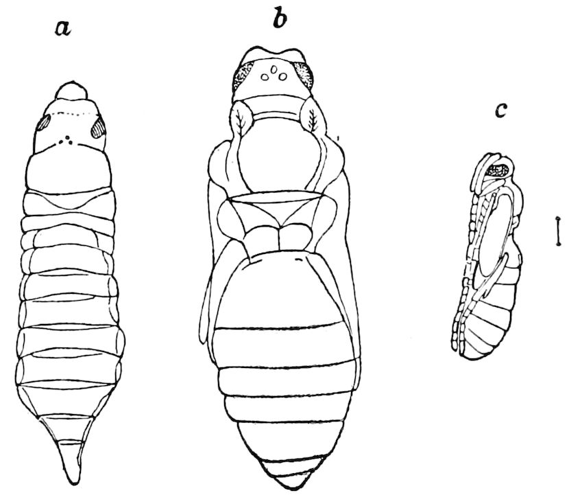
Fig. 38.—Larva (a) of a chalcid, about to pupate, with the head, including the eyes and three ocelli, in the prothoracic segment: b, c, pupa.
The composition of the head in the Hymenoptera.—Ratzeburg stated in 1832 that the head in the adult Hymenoptera (Cynips, Hemiteles, and Formica) does not correspond to that of the larva, but is derived from the head and the first thoracic segment of the larva. Westwood and also Goureau made less complete but similar observations, though Westwood afterwards changed his opinion, and the same view was maintained by Reinhard. Our own observations (as seen in Fig. 38) led us to suppose that this was a mistaken view; that the larval head, being too small to contain that of the semipupa, was simply pushed forward, as in caterpillars. Bugnion, however, reaffirms it in such a detailed way that we reproduce his account. He maintains that the views of Ratzeburg are exact and easy to verify in the chalcid genus Encyrtus, except, however, that which concerns the ventral part and the posterior border of the prothoracic segment.
As the time of transformation approaches, the head of the larva, he says, is depressed and soon concealed under the edge of the prothoracic segment; the latter elongates, becomes thicker and more convex, and within can be seen the two oculo-cephalic imaginal buds. The head of the perfect insect is derived not only from the head of the larva, but also from the portion of the prothoracic segment which is occupied by the buds, i.e. almost its entire dorsolateral face. But the hinder and ventral part of this segment (which contains the imaginal buds of the first pair of legs) takes no part in the formation of the head; these parts, according to Bugnion, towards the end of the larval period detaching themselves so as to become fused with the thorax and constitute the pronotum and the prosternum.
56
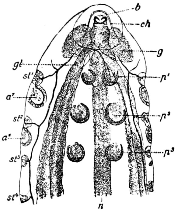
Fig. 39.—Anterior half of larva of Encyrtus, ventral face, showing the upper (wing) and lower (leg) thoracic imaginal buds: b, mouth; ch, chitinous arch; gl, silk gland; g, brain; n, nervous cord; a1, bud of fore, a2, bud of hind, wing; p1–p3, buds of legs; st1–st3, stigmata.
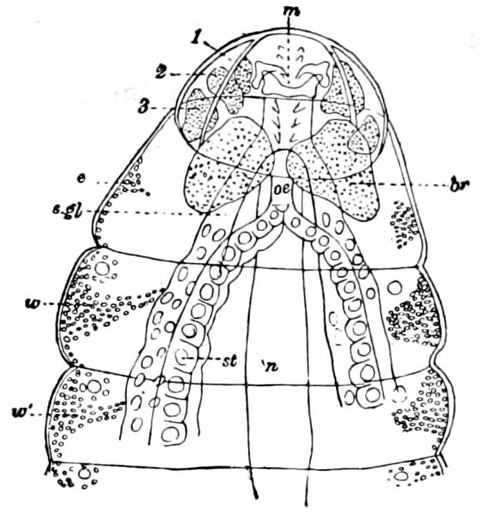
Fig. 40.—Anterior part of Encyrtus larva, 1.2 mm. in length; dorsal face; the cellular masses beginning to form the buds of the wings, eyes, and antennæ: o, eye bud; e, stomach.
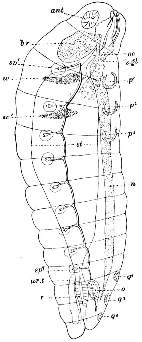
Fig. 41.—Older Encyrtus larva, lateral view, showing the buds of the antennæ (f), legs, and wings; oe, œsophagus; q1, q2, q3, buds of the genital armature; x, rudiment of the sexual gland (ovary or testis); u, urinary tube; i, intestine (rectum); a, anus.
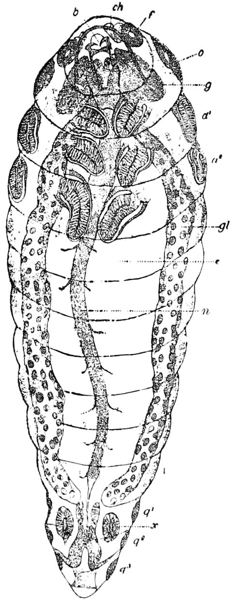
Fig. 42.—A still older larva, ready to transform. The imaginal buds of the antennæ, eyes, wings, and legs have become elongated; lettering as in Fig. 41.—This and Figs. 39–41 after Bugnion.
57
This mode of formation of the head may be observed still more easily in Rhodites, Hemiteles, and Microgaster, from the fact that their oculo-cephalic buds are much more precocious, and that the eyes are charged with pigment at a period when the insect still preserves its larval form.
“... I believe that this mode of formation of the head occurs in all Hymenoptera with apodous larvæ, in this sense; that a more or less considerable part of the first thoracic segment is always soldered to the head of the larva to constitute the head of the perfect insect. The arrangement of the nervous system is naturally in accord with this peculiarity of development, and the cephalic ganglia of the larva to which the ocular blastems later adapt themselves, are found not in the head, but in the succeeding segment (Figs. 39, 40, 41).
“Relying on these facts, I maintain that the encroachment of the head on the prothorax is a consequence of the preponderance in size of the brain, and indicates the superiority of the Hymenoptera over other insects....”
That the pronotum is derived from the larval prothoracic segment is proved by the fact that the first pair of stigmata becomes what authors call the “prothoracic” stigmata of the perfect insect. But Bugnion thinks that the projection which carries it, and which he calls the shoulder (Figs. 41 and 42), belongs to the mesonotum.
b. Appendages of the head
The antennæ.—These are organs of tactile sense, but also bear olfactory, and in some cases auditory organs; they are usually inserted between or in front of the eyes, and moved by two small muscles at the base, within the head. In the more generalized insects the antennæ are simple, many-jointed appendages, the joints being equal in size and shape. The antennæ articulate with the head by a ball and socket joint, the part on which it moves being called the torulus (Fig. 32, r). In the more specialized forms it is divided into the scape, the pedicel, and a flagellum (or clavola); but usually, as in ants, wasps, and bees, there are two parts, the basal three-jointed one being the scape, and the distal one, the usually long filiform flagellum. The antennæ, especially the flagellum, vary greatly in form in insects of different families and orders, this variation being the result of adaptation to their peculiar surroundings and habits. The number of antennal joints may be one (Articerus, a clavigerid beetle), or two in Paussus and in Adranes cœcus (Fig. 4312), where they are short and club-shaped; in flies (Muscidæ, etc.), they are very short and with few joints, and when at rest lying in a cavity adapted for their reception. In the lamellicorn beetles the flagellum is divided into several leaves, and this condition may be approached in the serrate or flabellicorn antennæ of other beetles. In Lepidoptera, and in certain saw-flies and beetles, they are either pectinate or bipectinate, being in one case at least, that of the Australian Hepialid (Abantiades argenteus), tripectinate (Fig. 44), and in the dipterous (Tachinid) genus Talarocera the third joint is bipectinate (Fig. 45). In Xenos and in Parnus they may be deeply forked, 58while in Otiocerus, two long processes arise from the base, giving it a trifid shape. In dragon-flies and cicadæ, they are minute and hair-like, though jointed, while in the larvæ of many metabolous insects they are reduced to minute three-jointed tubercles. In aquatic beetles, bugs, etc., the antennæ are short, and often, when at rest, bent close to the body, as long antennæ would impede their progress.
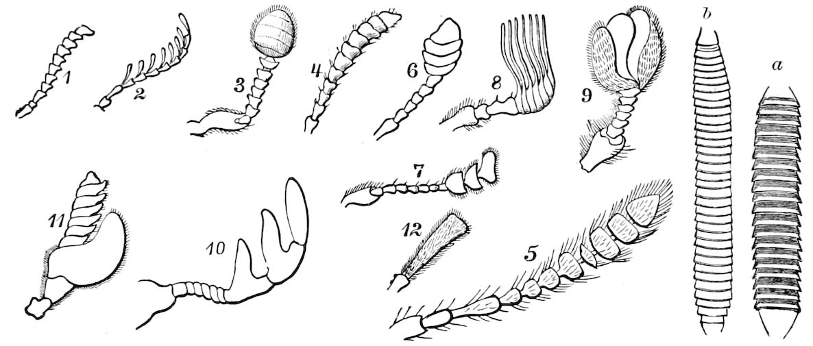
Fig. 43.—Different forms of antennæ of beetles: 1, serrate; 2, pectinate; 3, capitate (and also geniculate); 4–7, clavate; 8, 9, lamellate; 10, serrate (Dorcatoma); 11, irregular (Gyrinus); 12, two-jointed antenna of Adranes cæcus.—After LeConte. a, first joint of flagellum of antenna of Troctes silvarum; b, of T. divinatorius.—After Kolbe.
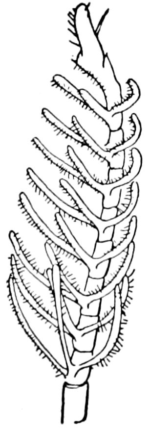
Fig. 44.—Tripectinate antenna of an Australian moth.
While usually more or less sensorial in function, Graber states that the longicorn beetles in walking along a slender twig use their antennæ as a rope-dancer does his balancing pole.
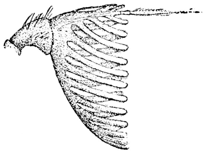
Fig. 45.—Antenna of Talarocera nigripennis, ♂.—After Williston.
Recent examination of the sense-organs in the antennæ of an ant, wasp, or bee enables us, he says, to realize what wonderful organs the antennæ are. In such insects we have a rod-like tube which can be folded up or extended out into space, containing the antennal nerve, which arises directly from the brain and sends a branch to each of the thousands of olfactory pits or pegs which stud its surface. The antenna is thus a wonderfully complex organ, and the insect must 59be far more sensitive to movements of the air, to odors, wave-sounds, and light-waves, than any of the vertebrate animals.
That ants appear to communicate with each other, apparently talking with their antennæ, shows the highly sensitive nature of these appendages. “The honey-bee when constructing its cells ascertains their proper direction and size by means of the extremities of these organs.” (Newport.)
How dependent insects are upon their antennæ is seen when we cut them off. The insect is at once seriously affected, its central nervous system receiving a great shock, while it gives no such sign of distress and loss of mental power when we remove the palpi or legs. On depriving a bee of its antennæ, it falls helpless and partially paralyzed to the earth, is unable at first to walk, but on partly recovering the use of its limbs, it still has lost the power of coördinating its movements, nor can it sting; in a few minutes, however, it becomes able to feebly walk a few steps, but it remains over an hour nearly motionless. Other insects after similar treatment are not so deeply affected, though bees, wasps, ants, moths, certain beetles, and dragon-flies are at first more or less stunned and confused.
The antennæ afford salient secondary sexual differences, as seen in the broadly pectinated antennæ of male bombycine moths, certain saw-flies (Lophyrus), and many other insects.
The mouth-parts, buccal appendages, or trophi, comprise, besides the labrum, the mandibles and maxillæ.
The mandibles.—These are true jaws, adapted for cutting, tearing, or crushing the food, or for defence, while in the bees they are used as tools for modelling in wax, and in Cetonia, etc., as a brush for collecting pollen. They are usually opposed to each other at the tips, but in many carnivorous forms their tips cross each other like shears. They are situated below the clypeus on each side, and are hinged to the head by a true ginglymus articulation, consisting of two condyles or tubercles to which muscles are attached, the principal ones being the flexor and great extensor (Fig. 48). They are solid, chitinous, of varied shapes, and in the form of the teeth those of the same pair differ somewhat from each other (Fig. 46 A). In the pollen-eating beetles (Cetoniæ) and in the dung-beetles (Aphodius, etc.) the edge is soft and flexible. In the males of Lucanus, etc. (Fig. 47), and of Corydalus (Fig. 29), they are of colossal size, and are large and sabre-shaped in the larvæ of water-beetles, ant-lions, Chrysopa, etc. where they are perforated at the tips, through which the blood of their prey is sucked.
While the mandibles are generally regarded as composed of a 60single piece, in Campodea and Machilis there appears to be an additional basal piece apparently corresponding to the stipes of the first maxilla, and separated by a faint suture from the molar or distal joint. In Campodea there is a minute movable appendage figured both by Meinert and by Nassonow, which appears to represent the lacinia of the maxilla (Fig. 48). Wood-Mason has observed in the mandibles of the embryo of a Javanese cockroach, Blatta (Panesthia) javanica, indications of “the same number of joints as in that of chilognathous myriopods, or one less than in that of Machilis.” Also he adds: “In both ‘larvæ’ and adults of Panesthia javanica a faint groove crosses the ‘back’ of the mandible at the base. This groove appears to be the remains of the joint between the third and apical segments of the formerly 4–segmented mandibles.”
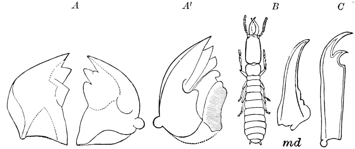
Fig. 46.—Various forms of mandibles. A, right and left of Termopsis. A′, showing at the shaded portion the “molar” of Smith. B, Termes flavipes, soldier; md, its mandible. C, Panorpa.
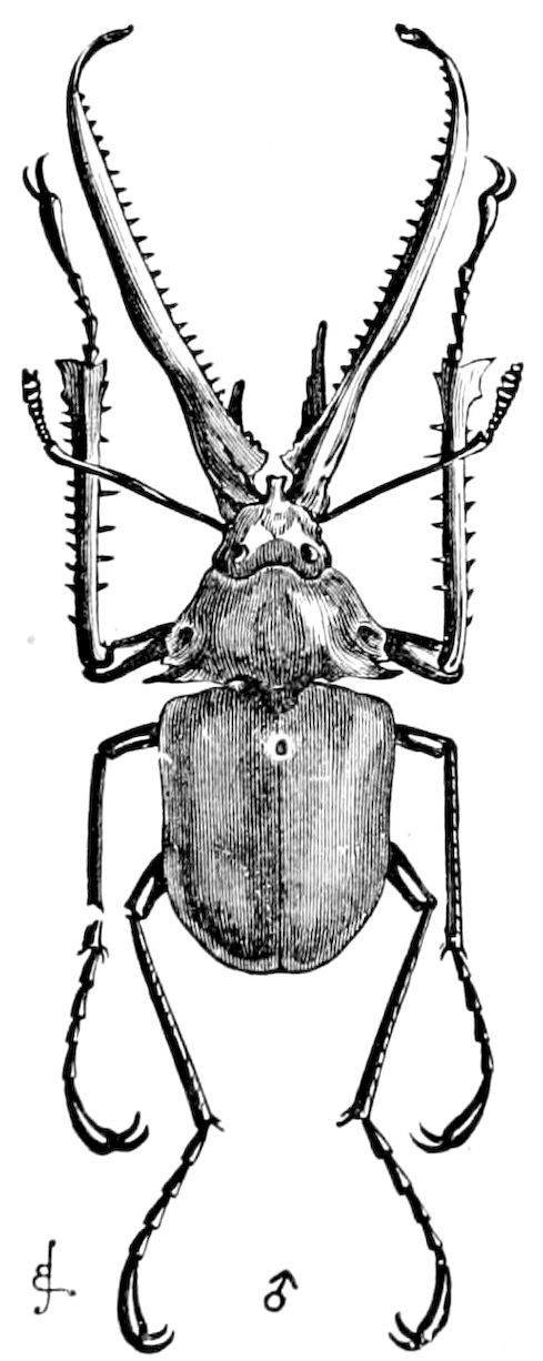
Fig. 47.—Chiasognathus grantii, reduced. Male.—After Darwin.
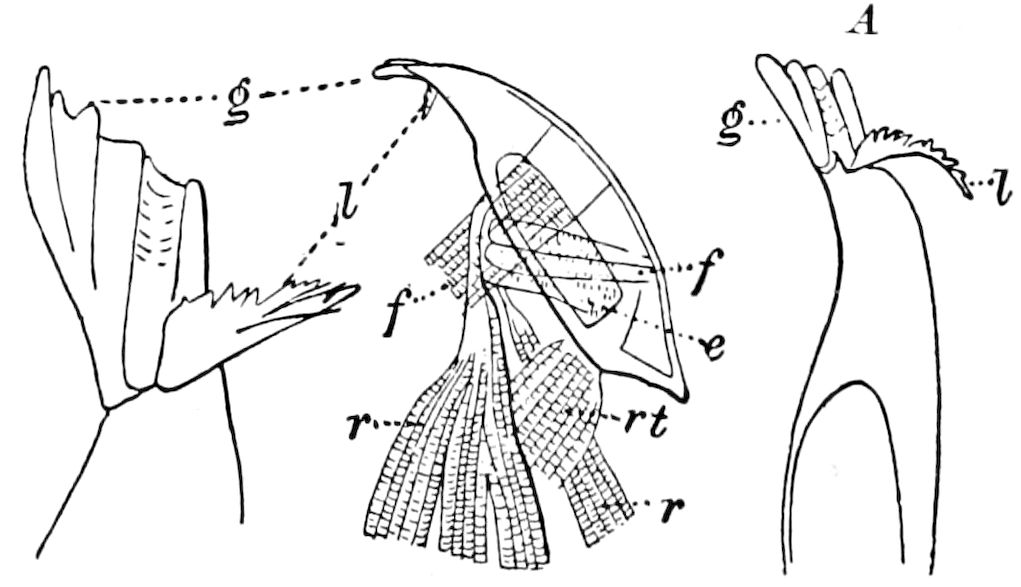
Fig. 48.—Mandible of Campodea: l, prostheca or lacinia; g, galea; f, f, flexor muscles; e, extensor; r, r, retractor; rt, muscle retaining the mandible in its place.—After Meinert. A, extremity of the same.—After Nassonow.
61
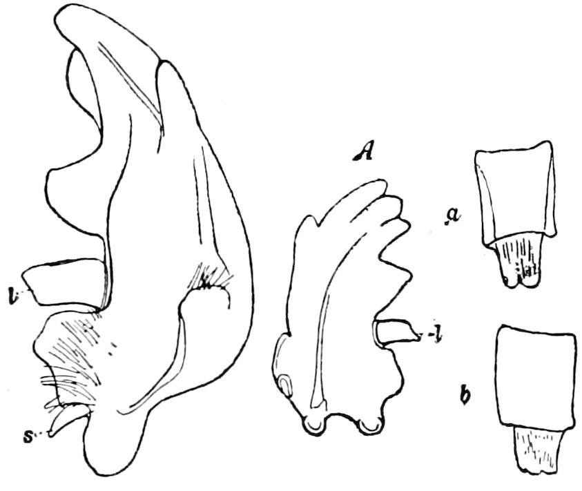
Fig. 49.—Mandible of Passalus cornutus with the prostheca (l): A, that of a Nicaraguan species; a, inside, b, outside view, with the muscle.
He also refers to the prostheca of Kirby and Spence (Fig. 49), which he thinks appears to be a mandibular lacinia homologous with it in Staphylinidæ and other beetles (J. B. Smith also considers it as “homologous to the lacinia of the maxilla”), and on examining it in P. cornutus and a Nicaragua species (Fig. 49), we adopt his view, since we have found that it is freely movable and attached by a tendon and muscle to the galea. In the rove beetles (Goërius, Staphylinus, etc.) and in the subaquatic Heteroceridæ, instead of a molar process, is a membranous setose appendage not unlike the coxal appendages of Scolopendrella, movably articulated to the jaw, which he thinks answers to the molar branch of the jaws in Blatta and Machilis. “It has its homologue in the diminutive Trichopterygidæ in the firmly chitinized quadrant-shaped second mandibular joint, which is used in a peculiar manner in crushing the food”; also in the movable tooth of the Passalidæ, and in the membranous inner lobe of the mandibles of the goliath-beetles, etc.
J. B. Smith has clearly shown that the mandibles are compound in certain of the lamellicorns. In Copris carolina (Fig. 50), he says, the small membranous mandibles are divided into a basal piece (basalis), the homologue of the stipes in the maxilla; another of the basal pieces he calls the molar, and this is the equivalent of the subgalea, while a third sclerite, only observed in Copris, is the conjunctivus, the lacinia (prostheca) being well developed. Smith therefore concludes “that the structure of the mandible is fundamentally the same as that of the labium and maxilla, and that we have an equally complex organ in point of origin. Its usual function, however, demands a powerful and solid structure, and the sclerites are in most instances as thoroughly chitinized and so closely united to the others that practically there is only a single piece, in which the homology is obscured.” (Trans. Amer. Ent. Soc., xix, pp. 84, 85. 1892.) From the studies of Smith and our observations on Staphylinus, Passalus, Phanæus, etc. (Fig. 50, A, B) we fully agree with the view that the mandibles are primarily 3–lobed appendages like the maxillæ. Nymphal Ephemerids have a lacinia-like process. (Heymons.)
62
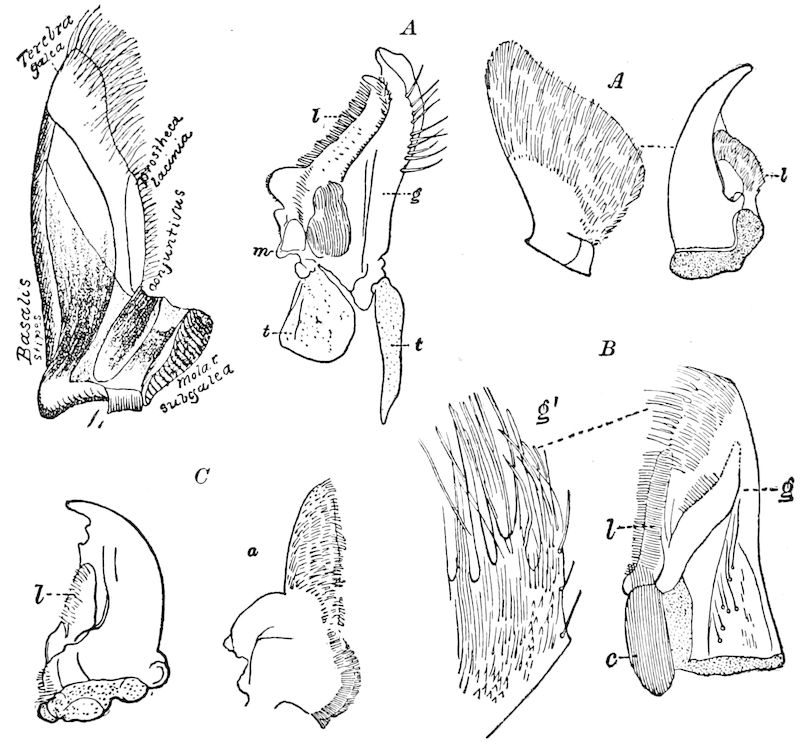
Fig. 50.—Mandible of Copris carolina.—After Smith. A′C. anaglypticus. A (figure to right), do. of Leistotrophus cingulatus; B, of Phanæus carnifex; g′, end of galea,—g, enlarged; c, conjunctivus. C, of Meloë angusticollis: l, lacinia; a, lacinia enlarged.
Mandibles are wanting in the adults of the more specialized Lepidoptera, being vestigial in the most generalized forms (certain Tineina and Crambus), but well developed in that very primitive moth, Eriocephala (Fig. 51). They are also completely atrophied in the adult Trichoptera, though very large and functional in the pupa of these insects (Fig. 52), as also in the pupa of Micropteryx (Fig. 53). They are also wanting in the imago of male Diptera and in the females of all flies except Culicidæ and Tabanidæ.
They are said by Dr. Horn to be absent in the adult Platypsyllus castoris, though well developed in the larva; and functional mandibles are lacking in the Hemiptera.
The first maxillæ.—These highly differentiated appendages are inserted on the sides of the head just behind the mandibles and the mouth, and are divided into three lobes, or divisions, which are supported upon two, and sometimes three basal pieces, i.e. the basal 63joint or cardo, the second joint or stipes, with the palpifer, the latter present in Termitidæ (Fig. 54, plpgr), but not always separately developed (Fig. 55). The cardo varies in shape, but is more or less triangular and is usually wedged in between the submentum and mandible. It is succeeded by the stipes, which usually forms the support for the three lobes of the maxilla, and is more or less square in shape.
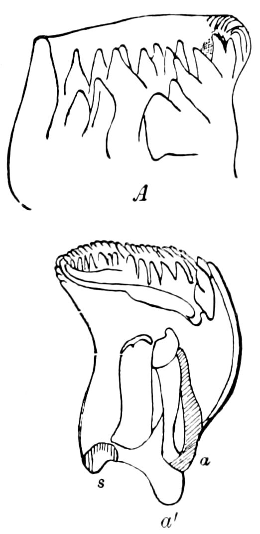
Fig. 51.—Mandible of Eriocephala calthella: a, a′, inner and outer articulation; s, cavity of the joint (acetabulum); A, end seen from one side of the cutting edge.—After Walter.
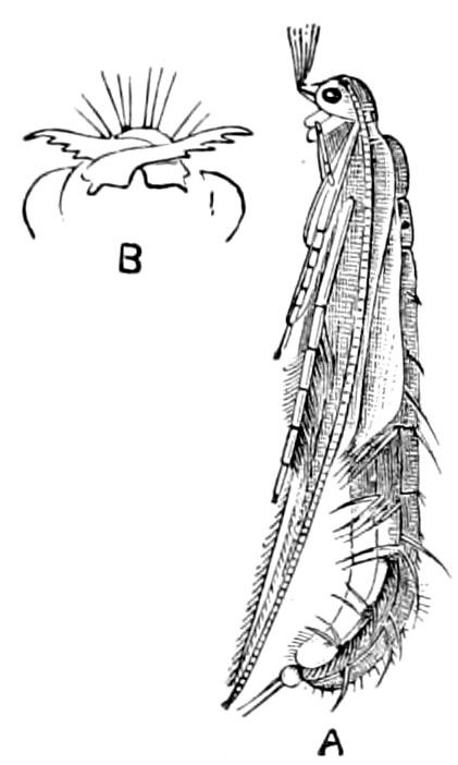
Fig. 52.—A, Pupa of Phryganea pilosa.—After Pictet. B, mandibles of pupa of Molanna angustata.—After Sharp.
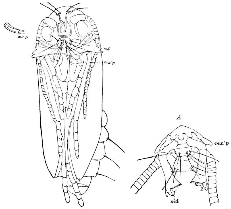
Fig. 53.—Pupa of Micropteryx purpuriella, front view: md, mandibles; mx.p, maxillary palpus, end drawn separately; mx.’p, labial palpi; lb, labrum; A, another view from a cast skin.
The three distal divisions of the maxilla are called, respectively, beginning with the innermost, the lacinia, galea, and palpifer, the latter being a lobe or segment bearing the palpus. The lacinia is more or less jaw-like and armed on the inner edge with either flexible or stiff bristles, spines, or teeth, which are very variable in shape and are of use 64as stiff brushes in pollen-eating beetles, etc. The galea is either single-jointed and helmet-shaped or subspatulate, as in most Orthoptera, or 2–jointed in Gryllotalpa, or lacinia-like in Myrmeleon (Fig. 55, C); or, in the Carabidæ (Fig. 56) and Cicindelidæ, it is 2–jointed and in form and function like a palpus.
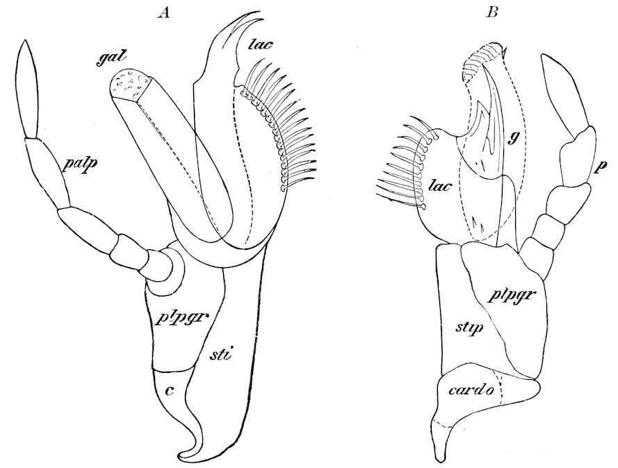
Fig. 54.—A, maxilla of Termopsis angusticollis. B, Termes flavipes: c, cardo; sti, stipes; plpgr, palpiger; palp, palpus; lac, lacinia; g, gal, galea.
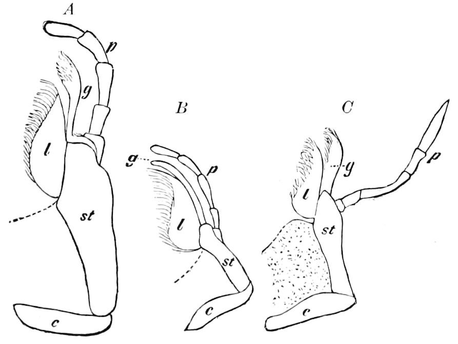
Fig. 55.—A, maxilla of Mantispa brunnea. B, Ascalaphus longicornis. C, Myrmeleon diversum. Lettering as in Fig. 54.
65
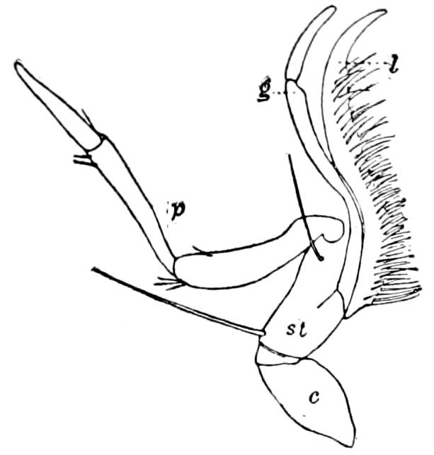
Fig. 56.—Maxilla of a carabid, Anophthalmus tellkampfii: l, lacinia; g, 2–jointed galea; p, palpus; st, stipes; c, cardo.
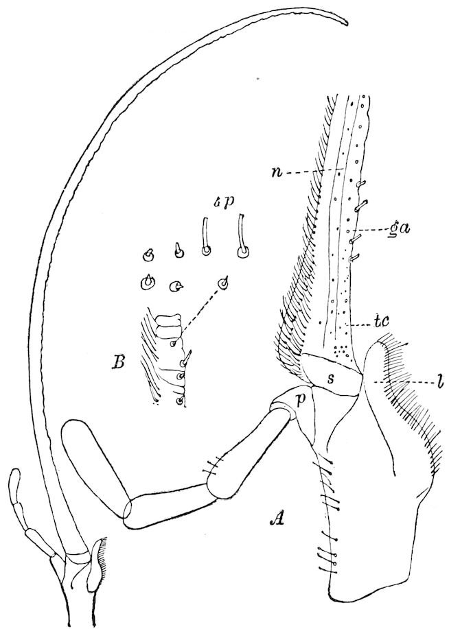
Fig. 57.—Maxilla of Nemognatha, ♀, from Montana. A, base of maxilla enlarged to show the taste-papillæ (tp) and cups (tc), on the galea (ga). B, part of end of galea to show the imperfect segments and taste-organs: n, nerve; a ganglionated nerve supplies each taste-papilla or cup; l, lacinia; p, palpifer; s, subgalea.
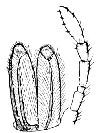
Fig. 58.—Maxilla of Panorpa.
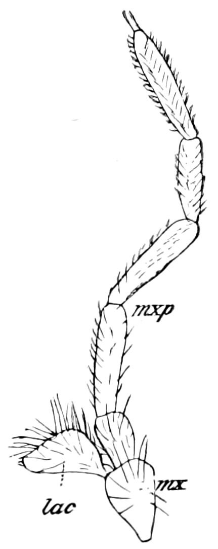
Fig. 59.—Maxilla of Limnephilus pudicus: mx, stipes; lac, galea.
The palpus is in general antenniform and is composed of from 1 to 6 joints, being usually 4– or 5–jointed, and is much longer than the galea. In the maxilla of the beetle Nemognatha (Fig. 57), the galea is greatly elongated, the two together forming an imperfect tube or proboscis and reminding one of the tongue of a moth, while the lacinia is reduced. In the Mecoptera the lacinia and galea are closely similar (Fig. 58); in the Trichoptera only one of the lobes is present (Fig. 59), while in the Lepidoptera the galea unites with its mate to form the so-called tongue (Fig. 60). The maxilla of the male of Tegeticula yuccasella is normal, though the galeæ are separate; but in the female, what Smith regards as the palpifer (the “tentacle” of Riley) is 66remarkably developed, being nearly as long as the galea (Fig. 61) and armed with stout setæ, the pair of processes being adapted for holding a large mass of pollen under the head.
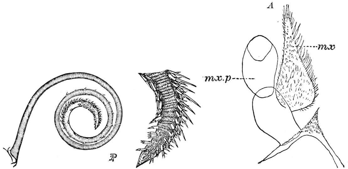
Fig. 60.—Tongue of Aletia xylina, with the end magnified.—Pergande del., from Riley. A, much reduced maxilla (mx) of Paleacrita vernata; mx.p, palpus.
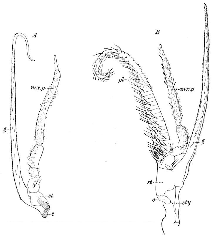
Fig. 61.—A, maxilla of Tegeticula yuccasella, ♂: g, galea. B, ♀: pl, enormously developed palpifer; mx.p, palpus; c, cardo; st, stipes; sty, stylus.
In coleopterous larvæ the maxillæ are 2–lobed 67(Fig. 62), the galea being undifferentiated, but in those of saw-flies the galea is present (Fig. 63, gal).
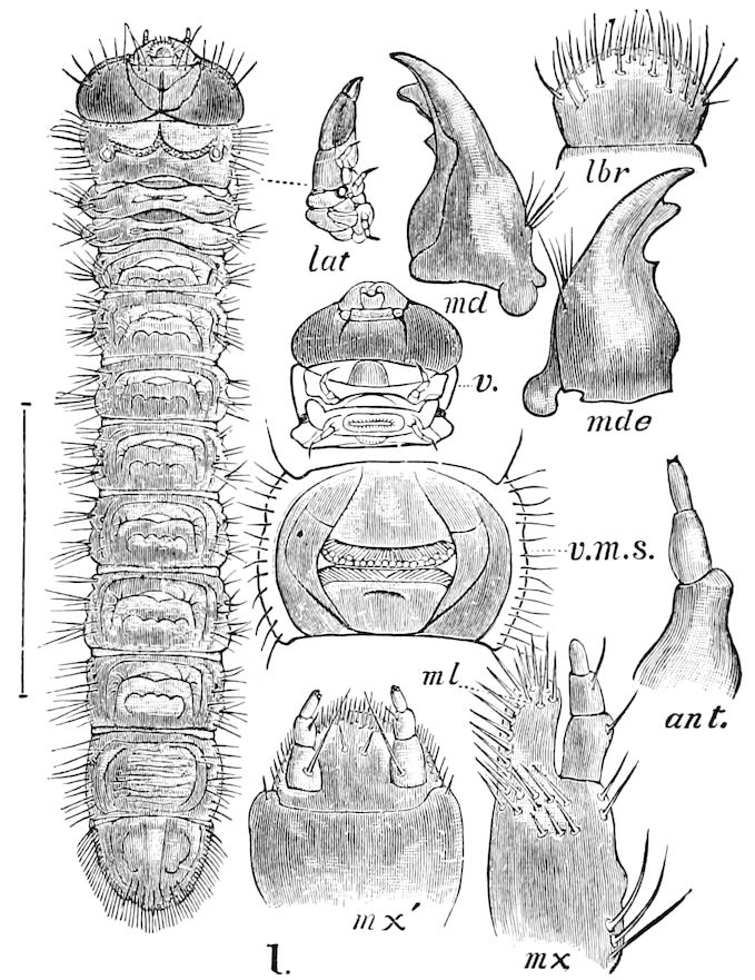
Fig. 62.—Larva of Rhagium lineatum: lat, lateral view of head and thoracic segments; mx, first maxilla; ml, undifferentiated lacinia and galea; v, under side of head and pro- and meso- thoracic segments; v.m.s., one of the middle ventral segments, magnified six times; mx′, 2d maxilla.
It now seems most probable that in the first maxillæ we have the primary form of buccal appendage of insects, the appendage being composed of three basal pieces with three variously modified distal lobes or divisions; and that the mandibles and second maxillæ are modifications of this type.
How wonderfully the maxillæ of the Lepidoptera are modified, and the peculiar shapes assumed in the Diptera, Hymenoptera, and other groups, will be stated in the accounts of those orders, but it is well to recall the fact that in the most primitive and generalized moth, Eriocephala, the lacinia is well developed (Fig. 64).
As Newport remarks, the office of the maxillæ in the mandibulate insects is of a twofold kind; since they are adapted not only for seizing and retaining the food in the mouth, but also as accessory jaws, since they aid the mandibles in comminuting it before it is passed on to the pharynx and swallowed. Hence, as the food varies so much in nature and situation, it will be readily seen that the maxillæ, especially their distal parts, vary correspondingly. Thus far no close observations on the exact use of the first and second maxillæ have been published.
The palpi also are not only organs of touch, but in some cases act as hands and also bear minute sense-organs, the function 68of which is unknown, but would appear to be usually that of smell.
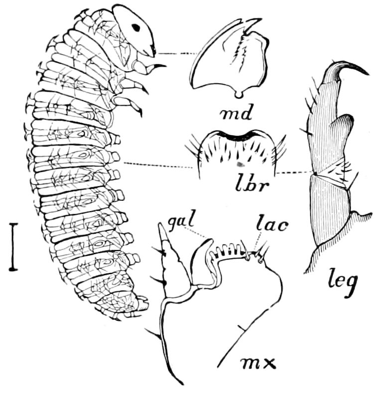
Fig. 63.—Selandria larva, common on Carya porcina, with details of mouth-parts: leg, leg; mx, maxilla; gal, galea; lac, lacinia.
The second maxillæ.—The “under-lip” or labium of insects is formed by the fusion at the basal portion of what in the embryo are separate appendages, and which arise in the same manner as the first maxillæ. They are invariably solidly united, no cases of partial or incomplete fusion being known. The so-called labium is situated in front of the gula or gular region, and is bounded on each side by the gena, or cheek. As already observed, the second maxillæ appear to be the appendages of the last or occipital segment of the head.
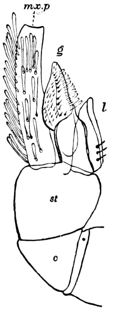
Fig. 64.—Maxilla of Eriocephala calthella: l, lacinia; g, galea; mx.p, maxillary palpus; st, stipes; c, cardo.—After Walter.
The second maxillæ are very much differentiated and vary greatly in the different orders, being especially modified in the haustellate or suctorial orders, notably the Hymenoptera and Diptera. In the mandibulate orders, particularly the Orthoptera, where they are most generalized and primitive in shape and structure, they consist of the following parts: the gula (a postgula is present in Dermaptera), submentum (lora of Cheshire, i, p. 91), mentum, palpifer, the latter bearing the palpi; the lingua (ligula) and paraglossæ, while the hypopharynx or lingua is situated on the upper side. The labial palpi are of the same general shape as those of the first maxillæ, but shorter, with very rarely more than three joints, though in Pteronarcys there are four. Leon has detected vestigial labial palpi in several Hemiptera (Fig. 73). As to the exact nature and limits of the gula, we are not certain; it is not always present, and may be only a differentiation of the submentum, or the latter piece may be regarded as a part of the gula.
We are disposed to consider the second maxillæ as morphologically nearly the exact equivalents of the first pair of maxillæ, and if we 69adopt this view it will greatly simplify our conception of the real nature of this complicated organ. The object of the fusion of the basal portion appears to be to form an under-lip, in order both to prevent the food from falling backwards out of the mouth, and, with the aid of the first pair of maxillæ, to pass it forward to be crushed between the mandibles, the two sets of appendages acting somewhat as the tongue of vertebrates to carry and arrange or press the morsels of food between the teeth or cutting edges of the mandibles.
The spines often present on the free inner edges of the first and second maxillæ (Figs. 54, 62) form rude combs which seem to clean the antennæ, etc., often aiding the tibial combs in this operation.
The submentum and mentum, or the mentum when no submentum is differentiated (with the gula, when present), appear to be collectively homologous with the cardines of the first pair of maxillæ, together with the palpifers and the stipites.[15] These pieces are more or less square, and have a slightly marked median suture in Termitidæ, the sign of primitive fusion or coalescence.
The most primitive form of the second maxillæ occurs in the Orthoptera and in the Termitidæ. The palpifer is either single (Periplaneta, Diapheromera, Gryllidæ) or double (Blatta orientalis, Locustidæ). In Prisopus the single piece in front of the palpifer is in other forms divided, each half (Blatta, Locustidæ, Acrydidæ) bearing the two “paraglossæ,” which appendages in reality are the homologues of the lacinia and galea of the first maxillæ.[16] In the Termitidæ (Fig. 65) the lingua is not differentiated from the palpifer, and the two paraglossæ (or the lamina externa and interna of some authors) with the palpus are easily seen to be the homologues of the three lobes of the first maxillæ. In the Perlidæ (Pteronarcys, Fig. 66) the palpifer is divided, while the four paraglossæ arise, as in Prisopus and Anisomorpha, from an undivided piece, the lingua not being visible from without. In the Neuroptera the lingua or ligula is a large, broad, single lobe, without “paraglossæ,” and the palpifer is either single (Myrmeleon, Fig. 67), or divided (Mantispa, Fig. 68). 70In Corydalus (Fig. 29) the palpifer forms a single piece, and the lingua is undivided, though lobed on the free edge.
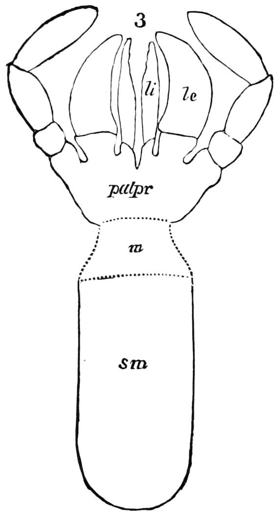
Fig. 65.—Second maxillæ of Termopsis angusticollis: li, the homologue of the lacinia; le, galea.
In the metabolic orders above the Neuroptera the lingua is variously modified, or specialized, with no vestiges of the lacinia or galea, except in that very primitive moth, Eriocephala, in which Walter found a minute free galea, me, and an inner lobe (Figs. 76, 77), the lacinia.
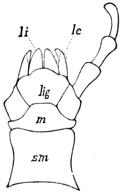
Fig. 66.—Second maxillæ of Pteronarcys californica.
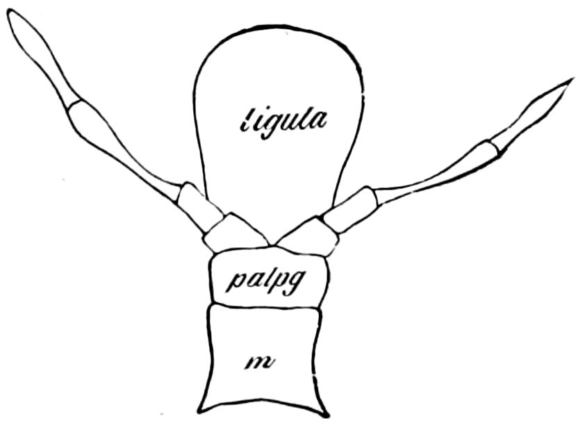
Fig. 67.—Second maxillæ of Myrmeleon diversum.
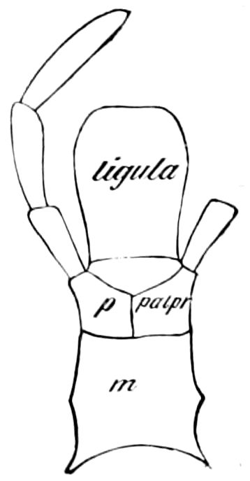
Fig. 68.—Second maxillæ of Mantispa brunnea.
The hypopharynx.—While in its most generalized condition, as in Synaptera, Dermaptera, Orthoptera, and Neuroptera, this anterior median fold or outgrowth of the labium forming the floor of the mouth may retain the designation of “tongue,” lingua, or ligula; in its more specialized form, particularly when used as a piercing or lapping organ, the use of the name hypopharynx seems most desirable. And this is especially the case since, like the epipharynx, it is morphologically a median structure, and while the epipharynx forms the soft, sensitive roof of the mouth, or pharynx; its opposite, the hypopharynx, rises as a fold from the floor of the mouth, forming in its most generalized condition a specialized fold of the buccal integument. In certain cases, as in the honey-bee, the very long slender “tongue” or hypopharynx is evidently, as in the case of the epipharynx, a highly sensitive armature of the mouth.
71In all insects this organ—whether forming a soft, tongue-like, anterior portion or fold of the labium, and “continuous with the lower wall of the pharynx,” or a hard, piercing, awl-like appendage (fleas and flies), or a long, slender, hairy or setose, trough-like structure like the “tongue” of the honey-bee—has a definite location at the end and on the upper side of the labium, and serves to receive at its base the external opening of the salivary duct.
The hypopharynx, as well shown in its lingua condition in Orthoptera, is continuous with and forms the anterior part or fold of the base of the coalesced second maxillæ. It does not seem to be paired, or to represent a pair of appendages.
Opinion regarding the homology of this unpaired piercing organ is by no means settled, and while there is a general agreement as to the nature of the paired mouth-parts, recent observers differ very much as to the morphology of the organ in question.
It is the langue or lingua of Savigny (1816), the ligula of Kirby and Spence (1828), the langue ou languette (lancette médiane du suçoir) of Dugès (1832), the lingua of Westwood (Class, ins., ii, p. 489, 1840), “the unpaired median piercing organ” (“the analogon of the epipharynx of Diptera”) of Karsten (1864), the “tongue” of Taschenberg (1880).
The name hypopharynx was first proposed by Savigny in 1816, who, after naming the membranous plate which has for its base the upper side of the pharynx, the epipharynx, remarks: “Dans quelques genres, notamment dans les Eucères, le bord inférieur de ce même pharynx donne naissance à un autre appendice plus solide que le précédent, et qui s’emboîte avec lui. Je donnerai à ce dernier le nom de langue ou d’hypopharynx. Voilà donc la bouche des Hyménoptères composée de quatre organes impaires, sans y comprendre la ganache ou le menton; savoir, la lèvre supérieure, l’épipharynx, l’hypopharynx, et la lèvre inférieure, et de deux organes paires, les mandibules et les mâchoires.”
As stated by Dimmock: “The hypopharynx is usually present in Diptera (according to Menzbier absent in Sargus), and contains a tube, opening by a channel on its upper surface; this channel extends back, more or less, from the tip, and is the outlet for the salivary secretion. The tip of the hypopharynx may be naked and used as a lance (Hæmatopota, according to Menzbier), or may be hairy (Musca). The upper side of the base of the hypopharynx is continuous with the lower wall of the pharynx; its under surface may entirely coalesce with the labium (Culex, male), may join the labium more or less, anterior to the month (Musca), or, if 72either mandibles or maxillæ are present, its base may join them (Culex, female).” (p. 43.)
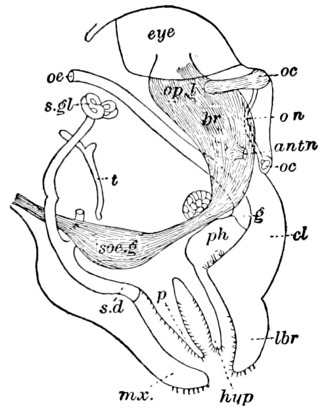
Fig. 69.—Section of head of Machilis maritima: hyp, hypopharynx; lbr, labrum; t, tentorium; ph, room in which the mandibles move on each other; p, paraglossa; mx, labium; sd, salivary duct; s.gl, salivary gland. oe, œsophagus.—After Oudemans.
We will now briefly describe the lingua, first of the mandibulate or biting insects, and then its specialized form, the hypopharynx of the haustellate and lapping insects.
The lingua (hypopharynx) exists in perhaps its most generalized condition in the Thysanura (Fig. 69), where it forms a soft projection, having the same relations as in Anabrus and other Orthoptera.[17]
In the cockroach (Fig. 70), as stated by Miall and Denny, the lingua is a chitinous fold of the oral integument situated in front of the labium, and lying in the cavity of the mouth. The common duct of the salivary glands enters the lingua, and opens on its hinder surface. The lingua is supported by a chitinous skeleton (Figs. 70, B; 82, shp). “The thin chitinous surface of the lingua is hairy, like other parts of the mouth, and stiffened by special chitinous rods or bands.” (Miall and Denny.)
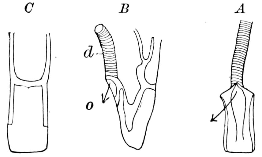
Fig. 70.—Hypopharynx of Periplaneta orientalis; the arrow points out of the opening of the salivary duct: A, origin of salivary duct. B, side view. C, front view.—After Miall and Denny.
In the Acrydiidæ (Melanoplus femur-rubrum) the tongue is a large, membranous, partly hollow expansion of the base of the labium. It may be exposed by depressing the end of the labium, when the opening of the salivary duct may be seen at the bottom or end of the space or gap between the hinder base of the tongue, and the inner anterior base of the labium, as shown by the arrows in Fig. 70. It is somewhat pyriform, slightly keeled above, and bearing fine stiff bristles, which, as they point more or less inwards, probably aid in retaining the food within the mouth. The 73base of the tongue is narrow, and extends back to near the pharynx, there being on the floor of the mouth, behind the tongue, two oblique, slight ridges, covered with stiff, golden-yellow hairs, like those on the tongue. The opening of the salivary duct is situated on the under or hinder side of the hypopharynx, between it and the base of the labium, the base of the former being cleft; the hollow thus formed is situated over the opening, and forms the salivary receptacle.
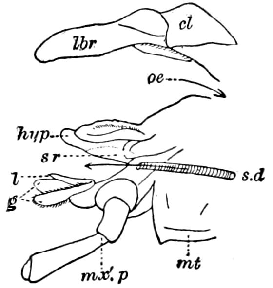
Fig. 71.—Section through the anterior part of the head of Anabrus (the mandibles removed), showing the relations of the hypopharynx (hyp) to the opening of the salivary duct (sd): g, galea; l, lacinia; mt, mentum; oe, œsophagus; lbr, labrum; cl, clypeus.
In the Locustidæ (Anabrus, Fig. 71) the tongue (hypopharynx) is a broad, somewhat flattened lobe arising from the upper part of the base of the mentum and behind the palpifer. This lobe is cavernous underneath, the hollow being the salivary receptacle (sr); the latter is situated over the opening of the salivary duct, which is placed between the base of both the hypopharynx and the labium. The salivary fluid apparently has to pass up and around on each side of the hypopharynx in order to mix with the food.
These relations in the Orthoptera are also the same in the Perlidæ, where the hypopharynx is well developed, forming an unusually large tongue-like mass, nearly filling the buccal cavity.
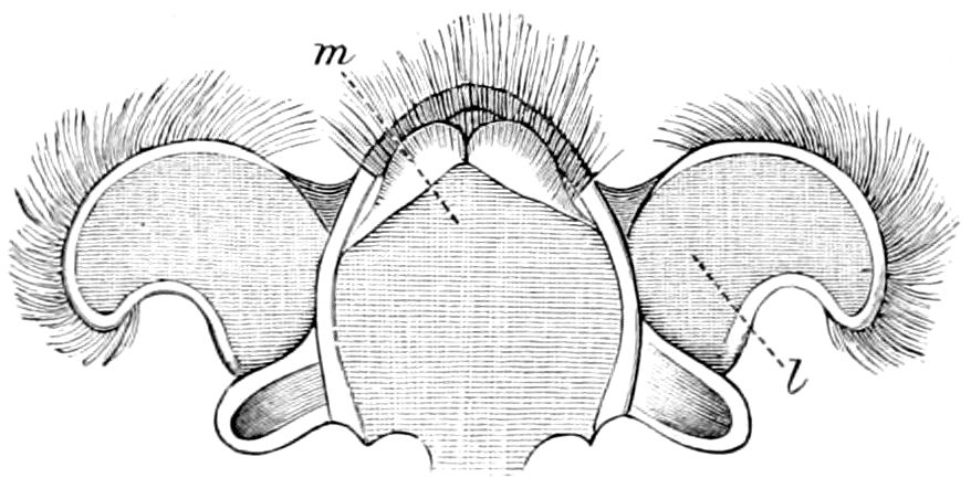
Fig. 72.—Lingua of a May-fly, Heptagenia longicauda, ×16: m, central; l, lateral pieces.—After Vayssière from Sharp.
In the Odonata the lingua is a small, rounded lobe, as also in the Ephemeridæ; in the nymph, however, of Heptagenia (Fig. 72) it is highly developed, according to Vayssière, who seems inclined to regard it as representing a pair of appendages. The tongue in Hemiptera is said by Léon to be present in Benacus griseus (Say) and to correspond to the subgalea of Brullé or hypodactyle of Audouin (Fig. 73), but this appears to correspond to the labium proper, rather than a true lingua, the latter not being differentiated in this order. In the Coleoptera the lingua is rather small. In beetles, as Anopthalmus (Fig. 74), it forms a setose lobe; and a well-developed nerve, the lingual nerve, passes to it, dividing at the end into several branches (n-l). In Sialis the lingua is short, much less developed than usual, being rounded, 74and bears on the edge what appear to be numerous taste-hairs, like those on the ends of the maxillary and labial palpi.
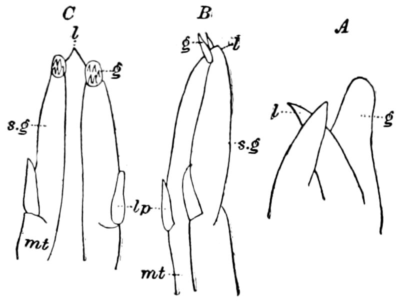
Fig. 73.—A, labium of Zaitha anura. B, of Z. margineguttata. C, of Gerris najas: mt, mentum; lp, labial palpi; sg, subgalea; l, lacinia (= intermaxillare and præmaxillare of Brullé); g, galea.—After Léon.
In the adult Panorpidæ the lingua is a minute, simple lobe.
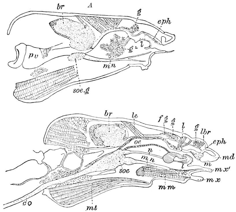
Fig. 74.—Section through head of a carabid, Anopthalmus telkampfii: br, brain; f. g, frontal ganglion; soe, subœsophageal ganglion; co, commissure; n. l, nerve sending branches to the lingua (l); mn, maxillary nerve; mx, 1st maxilla; mm, maxillary muscle; mx′, 2d maxilla; mt, muscle of mentum; le, elevator muscle of the œsophagus; l of the clypeus, and a third beyond raising the labrum (lbr); eph, epipharynx; g, g, salivary glands above; g2, lingual gland below the œsophagus (oe); m, mouth; pv, proventriculus; md, mandible. A, section passing through lingual gland (g2).
In the larval Trichoptera the spinneret is well developed, and in structure substantially like that of caterpillars, and it is plainly the homologue of the hypopharynx, receiving as it does the end of the silk-duct.
In the adult Trichoptera the hypopharynx is a very large, tongue-like, fleshy outgrowth, and is, both in situation and structure, since it contains the opening of the silk-duct, exactly homologous with the hypopharynx of insects of other orders, being somewhat intermediate between the fleshy tongue or lingua of the mandibulate insects, especially the 75Neuroptera, and the hypopharynx of the bees (Fig. 86). Lucas describes and figures it under the name of “haustellum,” but does not homologize it with the hypopharynx. The caddis-flies have been observed to drink water and take in both fluid and fine particles of solid food, and to use the haustellum for this purpose, the end being provided with minute sense-organs like those on the first maxillary lacinia, and possibly of a gustatory nature.
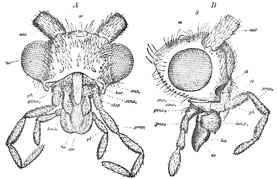
Fig. 75.—Head of Anabolia furcata: A, front view, showing the labrum removed. B, side view; ant, antenna; oc, ocellus; ol, labrum; gh, articulatory process; cmx1, cardo; stmx1, stipes; lemx1, outer lobe (galea); ptmx1, palpus of 1st maxilla; pl, palpus of 2d maxilla; ha, haustellum; so, gustatory pits; spr, opening of salivary duct; chsp, chitinous hook of the clasp; spr, furrow or gutter of the haustellum.—After Lucas.
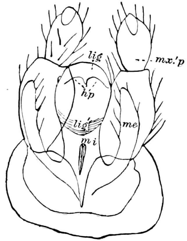
Fig. 76.—Hypopharynx of Eriocephala calthella: lig, ligula, its membranous hinder edge; lig′, anterior horny edge of the ligula-tube opening outwards; hp, contour of the hypopharynx; mi, mala interior (lacinia); me, mala exterior (galea), of second maxilla; mx′ p, labial palpus.—After Walter.
The spinneret of the larvæ of Lepidoptera is evidently the homologue of the hypopharynx of insects of other orders. It will be seen that the homology of the different parts is identical, the common duct of the silk-glands opening at the end of the hypopharynx, which here forms a complete tube or proboscis extending beyond the end of the labium, in adaptation to its use as a spinning organ.
Walter refers to Burgess’s discovery of a hypopharynx in Danais archippus, 76remarking that this organ in the adult Eriocephalidæ (Fig. 76) exhibits a great similarity to the relations observable in the lower insects, adding:—
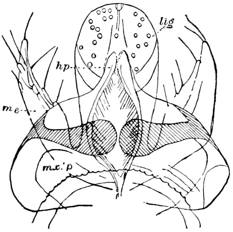
Fig. 77.— Labium of Micropteryx anderschella seen from within (the labial palpi (mx.′ p) removed to their basal joint). Lettering as in Fig. 76.—After Walter.
“The furrow is here within coalesced with the inner side of the labium, and though I see in the entire structure of the head the inner edge of the ligula tube extended under the epipharynx as far as the mandible, I must also accept the fact that here also the hypopharynx extends to the mouth-opening as in all other sucking insects with a well-developed under-lip, viz. the Diptera and Hymenoptera.”
He has also discovered in Micropteryx a paired structure which he regards as the hypopharynx (Fig. 77). As he states:
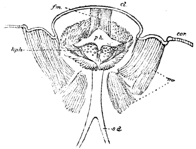
Fig. 78.—Hypopharynx (hph) of Danais: cl, clypeus; sd, salivary duct; m, labial palp muscles; fm, frontal muscle; ph, pharynx; cor, cornea.—After Burgess.
“A portion of the inner surface of the tube-like ligula is covered by a furrow-like band which, close to the inner side, is coalesced with it, and in position, shape, as well as its appendages or teeth on the edge, may be regarded as nothing else than the hypopharynx.”
A hypopharynx is also present in the highest Lepidoptera, Burgess having detected it in Danais archippus. He states that the hypopharynx forms the floor of the pharyngeal cavity; “it is convex on each side of a median furrow (Fig. 78, hph) and somewhat resembles in shape the human breast. The convex areas are dotted over with little papillæ, which possibly may be taste-organs.”
As a piercing organ the hypopharynx reaches its greatest development in the Siphonaptera and Diptera, where the chitinous parts are greatly hypertrophied, the fleshy tongue-like portion so developed in the mandibulate orders being greatly reduced. The chitinous parts are alike on each side of the median organ, being bilaterally symmetrical.
77
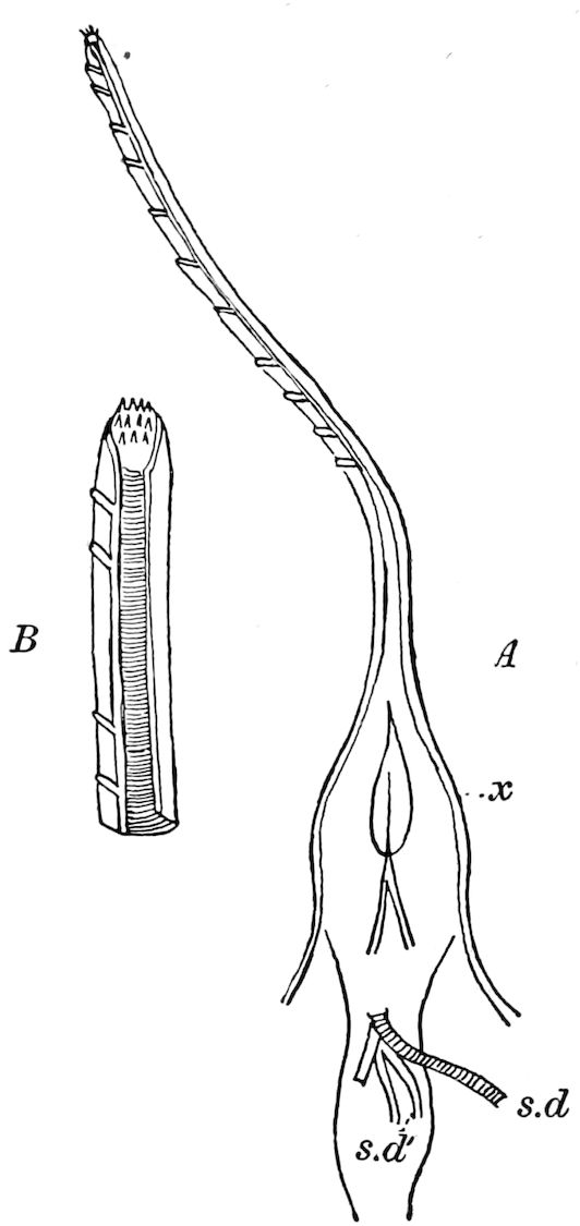
Fig. 79.—A, hypopharynx of Pulex canis: x, basal portion situated within the head; s. d, common duct of the four bladder-shaped salivary glands; s. d′, opening of the tubular salivary glands into the throat. B, end of the hypopharynx, showing the gutter-like structure and teeth at the end.—After Landois.
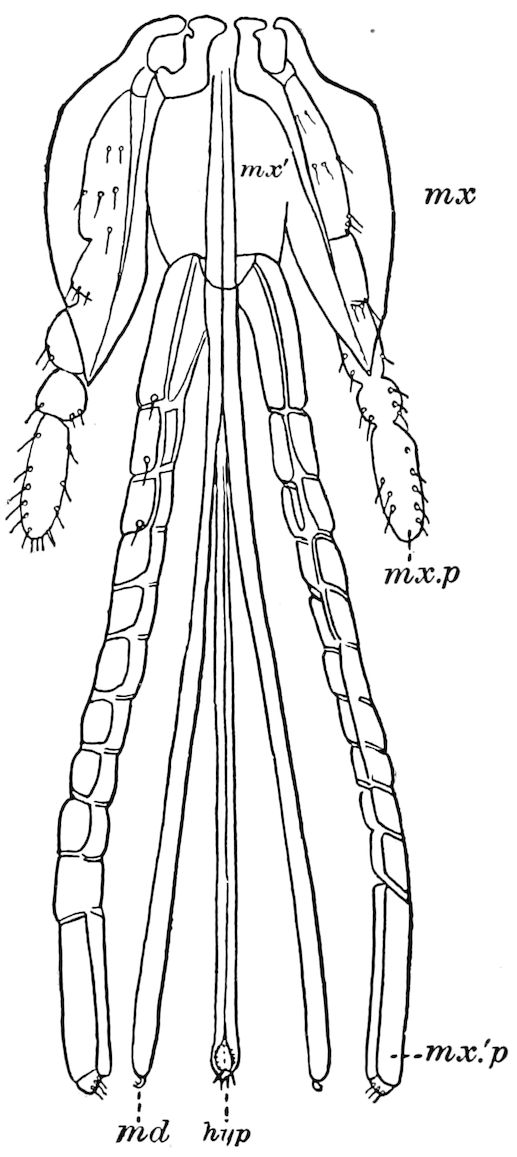
Fig. 80.—Beak of Vermipsylla: hyp, hypopharynx.—After Wagner.
In the fleas the hypopharynx is a large, slender, unpaired, long, chitinous trough, as long as the mandibles, and toothed at the end. Figures 79 and 80 show its relations to the other parts of the mouth; in Fig. 79, x, is seen where the salivary duct opens into the pharynx. Although this organ is not unanimously referred to the hypopharynx, yet from the description of Landois and others, it is evident that this structure does not correspond to the labrum or epipharynx, but belongs to or arises from the floor of the mouth, and, being in close relation to the labium, and also receiving the salivary duct, must be a true hypopharynx.
78In the Diptera the hypopharynx reaches its highest development as a large, stout, awl-like structure.
Meinert, in his detailed and elaborately illustrated work, Trophi Dipterorum (1881), has made an advance on our knowledge of the hypopharynx and its homologies, both by his evidently faithful descriptions and dissections, and by his admirably clear figures.
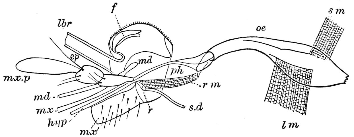
Fig. 81.—Culex pipiens, section of head: oe œsophagus; sm, upper muscle, lm, lower muscle of the œsophagus; ph, pharynx; rm, retractor muscle of the receptacle (r) of the salivary duct (s.d); lbr, labrum; ep, left style of the epipharynx; f, part of front of head.—After Meinert.
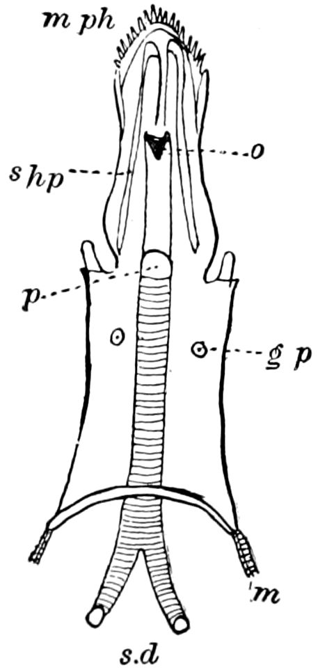
Fig. 82.—Pharynx and hypopharynx of Simulium fuscipes: lph, lower lamina of the pharynx; p, the salivary duct (s.d) perforating the pharynx; o, orifice of the duct; shp, styles of the hypopharynx; mph, membranous edge of the hypopharynx; m, protractor muscle of the pharynx; gp, gustatory papillæ.—After Meinert.
“The hypopharynx, a continuation of the lower edge (lamina) of the pharynx, most generally free, more or less produced, acute anteriorly, forms with the labrum the tube of the pump (antliæ). (The hypopharynx when obsolete, or coalesced with the canal of the proboscis, is the theca; in such a case the siphon or tube is formed by the theca and labrum.) Meanwhile the hypopharynx, the largest of all the trophi (omnium trophorum maximus), constitutes the chief piercing organ (telum) of Diptera. The hypopharynx is moved by protractor, most generally quite or very powerful, and by retractor muscles.
“The efferent duct of the thoracic salivary glands (ductus salivalis) perforates the hypopharynx, more or less near the base, that the saliva may be ejected through the canal into the wound, or that it may be conducted along the labella. Very rarely the salivary duct, perforating the hypopharynx, is continued in the shape of a free, very slender tube.
“The salivary duct behind the base of the hypopharynx forms the receptacle or receptaculum, provided with retractor and levator muscles.”
79
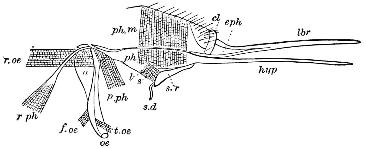
Fig. 83.—Labrum-epipharynx (lbr and eph) and hypopharynx (hyp) of Tabanus brominus: oe, posterior cylindrical portion of the œsophagus; a, anterior swollen portion of the same; ph, pharynx; ph.m, pharyngeal muscle; p.ph, protractor muscle of the pharynx; r.oe, retractor muscle of the œsophagus; r.ph, retractor muscle of the pharynx; f.oe, flexor muscle of the pharynx; t.oe, twisting muscle of the œsophagus; s.r, receptacle of the salivary duct; l, its elevator muscle; s, its retractor muscle; cl, clypeus.—After Meinert.
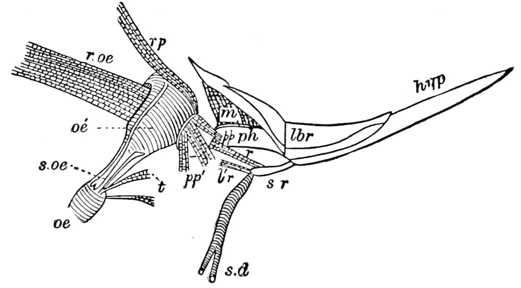
Fig. 84.—Œsophagus (oe), pharynx (ph) with epipharynx and labrum (lbr) of Asilus atricapillus: m, ph, pharyngeal muscle; sr, salivary receptacle; t, twisting; r, l′r, retractor muscles; other lettering as in Fig. 83.—After Meinert.
It has been carefully studied by Meinert in a species of Culex (Fig. 81), Simulium (Fig. 82), Tabanus (Fig. 83), and in Asilus (Fig. 84), where it is seen to attain enormous proportions. In the Hymenoptera, this organ in its most specialized condition is a trough-like rod, adapted for lapping nectar (Fig. 85, 86, hyp). The tongue or hypopharynx of the honey-bee has been elaborately described by Cheshire in his Bees and Bee Keeping.[18] He calls it the tongue or ligula. It is situated in a tube formed by the maxillæ and labial palpi, and can be partially retracted into the mentum. He states that it can move up and down in the tube thus formed, and then describes it as covered by a hairy sheath, its great elasticity being due to a rod running through its centre enabling it to be used as a lapping tongue. The sheath
80“passes round the tongue to the back, where its edges do not meet, but are continuous with a very thin plaited membrane (G, pm) covered with minute hairs. This membrane, after passing towards the sides of the tongue, returns to the angle of the nucleus, or rod, over the under surface of which it is probably continued. The rod passes through the tongue from end to end, gradually tapering towards its extremity, and is best studied in the queen, where I trace many nerve threads and cells. It is undoubtedly endowed with voluntary movement, and must be partly muscular, although I have failed completely in getting any evidence of striation. The rod on the underside has a gutter, or trough-like hollow (cd, the central duct) which is formed into a pseudotube (false tube) by intercrossing of black hairs. It will also be seen that, by the posterior meeting of the sheath, the space between the folded membrane (G, sd) becomes two pseudotubes of larger size, which I shall call the side ducts.
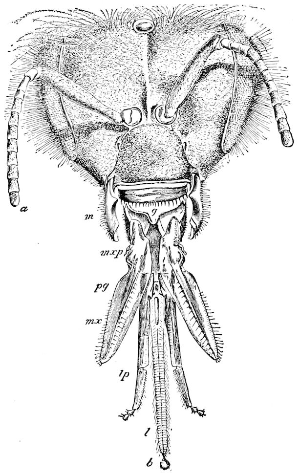
Fig. 85.—Head of honey bee, worker: a, antenna; g, epipharynx; m, mandible; mx, maxilla; mxp, maxillary palpus; pg, paraglossa; lp, labial palpus; l, hypopharynx; b, its spoon.—After Cheshire; from Bull. Div. Ent. U. S. Dept. Agr.
“These central and side ducts run down to that part of the tongue where the spoon, or bouton (K, Fig. 86) is placed. This is provided with very delicate split hairs (b, Fig. 86) capable of brushing up the most minute quantity of nectar, which by capillarity is at once transferred by the gathering hairs (which are here numerous, long, and thin) to two side groove-like forms at the back of the bouton, and which are really the opened-out extremity of the centre and side ducts, assuming, immediately above the bouton, the form seen in F, Fig. 86. The central duct, which is only from 1 600 inch to 1 1000 inch in diameter, because of its smaller size, and so greater capillary attraction, receives the nectar, if insufficient in quantity to fill the side ducts. But good honey-yielding plants would bring both centre and side ducts into requisition. The nectar is sucked up until it reaches the paraglossæ (pa, B, Fig. 86), which are plate-like in front, but membranous extensions, like small aprons, behind; and by these the nectar reaches the front of the tongue, to be swallowed as before described.”
81
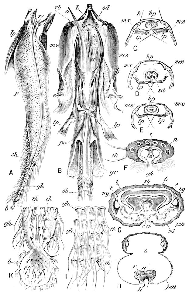
Fig. 86.—Tongue or ligula of the honey bee: A, under side of the tongue; lp, labial palpi; r, r, rod; p, pouch; sh, sheath; gh, gathering hairs; b, bouton or spoon. B, under lip or labium, with appendages, partly dissected; l, lora or submentum; a, a, retractor linguæ longus; sd, salivary duct; rb and b, retractor linguæ biceps; mx, maxillæ; lp, labial palpi; pa, paraglossa; gr, feeding groove; sh, sheath of ligula. C, D, E, sections of ligula; hp, hyaline plate of maxilla; h, hairs acting as stops; mx, maxilla; lp, labial palpi; sd, side duct. F, cross-section of extremity of tongue near the “spoon”; th, tactile hairs; r, rod; n, nucleus; gh, gathering hairs. G, cross-section of tongue without gathering hairs, × 400 times; sh, sheath; b, blood space; t, trachea: ng, gustatory nerve; cd, central duct; sd, lateral duct; pm, plaited membrane. H, same as G, but magnified two hundred times, and with pm, plaited membrane, turned outwards; h, closing hairs; lp, labial palpi; b, blood; n, nucleus; r, rod; h, closing hairs. I, small portion of the sheath; lettering as before. K, extremity of the tongue, with spoon; b, branching hairs for gathering.—After Cheshire.
82Cheshire then settles the question which has been in dispute since the time of Swammerdam, whether the bee’s tongue is solid or tubular. He agrees with Wolff that the duct is a trough and not a tube, and proves it by a satisfactory experiment. He remarks:
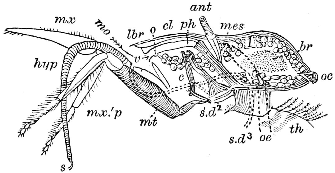
Fig. 87.—Longitudinal section through the head of the honey bee, ♀, just outside of right antenna: ant, antenna with three muscles attached to mes, mesocephalic pillar; cl, clypeus; lbr, labrum; 1, chyle-gland (system no. 1, of Siebold); o, opening of the same; oc, ocellus; br, brain; n, neck; th, thorax; oe, œsophagus; s.d2, s.d3, common salivary ducts of systems 2 and 3; v, salivary valve; c, cardo; ph, pharynx; mx′, labium; mx.′p, labial palpi; mt, mentum; mx, maxilla; hyp, hypopharynx; s, bouton.—After Cheshire.
“Bees have the power, by driving blood into the tongue, of forcing the rod out from the sheath, and distending the wrinkled membrane so that in section it appears as at H, Fig. 86, the membrane assuming the form of a pouch, given in full length at A. It will be seen at once that this disposition of parts abolishes the side ducts, but brings the central duct to the external surface. The object of this curious capability on the part of the bee is, in my opinion, to permit of cleaning away any pollen grains, or other impediment that may collect in the side ducts. The membrane is greasy in nature, and substances or fluids can be removed from it as easily as water from polished metal. If, now, the sides of a needle, previously dipped into clove oil in which rosanilin (magenta) has been dissolved, so as to stain it strongly red, be touched on the centre of the rod, the oil immediately enters, and passes rapidly upwards and downwards, filling the trough.”
Does the hypopharynx represent a distinct segment?—The facts which suggest that the hypopharynx may possibly represent a highly modified pair of appendages, arising from a distinct intermaxillary segment, are these: Heymons plainly shows that, in the embryo of Lepisma, the hypopharynx originates as a transverse segment-like fold in front of the 2d maxillary segment, and larger than it, and though he does not mention it in his text, it appears like the rudiment of a distinct segment; the hypopharynx of Ephemeridæ; arises and remains separate in the nymph from the labium (see Heymons’ Fig. 29, and there are two lateral projections; see also Fig. 72, and Vayssiere’s view that it may represent a pair of appendages; Kolbe also regards it as representing a third pair of maxillæ, his endolabium, p. 213). Though what is called an unpaired 83organ, it is composed of, or supported by, two bilaterally symmetrical styles, both in Myriopods (Fig. 6, labiella, stil) and in insects (Fig. 77, etc.). On the other hand, in the embryo of pterygote insects, an intermaxillary segment has not been yet detected.
LITERATURE OF THE MOUTH-PARTS OR BUCCAL APPENDAGES
a. General
Savigny, Jules-César. Mémoires sur les animaux sans vertèbres. 1re Part. Description et classification des animaux invertébrés et articulés, etc. Fasc. 1re. Mém. 1–2. Théorie des organes de la bouche des crustacés et des insectes. 12 Pl., Paris, 1816, pp. 1–117.
Gerstfeld, Georg. Ueber die Mundteile der saugenden Insekten. Dorpat, 1853.
Olfers, Ernestus V. Annotationes ad anatomiam Podurarum. Berolini, 1862, 4 Pls.
Gerstaecker, Carl Eduard Adolph. Zur Morphologie der Orthoptera amphibiotica. (Festschrift zur Feier des hundertjährigen Bestehens der Gesellschaft naturf. Freunde zu Berlin. 4º, 1873, pp. 39–59, 1 Taf.)
Muhr, Joseph. Die Mundteile der Orthoptera. Ein Beitrag zur vergleichenden Anatomie. (Jahrbuch “Lotos.” Prag, 1877, pp. 40–71, 8 Taf.)
Burgess, Edward. The anatomy of the head and the structure of the maxilla in the Psocidæ. (Proc. Boston Soc. Nat. Hist., xix, 1878, pp. 291–296, 1 Pl.)
Meinert, Fr. Sur la conformation de la tête et sur l’interpretation des organes buccaux chez les insectes, ainsi que sur la systématique de cette ordre. (Ent. Tidsskr., 1. Arg., 1880, pp. 147–150.)
—— Tungens udskydelighed hos Steninerne, en slaegt af Staphylinernes familie. (Vidensk. meddel. fra den naturh. Foren, 1884–1886, pp. 180–207, 2 Pls. Also Zool. Anzeiger, 1887, pp. 136–139.)
Müller, A. Vergleichend-anatomische Darstellung der Mundteile der Insekten. Villach, 1881, 3 Taf.
Kraepelin, Karl. Ueber die Mundwerkzeuge der saugenden Insekten. (Zool. Anzeiger, 1882, pp. 574–579.)
Dewitz, H. Ueber die führung an den Körperanhangen der Insekten. (Berlin. Zeitschr. xxvi., 1882, pp. 51–68, Figs.)
Wolter, Max. Die Mundbildung der Orthopteren mit specieller Berücksichtigung der Ephemeriden. 4 Taf. Greifswald, 1883.
Oudemans, J. T. Beiträge zur Kenntniss der Thysanura und Collembola. (Bijdragen tot de Dierkunde, pp. 149–226. Amsterdam, 1888, 3 Taf.)
Smith, John B. An essay on the development of the mouth-parts of certain insects. (Trans. Amer. Philosophical Soc., xix, pp. 175–198, 3 Pls.)
Also articles by Chatin, McLachlan, Riley, Wood-Mason.
b. Thysanoptera (Physapoda)
Jordan, Karl. Anatomie und biologie der Physapoda. (Zeitschr. f. wissens. Zool., xlvii, pp. 541–620, 3 Taf. 1888.)
Garman, H. The mouth-parts of the Thysanoptera. (Bull. Essex Inst., xxii, 4 pp., Fig. 1890.)
—— The asymmetry of the mouth-parts of Thysanoptera. (Amer. Naturalist, July, 1896, pp. 591–593, Fig.)
Bohls, J. Die Mundwerkzeuge der Physapoden. Dissertation Göttingen, 1891, pp. 1–36.
84Uzel, Heinrich. Monographie der Ordnung Thysanoptera. Königgrätz, 1895, pp. 472, 10 Taf., 9 Figs.
c. Hemiptera
Léon, N. Beiträge zur Kenntniss der Mundteile der Hemipteren. Jena, 1887, pp. 47, 1 Taf.
—— Labialtaster bei Hemipteren. (Zool. Anzeiger, pp. 145–147, 1892, 1 Fig.)
—— Beiträge zur Kenntniss des Labiums der Hydrocoren. (Zool. Anzeiger, März 29, 1897, pp. 73–77, Figs. 1–5.)
Geise, O. Mundteile der Rhynchoten. (Archiv f. Naturgesch., xlix, 1883, pp. 315–373, 1 Taf.)
Wedde, Hermann. Beiträge zur Kenntniss des Rhynchotenrüssels. (Archiv f. Naturgesch., li Jahrg., 1 Bd., 1885, pp. 113–148, 2 Taf.)
Smith, John B. The structure of the hemipterous mouth. (Science, April 1, 1892, pp. 189–190, Figs. 1–5.)
d. Coleoptera
Smith, John B. The mouth-parts of Copris carolina; with notes on the homologies of the mandibles. (Trans. Amer. Ent. Soc., xix, April, 1892, pp. 83–87, 2 Pls.)
e. Lepidoptera
Kirbach, P. Ueber die Mundwerkzeuge der Schmetterlinge. (Zool. Anzeiger, vi Jahrg., 1883, pp. 553–558, 2 Figs.)
—— Ueber die Mundwerkzeuge der Schmetterlinge. (Archiv f. Naturgeschichte, 1884, pp. 78–119, 2 Taf.)
Walter, Alfred. Palpus maxillaris Lepidopterorum. (Jenaische Zeitschr. f. Naturwiss, xviii, 1884, pp. 121–173, Taf.)
—— Beiträge zur Morphologie der Lepidoptera. I, Mundteile. (Jenaische Zeitschr. f. Naturwiss, xviii, 1885, pp. 751–807, 2 Taf.)
Breitenbach, W. Vorläufige Mitteilung über einige neue Untersuchungen an Schmetterlingsrüsseln. (Archiv f. mikroskop. Anatomie, xiv, 1877, pp. 308–317, 1 Taf.)
—— Untersuchungen an Schmetterlingsrüsseln. (Ibid., xv, 1878, pp. 8–29, 1 Taf.)
—— Ueber Schmetterlingsrüssel. (Entomolog. Nachr. 5 Jahrg., 1879, pp. 237–243, 1 Taf.)
—— Der Schmetterlingsrüssel. (Jenaische Zeitschr. f. Naturwiss, 1881.)
f. Siphonaptera
Kräpelin, K. Ueber die systematische Stellung der Puliciden. (Festschrift z. 50 jahr. Jubil. d. Realgymnas. Iohanneum, Hamburg, pp. 17, 1 Taf. 1884.)
Kellogg, V. L. The mouth-parts of the Lepidoptera. (Amer. Nat., xxix, 1895, pp. 546–556, 1 Pl. and Fig.)
g. Diptera
Menzbier, Michael Alexander. Ueber das Kopfskelett und die Mundwerkzeuge der Zweiflügler. (Bull. Soc. Imp. Natur. de Moscou, lv, 1880, pp. 8–71, 2 Taf.)
Dimmock, George. The anatomy of the mouth-parts and of the sucking apparatus of some Diptera. Boston, 1881, pp. 48, 4 Pls.
85Meinert, F. Fluernes Munddele. Trophi Dipterorum. Kjöbenhavn, 1881, 6 Pls.
—— Die Mundteile der Dipteren. (Zool. Anz. 1882, pp. 570–574, 599–603.)
Becher, E. Zur Kenntniss der Mundteile der Dipteren. (Denkschr. Akad. d. Wissensch. Wien., xlv, 1882, pp. 123–162, 4 Taf.)
Hansen, H. J. Fabrica oris dipterorum: Dipterernes mund: anatomisk og systematisk henseende. 1 Tabanidae, Bombyliidae, Asilidae, Thereva, Mydas, Apiocera. (Naturhist. Tidsskrift, 1883, xiv, pp. 1–186, Taf. 1–5.)
Kräpelin, Karl. Zur Anatomie und Physiologie des Rüssels von Musca. (Zeitschr. f. wissensch. Zool., xxxix, 1883, pp. 683–719, 2 Taf.)
McCloskie, George. Kraepelin’s Proboscis of the house-fly. (American Naturalist, xviii, 1884, pp. 1234–1244, Figs.)
Langhoffer, August. Beiträge zur Kenntniss der Mundtheile der Dipteren. Jena, 1888, pp. 1–32.
Smith, John B. A contribution toward a knowledge of the mouth-parts of the Diptera. (Trans. Amer. Ent. Soc., xvii, Nov. 1890, pp. 319–339, Figs. 1–22.)
h. Hymenoptera
Briant, Travers J. On the anatomy and functions of the tongue of the honey-bee (worker). (Journ. Linn. Soc., London, xvii, 1884, pp. 408–416, 2 Pls.)
Breithaupt, P. F. Ueber die Anatomie und die Funktionen der Bienenzunge. (Archiv f. Naturgesch., Jahrg. lii, 1886, pp. 47–112, 2 Taf.)
i. Larval stages
Brauer, F. Die Zweiflügler des kaiserlichen Museums zu Wien. III, Systematische Studien auf Grundlage der Dipterenlarven nebst einer Zusammenstellung von Beispielen aus der Litteratur uber dieselben und Beschreibung neuer Formen. (Denkschr. math.-naturwiss. Cl. k. Akad. Wiss. Wien, 1883, xlvii, pp. 100, 5 Taf.)
Dewitz, H. Ueber die Führung an den Körperanhangen der Insekten speziell betrachtet an der Legescheide der Acridier, dem Stachel der Meliponem und den Mundteilen der Larve von Myrmeleon, nebst Beschreibung dieser Organe. (Berliner ent. Zeitschr., xxvi, 1882, pp. 51–68.)
—— Die Mundteile der Larve von Myrmeleon. (Sitzungsber. d. Ges. naturforsch. Freunde zu Berlin, 1881, pp. 163–166.)
Redtenbacher, Josef. Uebersicht der Myrmeleonidenlarven. (Denkschrift, math.-naturwiss. Cl. k. Akad. Wiss. Wien, 1884, xlviii, pp. 335–368, 7 Taf.)
Schiödte, J. G. De metamorphosi Eleutheratorum. Bidrag til insekternes udviklingshistorie. (Kroyer’s Naturhist. Tidsskrift. Kjöbenhavn. 12 Teile mit 88 Taf., 1862–1883.)
j. Embryonic stages
Heymons, Richard. Grundzüge der Entwicklung und des Körpersbaues von Odonaten und Ephemeridem (Anhang zu den Abhandl. K. Akad. d. Wissens. Berlin, 1896, p. 22, 2 Taf. See Figs. 5, 29.)
—— Entwicklungsgeschichtliche Untersuchungen an Lepisma saccharina L. (Zeitschr. f. Wissens. Zoologie, lxii, 1897, p. 595, 2 Taf. See Fig. 10.)
86
THE THORAX AND ITS APPENDAGES
a. The thorax; its external anatomy
The middle region of the body is called the thorax, and in general consists of three segments, which are respectively named the prothorax, mesothorax, and metathorax (Figs. 88, 89, 98).
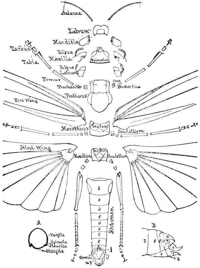
Fig. 88.—External anatomy of Melanoplus spretus, the head and thorax disjointed.
The thorax contains the muscles of flight and those of the legs, besides the fore intestine (œsophagus and proventriculus), as well as, in the winged insects, the salivary glands.
87In the more generalized orders, notably the Orthoptera, the three segments are distinct and readily identified.
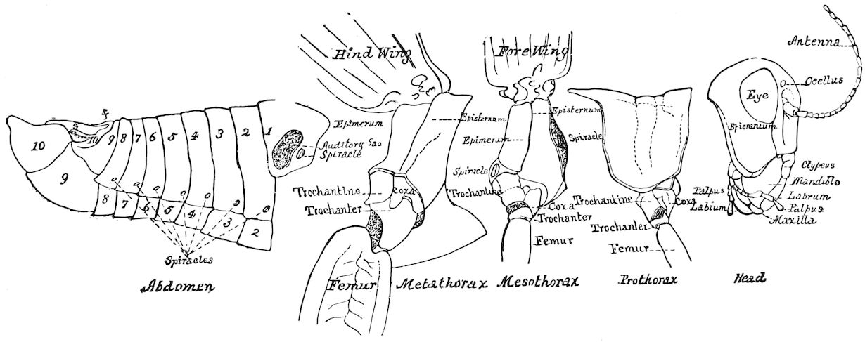
Fig. 89.—Locust, Melanoplus, side view, with the thorax separated from the head and abdomen, and divided into its three segments.
Each segment consists of the tergum, pleurum, and sternum. In the prothorax these pieces are not subdivided, except the pleural; in such case the tergum is called the pronotum. The prothorax is very large in the Orthoptera and other generalized forms, as also in the Coleoptera, but small and reduced in the Diptera and Hymenoptera. In the winged forms the tergum of the mesothorax is differentiated into four pieces or plates (sclerites). These pieces were named by Audouin, passing from before backwards, the præscutum, scutum, scutellum, and postscutellum. In the nymph stage and in the wingless adults of insects such as the Mallophaga, the true lice, the wingless Diptera, ants, etc., these parts by disuse and loss of the wings are not differentiated. It is therefore apparent that their development depends on that of the muscles of flight, of which they 88form the base of attachment. The scutum is invariably present, as is the scutellum. The former in nearly all insects constitutes the larger part of the tergum, while the latter is, as its name implies, the small shield-shaped piece directly behind the scutum.
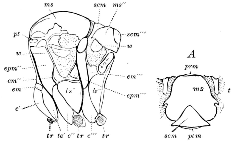
Fig. 90.—Thorax of Telea polyphemus, side view, pronotum not represented: em, epimerum of prothorax, the narrow piece above being the prothoracic episternum; ms, mesoscutum; scm, mesoscutellum; ms″, metascutum; scm‴, metascutellum; pt, a supplementary piece near the insertion of tegulæ; w, pieces situated at the insertion of the wings, and surrounded by membrane; epm″, episternum of the mesothorax; em″, epimerum of the same; epm‴, episternum of the metathorax; em‴ epimerum of the same, divided into two pieces; c′, c″, c‴, coxæ; te′, te″, te‴, trochantines; tr, tr, tr, trochanters. A, tergal view of the mesothorax of the same; prm, præscutum; ms, scutum; scm, scutellum; ptm, postscutellum; t, tegula.
The præscutum and postscutellum are usually minute and crowded down out of sight between the opposing segments. As seen in Fig. 90, the præscutum of most moths (Telea) is a small rounded piece, bent vertically down so as not to be seen from above. In Polystœchotes and also in Hepialus the præscutum is large, well-developed, triangular, and wedged in between the two halves of the scutum. The postscutellum is still smaller, usually forming a transverse ridge, and is rarely used in taxonomy.
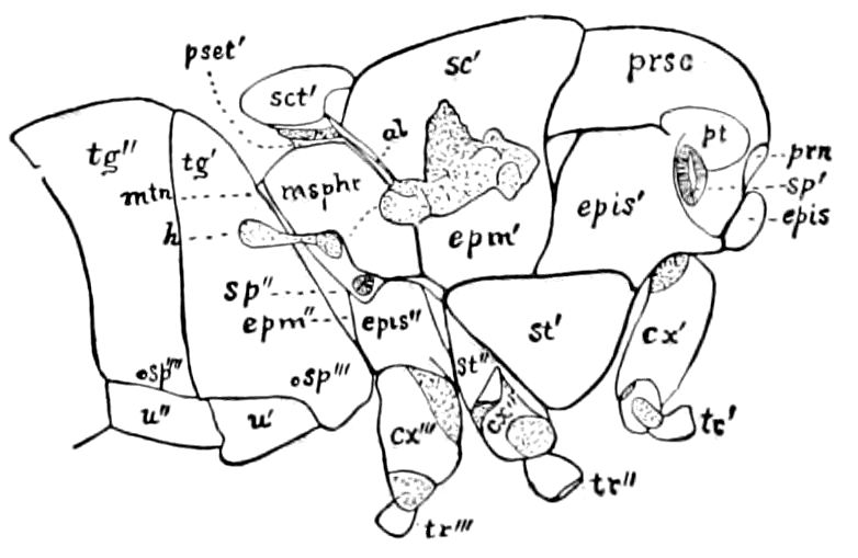
Fig. 91.—Thorax of the house-fly: prn, pronotum; prsc, præscutum; sc′, mesoscutum; sct′, mesoscutellum; psct′, postscutellum; al, insertion of squama, extending to the insertion of the wings, which have been removed; msphr, mesophragma; h, balancer (halter); pt, tegula; mtn, metanotum; epis, epis′, epis″, episternum of pro-, meso-, and metathorax; epm′, epm″, meso- and meta-epimerum; st′, st″, meso- and metasternum; cx′, cx″, cx‴, coxæ; tr′, tr″, tr‴, trochanters of the three pairs of legs; sp′, sp″, sp‴, sp‴′, sp‴″, first to fifth spiracles; tg′, tg″, tergites of first and second abdominal segments; u′, u″, urites.
The metathorax is usually smaller and shorter than the mesothorax, being proportioned to the size of the wings. In certain Neuroptera and in Hepialidæ and some tineoid moths, where the hind wings are nearly as large as those of the anterior pair, the metathorax is more than half or nearly two-thirds as large as the mesothorax. In Hepialidæ the præscutum is large and distinct, while the scutum is divided into two widely separated pieces. The postscutellum is nearly or quite obsolete.
The pleurum in each of the three thoracic segments is divided into two pieces; the one in front is called the episternum, since it 89rests upon the sternum; the other is the epimerum. To these pieces, with the sternum in part, the legs are articulated (Fig. 89).
Between the episterna is situated the breastplate or sternum, which is very large in the more primitive forms, as the Orthoptera, and is small in the Diptera and Hymenoptera.
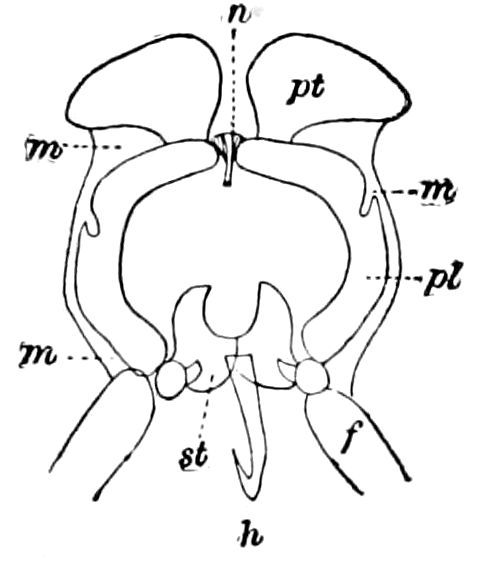
Fig. 92.—Prothorax of Geometra papilionaria: n, notum; p, pleura; st, sternum; pt, patagia; m, membrane; f, femur; h, a hook bent backwards and beneath, and connecting the pro- with the mesothorax.—After Cholodkowsky.
The episterna and epimera are in certain groups, Neuroptera, etc., further subdivided each into two pieces (Fig. 102). The smaller pieces, hinging upon each other and forming the attachments of the muscles of flight, differ much in shape and size in insects of different orders. The difference in shape and degree of differentiation of these parts of the thorax is mentioned and illustrated under each order, and reference to the figures will obviate pages of tedious description. A glance, however, at the thorax of a moth, fly, or bee, where these numerous pieces are agglutinated into a globular mass, will show that the spherical shape of the thorax in these insects is due to the enlargement of one part at the expense of another; the prothoracic and metathoracic segments being more or less atrophied, while the mesothorax is greatly enlarged to support the powerful muscles of flight, the fore wings being much larger than those appended to the metathorax. In the Diptera, whose hinder pair of wings are reduced to the condition of halteres, the reduction of the metathorax as well as prothorax is especially marked (Fig. 91).
The patagia.—On each side of the pronotum of Lepidoptera are two transversely oval, movable, concavo-convex, erectile plates, called patagia (Fig. 92). On cutting those of a dry Catocala in two, they will be seen to be hollow. Cholodkowsky[19] states that they are filled with blood and tracheal branches; and he went so far as to regard them as rudimentary prothoracic wings, in which view he was corrected by Haase,[20] who compares them with the tegulæ, regarding them also as secondary or accessory structures.
The tegulæ.—On the mesothorax are the tegulæ of Kirby (pterygodes of Latreille, paraptera of McLeay, hypoptère or squamule), which cover the base of the fore wings, and are especially developed in the Lepidoptera (Fig. 90, A, t) and in certain Hymenoptera (Fig. 95, c).
90The external opening of the spiracles just under the fore wings, is situated in a little plate called by Audouin the peritreme.
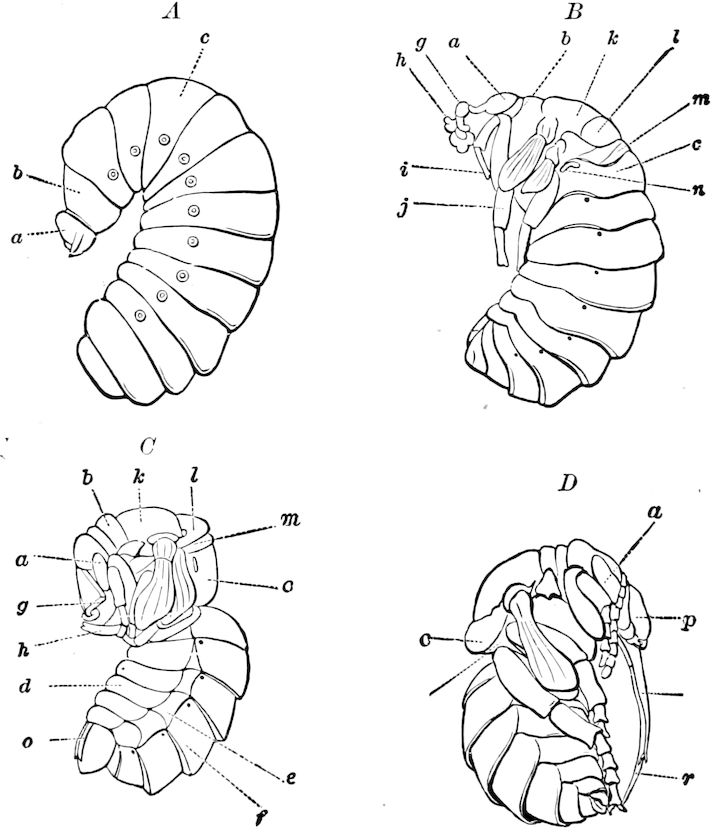
Fig. 93.—Transformation of the bumble bee, Bombus, showing the transfer of the 1st abdominal larval segment (c) to the thorax, forming the propodeum of the pupa (D) and imago; n, spiracle of the propodeum. A, larva; a, head; b, 1st thoracic; c, 1st abdominal segment. B, semipupa; g, antenna; h, maxillæ; i, 1st; j, 2d leg; k, mesoscutum; l, mesoscutellum; m, metathorax; d, urite (sternite of abdomen); e, pleurite; f, tergite; o, ovipositor; r, lingua; q, maxilla.
In the higher or aculeate Hymenoptera, besides the three segments normally composing the thorax, the basal abdominal segment is during the change from the larva to the pupa transferred to this region, making four segments. This first abdominal is called “the median segment” (Figs. 93–95). In such a case the term alitrunk has been applied to this region, i.e. the thorax, as thus constituted. Latreille wrongly stated that in the Diptera the first abdominal segment also entered into the composition of the thorax; but Brauer has fully disproved that view, as may be seen by an examination of his sketches which we have copied (Fig. 94).
91
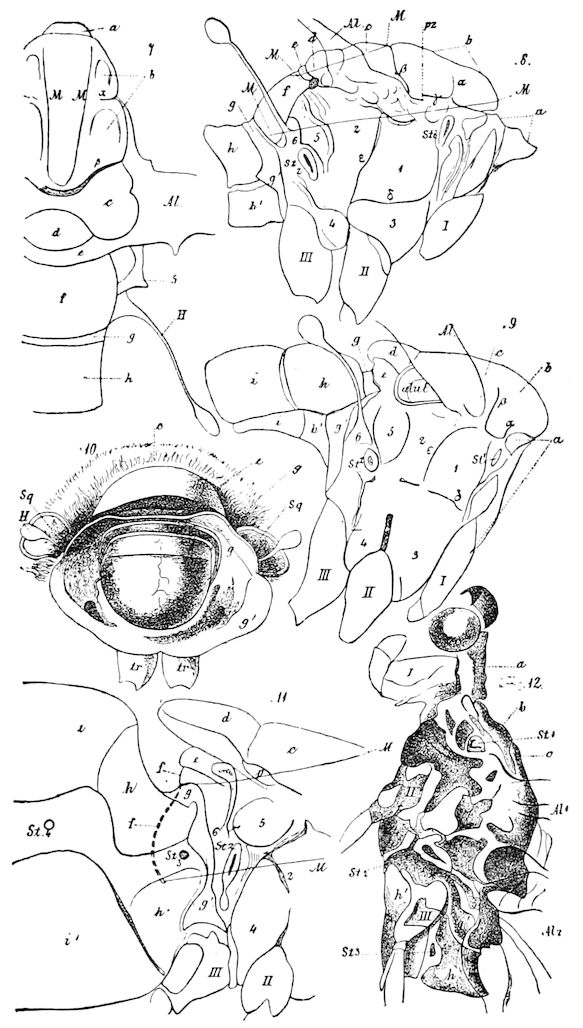
Fig. 94.—7, 8, thorax of Tipula gigantea; 9, of Leptis; 10, thorax of Tabanus bromius after the removal of the abdomen, in order to bring into view the inner mesophragma (f), and to show the extension of the metathorax g and g′; tr, trochanter; 11, hind end of the mesothorax, the entire metathorax, and the 1st and 2d abdominal segments of Volucella zonaria, seen from the side. The internal mesophragma (f), and the position of the muscle inserted in it, are indicated by the two lines M. p, Callus postalaris; pr (pz in 8), callus præalaris Osten Sacken (= “patagium” of some authors); g, metanotum; g′, metepimerum, “segment médiaire” of Latreille (wrongly considered by him to be the 1st abdominal segment); 4, metasternum (hypopleura of Osten Sacken); 5 (? “episternum of metathorax” (Brauer) = metapleura of Osten Sacken); 6, and also H, halter; st1, mesothoracic stigma; st2, metathoracic stigma; st3, first abdominal stigma; γ, dorsopleural; δ, sternopleural; ε, mesopleural sutures; h, 1st, i, 2d, abdominal segment; al, wing; alul, alula. 12, the head and the three thoracic rings, and the 1st abdominal segment of Ephemera vulgata, the connecting membranes are in white: a, prothorax; b, præscutum; c, scutum; d, scutellum; e, postscutellum; ps, postscutellum of mesothorax.—After Brauer.
92
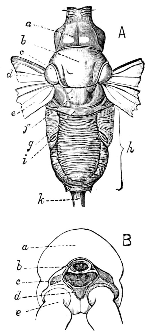
Fig. 95.—Alitrunk of Sphex chrysis: A, dorsal aspect; a, pronotum; b, mesonotum; c, tegula; d, base of fore,—e, of hind, wing; f, g, divisions of metanotum; h, median (true first abdominal) segment; i, its spiracle; k, second abdominal segment, usually called the petiole or first abdominal segment. B, posterior aspect of the median segment; a, upper part; b, superior,—c, inferior, abdominal foramen; d, ventral plate of median segment; e, coxa.—After Sharp.
The sternum is in rare cases subdivided into two halves, as in the meso- and metathorax of the cockroach; in Forficula the prosternum is divided into four pieces besides the sternum proper (Fig. 96); and in Embia, also, the sternites, according to Sharp, are complex.
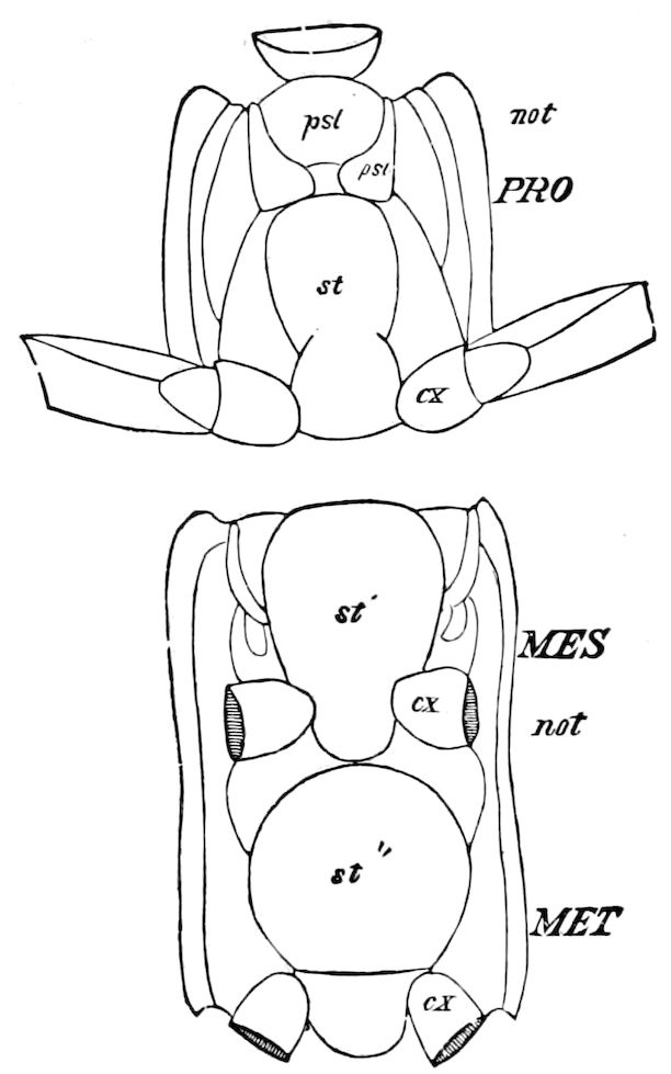
Fig. 96.—Sternal view of pro-, meso-, and metathorax of Forficula tæniata: pst, præsternum, divided into 4 pieces; st, pro-, st′, meso-, st″, metasternum; cx, coxa; not, notum.
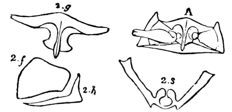
Fig. 97.—A, under surface of prothorax, or prosternum, of Dyticus circumflexis: 2.g, prosternum; 2.f, episternum; 2.h, epimerum; 2.s, antefurca or entothorax.
93
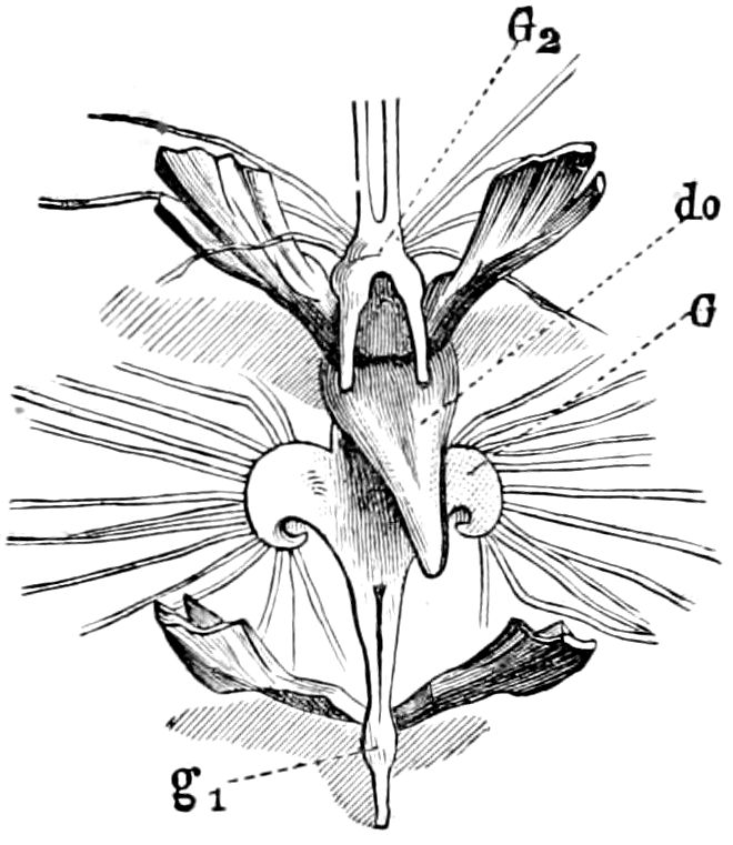
Fig. 98.—Meso- (G2) and metathoracic ganglia (G1), with the apodemes of Gryllotalpa.—After Graber.
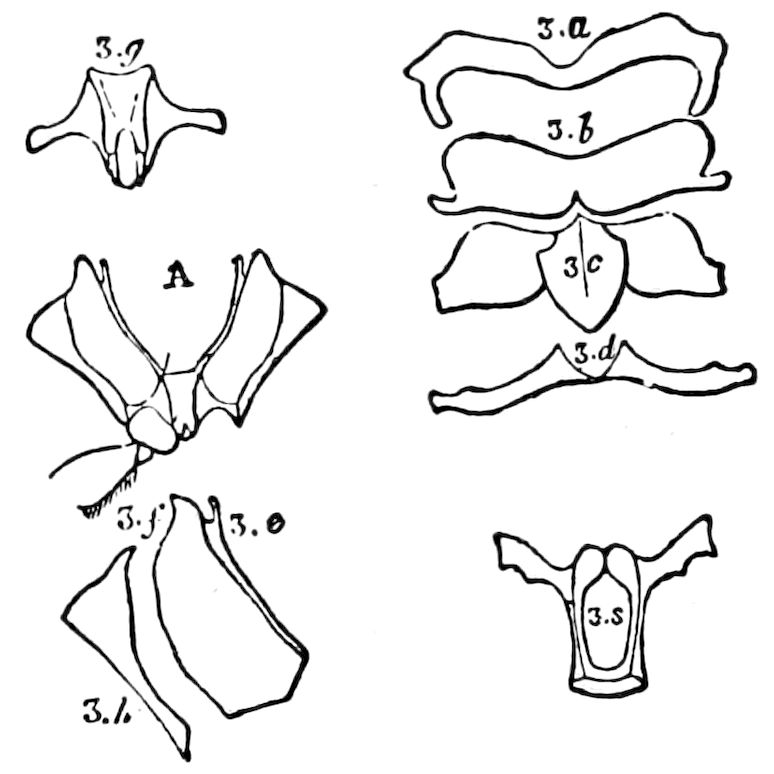
Fig. 99.—Parts of the mesothorax of Dyticus: A, mesosternum; 3.a, præscutum; 3.b, scutum; 3.c, scutellum; 3.d, postscutellum; 3.e, parapteron; 3.g, mesosternum; 3.f, episternum; 3.h, epimerum; 3.s, medifurca or entothorax.
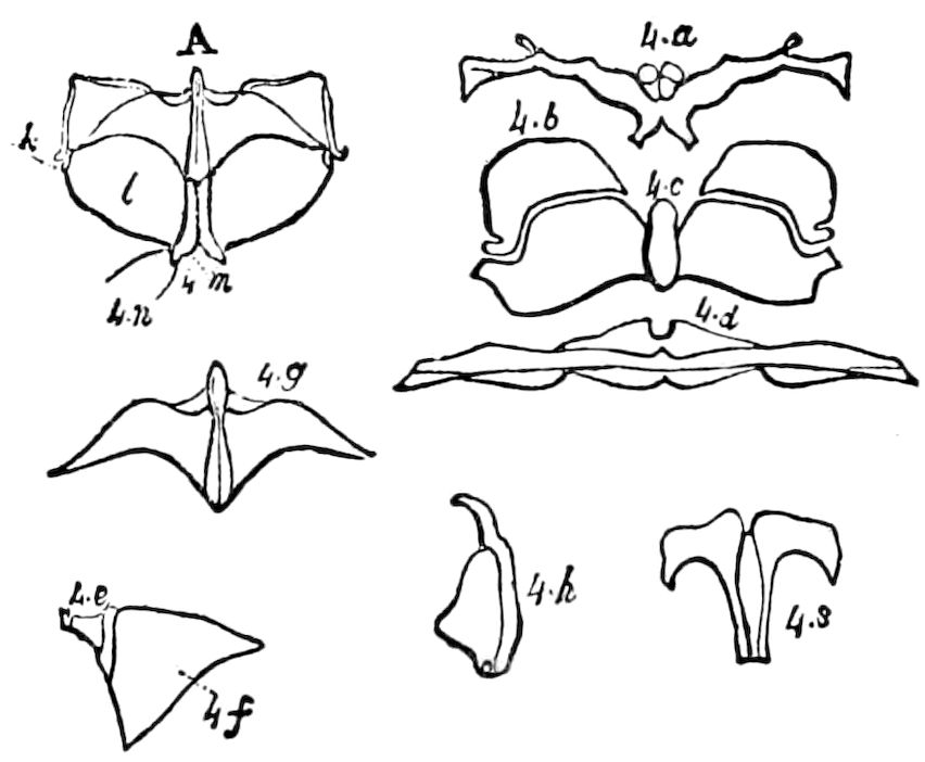
Fig. 100.—Parts of the metathorax of Dyticus: A, metasternum; 4.a, præscutum; 4.b, scutum; 4.c, scutellum; 4.d, postscutellum; 4.e, parapteron; 4.f, episternum; 4.g, metasternum; 4.h, epimerum; 4.s, postfurca.—This and Figs. 97 and 99 from Audouin, after Newport.
The apodemes.—The thorax is supported within by beam-like processes, or apodemes, which pass inward and also form attachments for the muscles. Those passing up from the sternum form the entothorax of Audouin, and the process of each thoracic segment is called respectively the antefurca, medifurca, and postfurca. In the Orthoptera (Caloptenus and Anabrus), the antefurca is large, thin, flattened, directed forward, and bounds each side of the prothoracic ganglion. In the Coleoptera two plates (Fig. 97, 2.s) arise from the inside of the sternum and “form a collar or leave a circular hole between them for the passage of the nervous cord” (Newport). The medifurca is a pair of flat processes which diverge and bridge the commissure, while the postfurca is situated under the commissure. In beetles (Dyticus) Newport states that it is expanded into two broad plates, to which the muscles of the posterior legs are attached. Graber also notices in the mole cricket between the apodemes of the meso- and metathorax, a flattened spine (Fig. 98, do) with two perforations through which pass the commissures connecting the ganglia. Besides these processes there are large, thin, longitudinal partitions passing down from the tergum (or dorsum), called phragmas; they are most developed in those insects which fly best, i.e. in Coleoptera (Figs. 97–101), Lepidoptera, Diptera, and Hymenoptera, none being developed in the prothorax. (The term phragma has also been applied to a partition formed by the inflexed hinder edge of this segment, and is present only in those insects in which the prothorax is movable.—Century Dictionary.) All these ingrowths 94may be in general termed apodemes. There are similar structures in Crustacea and also in Limulus; but Sharp restricts this term to minute projections in beetles (Goliathus) situated at the sides of the thorax near the wings. (Insecta, p. 103, Fig. 57.) The internal processes arising from the sternal region have been called endosternites.
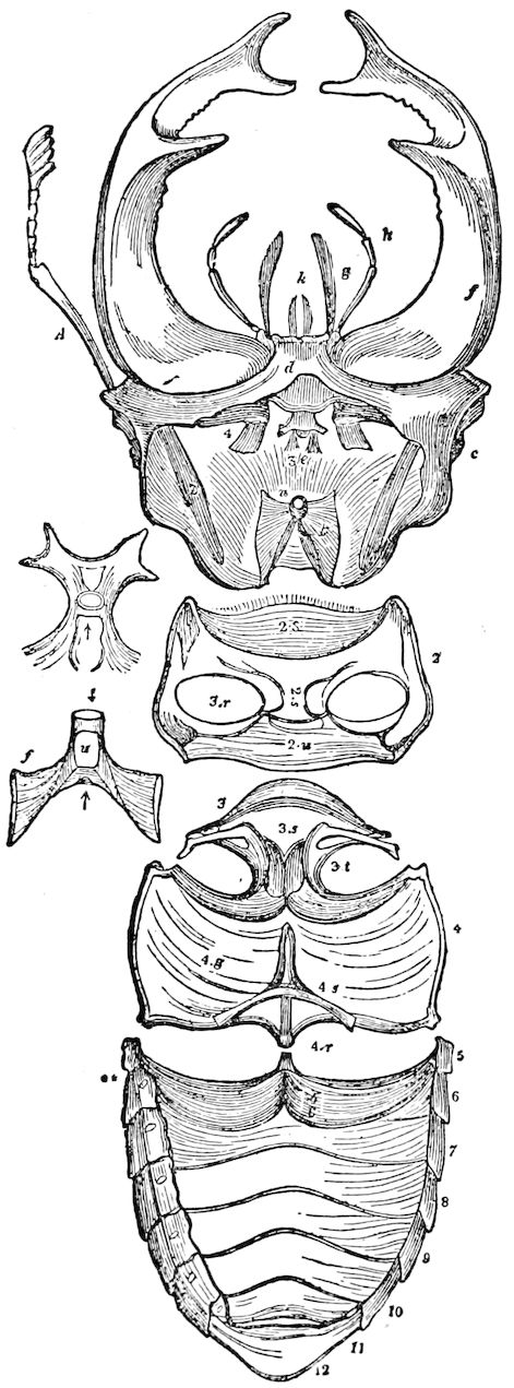
Fig. 101.—Internal skeleton of Lucanus cervus, ♂, head: A, antenna; f, mandible; d, mentum; 2, 4, tendons of mandible; f, u, t, parts of the tentorium; 3 e, labial muscles. Thorax: 2, prothorax; 3, 4, meso- and metathorax fused solidly together; 3 r, acetabulum of prothorax, into which the coxa is inserted; 2 s, sternum; 3t, acetabulum of mesothorax, 4r, of metathorax; 3 s, mesothoracic sternum fused with that of the metathorax (4g); 4 s, apodeme.—After Newport.
The acetabula.—These are the cavities in which the legs are inserted. They are situated on each side of the posterior part of the sternum, in each of the thoracic segments. They are, in general, formed by an approximation of the sternum and epimerum, and sometimes, also, of the episternum, as in Dyticus (Fig. 97, A). This consolidation of parts, says Newport, gives an amazing increase of strength to the segments, and is one of the circumstances which enables the insect to exert an astonishing degree of muscular power.
95
Tabular View of the Segments, Pieces, and Appendages of the Thorax
Name of Segment Pieces (Sclerites) Appendages
1. Prothorax Pronotum, sometimes differentiated into
Scutum 1st pair of legs
Scutellum Patagia
Episternum
Epimerum
Sternum
Antefurca
2. Mesothorax Præscutum
Scutum 2d pair of legs
Scutellum 1st pair of wings
Proscutellum Tegulæ
Episternum Squamæ (Alulæ)
Epimerum Peritreme
Sternum
Mesofurca
Mesophragma
Apodemes
3. Metathorax Præscutum
Scutum 3d pair of legs
Scutellum 2d pair of wings
Postscutellum (Halteres of Diptera)
Episternum
Epimerum
Sternum
Postfurca
Metaphragma
Apodemes
96
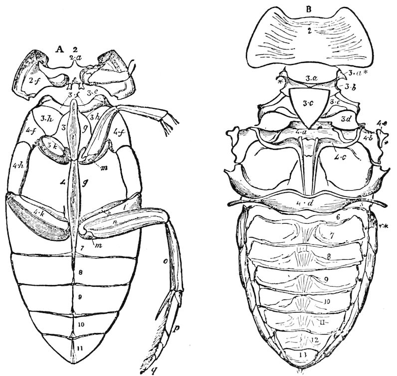
Fig. 102.—External anatomy of the trunk of Hydröus piceus: A, sternal—B, tergal aspect; 2, pronotum; 2 a, prosternum; 2 f, episternum; 3 a, præscutum; 3 b, scutum; 3 c, scutellum; 3 d, postscutellum; 3 g, mesosternum; 3 h, episternum; 3 f, epimerum; 3 i, crest of the mesosternum; 3 a, parapteron; 3 k, coxa; 4 a, metapræscutum; 4 b, metascutum; 4 c, metascutellum; 4 d, postscutellum; 4 e, tegula; 4 f, episternum; 4 h, epimerum; 4 g, metasternum; 4 i, crest of metasternum; 4 k and l, coxa; 4 m, trochanter; n, femur; o, tibia; p, tarsus; q, unguis; 7–11, abdominal segments.—After Newport.
b. The legs: their structure and functions
The mode of insertion of the legs to the thorax is seen in Figs. 90, 97, 101, and 103. They are articulated to the episternum, epimerum, and sternum, taken together, and consist of five segments. The basal segment or joint is the coxa, situated between the episternum and trochanter. The coxa usually has a posterior subdivision or projection, the trochantine; sometimes, as in Mantispa (Fig. 103), the trochantine is obsolete. We had previously supposed that the trochantine was a separate joint, but now doubt whether it represents a distinct segment of the leg, and regard it as only a subdivision of the coxa. It is attached to the epimerum, and is best developed in Panorpidæ, Trichoptera, and Lepidoptera. In the Thysanura the trochantine is wanting, and in the cockroach it merely forms a subdivision of the coxa, its use being to support the latter. The second segment is the trochanter, a more or less short spherical joint on which the leg proper turns; in the parasitic groups (Ichneumonidæ, etc., Fig. 104) it is usually divided into two pieces, though there are some exceptions. The trochanter is succeeded by the femur, tibia, and tarsus, the latter consisting of from one to five segments, the normal number being five. Tuffen West believed that the pulvillus is the homologue of an additional tarsal joint, “a sixth tarsal joint.” The last tarsal segment ends in a pair of freely movable claws (ungues), which are modified setæ; between the claws is a cushion-like 97pad or adhesive lobe, called the empodium or pulvillus (Fig. 105, also variously called arolium, palmula, plantula, onychium, its appendage being called paronychium and also pseudonychium). It is cleft or bilobate in many flies, but in Sargus trilobate. All these parts vary greatly in shape and relative size in insects of different groups, especially Trichoptera, Lepidoptera, Diptera, and Hymenoptera. In certain flies (e.g. Leptogaster) the empodium is wanting (Kolbe). By some writers the middle lobe is called the empodium and the two others pulvilli.
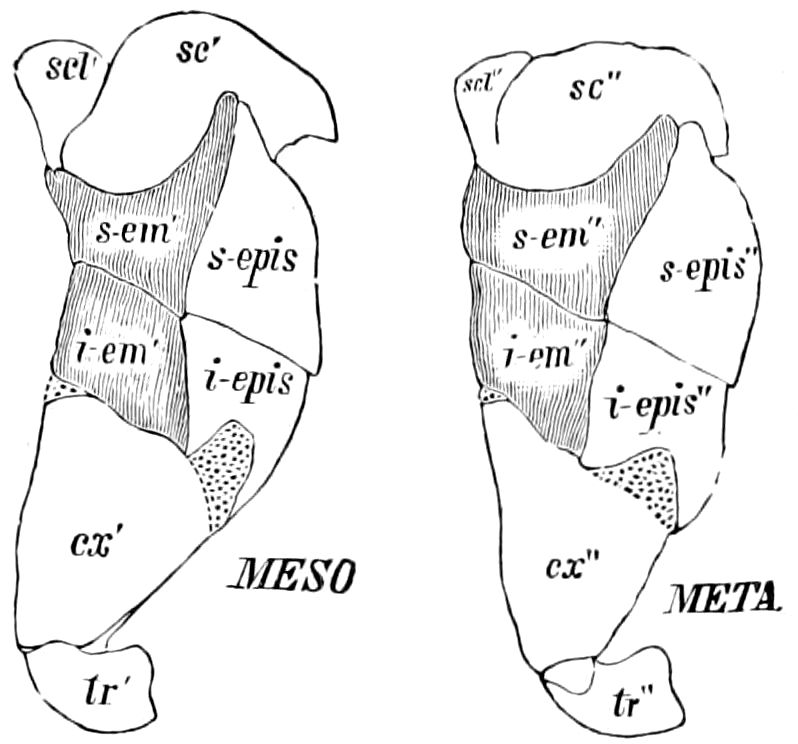
Fig. 103.—Side view of meso- and metathorax of Mantispa brunnea, showing the upper and lower divisions of the epimerum (s. em′, s. em″, i. em′, i. em″); s. epis, i. epis″, the same of the episternum.
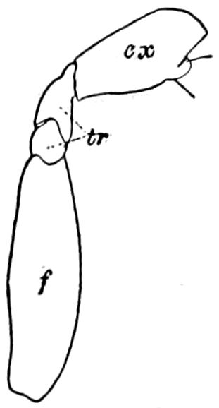
Fig. 104.—Divided (ditrochous) trochanter of an ichneumon: cx, coxa; tr, the two divisions of the trochanter; f, femur.—After Sharp.
The fore legs are usually directed forward to drag the body along, while the middle and hind legs are directed outward and backward to push the body onwards. While arachnids walk on the tip ends of their feet, myriopods, Thysanura, and all larval insects walk on the ends of the claws, but insects generally, especially the adults, are, so to speak, plantigrade, since they walk on all the tarsal joints. In the aquatic forms the middle and hind tarsi are more or less flattened, oar-like, and edged with setæ. In leaping insects, as the locusts and grasshoppers, and certain chrysomelids, the hind femora are greatly swollen owing to the development of the muscles within. The tibia, besides bearing large, lateral, external spines, occasionally bears at the end one or more spines or spurs called calcaria. The fore tibia also in ants, etc., bear tactile hairs, and chordotonal organs, as well as other isolated sense-organs (Janet), and, in grasshoppers, ears.
In the Carabidæ the legs are provided with combs for cleaning the antennæ (Fig. 107), and in the bees and ants these cleansing organs are more specialized, the pectinated spine (calcar) being opposed by a tarsal comb (Fig. 106, d; for the wax-pincers of bees, see g). In general the insects use their more or less spiny legs for cleansing the head, antennæ, palpi, wings, etc., and the 98adaptations for that end are the bristles or spinules on the legs, especially the tibiæ.
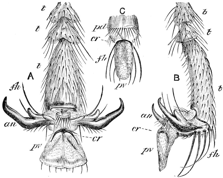
Fig. 105.—Foot of honey-bee, with the pulvillus in use: A, under view of foot; t, t, 3d–5th tarsal joints; a n, unguis; f h, tactile hairs; p v, pulvillus; cr, curved rod. B, side view of foot. C, central part of sole; pd, pad; cr, curved rod; pv, pulvillus unopened.—After Cheshire.
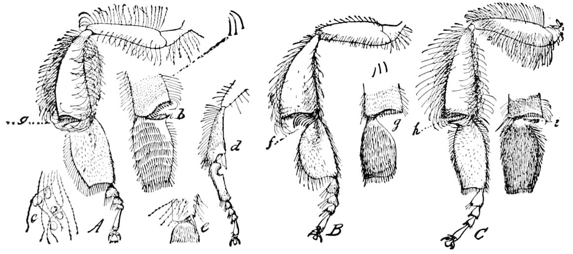
Fig. 106.—Modifications of the legs of different bees. A, Apis: a, wax-pincer and outer view of hind leg; b, inner aspect of wax-pincer and leg, with the nine pollen-brushes or rows of hairs; c, compound hairs holding grains of pollen; d, anterior leg, showing antenna-cleaner; e, spur on tibia of middle leg. B, Melipona: f, peculiar group of spines at apex of tibia of hind leg; g, inner aspect of wax-pincer and first tarsal joint. C, Bombus: h, wax-pincer; i, inner view of the same and first tarsal joint, all enlarged.—From Insect Life, U. S. Div. Ent.
Osten Sacken states that among Diptera the aerial forms (Bombylidæ, etc.) with their large eyes or holoptic heads, which carry with them the power of hovering or poising, have weak legs, principally fit for alighting. On the other hand, the pedestrian or walking Diptera (Asilidæ, etc.) “use the legs not for alighting only, but for running, and all kinds of other work, seizing their prey, carrying 99it, climbing, digging, etc.; their legs are provided not only with spines and bristles, but with still other appendages, which may be useful, or only ornamental, as secondary sexual characters.”
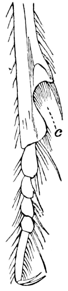
Fig. 107.—End of tibia and tarsal joints of Anophthalmus; c, comb.
Tenent hairs.—Projecting from the lower surface of the empodium are the numerous “tenent hairs,” or holding hairs, which are modified glandular setæ swollen at the end and which give out a minute quantity of a clear adhesive fluid (Figs. 108, 109, 130, 134). In larval insects, and the adults of certain beetles, Coccidæ, Aphidæ, and Collembola, which have no empodium, there are one or more of these tenent hairs present. They enable the insect to adhere to smooth surfaces.
Fig. 108.—Transverse section through a tarsal joint of Telephorus, a beetle: ch, cuticula of the upper side; m, its matrix; ch′, the sole; m′, its matrix; h, adhesive hair; h′, tactile hair, supplied with a nerve (n′), and arising from a main nerve (n); n″, ganglion of a tactile hair; t, section of main trachea, from which arises a branch (t′); dr, glands which open into the adhesive hairs, and form the sticky secretion; e, chitinous thickening; s, sinew; b, membrane dividing the hollow space of the tarsal joint into compartments. See p. 111.—After Dewitz.
Striking sexual secondary characters appear in the fore legs of the male Hydrophilus, the insect, as Tuffen West observes, walking on the end of the tibia alone and dragging the tarsus after it. The last tarsal joint is enlarged into the form of an irregular hollow shield. The most completely suctorial feet of insects are those of the anterior pair of Dyticus (Fig. 132). The under side of the three basal joints is fused together and enlarged into a single broad and nearly circular shield, which is convex above and fringed with fine branching hairs, and covered beneath with suckers, of which two are exceptionally large; by this apparatus of 100suckers the male is enabled to adhere to the back of its mate during copulation. The line branching hairs around the edge prevent the water from penetrating and thus destroying the vacuum, “while if the female struggle out of the water, by retaining the fluid for some time around the sucker, they will in like manner under these altered conditions equally tend to preserve the effectual contact.” (Tuffen West.)
Fig. 109.—Cross-section through tarsus of a locust: ch, cuticula of upper side,—ch′, ch″, ch‴, of sole; ch, tubulated layer; ch″, lamellate layer; ch‴, inner projections of ch″. Other lettering as in Fig. 101. See p. 113.—After Dewitz.
In the saw-flies (Uroceridæ and Tenthredinidæ) and other insects, there are small membranous oval cushions (arolia, Figs. 109 and 131) beneath each or nearly each tarsal joint.
The triunguline larvæ of the Meloidæ are so called from apparently having three ungues, but in reality there is only a single claw, with a claw-like bristle on each side.
Why do insects have but six legs?—Embryology shows that the ancestors of insects were polypodous, and the question arises to what cause is due the process of elimination of legs in the ancestors of existing insects, so that at present there are no functional legs on the abdomen, these being invariably restricted (except in caterpillars) to the thorax, and the number never being more than six. It is evident that the number of six legs was fixed by heredity in the Thysanura, before the appearance of winged insects. We had thought that this restriction of legs to the thorax was in part due to the fact that this is the centre of gravity, and also because abdominal legs are not necessary in locomotion, since the fore legs are used in dragging the insect forwards, while the two hinder pairs support and push the body on. Synchronously with this elimination by disuse of the abdominal legs, the body became shortened, and subdivided into three regions. On the other hand, as in caterpillars, with their long bodies, the abdominal legs of the embryo persist; or if it be granted that the prop-legs are secondary structures, then they were developed in larval life to prop up and move the abdominal region.
The constancy of the number of six legs is explained by Dahl as being in relation to their function as climbing organs. One leg, he says, will almost always be perpendicular to the plane when the animal is moving up a vertical surface; and, on the other hand, we know that three is the smallest number with which stable equilibrium is possible; an insect must therefore have twice this number, and the great numerical superiority of the class may be associated with this mechanical advantage. (This numerical superiority of insects, however, seems to us to be rather due to the acquisition of wings, as we have already stated on pages 2 and 120.)