PART III.—THE METAMORPHOSES OF INSECTS
Reading edition
We have seen that the embryo rapidly passes through extraordinary changes of form, and now, after hatching, especially in the insects with a complete metamorphosis, the animal continues to undergo striking changes in form, in adaptation to different modes of life.
The life of a winged insect, such as a butterfly, fly, or bee, may be divided into four stages: the embryo, or egg state, the larva, pupa, and imago,—the term metamorphosis being applied to the changes after birth, or post-embryonic stages of life. The transformations of the more specialized orders of insects involve wonderful changes of form, which are only paralleled in other types of animals by the metamorphoses of the echinoderms, of certain worms, and of the Crustacea, as well as by those of the frog. An insect, such as a butterfly or bee, during its post-embryonic life lives, so to speak, three different lives, having distinct bodily structures and existing under quite dissimilar surroundings and habits; so that a caterpillar is practically a different animal from the pupa, and the latter from the imago, with different organs, the appendages and other structures being so modified as to be, so far as regards their functions, radically different. These changes of functions or of habits have also been plainly enough the exciting cause of the divergency in structure of what fundamentally is one and the same organ, the change having been brought about by adaptation of the same organs to quite different uses.
The changes are not only observable in the body and its appendages, but also in the internal organs, and consequently are both structural and physiological. The term larva, as applied to the first stage of animals, is a very variable and indefinite one, that of insects in general being a much more highly organized animal than the larva of a worm, starfish, or crustacean.
a. The nymph as distinguished from the larval stage
As there is no marked difference between the different stages of the young in the insects with an incomplete metamorphosis (Heterometabola), 594the chief difference being the possession of the rudiments of wings and the absence of a resting stage, the terms larva and pupa are in reality scarcely applicable to them, and we much prefer the term nymph, first proposed by Lamarck for the active “pupa” of Orthoptera, Hemiptera, the Odonata and Ephemeridæ, and adopted in part by many. Indeed, in the more generalized and older orders, the larval and pupal stages are not differentiated, though the term larval, in its general sense, will probably always be used; just as we speak of the larval stages of worms, echinoderms, or Crustacea.
Eaton in his elaborate work on the Ephemeridæ employs the term nymph to designate all the aquatic or early stages in the development of the young after hatching, and he urges that the old-fashioned usage of larva and pupa seem scarcely worth retention. “Nymphs are young which live an active life, quitting the egg at a tolerably advanced stage of morphological development and having the mouth-parts formed after the same main type of construction as those of the adult insect.” The word nymph is used in the same sense by McLachlan, and by Cabot. Calvert also applies the term nymph “to the stage of odonate existence between the egg and the transformation into the imago.” On the other hand, Brauer applies the term nymph to the pupa of holometabolous insects. For larval Hyatt proposes the term nepionic.
b. Stages or stadia of metamorphosis
The intervals or periods between the moults or ecdyses of caterpillars and other eruciform larvæ are called stages or stadia; thus, as most caterpillars moult four times, we have five stages or stadia, or stage (stadium) I to V. As observed by Sharp, there is, unfortunately, no term in general use to express the form of the insect at the various stadia; “entomologists say, ‘the form assumed at the first moult,’ and so on.” Hence he adopts a term suggested by Fischer,[86] and calls the insect as it appears after leaving the egg the first instar, and what it is after the first moult the second instar, and so on; hence the pupa, or chrysalis, which assumed that condition after moulting five times would be the sixth instar, and the butterfly itself would be the seventh instar.
c. Ametabolous and metabolous stages
In the Synaptera development is direct, the young differing neither in form, structure, or habits from the adult. Hence they are said to be ametabolous. Since there is an absence of even a tendency to a partial metamorphosis, it is evident that the insects have not inherited a tendency to undergo a transformation, but that it is 595an adaptation induced in the hexapod type after the first winged insects appeared, and which became more marked in the more specialized insects and at a period comparatively late in geological history, i.e. perhaps at or soon after the beginning of the Carboniferous period.[87]
The transformations of the pterygote insects vary greatly in degree, and it is difficult to draw the line between the grades. Those in which the adults differ from the freshly hatched young only or mainly in having wings are generally said to have an incomplete or gradual metamorphosis. There is no inactive, resting, or pupal stage, and the wings are acquired only after successive moults. Insects with an incomplete metamorphosis are the Orthoptera, Dermaptera, Platyptera (Mallophaga, Plecoptera, Corrodentia, Embidæ), Ephemeridæ, Odonata, Thysanoptera, and Hemiptera, with the exception of the male Coccidæ, in which there is a resting or sub-nymph stage. As regards the number of moults in the Synaptera, Grassi states that in Campodea there is a single fragmentary ecdysis, while Sommers tells us that Macrotoma plumbea sheds its skin throughout life, even after attaining its full size.
As an example of the partial metamorphosis of the hemimetabolous insects we may select that of the locust, in which there are five moults and six stages (instars), as seen in Fig. 558, five of which are nymphal. In the first two stages there are no rudiments of wings, these appearing after the second moult. Besides the acquisition of wings there are slight differences after each moult, both in structure and color, besides size, so that we may always recognize the comparative age and the particular stage of growth of any individual.[88]
We have watched the development of Melanoplus spretus from the egg to the imago, and examined thousands of specimens which show the six stages. On the other hand, European authors differ as to whether there are three, four, or five moults in the migratory locust.[89] It is not improbable that, as is the case with many other insects, the number of moults may vary according to the temperature and food, variation in these agencies causing either retardation or rapidity in development.
Those with a complete metamorphosis are said to be metabolous or holometabolous. (Lang.)
596Leach[90] in 1815 gave the name of Ametabola to insects without, and Metabola to insects with a metamorphosis.
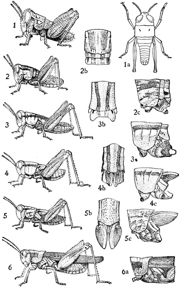
Fig. 558.—Partial metamorphosis of Melanoplus femur-rubrum, showing the five nymph stages, and the gradual growth of the wings, which are first visible externally in 3, 3b, 3c.—Emerton del.
597Latreille (1831) called insects with an incomplete metamorphosis homotenous (which means similar to the end of life), and those with a complete metamorphosis, polymorphous. For the different degrees of metamorphosis of insects he employed two terms: for the incomplete degree, metamorphosis dimidia, and for the total or pupal, metamorphosis perfecta.
Westwood in his Introduction to the Modern Classification of Insects (1839), taking into account the relation of the larva with the imago, divided insects into two divisions: the Heteromorpha, or those in which there is no resemblance between the parent and its offspring, and Homomorpha, in which the larva resembles the imago, except in the absence of wings.
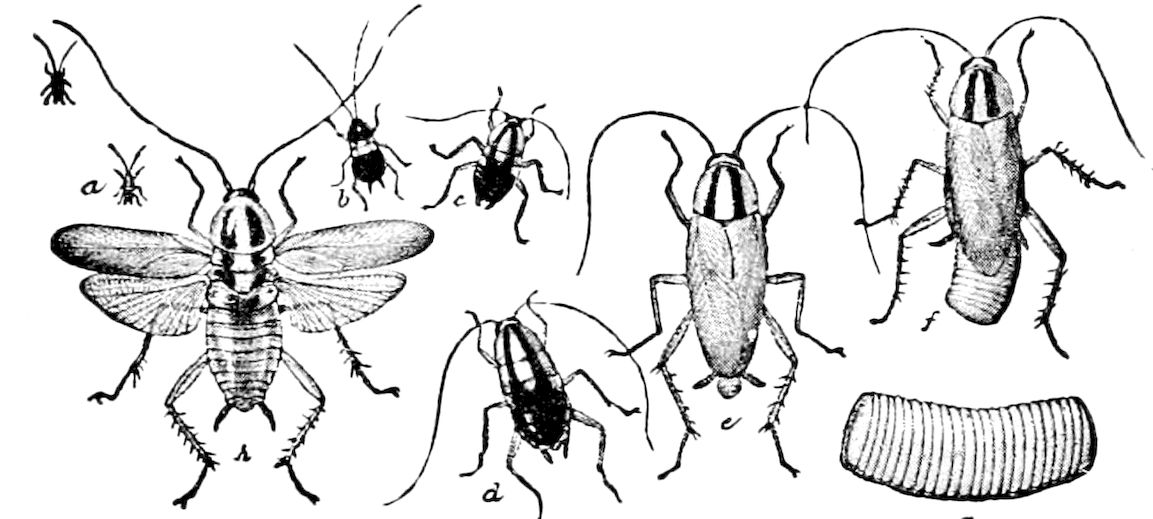
Fig. 559.—Manometabolous metamorphosis of the cockroach (Phyllodromia germanica) with its four nymphal stadia a-d; e, h, adult; f, female with egg-case; g, egg-case.—From Riley.
From the point of view of the degree of metamorphosis, insects have been divided into Heterometabola and Metabola.
I. Heterometabola.—This group may be divided as follows:
1. Manometabola,[91] embracing those forms with a slight or gradual metamorphosis, but which are active in all the stages, without any resting stage. The orders passing through this degree of metamorphosis are the following: Orthoptera, Dermaptera, Platyptera, Thysanoptera, and Hemiptera (Coccidæ excepted).
In all these groups, the only external differences of importance between the freshly hatched nymph and the adult is the presence of wings. The chief difference internally is the complete development of the sexual glands.
It should be observed, however, that in the last nymph stage of the Thysanoptera the articulations of the limbs are enveloped by a membrane and the wings enclosed in short fixed sheaths; the antennæ are turned back on the head, and the insect, though it moves about, is much more sluggish than in the other state. (Haliday.) Hence here we have a close approach to the following degree.
2. Heremetabola,[92] including those forms with a gradual though 598slight or incomplete metamorphosis, but with a quiescent or resting stage at the close of the nymph life. Lang has emphasized this stage, calling attention to the fact that the fore legs of the nymph of the 17–year Cicada, which lives underground on the roots of trees, are thick and adapted for digging. The transition from the nymph to the winged adult is signalized by the decided change in form of the fore legs, as well as by the acquisition of the wings. “The last larval stage is, then, what is called quiescent, i.e. the organization of the imago develops within the chrysalis at the expense of the accumulated reserve material.” (Lang.) There seems to be a resting stage, when the insect does not perhaps suck the sap from the roots, and awaits in its chamber its approaching change to the imago; but we should scarcely apply the term pupa to this stage, though the antennæ of the freshly hatched larva are larger and longer than in the fully grown nymph and are distinctly 8–jointed.
3. Hemimetabola.—In this division, so named by Brauer, the changes are more marked, though there is no truly inactive pupa-like stage. The orders are Perlaria (Plecoptera), Odonata, and Plectoptera (Ephemeridæ). The freshly hatched nymphs of these three groups are much alike in shape, that of Perlidæ, and indeed most of the Platyptera, being more generalized, unless we except that of Chloëon; all closely recall Campodea, and are therefore in the Campodea-stage. These nymphs are indeed more generalized than the freshly hatched nymph of Blattidæ, or any other of the orders mentioned except the Platyptera, to which perlids belong. They all have feet, and the body is more or less flattened. (Fig. 560.)
II. Holometabola.—In this division we have for the first time a true larva, and a pupa stage as distinguished from the imago. Moreover, the insect at each stage is distinguished by radical differences in form, surroundings, and in the nature of the food, while the pupa is inactive, usually immovable, and incapable of taking any food, and is often protected by a cocoon spun by the larva. The holometabolous orders are the Neuroptera, Coleoptera, Mecoptera, Trichoptera, Lepidoptera, Siphonaptera, Diptera, and Hymenoptera.
As we have among worms, echinoderms, and Crustacea certain exceptional species in a metamorphic group whose metamorphosis is suppressed, their development being direct, so there is in pterygote insects, though in a very much less degree, cases of direct development. In the wingless cockroaches such as Pseudoglomeris, etc., of the tribe of Periphæriides, in some of which, however, the males are winged, and in the Hemiptera, occur wingless forms such as the lice and bed-bug. The Mallophaga are all wingless, while certain Dermaptera (Chelidura, Anisolabis) are also apterous. The absence of wings in such cases is due to disuse from parasitism, or to a life under stones or in cracks and 599fissures, where the insects are driven to avoid their enemies, and hence do not need wings. The growth of wings and consequently the development of a metamorphosis is suppressed, so that, as Lang says, “in contrast to the original ametabola of the Apterygota, we have here an acquired ametabola.”
It is rare that, after the rudiments of wings have once appeared in the very young, they should disappear in the late nymph stage; this is, however, said by Walsh to be the case with the Ephemerid Bætisca (Fig. 440). This is a case of retardation in an acquired ametabolesis.
THE LARVA
The term larva is peculiarly applicable to the young of the holometabolous orders. The name (Latin, larva, a mask) was first given to the caterpillar because it was thought by the ancients to mask the form of the perfect insect. Swammerdam supposed that the larva contained within itself “the germ of the future butterfly, enclosed in what will be the case of the pupa, which is itself included in three or more skins, one over the other, that will successively cover the larva.” What led to his conception of the nature of these changes was probably his observations on the semitransparent larva of the gnat, in which the body and limbs of the pupa can be partially seen; for Weismann has shown that the great Dutch observer’s belief that the pupal and imaginal skins were in reality already concealed under that of the larva is partially founded in fact. Swammerdam states: “I can point out in the larva all the limbs of the future nymph, or Culex, concealed beneath the skin,” and he also observed beneath the skin of the larvæ of bees, just before pupating, the antennæ, mouth-parts, wings, and limbs of the adult. But, as we shall see farther on, the discovery by Weismann in the larva of the germs of the imago has completely changed our notions as to the nature of metamorphosis, and revolutionized our knowledge of the fundamental processes concerned in the change from larva to pupa, and from pupa to imago.
Not only are the larvæ of each order of insects characteristic in form, so that the grub or larva of beetles is readily distinguished from those of other orders, or the maggot of flies from the apodous larva of wasps and bees, but within the limits of the larger orders there is great diversity of larval forms, showing that they are the result of adaptation to their surroundings. This is especially the case with the larvæ of the Coleoptera, Lepidoptera, Diptera, and Hymenoptera.
In general, the larvæ of insects may be divided into two types,—the 600Campodea-form, or campodeoid, sometimes called thysanuriform, and the eruciform.
a. The Campodea-form type of larva
This is the most primitive and generalized type of larva (Fig. 560). A Campodeoid larva is one nearest in general shape to Campodea, the form which we have seen to be the nearest allied to the probable ancestor of the insects, and it also resembles the nymphs of the heterometabolous insects, before the appearance of their rudimentary wings.
Brauer, in 1869,[93] first suggested that the larvæ of a great number of insects may be traced back to Campodea and Iapyx. The Campodea-form larva is active, with a more or less flattened body, well developed mandibulate mouth-parts, and usually long legs. The nearest approach to the form of Campodea is the freshly hatched nymph of cockroaches (Blattidæ), Forficula, Perlidæ, Termitidæ, Psocidæ, Embidæ, Ephemeridæ, Odonata, especially the more generalized Agrionidæ, the nymphs of Hemiptera, the larvæ of certain Neuroptera, the active pedate larvæ of the more generalized Coleoptera, such as those of Carabidæ, Cicindelidæ, Dyticidæ, etc., and the first larva (instar) of Stylopidæ and Meloidæ (Fig. 560, d).
While the Campodea-shape is retained throughout nymphal life, of the orders above mentioned the Neuroptera and Coleoptera alone have a true resting pupal stage.
It should also be observed that great changes in the form of the nymph occur within the limits of the Orthoptera; the nymph of all the families except that of the Blattidæ, evidently the most generalized and primitive, being more or less specialized, while the nymphs of the other orders all vary in degree of specialization and modification. The process of adaptation once begun went on very rapidly, as it has in many other orders of insects, as well as in animals of other phyla.
601
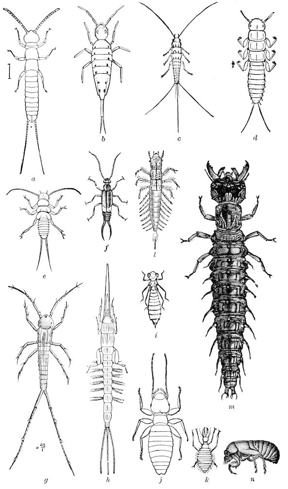
Fig. 560.—Examples of campodeoid nymphs and larvæ: a, Campodea; b, Podura (Degeeria); c, Lepisma; d, triungulin larva of Meloë; e, Perla; f, Forficula; g, Chloëon; h, May-fly (Palingenia); i, Æschna; j, Atropos; k, Myrmeleon; l, Sialis; m, Corydalus; n, Cicada.
602
b. The eruciform type of larva
Brauer also sagaciously pointed out that “a larger part of the most highly developed insects assume another larva form, which appears not only as a later acquisition, through adaptation to certain definite conditions, but also arises as such before our eyes. The larvæ of Lepidoptera, of saw-flies, and Panorpidæ show the form most distinctly, and I call this the caterpillar form (Raupenform). That this is not the primitive form, but one later acquired, we see illustrated in certain beetles. The larvæ of Meloë and of Sitaris, in their fully grown conditions, possess the caterpillar form, but the new-born larvæ of these genera show the Campodea-form. The last form is lost as soon as the larva begins its parasitic mode of life.... The larger part of the beetles, the Neuroptera (in part), the bees and flies (the last with the most degraded maggot form), possess larvæ of this second form.” In 1871 we adopted these views, giving the name eruciform to this type of larvæ, and afterwards Lubbock adopted Brauer’s views. Brauer considered that the eruciform larva was the result of living a stationary semi-parasitic life on plants, in carrion, or burrowing in the trunks and branches or leaves and buds of trees, where they do not have to move about in search of their food. The change from the Campodea-form to the eruciform larva is a process of degeneration and often of atrophy of the limbs, and, in the footless forms of dipterous and hymenopterous insects, of the gnathites, accompanied by a tendency of the body to become more or less cylindrical.
The first steps in the origination of the eruciform larva were apparently taken in the order Neuroptera, as restricted by Brauer and by myself, where, though the larvæ are campodeoid, there is a true resting pupal stage. The most generalized larval form is perhaps that of the Sialidæ (Fig. 560, l), in which the body tends to be slightly cylindrical, though the legs are long, and the gnathites well developed for seizing and biting their living prey. The terrestrial larvæ of the Hemerobiidæ, though modifications of the sialid larval form, are considerably specialized in adaptation to their active carnivorous habits. But the life-history of Mantispa, where there are two larval stages, gives us plainly enough the key to the mode in which the complete metamorphosis was brought about. The larva, born a true Campodea-like form, with large, long, 4–jointed legs, has a structure which would enable it to move about freely after its prey, beginning at once to live a sedentary life in the egg-sac of a spider; before the first moult it loses the use of its legs, while the antennæ are partly aborted. The result is that, owing to this change of habits and surroundings from those of its active ancestors, it changes its form, and the fully grown larva becomes cylindrical, with small slender legs, and, owing to the partial disuse of its jaws, acquires a small, round head.
603
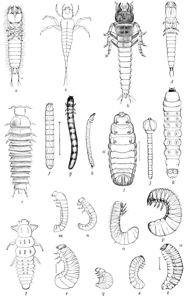
Examples of coleopterous larvæ, showing the passage from the campodeoid to the eruciform type of larvæ. Fig. 561.—Coleopterous larvæ showing passage from campodeoid to eruciform larvæ: a, b, Harpalus; c, Dyticus; d, Staphylinus; e, Silpha; f, Melanactes; g, Ludius; h, Elater; i, Donacia; j, Chrysobothris; k, Orthosoma; l, Coccinella; m, Byrrhus; n, Trox; o, p, Lachnosterna; q, Labidomera; r, Ptinus; s, Anobium; t, Balaninus (entirely apodous).
604Its antennæ, mouth-parts, and legs not only retarded in growth, but retrograding and becoming vestigial, the body meanwhile becoming fat and cylindrical, an apparent acceleration of growth goes on within, with probably an enlargement of the intestine and fat-body, and thus the pupal form is perfected while the larva is full-fed and quiescent. It is not improbable that in the primitive neuropteron, as the result of a mode of life like that of Mantispa, the quiescent life of the later stages graduated into a quiescent, inactive pupal life, allowing the changes going on in the internal organs to result in a complete metamorphosis, which was transmitted to the later Neuroptera, thus making the complete metamorphosis a fixed, normal condition. It thus appears that a change of habits and of food, and more especially the fact that the nymph became so surrounded with an abundance of food close at hand that it did not have to run actively about and seize it in a haphazard manner, were the factors bringing about a change from the Campodea-form nymph to the eruciform larva, thus inducing a hypermetamorphosis.
The larvæ of the Mecoptera (Panorpidæ, Fig. 562, b) are still more caterpillar-like, and besides their cylindrical body, rounded head, small short gnathites, small thoracic legs, they have what appear to be 2–jointed legs to each of the nine abdominal segments, and the close resemblance to caterpillars is farther carried out by the presence of a pair of prothoracic spiracles, none existing on the other two thoracic segments.
605
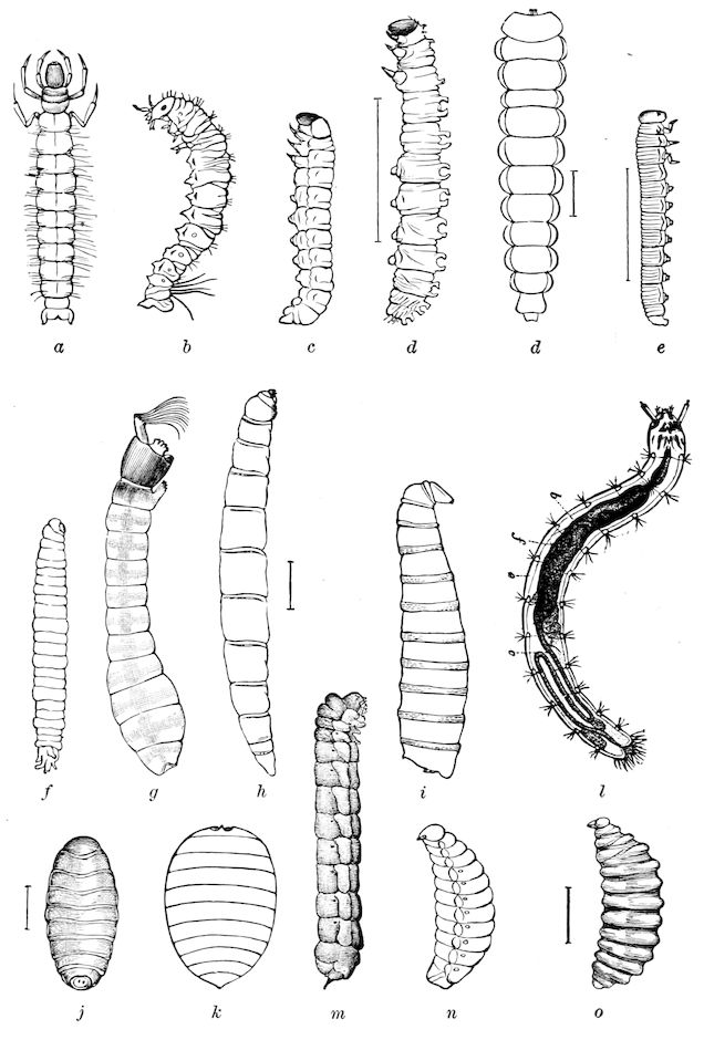
Fig. 562.—Examples of eruciform larvæ: a, Phryganea; b, Panorpa; c, Sesia; d, d, caterpillars; e, Selandria; f, Tipula; g, Simulium; h, Chionea; i, Musca; j, Tachina; k, Braula; l, flea; m, Tremex; n, coarctate larva of Meloë; o, bee (Andrena).
606In the Meloidæ (Fig. 560, d) and Stylopidæ the first larval stage is Campodea-form; the changes will be described in the subsequent section on Hypermetamorphosis, and while these cases of change from a campodeoid to an inactive eruciform larva are very salient, if we compare the graduated series of larval forms throughout the order of Coleoptera, as represented by the illustrations in Fig. 561, we shall see that in nearly, if not each, case the form of the boring or mining, or bark or bud or seed-inhabiting grub is the result of a change of habit and commissariat from active predaceous larvæ, like those of the Carabidæ and other adephagous families, together with those of the Staphylinidæ, with their flat body, big mandibles, and well-developed maxillæ, to the cylindrical bodies of such larvæ as those of Dermestes and Anthrenus, which live a more sedentary life, to the root-feeding wire-worm or elaterid larvæ, and scarabæid grubs, onward to the phytophagous Chrysomelidæ, with the mining and boring buprestids and cerambycids,—in all these forms we see a gradual atrophy of the legs, which is fully carried out in the vermiform or maggot-like larva of the weevils. These changes throughout the members of the entire order are epitomized in the life-history of the Meloidæ, in which there are three typical forms of larva: the Campodea-form (triungulin stage), eruciform (second or carabidoid stage), and vermiform (coarctate) larva.
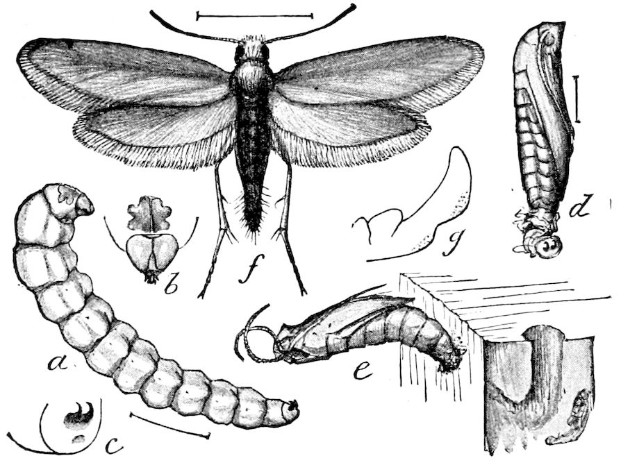
Fig. 563.—Prodoxus cinereus: a, apodous larva; b, head and prothoracic segment; c, anal hooks; d, pupa; e, cast pupal shell protruding from stalk of Yucca; f, female; g, side view of ♂ clasper.—After Riley, from Insect Life.
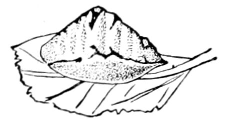
Fig. 564.—Larva of Limacodes scapha, nat. size.
In the Lepidoptera the eruciform, pedate type is adhered to throughout the order, with the rare exception of the nearly apodous mining larva of Prodoxus (Fig. 563, a), Phyllocnistis, and Nepticula, which have no thoracic legs, and the limacodid larvæ, whose abdominal legs are totally aborted, while the thoracic ones are much reduced (Fig. 564).
In the Hymenoptera the phytophagous forms are eruciform, while by the agency of the same factors as already mentioned, i.e. a sedentary or parasitic life and abundance of food within constant reach, the larvæ lose their legs and become vermiform.
607In the Diptera, which are the most highly specialized of insects, the maggot or vermiform shape, and absence of any legs, prevails throughout the order, though the eucephalous larvæ show their origin from a primitive eruciform type of larva. The highly specialized larvæ of the Culicidæ and Simuliidae are undoubtedly related to the earliest and most generalized types, while the maggots of the parasitic flies (Tachinidæ) and other muscids are later degradational forms, and the result of adaptation induced, as in the previous cases, by a sedentary or parasitic mode of life, living as they do immersed in an abundance of rich nitrogenous food, with the result that the mouth-parts have become atrophied by disuse, while the limbs have become entirely aborted, though the thoracic imaginal discs develop normally in the embryonic or pre-larval stages.
It appears, therefore, highly probable that the metamorphoses of insects are the result of the action of change of conditions, just as the polymorphism of Termites is with little doubt the result of differences of food and other conditions. These matters will be farther discussed under the head of Causes of Metamorphosis.
LITERATURE ON ANCESTRY OF INSECTS, ETC.
Müller, Fritz. Für Darwin, 1869, pp. 144, 67 Figs.
Brauer, Friedrich. Betrachtung ueber die Verwandlung der Insekten im Sinne der Descendenz-Theorie. (Verhandlung d. k.k. zool. bot. Gesell. Wien., 1869, 1 Taf., pp. 1–20.)
Packard, A. S. Amer. Naturalist, iii, p. 45, March, 1869.
—— Proc. Boston Soc. Nat. Hist., xiv, 1870, p. 61.
—— Amer. Nat., iv, Feb. 1871, p. 756; v, 1871, pp. 52, 567.
—— Embryological Studies. (Memoirs Peabody Acad. Sc. Salem, 1871–72.)
—— Our common insects, 1873, Chapter on Ancestry of Insects, pp. 175–178.
—— Third Report U. S. Ent. Commission, 1883, pp. 295–304.
Lubbock, John. On the origin of insects. (Journ. Linn. Soc., London, xl, 1873.)
—— Origin and metamorphoses of insects. (Nature, 1873 [in book form, 1874], pp. 108, 66 Figs.)
Mayer, Paul. Ueber Ontogenie and Phylogenie der Insekten. (Jena. Zeitschr. Wissens., x, 1876, pp. 125–221, 4 Taf.)
Hyatt, A., and Arms, J. M. Insecta. (Bost. Soc. Nat. Hist. Guides for science-teaching, viii.) Boston, 1890, pp. 300, 13 Pls., 223 Figs.
608
c. Growth and increase in size of the larva
The rapidity of growth and enormous increase in size in early life is especially noticeable in caterpillars and other phytophagous larvæ. The latest observations are those of Trouvelot on Telea polyphemus. When this silkworm hatches, it weighs 1 20 of a grain.
When
10 days old it weighs ½ a grain, or 10 times the original weight.
20 days old it weighs 3 grains 60 times the original weight.
30 days old it weighs 31 grains 620 times the original weight.
40 days old it weighs 90 grains 1800 times the original weight.
56 days old it weighs 207 grains 4140 times the original weight.
“When,” he says “a worm is 30 days old, it will have consumed about 90 grains of food; but when 56 days old, it is fully grown and has consumed not less than 120 oak leaves, weighing ¾ of a pound; besides this it has drank not less than ½ an ounce of water. So the food taken by a single silkworm in 56 days equals in weight 86,000 times the primitive weight of the worm. Of this about ¼ of a pound becomes excrementitious matter, 207 grains are assimilated, and over 5 ounces have evaporated.”[94]
Dandolo stated that the Asiatic silkworm (Bombyx mori) weighs on hatching not over 1 100 of a grain, but when fully grown about 95 grains. During this period, therefore, it has increased 9500 times its original weight, and has eaten 60,000 times its weight of food. Newport thought this estimate of the amount of food was a little too great. But comparing it with Trouvelot’s estimate for the American silkworm, which weighs when hatched five times as much, it would not appear to be so. Newport found that the larva of Sphinx ligustri at the moment of leaving the egg weighs about 1 80 of a grain, and when fully fed 125 grains, so that in the course of 32 days it increases about 9976 times its original weight. This proportion of increase is exceeded by the larva of Cossus ligniperda, which, boring in the trunks of trees, remains about three years in the larva state, and increases, according to Lyonet, to the amount of 72,000 times its first weight.
Newport adds that those larvæ in which the proportion of increase is the greatest, are usually those which remain longest in the pupa state, as in the silkworm. “Thus Redi observed in the maggots of the common flesh-flies a rate of increase amounting to about 200 times the original weight in 24 hours, but the proportion of increase in these larvæ does not at all approach that of the Sphinx and Cossus.” From his observations on the larva of one of the wild bees (Anthophora retusa) Newport believes that this is also the case with the Hymenoptera. The weight of the egg of this insect is about 1 150 of a grain, and the average of a full-grown larva 68 10 grains, so that its increase is about 1020 times its original weight; “which compared with that of the Sphinx of medium size, is but as 1 to 9¾, and to a Sphinx of maximum size only as 1 to a little more than 11.”
609The growth is most rapid after the last moult. “Thus a larva of Sphinx ligustri, which at its last change weighed only about 19 to 20 grains, at the expiration of eight days, when it was fully grown, weighed nearly 120 grains.” (Newport.)
d. The process of moulting (ecdysis)
Insects periodically shed the exoskeleton, together with the chitinous lining of their internal organs of ectodermal origin, which thus sloughed off are called the exuvia. The process in the locust has been described by Riley.[95] It occupies from half to three-quarters of an hour (Fig. 565). This process has naturally, from the ease with which it can be observed, been most frequently examined in the Lepidoptera, though careful and detailed observations of the inner and outer changes are still greatly needed, especially in other orders. In the caterpillar of most moths, especially one of the more generalized bombycine moths, on slipping out of its egg-shell the head is of enormous size as compared with the body, but the latter soon fills out after the creature has eaten a few hours; the head, of course, does not during this time increase in size, and the larvæ of different instars may be exactly distinguished, as Dyar has shown, by the measurements of the head.
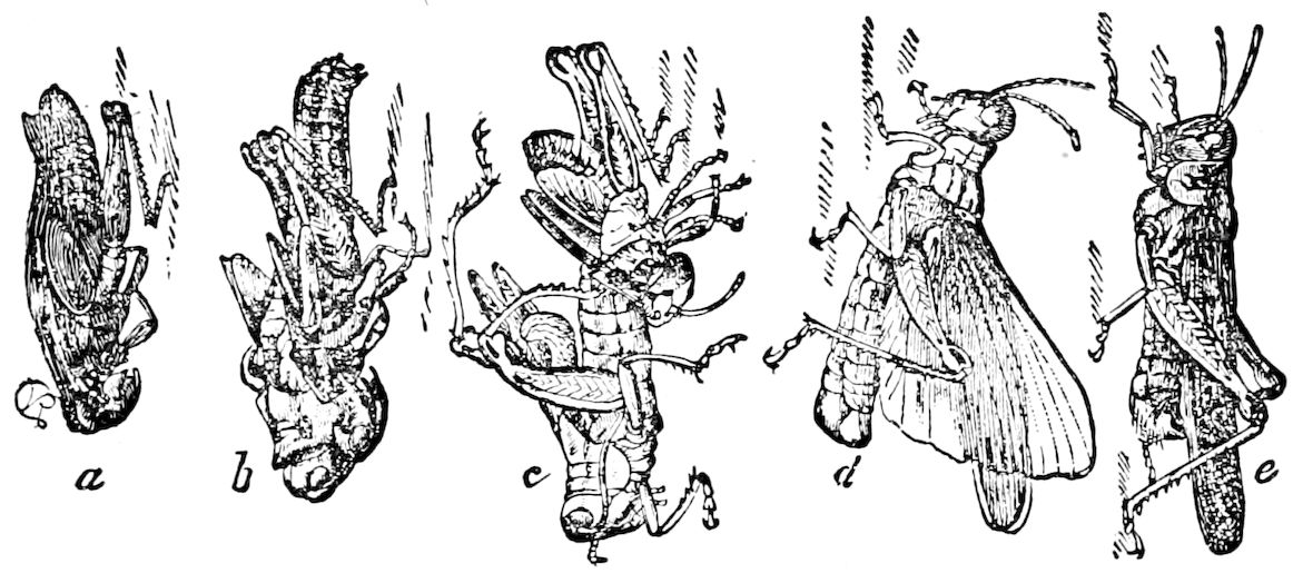
Fig. 565.—Process of moulting from nymph to imago in the locust (M. spretus): a, nymph with skin just split on the back; b, the imago drawing itself out, at c, nearly free; d, the imago, with wings expanded; e, the same with all parts perfect.—After Riley.
Before the caterpillar moults, it stops feeding, and the head is now small compared with the body; the head of the second instar is now large, situated partly under the much-swollen prothoracic segment, and pushes the head of the first instar forward.
Newport has well described the mode of shedding the skin in 610Sphinx ligustri, and his detailed description will apply to most lepidopterous larvæ.
The whole body is wrinkled and contracted in length, and there are occasionally powerful contractions and twitchings of its entire body; the skin becomes dry and shrivelled, and is gradually separated from the new and very delicate one of the next instar beneath. After several powerful efforts of the larva the old skin cracks along the middle of the dorsal surface of the mesothoracic segment, and by repeated efforts the fissure is extended into the 1st and 3d segment, while the covering of the head divides along the vertex and on each side of the clypeus. “The larva then gradually presses itself through the opening, withdrawing first its head and thoracic legs, and subsequently the remainder of its body, slipping off the skin from behind like the finger of a glove. This process, after the skin has once been ruptured, seldom lasts more than a few minutes. When first changed the larva is exceedingly delicate, and its head, which does not increase in size until it again changes its skin, is very large in proportion to the rest of the body.” (Art. Insecta, etc.)
Trouvelot’s account is more detailed and an advance on that of Newport’s view. He explicitly states, and we know that he was a very close observer, that the old skin is detached by “a fluid which circulates between it and the worm.” His account is as follows: The polyphemus worm, like all other silkworms, changes its skin five times during its larval life. The moulting takes place at regular periods, which comes around about every 10 days for the first four moultings, while about 20 days elapse between the fourth and fifth moulting. The worm ceases to eat for a day before moulting, and spins some silk on the vein of the under surface of a leaf; it then secures the hooks of its hind legs in the texture it has thus spun, and there remains motionless; soon after, through the transparency of the skin of the neck, can be seen a second head larger than the first, belonging to the larva within. The moulting generally takes place after four o’clock in the afternoon; a little before this time the worm holds its body erect, grasping the leaf with the two pairs of hind legs only; the skin is wrinkled and detached from the body by a fluid which circulates between it and the worm; two longitudinal bands are seen on each side, produced by a portion of the lining of the spiracles, which at this moment have been partly detached; meanwhile the contractions of the worm are very energetic, and by them the skin is pulled off and pushed towards the posterior part; the skin thus becomes so extended that it soon tears just under the neck, and then from the head. When this is accomplished the most difficult operation is over, and now the process of moulting goes on very rapidly. By repeated contractions the skin is folded towards the tail, like a glove when taken off, and the lining of the spiracles comes out in long white filaments. When about one-half of the body appears, the shell still remains like a cap, enclosing the jaws; then the worm, as if reminded of this loose skull-cap, removes it by rubbing it on a leaf; this done, the worm finally crawls out of its skin, which is attached to the fastening made for the purpose. Once out of its old skin, the worm makes a careful review of the operation, with its head feeling the aperture of every spiracle, as well as the tail, probably for the purpose of removing any broken fragment of skin which might have remained in these delicate organs. Not only is the outer skin cast off, but also the lining of the air-tubes and intestines, together with all the chewing organs and other appendages of the head. After the moulting, the size of the larva is considerably increased, the head is large compared with the body, but 8 or 10 days later it will look small, as the body will have increased very much in size. This is a certain indication that the 611worm is about to moult. Every 10 days the same operation is repeated. From the fourth moulting to the time of beginning the cocoon the period is about 16 days. (Amer. Naturalist, i, pp. 37, 38.)
Little has been recorded as to the exact mode of casting the larval skin in Coleoptera. Slingerland states that Euphoria inda when pupating sheds the larval skin off the anal end in the same way as in caterpillars, while in Pelidnota punctata the larval skin splits down the whole length of the back, retains the larval shape, and forms a covering for the pupa which remains inside. (Can. Entomologist, xxix, p. 52.) The old larval skin in the Coccinellidæ and certain Chrysomelidæ is retained crumpled up at the end of the body, while in Dermestes, Anthrenus, etc., it cloaks the pupa.
Not only is the integument, with its hairs, setæ, and other armatures, as well as the cornea or facets of the eyes, shed, but also all the lining or intima of those internal organs which have been originally derived by an ingrowth or invagination of the ectoderm are likewise cast off, with the probable exception, of course, of the mid-intestine, which is endodermal in its origin. Even so early an observer as Swammerdam noticed that the internal lining of the alimentary canal comes away with the skin. He states that the larva of Oryctes nasicornis sheds both the lining of the colon, and of the smaller as well as the larger branches of the tracheæ.
Careful observations are still needed on the internal changes at ecdysis of most insects. Newport seems to have observed more closely than any one else, notwithstanding the great number who have reared caterpillars but have not carefully observed these points, the extent of the process internally. He informs us: “The lining of the mouth and pharynx, with that of the mandibles, is detached with the covering of the head, and that of the large intestines with the skin of the posterior part of the body, and besides these also the lining of the tracheal tubes. The lining of the stomach itself, or the portion of the alimentary canal which extends from the termination of the œsophagus to the insertion of the so-called biliary vessels, is also detached, and becomes completely disintegrated, and appears to constitute part of the meconium voided by the insect on assuming its imago state.” (Art. Insecta, p. 876.) Newport states on another occasion that he had “noticed the remarkable circumstance [now explained by the fact that the mid-intestine is of endodermal origin] that the mucous lining of the true ventriculus was not cast off with the rest, but was discharged with the fecula.”[96] Burmeister also observed that the smaller tracheæ as well as the internal tunic of the colon of Libellulæ are shed.
In the apodous larvæ of Hymenoptera which live in cells, as we have observed in those of Bombus, during the process of moulting, the delicate skin breaks away in shreds, probably owing to the tension due to the unequal growth of the different parts of the body. “Thus after the skin beneath has fully formed, shreds of the former skin remain about the mouth-parts, the spiracles, and anus. Upon pulling upon these, the lining of the alimentary tube and tracheæ can be drawn out, sometimes, in the former case, to the length of several lines.”[97] We then added, “As all these internal systems of vessels are destined to change their form in the pupa, it may be laid down as a rule in the moulting of insects and Crustacea, that the lining of the internal organs, which is simply a continuation of the outer tegument, or arthroderm, is, in the process of moulting, sloughed off with that outer integument.” We have satisfied ourself that in the larvæ of the Lepidoptera (e.g. Datana) the tracheæ at the time of ecdysis 612undergo a complete histolysis, and arise de novo from hypodermal cells, the so-called spiral threads originating from elongated peritracheal nuclei. (See p. 449, Fig. 412.) This is undoubtedly also the case with the salivary ducts, which are strengthened and rendered elastic by tænidia like those of tracheæ. As the urinary tubes are diverticula of the proctodæum, itself an ectodermal invagination, they may also, though not lined with a chitinous intima, be renewed. With little doubt the intima of the ducts of poison, spinning, and most, if not all the other glands, though certainly the dermal glands, is exuviated. We have found that the lobster in moulting sheds, besides the skin with the most delicate setæ, the lining of the proventriculus, and the apodemes of the head and thorax, hence it is most probable that the tentorium of the head of insects as well as the apodemes and phragmas of the thorax are exuviated.
The formation of the inner skin, or that of any succeeding stage (instar), is due to the secretion of the structureless chitinous layer by the cells of the hypodermis, during the process of histogenesis. These cells at this time are very active, and the formation of the new layer of chitin arrests the supply of nourishment to the old skin, so that it dries, hardens, and with the aid of the fluid thrown out at this time separates from the new chitinous layer secreted by the hypodermis.
Mention of this fluid, which Newport was the first to observe, and which he says causes the separation of the old from the underlying fresh integument of the caterpillar, recalls a passage in Hatchett-Jackson’s Studies in the morphology of the Lepidoptera, which we quote on a succeeding page, where he calls attention to the formation of such a liquid, which in the reptiles facilitates the process of moulting, adding, “Whether such is the case with the moult of the caterpillar, I do not know.” Is it not also possible that the growth of the setæ or tubercles on the cuticle of the caterpillar may likewise serve to loosen and detach the overlying skin about to be cast off? After writing the foregoing, we find that Miall and Denny have suggested that the setæ of the cockroach probably serve the same purpose as the casting-hairs of the crayfish and reptiles.
It is well known that in the crayfish and in lizards the skin is first loosened by the growth of temporary hairs or setæ, which locally grow inward from the old cuticle and push the skin away when it is shuffled off by the movements of the body, jaws, and limbs, as well as the body in general.[98]
Such spines arise in the pupa of many insects, for Verhoeff finds that the spines and teeth of pupal fossorial and other Hymenoptera, as well as Coleoptera, function as moulting-processes for loosening and pushing off the last larval skin, rather than for locomotion. He also claims that the spines of the pupa of the dipterous Anthrax are both for locomotion and for boring, especially the spines on the head and tail. He therefore divides these pupal spines into helcodermatous (boring or tearing) and locomotor spines.
613Gonin has fully confirmed Newport’s discovery of the exuvial fluid. He states that during pupation the outside of the pupa, especially the parts of the head and thorax “is coated with a viscous liquid secreted by special glands.” The parts only harden subsequent to pupation after exposure to the air (p. 41). His observations were made under the direction of Professor Bugnion, who kindly writes us:—
“M. Gonin has proved the formation of a liquid which passes under the cuticle at the time of the last moult and facilitates exuviation. We think that this liquid is secreted by large cells (unicellular glands) which we see especially on the surface of segments 1–3. These cells form part of the hypodermis, and their pores open under the cuticle.”
In a subsequent letter enclosing a sketch kindly made for me by M. Gonin (Fig. 566), Professor Bugnion writes me Aug. 24, 1897, regarding the functions of the large hypodermal cells (l. hy), as follows: “It seems to me, in fact, after having again examined the sections, that the function of these cells is not sufficiently elucidated. Indeed these cells occur only in the section passing through the 1st segment, between the head and 1st thoracic segment. It would seem, if these cells supply the liquid which lubricates the surface at the time of ecdysis, that they should be spread over the entire surface of the body. Moreover, these cells have no distinct orifice, and although there is seen at times to issue streams of a substance (coagulated by the reagents), they cannot be compared with true unicellular glands like those of the epidermis of fishes, amphibians, etc.
“On the other hand, if it is the blood which oozes out on the surface (according to your hypothesis), it would seem that the loss of blood would cause the death of the larva. I believe then it is due to the secretion of the hypodermis which spreads over the whole surface when the cells are still soft (not yet hardened from contact with the air). At all events, there is a liquid spread over the surface; it is this liquid which glues the wings and the legs to the body at the moment the caterpillar issues from the rent in its skin. If at this instant we plunge the pupa in the water the liquid is dissolved, and the feet, wings, etc., are not glued to the body.”
Dr. T. A. Chapman also writes us: “There is no question about the existence of a fluid between the two skins at moulting. In hairy larvae the hairs are always wet at first, or if the skin be renewed rather more quickly than the larva does it naturally, the wetness of both surfaces is obvious. I do not know the nature of the fluid, but it is related to that which hardens into the dense pupal case, and also hardens in a less degree the skin of the larva. I suppose it must contain some chitin in a soluble form. If a newly cast larva skin be taken, there is no difficulty in extending the shrivelled mass to its full length and dimensions, but if a short time elapses, this chitin hardens, and the skin cannot be extended after soaking in water, alcohol, ammonia, or any other solvent I have tried.”
It has been stated that there is a subimaginal pellicle in Lepidoptera, but as Dr. Chapman writes me, “what has been observed has been some of the inner pupal dissepiments, such as the pupal cases of the under wings,” etc. They may be observed in the head of the tineid pupæ, and other small moths. We have thought that the 614delicate, purplish, powdery layer left in the cast shells of the pupæ of saturnians, Catocalæ, and other moths, might possibly be such a pellicle, but this view has been dispelled by the following statement of Professor Bugnion in a letter answering an inquiry whether he had noticed such a pellicle.
“A liquid which is secreted in a few minutes at the time of the last moult, forms in drying a yellowish layer spotted with black (in Pieris brassicæ). This layer extends around the entire pupa, and serves both to protect it and to glue together the wings, legs, etc., in their new position. The dried liquid on the surface of the pupa, and by means of which the appendages are glued to the surface, very likely corresponds to the pellicle of which you speak.” The newly exposed integument is at first pale and colorless, but soon assumes the hues peculiar to the species, and the insect, at first exhausted, after a short rest becomes active.
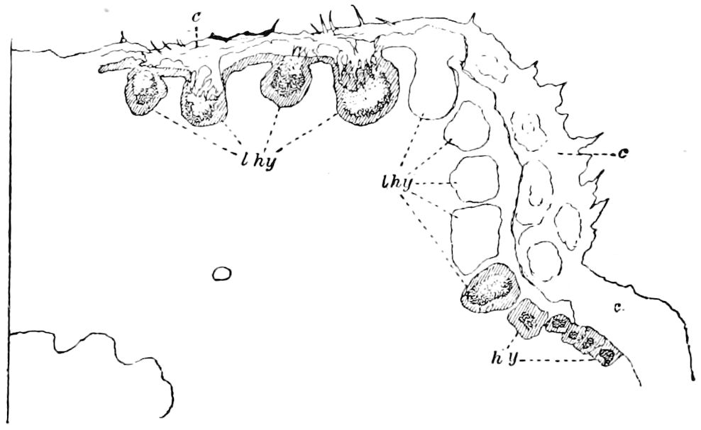
Fig. 566.—Transverse section through the prothoracic segment (ventral face) of larva of Pieris brassicæ, about 12 hours before pupation: c, cuticula; l. hy, large glandular (?) hypodermal cells; gradually passing into normal hypodermal cells (hy).—Gonin del.
E. Howgate has noticed under the microscope peculiar internal movements in a small immature transparent geometrid while moulting. “Each separate segment,” he says, “commencing at the head, elongated within the outer skin, whilst the next ones remained in their former state. Each segment in its turn behaved in this curious manner until the last was reached, when the motion was reversed and proceeded toward the head, when it was again reversed.... The whole proceeding appeared as if the larva was gliding within itself, segment after segment, the outer skin remaining as if held by the other segments, whilst the particular one in motion freed itself within. After remaining motionless for a short interval, the skin near the head swelled and burst, open at the back.... Presently out comes the head of the new caterpillar, pushing forward the old one.... After a short struggle the new true legs appear, pushing off and treading under foot the old ones. Then by violent wriggling movements the abdominal legs were extricated. Then all is clear, and the larva, which is quite exhausted, coils itself up and literally pants for breath.” (The Naturalist, November, 1885, No. 124, p. 366, quoted in Psyche, iv, p. 327, 1887.)
Since the worms and most other ametabolous invertebrates are not known to moult their integument, the body steadily increasing in size without frequent 615changes of skin, it seems that growth may go on and still be accompanied by considerable changes in shape of the body without change of skin. Frequent ecdyses appear, then, to be the result of the great and sudden changes of the body, necessitated by the adaptation of the animal to new or unusual conditions of life. In young Daphnia, a cladocerous crustacean, as many as eight moults were observed in a period of 17 days, and spiders frequently moult even after reaching their full size. The swollen bodies of the gravid female of Gastrophysa, Meloë, or of Termites, and of the honey ant show that the skin can stretch to a great extent, but in the metamorphoses of Crustacea and of insects, whose young are more or less worm-like or generalized in form, with fewer segments and appendages, or with appendages adapted for quite different uses from those of mature life, the necessity for a change of skin is seen to be necessary for mechanical reasons. Hence Crustacea and insects moult most frequently early in life, when the changes of form are most thoroughgoing and radical, while simple growth and increase in size are most rapid at the end of larval life, as seen both in shrimps and crabs, and in insects.
The hibernating caterpillars of certain butterflies are known to moult once oftener than those of the summer brood. Mr. W. H. Edwards has discussed the subject with much detail. “There seems,” he says, “to be a necessity with the hibernators of getting rid of the rigid skin in which the larva has passed the winter; that is, if the hibernation has taken place during the middle stages, as it does in Apatura and Limenitis. In these cases very little food is taken between the moult which precedes hibernation and the one which follows it, and the larva while in lethargy is actually smaller than before the next previous moult. The skin shrinks, and has to be cast off before the awakened larva can grow. Those species (observed) whose larva moults five times in the winter brood require but four moults during the summer.” He adds that while the larva is in lethargy, it is actually smaller than before the next previous moult. Dr. Dyar writes: “I think there is no doubt about the number of stages of arctian larvæ. They seem to have a great capacity of spinning out their life-history by interpolated stages (as regards width of head). I think it is because so many of them hibernate, and only a single brood extends throughout the season.” (Psyche iii, p. 161.)
On the other hand, it is difficult to understand why the caterpillars of arctians moult so frequently, nearly twice as often as in most other caterpillars, though the changes of form and armature are so slight.
Dr. Chapman also writes me: “Arctians resemble bears (Arctos), polar and others, in having long hairs to protect them during winter, and are, in fact, typically hibernators. Many of them have to half-hibernate, having warmth enough to keep them awake, but not enough food for growth, but their tissues, at least the chitinous ones of the cutis, and also probably, and perhaps especially, of the alimentary canal, become old and effete, and require the rejuvenescence acquired by a moult. Other smooth-skinned hibernators have similar capabilities.”
Chapman has shown in his paper on Acronycta that these caterpillars of this genus illustrate how larvæ may lose a moult, and they do so to acquire a sudden change of plumage.
The number of moults in insects of different orders.—It will be seen from the data here presented that the number of moults is as a rule greatest in holometabolic insects with the longest lives, and that an excessive number of ecdyses may at times be due to some physical cause, such as lack of food combined with low temperature.
616In Campodea there is a single fragmentary moult (Grassi), while the Collembola (Macrotoma plumbea) shed their skin throughout life. (Sommer.)
In the winged insects, especially Lepidoptera, the number of moults is dependent on climate. Insects of wide distribution growing faster in warmer climates consequently shed their skins oftener; for example, the same species may moult once oftener in the southern than in the northern States, as in the case of Callosamia promethea, which in West Virginia is double-brooded. Hibernating larvæ moult once oftener than those of the summer brood. (W. H. Edwards.) Weniger by rearing the larvæ of Antheræa mylitta and Eacles imperialis, and which, when reared under normal conditions, actually have six stages, found that when reared in a warm moist atmosphere of about 25° C. they have but five stages, i.e. moult but four times. In the hot and moist climate of Ceylon, A. mylitta has but five stages. (Psyche, v, p. 28.)
Among Orthoptera Acrydians moult five times; Diapheromera femorata but twice (Riley); a katydid (Microcentrum retinervis) moults four times (Comstock). Mantis religiosa, according to Pagenstecher, moults seven times, having eight stages, including that before the amnion is cast, but the first “moult” being an exuviation of the amnion, the number of stages is seven. Cockroaches (Periplaneta americana) are said by Marlatt to “pass through a variable number of moults, there being sometimes as many as seven.”
In the Homoptera there are, in general, from two to four moults; thus in Typhlocyba there are five stages, and in Aphis at least three, and in Psylla four during the nymphal state. Psocus has four. Riley states that the nymph of the female coccid, Icerya purchasi, sheds its skin three times, and that of the male twice. Notwithstanding its slow growth, Riley says, the 17–year Cicada moults oftener than once a year, and the number of larval stages probably amounts to 25 or 30 in all. The bed-bug sheds its skin five times; and with the last moult appear the minute wing-pads characteristic of the adult. In Conorhinus sanguisuga there are “at least two larval stages and pupal stages.” (Marlatt.)
In the dragon-flies moulting occurs, Calvert thinks, many times, since the rudiments of wings are said by Poletaiew to only appear in odonate nymphs after the third or fourth moult.
In the May-fly, Chloëon, the number of ecdyses is 20. The neuropterous Ascalaphus (Helecomitus) insimulans of Ceylon moults three times before pupating. Among the Mecoptera Felt has shown that Panorpa rufescens moults seven times.
617In Coleoptera the normal or usual number is not definitely known; Meloë moults five times, but this is a hypermetamorphic insect; Tribolium confusum has been carried by Mr. Chittenden through seven moults. Phytonomus punctatus, the clover-leaf weevil, moults three times, according to Riley, who has observed that Dermestes vulpinus passes through seven larval stages.
In the breeding jars, with plenty of food and a constant temperature of from 68° to 78° F., the larvæ cast their 1st skin in from four to nine days, the great majority moulting at seven days. Under the same conditions the 2d skin was cast at from four to seven days, the majority moulting at six days; the 3d skin at from three to six days, the majority moulting at five days; and the 4th skin at from three to six days, the majority moulting at five days; the 5th skin at from five to seven days, and the 6th skin at six days. There are thus seven larval stages. (Report for 1885, p. 260.)
Riley has ascertained that by rearing isolated larvæ of Tenebrio molitor, one after being kept nearly a year had moulted 11 times, when it died. A second larva, hatched June 5, had moulted 12 times by June 10 of the following year, (1877), when it also died. Of T. obscurus three larvæ were reared to the imago state. One moulted 11 times by Aug. 30 of the same year, pupated Jan. 20, 1877, and finally became a beetle Feb. 7, 1877. The other two both moulted 12 times, and reached the imago stage Feb. 18 and March 9, respectively. “All were, as nearly as possible, under like conditions of food and surroundings, and in all cases the moult that gave the pupa is not considered among the larval moults.”
Two larvæ of the museum pest (Trogoderma tarsale) were kept by Riley in a tight tin box with an old silkworm cocoon. “They were half-grown when placed in the box. On Nov. 8, 1880, there were in the box 28 larva skins, all very much of a size, the larva having apparently grown but little. The skins were removed and the box closed again as tightly as possible. Recently, or after a lapse of two years, the box was again opened and we found one of the larvæ dead and shrivelled up; but the other was living and apparently not changed in appearance. There were 15 larva skins in the box. He could not tell when the one larva died, but it is certain that within a little more than three and a half years, two larvæ shed not less than 43 skins, and that one larva did not, during that time, appreciably increase in size. We know of no observations which indicate the normal or average length of life, or number of moults in either Tenebrio or Trogoderma, but it is safe to assume from what is known, in these respects, of allied species, that in both the instances here referred to, but particularly in the case of Trogoderma, development was retarded by insufficient nutrition, and that the frequent moulting and slow growth resulted therefrom, and were correlated.”[99] Further observations such as these are greatly needed.
Of the Siphonaptera the common cat and dog flea (Pulex serraticeps) moults three times before pupating. (Howard.)
In Lepidoptera the usual or average number of moults is four, but the number varies considerably, the greatest number yet known occurring in Phyrrarctia isabella, which, Dr. Dyar informs me, moults 10 times.
From Dyar’s observations it appears that there are usually five 618larval stages, but six and seven stages are not infrequent, while there are seven in Seirarctia echo, eight in Ecpantheria scribonia, Scepis, and Apatelodes, and nine and ten in arctians, while the European Nola centonalis moults nine times, other species of this genus shedding their skins six times. (Buckler.) (Psyche, v, pp. 420–422.) Callosamia promethea appears, as a rule, to moult but three times. Orgyia antiqua was found by Hellins to moult from three to five times. Riley found that in O. leucostigma the males moult four times, the female four, but sometimes five times, while Dyar states that in O. gulosa the male larvæ moult three or four times, the female always four times; in O. antiqua, however, there are six stages, and in the female seven. Lithocolletis, Chambers thinks, as a rule, moults eight times, and Comstock thinks that L. hamadryadella casts its skin seven or eight times.
In the blow-fly (Calliphora) Leuckart and Weismann have inferred at least two moults, while Weismann suspected that there are as many as four. In Musca domestica we have observed that the larva moults three times; in Œstridæ there are three larval stadia. (Brauer.) In Corethra there are four larval moults, and Miall thinks there are probably as many in Chironomus. Passing to the phytophagous Hymenoptera, there are three moults or four larval stages in Nematus erichsonii, but Dyar informs us that less than four stages in saw-fly larvæ is very rare, that he has only one record of less than five, and that that is doubtful; “five for nematid, six and seven for others, is certainly the rule. The highest I have is the indication of 11 stages for Harpiphorus varianus, but this again is an inference only, and attended with doubt.” (Can. Ent., xxvii, p. 208.) In Bombus we have observed five different sizes of larvæ, and hence suppose the least number of ecdyses is five, while we are disposed to believe that this insect, as well as wasps and bees, in general shed their skins as many as eight times during their entire existence.
The honey-bee, Cheshire thinks, since he has found the old and ruptured pellicles, probably moults six times before it spins its cocoon, or passes into the semipupa condition. (Bees and Bee-keeping, p. 20.)
As to the cause of the great number of moults in the arctians and in the beetles experimented with by Riley, it would seem that cold and the lack of food during hibernation were the agents in arctians, and starvation or the lack of food in the case of the beetles, such cause preventing growth, though the hypodermis-cells retained their activity.
619Reproduction of lost limbs.—Here might be discussed the subject of the renovation or renewal of maimed or lost limbs, or the reparation of other injuries. As is well known, the cœlenterates, echinoderms, and worms under certain circumstances multiply by self-division, or if artificially mutilated, the parts are gradually restored by cell-proliferation or histogenesis. It is so with the antennæ and legs of crustaceans as well as the digits and tail of salamanders. The experiments first made by Le Pelletier[100] on spiders, and later by Heineken,[101] and others after him, on different spiders, as well as on Orthoptera and Hemiptera (Blatta, Reduvius, etc.), have proved that antennæ and legs and other external parts which have been injured or shortened, or entirely cut off in young individuals, are replaced at the next, or after successive moults, though generally in diminished size. This does not usually occur in adult life, and the process of reparation of lost parts is apparently due to the active growth of the cells of the parts affected during the process of moulting, when the histolysis of the maimed or diseased parts is succeeded by the rapid development of new cells, not only of the hypodermis, but also of the more specialized tissues within. And this tends to prove that such histolysis and making over of the muscles and other structures within occur especially in all metamorphic insects, and also in ametabolous forms, though the process has been most thoroughly examined in the Diptera, where these changes are more marked.
Gonin has found that the thoracic legs of the caterpillar correspond only to the tarsi of the imago (Fig. 608). It results, he says, from this fact that in accordance with the observations of Réaumur (which were wrongly interpreted by Newport and Künckel D’Herculais) that the amputation of the legs of the larva does not involve the entire leg, but only the extremity of the leg of the imago.
Formation of the cocoon.—While the larvæ of many insects, as those of the butterflies, suspend themselves before transforming, and spin no cocoon, or dig into the earth for protection and to secure an immunity from too great changes of temperature, a large proportion of the larvæ of metabolous insects which lead an inactive pupal life, line their earthen cells with silk, or spin a more or less elaborate case of silk, called the cocoon. We have seen that the inactive pupa of the male scale-insects is covered by the scale itself, or even in one case the insect forms a true cocoon of fibres of wax. The aquatic 620larvæ of the Neuroptera and Coleoptera creep out of the water, and by the movements of their bodies make a rude earthen cell in the bank, while that of Donacia spins a dense, leathery cocoon (Fig. 567) in the earth. The larvæ of the Embiidæ are protected by a cocoon, which they renew at each moult. Coniopteryx spins an orbicular cocoon, the Hemerobiidæ a spherical, dense, whitish one. The Trichoptera transform within their larval cases, which thus serve as cocoons, as do certain case-bearing Lepidoptera, notably the Psychidæ.
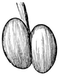
Fig. 567.—Cocoon (natural size) of Donacia proxima.
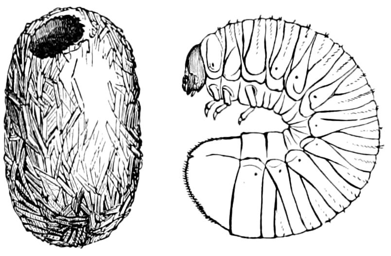
Fig. 568.—Cocoon and larva of Lucanus dama.
The pupa of certain leaf-eating beetles (Chrysomelidæ), as well as the Coccinellidæ, Dermestidæ, Hister, etc., are usually protected by the cast larval skin, which is retained, forming a rude shelter. While many beetles spin an oval cocoon (Gyrinus, Silphidæ), the wood-boring species make one of chips glued together, and that of Lucanus, which feeds on decayed wood, is lined with silk (Fig. 568). Anobium constructs a silken cocoon, interweaving the fine particles of its thin castings; the larvæ of weevils also usually spin silken cocoons.
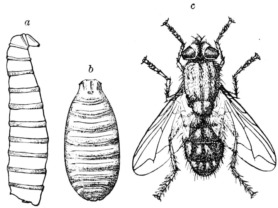
Fig. 569.—Larva (a), puparium (b), and imago (c) of Sarcophaga, enlarged.
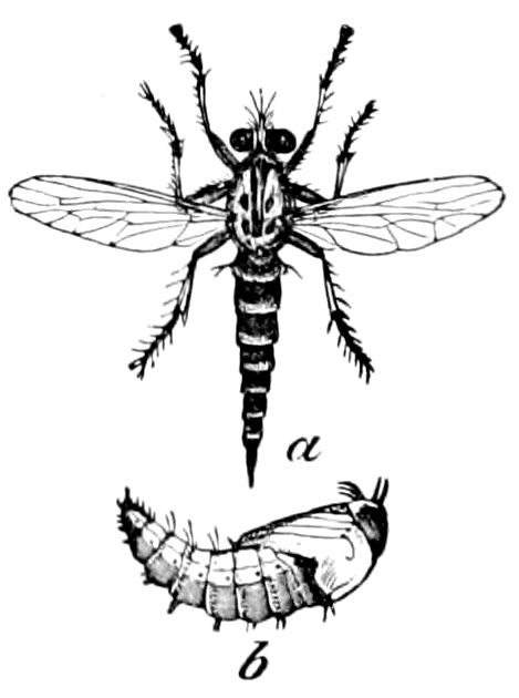
Fig. 570.—a, Erax bastardi; b, pupa.—After Riley.
The larval skin of the coarctate Diptera is retained as a protection for the soft-bodied pupa within, the old larval skin separating from 621the integument of the semipupa. To this cocoon-like covering of the coarctate pupa we have restricted the term puparium, originally used by Kirby and Spence to designate the pupa. The puparium is usually cylindrical or barrel-shaped, rounded at each end.
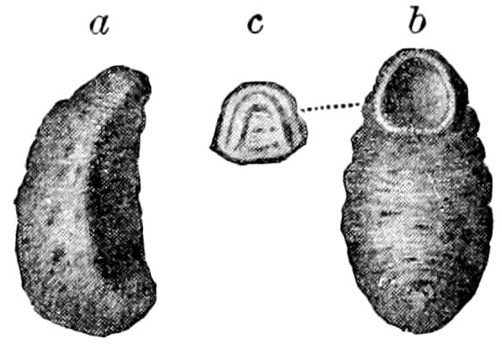
Fig. 571.—Puparium of Hypoderma bovis: a, side; b, ventral view, showing exit hole of adult; c, cap which splits off for exit of fly.—After Clark, from Osborn, Bull. 5, Div. Ent. U. S. Dept. Agr.
In the Diptera cyclorhapha, or common house and flesh flies, etc., the puparium remains in vital connection, by means of four tracheæ, with the enclosed pupa, which escapes from the case through a curved seam or lid at the anterior end and not by a slit in the back, as do the orthoraphous families, represented by the horse-fly (Tabanidæ, Asilidæ, Fig. 570), etc., where in some cases the obtected pupa remains within the loose envelope formed by the old larval skin, which Brauer calls a false puparium. The dry, hard puparium is burst open at the cephalic end when the fly emerges, by means of the frontal vesicle, which is distended with fluid (Fig. 571).
The exact mode of spinning the cocoon by caterpillars has been carefully observed by L. Trouvelot in the case of the polyphemus silkworm.
“When fully grown, the worm, which has been devouring the leaves so voraciously, becomes restless and crawls about the branches in search of a suitable place to build up its cocoon; before this it is motionless for some time, holding on to the twig with its front legs, while the two hind pair are detached; in this position it remains for some time, evacuating the contents of the alimentary canal until finally a gelatinous, transparent, very caustic fluid, looking like albumen, or the white of an egg, is ejected; this is a preparation for the long catalepsy that the worm is about to fall into. It now feels with its head in all directions, to discover any leaves to which to attach the fibres that are to give form to the cocoon. If it finds the place suitable, it begins to wind a layer of silk around a twig, then a fibre is attached to a leaf near by, and by many times doubling this fibre and making it shorter every time, the leaf is made to approach the twig at the distance necessary to build the cocoon; two or three leaves are disposed like this one, and then fibres are spread between them in all directions, and soon the ovoid form of the cocoon distinctly appears. This seems to be the most difficult feat for the worm to accomplish, as after this the work is simply mechanical, the cocoon being made of regular layers of silk united by a gummy substance. The silk is distributed in zigzag lines of about one-eighth of an inch long. When the cocoon is made, the worm will have moved his head to and fro, in order to distribute the silk, about 254,000 times.
“After about half a day’s work, the cocoon is so far completed that the worm can hardly be distinguished through the fine texture of the wall; then a gummy resinous substance, sometimes of a light-brown color, is spread all over the inside of the cocoon. The larva continues to work for four or five days, hardly taking 622a few minutes of rest, and finally another coating is spun in the interior, when the cocoon is all finished and completely air tight. The fibre diminishes in thickness as the completion of the cocoon advances, so that the last internal coating is not half so thick and so strong as the outside ones.” (Amer. Naturalist, i, p. 86.)
The mode of spinning the cocoon of an ichneumon (Microgaster) parasitic on Philampelus has been well described by John P. Marshall, as follows:—
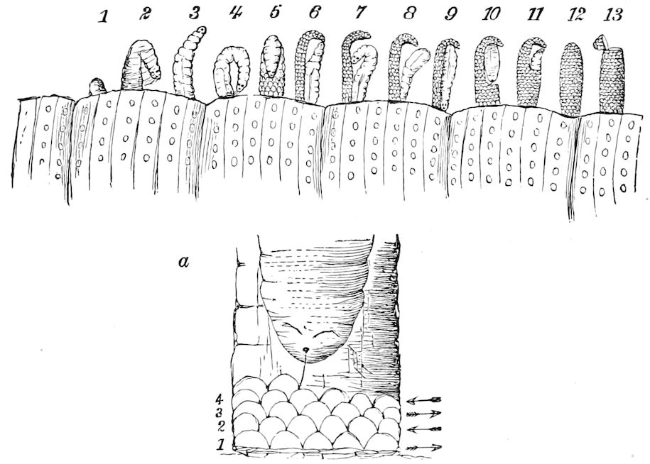
Fig. 572.—Microgaster larvæ; spinning their cocoons: a, enlarged view of 5.—After Marshall.
The first appearance of the parasite is represented in Fig. 572, 1. A warty excrescence appears on the back of the caterpillar, which slowly emerges until it is seen to be a larva enclosed in a delicate transparent membrane, as represented in 2. This it soon succeeds in bursting, and, rising to its full length, balances itself a moment as in 3, then, bending double, it ejects from its mouth a glairy liquid, which instantly changes to silk, and fastens the posterior end to the skin of the caterpillar, as shown in 4, side view. It now begins to spin its cocoon by attaching a silken thread to the silky mass by which it had previously fastened itself to the caterpillar, and forming a series of loops of uniform size, first from right to left, and then back again from left to right, as represented in the front view, 5, and better in the enlarged view, 5a, the arrow heads showing the direction in which the head of the larva moved while forming the loops. The ends of the series, numbered 1, 2, 3, 4, are fastened to the edges of the ventral side of the body, which thus serves as a measure of the width of the cocoon, and also acts as a support for the frail fabric in the first stages of spinning. After the larva has fastened the fabric as far up on its ventral surface as it can, conveniently, it then begins to spin free, as shown in the side view, 6, where it is represented as just completing the first half of its cocoon, which 623resembles in form a slipper. This accomplished, the larva ceases to spin for the time being, bends its head, as in 7, towards its ventral surface, and pushes the half cocoon free from its body. The form of the silken fabric enables it to stand unsupported, while the larva, sliding its head down to the base, holds on firmly until it swings its posterior end into the toe of the slipper.
Figure 572, 8, shows it in the act of changing end for end, and in 9 the larva is seen erect, beginning at the base to complete the other half of its cocoon; 10 shows the larva contracting its body as it spins upward for about half the length of the cocoon, when it again changes end for end, as shown in 11, where it is beginning at the upper part to unite the two sides, finally enclosing itself as represented in 12.
It may now be seen, under the microscope, through the meshes of its cocoon actively engaged in lining the interior with layers of very fine silk ejected from its mouth in great abundance. One half of the cocoon is first lined by a forward and back movement of its head, and then reversing its position, it lines the other half in a similar manner.
In one case the larva was disengaged from the skin of the caterpillar, after beginning its cocoon. It, however, began again, and spun a portion while lying on the table. This was removed, when it began a third time, and completed its cocoon.
In about 10 days the insect made its appearance through a hole in the upper end, as represented in 13. The top was eaten off in a perfect circle and hung by a few threads, so as to resemble a lid as it was thrown back.
One caterpillar observed had between 300 and 400 cocoons on its back and sides, and another was dissected after more than 30 larvæ had escaped, and 130 were discovered in the soft integuments of the back.
The figures from 1 to 13 are magnified five diameters, but in order to observe the spinning of the cocoon a power of 50 is required. (Amer. Naturalist, xii, pp. 559, 560.)
Certain differences observed by W. A. Buckhout in a Microgaster parasitic on the different species of Macrosila, are referred to in the same volume, p. 752.
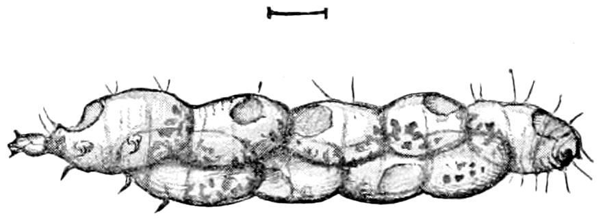
Fig. 573.—Body of larva of Lithocolletis. swollen and filled with cocoons of Copidosoma, enlarged.
While those chalcidid larvæ which feed internally on their host, as a rule, transform into naked, more or less coarctate pupæ, Howard states that the larvæ of Copidosoma, Bothriothorax, Homalotylus, and perhaps others, which are much crowded within their host, cause a marked inflation of the body of the latter (Figs. 573, 574). The nature of this cocoon-like cell, and how it is produced, is unknown. “Its structure shows it not to be silk, nor yet the last larval skin of the parasite, and whether it is an adventitious tissue of the host-larva or a secretion of the parasite, or is explicable upon other grounds, I cannot say.”
The silken cocoon of an aphidiid ichneumon has been found by Miss Murtfeldt, and also by Dr. Riley, under a rose aphid in which it had lived, and referred by Howard to the genus Praon (Fig. 575).
Sanitary conditions observed by the honey-bee larva, and admission of air within the cocoon.—Cheshire has observed that after the larva of the honey-bee has spun its cocoon or silken lining of its cell, it 624observes the following means of preserving cleanliness. The food given to the larva, especially during the latter part of the growing period, contains much pollen, the cases of the grains of which consist of cellulose, which is indigestible.
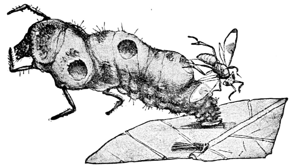
Fig. 574.—Coccinellid larva infested by Homalotylus obscurus, enlarged.
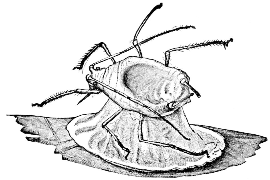
Fig. 575.—Cocoon of Praon under the body of a dead Aphis, enlarged.—This and Figs. 573 and 574 after Howard, from Insect Life.
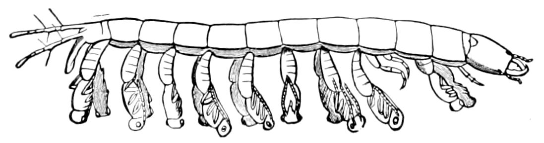
Fig. 576.—Pupation of Proctotrupes in the body of a larva of a beetle, representing a case mentioned by Dr. Sharp, where the parasites have pupated on the outside of the host, a pair of each attached to nearly each segment of the body of their host.—After Sharp.
“These cases, with other refuse matters, collect in quantity within the bowel, which becomes distended, since it has no opening. The imprisoned larva, having little more than enough room for turning, must be freed of these objectionable residua.... In a word, the larva turns its head upon its stomach, and pushes the former towards the base of the cell until its position is reversed, the tail being outwards; and, thus placed, it laps up all residue of food, especially from 625its old clothes previously referred to, until they are dried, and practically occupy no space. It now throws up its stomach and bowel, with all their contents, and without detaching them from its outer skin, which is moulted as before, but in this instance to be pressed against the cell, so as to form for it an interior lining. The dejectamenta of the bowel in this way lie between the cast skin and cell-wall (as seen at e, Fig. 577), and so the larva remains absolutely unsoiled. It now turns its head and resumes its old position, joining its cocoon to the edges of its last cast skin, so that its habitation is relined, it is cleansed, and air can still pass to it through the imperceptible openings left by the bees in the sealing. This point is of radical importance, since breathing is carried on pretty rapidly during the latter part of its subsequent transformations, the absorbed oxygen permitting then of a production of heat, and causing also considerable diminution in weight.”
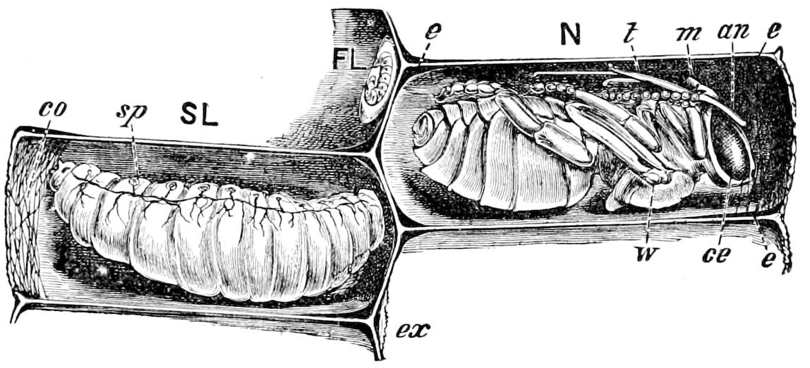
Fig. 577.—Larva and pupa of honey-bee in their cell: SL, spinning-larva; N, pupa; FL, young feeding larva; co, cocoon; sp, spiracles; t, tongue; m, mandible; an, antenna; w, wing; ce, compound eye; e, excrement; ex, exuvium.—After Cheshire.
As to the passage of air into the bee’s cocoon, Cheshire states that before the cocoon can be built, a cover, technically called sealing, is put over the larva by its nurses. These covers are made of pollen and wax, and are pervious to the air. They are more convex and regular in form than those sealing in the honey.[102]
THE PUPA STATE
The word pupa is from the Latin meaning baby. Linnæus gave it this name from its resemblance to a baby which has been swathed or bound up, as is still the custom in Southern Europe. The term pupa should be restricted to the resting inactive stage of the holometabolous insects.
Lamarck’s term chrysalis was applied to the complete or obtected pupa of Lepidoptera and of certain Diptera, and mumia, a mummy, to the pupæ of Coleoptera, Trichoptera, and most Hymenoptera. Latreille (1830) also restricted 626the term pupa to the “oviform nymph,” or puparium, of Diptera. Brauer applies the term nymph to the pupa of metabolous insects.
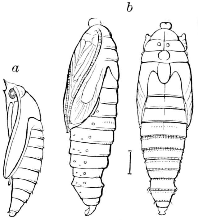
Fig. 578.—Pupa obtecta: a, of Sesia, with its cocoon-cutter on the head; b, of Tortrix vacciniivorana.
The typical pupa is that of a moth or butterfly, popularly called a chrysalis. A lepidopterous pupa in which the appendages are more or less folded close to the body and soldered to the integument, was called by Linnæus a pupa obtecta; and when the limbs are free, as in Neuroptera, Mecoptera, Trichoptera, and the lepidopterous genus Micropteryx it is called a pupa libera (Fig. 579). When the pupa is enclosed in the old larval skin, which forms a pupal covering (puparium), the pupa was said by Linnæus to be coarctate. The pupa of certain Diptera, as that of the orthoraphous families, is nearly as much obtected as that of the tineoid families of moths, especially as regards the appendages of the head; the legs being more as in pupæ liberæ (Fig. 580).
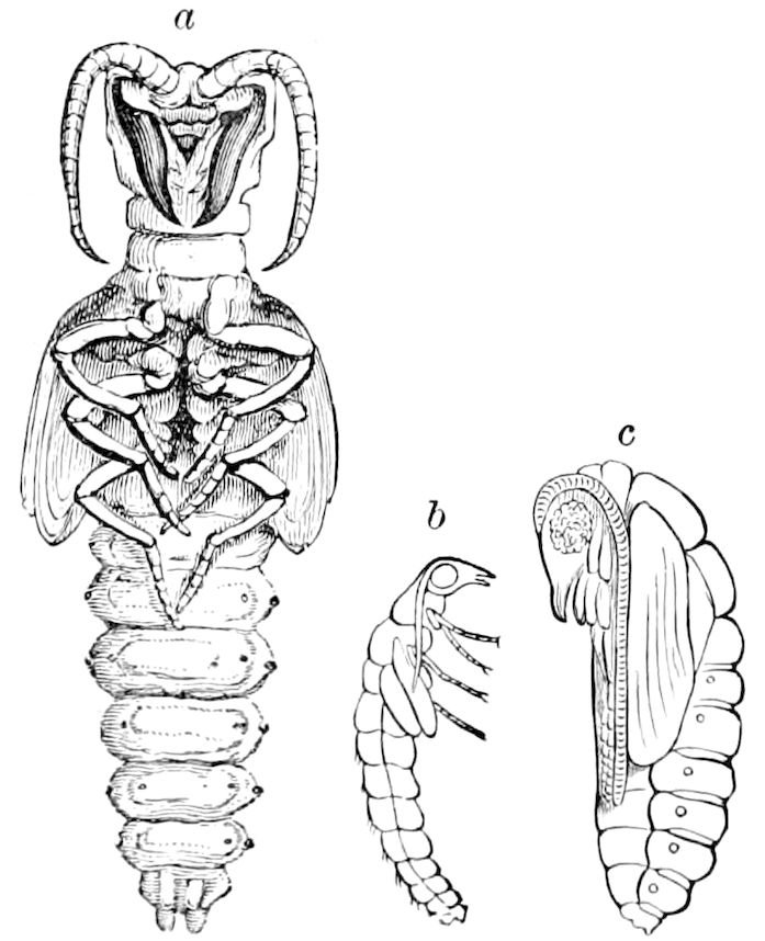
Fig. 579.—Pupa libera of neuropterous insects a, Corydalus cornutus; b, Sialis; c, Hemerobius.
The male Coccid anticipates the metabolous insects in passing through a quiescent state, when, as Westwood states, it is “covered by the skin of the larva, or by an additional pellicle.” The body appears to be broad and flat, the antennæ and fore legs resting under the head, while the two hinder pairs of legs are appressed to the under side of the body. There is but a slight approach to the pupa libera of a metabolous insect.
Riley states that the male larva of Icerya purchasi forms a cocoon waxy in character, but lighter, more flossy, and less adhesive than that of the female egg-cocoon. It melts and disappears when heated, proving its entirely waxy nature. When the mass has reached the proper length, the larva casts its skin, which remains in the hind end of the cocoon, and pushes itself forward into the middle of the cocoon. The pupa (Fig. 581) is of the same general form and size as the larva. All the limbs are free and slightly movable, so that they vary in position, though ordinarily the antennæ are pressed close to the side, 627as are the wing-pads; the front pair of legs are extended forward. “If disturbed, they twist and bend their bodies quite vigorously.” The pupa state lasts two or three weeks. A similar pupa is that of Icerya rosæ. (Riley and Howard.)
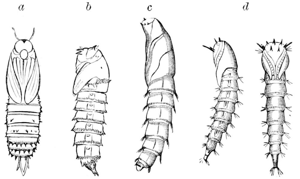
Fig. 580.—Pupa obtecta of Diptera: a, Ptychoptera; b, Tabanus atratus; c, Proctacanthus philadelphicus; d, Midas clavatus.
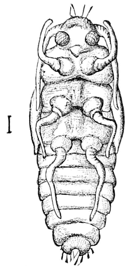
Fig. 581.—Pupa libera of Icerya purchasi, ventral view.—After Riley, Insect Life.
The metamorphosis of Aspidiotus perniciosus is of interest. The male nymph differs much after the first moult from the female, having large purple eyes, while the female nymph loses its eyes entirely. It passes into what Riley terms the pro-pupa (Fig. 582, b), in which the wing-pads are present, while the limbs are short and thick. The next stage is the “true pupa” (Fig. 582, c, d), in which the antennæ and legs are much longer than before. There is no waxy cocoon, but only a case or scale composed of the shed larval skin, i.e. “with the first moult the shed larval skin is retained beneath the scale, as in the case of the female; with the later moultings the shed skins are pushed out from beneath the scale,” and when they transform into the imago they “back out from the rear end of their scale.”
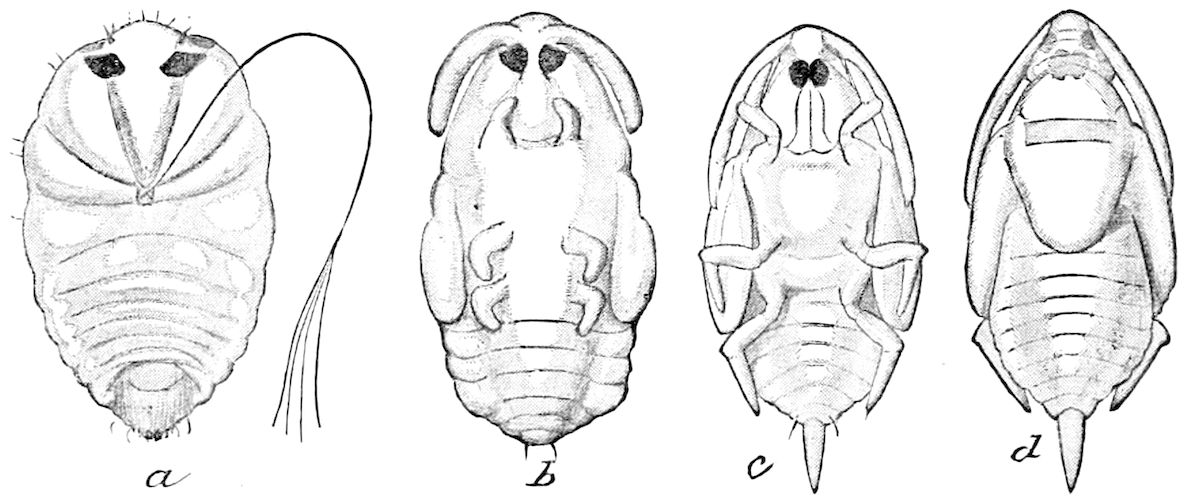
Fig. 582.—Aspidiotus perniciosus, development of male insect: a, ventral view of larva after first moult; b, the same, after second moult (pro-pupa stage); c and d, true pupa, ventral and dorsal views. All greatly enlarged.—After Riley.
628The pupæ of Coleoptera and of Hymenoptera, though there is, apparently, no near relationship between these two orders, are much alike in shape, and, as Chapman pertinently suggests, those of both orders are helpless from their quiescence, and hence have resorted for protection to some cocoon or cell.
But it is quite otherwise with the pupæ of Lepidoptera and Diptera, which vary so much in adaptation to their surroundings, and hence afford important taxonomical and phylogenetic characters. This, as regards the Lepidoptera, was almost wholly overlooked until Chapman called attention to the subject, and showed that the pupæ had characters of their own, of the greatest service in working out the classification, and hence the phylogeny, of the different lepidopterous groups. We have, following the lead of Chapman, found the most striking confirmation of his views, and applied our present knowledge of pupal structures to dividing the haustellate Lepidoptera into two groups,—Paleolepidoptera and Neolepidoptera.
The pupæ of the Neuroptera, Coleoptera, and Hymenoptera differ structurally from the imago, in the parts of the head and thorax being less differentiated. Thus in the head the limits or sutures between the epicranium and clypeus, and the occiput and gula, are obscurely marked, while the tergal and pleural sclerites of the imago are not well differentiated until the changes occurring just before the final ecdysis.
It is easy, however, to homologize the appendages of the pupæ with those of the imago of all the holometabolous orders except in the case of the obtected pupa of the Lepidoptera (and probably of the obtected dipterous pupæ), where the cephalic appendages are soldered together.
That the appendages of the lepidopterous pupa are, as generally supposed, merely cases for those of the imago has been shown by Poulton to be quite erroneous. He says: “If we examine a section of a pupal antenna or leg (in Lepidoptera), we shall find that there is no trace of the corresponding imaginal organ until shortly before the emergence of the imago. In the numerous species with a long pupal period, the formation of imaginal appendages within those of the pupa is deferred until very late, and then takes place rapidly in the lapse of a few weeks. This also strengthens the conclusion that such pupal appendages are not mere cases for the parts of the imago, inasmuch as these latter are only contained within them for a very small proportion of the whole pupal period.” On the other hand, Miall and Hammond claim that there is a strong superficial contrast as to the formation of the imaginal organs, between Lepidoptera 629and tipularian Diptera, the appendages, wings, and compound eyes being substantially those of the imago. “With the exception of the prothoracic respiratory appendages and the tail-fin, there is little in the pupa of Chironomus which does not relate to the next stage.”
The exact homology of the “glazed eye” of the lepidopterous pupæ and of the parts under the head, situated over the maxillæ, is difficult to decide upon, and these points need farther examination. In the dipterous pupa it is interesting to observe that the halteres are large and broad, which plainly indicates that they are modified hind wings. The number and arrangement of the spiracles is different in pupæ from those of the larva and imago.
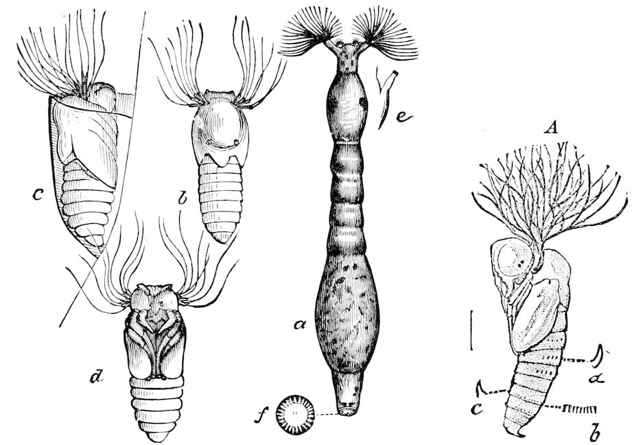
Fig. 583.—Simulium piscicidium: a, larva; b, c, d, pupa; e, thoracic leg; f, row of bristles at end of body. A, S. pecuarum, pupa; a, b, c, adminicula.—After Riley.
There are also secondary adaptive structures peculiar to the pupa, which are present and only of use in this stage. These are the thoracic, spiracular, or breathing appendages of the aquatic Diptera (Fig. 583), the various spines situated on the head or thorax, or on the sides, or more often at the end of the abdomen, besides also the little spines arranged in more or less circular rows around the abdominal segments, the cocoon-breaker, and the cremaster of many pupæ.
In the pupa of certain Diptera, there is a terminal cremaster-like spine, as in that of Tipula eluta (Fig. 584), Tabanus lineola (Fig. 585), besides adminicula or locomotive spines like those of lepidopterous pupæ (Fig. 580, a, b, c).
630
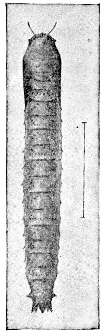
Fig. 584.—Pupa of Tipula eluta.
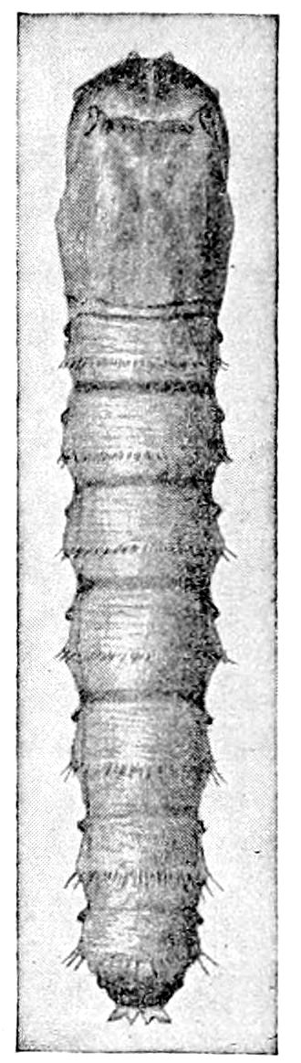
Fig. 585.—Pupa of Tabanus lineola.—This and Fig. 584 after Hart.
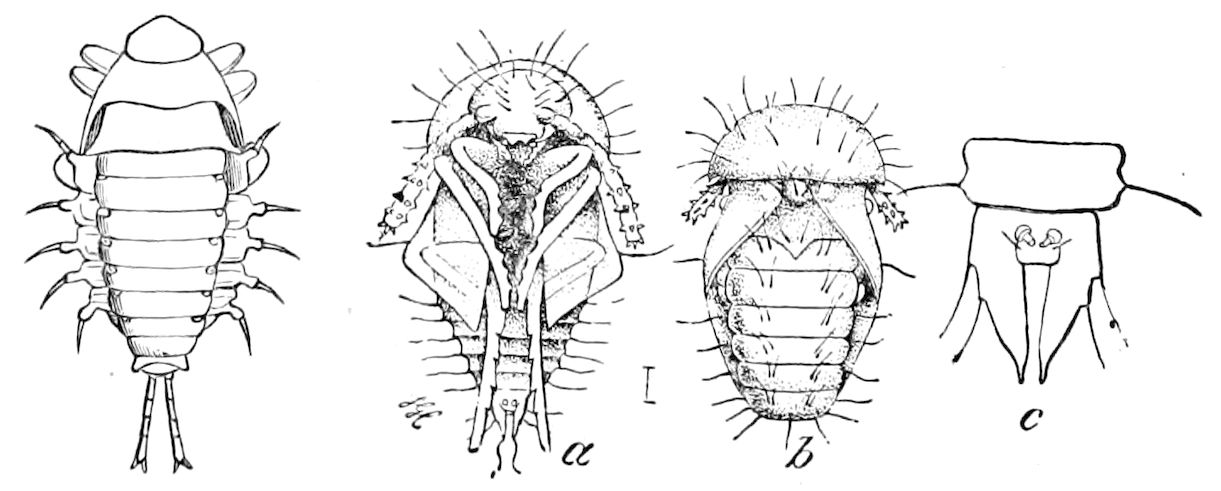
Fig. 586.—Pupa of Galerita lecontei, and of Adelops hirtus (a, b, c).—After Hubbard.
The pupæ of Coleoptera are variously spined or hairy (Fig. 586). Those of Hydrophilus and of Hydrobius are provided with stout spines on the prothorax and abdomen which support the body in its cells, so that, as Lyonet first showed, though surrounded on all sides by moist earth, it is kept from contact with it by the pupal spines; other pupæ of beetles, such as that of the plum weevil, which is also subterranean, possess similar spines. The abdomen of many coleopterous pupæ, such as those of Carabidæ, end in two spines, to aid them in escaping from their cells in wood or in the earth; others have stiff bristles, and others spines along each side of the abdomen (Fig. 586). All these structures are the result of a certain amount of activity in what we call quiescent pupæ, but most of these are for use at the end of pupal life, at the critical moment when by their aid the insect escapes from its cocoon or subterranean cell, or if parasitic, bores out of its host.
If we are to account for the causes of their origin, we are obliged to infer that they are temporary deciduous structures due to the need of support while the body is subjected to unusual strains and stresses in working its way out of its prison in the earth, or its cell within the stems and trunks of plants and similar situations. They are pupal inheritances or heirlooms, and well illustrate the inheritance of characters acquired during a certain definite, 631usually brief, period of life, and transmitted by the action of synchronous heredity.
The pupæ of certain insects are quite active, thus that of Raphidia, unlike that of Sialis, before its final ecdysis regains its activity and is able to run about. (Sharp, p. 448.)
a. The pupa considered in reference to its adaptation to its surroundings and its relation to phylogeny
The form of the pupa is a very variable one, as even in Lepidoptera it is not entirely easy to draw the line between a pupa libera and a pupa obtecta (Fig. 578); and though the period is one of inactivity, yet when they are not in cocoons or in the earth in subterranean cells, their form is more or less variable and adapted to changes in their surroundings. Even in the obtected pupa of butterflies, there is, as every one knows, considerable variability of shape and of armature, which seems to be in direct adaptability to the nature of their environment. Scudder has well shown that in certain chrysalids, such as those of the Nymphalidæ, which are variously tuberculated, and hang suspended by the tail, and often hibernate, these projections serve to protect the body. All chrysalids with projections or ridges on different parts of the body, being otherwise unprotected, move freely when struck by gusts of wind, hence “the greater the danger to the chrysalis from surrounding objects, the greater its protection by horny tubercles and roughened callous ridges.” The greater the protection possessed in other ways, as by firm swathing or a safe retreat, the smoother the surface of the body and the more regular and rounded its contours. The tendency to protection by tubercles is especially noticeable in certain South American chrysalids of nymphalid butterflies. This response to the stimuli of blows or shocks is also accompanied by a sensitiveness to the stimulus of too strong light.
Previously Scudder[103] had made the important suggestion that the smooth crescent-shaped belt of the “glazed eye” or “eyepiece” of chrysalids is, as an external covering of the eye, midway between that of the caterpillar and the perfect insect, and he asks: “May it not be a relic of the past, the external organ of what once was? 632And are we to look upon this as our hint that the archaic butterfly in its transformations passed through an active pupal stage, like the lowest insect of to-day, when its limbs were unsheathed, its appetite unabated?” etc. Scudder also shows that “the expanded base of the sheath covering the tongue affords protection also to the palpi which lie beneath and beside the tongue.”
All this tends to show the importance of studying the structure of the pupa, in order to ascertain how the pupal structures have been brought about, with the final object of discovering whether the pupæ of the holometabolic insects are not descended from active nymphs, and if so, the probable course of the line of descent.
b. Mode of escape of the pupa from its cocoon
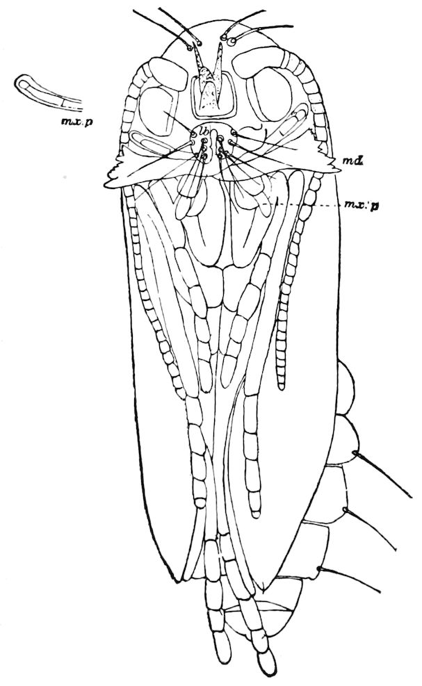
Fig. 587.—Pupa of Micropteryx purpuriella, front view: md, mandibles; mx. p, maxillary palpus, end drawn separately; mx.′ p, labial palpi; lb, labrum.
“In all protected pupæ,” as Chapman says, “the problem has to be faced, how is the imago to free itself from the cocoon or other envelope protecting the pupa.” In the Coleoptera and Hymenoptera the imago becomes perfected within the cocoon or cell, as the case may be, and as Chapman states, “not only throws off the pupal skin within the cocoon, but remains there till its appendages have become fully expanded and completely hardened, and then the mandibles are used to force an outlet of escape,” and he calls attention to the fact that “in many cases, even in some entire families, they are of no use whatever to the imago except in this one particular,” and he cites the Cynipidæ as perhaps the most striking instance of this circumstance.
In those Neuroptera which spin a silken cocoon, e.g. the Hemerobiidæ, the Trichoptera, 633and in Micropteryx (Fig. 588), the jaws used by the pupa for cutting its way out of the cocoon are even larger in proportion than in the pupa of caddis-flies (Fig. 588), being of extraordinary size.
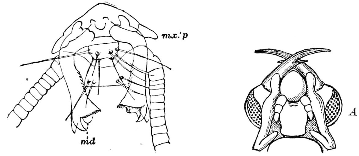
Fig. 588.—Mandibles (md) of Micropteryx purpuriella, enlarged.—Author del. A, pupal head of a hydropsychid caddis-fly, showing the large mandibles.—After Reaumur, from Miall.
In Myrmeleon the pupa pushes its way half out of the cocoon, and then remains, while the imago ruptures the skin and escapes (Fig. 589, a).
Thus in the Neuroptera and Trichoptera we have already established the more fundamental methods of escape from the cocoon, which we see carried out in various ways in the more generalized or primitive Lepidoptera.
The most primitive method in the Lepidoptera of escaping from the cocoon seems to be that of Micropteryx.
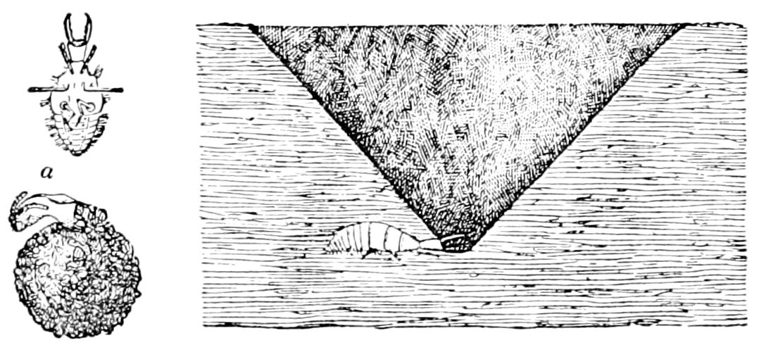
Fig. 589.—Larva of Myrmeleon with (a) its cocoon and cast pupa-skin.
“In this genus,” says Chapman, “though it is nominally the pupa that escapes from the cocoon, it is in reality still the imago, the imago clothed in the effete pupal skin. To rupture the cocoon it uses not its own jaws, but those of the pupal skin, energizing them, however, in some totally different way from ordinary direct muscular action, their movements being the result of the vermicular movements of the pupa, acting probably by fluid pressure on the articular structure of the jaws, by some arrangement not altogether different perhaps from the frontal sac of the higher Diptera. In the Micropteryges the jaws of the pupa not only rupture the cocoon, but appear to be the most active agents in dragging the pupa through the opening in the cocoon and through any superincumbent earth, being merely assisted by the vermicular action of the abdominal 634segments, and we find in accordance with this circumstance that the pupal envelope is still very thin and delicate, and has little or no hardening or roughness by which to obtain a leverage against the walls of the channel of escape.” (Trans. Ent. Soc. London, 1896, pp. 570, 571.)
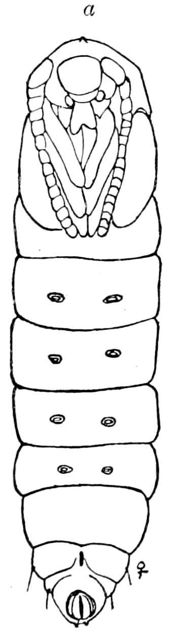
Fig. 590.—Pupa of Talæporia: a, cocoon-cutter; with vestiges of four pairs of abdominal legs, and the cremaster.
Some sort of a beak or hard process, more or less developed, according to Chapman, adapted for breaking open the cocoon exists in nearly all the Lepidoptera with incomplete pupæ (pupæ incompletæ), except the limacodid and nepticulid section. “In all these instances the pupa emerges from the cocoon precisely as in the Micropteryges, that is, the moth it really is that emerges, but does so encased in the pupal skin. To achieve this object, it seems to have been found most efficient to have three, four, or five abdominal segments capable of movement, but to have the terminal sections (segments) soldered together.”
This cocoon-breaker, as we may call it, is especially developed in Lithocolletis hamadryadella. As described by Comstock, it forms a toothed crest on the forehead which enables it to pierce or saw through the cocoon.
“Each pupa first sawed through the cocoon near its juncture with the leaf and worked its way through the gap, by means of the minute backward-directed spines upon its back, until it reached the upper cuticle of the leaf. Through this cuticle it sawed in the same way that it did through the cocoon. The hole was in each case just large enough to permit the chrysalis to work its way out, holding it firmly when partly emerged. When half-way out it stopped, and presently the skin split across the back of the neck and down in front along the antennal sheaths, and allowed the moth to emerge.”[104]
We have observed and figured the cocoon-breaker in Bucculatrix, Talæporia (Fig. 590, a), Thyridopteryx, and Œceticus, and rough knobs or slight projection answering the purpose in Hepialidæ, Megalopyge, Zeuzera, and in Datana.[105] See also the spine on the head of Sesia tipuliformis (Fig. 578).
The imago of the attacine moths cuts or saws through its cocoon by means of a pair of large, stout, black spines (sectores coconis), one on each side of the thorax at the base of the fore wings (Fig. 591), and provided with five or six teeth on the cutting edge (C, D).
635
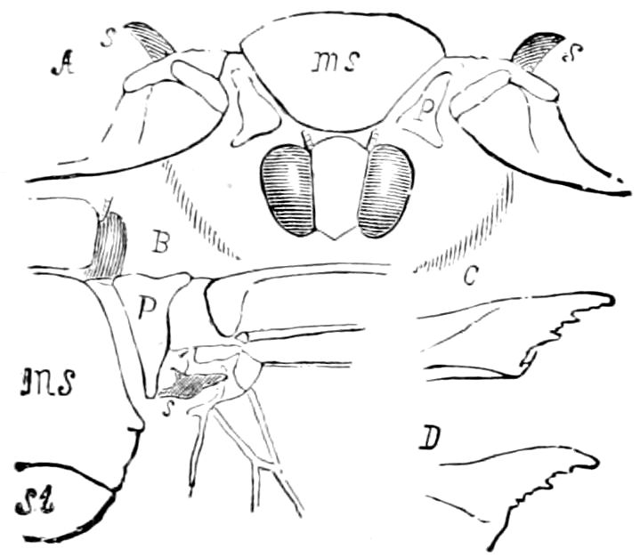
Fig. 591.—Cocoon-cutter of the Luna moth: front view of the moth with the shoulders elevated and the rudimentary wings hanging down: s, cocoon-cutter; p, patagium. B, represents another specimen with fully developed wings: ms, scutum; st, scutellum of the mesothoracic segment; s, cocoon-cutter, which is evidently a modification of one of the pieces at the base of the fore wings; it is surrounded by membrane, allowing free movement. C and D, different views of the spine, magnified, showing the five or six irregular teeth on the cutting edge.