PART II. EMBRYOLOGY OF INSECTS
Reading edition
a. The egg
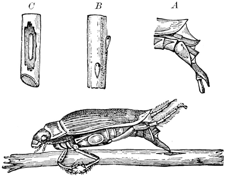
Fig. 482.—Female Dyticus, laying eggs: A, ovipositor extended. B, egg of Notonecta, attached to stem of rush. C, egg of Dyticus, laid in excavation in rush.—After Régimbart, from Miall.
Insects as a rule arise from eggs which are laid in a great variety of situations, those species which are viviparous being exceedingly few in number compared with the class as a whole. It is noteworthy that Leydig has found in the same Aphis, and even in the same ovary, an egg-tube producing eggs, while a neighboring tube was producing viviparous individuals.[77] The viviparous species are confined to certain May-flies, the Aphidæ, Diptera (Sarcophaga, Tachinidæ, Œstridæ, and Pupipara), and to certain Coleoptera (Stylopidæ and some Staphylinidæ).
The number of eggs laid varies from a very few, as in the Collembola and in the Psocidæ, or 15 or even less in certain fossorial wasps, and from 20 to 35 in some locusts to many thousands in the social insects, the honey-bee laying by estimate over 1,000,000 eggs in the course of her life. Dr. Sharp thinks that from 50 to 100 may perhaps be taken as an average number for one female to produce. The eggs of insects with a complete metamorphosis are said by Brauer to be smaller in proportion to the parent than those laid by ametabolous or heterometabolous insects. In this respect the insects are paralleled by the birds, the highest forms laying smaller eggs than the water birds, ostrich, Apteryx, etc.
516
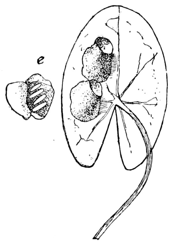
Fig. 483.—Eggs (e) of Hydrobius (?) and their capsules, from which the larva, Fig. 452, hatched.—Emerton del.
The egg, or ovum, when laid is not always ripe or perfect, but, as in those of ants, continues to grow after oviposition. Others are laid some time after the embryo has begun to form; and in the flesh-flies the larva hatches before the egg is deposited.
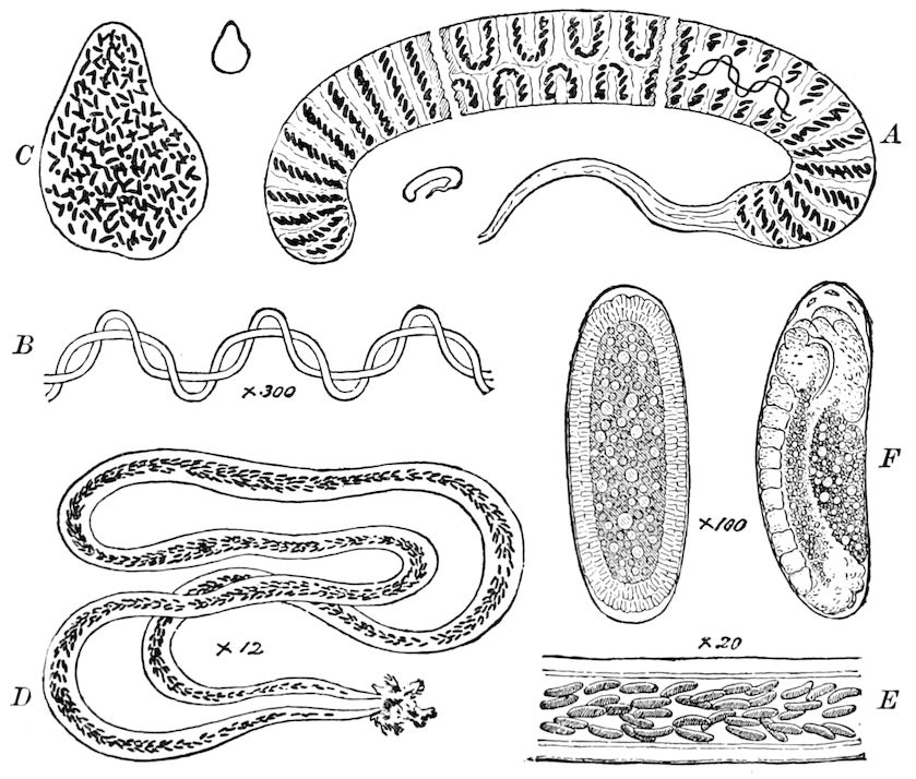
Fig. 484.—Egg-masses of Chironomus: A, string of eggs of C. dorsalis, divided into sections to show both sides. B, twisted fibres which traverse the string of eggs. C, egg-mass of Chironomus (sp). D, egg-mass of a third species. E, part of D, more highly magnified. F, developing eggs, two stages.—After Miall.
Insects as a rule instinctively lay their eggs near or upon objects destined to be the food of the larva; those of caterpillars on leaves, those of many flies on meat or carrion, those of Copris and other dung-beetles in dung, those of aquatic insects in water, while many oviposit in the earth or in plants (Fig. 482), or in the bodies of animals destined to be the hosts of the parasitic larvæ. As the eggs are preyed upon by mites and other animals, the contrivances and modifications of the mode of egg-laying, and the situations in which they are placed, are almost endless. Many insects lay their eggs in a mass, covered with a gummy substance; 517or those laid in the water, as the eggs of dragon-flies, caddis-flies, Chironomus (Fig. 484), etc., are enveloped by a jelly-like mass.
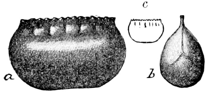
Fig. 485.—Egg-capsule of Periplaneta americana: a, side; b, end view; c, natural size.—After Howard and Marlatt, Bull. 4, Div. Ent. U. S. Dept. Agr.
The oötheca of the cockroach (Fig. 485) is a solid, dense case, which, after being carried about by the mother, can be left without harm in the crevices of the floors of houses. The oötheca of Mantis (Fig. 486) is formed by a large mass of frothy matter, which hardens and is attached to stems of plants.
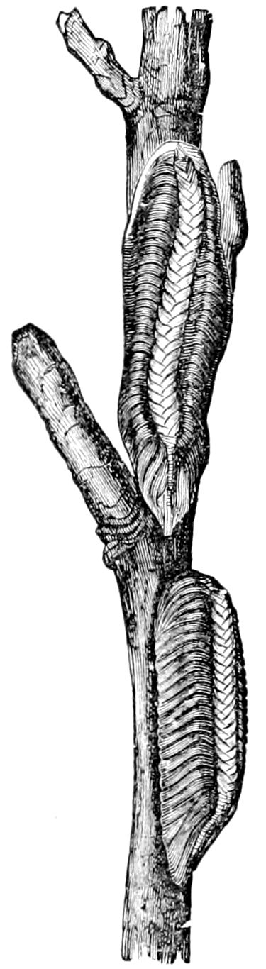
Fig. 486.—Egg-capsules of Mantis carolina.—After Riley.
On the other hand, the female “walking-stick” (Diapheromera femoratum) drops her eggs, says Riley, loosely upon the ground, from whatever height she may happen to be, and “one hears a constant pattering, not unlike drops of rain, that results from the abundant dropping of these eggs, which, in places, lay so thick among and under the dead leaves that they may be scraped up in great quantities.” (Report for 1879.)
The eggs of the lace-winged flies are supported on pedicels, above the reach of ovivorous mites.
The female Chrysopa usually lays between 40 and 50 eggs. In one case, we observed that 18 egg-stalks were deposited, but there were only nine well-formed eggs in the batch, and nine eggless stalks, some only half the usual height, others with the knob of cement at the end to which the egg is ordinarily fastened. The eggs are evidently stuck on to the end of the pedicel after the latter has been formed, as, in one instance, an egg was glued to the stalk very much out of centre, the insect’s abdomen not having been aimed straight, so to speak, at the mass of cement.
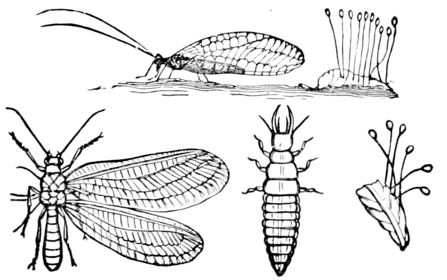
Fig. 487.—Eggs of Chrysopa, with larva and fly.
The eggs of Rhodites are fixed to a long stalk thickened at the end; those of Inquilines and certain Chalcids (Leucospis gigas, Fig. 489, A) are also stalked; and the use of this stalk in the eggs of Cynips (E) is thought by Adler to be respiratory, while, also, he states that the egg-cavity communicates with the egg-stalk, so that a part of the egg-contents can pass into the latter, and this happens at the laying of each egg. The egg of certain ichneumons (Paniscus, Fig. 488) ends in a short stalk, which is inserted 518in the skin of the caterpillar destined to serve as the host of the parasite, the eggs, as stated by De Geer, being retained more firmly in the integument by the stalk so swelling as to form two knobs (Fig. 498, c).
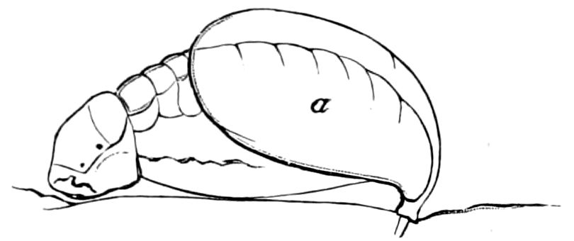
Fig. 488.—Young larva of Paniscus in position of feeding on the skin of a caterpillar: a, the egg-shell.—After Newport, from Sharp.
Certain Homoptera also have stalked eggs, as those of Psylla pyricola (Fig. 489, B), those of Aleyrodes citri (C, a, b), and of an allied form, Aleurodicus cocois (D), and those of Corixa (Fig. 493).
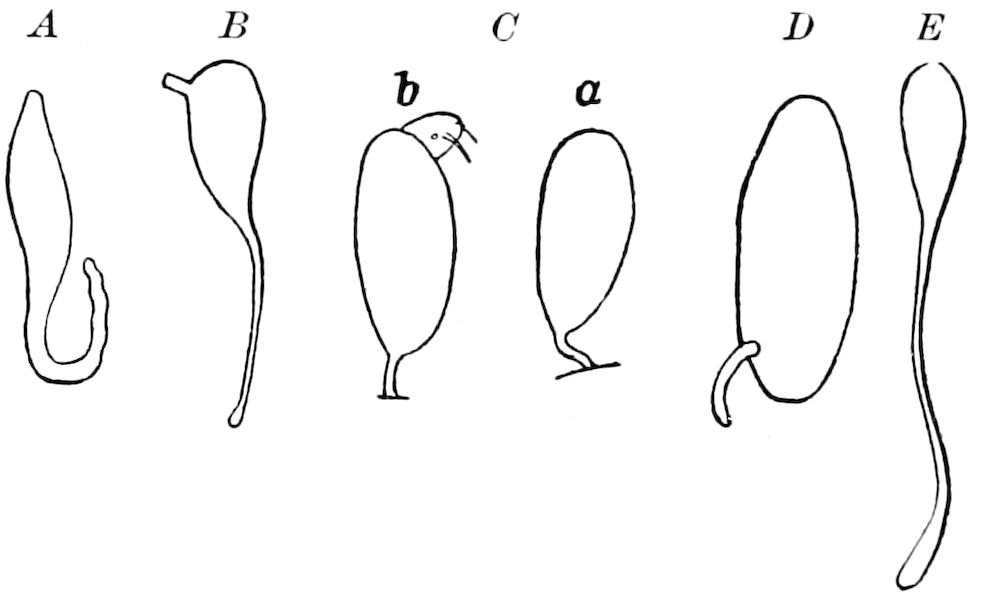
Fig. 489.—Stalked eggs: A, of a Chalcid (after Fabre); B, of Psylla (after Slingerland); C, of Aleyrodes; D, of Aleurodicus (after Riley and Howard); E, of Dryophanta scutellaris (after Adler).
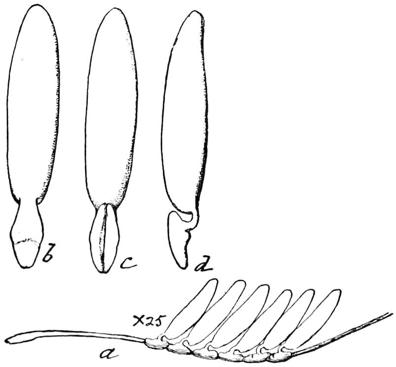
Fig. 490.—Eggs of ox bot-fly, enlarged.—After Riley.
Reference should also be made to the eggs of lice, which are oval and attached to the hairs of their host. Those of the ox bot-fly (Hypoderma lineata) are usually placed four to six together, and fastened to a hair. The lower portion of the egg is admirably adapted for clasping a hair. “It consists of two lobes, forming a bulbous enlargement, which is attached to the egg by a broad, but rather thin, neck, so that, when the latter is viewed sidewise, it appears as a slender pedicel” (Fig. 490, a-d). (Riley in Insect Life, iv, p. 307.) The egg of another fly (Drosophila ampelophila, Fig. 491) bears a pair of long, slender appendages near the anterior end. “The egg is inserted into the soft pulp of the decaying fruit; these appendages leave the ovipositor last, and are spread out upon the surface of the mass. They, in this way, serve to keep the egg in place, and thus insure the emergence of the larva into the open air instead of into the more or less fluid mass in which the egg is situated. The larva issues from the egg just above the base of these appendages.” (Comstock.)
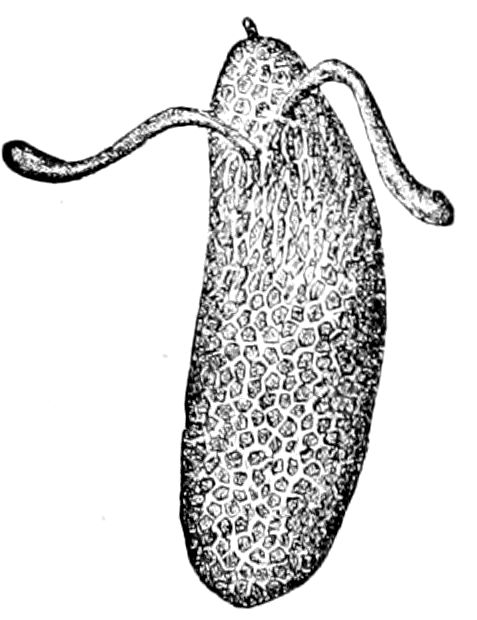
Fig. 491.—Egg of Drosophila.—After Comstock.
Mode of deposition.—The exact process of oviposition has been rarely observed, or at least not observed in detail, 519and further observations are much needed. In the cockroach (Phyllodromia), Wheeler has seen the eggs pass out of the oviduct and become arranged in the oötheca, in a way similar to that in the account published by Kadyi on Periplaneta.
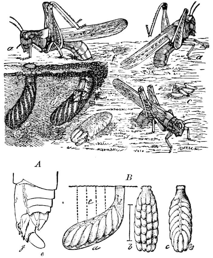
Fig. 492.—Rocky Mountain locust (aa) depositing its eggs (c); d, the earth partially removed, showing (e) an egg-mass already in place, and (d) one being placed; f shows where such a mass has been covered over. A, oviposition; j, position of oviduct; g, egg-guide; e, egg. B, egg-mass of the same; a, from side, b, from beneath, c, from above.—After Riley.
“When about to form the capsule, the female Blatta closes the genital armature, and the two folds of the white membrane which lines the oöthecal cavity close vertically in the middle line. Then some of the contents of the colleterial glands are poured into the chamber, and bathe the inner surface of the posterior wall. The first egg glides down the vagina from the left ovary, describes an arc, still keeping its germarium-pole uppermost, after having pressed the micropylar area against the mouth of the spermatheca, passes to the right side of the back of the chamber, and is placed perpendicularly two-thirds to the right of the longitudinal axis of the insect’s body. The next egg comes from the right ovary, describes an arc to the opposite side of the body, decussating with the path of the first egg, and is placed completely on the left side of the median line. The third egg comes from the left ovary, and is made to lie completely on the right side of the median line; and so the process continues, the ovaries discharging the eggs alternately, and each egg describing an arc to the opposite side of the capsule. The oöthecal chamber soon becomes too small to contain all the constantly accumulating eggs, so the anal armature opens and allows the end of the capsule to project. A raised line, the impression of the edges of the white membrane, runs down the end of the capsule. The last egg deposited comes from the right ovary, and lies two-thirds on the left, and one-third to the right, of the median line. As soon as the egg is laid, a further discharge from the colleterial glands spreads over the vaginal or anterior wall of the cavity, and becomes evenly continuous with the secretion which has before been spread over the back and the sides of the capsule by the white membrane.
“The crista, a cord-like ridge running the full length of the dorsal surface of the capsule, is a thick-walled tube, either half of which is formed by the edge of the side walls of the capsule split into two laminæ. The rhythmical clasping 520of the three pairs of palpi which guard the vaginal opening is registered in an exquisite pattern on the inner face of either half of the crista.”[78]
The mode of oviposition in the locust has been fully described by Riley, who states that the eggs pass down and out of the oviduct, and “guided by a little finger-like style” (Fig. 298), they pass in between the horny valves of the ovipositor, and issue at their tips amid the mucous fluid which forms the egg-capsule (Fig. 492).
Vitality of eggs.—It is well known that the eggs of phyllopod and other fresh-water Crustacea have wonderful vitality, withstanding extreme dryness for several years, at least from two to ten. Such cases are unknown among insects. It has been observed, however, by T. W. Brigham, and also by L. Trouvelot, that the eggs of the walking-stick (Diapheromera femorata) for the most part hatch only after the interval of two years.[79]
The eggs of Bittacus are said by Brauer to lie over unhatched for two years; indeed, the first condition of their hatching is a complete drying of the earth in which the eggs lie, the second is a succeeding thorough wetting of the ground in spring.
Appearance and structure of the ripe egg.—The eggs of insects are on the whole rather large in proportion to the size of the parent, especially so in many minute forms, as the fleas, lice, etc.
Their general shape is spherical or oval, often cylindrical; where the eggs are long and cylindrical a dorsal and ventral side can be distinguished (Fig. 502). They are in the Tortricidæ and Limacodid moths flattened, thin, and scale-like. In the eggs of locusts and grasshoppers, as well as certain Diptera, the ventral side of the embryo corresponds to the convex side, and the concave side of the egg to the dorsal region of the embryo (Figs. 502 and 493).
There is an anterior and posterior end or pole, the anterior end being that which in the body of the parent lies towards her head, or towards the upper or distal end of the ovarian tube. Towards this end lies in the later stages of embryonic life the head-end of the embryo, while the posterior end of the embryo is turned towards the hinder pole of the egg (Figs. 493 and 520).
The egg-shell and yolk-membrane.—The ripe egg is protected by two membranes: 1, an inner or vitelline membrane or oölemma (dh) (Fig. 500, d), produced in the egg by a hardening of the outer layer, and 2, the outer or chorion (c), which is secreted by the cells of the ovarian follicle. The latter is divided into two layers: an inner, the endochorion, and an outer, the exochorion.
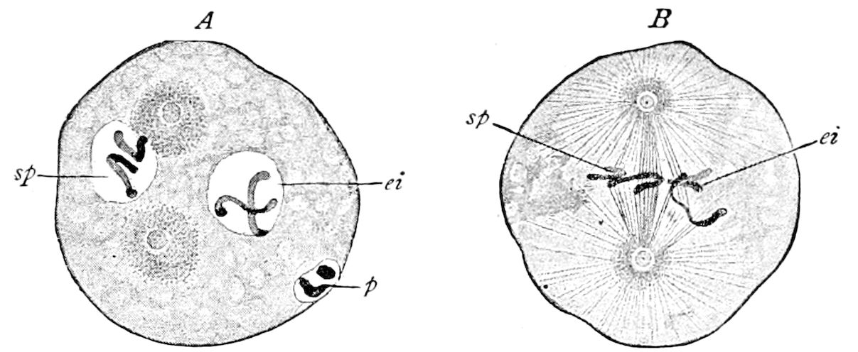
Fig. 503.—Fertilization of the egg of a round-worm (Ascaris megalocephala): A, the ends (centrosomes) of the spindle formed. B, the spindle completed; sp, sperm-nucleus, with its chromosomes; ei, egg-nucleus; p, polar bodies.—After Boveri, from Field’s Hertwig.
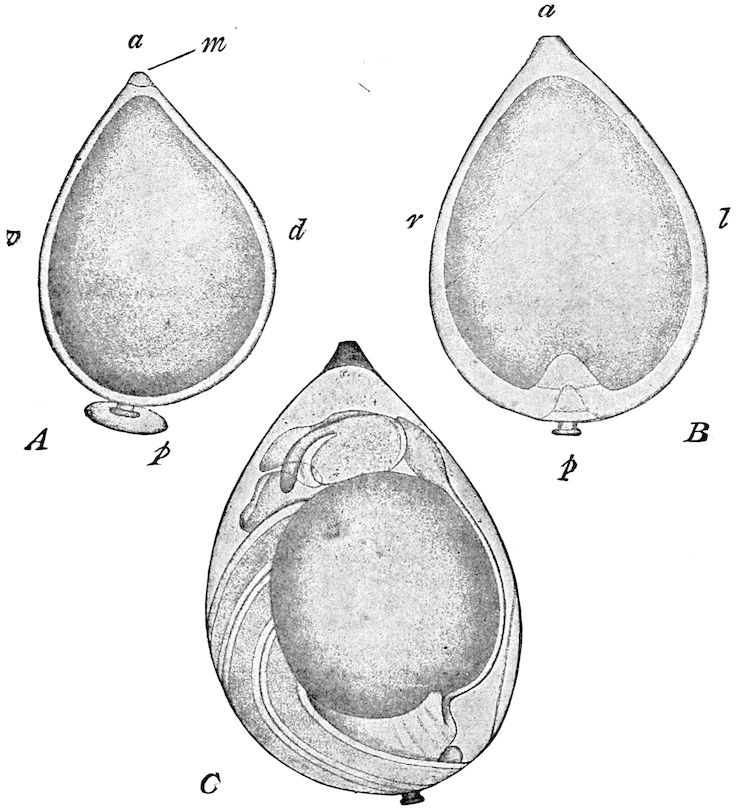
Fig. 493.—Eggs of Corixa: A, early stage before formation of the embryo, from one side. B, the same viewed in the plane of symmetry. C, the embryo in its final position; a, anterior, p, posterior, end; l, left, r, right, v, ventral, d, dorsal, aspect. (The letters refer to the final position of the embryo, which is nearly diametrically opposite to that in which it first develops); m, micropyle; p, pedicle.—After Metschnikoff, from Wilson.
521
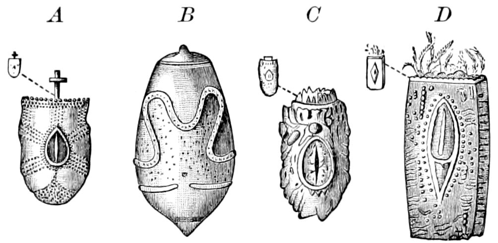
Fig. 494.—Eggs of Phasmidæ: A, Lonchodes duivenbodi. B, Platycrania edulis. C, Haplopus grayi. D, Phyllium siccifolium.—After Kaup, from Sharp.
While the yolk-membrane is usually a completely homogeneous, thin, structureless membrane, the chorion or shell of the egg is usually covered with a network of ridges enclosing polygonal areas, varying in shape according to the species or genus. These external markings are due to the impress of the cellular structure of the epithelium of the ovarian follicle.
In the chorion of the cockroach the surface appears to be finely granular, the finest granules being arranged in large, more or less regularly hexagonal areas, which are bounded by narrow, dark spaces, containing somewhat larger though less dense granules. The surface of the eggs of certain Phasmids are variously sculptured (Fig. 494).
The true structure of the chorion can only be, as Wheeler observes, seen in cross-sections, as shown by Blochmann, and also by Wheeler. The chorion consists of two chitinous laminæ kept in close apposition by means of numerous minute trabeculæ or pillars. It is the ends of these pillars that look like granules. In the spaces between the hexagonal areas, the trabeculæ are more scattered and individually thicker than those of the hexagons.
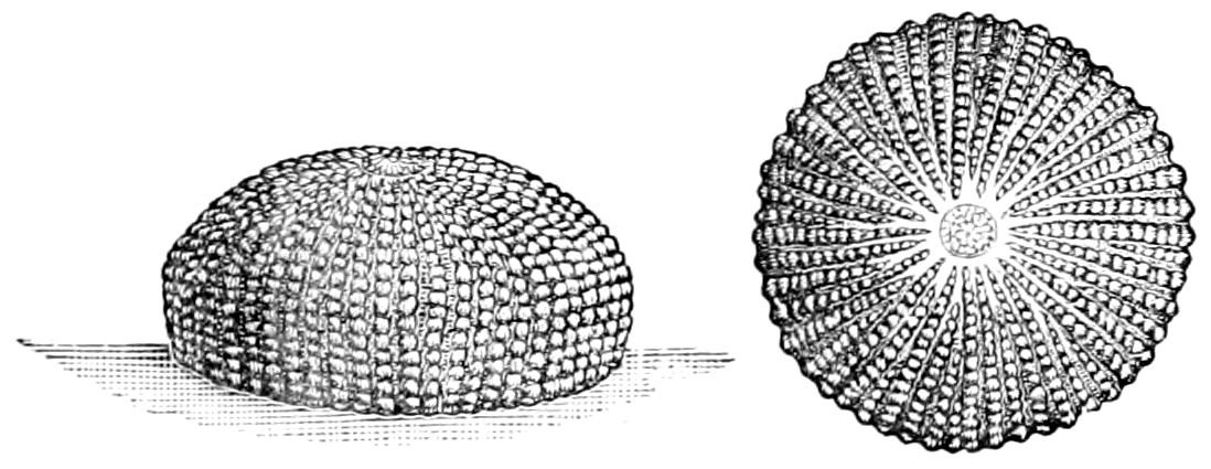
Fig. 495.—Egg of cotton-worm moth, Aletia: a, top view, showing the micropyle.—After Comstock.
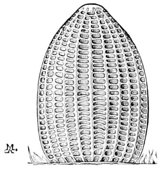
Fig. 496.—Egg of Danais archippus.—After Riley.
These markings are of singular beauty and complexity in the eggs of many Lepidoptera, whose ova are variously ribbed, forming a beautiful fretwork of raised lines (Figs. 495 and 496), while in the Diptera and Hymenoptera the chorion is less solid, and usually smooth under low powers. The exochorion of the egg of the house and meat fly (C. vomitoria) is pitted with elongated hexagonal depressions, which cross the egg transversely. That of the honey-bee is also divided into long hexagonal areas (Fig. 497).
522
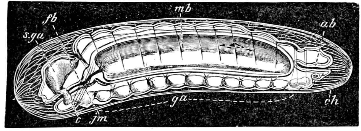
Fig. 497.—Egg with embryo of honey-bee, × 40: ch, chorion; ga, ganglia; s. ga, brain; jm, jaw-muscles forming; c, œsophageal collar; fb, fore intestine; mb, mid-intestine; ab, hind-intestine.—After Cheshire.
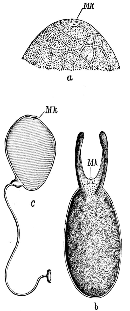
Fig. 498.—Micropyle (Mk) of eggs; a, of a fly, Antomyia; b, Drosophila cellaris; c, stalked egg of Paniscus testaceus.—After Leuckart, from Perrier.
When the eggs are deposited in exposed places, and remain in such situations for several days, or weeks, or even through the winter, the shell is either solid and strengthened by the ribs and ridges; or the shell, if of winter eggs, is unornamented, and is dense and solid, to resist extremes in temperature or the attacks of egg-eating birds, mites, etc.
The micropyle.—This is an opening or canal, or, as in most insects, a group of canals situated at the anterior end of the egg for the entrance of the spermatozoa during the process of fertilization of the ovum (Fig. 498). In Acrydians, however, the micropyle is situated at the posterior end of the egg. The micropyle (Fig. 499) is a complicated apparatus within whose circumference the vitelline membrane appears to be firmly attached to the chorion, so that the perforation passes through the chorion as well as the yolk-membrane.
The micropyles of the cockroach are probably as simple and generalized as in any insect. Wheeler states that they are in Phyllodromia scattered over the end of the egg, “over a quadrant of the upper hemisphere, where the beautiful hexagonal pattern of the chorion gives away to an even trabeculation.” The micropyles are wide-mouthed, very oblique, funnel-shaped canals, perforating the chorion, the apertures of the funnels appearing under a low power as 523clear, oval spots, the long axis of which is parallel to the long axis of the egg.
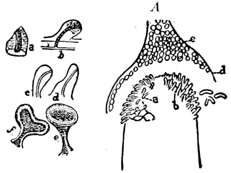
Fig. 499.—a, fragment of a micropylar papilla, showing its lumen; b, optical section of another papilla, in this one the lumen extends to the vitelline membrane, but does not pass beyond it; c, d, e, and f, papillæ of different forms. A, anterior end of an ovarian egg, showing mode of growth of the micropylar papillæ: a, b, two successive stages; c, surface view of modified papillæ from the lower edges of the cap; d, tunica propria of the ovariole; e, remnant of the cell-mass that secreted (?) the micropylar cap.—After Ayers.
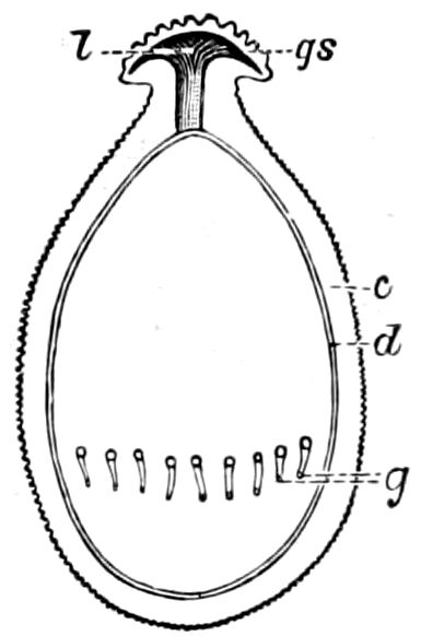
Fig. 500.—Egg of Perla maxima: c, chorion; d, oölemma; gs, glass-like covering of micropyle; l, cavity under same; g, canals penetrating chorion.—After Imhof, from Sharp.
“With a higher power the tube of each funnel is clearly visible as a thin canal which dilates rapidly into the large oval aperture on the outer face of the chorion. The narrow tube is sometimes fully as long as the large orifice. The micropylar perforations are all directed from the germarium to the vaginal pole of the egg. Hence a line, the hypothetical path of the spermatozoön, drawn through one of these oblique micropyles, and continued into the egg, would strike the equatorial plane. The female pronucleus, as we shall see further on, moves in this plane.” (Wheeler, p. 289.)
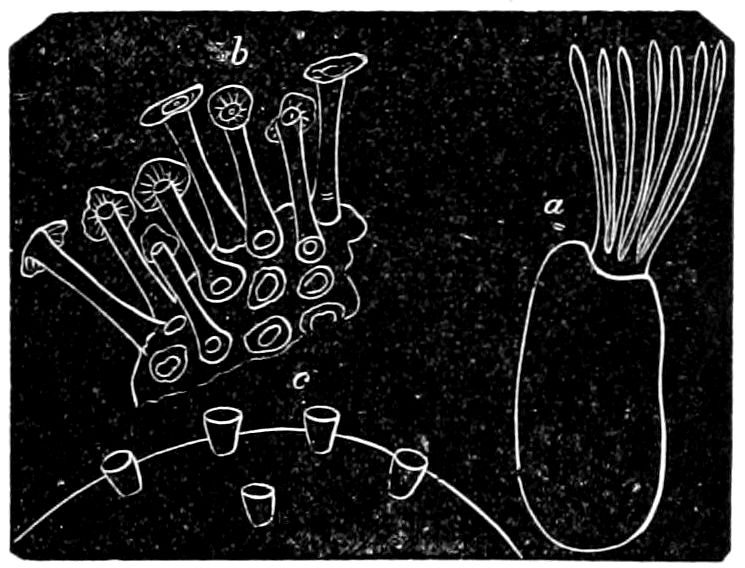
Fig. 501.—Micropyles: a, of Nepa cinerea; b, of Locusta viridissima; c, of a bug (Pyrrhocoris apterus).—From Gerstäcker.
The micropylar region is generally, at least in Orthoptera and Odonata, covered by a gelatinous cap (Figs. 499 and 500, gs), which may form a covering membrane which extends over a large part of the egg, or may envelop the entire outer surface. In some cases micropyles are scattered over the entire surface of the egg, but usually the perforation is situated at the end, and is often guarded by raised processes, either one or several, like bristles, or toadstools, etc., these being especially characteristic of the eggs of certain Hemiptera (Nepa, Fig. 501, a, and Ranatra), or the region is variously sculptured, as in the eggs of butterflies. In the micropylar apparatus of Œcanthus the papillæ have a distinct lumen (Fig. 499), or a channel for the ingress of the male filament.
524
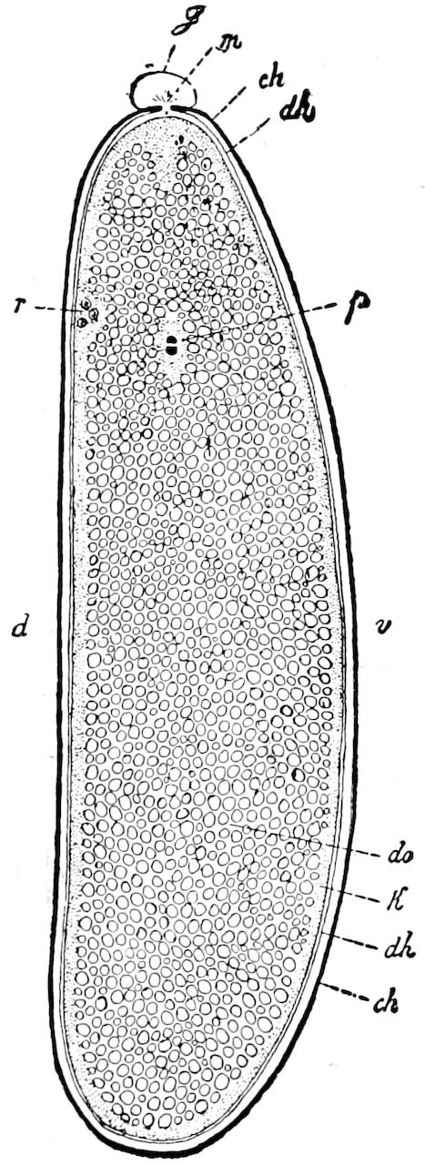
Fig. 502.—Diagrammatic median section through egg of Musca in stage of fertilization (incorporating the figures of Henking and Blochmann): ch, chorion; d, dorsal; v, ventral side of the egg; dh, yolk-membrane; do, nutritive yolk; g, gelatinous cap over the micropyle (m); K, outer layer of plasma (Keimhautblastem); p, male and female pronucleus before copulation; r, directive body (Richtungskörper).—After Korschelt and Heider.
Another use of the micropylar apparatus noticed by Ayers in the egg of the tree-cricket is that it “serves as a thick, roughened plate, against which the insect may push when ovipositing, without injury to the egg, and without danger that the ovipositor slips from its place.” In Chrysopa eggs the micropyle forms a conspicuous button-like knob, resembling the finely milled head of a certain kind of screw.
Internal structure of the egg.—The egg-contents are surrounded by an outer layer of protoplasm or formative yolk, which is separate from the inner parts of the egg (Fig. 502, do), the latter being mostly composed of the nutritive yolk-element. The superficial protoplasmic layer, called by Weismann Keimhautblastem (K) is, in a few cases, afterwards entirely lost, but in most instances forms a very thin layer of clear protoplasm, slight in extent compared with the yolk-mass within.
The eggs of insects are rich in yolk, only certain eggs, such as those of the Aphides and the egg parasites (Proctotrypidæ) being poor in yolk. The eggs of heterometabolous insects have been said by Brauer to contain relatively more yolk than those of the Metabola, particularly the Diptera; though, as Wheeler observes, this rule has some exceptions, the eggs of the 17–year Cicada being very numerous and small.
This he thinks is a greater advantage to the insect than the production of a few large eggs, “when we consider the extremely long period of larval life and the vicissitudes to which the larvæ may be subjected during all this time.” “Similarly, Meloë angusticollis produces a large number of very small eggs, while the eggs of the smaller beetles (Doryphora, e.g.) are much larger. But 525Meloë is a parasitic form, and probably only a few of its many offspring ever succeed in gaining access to the egg of the bee.”
In the eggs of Chrysopa the yolk-granules are remarkably small, so that the primitive band is in strong contrast to the yolk in color and density. When crushed, the yolk does not flow out as a liquid, but in a pasty mass, and we have questioned whether, as in the eggs of Limulus, whose yolk is solid with fine granules, the denseness of the yolk is not connected in the way of cause and effect with their exposed situation.
The central or yolk-mass (Fig. 502, do) consists chiefly of rounded masses of yolk, with fat-globules, between which extends a fine network of protoplasm.
The elements of the yolk are spherical and strongly refractive, by pressure becoming polygonal structureless homogeneous bodies.
The germinal vesicle of the ripe insect-egg lies in the centre of the yolk, where it appears as a large vesicle-like cell-nucleus containing a few chromatin elements.
b. Maturation or ripening of the egg
Before the eggs of animals can be fertilized, they require in some observed cases, and probably in animals in general, to undergo a series of changes, which, as observed in the starfish, etc., consists in the replacement of the germinal vesicle by a very much smaller egg-nucleus, and also at the same time the construction at one pole of the egg of the directive or polar bodies (Fig. 502, r). Towards the end of the ripening process of the insect egg this vesicle, according to Blochmann, passes to the dorsal surface of the egg, and is transformed into the directive spindles (Richtungspindel).
c. Fertilization of the egg
The egg next requires the penetration and admission into the yolk-interior of a spermatozoön.
This process is essentially in insects, as in other animals, the fusion of the sperm-nucleus with the nucleus of the egg. Under normal conditions but a single spermatozoön is required for fertilization. As shown by Hertwig, in the sea-urchin, after the spermatozoön has penetrated into the egg, the head, and the small rounded body, called a centrosome, can still be recognized, but the tail becomes fused with the yolk of the egg. In the protoplasm of the egg (called cytoplasm) the achromatic end of the sperm-nucleus gives rise to 526conspicuous rays, like those observed in ordinary cell-division. Preceded by these rays, the sperm-nucleus or male pronucleus (Fig. 502, p) moves towards the nucleus of the egg, and finally fuses with it, thus forming a new single nucleus. This latter, which is called “the cleavage nucleus,” rapidly forms a nuclear or “cleavage spindle” (Fig. 503). This act gives an impulse to the cleavage of the egg, which is the first step in the formation of the embryo. All these changes have yet to be worked out in detail in insects by microscopic sections of the egg, whose generally hard and opaque egg-shells present great obstacles to such work.
d. Division and formation of the blastoderm[80]
In insects as in most other Arthropoda the segmentation of the yolk is superficial and not total. The ovum is centrolicithal, i.e. the yolk is concentrated at the centre of the egg, and surrounded by a peripheral layer of transparent protoplasm (the Keimhautblastem).
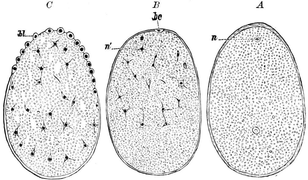
Fig. 504.—Formation of the blastoderm of Pieris cratægi: A, longitudinal section through the egg, with two masses of protoplasm in the yolk. B, a blastoderm-cell at the upper end. C, a later stage, with more blastoderm-cells.—After Bobretsky.
The first step in segmentation is the movement of the first division-nucleus (i.e. that in the fertilized egg arising from the union of the sperm-nucleus with the female pronucleus) towards the interior of the egg in order to multiply itself by the mode of indirect nuclear division (Figs. 504, A, and 507).
527
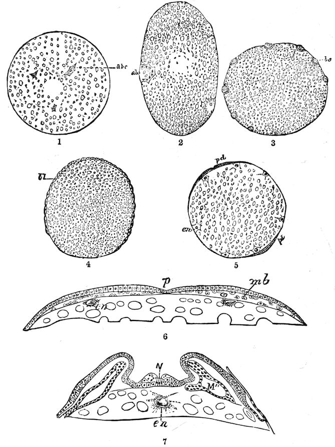
Fig. 505.—Embryology of the mole-cricket: 1, egg in which the amœboid nuclei (abc) are moving toward the surface; 2, egg in which the nuclei (abc) have reached the surface, and show an active nucleus-formation; 3, the blastoderm-cells have no nucleus, and are placed at equal distances apart; 4, the blastoderm-cells now forming a continuous layer; 5, cross-section of the egg with blastodermic disk, also showing the disposition of the endodermal cells; 6, cross-section of the blastodermic disk, with the myoblast cells (mb) already formed; 7, cross-section through the thorax of the embryo, the body-cavity extended into the limbs.
Lettering.
abc,
amœboid blastodermic cells.
bc,
blastoderm-cells.
bl,
blastoderm.
en,
endodermal cells.
M″,
cavity of the myoblast.
mb,
myoblast cells.
N,
nerve-furrow.
P,
primitive groove.
pd,
primitive disk.
529
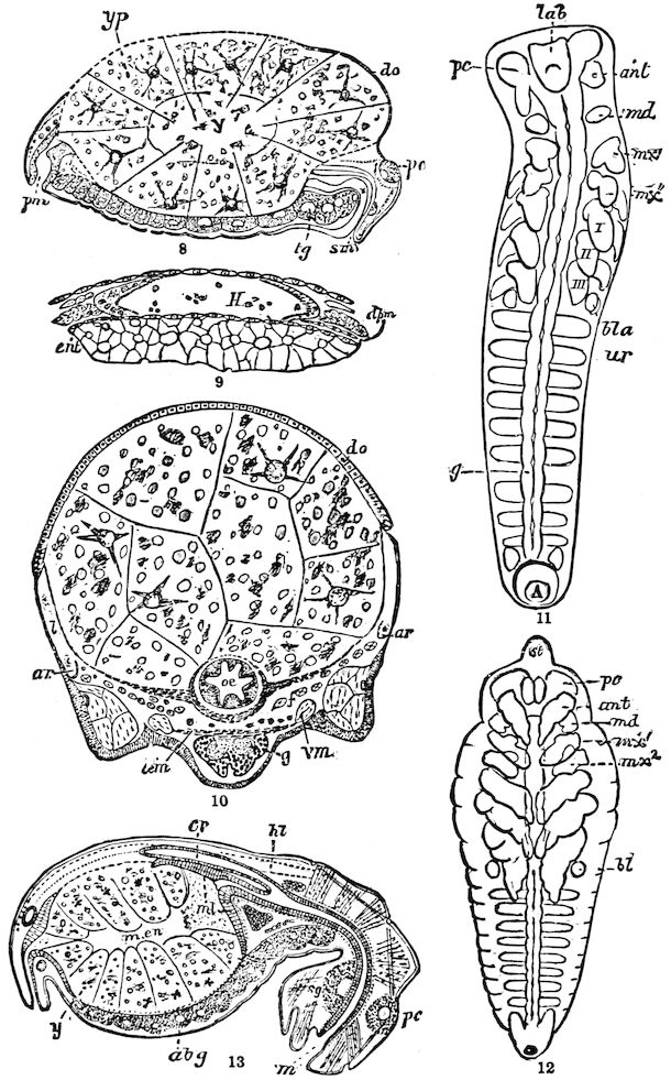
Fig. 505 concluded.—Later stages in the embryology of the mole-cricket: 8, longitudinal section of the embryo; the yolk-pyramids (yp) form a common inner yolk-mass (y). 9, section through the heart; H, cavity of the heart; the two halves of the heart-sinuses having united dorsally, ventrally they are still open and are bounded by the walls of the mesenteron. 10, cross-section of an embryo, showing the blood-lacunæ separated on the back by the dorsal organ (do); the intestinal fasciated layer (Darmfaserblatt) has not completely enclosed the yolk. 11, embryo completely segmented, with the rudiments of the appendages, labrum (lab), and nervous ganglia (pc-ng). 12, a more advanced embryo, showing the stomodæum (st) indicated as a frontal protuberance. 13, section through the recently hatched larva, showing the cells of the mesenteron or chyle-stomach, and the cellular layer on the front surface, also the proventriculus or crop.
Lettering.
ant,
antenna.
ar,
arterial sinus.
bl,
blastoderm.
bla,
abdominal vesicles.
cr,
proventriculus, or crop.
dm,
ventral diaphragm.
do,
dorsal organ.
d pm,
dorsal diaphragm.
ent,
enteric layer.
f,
fat-body.
g,
ventral ganglion.
H, ht,
heart.
l,
lacuna.
m,
mouth.
md,
mandible.
m.en,
mesenteron.
mx′,
1st maxilla.
mx″,
labium, or 2d maxilla.
ml,
leaf-like portion of mesenteron.
oe,
œsophagus.
pc,
procerebrum.
pm,
proctodæum.
sg,
subœsophageal ganglion.
sm,
stomodæum.
tg,
thoracic ganglion.
vm,
ventral muscle.
y,
yolk.
yp,
yolk-pyramids.
I,
1st pair of feet.
II,
2d pair of feet.
III,
3d pair of feet.
—After Korotneff.
The origin of numerous division-nuclei as the offspring of the first has been observed to take place in the eggs of those insects (Aphides, Cecidomyia, and Cynips) which have a slight amount of yolk. Yet in the large, ordinary eggs of insects with an abundance of yolk there is no doubt, say Korschelt and Heider, that these numerous division-nuclei, which soon after the process of oviposition are scattered within the egg between the yolk-spheres, and are enveloped by a star-shaped protoplasmic layer, and which constitute the formative elements of the blastoderm,—there is no doubt but that they have practically arisen through indirect nuclear division from the first division-nucleus.
The process of formation of the blastoderm in ordinary eggs with abundant yolk was first observed by Bobretsky in the eggs of a moth (Porthesia) and Pieris, also by Graber, and more recently by Blochmann in Musca, and by Heider in Hydrophilus.
In the earliest stage observed by Bobretsky there first appear after fertilization a few (the smallest number four) cell-like, minute amœboid masses of protoplasm, each with a distinct nucleus. A few (one at least) of these bodies gradually pass out of the centre of the yolk to the surface of the egg (Fig. 504, A, n), these becoming larger and rounder, and from one or two of these nuclei (B, bc) the blastoderm originates (C, bl). Those nuclei remaining in the yolk increase in number and afterwards become the nuclei of rounded masses of yolk-granules, forming the so-called yolk-spheres which Bobretsky regards as true cells.
To the few blastoderm cells situated on the upper end of the egg are added others which continue to pass from the yolk to the periphery, and then the blastoderm spreads out farther and farther from the upper end of the egg until finally it covers or envelops the whole yolk. This layer of cells is called the blastoderm.
As to the origin of the primitive amœboid cells, Bobretsky is in doubt, but is disposed to think that they are the result of the subdivision of the germinative vesicle or nucleus of the ovarian egg-cell. In this connection may be quoted the observations of Graber, who states that an examination of the ovarian cell at an early period has revealed the presence, in the centre of the yolk, of a number of amœboid cells, which appear to have been formed by the division of the germinal vesicle. These “primary embryonic cells” have a relatively large nucleus and a number of nucleoli. Several may be seen to unite with one another by means of their pseudopodia, and they may also be observed to undergo division. With this account may be compared the results obtained by Korotneff in his work on the embryology of the mole-cricket (Fig. 505).
530
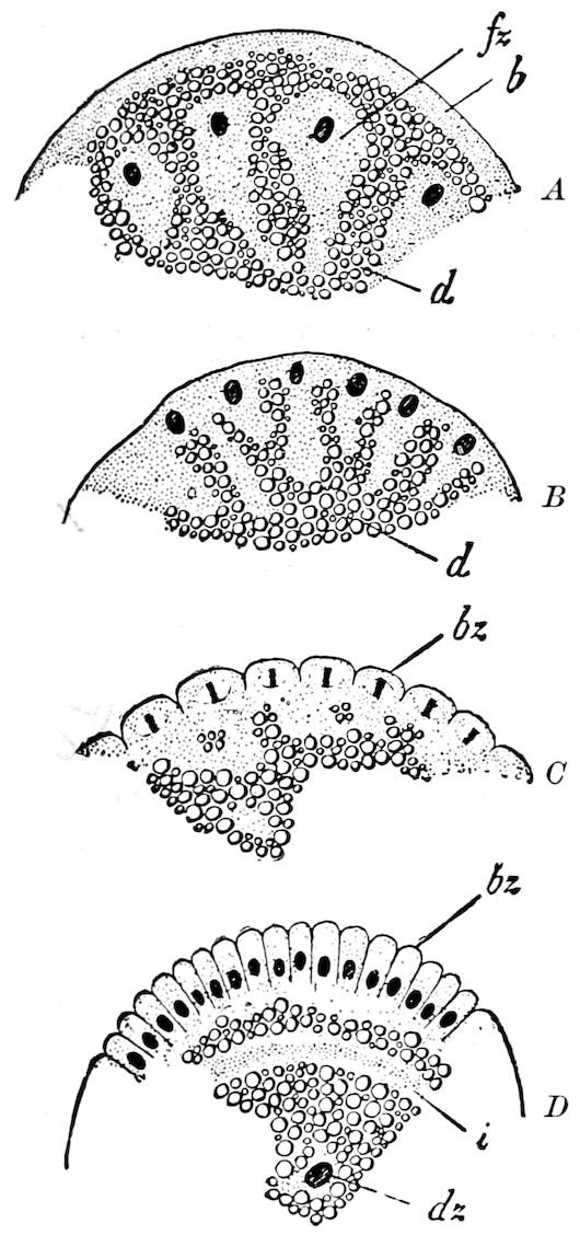
Fig. 506.—Four successive stages in the formation of the blastoderm of Calliphora vomitoria (the figures represent segments of cross-sections through the fly’s egg): A, the nuclei of the division-cells have arranged themselves parallel with the outer surface of the egg. B, the division-cells fused with the “keimhautblastem.” C, the outer surface becomes furrowed by indentations; all the nuclei of the blastoderm-cells in process of division. D, the blastoderm-cells form a high cylinder-epithelium: b, “keimhautblastem”; bz, blastoderm-cells; d, nutritive yolk; dz, yolk-cell; fz, so-called division-cell; i, inner “keimhautblastem.”—After Blochmann, from Korschelt and Heider.
The result of these and of later observations, especially those of Blochmann on Musca, and those of Heider on Hydrophilus, show that the division-nuclei lie near the centre of the egg, along the longitudinal axis (Fig. 507, A). Each of these nuclei is enveloped by a star-shaped mass of protoplasm, and on the whole resembles a wandering amœboid cell. These isolated masses of protoplasm are all connected by a fine network of rays, which unite to form within the yolk a syncytium. Afterwards, in the later stages, these division-cells, as they may be, though somewhat incorrectly, regarded, move nearer the periphery and arrange themselves into a plane parallel with the surface (Figs. 506, A, 507, B). Continuing to divide, they reach the surface and fuse with the peripheral protoplasmic layer (Figs. 506, B, 507, C). Then follows the division into single cell-territories (Figs. 506, B, 507, C), corresponding to the division-nuclei, through the appearance of furrows which pass in from the outer surfaces of the egg into the interior and gradually penetrate the entire “keimhautblastem.” In this way the surface of the egg is covered with an epithelium (blastoderm). In many insects the so-called inner “keimhautblastem” (Fig. 506, D, i) is formed by the separation of a layer of protoplasm which contains larger granules and are accumulated between the blastoderm and the upper surface of the central nutritive yolk-mass. By the addition of this plasmic layer the cells of the blastoderm increase 531in height, and now form a cubical or cylinder epithelium, which continuously envelops the surface of the egg. (Korschelt and Heider.)
e. Formation of the first rudiments of the embryo, and of the embryonic membranes
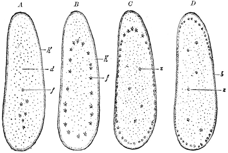
Fig. 507.—Formation of the blastoderm in Hydrophilus: b, completed blastoderm; d, yolk; f, so-called division-cells; k, “keimhautblastem”; z, yolk-cells.—After Heider, from Korschelt and Heider.
The embryo first arises as a whitish streak or band-like thickening on the ventral side of the egg, and is variously called the “primitive streak,” “primitive band,” “germinal band,” or “embryonal streak.” In most cases the primitive band is divided at regular intervals by transverse furrows, indicating the limits of what are to be the body segments.
Cross-sections (Fig. 509) show that the band is composed of several layers, i.e. an outer layer (ectoderm) and an inner layer which comprises the endoderm and mesoderm, and so long as these two layers are not sharply differentiated from one another, this second layer may be called, with Kowalevsky, “the inner lower layer, or ento-mesoderm” (Figs. 508, 509, B, C, u).
It is characteristic of insects, only rarely occurring in other arthropods (e.g. the scorpion), that the primitive streak is not situated on the surface of the egg, but becomes overgrown by a folded structure (Fig. 508, af) rising from its edges, the amnion-fold, so 532that it appears somewhat depressed or sunken in under the upper surface of the yolk. While the amnion-folds are extending from all sides over the primitive band, there becomes formed under it, by the invagination of the outer surface of the egg, a cavity, the amnion-cavity (ah), which, when the amnion-fold has completely overgrown the primitive band and united together (Fig. 509, C), appears completely closed from without.
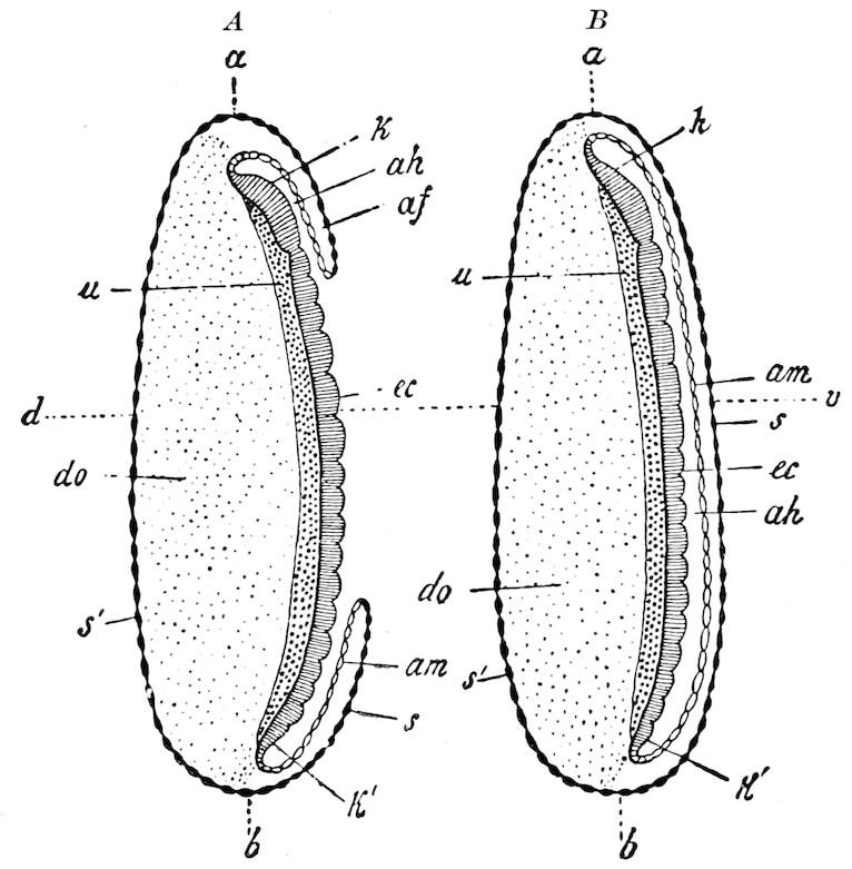
Fig. 508.—Two schematic median sections through an insect-embryo to represent the development of the embryonal membranes. In A the primitive streak is not wholly overgrown by the amnion-fold. In B the amnion-folds have united with each other and completely overgrown the primitive streak: a, fore, b, hind, egg-pole; v, ventral side; d, dorsal side; af, amnion-folds; ah, amnion-cavity; am, amnion; do, yolk; ec, ectoderm; k, head-end, k′, hinder-end, of the primitive streak; s, the part of the serosa arising from the amnion-fold; s′, the part of the serosa arising from the unaltered blastoderm; u, lower layer.—After Korschelt and Heider.
Formation of the embryonic membranes.—The amnion-folds finally completely overgrow the primitive band (Fig. 509, B and C), and form the embryonal membranes. The primitive band is seen after its completion to be overgrown by a double cellular epithelial membrane. The outer of these two membranes, that which arises from the outer leaf or layer of the amnion-fold, is the serosa (Figs. 508, B; 509, C, s; 510). This passes continuously into the unchanged part of the blastoderm, which has no part in the formation of the 533primitive band and germ-layers, and which covers the outer surface of the yolk. Thus the serosa, which is usually held to include this portion also of the blastoderm, forms a closed sac which covers the whole surface of the egg, with one part extending over the surface of the yolk, and the other over the primitive band (Fig. 510).
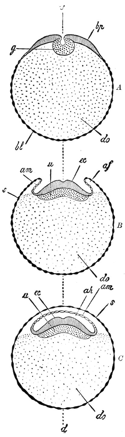
Fig. 509.—Diagrammatic cross-section through three successive stages of the primitive streak, and growing embryonal membranes of insect-embryos. A, formation of the ventral plate and of the gastrula invagination (g). B, upward growth of the amnion-folds (af). C, complete overgrowth of the primitive band through the amnion-folds: v, ventral side; d, dorsal side; af, amnion-folds; ah, amnion-cavity; am, amnion; bl, blastoderm; bp, ventral plate; do, yolk; ec, ectoderm; s, serosa; u, under or inner layer.—After Korschelt and Heider.
The inner of the two layers, called the amnion (Fig. 509, am), is more closely connected with the embryo. The amnion and ectoderm of the primitive band together form a completely closed sac, whose lumen forms the amniotic cavity. Originally connected with the serous membrane, it splits off from the primitive band about the time the appendages begin to bud out, and continues to closely envelop the body and appendages, as seen in Fig. 509. Both of these membranes are, before the time of hatching, either absorbed, or, as in Lepidoptera, retained. The amnion is retained until after hatching in the locust, etc. In certain Coleoptera the serosa is retained, and the amnion is absorbed (Fig. 532), while in Chironomus and the Trichoptera the serosa is absorbed, and the amnion retained, with the egg-shell or chorion. 534Hence we have eight layers in the winged insects[81] during embryonic life:
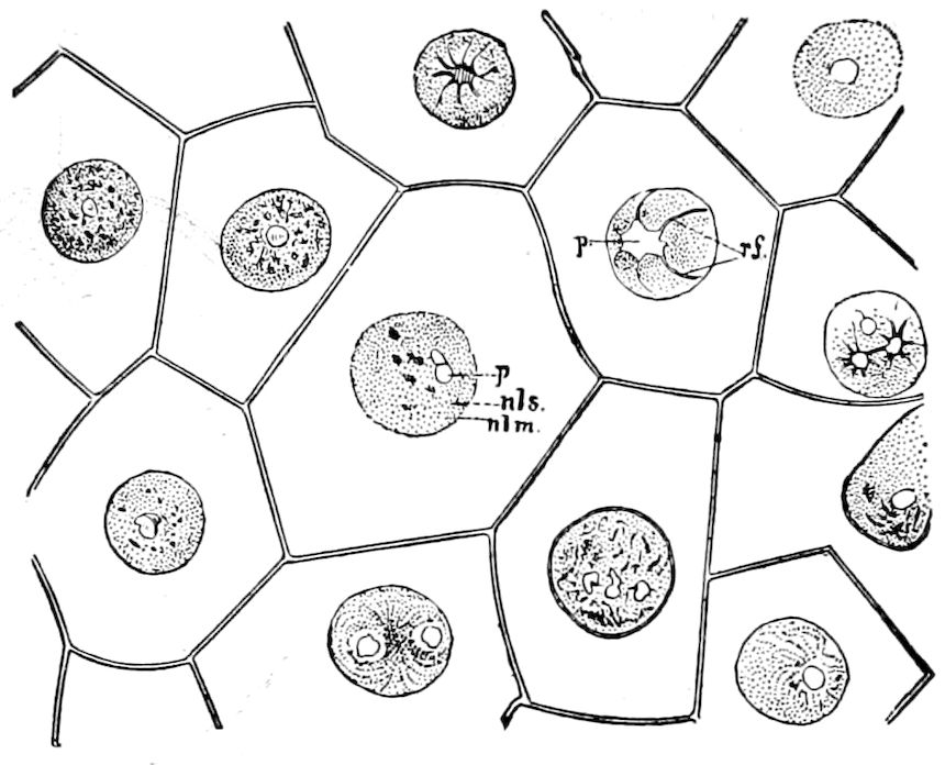
Fig. 510.—Surface view of fresh serosa from an Œcanthus, treated with acetic carmine; the blastoderm completely formed, × 500: p, polar body; rf, radiating fibres; nls, nuclear substance; nlm, nuclear membrane.—After Ayers.
1. Exochorion. (Remains of the epithelium of the ovarian follicle.) 2. Chorion. (Egg-shell or cuticle secreted in the ovarian follicle.) 3. Vitelline membrane. (Primary egg-membrane. Yolk-skin or membrane.) 4. Serous or outer germ-membrane. (Serosa.) } 5. Amnion or inner germ-membrane. } Derived from the blastoderm. 6. Ectoderm. } } 7. Mesoderm. } Embryo. } 8. Endoderm. } }
In the embryo of Xiphidium and Orchelimum Wheeler has found and described with much detail a membranous structure which he calls the indusium. “The organ,” he says, “appears to have been retained by the Locustidæ, and completely lost by the embryos of other winged insects.” It arises in Xiphidium, as a simple circular thickening of the blastoderm, between and a little in front of the procephalic lobes (Figs. 511, 512, A-E), and afterwards spreads over nearly the whole surface of the egg, leaving the poles uncovered, as in Fig. 513, where it is divided into two further membranes, the inner and outer indusium, the former lying in contact with the amnion. After this the serosa “is excluded from taking any part in the development of the embryo; both its position and function are now usurped by the inner indusium.”
535Hence in an egg of the Locustidæ Wheeler distinguishes, passing from within outward in a median transverse section of the egg, the following envelopes:
1. The chorion. 2. The blastoderm-skin-like cuticle secreted by the serosa. 3. The serosa. 4. The outer indusium. 5. A layer of dark granular secretion (probably some urate). 6. The cuticle secreted by the inner indusium. 7. The inner indusium. 8. The amnion. While envelopes 1–7 invest the whole egg; layer 8, the amnion, covers only the embryo.
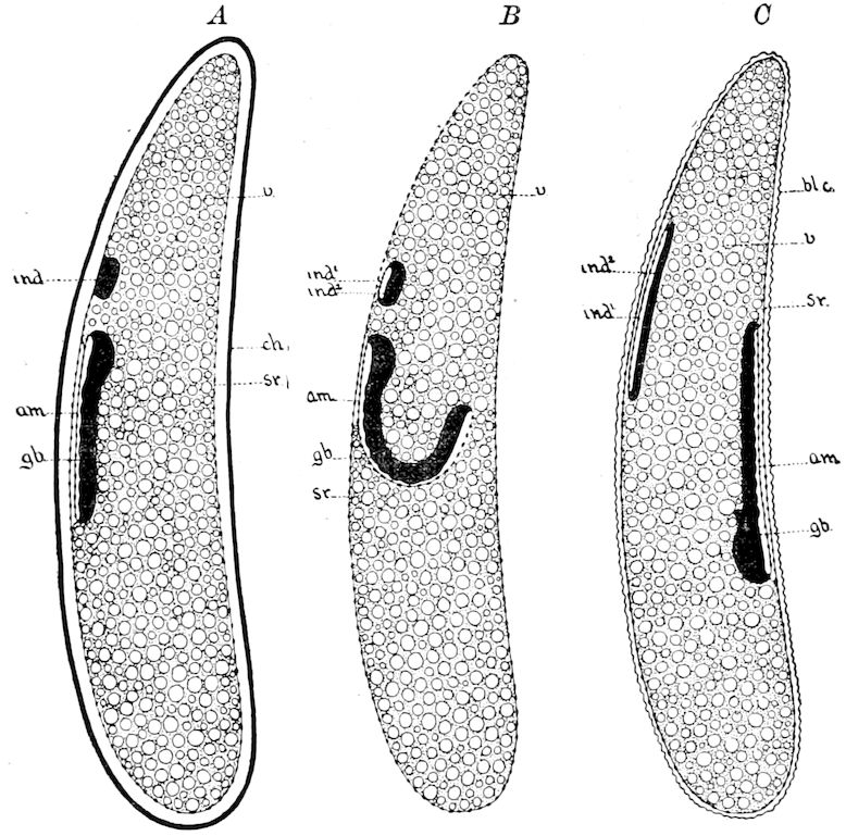
Fig. 511.—Diagrams illustrating the movements and envelopes of the embryo of Xiphidium: A, after the closure of the amnioserosal folds. B, during the embryo’s passage to the dorsal surface. C, just after the straightening of the embryo on the dorsal surface; ind, indusium afterwards forming ind1, the inner, and ind2, the outer indusium; ch, chorion; sr, serosa; am, amnion; gb, germ-band; v, yolk; bl. c, blastoderm membrane.
Wheeler further suggests that the so-called micropyle of the Collembola (Anurida), which has been homologized with the “dorsal organ” of Crustacea, is a possible homologue of the indusium, as also the “primitive cumulus” of spiders, and the “facette” or “cervical cross” of Pentastomids described by Leuckart and also by Stiles.
The gastrula stage.—The primitive band invaginates so as to give the opportunity for the formation of the inner layer. This invagination, which at a certain stage is established along the whole length of the primitive band, forms a median furrow and may be 536regarded as the gastrula-invagination of insects. The lower (inner) layer thus arising afterwards spreads out under the entire primitive band (Fig. 509, B and C), the edges of which become bordered by the growing amnion-fold. (Korschelt and Heider.)
In certain forms the primitive band arises from several separate rudiments which afterwards unite. Thus in Musca and Hydrophilus the anterior and posterior ends develop first, and in Hydrophilus the procephalic lobes originate independently of the rest of the band. In the Aphides, also, according to Will, these lobes arise independently, afterwards uniting with the primitive band.
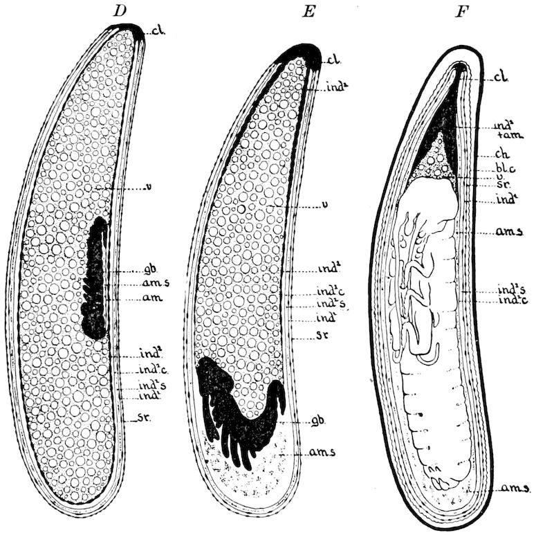
Fig. 512.—Diagrams illustrating the movements and envelopes of the embryo of Xiphidium: D, the stage of the shortened embryo on the the dorsal yolk. E, embryo returning to the ventral surface. F, embryo nearly ready to hatch; ch, chorion; b. lc, blastoderm membrane; sr, serosa; ind1, outer indusium; ind2, inner indusium; ind2 + am, inner indusium and amnion fused; am, amnion; ind1 c, cuticle of the inner indusium; ind2 s, granular secretion of the inner indusium; am. s, amniotic secretion; v, yolk; cl, columella; gb, primitive band.
Division of the embryo or primitive band into body-segments.—Meanwhile the primitive band grows at the expense of the yolk, spreading out more and more over its surface, until in certain cases (Coleoptera, Diptera, Siphonaptera, and Trichoptera) it lies like a broad ribbon over the yolk, so that the two ends nearly meet on the dorsal side. By this time it becomes divided by transversely impressed lines into segments, which correspond to those of the larva and adult. The first of these segments is divided into two broad 537and flaring flaps, which are called the procephalic lobes. It becomes the antennal segment.
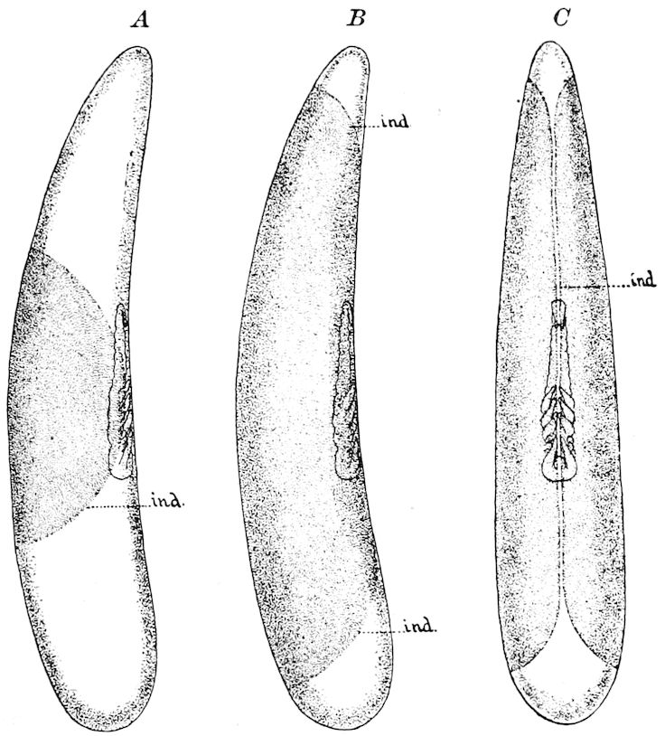
Fig. 513.—Two stages in the spreading of the indusium. A, lateral view of egg just after the arrival of the embryo on the dorsal yolk. B, lateral view of the egg with the indusium nearly reaching the poles. C, same egg seen from the dorsal surface.
The mouth (stomodæum) now develops, and is situated at the anterior,[82] and the rectum (proctodæum,) at the posterior pole, or end of the primitive band.
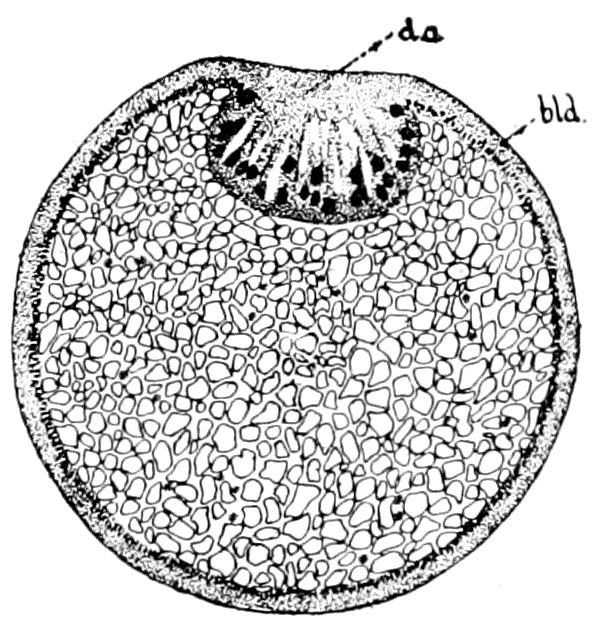
Fig. 514.—Median section of the egg of Anurida maritima: do, “micropyle”; bld, blastoderm.—This and Figs. 511–513, after Wheeler.
In Blatta, Hydrophilus, the Trichoptera, and the Lepidoptera the hindermost part of the primitive band is turned in ventrally (Figs. 534, C).
The preceding account of the relations of the primitive band to the yolk does not apply to all insects, since there are variations which appear to depend on the form of the egg, and on the amount and distribution of the yolk-masses. In certain Coleoptera, the primitive band sinks down and thus becomes immersed into the yolk. In Donacia (Kölliker and Melnikow) and Hydrophilus (Heider), and in the Chrysomelidæ and 538Attelabus, a weevil, as we have observed, the primitive band rests on the outside of the yolk, but in Telephorus fraxini it is immersed. In the Hemiptera it is immersed (Fig. 516), but there is much variation in this respect, the degree of immersion being most marked in the Coccidæ (Aspidiotus), and least so in Corixa. Besides the position of the primitive band, there are in Odonata and Hemiptera differences in the origin of the primitive band itself and of the embryonic membranes.
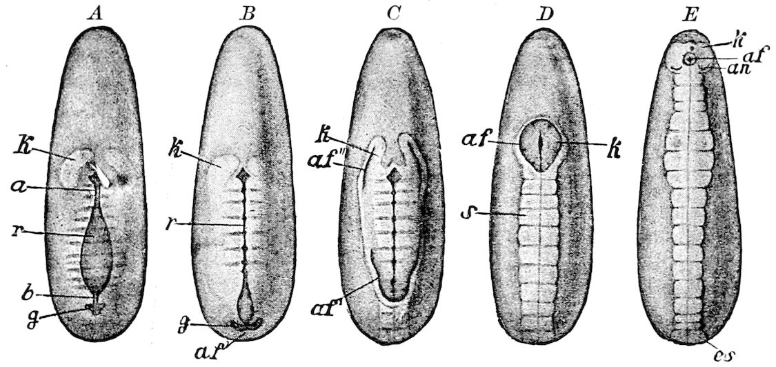
Fig. 515.—Ventral view of five developmental stages of Hydrophilus: a and b, places at which the blastopore contracts; af, edge of the amnion-fold; af′, caudal fold; af″, paired head-fold of the amnion; an, antenna; es, last segment; g, pit-like invagination (first indication of the amniotic cavity); k, head-lobes; r, furrow-like invagination; s, portion of the primitive streak covered by the amnion.—After Heider, from Lang.
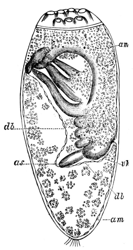
Fig. 516.—Embryo of the louse: am, serosa; db, amnion; as, antenna; vk, clypeus.—After Melnikow.
Korschelt and Heider divide the early embryo of insects into two types:
1. Into those with a superficial primitive band; viz., where there is no passage of yolk-elements into the space between the amnion and serosa. The primitive band has in such cases a relatively superficial position (Figs. 508, 509, 521, 535). Examples are certain Orthoptera (Blatta, Œcanthus, Mantis, Gryllotalpa), also certain Hemiptera (Corixa), certain Coleoptera, and the Trichoptera, Diptera, and Hymenoptera.
2. Into those with an immersed primitive band, with the space between the serosa and amnion filled with yolk (Figs. 517, 518, 534). Examples are the orthopterous Stenobothrus, Odonata, many Hemiptera (the Pediculina and Pyrrhocoris), the Coleoptera already mentioned, and Lepidoptera.
It should be observed, however, that these differences are of little phylogenetic or taxonomic value, since genera of the same order, notably the Coleoptera, differ as to the position of the primitive band, so also two orders so nearly allied as the Trichoptera and Lepidoptera.
Differences between the invaginated and overgrown primitive band.—In respect to the mode of origin of the primitive band and its relative position, 539there are two opposite types, though connected by transitional forms. In the one case the primitive band, i.e. its ventral portion, the “ventral plate” (Fig. 518, b, p) is pushed in or invaginated in the interior of the egg; in the other case it becomes overgrown by the folds of the amnion arising from its edges.
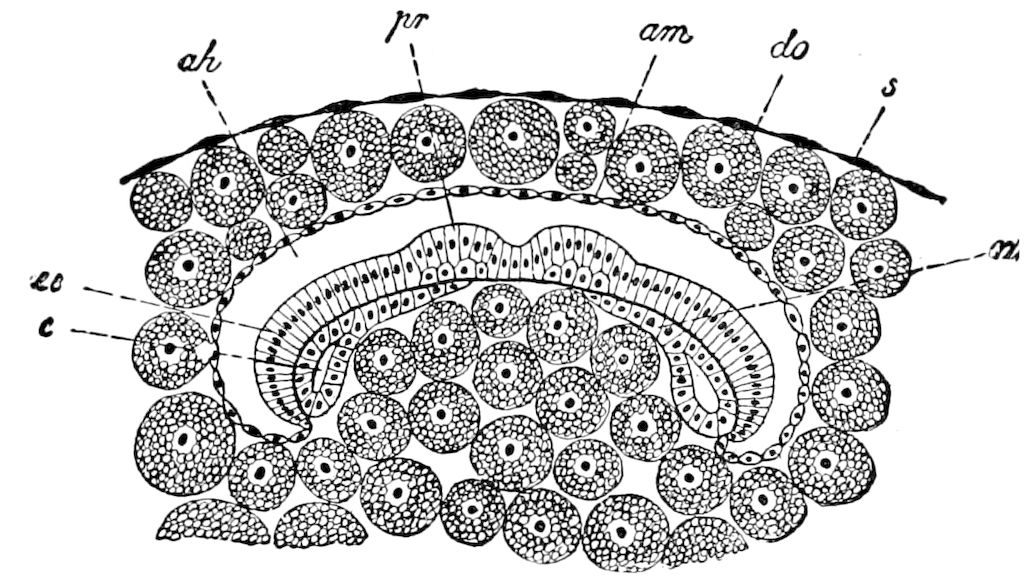
Fig. 517.—Primitive streak of a lepidopter in cross-section: ah, amniotic cavity; am, amnion; c, cœlomic cavity; do, nutritive yolk, divided into single nucleated masses; ec, ectoderm; m, mesoderm; pr, primitive thickenings of the ventral nervous cord; s, serosa.—Combined figure after those of Brobretsky and Hatschek, from Korschelt and Heider.
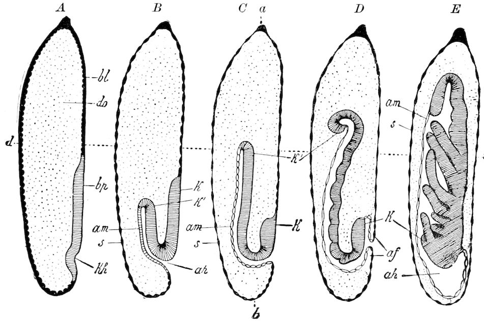
Fig. 518.—Five diagrammatic median sections representing the growth of a dragon-fly (Calopteryx). A-C, development of the primitive streak (k, k′) by invagination. D, the amnion-fold (af), growing over the head-end of the primitive streak. E, closing of the opening of the amnion-cavity (ah): v, ventral, d, dorsal side; a, fore, b, hind end of egg; bl, blastoderm; bp, ventral plate; do, yolk; k, head-end, k′, caudal end, of the primitive streak; kh, germinal thickening or initial point of invagination; s, serosa.—After Brandt, from Korschelt and Heider.
In insects with an overgrown primitive band, the band at the beginning is generally short and always situated on the ventral side of the egg, with the head-end looking forward, and remains in this position throughout embryonic 540life. There is no revolution of the embryo. The embryonal membranes arise through the formation of folds.
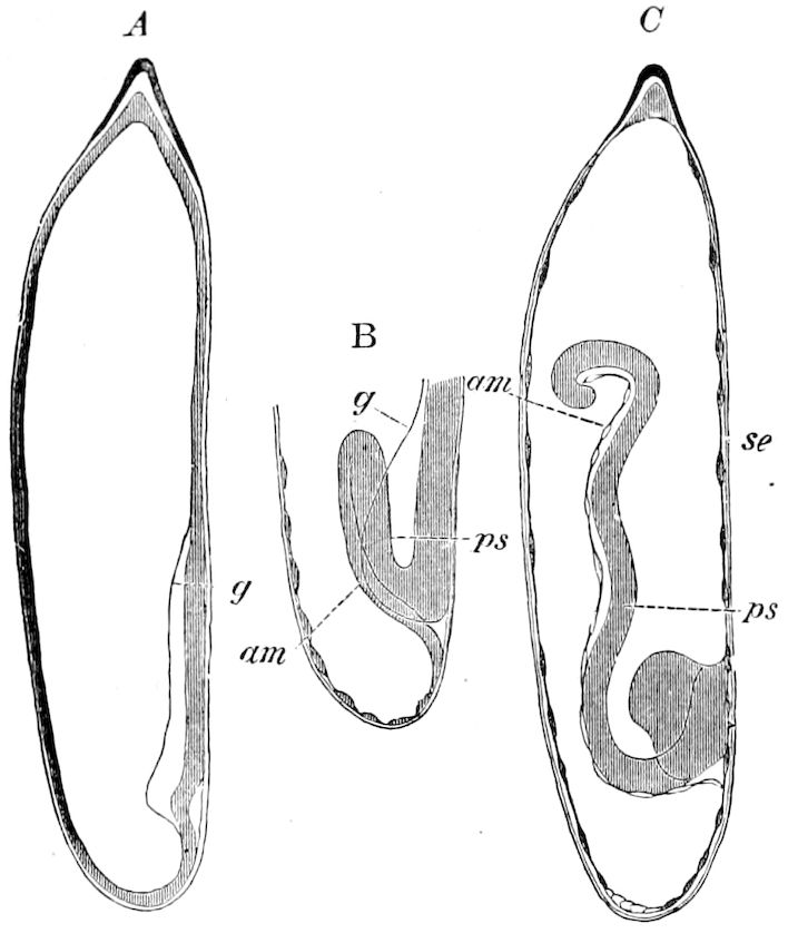
Fig. 519.—Three embryonic stages of Calopteryx: am, amnion; g, edge of the ventral plate; ps, germ of primitive band; se, serosa.—After Brandt, from Balfour.
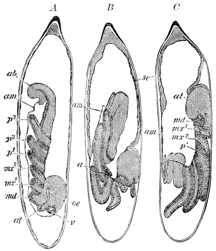
Fig. 520.—Three farther stages of growth of Calopteryx. B and C show the inversion of the embryo: a, opening of the amniotic-cavity, out of which the embryo emerges; ab, abdomen; am, amnion; at, antenna; md, mandible; mx1, mx2, 1st and 2d maxillæ; œ, œsophagus; p1, p2, p3, legs; se, serosa; v, anterior end of the primitive streak.—After Brandt, from Balfour.
In insects with an invaginated primitive band, of which the Odonata afford examples, the first rudiment of the primitive band is in the form of a ventral plate of slight extent passing ventrally in the hinder half of the egg, in whose posterior section a process of invagination (Fig. 518, A, kh), soon occurs. The cavity of this invagination is the first indication of the amnion-cavity (Fig. 518, B, ah), while its wall in its thickened ventral part (K′) is concerned in the formation of the primitive band, and, in its dorsal thin part, in the formation of the amnion (B, C, am).
Revolution of the embryo where the primitive band is invaginated.—At first the head-end of the embryo is directed towards the posterior end of the egg, as in dragon-flies (Fig. 518). Also that surface of the primitive band which afterwards faces the ventral, is at first turned towards the dorsal side of the egg. In order to bring the primitive band into the later relations, there must occur the process of revolution, or turning, of the embryo. The somewhat advanced embryo of the Odonata, after the appearance of the head and thoracic appendages, undergoes a rotating motion around its transverse axis, and at the same time turns out of the amniotic cavity (Fig. 520, B). This process is so managed that near the head-region, the amnion and serosa, there closely situated to each other, are fused together, and at this 541place tear or burst open. Through this rent (a), in the same place in which the original invagination-opening was situated, the amniotic cavity again opens, and through the opening thus formed first the head and then the succeeding segments of the primitive band (Fig. 520, B) pass out, and remain there while the head passes on to the anterior pole of the egg on the ventral side, the embryo thus assuming a position like that of other insects. (Kowalevsky.)
In the parasitic Hemiptera (Pediculina), according to Melnikow, the opening in the membranes near the head remains permanent, and the embryo becomes everted through it, while the yolk, enclosed in the continuous membrane formed by the amnion and serous membrane, forms a yolk-sac on the dorsal surface. The same process occurs in Mallophaga, and also in Œcanthus, as described by Ayers (Fig. 521). Generally as soon as the embryo passes out of the amniotic cavity the latter soon becomes smaller and finally completely disappears.
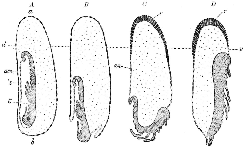
Fig. 521.—Revolution of the embryo of Œcanthus (diagrammatic): a, fore, b, hind end of egg; am, amnion; d, dorsal, v, ventral side of egg; k, primitive streak; r, dorsal plate (originating by the contraction of the serosa (s)).—After Ayers, from Korschelt and Heider.
As the embryo grows, and the sides grow up and the back closes over, the contents of the yolk-sac are soon taken up and absorbed in the intestinal cavity, which communicates with it.
In Phyllodromia, according to Wheeler, the process of revolution is “hurried through by the embryo from the beginning of the 16th to the end of the 17th day.” Several successive stages are represented in Fig. 522. In the 15th day the embryo still occupies the middle of the ventral surface of the egg. Soon the envelopes (amnion and serosa, as) rupture, an irregular slit being formed, and soon the egg and embryo are as seen in Fig. 522, B, the embryo standing out free from its envelopes on the yolk, and the edges of its dorsal growing walls (b) are distinctly marked. The tail now lies at the caudal end of the egg (Fig. 522, C). By the 17th day the walls have closed in the median dorsal line, and the embryo has grown in length to such an extent as to bring its head to the cephalic pole (Fig. 522, E).
Korschelt and Heider consider, since the primitive band of the chilopod myriopods (Geophilus) is curved in at the middle and sinks into the interior of the 542yolk, that in insects the invaginated primitive band is the ancestral or primitive one, the overgrown primitive band being derived from it. The overgrown primitive band by its position may also be better insured against certain mechanical attacks, perhaps also against the danger of drying up.
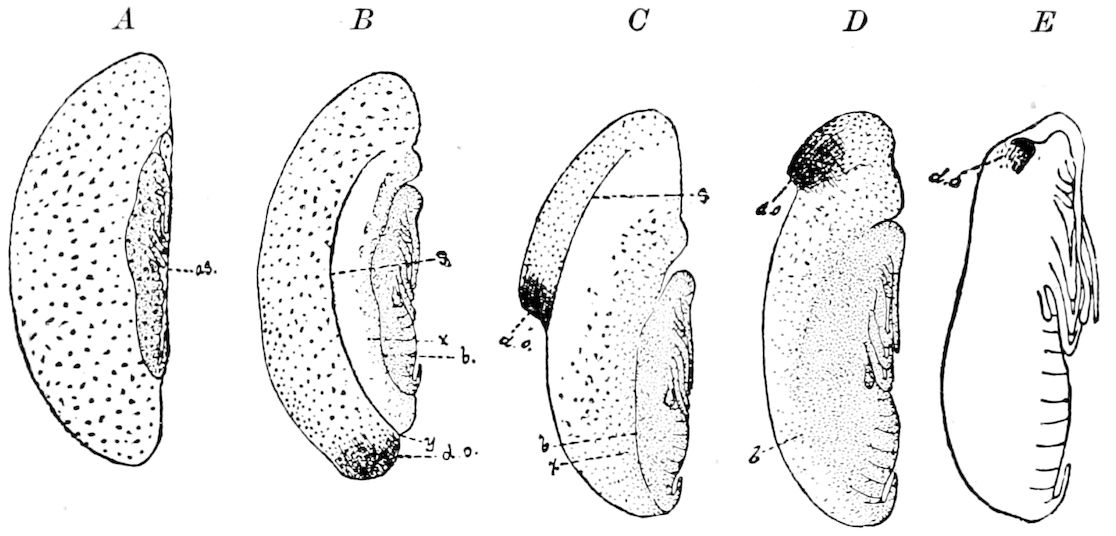
Fig. 522.—Embryo of Phyllodromia, 15 days old; revolution about to begin. The stages in revolution are represented, after the rupture of the amnion and serosa, in A to E, which are from embryos 16, 16½, 16¾, and 17 days old respectively: as, amnion and serosa; s, edge of serosa; b, dorsal growing body-wall; d.o, dorsal organ; x, clear zone covered with scattered amniotic nuclei.—After Wheeler.
f. Formation of the external form of the body
Origin of the body-segments.—As we have seen, the first traces of segments appear very early, the primitive band being divided by superficial transverse furrows into segments. This segmentation into arthromeres (somites or metameres) can be observed in Hydrophilus and Chalicodoma at a time when gastrulation begins (Figs. 515, 536). The segmentation extends not only across the median portion of the primitive band, through whose invagination the inner layer (endomesoderm) results, but also to the lateral portions which become a part of the ectoderm of the primitive band. These transverse furrows correspond to thinner places in the epithelium, which in this stage forms the embryonal rudiment. It thus happens that, in the forms named, after the end of gastrulation not only the ectoderm, but also the endomesoderm, is already segmented.
So early an appearance of segmentation as that observed in Hydrophilus and Chalicodoma we must regard as a falsification of the process of development due to heterochrony. We must consider the conditions observed in other forms as the primitive ones, in which (as, for example, in Lina and in Stenobothrus, according to Graber) the gastrulation and separation of the ectoderm occurs in the still unsegmented primitive band, the division into segments occurring in later stages (Fig. 524). In these forms, then, the segmentation affects the invaginated endomesoderm, as well as the ectoderm. (Korschelt and Heider, p. 789.)
543
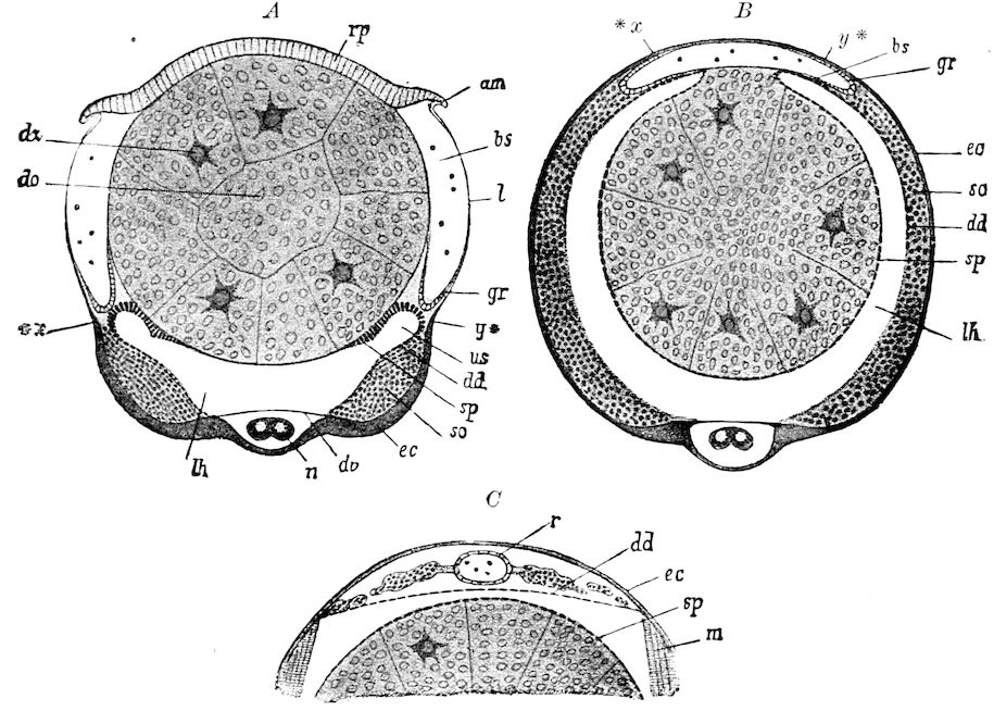
Fig. 523.—Diagrammatic cross-section through three successive stages of Gryllotalpa, showing the formation of the heart. (Compare Fig. 505.) The germs of the glandular intestinal layer (darmdrüsenblatt) are omitted. A, earliest stage; the primitive streak extends from *x to y*. The embryonal membranes are torn and pressed against the back: am, edge of the rent; rp, dorsal plate (serosa); l, lamella (amnion turned up) standing in connection with the ectoderm of the primitive streak. B, second stage; the primitive streak has completely grown around the yolk; the dorsal organ is absorbed. C, third stage, dorsal portion; the formation of the heart is finished: am, vestige of the amnion-fold; bs, blood-sinus; dd, rudiment of the dorsal diaphragm; dv, ventral diaphragm (compare Fig. 505); do, yolk; dz, yolk-cells; ec, ectoderm; gr, vascular groove (rudiment of the heart); l, lamella of the upturned amnion; lh, definite body-cavity; m, transverse muscle; n, nervous cord; r, heart; rp, dorsal plate; sp, splanchnic; so, somatic layer of the mesoderm; us, primitive segmental cavity; *x, y*, lateral terminations of the primitive streak.—After Korotneff, from Korschelt and Heider.
In the completely segmented primitive band may be distinguished two regions of a peculiar appearance (Figs. 515, 527), one at the anterior, and the other at the hinder end. The anterior, the primary head-section, contains the mouth-opening, and is characterized by its lateral expansions, or procephalic lobes. The other section, or posterior section, the so-called anal segment or telson, contains the anus. Between the two sections lies the segmented primary trunk-segment, which in insects consists of 17 segments. Of these the three most anterior are those destined to bear the mandibles and two pairs of maxillæ; the three following are the thoracic, which are succeeded by 10 abdominal segments, besides the 11th or telson (pygidium, or suranal plate).
544It is now generally believed that there are primarily eleven abdominal segments, while Heymons has detected twelve in the embryos of Blattids and Forficula (see p. 162). In the later stages of embryonic development the number of abdominal segments is diminished, the 10th and 11th abdominal segments becoming fused. In Hydrophilus and Lina this is the case, but according to Graber, in the Lepidoptera there is a fusion of the 9th and 10th abdominal segments, the llth remaining free.
According to Wheeler, in Doryphora, and also in Chalicodoma (Carrière), between the primary head-region and the mandibular segment is interpolated a rudimentary and transitory body-segment, the premandibular segment. According to Carrière this segment corresponds to a rudimentary pair of limbs, and also to a ganglion, which participates in the formation of the œsophageal commissure (see p. 51).
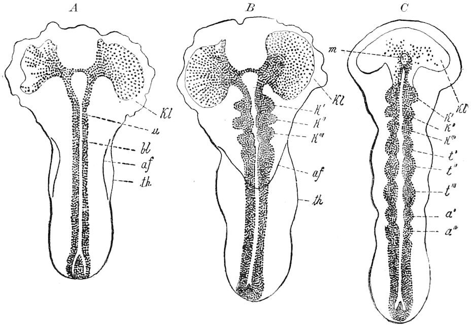
Fig. 524.—Three embryonic stages of a leaf-beetle (Lina): A, unsegmented primitive streak; in B and C the segmentation becomes distinct on the lower layer (u). B, with the germs of the gnathal segments (k′-k‴), and in C the three thoracic segments (t’-t‴), with the first two abdominal segments (a′, a″): bl, blastopore; kl, head-lobes; th, extension of the primitive streak into the thoracic region.—After Graber, from Korschelt and Heider.
The procephalic lobes.—The head-lobes, or procephalic lobes, appear at a very early period (Fig. 524, kl), before any traces of the segments of the trunk region. Ayers has shown that in Œcanthus the primitive band, in its earliest condition and before the appearance of the head-lobes, is a simple oval plate or almond-shaped thickening near the posterior end of the egg (Fig. 525, 1, 2). This plate is “soon divided into two tolerably well-marked regions by the enlargement of the head-end,” the first indication of the head-lobes 545(3). A depression next forms in what is to be the middle of the forehead. “It indicates the position of the future labrum, and forms the inner boundaries of the two cephalic ganglia, which are developed on either side of this depression at a much later stage.” Almost simultaneously with the appearance of this depression, two lateral folds are formed in the trunk portion of the band, which are the first indications of the maxillary and thoracic regions, the abdominal portion not yet showing traces of a division into segments (Fig. 525, 5). The thickened outer edges of the head-fold next gradually grow in towards the median line (Fig. 525, 5), and bend forward towards the region of the future mouth. The rounded angle made by the posterior end of the head-fold is the first indication of the antennæ. The embryo is now composed of four well-marked regions: cephalic, maxillary, thoracic, and abdominal. The primitive band then grows much longer, the primitive mouth and anus appear, and the appendages bud out, and eventually the embryo revolves and appears on the ventral side of the egg (Fig. 525, 6).
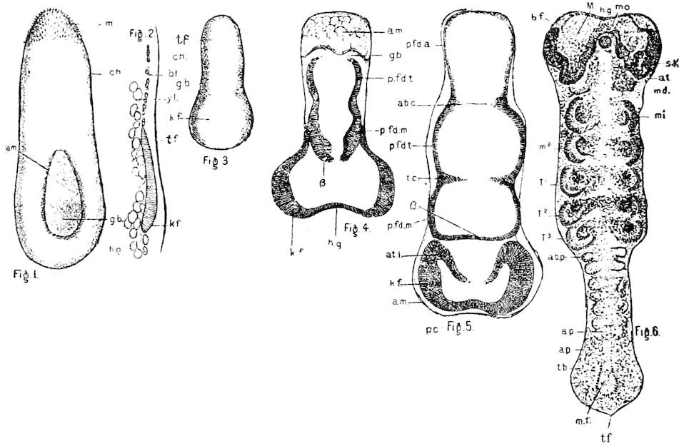
Fig. 525.—Early stages in the embryology of Œcanthus niveus. Fig. 1, the youngest observed primitive band, the serosa not yet formed; 2, longitudinal optic section (diagrammatic) of Fig. 1; 3, the primitive band after the appearance of the head-fold, which is indicated at this time by the more rapid growth and consequent greater breadth of the lower end of embryo, x 25; 4, a young embryo after the appearance of the primitive segment-folds, x 50; 5, a more advanced embryo, with the antennal folds distinctly marked off; the free ends of the primitive folds have united across the embryo posterior to the antennal folds, x 50; 6, ventral view of the embryo with the appendages budding out, x 25 (the embryo in this stage lies dormant through the six colder months of the year): am, amnion; m, micropylar end; ch, chorion; gb, primitive band; bf, brain-fold; yl, yolk; tf, caudal fold; kf, head-fold (procephalic lobe); p.fd.t, primitive thoracic fold; p.fd.m, primitive maxillary fold; p.fd.a, primitive abdominal fold; ab.c, abdominal constriction; t.c, thoracic constriction; at.l, antennal lobe; M, mesoderm; h.g, head groove; mo, mouth; sk invagination of ectoderm to form head-apodeme; md, rudiment of mandible; m1, 1st, m2, 2d maxilla; T1–T5, legs: ab.p, 1st abdominal appendage; ap, other appendages; tb, caudal expansion; mf median furrow; B, primitive unpaired organ (metastomum).—After Ayers.
546These primitive regions of the primitive band, before the segments are formed, are called by Graber macrosomites, and the secondary segments into which they divide (which afterwards become the body-segments), microsomites. The macrosomites are peculiar to insects, and may be an inheritance from a hypothetical ancestral form. With Korschelt and Heider, we should hardly share this view.
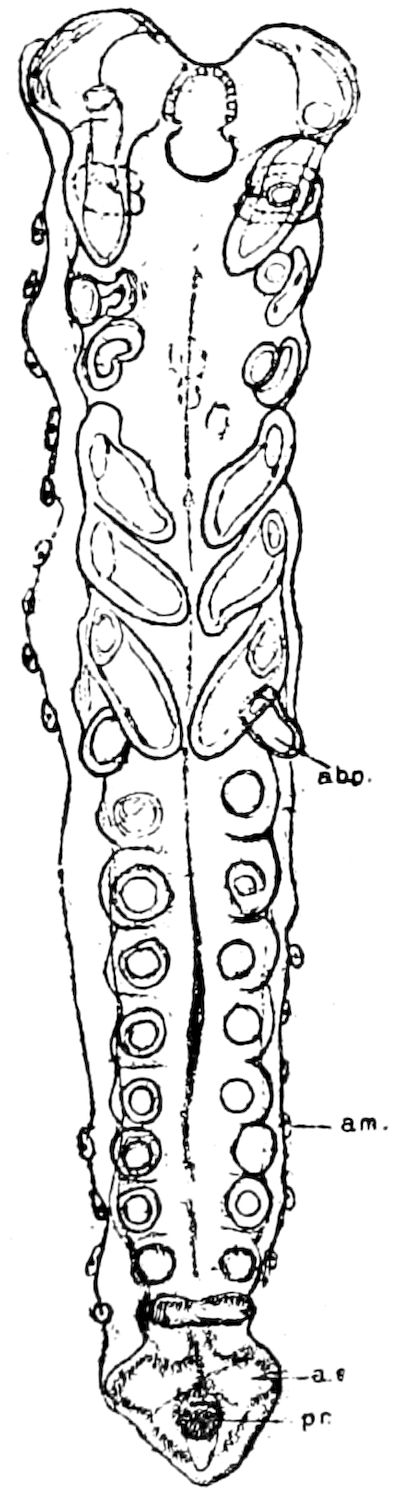
Fig. 526.—Older embryo of Œcanthus with the appendages budded out, those of the abdomen distinct: abp, first pair; a.s, anal stylet; pr, proctodæum; am, amnion.—After Ayers.
Our observations on locusts show clearly (1) that the procephalic lobes are the pleural portion of the first cephalic or antennal segment; (2) that the antenna is an appendage or outgrowth of the procephalic lobes; (3) that the eyes are a specialized group of epidermal cells of the upper part of the procephalic lobes, and are not homologues of the antennæ or of the appendages in general; and (4) it seems to follow from a study of the relations and mode of development of the clypeus and labrum, that they arise between the procephalic lobes, and probably represent the tergal part of the antennal segment, forming the roof of the mouth, i.e. closing in from above the pharynx.
In general the formation of the body-segments into the primitive band is in succession from before to the hinder end. This successive appearance has been observed by Graber in genera of different orders (Stenobothrus, Lina, and Hylotoma). For example, in the beetle Lina, after the appearance of the mandibular and two maxillary segments, appear the three thoracic segments, together with the two anterior abdominal segments, the other abdominal segments arising afterwards. In other cases, the formation of segments seems to be simultaneous along the entire length of the primitive band. An exception to the rule has been noticed by Heider in Hydrophilus, as in this beetle the development of the segments of the middle region appears somewhat delayed, while the fore and hind parts of the primitive band are more rapid in development. In Pieris, according to Graber, the thoracic segments are more rapidly developed than the others; soon after, the gnathic segments (mandibles and two pairs of maxillæ) appear, and finally the abdominal segments are formed.
547Fore-intestine (stomodæum) and hind-intestine (proctodæum), Labrum.—The digestive canal of insects consists, as in other animals, of three portions, the fore, mid, and hind gut or intestine. The next change after the completion of the segments of the primitive band is the development of the fore and hind intestine and the appendages. The fore-intestine (stomodæum) arises as an invagination in the area of the primary head-section, and the hind-intestine (proctodæum) in the terminal section (Figs. 300 and 546).
In insects generally the formation of the fore-intestine occurs earlier than that of the hind-intestine. An exception was discovered by Graber and also by Voeltzkow in Muscidæ, where the proctodæum appears earlier.
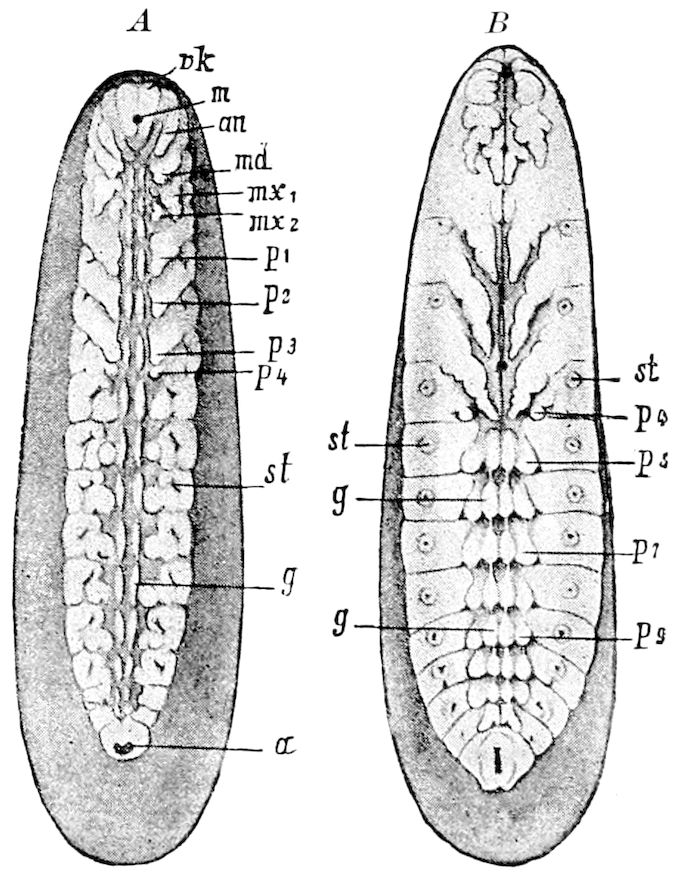
Fig. 527.—Rudiments of the appendages of the embryo of Hydrophilus: an, antenna; md, mandibles; mx1, 1st, mx2, 2d maxilla; vk, clypeal region; m, mouth; p1-p3, legs; p4-p9, rudiments of abdominal appendages, 1–9; st, stigma; a, anus.—After Heider, from Lang.
Usually at the time of origin of the stomodæum a projection arises at the anterior edge of the primary head-region, the so-called forehead (Fig. 527, vk), which is the common rudiment of the clypeus and labrum. In many cases (certain Coleoptera and Lepidoptera) these rudiments first assume the form of paired hooks (see Figs. 83, 102, 104, 105, of Graber’s Keimstreif der Insekten, also Figs. 529 and 546), which afterwards, by fusion in the median line, become single, though notched in the middle; but in the more generalized Blatta and Mantis, as well as in bees, the rudiment is single at the outset.
The view advanced by Patten, and also by Carrière, that the labrum is a first pair of antennæ, is scarcely tenable, and we quite agree with Korschelt and Heider in regarding the clypeo-labral region as homologous with the upper lip of Crustacea, and, we may add, of Merostomes and of Trilobites.
It should be observed that in many insects, in their earlier embryonic state, directly behind the mouth arises, from paired rudiments, what seem provisional lower lip structures (not to be confounded with the 2d maxillæ of insects). This under lip structure was first discovered by Bütschli in the bee (his inner or 2d 548antennæ), and afterwards by Tichomiroff in Lepidoptera. Heider, in his work on Hydrophilus, describes it as the “lateral mouth-lips,” while, more recently, Nusbaum has observed it in Meloë. This under lip structure may be regarded as analogous to the paragnaths of Crustacea, although to attempt to homologize it with these seems useless. (Korschelt and Heider.)
Completion of the head.—Sufficient attention has not been paid to this subject by embryologists. The head is at first, dorsally, mostly composed of the head-lobes, or antennal segment only, and the dorsal or tergal portion of the oral appendages develop at a later period. We have observed in the embryo of dragon-flies (Æschna) that the tergites of the mandibles and first maxillæ are simultaneously fused with the head-lobes, while the much larger tergal region of the 2d maxillæ remains for some time separate from the anterior part of the head, and is continuous with the thoracic segments, and it is only just before hatching that this segment becomes fused with the rest of the head (Fig. 36). In a sense, the 2d maxillary segment when it is free from the head reminds us of the foot-jaw, or 5th segment of chilopod myriopods (see also p. 53).
g. The appendages
As we have seen, nearly or quite simultaneously all the limbs as a rule bud out from each side of the median line of the primitive band. They arise as saccular evaginations or outgrowths of the ectoderm, directed a little backwards. They are at first filled with mesoderm cells, and in the Orthoptera diverticula of the cœlom-sac are taken up into the rudimental limbs, as in Peripatus and Myriopoda. (Graber, Cholodkowsky.) As the antennæ, mouth-parts, legs, and abdominal appendages are all alike at first, their strict homology with one another is thus demonstrated. In insects never more than a single pair of limbs is known to arise from one segment.
The cephalic appendages.—The antennæ evidently arise from the hinder edge of the procephalic lobes (Fig. 527, an). As in Limulus, the first pair of appendages are at first postoral (Fig. 528, at), afterwards moving forward owing to changes in the relative proportions of the parts of the head, and they are in all respects, in their development and position in relation to the segment from which they arise, homologous with the appendages succeeding them.
The occurrence of rudiments of a pair of preantennal appendages in Chalicodoma which is claimed by Carrière, needs confirmation, as other embryologists have not observed them.
549The postoral appendages of the head are the mandibles and the 1st and 2d maxillæ, besides the supposed premandibular segment already referred to on pp. 50–54, which only temporarily exists.
The trophi or oral appendages are all alike at first, but soon differ in shape, acquiring their characteristic form shortly before the embryo leaves the egg. The mandibles of Œcanthus are said by Ayers at the time of revolution of the embryo to be slightly bilobed, and in his Fig. 5, Pl. 19, they are represented as deeply trilobed, but in general they are undivided. The 1st maxillæ are at this time distinctly trilobed. The 2d maxillæ are separate, and distinctly though unequally bilobed, becoming united shortly before birth. In the embryos of dragon-flies they are at an early date very large and long, and directed backwards, and are not fused together until just before hatching, when the extraordinary mask-shaped labium is fully developed.
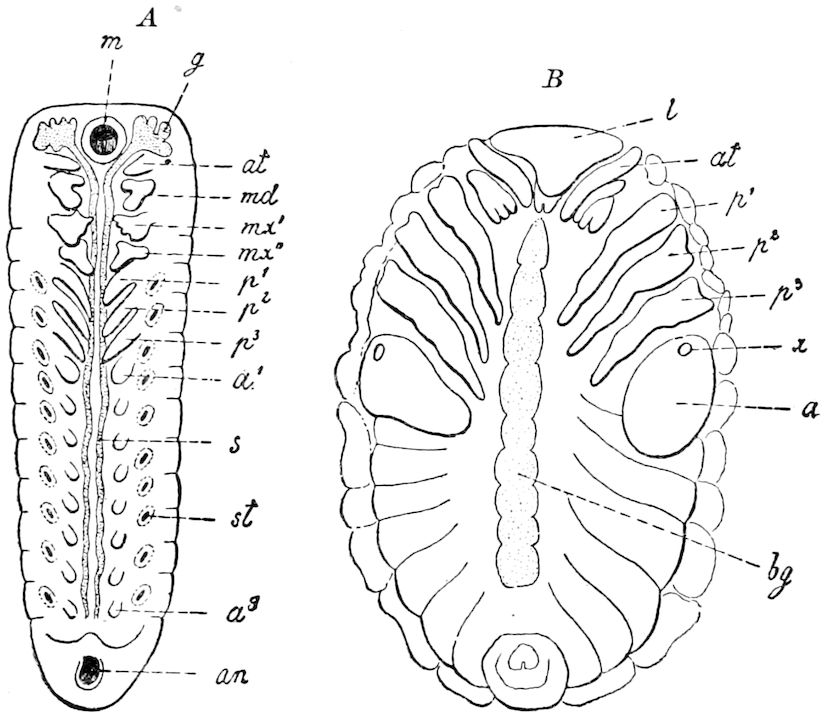
Fig. 528.—Two embryonic stages of the primitive streak of Melolontha. A, younger stage, with rudiments of eight pairs of abdominal appendages (a1–a8). B, older stage, the primitive band now very broad: a, 1st abdominal appendage, in B sac-like; x, place of adhesive disc; g, brain; l, clypeo-labrum; s, lateral cord of the ventral nervous cord; other lettering as in previous figures.—After Graber, from Korschelt and Heider.
The distal parts of the labium, such as the ligula, palpifer, and palpus are elaborated before the mentum and submentum. Many details as to the final changes in the mouth-parts before hatching remain to be worked out.
550The thoracic appendages.—The three pairs of legs arise at the same period and in the same manner in all insects; it is not until the end of embryonic life that they become jointed, and that the claws and onychia are developed. Especial attention has not yet been given to the details of the development of the parts of the last joint of the tarsus.
In many forms the antennæ are the first to appear, the mandibles, maxillæ, and legs appearing at a latter date, though simultaneously. It is thus in Stenobothrus, Hydrophilus, and Melolontha. In Lina, according to Graber, the mandibles precede the antennæ in appearance. In the Libellulidæ, according to Brandt, the legs first appear, then the jaws, and lastly the antennæ. This did not seem to be the case in the embryos of Æschna observed by us, although our observations were more superficial.
On the other hand, in those insects whose larvæ are footless, the rudiments of the legs are retarded and aborted just before hatching (fossorial Hymenoptera and Apidæ), or the rudiments of the legs are not developed at all.
The abdominal appendages.—These appear soon after the thoracic limbs, corresponding in most cases to the latter in shape and position, and their position in the embryo is a matter of the greatest interest. Von Rathke was the first embryologist to detect those of the first abdominal segment, in his examination of the development of Gryllotalpa. Long afterwards Bütschli detected them in the embryo of the honey-bee, observing a pair on each segment. Patten observed them in Trichoptera; Kowalevsky first perceived them in Lepidoptera, Tichomiroff confirming his observations. Graber, Ayers, and Wheeler have observed them in Orthoptera and Coleoptera, and the latter has detected them also in Hemiptera and Neuroptera; and while they do not arise in the embryos of Diptera and of Siphonaptera, they are to be looked for in any or all the lower or more generalized orders.
As the result of these discoveries of polypodous embryos occurring in all but the most specialized order (Diptera), it appears to be a rational deduction that the winged insects have descended from insects in which there were functional legs on each abdominal segment. Such an ancestor was the forerunner of the Thysanura, in which abdominal locomotive appendages still survive, though in a modified, more or less aborted condition. This polypodous ancestral form was apparently allied to Scolopendrella, which has a pair of functional legs on each abdominal segment.
The subject, then, of polypodous embryo insects is one of special interest, and has attracted much attention from Graber, Wheeler, Haase, and others. That these are genuine, though transitory appendages, is shown by the fact that certain pairs persist throughout adult life. The embryology of the Thysanura 551when worked out will throw much light on this subject, but we know that the spring (elater) of Collembola (and possibly the collophore) and the cerci of the winged insects are survivals of these limbs. That the three pairs of appendages forming the ovipositor, or sting, are most probably derived from these appendages is claimed by Wheeler (p. 167), and seems proved by the fact that Ganin and also Bugnion has detected three pairs of imaginal disks in the embryo of parasitic Hymenoptera. Hence the abdominal appendages may ultimately be found to arise in nearly all cases from imaginal disks like those giving origin to the cephalic and thoracic appendages.
As regards the Diptera, Pratt has observed that each of the three thoracic and eight abdominal segments of the embryo brachycerous Diptera (seen especially well in Melophagus) has two pairs of imaginal disks, a dorsal and a ventral pair. He thinks there is no doubt but that the ventral abdominal disks are homologous with the rudimentary appendages which appear in the embryos of all other insects, though not in the brachycerous dipters.
Appendages of the first abdominal segment (pleuropodia).—As early as 1844 Rathke observed in the embryo of the mole-cricket a pair of appendages on the 1st abdominal segment, which he described as mushroom-shaped bodies, and supposed to be embryonic gills. They are called pleuropodia by Wheeler, who, with Patten, Graber, and Nusbaum, ascribes a glandular function to them, while Wheeler suggests that they were odoriferous repugnatorial organs. In Blatta (Phyllodromia) they are of large size, in Melolontha enormous (Fig. 528, B) and filled with blood. Wheeler distinguishes as varieties, beside the mushroom-shaped appendages of Gryllotalpa and Hydrophilus, the reniform (Œcanthus), the broadly pyriform (Blatta), and the elongate pyriform (Mantis carolina). In the European Mantis they are most limb-like, with a digitiform continuation divided by a constriction into two sections. (Graber.) In Meloë they assume the shape of a stalked cup. (Nusbaum.) In the bee and in Lepidoptera the pleuropodia are not present, though the temporary appendages on the succeeding segments appear; Carrière, however, found them on the two first abdominal segments of very young larvæ of the wall-bee (Chalicodoma).
Their cellular structure is peculiar, and they are either formed by evagination or invagination, those of the latter type being subspherical and solid. Those of the former type have a cavity communicating by means of a narrow duct through the peduncle with the body-cavity (Blatta). No tracheæ, nerves, or muscles enter them, though blood-corpuscles have been seen in the cavities. “In some species the pleuropodia produce a secretion from the ends of their enlarged cells. This secretion may be a glairy albuminoid substance (Cicada, Meloë), a granular mass (Stenobothrus), a bundle of threads (Zaitha), or a thick, striated, cuticula-like mass 552(Acilius).” They attain their greatest size during the revolution of the embryo, and they are “mere rudiments of what were probably in remote ages much larger and more complex organs.” (Wheeler.)
Lameere has observed that in Phyllodromia the first pair of abdominal appendages, after becoming of considerable size, undergo an enlargement at their free end, become detached, and fall into the amnion.
Wheeler also calls attention to the homology of these pleuropodia with the 1st abdominal appendages of Campodea, shown by Haase to be originally glandular, but with at present a respiratory function. In the embryos of later, higher orders of insects, these appendages are in size and shape similar to those of the succeeding segments. (See also p. 164.)
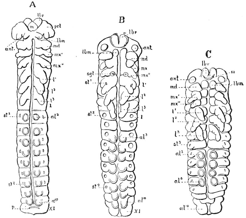
Fig. 529.—Primitive band of Bombyx mori, showing the temporary legs on abdominal segments 2–11: A, early stage, in which the abdominal legs al2–al10 appear. B, later stage, when they are very faint and all except al3–al6 and al10 are about to disappear. C, the persistent abdominal legs al3–al6 and al10; st2, st9, the 2d and 3d pair of stigmata; sgl, silk duct.—After Tichomiroff.
Are the abdominal legs of larval Lepidoptera and phytophagous Hymenoptera true limbs?—The presence of these abdominal legs in the embryos of Sphinx (Kowalevsky), of Bombyx mori (Tichomiroff), and both Bombyx mori and Gastropacha quercifolia (except those of the first segment), as well as in Hylotoma, which has 11 pairs of such appendages, has suggested that the prop or prolegs of caterpillars and saw-fly larvæ are survivals of these outgrowths, and not secondary, adaptive structures. Opinions on this point vary. Balfour, and also, more recently, Cholodkowsky, hold that the prolegs are survivals of the embryonic appendages. Graber cautiously, after a 553lengthy and interesting discussion, says that the question cannot be, in the present state of our knowledge, solved. He, however, seems inclined to believe that the prolegs are not merely secondary structures, and that the rudiments of limbs may remain for a long time in a latent state before their final development. Korschelt and Heider are disposed to regard the abdominal appendages of Lepidoptera and Hymenoptera as true limbs, referring to Balfour’s statement that in the Crustacea there are different examples of the loss and later appearance of limbs, such as the loss of the mandibular palpi of the zoëa of decapods, and the loss in the zoëa of appendages in the Erichthus form of the Squilla larva corresponding to the third pair of maxillipedes and first two pairs of legs of Decapoda, and which are afterwards reproduced; similar cases occurring in the Acarina. In the wasps and bees also, as is well known, the imaginal disks of the thoracic appendages appear, the legs themselves being suppressed in the larva (the imaginal disk probably existing in an indifferent state), to reappear in the pupa and imago. It does not, however, necessarily follow that the numerous pairs of hooked ventral tubercles of certain dipterous larvæ (Ephydra) are true appendages.
It seems to us that it is a strong argument for the view that these prolegs are survivals of primitive limbs, that from similar embryonic paired outgrowths on different segments arise the spring of Podurans, the anal cerci, and three pairs of appendages forming the ovipositor, and the anal legs of the Corydalus larva, as well as those of caddis-worms; at least five abdominal segments throughout the class of insects as a whole bearing appendages in the adult.
On the other hand the view of Haase, that the prolegs of caterpillars are secondary, adaptive characters, is supported by the fact of the rapidity with which two pairs on the 3d and 4th segments nearly disappear in the larvæ of certain Noctuidæ (Catocala, etc.), a reduction evidently due to disuse.
The tracheæ.—The tracheal system arises as ectodermal invaginations on one side of the appendages, appearing soon after the latter. The earliest condition of the tracheal invagination is seen in section at Fig. 539, E, tr; as it deepens, it sends off diverticula or tracheal branches, while the narrow mouth of the invagination forms the stigma. The cup-like cavities situated serially one behind the other, and arising from the single tracheal invaginations, become at the end or bottom of the cup elongated along the length of the body and fused together at their ends; then the two longitudinal stems of the system arise, by a breaking through at the place where the 554original invagination had become fused, thus forming a continuous tube, the lumina opening into each other. (Bütschli.)
The cuticular tracheal intima is differentiated late in embryonic life. The entrance of the air is accomplished in part before the embryo hatches, the air being derived from the tissues and fluids of the body.
The farther development of the tracheal branches is due to the progressive formation of diverticula. The branches thus arising are intercellular formations. On the other hand, the finest twigs are intercellular structures. However, as Schaeffer states, the differences between the two modes of formation are not important.
Wheeler mentions the existence of “two pairs of very indistinct tracheal openings in the 10th and 11th somites” of the abdomen of Doryphora (Fig. 546, t19, t20), and Heider believes that they exist in Hydrophilus.
The tracheal invaginations as a rule begin to appear after the appendages commence to bud out. An exception is met with in the bee (Apis), where the tracheal ingrowths are seen before the rudiments of the legs. Most of the tracheal invaginations appear simultaneously. Only rarely do we see an indication of their successive development from before backwards. Thus in Hydrophilus, Graber observed that the mesothoracic stigmata appeared somewhat earlier than those of the other segments.
h. Nervous system
The rudiments of the nervous and tracheal systems essentially contribute to the building up of the relief of the primitive band of insects. The nervous system is the earliest to appear, being indicated very early, in fact before the appendages begin to grow out. The first traces of the nervous system are two ridges extending along the primitive band, the depression between them being called the primitive furrow. At an early period the segmentation is observed in the primitive ridges, while widened spaces (the rudiments of the ventral ganglia) alternate segmentally with the narrow places which are the incipient longitudinal commissures (Fig. 527, A, g).
The primitive ridges extend anteriorly into the head-lobes; this part must be regarded as the rudiment of the œsophageal commissure. The rudiments of the brain are from their first appearance directly connected with the ventral chain of ganglia.[83]
555Completion of the definite form of the body.—This is accomplished by the growth of the primitive band around the yolk, the band widening, so that its edges behind the head extend up, and finally meet on the back, forming the back or tergum of the embryo, thus enclosing the yolk (Fig. 530, F). The tergal wall of the head is due to the dorsal growth of the head-lobes, and of the clypeo-labral region. In the course of this process the anterior end of the primitive band becomes turned up dorsally, forming a dorsal curve or bend. By this bending up of the primitive band the forehead nearest the mouth forms a transverse ridge, the labrum, while the basal or earlier part of the forehead now is differentiated into the clypeus. This clypeo-labral region likewise forms the roof or palatal region of the mouth. The head-lobes cause by this dorsal growth a rotating motion which carries the rudimental antennæ back over the mouth.
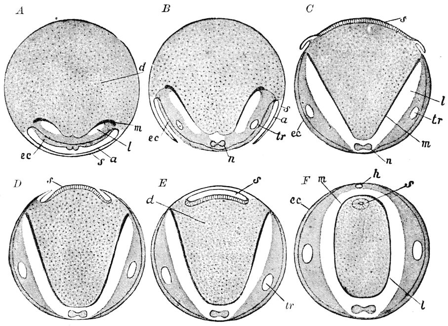
Fig. 530.—Diagram of the formation of the dorsal organ in Hydrophilus. A, cross-section through an egg, whose primitive streak is still covered over by amnion (a) and serosa (s). B, amnion and serosa are grown together in the middle line, then separated and drawn back to form a fold on each side. C, by the contraction of the serosa (s),which becomes converted into the dorsal plate, the folds become drawn up dorsally. D, the contracted serosa becomes partly overgrown by the folds. E, the folds grow together to form the dorsal tube. F, the mid-gut has closed over dorsally and enclosed the dorsal tube (s): a, amnion; d, yolk; ec, ectoderm; h, heart; l, body-cavity; m, rudiment of the mid-gut; n, nervous system; s, serosa (in C and D = dorsal plate, in E and F, dorsal tube); tr, the chief tracheal stem.—After Graber and Kowalevsky, from Lang, and Korschelt and Heider.
556The gnathal or post-antennal segments at first bear but a small part in completing the tergal region of the head, but shortly before hatching the mandibles and their muscles enlarge, giving fulness to the upper and back part of the head.
i. Dorsal closure and involution of the embryonic membranes
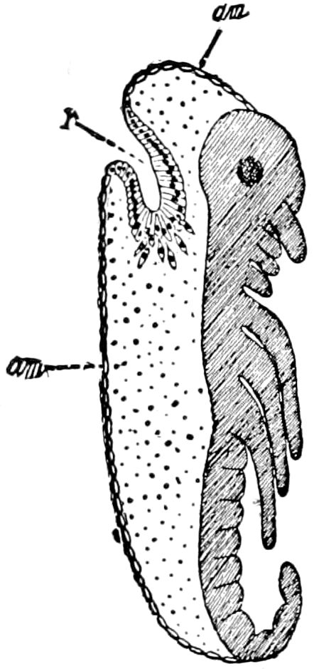
Fig. 531.—Schematic figure of the formation of the dorsal tube by invagination of the dorsal plate (transformed serosa); following after stage Fig. 520, C, and Fig. 521, D; am, amnion (now forming the provisional dorsal closure); r, dorsal tube, whose cells are already breaking away.—After Korschelt and Heider.
In most other Arthropoda (Crustacea, Arachnida, Myriopoda, etc.) development goes on by the formation of a so-called primitive band, but without the appearance of peculiar embryonic membranes. The outer surface of the entire egg becomes, then, in part covered by the band-like embryonic germ, and partly by a portion of the blastoderm which remains unchanged. The dorsal region is formed by the widening and spreading of the primitive band over the greater part of the surface of the egg, while the area of the unchanged section of the blastoderm continually becomes more restricted. It is generally accepted that the latter is concerned in the dorsal closure, because, together with a histological transformation, it becomes involved in the formation of the ectoderm of the primitive band.
A similar form of retrograde structure possibly occurs in the embryos of Poduridæ, in which a dorsal organ has been observed to develop in an early embryonic stage, which bears some relation to the cuticula enveloping the embryo, but whose significance is in general rather obscure.
In most insects the relations are more complicated, since in such cases, the amnion-folds rise on the edges of the primitive band and of the unchanged section of the blastoderm, whose retrograde development is intimately connected with the closure of the back.
A very simple case of dorsal closure, but which certainly is not a primitive one, occurs in Muscidæ and certain other Diptera whose amnion-folds are developed in a rudimentary way. In this case (according to Kowalevsky and Graber), the amnion-folds become smoothed out again. Amnion and serosa become then a simple epithelium, which throughout corresponds to the unmodified type of blastoderm of Crustacea, Arachnida, and Myriopoda, and here seems to share in the formation of the back. More complicated and very manifold relations of dorsal closure and involution of the embryonal membranes occur in other insects, of which Korschelt and Heider distinguish four different types:
5571. Involution under the formation of a continuous dorsal amnion-serosa-sac (Odonata).
2. Involution with exclusively dorsal absorption of the amnion (Doryphora).
3. Involution with exclusively dorsal absorption of serosa and separation of the amnion (Chironomus and Trichoptera).
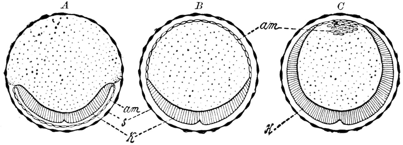
Fig. 532.—Diagram of the formation of the dorsal walls in Doryphora in cross-sections: am, amnion; in B, serving as a provisional dorsal closure, in C, about to break up; k, primitive band; s, serosa.—After Wheeler, from Korschelt and Heider.
4. Involution with separation of both embryonic membranes (Lepidoptera and Hymenoptera, Hylotoma).
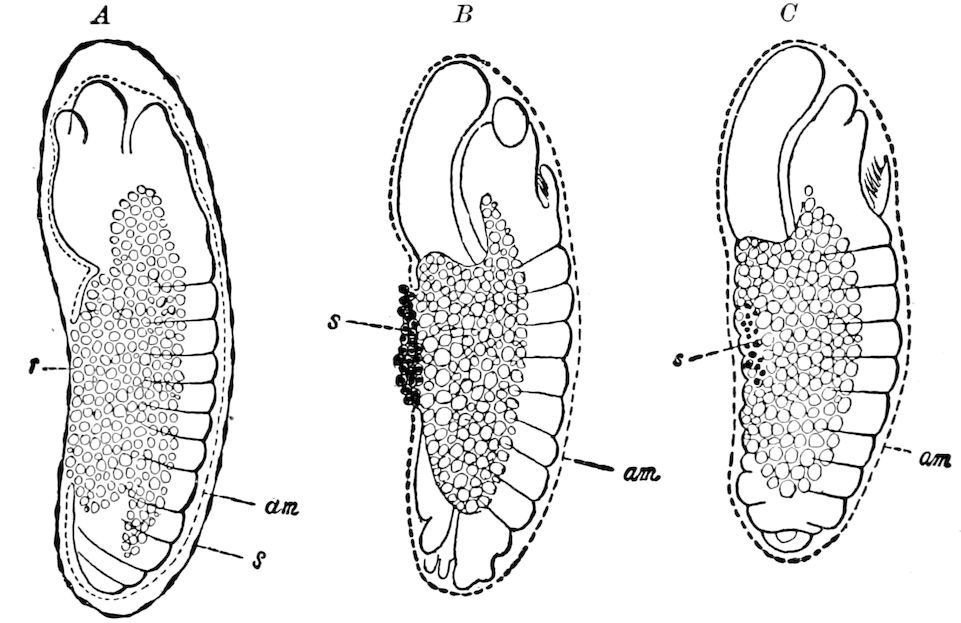
Fig. 533.—Involution of the embryonic membranes of Chironomus: am, amnion; r, dorsal umbilicus; s, serosa, which has withdrawn into the region of the dorsal umbilicus, and in C has passed into the interior of the embryo.—After Graber, from Korschelt and Heider.
The first type occurs in the most primitive order of winged insects. The second type (Coleoptera) appears to be an independently inherited form of dorsal closure. In the first type, the formation of the amnion-serosa-sac is initiated by a rupture of the two fused embryonic membranes. This rupture in the ventral middle line occurs in Odonata only in the region of the head-section. 558In the second type only the amnion, in the third only the serosa are concerned in this rupture, while in the fourth type both membranes remain intact until the slipping out of the larva. (Korschelt and Heider.)
j. Formation of the germ-layers
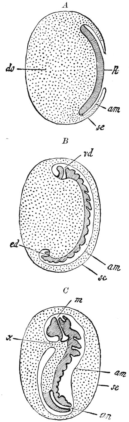
Fig. 534.—Diagram showing the formation of the embryonic membranes in Lepidoptera (A, after Kowalevsky, B and C, after Tichomiroff): k, primitive band; am, amnion: se, serosa; do, yolk; vd, invagination of the fore-gut, ed, of the hind-gut; m, mouth; an, anus; x, dorsal umbilical passage.—From Korschelt and Heider.
The older views on the structure of the layers of the primitive band of insects were thoroughly unsatisfactory. Bütschli first found that in the bee, by a kind of folding process, an inner layer of the primitive band arose. Soon afterwards Kowalevsky, by the employment of section-cutting and thorough researches, laid the foundation of a more exact knowledge of these layers. He found that in Hydrophilus a furrow extended along the whole length of the primitive band (Fig. 515, A, B, r), which, while invaginating or sinking in, gave rise to the inner layer of the primitive band, i.e. the common rudiment of endoderm and mesoderm (Fig. 539, A-C).
Kowalevsky also found similar conditions in the honey-bee (Apis), Lepidoptera, and other forms. The furrow above mentioned must be regarded as a very long gastrula invagination, extending along the entire ventral side of the embryo, and the edges of the furrow as a long-drawnout blastopore. The tube arising in Hydrophilus through the closing of the furrow we may regard as a primitive intestinal canal.
The first rudiment of the gastrula furrow appears in insects as two folds extending along both sides of the median line in the thickened ventral plate (Fig. 536, f), through whose 559formation a more median section of the ventral plate, the so-called middle plate (m), becomes separated from the side plates (s). As the middle plate curves in and becomes overgrown by the folds forming the edges of the blastopore, the gastrula-tube (Fig. 539, A, r) is formed, and furnishes the rudiments of the lower (inner) layer. The ectoderm, then, according to Heider, arises from the lateral plates of the primitive band. The growth of the edges of the blastopore, by which the closure of the gastrula-tube is effected, takes place latest in the region of the most anterior part of the furrow (Fig. 515, B and C), corresponding to that place in the primitive band in which the stomodæum afterwards develops.
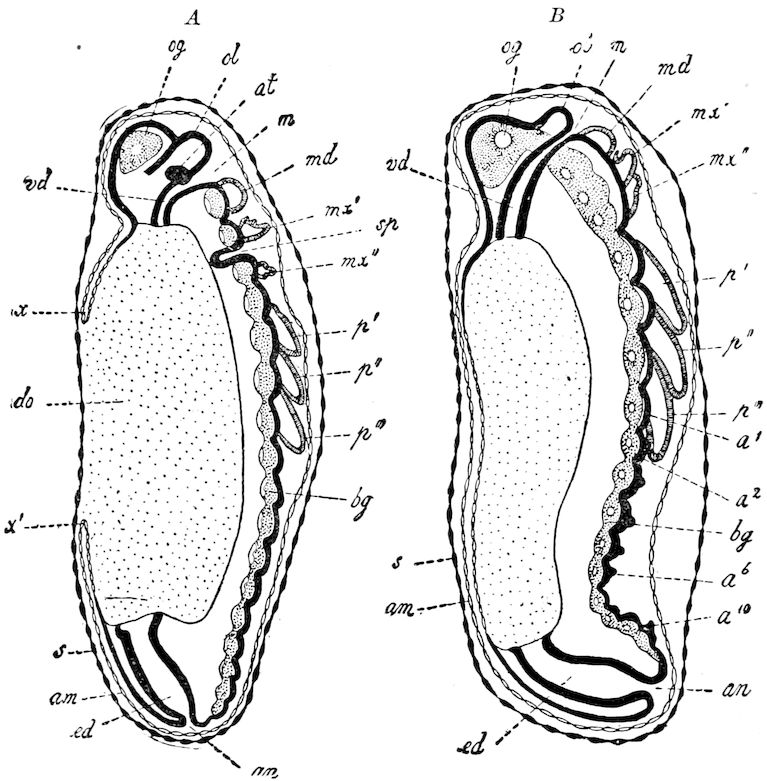
Fig. 535.—Two embryonic stages of a saw-fly (Hylotoma berberidis) in schematic median section: a1–a10, 1st to 10th abdominal segments; bg, ventral nervous cord; og, brain; ol, germ of labrum; sp, salivary gland; ed, hind-gut; x, x′, inner folds of amnion: other letters as before.—After Graber, from Korschelt and Heider.
560
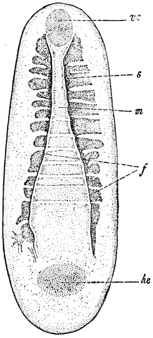
Fig. 536.—Gastrula stage of the wall-bee (Chalicodoma), so-called flask-shaped stage: f, folds which on each side border the middle plate (edge of the blastopore); m, the partly segmented middle plate (here = rudiment of the mesoderm); s, the segmented lateral plate (becoming afterwards the ectoderm of the primitive band); ve, fore, he, hinder entodermal rudiment.—After Carrière, from Korschelt and Heider.
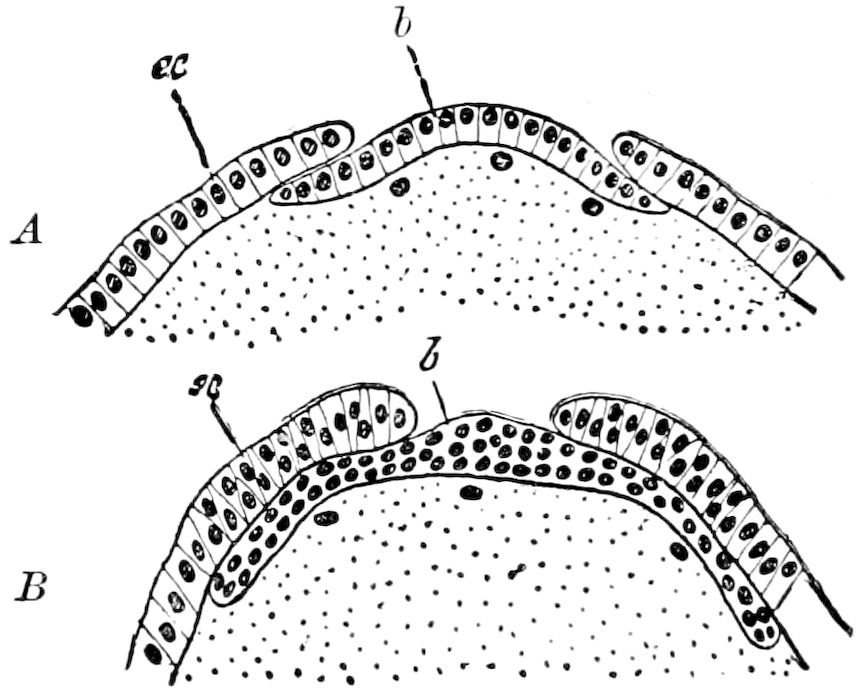
Fig. 537.—Two successive stages in the gastrulation of Apis. Cross-section through the primitive band: b, lower (inner) layer; ec, ectoderm.—After Grassi, from Korschelt and Heider.
During the invagination of the middle plate and its transformation into the gastrula-tube a change takes place in its histological character (Fig. 539, A and B). While it originally consists of a high cylinder epithelium, which after farther changes becomes divided into several layers, since the wedge-shaped single cells push themselves over each other, the cells in later stages become more and more cubical or irregularly polygonal (Fig. 539, B), and are irregularly arranged. At the same time the gastrula-tube is compressed in a dorso-ventral direction. While it in this way spreads out laterally under the side plates (ectoderm), its originally circular primitive lumen passes into the form of a horizontal fissure, which in Hydrophilus long remains as the boundary between the two layers of the inner (or lower) membrane. (Korschelt and Heider.)
There are numerous variations of the process of gastrulation, which are by Korschelt and Heider divided into three types, as follows:—
1. Through invagination and formation of a tube (Fig. 539, A, Hydrophilus, Musca, Pyrrhocoris, etc.).
2. By a lateral overgrowth (Fig. 537, Lepidoptera and Hymenoptera).
3. By an inward growth of cells from a median furrow (Aphides and Trichoptera).
In Doryphora and Lina (Fig. 524) the hinder end of the gastrula furrow is forked.
561
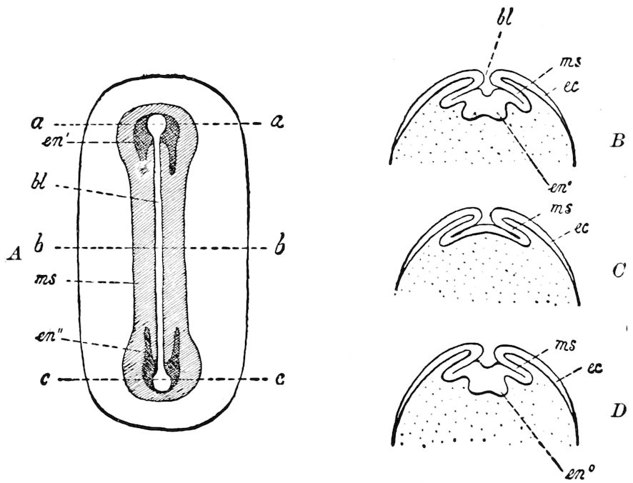
Fig. 538.—Diagrammatic sketch of the formation of the germinal layers in Doryphora: A, view of upper surface. B, cross-section through the fore end of the primitive streak at the line a-a. C, section through the middle of the primitive streak corresponding to the line b-b. D, section through the hinder end of the primitive band corresponding to the line c-c: bl, blastopore; ec, ectoderm; en′, anterior U-shaped; en″, hinder U-shaped germ of the endoderm; ms, mesoderm.—After Wheeler, from Korschelt and Heider.
The cellular layer arising from the gastrula invagination (lower layer) forms the common germ of the endoderm and mesoderm. It has only recently become known how these two germ-layers of insects have become differentiated. Kowalevsky first discovered in Musca that the greatest part of the lower (inner) layer yielded mesoderm exclusively, and that a cell-mass only corresponding to the most anterior and posterior end of the primitive band was used in the formation of the endoderm. We must therefore, in insects, speak of a fore and a hinder endodermal rudiment. In proportion, now, as the ectodermal invaginations, which are destined to form the stomodæum and the proctodæum sink beneath the surface of the embryo, the cell-masses of which the two endodermal rudiments are composed are pushed farther in, and a separation between them and the mesoderm is thus effected. The two endodermal rudiments now form accumulations of cells which lie closely adjacent to the blind ends of the stomodeal and the proctodeal invaginations. They soon widen out into two hour-glass-shaped rudiments, which are directed with their concavities towards each other, but with their convex side towards the nearest pole of the egg. They soon change their form; two lateral stripes grow out from them, and each now assumes the form of a U (Fig. 538, en′). The limbs of the fore 562and hind U-shaped rudiment are directed toward each other, and grow towards each other until they meet, and are fused together. Thus the endodermal rudiments arising out of the fusion of the two U-shaped rudiments form two stripes extending along the primitive band and situated mostly under the primitive segments. At the two ends the endodermal rudiment fuses with the stomodeal and proctodeal invaginations. These lateral endodermal streaks now spread out, and gradually begin to grow over the yolk, on whose outer surface they lie. This overgrowth makes the greater advance on the ventral side, so that the two endodermal streaks first unite in the ventral median line and afterward in the dorsal. The yolk in this way passes completely into the interior of the rudiment of the mid-intestine.
Kowalevsky has already proved that it is the median parts only of the inner layer which at the two ends of the primitive band become separated as endodermal rudiments through the advance of the stomodeal and proctodeal invaginations: the lateral portions become mesoderm.
Kowalevsky has compared the germ-layers of insects with those of Sagitta. This comparison is supported by the later researches of Heider and of Wheeler on Coleoptera. (See Korschelt and Heider, p. 809.)
Relations somewhat different from the common type of formation of germ-layers occur in Hymenoptera. Kowalevsky and also Grassi agree that here also the endoderm originally forms a part of the lower (inner) layer. But the separation of the endoderm from the mesoderm goes on in Apis in such a way that the two ends of the inner layer pass up to the dorsal side of the egg, where the fore and hind rudiments of the endoderm extending along the back of the embryo grow together. When the two horseshoe-shaped rudiments have met each other and become fused, the enclosing of the yolk begins, which accordingly here proceeds from the dorsal towards the ventral side, instead of vice versa. As a result the endodermal cell-layer in Apis (and also Chalicodoma) at first does not lie under the primitive band, but on the dorsal side of the egg under that flat epithelium, which, arising from the amnion-fold, completes the provisional closure of the back.
The yolk-cells and secondary yolk-segmentation are discussed by Korschelt and Heider at this point. The yolk-cells are elements scattered throughout the yolk and which partly remain in the yolk during the formation of the blastoderm (Fig. 507, C and D), but which in part through a later immigration pass out of the blastoderm into the yolk. Graber has proved the fact of the migration of cells from the lower layer into the yolk, and his observations have been confirmed by other authors. Indeed, in certain cases (Melolontha), these later immigrant cells are clearly distinguishable by their histological characters from those originally found in the yolk.
The yolk-cells are regularly scattered throughout the yolk. Their use to the embryo lies in the fact that they absorb the particles of yolk, which they digest and thus reduce to a fluid condition. It usually happens that after the complete formation of the primitive band there results a delimitation of the areas enclosing each yolk-cell, and this occurrence is called secondary yolk-division. 563In special cases (Apis, Musca) this occurrence seems not to take place. The yolk-cells are still, after the complete formation of the mid-intestine, to be recognized in the yolk-remnants filling the interior of the same, and gradually become absorbed.
k. Farther development of the mesoderm. Formation of the body-cavity
We have seen that by means of an invagination extending throughout the entire length of the primitive band a layer of cells is produced which soon spreads out on the inner side of the band and thus forms a second lower (inner) layer (Fig. 539, C). From this inner layer is separated at the anterior and posterior ends of the primitive band, the endoderm, which lies in direct contact with the invaginations of the proctodæum and stomodæum. The remainder, by far the most extensive part of the inner layer, is the mesoderm.
The mesoderm now becomes divided into two lateral streaks (mesodermal streaks), by the withdrawal of its cells from the median line (Fig. 539, D). This withdrawal is not, however, always a complete one. In the free median space thus formed, the yolk often forms the so-called median yolk-ridge. Segmentally arranged cavities soon appear in the lateral region of the mesoderm (the primitive segmental cavities), and the bordering mesoderm-cells arrange themselves in the form of an epithelium, and constitute the wall of the primitive segments or cœlom-sac. (Korschelt and Heider).
The primitive segmental cavities in general arise through a split in the mesoderm. In Phyllodromia, according to Heymons, the primitive segments are very extensive. The mesoderm, at the time of the formation of the rudiments of the appendages, is raised with the ectoderm from the surface of the yolk, and in this way there arise in each segment cavities, which, since they are surrounded by mesodermal elements, become the closed cœlom-sacs (Fig. 540, c, c′, c″).
The cœlom-sacs differ in different groups. They are largest in Orthoptera (Phyllodromia), where they take up almost all the cell material of the mesoderm in their formation, and exhibit certain conditions recalling those of Peripatus. The very large primitive segmental cavities, which in Orthoptera also extend into the rudiments of the appendages (Fig. 540, B, ex), in their later stages are, through the formation of a constriction, divided into a dorsal and a ventral half (Fig. 540, B, c′, c″). The ventral portions of these cavities extending into the extremities soon disappear, while the cells of their walls lose their epithelial nature, and group themselves irregularly into a sort of mesenchym. In this tissue, then, arises, partly through a separation among its cells, partly through the elevation of the same from the upper surface of the yolk, the definite body-cavity. The dorsal portions of the primitive segmental cavities remain unchanged a longer time in order to play a rôle in the formation of the intestinal muscular layer, of the heart, pericardial septum, and sexual organs.