PART I.—MORPHOLOGY AND PHYSIOLOGY (continued 8)
Reading edition
The mid-intestine.—This section of the digestive canal, often, though erroneously, called the “chylific stomach” or ventriculus, differs not only in its embryonic history, but also in its structure and physiology from the fore and hind intestine of arthropods, and also presents no analogy to the stomach of the vertebrate animals. In insects it is a simple tube, not usually lined with chitin, since it is not formed by the invagination of the ectoderm, as are the fore and hind intestine, the absence of the chitinous intima promoting the absorption of soluble food. Into the anterior end either open two or more large cœcal tubes (Fig. 299), or its whole outer surface is beset with very numerous fine glandular filaments like villi (Fig. 317 and Fig. 329).
The mid-intestine varies much in size and shape; it is very long in the lamellicorn beetles (Melolontha and Geotrupes), and while in Meloë it is very large, occupying the greatest part of the body-cavity, in the longicorn beetles and in Lepidoptera it is very small. The pyloric end consists of an internal circular fold projecting into the cavity. In the Psocidæ (Cæcilius) the pyloric end is prolonged into a slender tube nearly as long as the larger anterior portion.
The limits between the mid and hind intestine are in some insects difficult to define, the urinary tubes sometimes appearing to open 315into the end of the mid-intestine (“stomach”). The latter also is sometimes lined with an intima. The limits are also determined by a circular projection, directly behind which is an enlargement of the intestine in the shape of a trench (rigole), or circular cul-de-sac (the “pyloric valvule” of some authors, including Beauregard), while the walls of the small intestine contract so as to produce a considerable constriction of the cavity of the canal. This constriction exactly coincides with the beginning of the double layer of circular muscles in the wall of the small intestine. An internal layer, which is the continuation of the circular muscles of the chylific stomach, and an external layer much more developed probably belong to this part of the alimentary canal. Since the homologue of the circular fold occurs in the locust as well as in Diptera, it is probably common to insects in general.
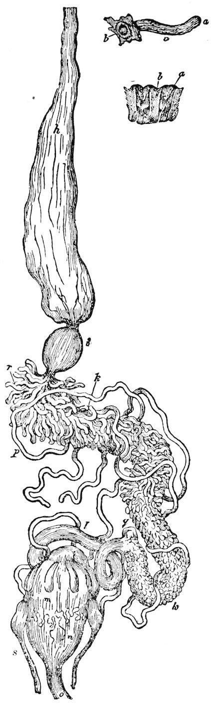
Fig. 317.—Digestive canal of Carabus monilis: h, œsophagus; i, gizzard or proventriculus; k, “stomach,” with its cœca (r); p, urinary tubes; q, their point of insertion; m, n, colon, with cœcal glands; s, anal glands; a, b, c, a gastric cœcum; a, b, portion of lining of gizzard.—After Newport.
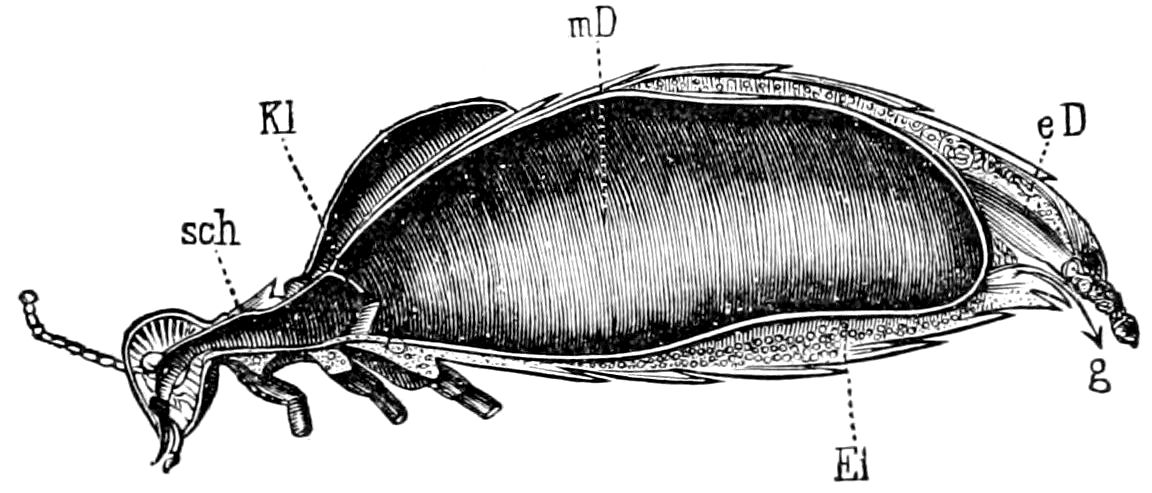
Fig. 318.—Digestive canal of Meloe: sch, œsophagus; Kl, œsophageal valve; mD, mid-intestine; eD, hind-intestine; Ei, eggs; g, sexual opening.—After Graber.
Gehuchten adds that the limit set by the circular projection does not exactly coincide with the opening into the intestine of the urinary tubes and the two annexed glands. He shows by a section (his Fig. 133) that the tubular glands open into the alimentary canal 316in front of the circular fold. It is the same with the Malpighian tubes. They are not, therefore, he claims, dependences of the terminal intestine, but of the mid-intestine. Beauregard has observed the same thing in the vesicating insects (Meloidæ). The Malpighian tubes, he says, open into the “chylific stomach” before the valvular crown. This arrangement does not seem to be general, because, according to Balbiani, the Malpighian vessels open into the beginning of the intestine in Cryptops. Compare also Minot’s account of the valve in locusts separating the stomach from the intestine, and in front of which the urinary or Malpighian tubes open.
Histology of the mid-intestine.—The walls of the stomach are composed of an internal epithelium, a layer of connective tissue, with two muscular layers, the inner of which is formed of unstriated circular muscular fibres, and the outer of striated longitudinal muscular fibres.
In the cockroach short processes are given off from the free ends of the epithelial cells, as in the intestine of many mammals and other animals. “Between the cells a reticulum is often to be seen, especially where the cells have burst; it extends between and among all the elements of the mucous lining, and probably serves, like the very similar structure met with in mammalian intestines, to absorb and conduct some of the products of digestion.” (Miall and Denny.)
Gehuchten shows that the epithelial lining of the mesenteron (chylific stomach) of the dipterous larva Ptychoptera is composed of two kinds of cells, i.e. secreting or glandular cells and absorbent cells, the former situated at each end of the stomach, and the absorbent cells occupying the middle region. The part played by these cells in digestion will be treated of beyond in the section on digestion. (See p. 327.)
The hind-intestine.—In many insects this is divided into the ileum, or short intestine, and the long intestine. The limit between the intestine and stomach is externally determined by the origin of the urinary tubes, which are outgrowths of the anterior end of the proctodæum. Like the fore-intestine the hind-intestine is lined with a thick muscular layer, and, as Gehuchten states, the passage from the epithelial lining of the stomach (mid-intestine) to the muscular lining of the intestine is abrupt.
Large intestine.—In Ptychoptera, as described by Gehuchten, there are no precise limits between the small and large intestine; the epithelium of the large intestine has a special character, and its constituents present a close resemblance to the absorbed cells of the chylific stomach, being like them large and polygonal. The muscular layer is not continuous, and is formed of longitudinal and circular fibres, the latter being the larger.
317The ileum—Though in most insects slender, and therefore called the small intestine, the ileum is in locusts (Fig. 298) and grasshoppers (Anabrus, Fig. 299) as thick as the stomach. In many carnivorous beetles (Dyticus, Fig. 320, il, and Necrophorus) it is very long, but rather slender and short in the Carabidæ and Cicindelidæ, as well as those insects whose food is liquid, such as Diptera. In the Lepidoptera it varies in length, being in Sphinx quite long and bent into seven folds (Fig. 309), while it is very short in the Psocidæ, Chrysomelidæ, and Tenthredinidæ.
In the locust the ileum is traversed by six longitudinal folds with intervening furrows; outside of each furrow is a longitudinal muscular band. Seen from the inner surface the epithelium has an unusual character, the cells in the middle of each of the flat folds being quite large, polygonal in outline, while towards the furrows the cells become very much smaller. The walls are double when seen in transverse section, the inner layer consisting of epithelial cells resting on connective tissue, the outer layer formed of circular muscles. The cuticula is thin, but probably chitinous; it resembles that on the gastro-ileal folds, except that there are no spinules, but unlike the cuticula of the stomach it extends equally over the folds and the furrows. (Minot.) In the cockroach the junction of the small intestine with the colon is abrupt, a well-developed annular fold assuming the nature of a circular valve. (Miall and Denny.)
The gastro-ileal folds.—In the locust the intestine is separated from the chylific stomach by what Minot calls “the gastro-ileal folds,” which form a peculiar valve. The urinary vessels open just underneath and in front of this valve. In Melanoplus, and probably in the entire family of Acrydiidæ, they are indicated as “dark spots, round in front and lying at the anterior end of the ileum so as to form a ring around the interior of the intestine.” They are 12 in number, and all alike. They are pigmented and round in front where they are broadest and stand up highest; they narrow down backwards, the pigment disappears, and they gradually fade out into the ileal folds. Directly beneath them, and just at the posterior end of the stomach, there is a strong band of circular striated muscular fibres. The epithelium of these folds is covered with minute conical spines, which are generally, but not always, wanting between the folds. (Minot.)
The colon.—This section of the intestine (Fig. 319) is sometimes regarded as a part of the rectum. In the locust the six longitudinal folds of the ileum are continued into the colon, but their surface, instead of being smooth as in the ileum, is thrown up into numerous 318irregular curved and zigzag secondary folds. The cells of the epithelium are of uniform size, and the layer is covered by a highly refringent cuticula without spines; and, like that in the ileum, it rests on a layer of connective tissue, beyond which follows (1) an internal coat of longitudinal, and (2) an external coat of circular striated muscular fibres. (Minot.)
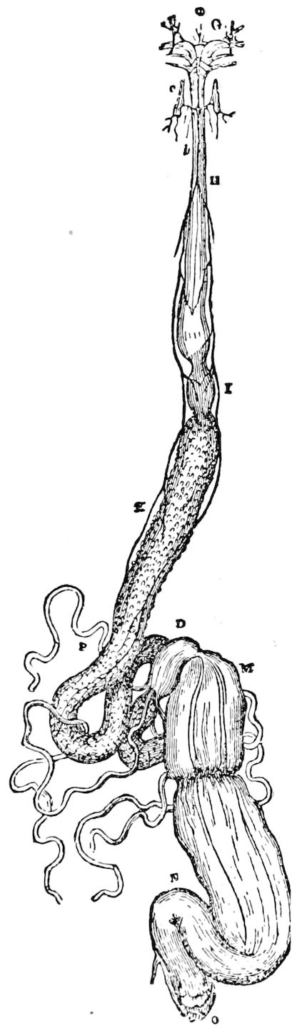
Fig. 319.—Digestive canal of Lucanus cervus: G, anterior muscles of the pharynx; H, œsophagus; I, gizzard; K, chyle-stomach; L, ilium; M, colon (cœcal part of); N, colon; O, rectum; a, frontal ganglion of the vagus; b, vagus nerve; c, anterior lateral ganglion connected with the vagus.—After Newport.
In butterflies (Pontia brassicæ), in Sphinx ligustri, and probably in most Lepidoptera the colon is distinct from the rectum, and is anteriorly developed into a very large more or less pyriform or bladder-like cæcum (Figs. 309, 310), which in certain Coleoptera (Dyticus, Fig. 320, d; Silpha, Necrophorus, etc.) is of remarkable length and shape; it also occurs in Nepidæ (Fig. 327). In the cockroach a lateral cæcum “is occasionally, but not constantly, present towards its rectal end,” and a constriction divides the colon from the rectum. (Miall and Denny.)
The rectum.—The terminal section of the hind-gut varies in length and size, but is usually larger than the colon, and with thick, muscular walls. In Lepidoptera it is narrow and short.
The rectum is remarkable for containing structures called rectal glands (Fig. 298). Chun describes those of Locusta viridissima as six flat folds, formed by a high columnar epithelium and a distinct cuticula; there is a coat of circular bands corresponding to the furrows between the glands. Minot states that this description is applicable to the locusts (Acrydiidæ) he has investigated, the only difference being in the structural details of the single layers. He claims that the rectal folds “do not offer the least appearance of glandular structure,” neither is their function an absorbent one, as Chun supposed. From their structure and position Fernald regards the rectal glands of Passalus as acting like 319a valve, serving to retain the food in the absorptive portions of the digestive tract till all nutriment is extracted.
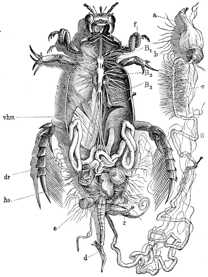
Fig. 320.—Dyticus marginalis, ♂ opened from the back: a, crop; b, proventriculus; c, mid-intestine beset with fine cœcal glands; d, long cœcal appendage of the colon; apodemes; B1-B3, apodemes; vhm, coxal extensor muscle, moving the hind leg; ho, testis; dr, accessory gland; r, penis; e, reservoir of the secretion of the anal gland.—After Graber.
The epithelial folds of the larvæ of dragon-flies serve as organs of respiration, the water being admitted into this cavity, and when forcibly expelled serving to propel the creature forward. Paired and single anal glands (repugnatorial) enter the rectum of certain Coleoptera (Figs. 302, l; 317, s; 320, e).
The vent (anus).—The external opening of the rectum is situated in the end of the body, in the vestigial 10th or 11th abdominal segment, 320and is more or less eversible. It is protected above in caterpillars, and other insects with 10 free abdominal segments, by the suranal plate. It is bounded on the sides by the paranal lobes, while beneath is the infra-anal lobe.
The anus is wanting in certain insects, and where this is the case the hind-gut, owing to a retention of the embryonic condition, is usually separated from the mid-intestine. (See p. 300.)
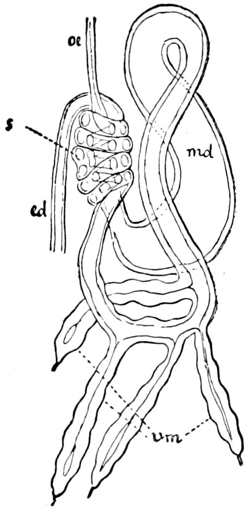
Fig. 321.—Enteric canal of Psyllopsis fraxinicola: œ, œsophagus; md, mid-intestine; ed, hind-intestine; vm, urinary vessels; s, the coil formed by the hind-intestine and the most anterior part of the mid-intestine.—After Witlaczil, from Lang.
Some remarkable features of the digestive canal in hemipterous insects are noteworthy. In the Coccidæ, according to Mark, the anterior end of the long mid-intestine forms, with the hinder end of the œsophagus, a small loop, whose posterior end is firmly grown to the wall of the rectum, and forms a cup-like invagination of the latter. Then the rest of the tube-like stomach turns sidewise and forms a large loop, which turns back on itself and occupies a large part of the body-cavity. This loop receives on the anterior end, near the œsophagus, the two urinary vessels, and forms just below the opening into the rectum a short cæcum.
In other homopterous genera (Psyllidæ and some Cicadidæ) Witlaczil describes nearly the same peculiarity, the mid-gut and part of the intestine forming a loop growing together for a certain distance and winding round each other (Fig. 321).
Histology of the digestive canal.—In all the divisions of the digestive canal of insects the succession of the cellular layers composing it is the same: 1st, a cuticula; 2d, an epithelium; 3d, connective tissue; 4th, muscular tissue. In the locust, the first division of the canal (fore-gut), there are two muscular coats, an internal longitudinal and an external circular coat; the fibres are all striated. The lining epithelium is not much developed, but forms a thick, hard, and refringent cuticula, which is thrown up into spiny ridges. In the second division (mid-gut, “stomach”) the epithelium is composed of very high columnar cells, which make up the greater part of the thickness of the walls, while the cuticula is very delicate, slightly refringent, with no ridges, and is probably not chitinous; the fibres of the muscular coats are not striated, while this division is also distinguished by the presence of glandular follicles and folds. The stomach and the cæcal appendages have all these peculiarities in common, while no other part of the canal is thus characterized.
321The third division (intestine and rectum) is composed of an epithelium, the cells of which are intermediate in size between those of the fore and mid gut. The cells are often pigmented, and they are covered by a much thicker cuticula than that of the stomach, but which is not so thick and hard as that of the œsophagus and proventriculus. The very refringent cuticula is not thrown up into ridges, though in some parts it is covered with delicate conical spines, which are very short. “The epithelium and underlying connective tissue (tunica propria) are thrown up into six folds, which run longitudinally, being regular in the ileum and rectum (as the rectal glands), but very irregular in the colon. Outside the depression between each two neighboring folds there is a longitudinal muscular band, these making six bands. This peculiar disposition of the longitudinal muscles does not occur in any other part of the canal; it is, therefore, especially characteristic of the third division.” (Minot.)
LITERATURE ON THE ORGANS OF DIGESTION
Treviranus, G. R. Resultate einiger Untersuchungen über den inneren Bau der Insekten. (Verdauungsorgane von Cimex rufipes.) (Annal. d. Wetterau. Gesells., 1809, i, pp. 169–177, 1 Taf.)
Ramdohr, C. A. Abhandlungen über die Verdauungswerkzeuge der Insekten. 1811, vii, pp. 221, 30 Taf.
Dutrochet, R. J. H. Mémoire sur les métamorphoses du canal alimentaire dans les insectes. (Journal de Physique, 1818, lxxxvi, pp. 130–135, 189–204; Meckel’s Archiv, 1818, iv, pp. 285–293.)
Suckow, F. W. L. Verdauungsorgane der Insekten. (Heusinger’s Zeitschr. f. organ. Physik., 1828, iii, pp. 1–89.)
Doyère, L. Note sur le tube digestif des Cigales. (Ann. Sc. nat. Zool., 1839, 2 Sér., xi, pp. 81–85.)
Grube, A. E. Fehlt den Wespen- oder Hornissenlarven ein After oder nicht? 1 Taf. (Müller’s Archiv für Physiol., 1849, pp. 47–74.)
Sirodot. Recherches sur les sécrétions chez les insectes. (Ann. Sc. nat. Zool., 1858, 4 Sér., x, pp. 141–189, 251–328, 12 Pls.)
Milne-Edwards, H. Leçons sur la physiologie et l’anatomie comparée, v, 1859, pp. 498–536, 581–638.
Dufour, L. Recherches anatomiques sur les Carabiques et sur plusieurs autres insectes Coléoptères. Appareil digestif. (Ann. Sc. nat., ii, 1824, pp. 462–482, 2 Pls.; iii, 1824, pp. 215–242, 5 Pls., pp. 476–491, 3 Pls.; iv, 1824, pp. 103–125, 4 Pls.; iv, 1825, pp. 265–283.)
—— Recherches anatomiques sur l’Hippobosque des chevaux. (Ann. Sc. nat., 1825, vi, pp. 299–322, 1 Pl.)
—— Description et figure de l’appareil digestif de l’Anobium striatum. (Ibid., xiv, 1828, pp. 219–222, 1 Pl.)
—— Recherches anatomiques sur les Labidoures. Appareil de la digestion. (Ibid., xiii, 1828, pp. 348–354, 2 Pls.)
322—— Recherches anatomiques et considerations entomologiques sur quelques insectes Coléoptères, compris dans les familles des Dermestins, des Byrrhiens, des Acanthopodes et des Leptodactyles. Appareil digestif. (Ibid., Sér. 2, Zool., i, 1834, pp. 67–76, 2 Pls.)
—— Résumé des recherches anatomiques et physiologiques sur les Hémiptères. (Ibid., pp. 232–239.)
—— Mémoire sur les métamorphoses et l’anatomie de la Pyrochroa coccinea. Appareil digestif. (Ibid., Sér. 2, Zool., xiii, 1840, pp. 328–330, 334–337, 2 Pls.)
—— Histoire comparative des metamorphoses et de l’anatomie des Cetonia aurata et Dorcus parallelepipedus. Appareil digestif. (Ibid., Sér. 2, Zool., 1824, xviii, pp. 174–176, 2 Pls.)
—— Anatomie générale des Diptères. Appareil digestif. (Ibid., Sér. 3, Zool., i, 1814, pp. 248, 249.)
—— Histoire des métamorphoses et de l’anatomie du Piophila petasionis. Appareil digestif. (Ibid., Sér. 3, Zool., i, 1844, pp. 372–377, 1 Pl.)
—— Études anatomiques et physiologiques sur les insectes Diptères de la famille des Pupipares. Appareil digestif. (Ibid., Sér. 3, Zool., iii, 1845, pp. 67–73, 1 Pl.)
—— Recherches sur l’anatomie et l’histoire naturelle de l’Osmylus maculatus. Appareil digestif. (Ibid., Sér. 3, Zool., ix, 1848, pp. 346–349, 1 Pl.)
—— Études anatomiques et physiologiques, et observations sur les larves des Libellules. Appareil digestif. (Ibid., Sér. 3, Zool., xvii, 1852, pp. 101–108, 1 Pl.)
—— Recherches anatomiques sur les Hyménoptères de la famille des Urocerates. Appareil digestif. (Ibid., Sér. 4, Zool., i, 1854, pp. 212–216, 1 Pl.)
—— Fragments d’anatomie entomologique. Sur l’appareil digestif du Nemoptera lusitanica. (Ibid., Sér. 4, viii, 1857, pp. 6–9, 1 Pl.)
—— Recherches anatomique et considerations entomologiques sur les Hémiptères du genre Leptopus. Appareil digestif. (Ibid., Sér. 4, Zool., 1858, x, pp. 352–356, 1 Pl.)
—— Recherches anatomiques sur l’Ascalaphus meridionalis. Appareil digestif. (Ibid., Sér. 4, xiii, 1860, pp. 200–202, 1 Pl.)
Leydig, F. Zur Anatomie von Coccus hesperidum. (Zeitschr. f. wissens. Zool., v, 1853, pp. 1–12, 1 Taf.)
Lubbock, J. On the digestive and nervous System of Coccus hesperidum. (Proc. Roy. Soc., ix, 1886, pp. 480–486; also Ann. Mag. Nat. Hist., 1859, Ser. 3, iii, pp. 306–311.)
Scheiber, S. H. Vergleichende Anatomie und Physiologie der Œstriden-Larven. V. Das chylo- und uropœtische System. (Sitzber. d. k. Akad. d. Wissens. Wien. Math.-naturwiss. Cl., 1862, xlv, pp. 39–64, 1 Taf.)
Gerstaecker, A. Bronn’s Klassen und Ordnungen des Tierreichs. V. Gliederfüssler. (Ernährungsorgane, pp. 87–105.)
Graber, V. Zur naheren Kenntnis des Proventriculus und der Appendices ventriculares bei den Grillen und Laubheuschrecken. (Sitzber. d. k. Akad. d. Wissensch. Wien. Mathem.-naturwiss. Cl., lix, 1869, pp. 29–46, 3 Taf.)
—— Ueber die Ernährungsorgane der Insekten und nächstverwandten Gliederfüssler. (Mitteil. d. naturwiss. Vereins für Steiermark. Graz, 1871, ii, pp. 181, 182.)
—— Verdauungssystem des Prachtkäfers. (Ibid., Graz, 1875.)
—— Die Insekten., i, 1877. (Verdauungsapparat, pp. 308–328.)
Wilde, K. F. Untersuchungen über den Kaumagen der Orthopteren. (Archiv f. Naturgesch., xliii Jahrg., 1877, pp. 135–172, 3 Taf.)
323Simroth, H. Ueber den Darmkanal der Larven von Osmoderma eremita mit seinen Anhängen. (Giebel’s Zeitschr. f. d. ges. Naturwiss., 1878, li, pp. 493–518, 3 Taf.)
Müller, H. Ueber die angebliche Afterlösigkeit der Bienenlarven. (Zool. Anzeiger, 1881, pp. 530, 531.)
Schiemenz, Paulus. Ueber das Herkommen des Futtersaftes und die Speicheldrüsen der Bienen, nebst einem Anhänge über das Riechorgan. (Zeitschr. f. wissens. Zool., xxxviii, 1883, pp. 71–135, 3 Taf.)
Rovelli, G. Alcune ricerche sul tubo digerente degli Atteri, Ortotteri e Pseudo-Neurotteri. (Como, 1884, p. 15.)
Beauregard, H. Structure de l’appareil digestif des Insectes de la tribu des Vésicants. (Compt. rend. Acad. Paris, 1884, xcix, pp. 1083–1086.)
—— Recherches sur les Insectes vésicants., 1 Part, Anatomie. (Journ. Anat. Phys. Paris, 1885, xxi Année, pp. 483–524, 4 Pls.; 1886, xxii Année, pp. 85–108, 242–284, 5 Pls.)
—— Les Insectes vésicants, Paris, 1890, Chap. III, Appareil digestif, pp. 63–99; (Phénomènes digestifs, pp. 161–170; Pls. 6–9.)
Wertheimer, L. Sur la structure du tube digestif de l’Oryctes nasicornis. (Compt. rend. Soc. Biol. Paris, 1887, Sér. 8, iv, pp. 531, 532.)
Kowalevsky, A. Beitrage zur Kenntniss der nachembryonal Entwicklung der Musciden. (Zeitschr. f. wissens. Zool., xlv, 1887, pp. 542–594, 5 Taf.)
Schneider, A. Ueber den Darm der Arthropoden, besonders der Insekten. (Zool. Anzeiger, 1887, x Jahrg., pp. 139, 140.)
—— Ueber den Darmkanal der Arthropoden. (Zool. Beiträge von A. Schneider, ii, 1887, pp. 82–96, 3 Taf.)
Fritze, A. Ueber den Darmkanal der Ephemeriden. (Berichte der Naturforsch.-Gesellsch. zu Freiburg i. Br., 1888, iv, pp. 59–82, 2 Taf.)
Emery, C. Ueber den sogenannten Kaumagen einiger Ameisen. (Zeitschr. f. wissens. Zool., 1888, xlvi, pp. 378–412, 3 Taf.)
Meinert, F. Contribution à l’anatomie des Fourmilions. (Overs. Danske Vidensk. Selsk. Forhandl. Kjöbenhavn, 1889, pp. 43–66, 2 Pls.)
Mingazzini, P. Richerche sul canale digerente dei Lamellicorni fitofage (Larve e Insetti perfetti). (Mitteil. Zool. Station zu Neapel, ix, 1889–1891, pp. 1–112, 266–304, 7 Pls.)
Fernald, Henry T. Rectal glands in Coleoptera. (Amer. Naturalist, xxiv, pp. 100, 101, Jan., 1890.)
Visart, O. Digestive canal of Orthoptera. (Atti Soc. Toscana Scient. Natur., vii, 1891, pp. 277–285.)
Eberli, J. Untersuchungen an Verdauungstrakten von Gryllotalpa vulgaris. (Vierteljahresschr. d. Naturforsch. Gesells. Zurich, 1892, Sep., p. 46, Fig.)
Holmgren, Emil. Histologiska studier öfver några lepidopterlarvers digestionskanal och en del af deras Körtelartade bildningar. (Ent. Tidskr. Årg. xiii, pp. 129–170, 1892, 6 Pls.)
Ris, F. Untersuchung über die Gestalt des Kaumagens bei den Libellen und ihren Larven. (Zool. Jahrb. Abth. Syst., ix, 1896, pp. 596–624, 13 Figs.)
See also the works of Straus-Dürckheim, Newport, Mark, Witlaczil, Vayssière, Landois, Jordan, Oudemans, Berlese, List, Grassi, Verson, Miall and Denny, Leidy, Cheshire, Kowalevsky, Gehuchten, Locy, etc.
324
b. Digestion in insects
For the most complete and reliable investigation of the process of digestion, we are indebted to Plateau, whose results we give, besides the conclusions of later authors:
In mandibulate or biting insects, the food is conducted through the œsophagus by means of the muscular coating of this part of the digestive canal. Suctorial insects draw in their liquid food by the contractions followed by the dilatations of the mid-intestine (chylific stomach). Dragon-flies, Orthoptera, and Lepidoptera swallow some air with their food.
Where the salivary glands are present, the neutral alkaline fluid secreted by them has the same property as the salivary fluid of vertebrates of rapidly transforming starchy foods into soluble and assimilable glucose. In such forms as have no salivary glands, their place is almost always supplied by an epithelial lining of the œsophagus, or, as in the Hydrophilidæ, a fluid is secreted which has the same function as the true salivary fluid.
Nagel states that the saliva of the larva of Dyticus is powerfully digestive, and has a marked poisonous action, killing other insects, and even tadpoles of twice the size of the attacking larva, very rapidly. The larvæ not only suck the blood of their victims, but absorb the proteid substances. Drops of salivary juice seem to paralyze the victim, and to ferment the proteids. The secretion is neutral, the digestion tryptic. Similar extra-oral digestion seems to occur in larvæ of ant-lions, etc. (Biol. Centralbl., xvi, 1896, pp. 51–57, 103–112; Journ. Roy. Micr. Soc., 1896, p. 184.)
In carnivorous insects and in Orthoptera, the œsophagus dilates into a crop (ingluvies) ended by a narrow, valvular apparatus (or gizzard of authors). The food, more or less divided by the jaws, accumulates in the crop, which is very distensible; and, when the food is penetrated by the neutral or alkaline liquid, there undergoes an evident digestive action resulting, in carnivorous insects, in the transformation of albuminoid substances into soluble and assimilable matter analogous to peptones, and, in herbivorous insects, an abundant production of sugar from starch. This digestion in the crop, a food-reservoir, is very slow, and, until it is ended, the rest of the digestive canal remains empty.
“Any decided acidity found in the crop is due to the injection of acid food; but a very faint acidity may occur, which results from the presence in the crop of a fluid secreted by the cæcal diverticula of the mesenteron.” (Miall and Denny.)
325When digestion in the crop is accomplished, the matters are subjected to an energetic pressure of the walls through peristaltic contractions, and then, guided by the furrows and chitinous teeth, pass along or gradually filter through the valvular apparatus or proventriculus, whose function is that of a strainer.
At the beginning of the “chyle-stomach” (mesenteron) of Orthoptera are glandular cæca which secrete a feebly acid fluid. This fluid emulsifies fats, and converts albuminoids into peptones. It passes forwards into the crop, and there acts upon the food.
In the mesenteron (mid-intestine) the food is acted upon by an alkaline or neutral fluid, never acid, either secreted, as in Orthoptera, by local special glands, or by a multitude of minute glandular cæca, as in many Coleoptera, or by a simple epithelial layer. It has no analogy with the gastric juices of vertebrates; its function differs in insects of different groups; in carnivorous Coleoptera it actively emulsionizes greasy matters; in the Hydrophilidæ it continues the process of transformation of starch into glucose, begun in the œsophagus. In the Scarabæidæ, it also produces glucose, but this action is local, not occurring elsewhere; in caterpillars, it causes a production of glucose, and transforms the albuminoids into soluble and assimilable bodies analogous to peptones, and also emulsionizes greasy matters. Finally, in the herbivorous Orthoptera there does not seem to be any formation of sugar in the stomach itself, the production of glucose being confined to the crop (jabot).
When digestion in the crop is finished, the proventriculus relaxes, and the contents of the crop, now in a semi-fluid condition, guided by the furrows and teeth, passes into the mesenteron, which is without a chitinous lining, and is thus fitted for absorption.
The contents of the mid-intestine (chylific stomach) then slowly and gradually pass into the intestine, the first anterior portion of which, usually long and slender, is the seat of an active absorption. The epithelial lining observed in certain insects seems, however, to indicate that secondary digestion takes place in this section. The reaction of the contents is neutral or alkaline.
The second and larger division of the intestine only acts as a stercoral reservoir. (The voluminous cæcum occurring in Dyticidæ, Nepa, and Ranatra, whether full or empty, never contains gas, and it is not, as some have supposed, a swimming-bladder.) The liquid product secreted by the Malpighian tubes accumulates in this division, and, under certain circumstances, very large calculi are often formed. In his subsequent paper on the digestion of the cockroach, Plateau states that in the intestine are united the residue of 326the work of digestion and the secretion of the urinary or Malpighian tubes, this secretion being purely urinary.
These organs are exclusively depuratory and urinary, freeing the body from waste products of the organic elements. The liquid they secrete contains urea (?), uric acid and abundant urates, hippuric acid (?), chloride of sodium, phosphates, carbonate of lime, oxalate of lime in quantity, leucine, and coloring-matters.
The products of the rectal or anal glands vary much in different groups, but they take no part in digestion, nor are they depuratory in their nature.
Insects have nothing resembling chylific substances.[51] The products of digestion, dissolved salts, peptones, sugar in solution, emulsionized greasy matters, pass through the relatively delicate walls of the digestive canal by osmose, and mingle outside of the canal with the blood.
Whatever substances remain undigested are expelled with the excrements; such are the chitin of the integuments of insects, vegetable cellulose, and chlorophyll, which is detected by the microspectroscope all along the digestive canal of phytophagous insects.
In his experiments in feeding the larvæ of Musca with lacmus, Kowalevsky found that the œsophagus, food-reservoir, and proventriculus, with its cæcal appendages, always remained blue, and had an alkaline reaction; the mid-intestine, also, in its anterior portion, remained blue, but a portion of its posterior half became deep red, and also exhibited a strong reaction. The hind-intestine, however, always remained blue, and also had an alkaline reaction. (Biol. Centralbl., ix, 1889, p. 46.)
The mechanism of secretion.—Gehuchten describes the process of secretion in insects, the following extract being taken from his researches on the digestive apparatus of the larva of Ptychoptera. The products of secretion poured into the alimentary canal are more or less fluid; for this reason, it is impossible to say when an epithelial cell at rest contains these products. For the secreting nature of these cells is only apparent at the moment when they are ready 327for excretion; then the cellular membrane swells out, and a part of the protoplasmic body projects into the intestinal cavity.
Before going farther, the terms secretion and excretion should, he says, be defined. With Ranvier, he believes that the elaboration in the protoplasm of a definite fluid substance is, par excellence, the secretory act, while the removal of this substance is the act of excretion.
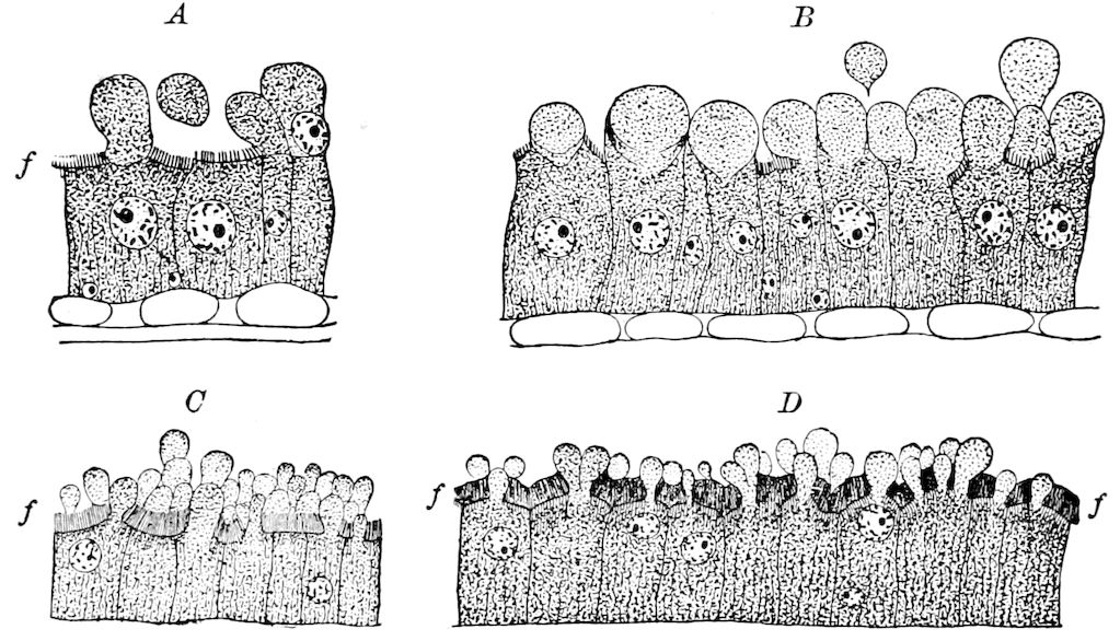
Fig. 322.—Different phases of the mechanism of secretion and of excretion.—After Gehuchten.
A glandular cell of the chylific stomach, when at rest, is always furnished with a striated “platform,” or flat surface, or face, on the side facing the cavity of the stomach, and the free edge of the platform, or plateau, is provided with filaments projecting into the digestive cavity (Fig. 322, f). These glandular cells, when active, differ much in appearance. In a great number, the platform (plateau) has disappeared, and is replaced by a simple, regular membrane. During the process of secretion, a finely granular mass, in direct continuity with the protoplasm, swells, and raises the membrane over the entire breadth of the cell, causing it to project into the intestinal cavity (Fig. 322, A, B). These vesicles, or drops of the secretion, whether free or still attached by a web to the cells, are clear and transparent in the living insect, but granular in the portions of the digestive canal fixed for cutting into sections. Gehuchten then asks: “How does a cell gorged with the products of secretion empty itself?” Both Ranvier and also Heidenhain believe that one and the same glandular cell may secrete and excrete several times without undergoing destruction, but their researches 328made on salivary glands have not answered the question. Gehuchten explains the process thus: when the epithelial cell begins to secrete, the clear fluid elaborated in the protoplasm of the cell increases the intracellular tension, until, finally, the fluid breaks through certain weak places in the swollen basal membrane of the platform, and then easily passes through the closely crowded filaments, and projects out into the intestinal cavity as a pear-shaped vesicle of a liquid rich in albumens at first attached to the free face of the cell, but finally becoming free, as at Fig. 322, A, B.
When the elaboration of the substance to be secreted is more active, the mechanism of the secretion is modified. The basal membrane of the platform may then be raised at several places at once; instead of a single vesicle projecting into the intestinal cavity, each cell may present a great number more or less voluminous. If all remain small and rapidly detach themselves from the glandular cell, the filaments of the platform are simply separated from each other at different points of the free face, as in Fig. 322, C. On the other hand, when the different vesicles of a single cell become larger, the filaments of the platform are compressed and crowded against each other in the spaces between the vesicles remaining free, and the undisturbed portions of the platform appear homogeneous (Fig. 322, D). After the excretion of the secretory products by this process of strangulation, the cell then assumes the aspect of a glandular cell at rest, and may begin again to form a new secretion.
To sum up: The process of excretion may occur in two ways:
1. Where the membrane ruptures and the substances secreted are sent directly out into the digestive cavity. 2. Where the vesicles become free by strangulation, floating in the glandular or intestinal cavity, and ending by rupturing and coming into contact with the neighboring vesicles or with the food.
Absorbent cells.—Besides the glandular or secreting cells in Ptychoptera, there is between the two regions of the chyle-stomach lined with these cells a region about a centimetre long composed of absorbent cells. The absorbent cells are very large, polygonal, and contain a large nucleus, in which is a striated convoluted chromatic cord.
The food on entering the chyle-stomach is brought into contact with the products secreted in the proventriculus, in the first part of the chyle-stomach, and in the tubular glands. These products of secretion act on the food, extracting from them useful substances which they render soluble. These substances, after having been absorbed by the absorbent cells in the middle region of the stomach, 329undergo special modifications, and are transformed into solid products, which are situated at the bottom of these cells. Afterwards the alimentary substances freed from a portion of their useful substances are again placed in contact with the products of secretion in the distal part of the chylific ventricle, and reach the terminal part of the intestine.
“The products of secretion,” adds Gehuchten, “diverted into the intestinal canal do not come into immediate contact with the alimentary substances; they are separated from it by a continuous, structureless, quite thick membrane (the peritrophic membrane), which directly envelops the cylinder of food matters, extending from the orifice of the œsophageal valvule to the end of the intestine. Between this membrane and the free face of the epithelial lining there exists a circular space, into which are thrown and accumulate the excreted substances. The latter then cannot directly mingle with the aliments; but when they are liquid they undoubtedly pass through this membrane by osmose, and thus come into contact with the nutritive substances. It is the same with the products of absorption. The absorption of soluble products of the intestinal cavity is not then so simple a phenomenon as it was at first thought to be, since these products are nowhere brought into immediate contact with the absorbent cells” (pp. 90, 91).
The most recent authority, Cuénot, states that absorption of the products of digestion takes place entirely in the mid-intestine, and in its cæca when these are present. The mid-intestine exercises a selective action on the constituents of the food comparable to the action of the vertebrate liver.
LITERATURE ON THE PHYSIOLOGY OF DIGESTION
Davy, J. Note on the excrements of certain insects, and on the urinary excrement of insects. (Edinburgh New Phil. Journ., 1846, xl, pp. 231–234, 335–340; 1848, xlv, pp. 17–29.)
—— Some observations on the excrements of insects, in a letter addressed to W. Spence. (Trans. Ent. Soc. London, Ser. 2, iii, 1854, pp. 18–32.)
Bouchardat, A. De la digestion chez le ver à soie. (Revue et Mag. de Zool., Sér. 2, 1851, iii, pp. 34–40.)
Lacaze-Duthiers, H., et A. Riche. Mémoire sur l’alimentation de quelques insects gallicoles et sur la production de la graisse. (Ann. Scienc. natur., 1854, Sér. 4, ii, pp. 81–105.)
Basch, S. Untersuchungen über das chylopoetische und uropoetische System von Blatta orientalis. (Sitzungsber. d. math.-naturwiss. Classe d. Akad. d. Wissensch. Wien., 1858, xxxiii, pp. 234–260, 5 Taf.)
Lambrecht, A. Der Verdauungsprozess der stickstoffreichen Nährmittel, welche unsere Bienen geniessen, in den dazu geschaffenen Organen derselben. (Bienenwirtschaftl. Centralbl., viii Jahrg., 1872, pp. 73–78, 83–89.)
330Plateau, F. Recherches sur les phénomènes de la digestion chez les insectes, (Mém. Acad. roy. de Belgique, Sér. 2, xli, 1 Part, 1873, pp. 124, 3 Pls.)
—— Note additionelle au mémoire sur les phénomènes de la digestion chez les insectes. (Bull. Acad. roy. de Belgique, Sér. 2, xliv, 1877, pp. 710–733.)
Tursini, G. Fr. Un primo passo nella ricerca dell’ assorbimento intestinale degli artropodi. (Rend. d. R. Accadem. di Sc. fis. e matemat. di Napoli, 1877, xvi, pp. 95–99, 1 Pl.)
Jousset de Bellesme, Physiologie comparée. Recherches expérimentales sur la digestion des insectes et en particulier de la blatte. Paris, 1876, vii, and 96 pp., 3 Pls.
—— Recherches sur les fonctions des glandes de l’appareil digestif des Insectes. (Compt. rend., lxxxii, Paris, 1876, pp. 97–99.)
—— Travaux originaux de physiologie comparée. (i, Insectes, Digestion, Métamorphoses.) Paris, 1878, 5 Pls.
Simroth, H. Einige Bemerkungen über die Verdauung der Kerfe. (Zeitschr. f. d. gesammten Naturwiss., xli, 1878, pp. 826–831.)
Krukenberg, C. Fr. W. Versuche zur vergleichenden Physiologie der Verdauung und vergleichende physiologische Beiträge zur Kenntnis der Verdauungsvorgange. (Untersuch, a. d. physiolog. Institut d. Universität Heidelberg, 1880, i, 4, pp 327, Figs.; ii, 1, p. 1, Figs.)
Metschnikoff, E. Untersuchungen über die intrazelluläre Verdauung bei wirbellosen Tieren. (Arb. d. zool. Instit. Wien., 1883, v, pp. 141–168, 2 Taf.)
Locy, William A. Anatomy and physiology of the family Nepidæ. (Amer. Naturalist, xviii, 1884, pp. 250–255, 353–367, 4 Pls.)
Vangel, E. Beiträge zur Anatomie, Histiologie, und Physiologie des Verdauungsapparates des Wasserkäfers, Hydrophilus piceus. (Termész. Füzet., x, 1886, pp. 111–126 (in Hungarian); pp. 190–208 (in German), 1 Pl.)
Schönfeld. Die physiologische Bedeutung des Magenmundes der Hönigbiene. (Archiv f. Anat. u. Physiol., Physiol. Abt., 1886, pp. 451–458.)
Faussek, V. Beiträge zur Histiologie des Darmkanals der Insekten. (Zeitschr. f. wiss. Zool., 1887, xl, pp. 694–712, 1 Taf.; Abstract in Zool. Anz., Jahrg. x, pp. 322, 323, 1 Taf.)
Frenzel, J. Ueber Bau und Thätigkeit des Verdauungskanals der Larve des Tenebrio molitor, mit Berücksichtigung anderer Arthropoden (Berlin. Ent. Zeitschr., 1882, pp. 267–316, 1 Taf.); Inaug.-Diss. Göttingen, 1882.
—— Einiges über den Mitteldarm der Insekten, sowie über Epithel-regeneration. (Archiv f. Mikrosk. Anat., 1885, xxvi, pp. 229–306, 3 Taf.)
—— Zum feineren Bau des Wimperapparates. (Ibid., 1886, xxviii, pp. 53–80, 1 Taf.)
—— Die Verdauung lebenden Gewebes und die Darmparasiten. (Archiv f. Anat., 1891.)
Gehuchten, A. van. Recherches histologiques sur l’appareil digestif de la Ptychoptera contaminata, I Part. Étude du revêtment épithélial et recherches sur la sécrétion. (La Cellule, 1890, vi, pp. 183–291, 6 Pls.)
Cuénot, L. Études physiologiques sur les Orthoptères. (Arch. Biol., xiv, 1895, pp. 293–341, 2 Pls.)
Needham, James G. The digestive epithelium of dragon-fly nymphs. (Zool. Bull., i, 1897, Chicago, pp. 104–113, 10 Figs.)
With the writings of Mingazzini (see p. 323), Kowalevsky, Ranvier, Haidenhain, Beauregard (p. 323), Sadones.
331
THE GLANDULAR AND EXCRETORY APPENDAGES OF THE DIGESTIVE CANAL
Into each primary division of the digestive canal open important glands. The salivary and silk-glands are offshoots of the œsophagus (stomodæum); the cœcal appendages open into the stomach (mesenteron), while the urinary tubes grow out in embryonic life from the primitive intestine (proctodæum), and there are other small glands which are connected with the end of the hind-intestine.
a. The salivary glands
We will begin our account of these glands with those of the Orthoptera, where they are well developed. In the cockroach a large salivary gland and accompanying reservoir lie on each side of the œsophagus and crop. The gland is a thin, leaf-like, lobulated mass, divided into two principal lobes. These open into a common trunk, which after receiving a branch from a small accessory lobe, and from the salivary reservoir, unites with its fellow to form the unpaired salivary duct which opens into the under side of the lingua. Each salivary reservoir is a large oval sac with transparent walls. (Miall and Denny, also Figs. 299, sr, and 327.) The ducts and reservoirs have a chitinous lining, and the ducts are, like the tracheæ, surrounded by a so-called spiral thread, or by separate, incomplete, hooplike bands, which serve to keep the duct permanently distended. In the locust (Fig. 298) the lobules are more scattered, forming small separate groups of acinose glands. In the embryo of Forficula Heymons has observed a pair of salivary glands opening on the inner angle of the mandibles, a second pair opening in the second maxillæ, while a third pair of glands, whose function is doubtful, is situated in the hinder part of the head, opening to the right and left on the chitinous plate (postgula) behind the submentum. In Perla, there are two pairs segmentally arranged (Fig. 343).
332
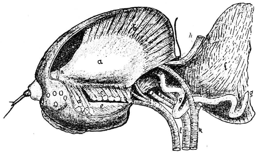
Fig. 323.—Left side of the head of the silkworm: a, adductor muscle of the mandible, from which the muscular fibres have been removed; b, upper fibres of the same; c, lower fibres cut away to show the adductor muscle (e); d, fibres inserted on the accessory adductor lamella; f, œsophagus, much swollen; g, salivary gland; h, dorsal vessel; i, l, tracheæ of the mandibular muscles; k, trachea; n, optic nerve.—After Blanc.
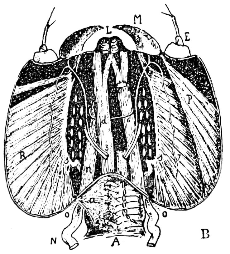
Fig. 324.—Lower side of the head of the silkworm exposed, the spinning apparatus, the œsophageal ganglion, and the adductor of the left mandible removed: M, mandible; P, abductor of the mandible; R, adductor; N, salivary gland attached at O to the edge of the adductor muscle; o, o, transverse portion of the “hyoid”; 3, masticator nerve and its recurrent branch (7); L, tongue cut horizontally.—After Blanc.
Here we might refer to a pair of glands regarded by Blanc as the true salivary glands. They do not appear to be the homologues of the salivary glands of other insects, though probably functioning as such. The functional salivary glands of lepidopterous larvæ have been overlooked by most entomotomists, and the spinning glands have been, it seems to us, correctly supposed to be modified salivary glands. Lucas also regards those of case-worms (Trichoptera) as morphologically salivary glands. Those of the silkworm were figured by Réaumur (Tom. i, Pl. v, Fig. 1), but not described; while those of Cossus, which are voluminous, were regarded by Lyonet as “vaisseaux dissolvans.” Dr. Auzoux (1849), in his celebrated model of the silkworm, represented them accurately, while Cornalia briefly described them as opening into the mouth. The first satisfactory description is that of Blanc (1891), who states that in the silkworm “the two salivary glands” are small, flexuous, yellow tubes, which occupy a variable position on the sides of the œsophagus (Fig. 323). The glandular portion passes into the head, ending at the level of the adductor plate of the mandibles (Fig. 324, o), and entering the buccal cavity at the base of the mandible, as seen in Fig. 323. It is plain, when we recognize the direct homology of the silk-glands of the caterpillars with the salivary glands of other insects, and of the spinneret with the hypopharynx, that 333these so-called “salivary glands” in lepidopterous larvæ are different structures. They are probably modified coxal glands, belonging to the mandibular segment.
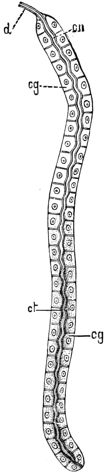
Fig. 325.—One of the two salivary glands of Cæcilius burmeisteri: d, excretory duct; cn, the lumen or canal; cg, gland-cells; ct, salivary fluid.—After Kolbe.
The polygonal epithelial cells of these glands contain branched nuclei, recalling those of the spinning-glands. In those caterpillars which feed on leaves, the salivary glands are slightly developed, but in such as bore into and eat wood, as the Cossidæ, the glands are, as figured by Lyonet, very large, forming two sausage-shaped bodies passing back to the beginning of the mid-intestine, each ending in a long convoluted filament. The salivary glands of the imago are very long and convoluted (Fig. 310, sd).
In the Panorpidæ these glands differ in the sexes, the males having three pairs of very long tortuous tubes, while, in the females, they are reduced to two indistinct vesicles. (Siebold.)
In the Diptera in general there are two pairs, one situated in the beak, the other in the thorax. In the larvæ there is a single pair (Fig. 341). Kraepelin describes a third pair in the Muscidæ at the point of transition from the fulcrum to the œsophagus, but Knüppel has apparently found only what may be fat cells at this point, so that the supposed presence of a third pair in Diptera needs confirmation. In the Psocidæ there are two salivary glands, of simple tubular shape (Fig. 325).
In the Nepidæ the salivary glands are four in number, and of conglomerate structure, two being long and extending back into the beginning of the abdomen, while the other two are about one-fourth as long. (Figs. 327, 328.) In Cicada, besides a pair of simple tortuous tubes, there is in the head another pair of glands, each composed of two tufts of short lobes, situated one behind the other. (Dufour.) In many Hemiptera (Pyrrhocoris, Capsus, etc.) there is but a single pair, each gland consisting of four lobes; in the Coccidæ each gland is divided into two lobes (Fig. 326); in the Aphidæ, according to Witlaczil, they consist of two lobes grown together. In the Psyllidæ they are said to be absent.
In Phylloxera vastatrix the saliva is forced through a salivary 334passage out of the duct and into the mouth by a pumping apparatus furnished with special muscles. (Krassilstschik.)
In the Odonata acinose glands are present in the imago, but not in the nymph until in its last stage, Poletaiew accounting for their absence in the earlier stages by the fact that the larva swallows more or less water while taking its food.
In the Coleoptera, as we have observed in Anopthalmus, there are three pairs of salivary glands (Fig. 74). In the Blapsidæ these glands consist of many ramifying tubes united on each side of the œsophagus into a single duct; in others they are but slightly developed, while in still others they are wanting.
The salivary glands are most highly differentiated in the Hymenoptera, and especially in the bees (Bombus and Apis), where Schiemenz found not less than five systems of glands (Fig. 329; also 87), of which four systems are paired. One pair of these glands lies in the tongue, three in the head, and one in the thorax.
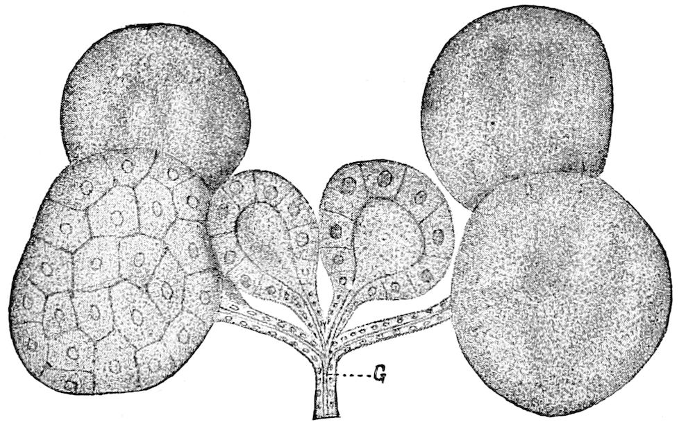
Fig. 326.—Acinous salivary glands of Orthezia cataphracta. In some acini the nuclei and boundaries of the cells are shown.—After List, from Field’s Hertwig.
System I is situated in the head, and consists of unicellular glands; the duct from each cell leads into a common, strongly chitinized duct, opening into the gullet.
System II, composed of acinose glands, lies also in the head; its duct is united with that of System III, situated in the thorax. (Fig. 329, 2, 3.)
System IV is situated at the base of the upper surface of the mandibles, and forms a delicate sac lined within with glandular cells; its duct opens at the insertion of the mandibles.
System V lies in the beak, and is a single gland consisting of unicellular glands; it opens into the common opening of Systems II and III. This system is wanting in the honey-bee, but occurs in Bombus and other genera.
335
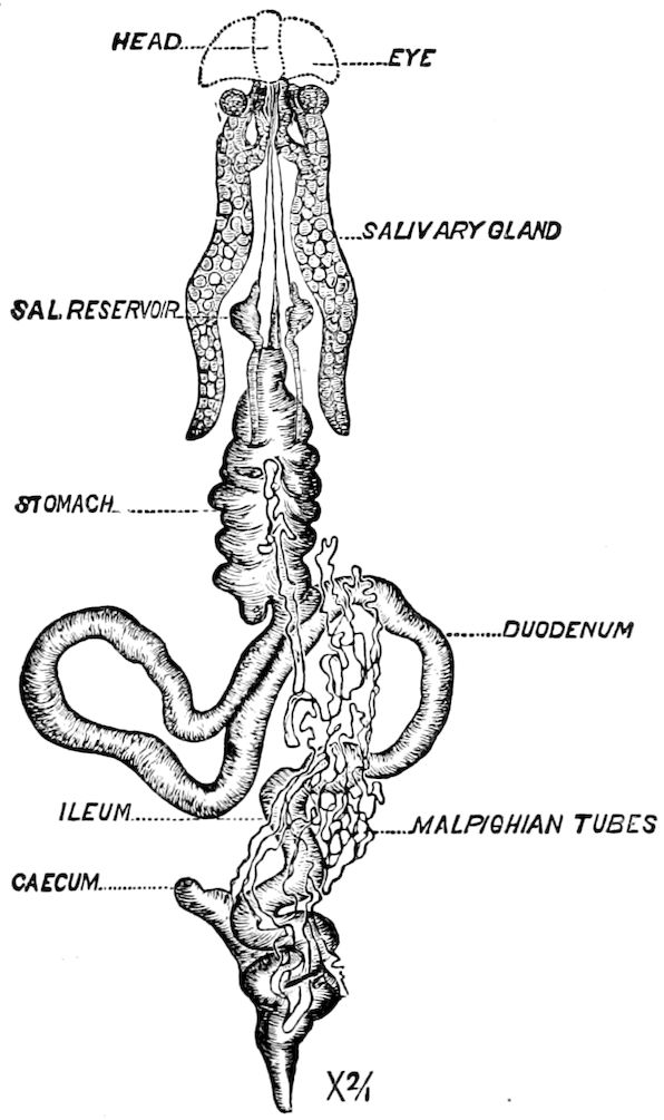
Fig. 327.—Appendages of digestive canal of Belostoma.—After Locy.
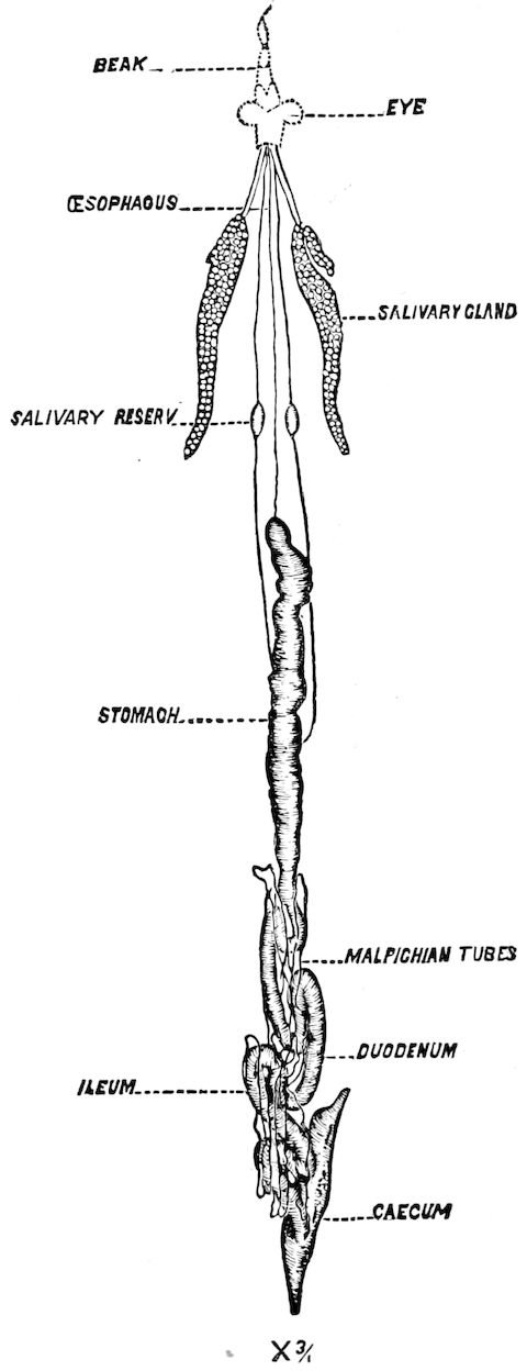
Fig. 328.—Salivary and other glands of Ranatra.—After Locy.
In all the five systems there constantly occur three cellular layers: the intima, epithelial, and propria. As regards their origin Schiemenz states that Systems I and IV are new structures, that System III arises in part, and Systems II and V wholly, from the silk-glands of the larva. As the glands differ much in the sexes, and in different species and genera, Schiemenz believes that their function is very manifold.
In addition to those previously discovered by Schiementz, Bordas has detected two additional pairs of salivary glands in the worker and male honey-bee, i.e. the internal mandibular and sublingual glands, so that in Apis there are in all six pairs, and apparently one unpaired.
The delicate chitinous external layer of the gland is perforated by many very fine pores through which the salivary fluid secreted 336by the epithelial cells passes into the salivary duct. The glands are externally bathed by the blood.
In many insects, including lepidopterous larvæ, the single median opening of the salivary duct is converted into a spraying apparatus.
In the adult Lepidoptera, according to Kirbach:—
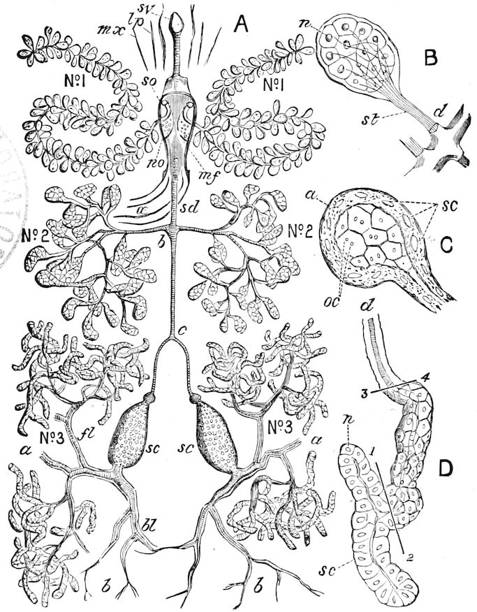
Fig. 329.—Salivary glands of the honey-bee: systems No. 1–3, × 15: sv, salivary valve (of systems 2 and 3) at base of tongue; lp, labial palpus; mx, maxilla; so, salivary opening of system 1 in hypopharyngeal plate; no, openings in plate for termination of taste-nerve; œ, œsophagus; sd, salivary duct; b, junction of ducts of system No. 2; c, junction of ducts of system No. 3; sc, sc, salivary sacs; fl, front lobe; bl, back lobe; a, chitinous duct, with spiral thread. B, single acinus of system No. 1, × 70: n, nucleus; st, salivary tract; d, large duct. C, single pouch, or acinus, from system No. 2: a, propria or outer membrane; sc, secreting cells. D, termination of system No. 3:1,2,3,4, lines marking end of section; d, duct in section; sc, secreting cells in section; n, nucleus.—After Cheshire.
“Its lower half forms a thick chitinous gutter, with a concave cover above, in which the similarly shaped upper half lies encased, so that between the two only a small semicircular opening remains. Powerful muscles extend from the cover to the lower side and to the two ridges of the bottom plate; through their contraction the upper channel is elevated, and presses out of the hinder part of the ducts into the space thus formed a great quantity of the saliva, which by allowing the contraction of the cover-muscle through the crevice-like opening, which is situated in the lower edge of the mouth-opening, becomes 337squeezed out in order either to mix with the fluid where the 2d maxillæ fuse, passing up into the canal in the proboscis, or to penetrate into and thus dilute the semi-fluid or solid substances taken, into the proboscis.”
The morphology and general relations of the salivary glands have been sketched out by Hatschek, Patten, and by Lucas, from observations on those of the case-worms or larval Trichoptera.
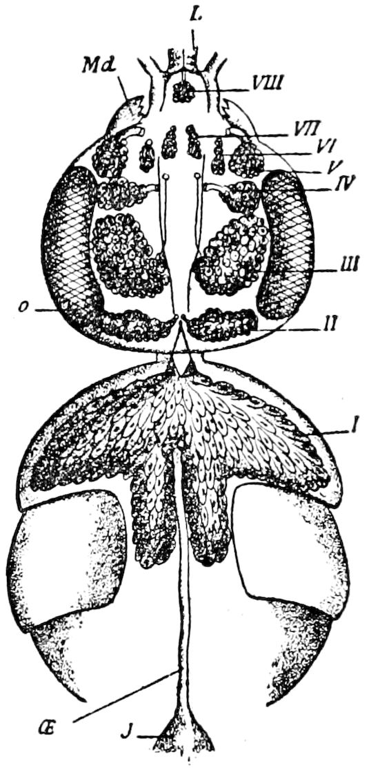
Fig. 330.—Eight pairs of glands of Andrena: I, thoracic; II, postcerebral; III, supracerebral; IV, lateropharyngeal; V, mandibular; VI, internomandibular; VII, sublingual; VIII, lingual; Md, mandible; L, tongue; o, eye; œ, œsophagus; J, honey-sac.—After Bordas.
Patten states that the spinning-glands in Neophylax are formed by a pair of ectodermal invaginations on the ventral side of the embryo, between the base of the 2d maxillæ and the nervous cord. They increase rapidly in length, and “they also unite to form a common duct, which opens at the end of the upper lip.”
The salivary glands in the same insect are “formed by invagination of the ectoderm on the inner sides of the mandibles, in the same manner as are the spinning glands.”
Lucas has shown that in trichopterous larvæ (Anabolia) there are three pairs of salivary glands in the head, which are serially arranged. The first pair belong to the mandibular, the second pair to the 1st maxillary, and the third pair, or spinning glands, to the 2d maxillary segment. The first or mandibular glands open into the mouth at the base of the mandibles directly behind the dorsal condyle. The second pair open between the 1st and 2d maxillæ; at the base of the latter, near the ventral condyle of the mandibles. The third pair open into the hypopharynx, which is modified to form the spinneret. Lucas agrees with Korschelt in regarding them as modified coxal glands, Schiemenz having previously regarded the headglands of the imago of the bee as belonging to the segments bearing the three pairs of buccal appendages, so that each segment originally contained a pair of glands. It is thus proven that the silk-glands are modified salivary glands adapted to the needs of spinning larvæ, and indeed in the imago the sericteries revert to their primitive shape and use as salivary glands.
The serial arrangement of the salivary glands in the Hymenoptera, where the number varies from five to ten pairs, is clearly proved by Bordas. He has detected five more pairs than were previously known, and names the whole series as follows:—1, the thoracic salivary glands, which are larger than the others, and nine other pairs, which are all contained in the head as follows: 2, postcerebral; 3, supracerebral; 4, lateropharyngeal; 5, mandibular; 6, internomandibular, situated on the inner side of the base of mandible; 7, sublingual; 8, lingual (these and 1 to 7 common to all Hymenoptera); 9, paraglossal (in Vespidæ); 10, maxillary (very distinct in most wasps). These glands do not all occur in the same species, being more or less atrophied.
Bordas further shows the segmental arrangement of the cephalic glands by stating that the supracerebral glands correspond to the antennal segment, the sublingual glands to the labial, the mandibular glands (external and internal) 338to the mandibular segment, the maxillary glands to the 1st maxillary segment, the lingual glands to the 2d maxillary segment, while the thoracic and postcerebral salivary glands, he thinks, correspond to the ocular segment, a view with which we are indisposed to agree, although conceding that each of the six segments of the head has in it at least one pair of salivary glands.
Functions of the different salivary glands in Hymenoptera.—The secretion of the thoracic glands is feebly alkaline. The postcerebral salivary glands, considered by Ramdohr to be organs of smell, secrete, like the preceding, a distinctively alkaline fluid, which mingles with the products of the thoracic glands. The supracerebral glands, also equally well developed in all Hymenoptera, though much atrophied in the females and especially the males of Apis mellifica, also in the Vespinæ and Polistinæ, secrete an abundant, feebly acid liquid, which is actively concerned in digestion.
As to the mandibular glands, which Wolf supposed to be olfactory organs, their acid secretion, though smelling strongly, acts energetically on the food as soon as introduced into the mouth.
The sublingual glands, atrophied in most Apidæ, but relatively voluminous in Sphegidæ, Vespinæ, Polistinæ, Crabronidæ, etc., empty their secretion into a small prebuccal excavation, where accumulate vegetable and earthy matters collected by the tongue, and the saliva secreted by these glands, acts upon them before they pass into the pharynx. The lingual glands secrete a thick, sticky liquid, which causes foreign bodies to adhere to the tongue, and also agglutinates alimentary substances. The uses of the other glands, maxillary and paraglossal, are from their minuteness undetermined. (Bordas.)
LITERATURE ON THE SALIVARY GLANDS
Leydig, F. Zur Anatomie der Insekten. (Archiv Anat. und Phys. 1859.)
—— Untersuchungen zur Anatomie und Histiologie der Tiere. Bonn, 1883, pp. 174, 8 Taf.
—— Intra- und interzellulare Gänge. (Biolog. Centralblatt, x, 1890, pp. 392–396.)
Dohrn, A. Zur Anatomie der Hemipteren. (Stettin. Entom. Zeit., 1866, salivary glands, pp. 328–332.)
Kupffer, C. Die Speicheldrüsen von Periplaneta orientalis und ihr Nervenapparat. (Beiträge zur Anatomie und Physiol., 1875.)
Schiemenz, P. Ueber das Herkommen des Futtersaftes und die Speicheldrüsen der Biene. (Zeitschr. f. wissens. Zool., xxxviii, 1883, pp. 71–135, 3 Taf.)
Korschelt, E. Ueber die eigentümlichen Bildungen in den Zellkernen der Speicheldrüsen von Chironomus plumosus. (Zool. Anzeiger, 1884, pp. 189–194, 221–225, 241–246.)
Hofer, B. Untersuchungen über den Bau der Speicheldrüsen und des dazu gehörenden Nervenapparates von Blatta. (Nova Acta d. Kais. Leopold.-Carol. Deutsch. Akad. d. Naturforscher, li, 1887, pp. 345–395, 3 Taf.)
Knüppel, A. Ueber Speicheldrüsen von Insekten. (Archiv für Naturg., 1887, Jahrg. 52, pp. 269–303, 2 Taf.)
Blanc, Louis. La tête du Bombyx mori à l’état larvaire, anatomie et physiologie. (Extrait des Travaux du Laboratoire d’Études de la Soie, 1889–1890; Lyon, 1891, p. 180, many figs.)
Bordas, L. Anatomie des glandes salivaires des Hyménoptères de la famille des Ichneumonidæ. (Zool. Anzeiger, 1894, pp. 131–133.)
—— Glandes salivaires des Apides, Apis mellifica. ♂ and ♀. (Comptes rendus Acad. Sc., Paris, cxix, pp. 363, 483, 693–695, 1894; also two articles in Bull. Soc. Philomath. Paris, 1894, pp. 5, 12, 66.)
339—— Appareil glandulaire des Hyménoptères. (Ann. Sc. Nat. Zool., xix, Paris, 1894, pp. 1–362, 11 Pls.) (See also p. 366.)
Berlese, Antonio. Le cocciniglie Italiane viventi sugli agrumi. Firenze, 1896, 12 Pls. and 200 Figs.
With the writings of Mark, Minot, Locy, List, Krassilstschik, Nagel (1896).
b. The silk or spinning glands, and the spinning apparatus
The larvæ of certain insects, chiefly those of the Lepidoptera, possess a pair of silk or spinning glands (sericteries) which unite to form a single duct opening in the upper lip at the end of the lingua, which is modified to form the spinneret. (See pp. 71, 75.) All caterpillars possess them, and they are best developed in the silkworms, which spin the most complete cocoon. Silk-glands also occur in the larvæ of the Tenthredinidæ, in the case-worms or larval Trichoptera, also in certain chrysomelid beetles (Donacia, Hæmonia), and in a weevil (Hypera). In a common caddis-worm (Limnophilus) the glands are of a beautiful pale violet-blue tint, and two and a half times as long as the larva itself; viz. the body is 20 mm. and the glands 55 mm. in length.
In caterpillars the glands are of tubular shape, shining white, and much like the ordinary simple tubular salivary glands of the imago. When only slightly longer than the body they are twice folded, the folds parallel and situated partly beneath and partly on the side of the digestive canal; not usually, when folded in their natural position, extending much behind the end of the stomach; but in the silkworms they are so long and folded as to envelop the hinder part of the canal. In geometrid caterpillars the glands when stretched out only reach slightly beyond the end of the body; in Datana they are half again as long as the body. Helm thus gives their relative length in certain Eurasian caterpillars, and we add that of Telea polyphemus:—
Vanessa io length of body 32 mm.; of the silk glands 26 mm.
Smerinthus tiliæ length of body 63 mm.; of the silk glands 205 mm.
Bombyx mori length of body 56 mm.; of the silk glands 262 mm.
Antheræa yamamaya length of body 100 mm.; of the silk glands 625 mm.
Telea polyphemus length of body 60 mm.; of the silk glands 450 mm.
Thus in Telea the silk-glands are about 18.50 inches in length, being about seven times as long as the body.
For the most complete accounts of the spinning glands of Lepidoptera and their mechanism we are indebted to Helm and to Blanc, and for that of the Trichoptera to Gilson.
340The unpaired portion, or spinning apparatus (filière of Lyonet), is divided into two portions; the hinder half being the “thread-press,” the anterior division the “directing tubes.” The silk material, stored up in the thickest portion of the glands, passes into the thread-press (Fig. 334, A), which is provided with muscles which force the two double ribbon-like threads through the directing tube, as wire is made by molten iron being driven through an iron plate perforated with fine holes. The entire spinning apparatus, or filator, as we may call it, is situated in the tubular spinneret. The opening of the spinneret is directed anteriorly, and the anterior end of the directing tube passes directly into this opening so that the directing tube may be regarded as an invagination of the lingua.
The silk thread which issues from the mouth of the spinneret is, as Leeuwenhoek discovered, a double ribbon-like band, as may be seen in examining the silk of any cocoon.
The process of spinning.—Since the appearance of Helm’s account, Gilson, and also Blanc, have added to our knowledge of the way in which the silk is spun and of the mechanism of the process. Gilson has arrived, in regard to the function of the press or filator, at the following conclusions: 1, the press regulates the thread, it compresses it, gives it its flattened shape; 2, it regulates the layer of gum[52] (grès) which surrounds the thread; 3, it may render the thread immovable by compressing it as if held by pincers.
The process of spinning in the silkworm, says Blanc, comprises all the phenomena by which the mass of silk contained in the reservoir is transformed into the silk fluid of which the cocoon is spun. The excretory canals each contain a cylindrical thread of silk having a mean diameter of 0.2 mm. and surrounded by a layer of gum (grès) which in the fresh living organ exactly fills the annular space situated between the fibroin cylinder and the wall. Arrived within the common duct, the two threads receive the secretion of Filippi’s gland, where the silken fluid is formed, but has not yet assumed its definite external characters. The two threads press through the common canal and arrive at the infundibulum (Fig. 334, c) of the press, at 341the bottom of which is situated the orifice of the spinning canal, almost completely divided into two by the sharp edge of the rachis (Figs. 334, a, 335, l). The threads each pass into one of the two grooves, and the layer of gum (grès) fills the rest of the canal of the press or filator.
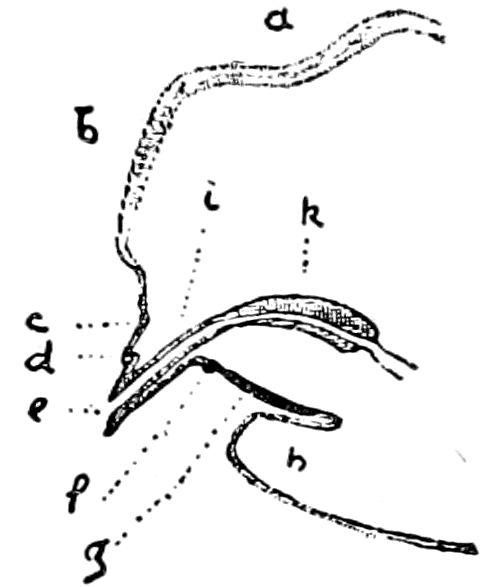
Fig. 331.—Longitudinal section of the spinneret: a, horizontal portion of the tongue; b, vertical portion; c, f, circle of the tongue; d, tongue-pad; e, orifice of the spinneret; g, body of the lyre; h, prebasilar membrane forming a fold; i, internal canal of the spinneret; k, filator.—After Blanc.
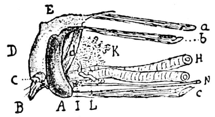
Fig. 332.—The lower lip (labium) of Bombyx mori, isolated, seen from the left side: A, lyre; B, spinneret; C, labial palpus; D, vertical part of the labium; E, horizontal part of the same; H, L, silk-canal; K, right gland of Filippi; L, canal of the left gland; N, labial nerve; a, oblique fibre of the elevator of the labium; b, right fibre of the same; c, depressor of the labium; d, superior spinning muscles.
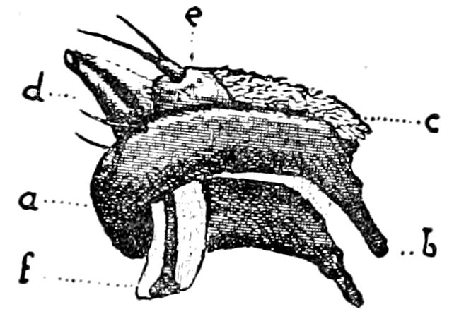
Fig. 333.—The labium in a horizontal position, seen from the side: f, the filator or press situated under the external part of the spinneret (d), between the branches (b), of the lyre (a); e, labial palp; c, tongue.
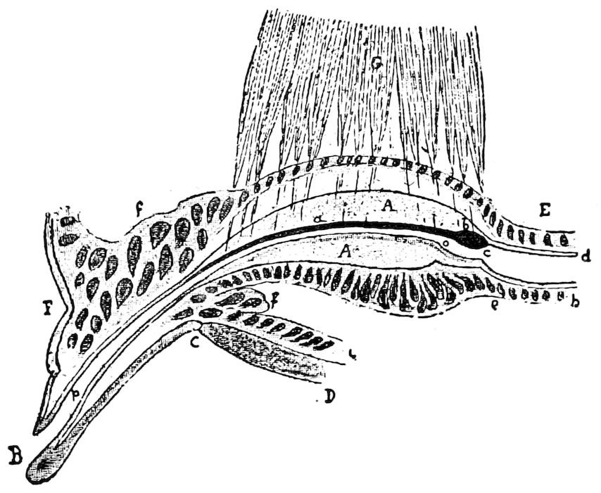
Fig. 334.—Longitudinal section of the spinneret and press (filator): A, filator or press; B, spinneret; C–D, body of the lyre; F, lower part of the labium; E, common canal; eh, its epithelium; G, superior muscle of the press; a, rachis; b, its posterior enlargement; c, infundibulum; d, cuticle; o, orifice of the spinning canal; op, central canal of the lyre and of the spinneret; fi, hypodermis of the lyre; f, f, hypodermic pad of the lyre.
The silken substance is then pressed by the more or less powerful contractions of the muscles of the filator, so 342that the passage of the threads is facilitated. If the muscles totally contract, the spinning canal is opened wide, the threads pass easily upwards and assume the form of a triangular prism (Fig. 336).
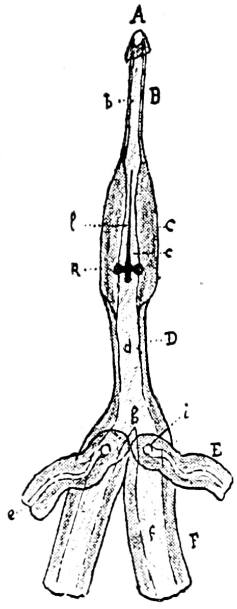
Fig. 335.—Spinning apparatus, seen from above: A, opening of the spinneret; B, central canal of the spinneret (C); D, common canal; E, canal of Filippi; F, excretory canal of a silk-gland; i, orifice of the canal of Filippi’s gland; l, rachis; k, ring of the infundibulum; b, c, d, e, f, cavity of the different canals; h, spur which separates the two excretory canals.—This and Figs. 331–334 after Blanc.
If this contraction diminishes, the chitinous wall of the spinneret comes together, owing to its elasticity; the ceiling of the canal approaches the floor; the cavity tends to take the form of a semicircular slit, and the threads are compressed, flattened. As each mass or thread of silk is much more voluminous than the canal, except when the latter is extremely dilated, it follows that the two threads are always compressed, or squeezed together, and that each of them is compelled to mould itself in the groove it occupies and to take its shape. Hence the variations in the appearance of the two masses or divided portions of silk, which as stated present all grades between the form of an isosceles-triangular prism and that of a nearly flat ribbon; but this last case is quite rare. The use of the spinneret, then, is to compress the thread and to change its form more or less considerably, at the same time as it diminishes its diameter.
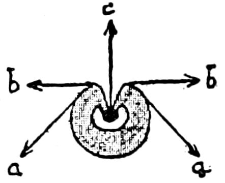
Fig. 336.—Diagram of the press and its muscles: a, lower; b, lateral; c, upper muscles of the press.—After Blanc.
Moreover, this constant compression of the thread as it passes through the press keeps it in a certain state of tension so as to allow the caterpillar while spinning to firmly hold its thread.
Finally, when the worm suspends the contraction of its spinning muscles, the press flattens, vigorously compresses the thread, and arrests its motion, in such a way that if there was a strain on the silken fluid (bave), it would break rather than oblige the caterpillar to let go any more of it.
The press does not act directly on the silken thread, but through the gummy layer (grès) which transmits over the whole surface of the silken fluid (brin) the pressure exerted on it. After having overcome this difficult passage, the silk thread has acquired its definite form; it rapidly passes out of the spinneret.
343How the thread is drawn out.—Having seen, says Blanc, how the two masses of silk (brins), in passing through the spinning apparatus (or press), join each other, constituting the frothy silken fluid, thus becoming modified in form, it remains to examine the way in which the thread is drawn out of the spinneret. If we examine a caterpillar while spinning, it will be seen that in moving its head it draws on the frothy mass of silk fixed to the web of the cocoon. This traction certainly aids very much the exit of the thread, but it is not the only cause.
The silk, Blanc affirms, is pushed out by a force a tergo, developed by different agents, such as the pressure of the distended cuticle or the silky mass contained in the reservoir, as seen in the section of a worm which has spun its cocoon. But if we consider a caterpillar before it has begun to spin, it is difficult to explain the mechanism of spinning. As Blanc has often observed, in making sections of the heads of silkworms, two cases arise. Sometimes the worm has already spun a little, and a certain length of the frothy silk (bave) issues from the orifice of the spinneret, where it forms a small twisted bundle. At other times the worm has not spun since its last moult or the frothy mass of silk has broken within the head, and we find the end in the common tube. In the first of these two cases, the worm, dilating its press, is able by a general contraction to discharge a little of the gritty material (grès) which lines the ball of silk hanging at the end of the spinneret. It can also reject a certain quantity of the secretion of Filippi’s glands and thus soften the gritty substance. The little plug of silk can then adhere to the body with which it comes in contact.
In the same case it is necessary that the two bits or portions of silk traverse the press, and this normally has a calibre less than their diameter. The worm should then distend the spinning tube as much as is practicable, so as to make the openings as large as possible. It has been stated that the press is, in this condition, at least as large as the mass of frothy silk. This Blanc believes (although Gilson thinks otherwise) is pushed by a force a tergo, and reaches the funnel of the spinning canal; its two bits of silk (brins) unite there, penetrate into the canal itself, and, owing to successive impulses produced by the general contractions of the worm, press through and pass out of the spinneret.
While the silkworm is engaged in spinning its cocoon, the spinneret and press execute very varied movements, determined by the elevator, depressor, retractor, and protractor muscles of the labium, as well as those of the press. These movements, originally very 344numerous, may combine among themselves, so that the spinneret is susceptible of assuming during the process of spinning still more diverse positions.
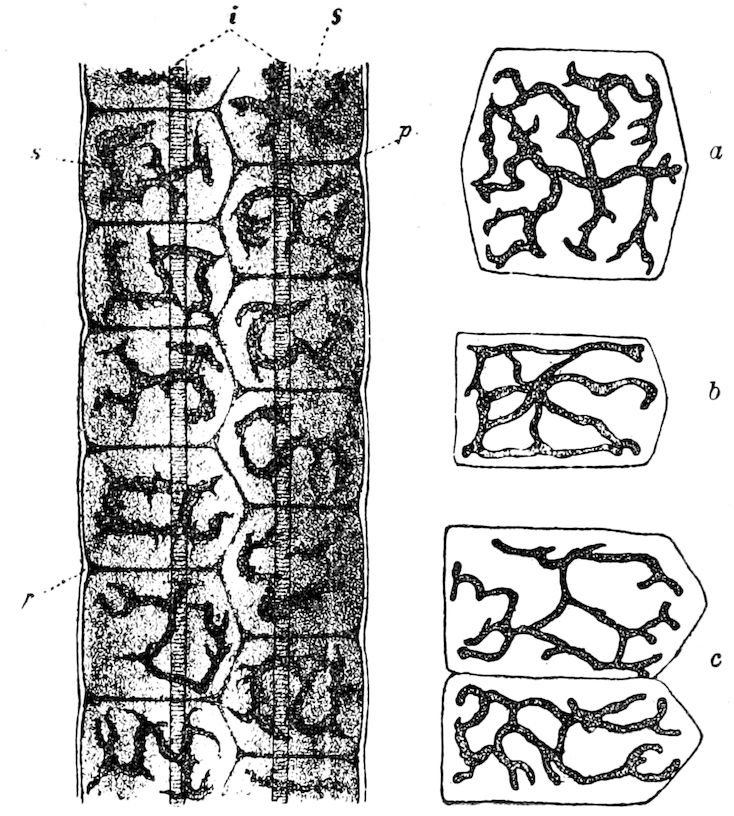
Fig. 337.—Portion of the silk-gland of Bombyx mori: p, tunica propria; i, tunica intima; s, secretion-cell with branched nuclei; a, separate secretion-cell from the anterior part of the silk-gland of Amphidasis betularia; b, the same of Vanessa urticæ; c, the same in Smerinthus tiliæ.—After Helm.
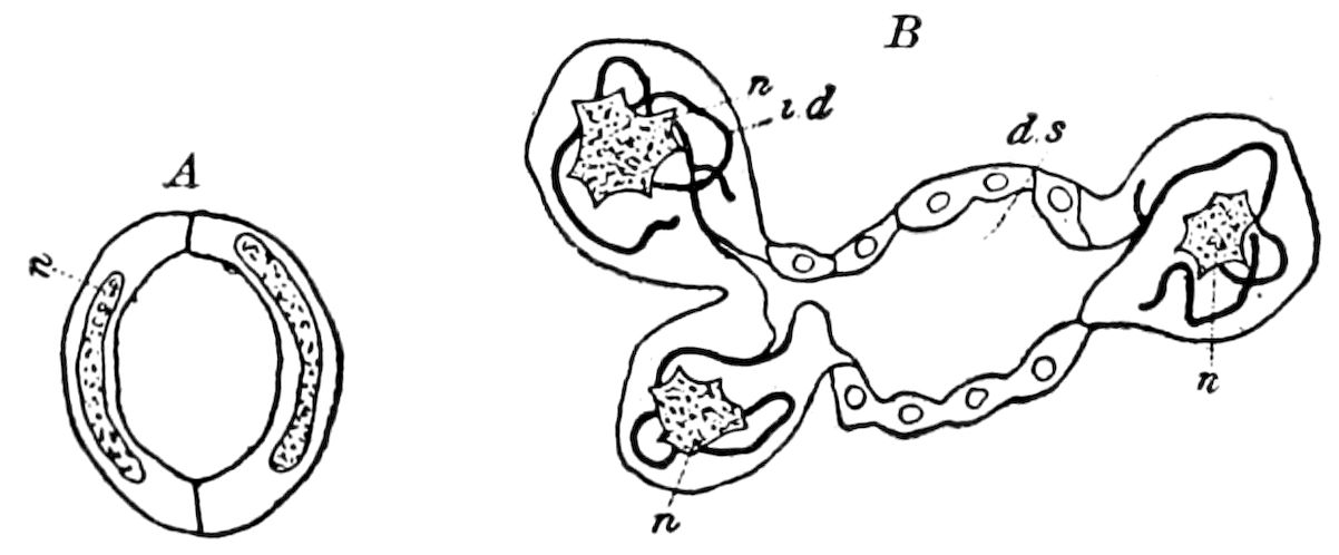
Fig. 338.—A, section of gland of lepidopter: B, section of silk-gland of a saw-fly larva; n, nucleus; i.d, canals; d.s, cavity.—After Gilson.
Histologically the silk-glands are composed of three layers,—the outer, or tunica propria (Fig 337); the inner, the tunica intima; the middle layer being composed of extraordinarily large epithelial cells which can be seen with the naked eye, and are also remarkable for the branched shape of the nuclei (a, b, c, 337), the branches being 345more or less lobed, and the larger the cells the more numerous are the branches of the nucleus. Gilson[53] finds that those of Trichoptera, Lepidoptera, Diptera, and Hymenoptera ordinarily consist of a small number of cells; and it is quite common, he says, to find only two cells in a transverse section (Fig. 338, A). In the Tenthredinidæ, however, “the organ still consists of a tube, the wall of which is composed of flat cells, but in addition to that, two series of spheroidal cells are attached to the sides. Each of these cells contains a system of tiny canals running through their cytoplasm (B, i. d). These cells are the secreting elements; they continually cast the silk substance into the tube.” A peculiarity of the tunica intima is its distinct transverse striation.
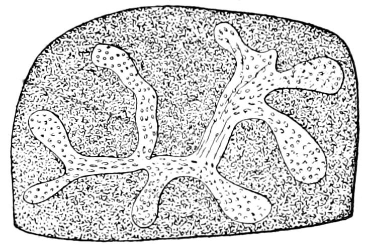
Fig. 339.—Branching nucleus of spinning gland of Pieris larva.—After Korschelt, from Wilson.
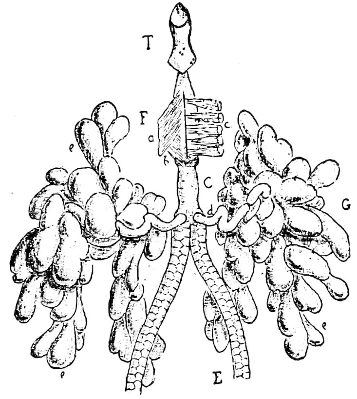
Fig. 340.—Filippi’s glands (G) isolated and seen from above: e, e, its lobules; d, its excretory canal; E, silk-duct; C, common canal; c, upper spinning muscle; b, lower muscle; a, lateral muscle; T, spinneret.—After Blanc.
The lining of the glands and of their common duct is moulted when the caterpillar casts its skin, and this, as well as the mode of development, shows that the glands are invaginations of the ectoderm. Gilson finds that the silk-glands and silk-apparatus of Trichoptera are very similar to those of caterpillars, and that the silk is formed in the same way.
Appendages of the silk-gland (Filippi’s glands).—In most larvæ there is either a single or a pair of secondary glands which open into the spinning glands near their anterior end. They are outgrowths of the gland provided with peculiarly modified excretory 346cells or evaginations of the entire glandular epithelium. Those of Bombyx mori (Fig. 340) are very well developed, and, according to Blanc, form two whitish, lobulated masses in the labium on each side of the common duct of the spinning gland. Externally they appear to be acinose; but their structure, as described by Blanc and by Gilson, is very peculiar. Helm thinks, with Cornalia, that the function of these glands is to secrete the adhesive fluid which unites the silk threads, and also to make the silk more adhesive in the process of spinning, but Blanc states that this is done before the thread passes into the common excretory tubes, and he is inclined to think that the secretion serves to lubricate the spinneret, and thus to facilitate the passage of the thread. On the other hand, in certain caterpillars these glands are situated quite far from the spinning apparatus.
The silk-glands in the pupa state undergo a process of degeneration, and finally completely disappear. They are specific larval organs evolved in adaptation to the necessity of the insect’s being protected during its pupal life by a cocoon. (Helm.)
Morphologically the silk-glands are by Lang regarded as modified coxal glands, and homologues of the setiparous parapodial glands of chætopod worms, the coxal glands of Peripatus, and the spinning glands of spiders.
In Scolopendrella, spinning glands are situated in the two last segments of the body, opening out at the end of the cercopods (Fig. 15, s.gl), and the larvæ of the true Neuroptera (Chrysopa, Myrmeleon, etc.) which spin cocoons, have spinning glands opening into the rectum. The silk forming the cocoon of the ant-lion, as Siebold and the older observers have stated, is secreted by the walls of the rectal or anal sac. Siebold (Anatomy of the Invertebrates, p. 445) states that in the larva of Myrmeleon, the silk-apparatus is very remarkable, “for the rectum itself is changed into a large sac and secretes this substance which escapes through an articulated spinneret projecting from the opening of the anus”[54] (Fig. 307, e). The larvæ of the Mycetophilidæ have spinning glands at the hinder end of the body, as also the imago of the female of the tineid moth Euplocamus. (Kennel.) The larvæ of ichneumons, wasps, bees, of Cecidomyia, and other Diptera, spin silken cocoons, but their glands have not yet been examined.
It should also be observed that during the process of pupation the larvæ of butterflies, of certain flies (Syrphus), and beetles (Coccinellidæ and some Chrysomelidæ) attach themselves by silk spun from the anus, so that the pupa is suspended by its tail; such glands are probably homogenetic with the coxal glands.
The silk in its fluid or soft state is mucilaginous, and according to Mulder, in the silkworm consists of the following substances, varying somewhat in their relative proportions by weight:
Silk-fibre material 53.67
Glue (Leim) 20.66
Protoplasm 24.43
Wax 1.39
Coloring matter 0.05
Fat and resin 0.10
347
LITERATURE ON THE SPINNING GLANDS
Helm, E. Anatomische und histiologische Darstellung der Spinndrüsen der Schmetterlingsraupen. (Zeitschr. f. wissens. Zool., xxvi, 1876, pp. 434–469, 2 Taf.)
Lidth de Jeude, Th. W. van. Zur Anatomie und Physiologie der Spinndrüsen der Seidenraupe. (Zool. Anzeiger, 1878, pp. 100–102.)
Engelmann, W. Zur Anatomie und Physiologie der Spinndrüsen der Seidenraupe. (Onderz. Phys. Lab. Utrecht, iii, 1880, pp. 115–119.)
Joseph, G. Vorläufige Mitteilung über Innervation und Entwickelung der Spinnorgane bei Insekten. (Zool. Anzeiger, 1880, pp. 326–328.)
Poletajew, N. Ueber die Spinndrüsen der Blattwespen. (Zool. Anzeiger, 1885, pp. 22–23.)
Meinert, Fr. Contribution à l’anatomie des fourmilions. (Overs. Danske Vidensk. Selsk. Forh. Kjöbenhavn, 1889, pp. 43–66, 2 Pls.)
Blanc, Louis. Étude sur la sécrétion de la soie et la structure du brin et de la bave dans le Bombyx mori. Lyon, 1889, pp. 48, 4 Pls.
—— La tête du Bombyx mori à l’état larvaire. Anatomie et physiologie. (Extrait du volume des Travaux du Laboratoire d’Études de la Soie. Années 1889–1890, Lyon, 1891, pp. 180, 95 figs.)
Gilson, G. Recherches sur les cellules sécrétantes. La soie et les appareils séricigènes: I. Lépidoptères. (La Cellule, 1890, vi, pp. 115–182, 3 Pls. I, Lépidoptères (suite); II, Trichoptères. Ibid., x, pp. 71–93, 1893, 1 Pl.)
Garman, H. Silk-spinning dipterous larvæ (Science, xx, 1893, p. 215).
Also the writings of Meckel, Pictet, Duméril, Klapálek, Wistinghausen, Loew, Hagen, Fritz Müller, Kolbe, McLachlan, de Selys-Longchamps.
c. The cæcal appendages.
These diverticula of the mid-intestine (“stomach”) are appended to the anterior end, and in the living, transparent larva of Sciara, which has two large, long, slender cœca (Fig. 341), the partly digested food may be seen oscillating back and forth from the anterior end of the stomach into and out of the base of each cæcum. In the Locustidæ (Anabrus, Fig. 299) and Gryllidæ (Fig. 344, e) there are two large, short cæca, and in the locusts (Caloptenus) there are six cæca, while cockroaches have eight. In the Coleoptera (Carabidæ and Dyticidæ) these large cæca appear to be replaced by very numerous slender, minute villi or tubules, which arise from the anterior part of the stomach (Figs. 317, r, also 342).
These cæca differ in structure from the stomach, as shown by Graber, as well as by Plateau and by Minot. The latter states that a single transverse section of one of the diverticula of the locust demonstrates at once that its structure is entirely different from that of the stomach.
348

Fig. 341.—Larva of Sciara: s.gl, salivary gland; ur.t, urinary tubes; i intestine; st, stomach; cae cæcal appendages; t, testis.
Its inner surface is thrown up into longitudinal folds, generally twelve in number. These folds shine through the outer walls, and are accordingly indicated in the drawings of Dufour, Graber, and others. The entire cæcum has an external muscular envelope, outside of which are a few isolated longitudinal muscular bands. The folds within are formed mainly by the high cylindrical epithelium which lines the whole interior of the cavity. Tracheæ ramify throughout all the layers outside the epithelium. There are appearances of glandular follicles in the bottom of the spaces between the folds. (Minot.)
Burmeister supposed that these cæca were analogous to the pancreas, and this view has been confirmed by Hoppe Seyler, Krukenberg, Plateau, and others, who claim that the digestive properties of the fluid secreted in them agrees with the pancreatic fluid of vertebrates.
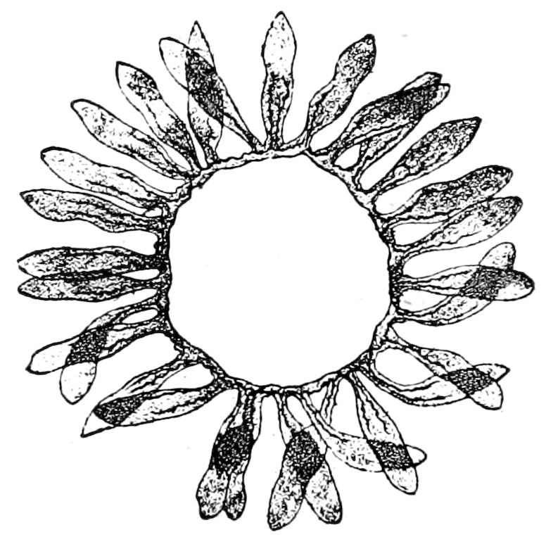
Fig. 342—Cross-section of mid-intestine of Acilius sulcatus, showing the arrangement of the cæca, two tracheæ passing into each cæcum.—After Plateau.
d. The excretory system (urinary or Malpighian tubes)
The excretory matters or waste products of the blood tissue of worms are carried out of the body by segmentally arranged tubes called nephridia. As a rule they arise in the blood sinuses of the body and open externally through minute openings in the skin. As there is a pair to each segment (in certain oligochete worms two or three pairs to a segment), they are often called segmental organs. In the annulate worms each segment of the body, even the cephalic or oral segment, originally contains a pair of these excretory organs. These 349vessels may have survived in myriopods and perhaps do exist in insects as urinary tubes, and also occur in many of the Arachnida, and thus are characteristic of each important class of land arthropods, but are either wanting or are very rudimentary or much modified in the marine classes, notably the Crustacea and Merostomata (Limulus), where they are represented by the shell-glands of Copepoda, green glands of the lobster, and the brick-red glands of Limulus.
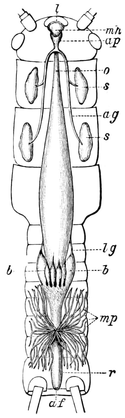
Fig. 343.—Digestive canal of Perla maxima: l, upper lip; mh, buccal cavity; ap, common end of salivary ducts (ag); o, œsophagus; s, s, salivary glands, arranged segmentally; b, cæca of chyle-stomach; lg, their ligaments of attachment; mp, urinary tubes; r, rectum; af, anal orifice.—After Imhof, from Sharp.
In the earliest tracheate arthropod, Peripatus, these tubes are well developed and are highly characteristic, each segment behind the head bearing a pair (Fig. 4, so4-so9). It has been suggested by some, but not yet proved, that the urinary tubes of insects are morphologically the same as the segmental organs of worms and of Peripatus; but there are no facts directly supporting this view, and, as Sograff states, it is a pure hypothesis and can only be confirmed or disproved by very detailed researches on the development of the urinary tubes of myriopods and of insects. Others regard them as probably homologous with the tracheæ, since they have a similar origin. As, however, they arise in the embryo as outgrowths of the proctodæum they may have arisen in myriopods and insects independently, and not be vermian heirlooms.
While in worms and in Peripatus a pair of these segmental organs occur in each segment, in insects this serial arrangement is not apparent; those with a purely excretory function are not segmentally arranged, with outlets opening externally, but arise as outgrowths of the hind-intestine or proctodæum of the embryo, not being segmentally arranged. The place of their origin is usually the dividing line between the mid and hind intestine (Fig. 343, mp); this applies to Scolopendrella (Fig. 15, urt) as well as to insects.
The urinary tubes are usually long, slender, blind, tubular glands varying in number from two to over a hundred, which generally arise at the constriction between the mid and hind intestine, and which lie loosely in the cavity of the body, often extending towards the head, and then ending near the rectum (Figs. 301, 310, vm). 350They were first discovered by the Italian anatomist Malpighi, after whom they were called the Malpighian tubes. While at first generally regarded as “biliary” tubes, they are now universally considered to be exclusively excretory organs, corresponding to the kidneys of the higher animals.
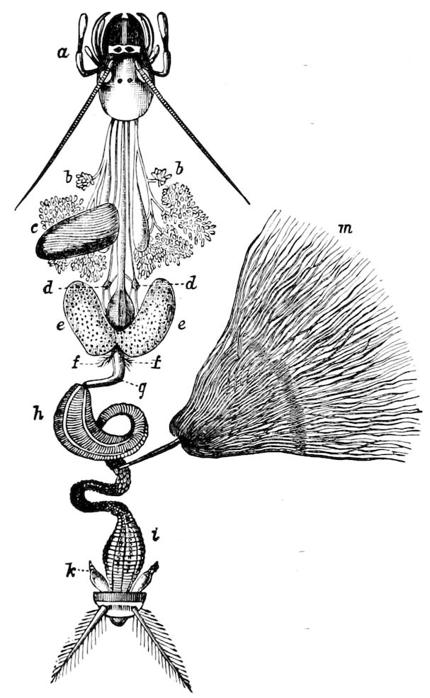
Fig. 344.—Digestive canal and appendages of the mole-cricket; a, head: b, salivary glands and receptacle; c, lateral pouch; d, stomatogastric nerves; e, anterior lobes of stomach; f, peculiar organ; g, neck of stomach; h, plicate part of same; i, rectum; k, anal gland; m, urinary tubes.—After Dufour, from Sharp.
Usually arising from the anterior end of the hind-intestine where it passes into the mid-intestine, in certain forms they shift their position, in some Hemiptera (Lygæus, Cimex) opening into the rectum, while in the Psyllidæ they arise from the slender hinder part of the mid-intestine, being widely separated at their origin. (Fig. 321.)
The length varies in different groups; where they are few in number (two to four, six to eight), they are very long, but where very numerous they are often short, forming dense tufts, each tuft connecting with the intestine by a common duct (ureter), or, as in the mole-cricket, the numerous tubes empty into a single duct (Fig. 344); in the locusts (Acrydiidæ), however, they are arranged in 10 groups, each group consisting of about 15 tubes, making about 150 in all; and are much convoluted and wound irregularly around the digestive canal, and when stretched out being about as long as the entire body.
The urinary tubes occur in twos, or in multiples of two, though a remarkable exception is presented in the dipterous genera Culex and Psychodes, in which there are five tubes; the young and fully grown larvæ, as well as the pupa and imago of Culex, having this number (Fig. 433, mg.)
351In many insects (Pentatoma, Cimex, Velia, Gerris, Haltica, Donacia, and often in caterpillars), the vessels open into a sort of urinary bladder connecting with the intestine on one side.
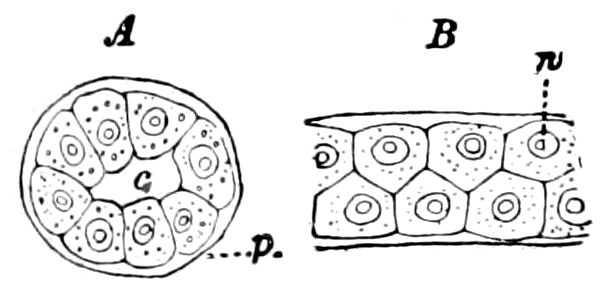
Fig. 345.—A, section of urinary tube of Periplaneta; B, part of tube of Perla; p, peritoneal membrane; c, cavity or lumen; n, nucleus of a secreting cell.—After Schindler.
In the larvæ of some insects the blind ends of the tubes are often externally bound to the rectum, in the silkworms being attached by fine threads to the intestine, while in some flies (Tipula and Ctenophora), two vessels may unite to form a loop. In all larval Cecidomyiæ, the two tubes are united to form a loop which curves backward, opening near the vent, the proctodæum being very short. (Giard.)
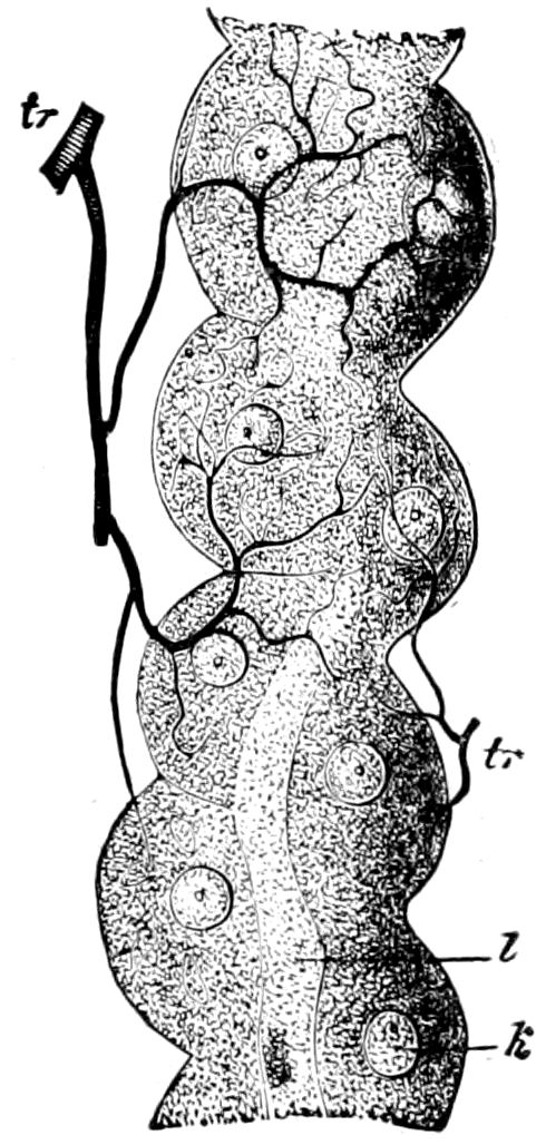
Fig. 346.—Portion of a urinary tube of Calliphora vomitoria: tr, trachea; l, lumen; k, nucleus.—After Gegenbaur.
While usually the urinary vessels form simple tubes, in many species of Lepidoptera and Diptera they are branched, thus resembling those of spiders and scorpions. Moreover, in many Lepidoptera and Diptera (Fig. 308), the tubes are not simple, but are lobulated, and in some Hemiptera (Pentatoma, Notonecta, and Tettigonia) are twisted or lace-like. In rare cases there are two kinds of urinary tubes; in Melolontha vulgaris, two of them are partly lobulated and yellow, while the other two are simple and white. Their color in beetles varies, some being whitish or yellowish; in Geotrupes, Dyticidæ, Hydrophilidæ, etc., reddish brown; in Gryllotalpa as well as Locusta viridissima, there are two different kinds of vessels, differing in contents and in color (white or yellow), as well as histologically. (Schindler.)
The exterior of the tubes is richly provided with tracheæ, which often form a web around them, and the fine branches often seem to attach them to the intestine. In Acheta they are enveloped by a very delicate, loose network of muscular fibres. (Schindler.)
The urinary tubes consist, according to Schindler, of at least three cellular layers (Fig. 345):—
1. An external, connective, nucleated membrane, the peritoneal membrane.
2. A very delicate homogeneous basal membrane, the tunica propria.
3. A single layer of large polygonal excretory cells.
4. Lining the internal canal a chitinous layer penetrated by pore-canals, the intima often wanting.
The secretory cells are usually of the same size, but in many cases are relatively small; sometimes four to six or more form the periphery of the canal, sometimes three or only two. In some insects the cells are so very large that a single cell forms the entire periphery. The nuclei in the Lepidoptera (Papilio, Pontia, Cossus) are large and irregularly branched.
The excretions of the Malpighian vessels, derived from the blood and from the fat-body, are more or less fluid and granular, sometimes pulpy. From the cells they pass into the canal, thence into the intestine, and thence out of the body. How, says Kolbe, the secretion passes into the intestine, whether by the contraction of the fine fibrillæ of the peritoneal membrane, or by the external pressure of the other organs, or by the pressure of the secretory matter behind, 352is not yet known. Grandis observed in living Hydrophilus that the urinary tubes moved, without the muscles seeming to show what caused the motion. Moreover, the cells incessantly changed their form. At a lower temperature such motions ceased. The tracheæ, ending freely in the cells, did not anastomose. (Kolbe.)
The different colors of the tubes (white, yellow, red, brown, or green) is due to the hue of the excretions, and is independent of the color of the blood and of the urinary substances held in the secreted matter.
Schindler found that insects of different stages, collected in winter, differed very much in their urinary secretions, the tubes in the adults being entirely empty, while in the larvæ they were filled full, so that he concluded that in the former the process of excretion during the winter hibernation is very slow, but in the latter very rapid.
As to the activity of the urinary vessels the following experiments will throw some light. Tursini fed a Pimelia with fuchsin; its urinary tubes were consequently colored red. Schindler fed insects with indigo-carmine, which was excreted by the urinary tubes; Kowalevsky arrived at the same results, which seems to prove that these vessels are analogous to the kidneys of vertebrates. Moreover, Schindler injected through the side of the first abdominal segment into the cavity of the body of a Gryllotalpa a concentrated solution of sodium salt of indigotin-disulphonic acid. After one or two hours the external portion of the epithelium of the urinary vessels was stained deep blue, while the inner portion remained of the normal transparency; the nuclei being for the most part deeply stained. Between one and two days after, the staining matter had not yet wholly passed through the central canal, the surface recently stained still appearing light blue.
The solid contents of the urinary tubes consist partly of crystals, which occur singly in the epithelial cells, or form scattered masses when situated in the central canal. Besides tabular rhombic crystals, there occur concretions which contain uric acid, and probably consist of urate of soda, also octahedral crystals of chloride of soda, and quadro-pyramidal crystals of oxalate of lime. Also acicular prisms occur; besides chloride of soda, phosphates, carbonate of lime, oxalate of lime in quantity, leucine, coloring matters, etc.; while the fluid secretion also contains urea (?), uric acid, and abundant urates; uric acid crystals were precipitated by the addition of acetic acid, and by adding hydrochloric acid crystals belonging to the dimetric system were formed. The often numerous spheroidal small granules are biurate of soda and biurate of ammonia. Pale, concentrically banded concretions are leucine pellets.
According to Kölliker the contents of the urinary vessels[55] in general are: (1) round granules of urate of soda and urate of ammonia; (2) oxalate of lime; 353and (3) pale transparent concretions of leucine. Crystals of taurin are also said to occur. (Claus’ Zoölogy, p. 531.)
Although uric acid is characteristic of the urinary tubes, yet sometimes it is wanting in them, while uric acid substances in quantity occur in the fat-body or in the mid-intestine.
In the living insect the urinary tubes remove urates from the blood; “the salts are condensed and crystallized in the epithelial cells, by whose dehiscence they pass into the central canals of the tubules and thence into the intestine.” (Miall and Denny.)
The process of excretion is carried on not only by the urinary tubes, but also, as Cuénot has recently shown (1896) in Orthoptera, by the pericardial cells and certain cells of the fat-bodies. In the last-named cells urates are stored throughout life; the pericardial cells apparently secrete but do not store waste products, which are finally eliminated by the urinary tubes, the latter constantly eliminating waste.
Primitive number of tubes.—Wheeler considers the primitive number of urinary tubules to be six, other authors regarding two pairs as the primary or typical number; and while Wheeler agrees that the more ancestral tracheate arthropods had but a single pair, Cholodkowsky supposes the primitive number in insects themselves to be a single pair. This view is strengthened by the fact that Scolopendrella has but a single pair (Fig. 15).
While Peripatus has no urinary tubes, in Myriopods a single pair arises, as in insects, from the hind-intestine.
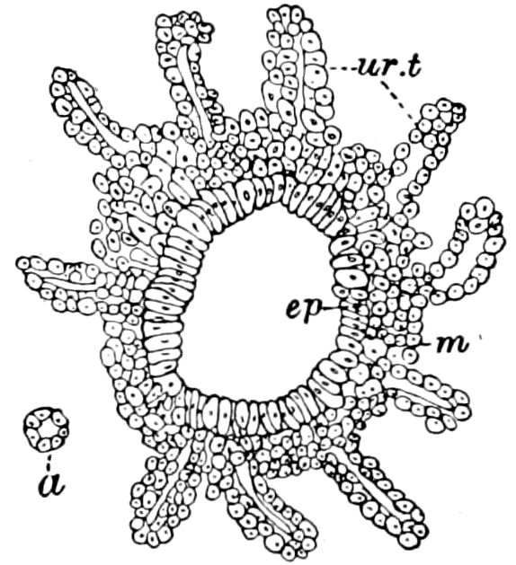
Fig. 347.—Section of proctodæum of embryo locust, showing origin of urinary tubes (ur.t); ep, epithelial or glandular layer; m, cells of outer or muscular layer; a, section of a tube.
When in insects the number of these tubes is few, they are, with rare exceptions, arranged in pairs, so that Gegenbaur and others have considered this paired arrangement as the primitive one. When the tubules are very numerous in the adult, as in Orthoptera, the embryos and larvæ have a much smaller number, Wheeler stating that “in no insect embryo have more than three pairs of these vessels been found.” We have observed 10 primary tubes in the embryo of Melanopus (Fig. 347), from each of which afterwards arise 15 secondary tubules. In the Termites, only, do the young forms have more urinary tubes than the adults.
In Campodea there are about 16 urinary tubes and in Machilis either 12 (Grassi) or 20 (Oudemans); but in other Thysanura the number is much less, Lepisma having either four, six, or eight, according to different authors, and both Nicoletia and Lepismina having six, opening separately into the hind-intestine. On the other hand, these organs have not yet been detected in Japyx. Whether they exist at all in the Collembola, which are degenerate forms, is doubtful. The weight of opinion denies their existence, though they may yet be found existing in a vestigial condition. They are said by Tullberg and by Sommer to exist in Podura, but are of peculiar shape.
Coming now to the winged insects, in what on the whole is perhaps the lowest or most generalized order, the Dermaptera, the number is over 30, and their insertions regularly encircle the intestine. (Schindler.) In the most ancient and generalized family of Orthoptera, the Blattidæ, Schindler detected from 60 to 70 tubes, but in a nymph of Periplaneta not quite 10 mm. in length he 354found from 16 to 18, and in nymphs 4 to 5 mm. long there were only eight vessels; while Wheeler has found in the embryo of Phyllodromia germanica but four tubes. In the adult Acrydiidæ there are as many as 150, in the Locustidæ between 40 and 50, and in the Gryllidæ about 100.
The Ephemeridæ with about 40, the Odonata with 50 to 60 tubules, the Perlidæ with from 50 to 60, are polynephrious; while the Termitidæ and Psocidæ are oligonephrious, the former having from six to eight and the Psocidæ only four tubes. So also all the other orders not mentioned, except the Hymenoptera, have few of these tubes. The Hemiptera, with none in Aphidæ, a single pair in the Coccidæ, and two in all the rest of the order, have the fewest number.
In the Neuroptera there are from six to eight, while in a larva, possibly that of Chauliodes, Wheeler finds the exceptional number of seven.
The closely allied order Mecoptera (Panorpidæ), and also the Trichoptera, agree with the Neuroptera (Sialis) in having six. According to Cholodkowsky all Lepidoptera have six of these vessels, except Galleria, which has but four. He finds that in Tinea biselliella (also T. pellionella and Blabophanes rusticella) the larva has six vessels, which, however, undergo histolysis during pupation, a single pair arising in their stead. On this account he regards the primitive number of urinary tubes as two, or a single pair, this return from six vessels in the larva to two in the imago being considered a case of atavism.
In the Coleoptera, the number of urinary tubes is from four to six; in what few embryo beetles have been examined (Doryphora, Melolontha), there are six vessels, but in the embryo of Dyticus fasciventris, Wheeler has detected only four, this number being retained in the adult. He thinks that in beetles in general, a pair of vessels must be “suppressed during post-embryonic development, presumably in early larval life.”
In Diptera and Siphonaptera, the number four is very constant, there being, however, a fifth one in Culex and Psychoda (Fig. 400.)
The number of these vessels is very inconstant in the Hymenoptera, varying from six (Tomognathus, an ant, worker) to 12 (Myrmica), and in Apis reaching the number of 150.
In the embryo of the honey-bee and wall-bee (Chalicodoma), there are only four; we still lack any knowledge of the number in embryo saw-flies.
The following is a tabular view of insects with few urinary tubes (Oligonephria) and many (Polynephria). It will be seen that the number has little relation to the classification or phylogeny, insects so distantly related as the Orthoptera and Hymenoptera being polynephrious:—
Oligonephria
Collembola, 2 (Podura), Tullberg and also Sommer.
Thysanura, 4 (Lepisma); in Campodea, 16; in Machilis, 12 or 20; wanting in Japyx.
Psocidæ, 4.
Termitidæ, 6 (many in the young, Rathke).
Mallophaga, 4.
Physapoda, 4.
Hemiptera, 2 (Coccidæ, none in Aphidæ).
Neuroptera, 6–8. (In Sialidæ and Rhaphididæ 6; in Myrmeleonidæ and Hemerobiidæ, 8).
Trichoptera, 6.
Mecoptera, 6.
Lepidoptera, 2–4–6 (2 in Tinea, Tineola, and Blabophanes; in Pterophorus and Yponomeuta, 4).
355Coleoptera, 4–6; never more.
4
Carabidæ,
Dyticidæ,
Staphylinidæ,
Gyrinidæ,
Palpicornes,
Lamellicornes,
Cantharidæ,
Buprestidæ
(in larva, 6; in beetle, 4).
6
Byrrhidæ,
Nitidulidæ,
Dermestidæ,
Cleridæ,
Meloidæ,
Pyrochroidæ,
Bruchidæ,
Bostricidæ,
Cerambycidæ
Chrysomelidæ,
Coccinellidæ.
Diptera, branching into 4 (Gegenbaur); in Culicidæ, and Psychoda, 5.
Siphonaptera, 4.
Polynephria
Orthoptera, 100–150. (In embryo Blattids, 4; in embryo locust, 10; in nymph of Gryllotalpa, 4.)
Dermaptera, “over 30” (Schindler).
Perlidae, 50–60.
Plectoptera (Ephemeridæ), 40.
Odonata, 50–60.
Hymenoptera, 20–150. (In embryo bees only 4; Cynipidæ, Ichnenumonidæ, and Formicidæ have the smallest number, 6–12.)
Here should be mentioned the singular fact discovered by Koulaguine that in the larva of Microgaster, the urinary tubes have no connection with the intestine, but open dorsally on the outside of the body on each side of the anus. Ratzeburg had stated that the last segment of the body was in the form of a vesicle. Koulaguine now shows that this vesicle is in reality the end of the intestine opening upwards; as the result of this dorsal opening of the intestine the Malpighian vessels open on the sides of the oval vent, and have no connection with the intestinal canal. Whether this is of morphological import, or is only a secondary adaptation, Koulaguine does not state, his paper being a preliminary abstract.
Wheeler thus sums up our present knowledge regarding the number and homologies of the Malpighian or urinary tubes:
1. It is very probable that the so-called Malpighian vessels of Crustacea and Arachnida are not the homologues of the vasa Malpighi of the Eutracheata (insects and myriopods).
2. The Malpighian vessels of the Eutracheata arise as paired diverticula of the hind-gut and are, therefore, ectodermal.
3. In no insect embryo are more than six vessels known to occur; although frequently only four are developed.
4. The number six occurs either during embryonic or post-embryonic life in members of the following groups: Apterygota, Orthoptera, Corrodentia; Neuroptera, Panorpata, Trichoptera, Coleoptera, Lepidoptera, and Hymenoptera.
5. The number four seems to be typical for the Corrodentia, Thysanoptera, Aphaniptera, Rhynchota, Diptera, and Hymenoptera.
6. The embryonic number in Dermaptera, Ephemeridea, Plecoptera, and Odonata has not been ascertained, but will probably be found to be either four or six.