PART III.—THE METAMORPHOSES OF INSECTS (continued 3)
Reading edition
679
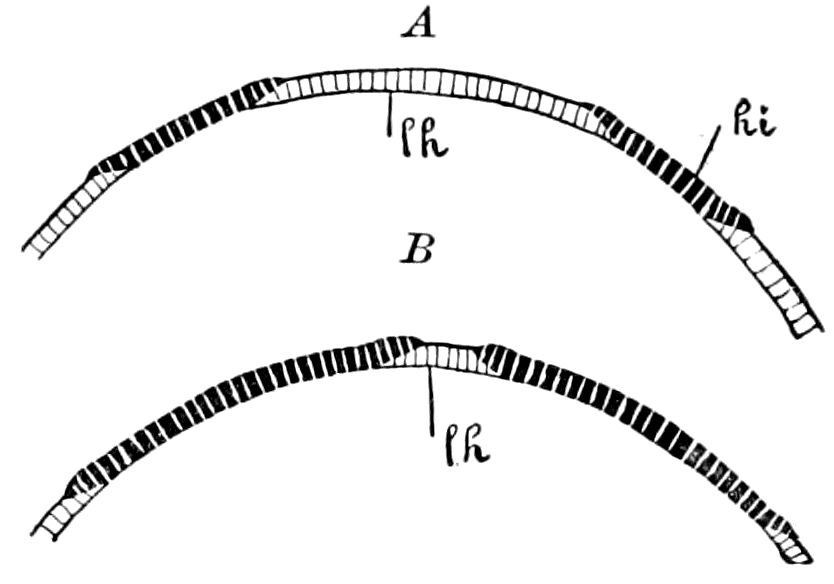
Fig. 631.—Diagram of the formation of the imaginal hypodermis on the abdomen of Muscidæ: hi, imaginal buds of the hypodermis; lh, larval hypodermis.—After Lang.
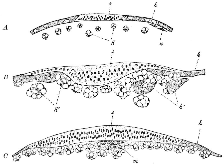
Fig. 632.—Section through the abdominal bud of the hypodermis of Musca: A, of the larva; B and C, of the pupa; h, larval hypodermis; h′ separated portion of the same attacked by phagocytes; i, imaginal bud; k, phagocytes with what are called cell-wrecks or fragments (so-called granulated cells); k′, phagocytes enclosing hypodermal nuclei; m, mesoderm-germ of the imaginal bud; w, wandering cells.—After Kowalevsky, from Korschelt and Heider.
The hypodermis.—The hypodermis of the imago arises through an extension of the ectodermal part of the imaginal buds. We have already mentioned this for the thorax. As the appendages of the thorax in the pupa gradually attain perfection, the hypodermis layer spreads from the place of their insertion, the layer consisting of numerous small cells whose origin we must refer to the peripodal membrane. This layer continues to spread over the surface of the pupal thorax, while at the same time the area of the larval hypodermis, consisting of large cells, is seen to diminish. Hence the thin edge of the newly-formed hypodermis (Fig. 631, hi) slowly grows into the space between the superficial cuticula and the larval hypodermis (Fig. 632, h), so that at this place the old hypodermis undergoing destruction eventually lies on the inner side of the newly-formed epithelial layer (B). We therefore see from this that, during the replacement of the old hypodermis by the new, the continuity of the superficial epithelium is never interrupted. Since the edges of the two kinds of hypodermis overlap, the surface of the body is nowhere bare of epithelium. The dissolution of the larval 680hypodermis is accomplished under the influence of the leucocytes (Fig. 632, k), which attack the larval hypodermis-cells and absorb their contents piece by piece, and so fill themselves with bits of the hypodermis-cells and their nuclei; since these fragments have the shape of roundish granules, they were called by Weismann granule-balls. These granule-balls, which fill the body-cavity of the later pupal stage, are nothing else than the leucocytes (blood corpuscles) which have absorbed the fragments of tissue of the larval body.
It should here be said that the destruction of the larval tissues is not to be attributed to the previous death of the cells, but is the result of the action of the leucocytes on tissues which, though weakened in their vital power, are still living. While the completely healthy, active tissues, i.e. those of the imaginal buds, withstand the attacks of the leucocytes, the less healthy larval tissues are by the attacks of the leucocytes divided into fragments and eaten and digested by them. This process is most marked in the histolysis of the larval muscles. The destruction of most of the larval organs depends, therefore, on the capacity of the amœboid blood-corpuscles for taking food and on intracellular digestion, as was first shown by Metschnikoff, who has given to these leucocytes the name of “phagocytes.”
This process of histolysis goes on in the same way in the head and abdomen as in the thorax. In the abdomen, as Ganin first proved, there are in each of the eight segments of which it consists in the larva four small cellular islets or imaginal buds (Figs. 631, hi, 632, i), from which originate the new hypodermis.
Van Rees has lately found in the abdominal segments another pair of smaller imaginal buds. The four imaginal buds occurring in the last segment are situated close to each other, encircling the anal opening (Fig. 633, ims), and take part in the formation of the hind-intestine, the rectal pouches and rectal papillæ. To this segment also belong the two pairs of imaginal genital buds (rudiments of the external sexual organs) which were first found by Künckel d’Herculais in Volucella.
The newly formed hypodermis spreads rapidly over the outer surface of the body, so that hypodermal areas corresponding to the separate imaginal buds soon unite. Simultaneously with this completion of the definite epithelial layer the larval hypodermis becomes completely destroyed by the phagocytes.
The muscles.—A similar process of destruction by phagocytes affects the greater number of the larval muscles, except the three pairs of thoracic muscles employed in respiration, and which pass 681intact from the larva to the imago. Indeed, the dissolution of the muscles is the first process which occurs in the metamorphosis. The destruction of the larval muscles is accomplished in such a way that, a great number of leucocytes which have collected on the surface of the muscular fibres, press through the sarcolemma and enter within the muscular tissue, filling the spaces formed between them, By this means the muscles break up into a number of rounded particles which are taken into the interior of the leucocytes. Thus a collection of granule-balls arise from the muscles, which finally separate from each other and become scattered throughout the body-cavity of the pupa. In the same way as the muscular substance, the muscle-nuclei are taken up and digested by the phagocytes.
The imaginal muscles develop from the definitive mesoderm which has originated from the mesoderm of the imaginal buds (Fig. 632, C, m).
The digestive canal.—As in the hypodermis and muscles, the histolysis of the larval digestive tract and its new formation from separate imaginal buds go on simultaneously, so that the continuity of the process is not interrupted.
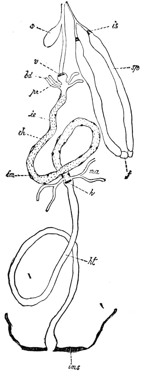
Fig. 633.—Digestive tract of a Musca larva with the imaginal germs: bd, cœca; s, food-reservoir; is, imaginal ring of the salivary gland (sp); f, fat-cells at the end of the salivary gland; pr, proventriculus; r, its ring; ie, imaginal cells of the mid-intestinal epithelium; ch, chyle-stomach; ma, urinary tubes; im, imaginal cells of the mid-intestinal, muscular layer; ims, binder, abdominal, imaginal buds; h, hind intestinal, imaginal bud; ht, hind-intestine.—After Kowalevsky, from Korschelt and Heider.
The imaginal buds of the much-shortened pupal digestive canal occur in the mid-intestine (stomach) in the form of numerous scattered groups of cells (Fig. 633, ie), and in the fore- and hind-intestine in the form 682of rings (v and h) of imaginal tissue. The imaginal ring of the fore-intestine (v) lies in the region of the proventriculus (pr, compare Fig. 635, im), while that of the hind-intestine is situated directly behind the base of the urinary tubes. The regeneration of these two parts of the digestive canal is not entirely accomplished by these two rings, but the imaginal rudiments of the neighboring parts of the outer surface of the body also have a share in it. Thus it appears that the foremost part of the œsophagus is built up from the imaginal buds in the region of the mouth, while the imaginal buds surrounding the anus in the 8th abdominal segment (Fig. 633. ims) produce by invagination the rectal pouches, together with the rectal papillæ.
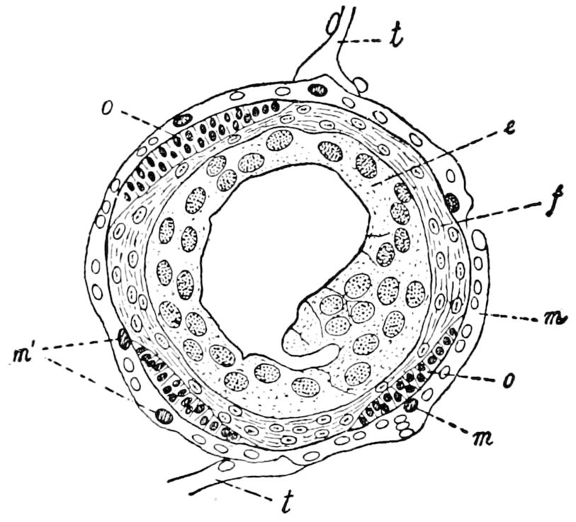
Fig. 634.—Cross-section through the mid-intestine of pupa of Musca: e, rejected and degenerate epithelium of the larval stomach; f, cellular layer newly formed around the same; m, muscular layer; m′, imaginal cell of m; o, imaginal bud of the mid-intestinal epithelium; t, tracheal stem.—After Kowalevsky, from Korschelt and Heider.
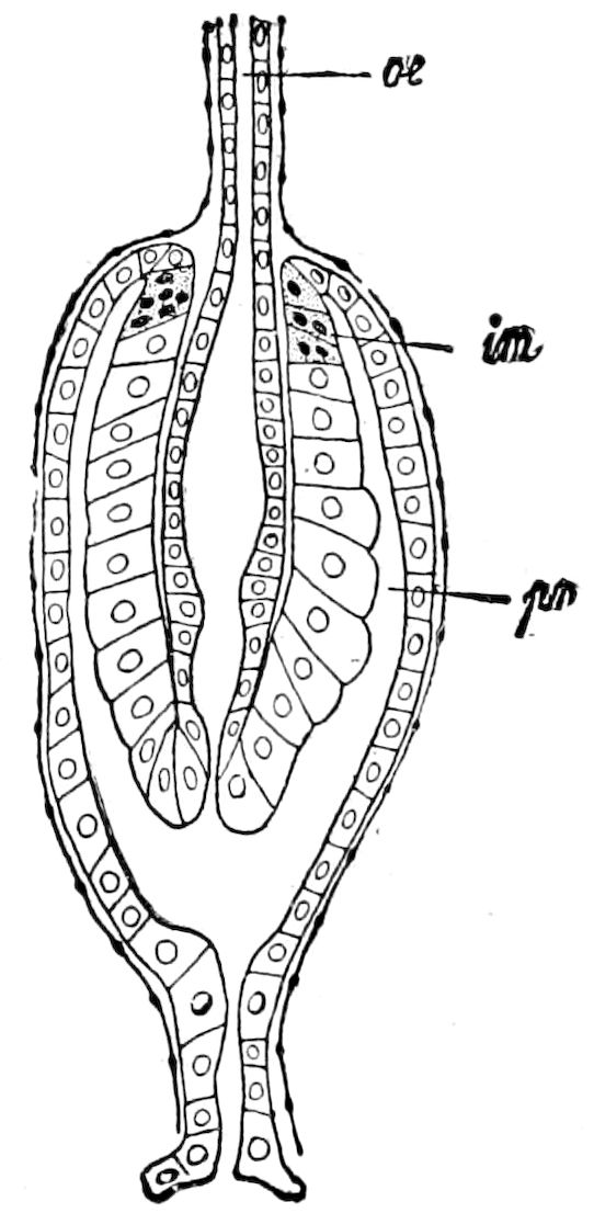
Fig. 635.—Longitudinal section through the proventriculus of a muscid larva: im, fore-intestinal, imaginal ring; oe, œsophagus; pr, proventriculus.—After Kowalevsky, from Korschelt and Heider.
The formation of the mid-intestine (stomach) takes place in such a way that the island-like imaginal buds spread out by cell-multiplication over the outer or basal surface of the larval mid-intestinal epithelium (Fig. 634, o), until they finally unite, so as to form the wall of the imaginal mid-intestine (stomach). At the same time the entire larval epithelium (e) is cast in the interior and forms the so-called yellow body, which becomes surrounded by a layer of small cells and a jelly-like mass, and remains until its destruction in the pupal stomach. The larval muscular layer (m) remains intact as long as the imaginal mid-intestine is not fully developed, when 683it is attacked and destroyed by phagocytes. The final muscular layer arises from single cells lying on the outer surface of the imaginal buds (Figs. 633, im, 634, m′), which should be regarded as special imaginal cells of the mid-intestinal muscular layer.
The transformation of the fore-intestine is introduced by a degeneration of the proventriculus and sucking stomach. The proventriculus (Fig. 635, pr), which had been formed from a circular fold of the fore-intestine, disappears by the smoothing out of this folded structure. The sucking stomach also similarly degenerates by withdrawing gradually into the œsophagus, so that instead of the original diverticulum there remains only an enlargement of the œsophageal cavity. At the same time this part of the canal is attacked and destroyed by phagocytes, while the destroyed portions become replaced by the gradually extending imaginal parts of the wall. The imaginal ring of the fore-intestine (Fig. 635, im), which, according to Kowalevsky, is concerned in the formation of a great part of the definitive œsophagus, becomes closed at its hinder end so that the communication with the mid-intestine appears to be interrupted.
The hind-intestine of the imago is rebuilt in exactly the same manner. Here also the imaginal ring widens and forms a tube, which while it grows around the openings into the urinary tubes, closes itself against the mid-intestine, while behind it remains in connection with the larval hind-intestine. In a similar way the larval hind-intestine is attacked by the growth from behind of an imaginal ring, which proceeds from imaginal buds near the anus, until finally, when the entire larval hind-intestine is reduced to granule-balls, the two imaginal sections of the tube are brought into contact with each other. (Kowalevsky in Korschelt and Heider.)
The larval salivary glands (Fig. 633, sp) are completely destroyed by phagocytes. Then succeeds the new formation of these glands from imaginal buds, which, according to Kowalevsky, form rings situated at their anterior ends.
The nature of the transformation undergone by the urinary tubes is not yet well ascertained. According to Van Rees, there is in this case perhaps a regeneration of the larval cells by division, but on the other side there may be a histolysis of these elements.
The above-described method of transformation of the digestive canal seems, according to Korschelt and Heider, to be very common among the holometabolic insects. It has not only been observed in the Diptera, but also in the Lepidoptera (Kowalevsky, Frenzel), Coleoptera (Ganin), and Hymenoptera (Ganin). The stripping off of the epithelium of the mid-intestine was found by Kowalevsky to occur also in Corethra, Culex, and Chironomus.
The tracheal system.—As we have seen (p. 448), the tracheal system of caterpillars just before pupation undergoes disintegration, 684accompanied by a reformation of the peritoneal membrane and tænidia. The larval ectotrachea undergoes histolysis, that of the imago being meanwhile formed; the larval tænidia also break up, dissolve, and are replaced by new tænidia which arise from the nuclei of the peritoneal membrane. That the tracheal system in the Muscidæ during metamorphosis undergoes a transformation is shown, as Korschelt and Heider claim, by the entirely different shape of the system in the maggot, the pupa, and the fly. The air is admitted to the tracheal system of the maggot, not by lateral openings, but through the two stigmata at the end of the body. On the other hand, the pupa breathes by prothoracic spiracles, while the fly has six pairs of lateral stigmata of the normal structure. There may be in the larva and pupa vestigial closed stigmata, as there are in the thorax of caterpillars, with tracheal branches leading to where were once functional stigmata. These stigmatal branches, as well as some other portions of the tracheal system already observed by Weismann, seem, according to Van Rees, to function as imaginal buds for the regeneration of the tracheal matrix, while frequently also a regeneration of this epithelium, by a simple repeated division of cells, may be recognized. The disintegration of the tracheal system is accomplished by means of phagocytes in the manner already described.
The nervous system.—The central nervous system passes directly from the larval into the imaginal stage, since it must continue to exercise most of its functions throughout metamorphosis, though it undergoes important changes of form and position. At the same time, certain histological transformations occur which may be regarded as a histolysis. Such is the destruction and rebuilding in the interior of the organs, which, however, preserve their continuity. Every case of destruction of tissues in the pupa has come to be regarded as a histolysis.
The problem of the transformation of the peripheral nervous system is not yet well understood. Although during the destruction of the larval muscles the motor nerves also in part degenerate, in the case of the nerves distributed to the appendages the conditions are different, as these may be recognized in the larva in the form of the nerve-threads which place the imaginal buds in connection with the central nervous system. These threads, according to Van Rees, pass from the larva into the pupa and imago, so that with the farther development of the rudiments of the extremities, only the distal part of the nerves belonging to them are to be regarded as new formations. (Korschelt and Heider.)
685The fat-body.—The larval fat-body is also destroyed through the activity of the leucocytes in the same way as the other tissues. The reformation of the fat-body seems to begin in the mesoderm of the imaginal buds. Possibly, also, the masses or collections of embryonic cells which are regarded by Schaeffer as “blood-forming cells,” may serve to regenerate the fat-body. At all events, they have been derived from the mesodermal tissues. Though Wielowiejsky saw the fat-body of Corethra arising from a cell-layer situated under the hypodermis, yet it is not necessary to regard this observation as favorable to the view of Schaeffer that in Musca the larval fat-body is derived in part from the hypodermis, and in part from the tracheal matrix, thus from the ectodermal tissues. (Korschelt and Heider.)
Definitive fate of the leucocytes.—We have seen that the formation of the organs of the imago originates in the imaginal buds, in all cases where these do not pass directly from the larva into the pupa. The leucocytes, whose numbers in the pupa are greatly increased, take no direct part in the formation of the tissues. Their importance seems to lie in this, that they destroy those larval organs doomed to destruction, the parts of which they take in and digest, and possibly, by their powers of locomotion, convey particles of food to the developing organs.
What, on the other hand, is the fate of the leucocytes after the developmental processes in the pupa have ceased? There can be no doubt that a part of the so-called granule-cells are again transformed into normal blood-corpuscles. Another, and, as it seems, more considerable, share suffer degeneration. Finally, the leucocytes themselves serve as nourishment for the newly formed tissues. Of interest in this direction is the observation of Van Rees, that numerous leucocytes finally pass into the newly-formed hypodermis and then degenerate in crevices between the hypodermis-cells. (Korschelt and Heider.)
It has been suggested by Van Rees that the phagocytes attack all the larval organs indiscriminately, but that the active metabolism of the imaginal buds preserves them from these attacks. He also thinks that Kowalevsky is probably right in supposing that the buds render themselves immune by some poisonous secretion.
Pratt, however, thinks that the supposition of a protecting or poisonous secretion is scarcely necessary to account for the phenomenon, and suggests that the larval tissues are a prey to the phagocytes, because at the end of larval life they become functionless and inactive, so as to become an easy prey to phagocytes or disintegrating influences of any sort. On the other hand, the imaginal buds “in which there is an exceedingly active metabolism, and all the larval 686organs which remain functional during the metamorphosis are immune from the attacks of the phagocytes. The heart in the muscids continues to beat, as Künckel d’Herculais has observed, during the entire period of the metamorphosis, with the exception of a day or two in the latter half of it. The nervous system must continue functional during the entire time. The three pairs of thoracic muscles which pass intact from the larva to the imago are probably employed in respiration during the metamorphosis. The reproductive glands are, like the imaginal disks, rapidly growing organs.” He adds that among the other holometabolic insects many or most of the larval organs remain functional during metamorphosis, hence there is but little histolysis. “But the larval intestine would always necessarily become unfunctional, and, as we have seen, Kowalevsky is of the opinion that the larval mid-gut in all holometabolic insects contains imaginal disks, and undergoes degeneration during the metamorphosis.”
The post-embryonic changes and imaginal buds in the Pupipara (Melophagus).—The sheep-tick (Melophagus) is still more modified than the Muscidæ; the larva is apodous and acephalous, but, as Pratt observes, much less highly specialized than those of muscids, and in respect to the position of the thoracic buds it closely resembles Corethra. They lie just beneath the hypodermis in two very regular rows, and not in the centre of the body, as in Musca (Figs. 628, 636, C). While, however, in Corethra all the thoracic buds are of larval origin, arising after the last larval moult, in Melophagus, on the other hand, each of these buds, except the dorsal prothoracic, arises in the embryo, as is also the case in Musca.
In the cephalic buds the conditions are similar to those in Musca, but still more complicated. Instead of a single pair of head-buds, there are two pairs, one dorsal and one ventral. “The dorsal pair corresponds to the muscidian head-disks in every respect; they are destined to form the dorsal and lateral portions of the imaginal head, together with the compound eyes. The ventral head-disks have no counterpart in Musca. The fate of these disks or buds is to form the ventral portion of the head, the paired projections forming the rudiments of the proboscis.
“The formation of the head-vesicle proceeds in a way similar to that in Musca. The ventral disk fuses early at its lateral edges with the dorsal pair. The communications between both ventral and dorsal disks and the pharynx rapidly widen (in the old larva they have already become very large), and soon the disks and pharynx form together a single vesicle, which is the head-vesicle.” The imaginal buds of the abdomen Pratt finds to be exactly as in the corresponding ones of Musca.
In the embryo of Melophagus the cephalic and thoracic imaginal buds first appear as local thickenings, followed by the invagination of the ectoderm; the cephalic buds first appear very early in the 687ontogeny of the insect (Fig. 636, C), just as the germs of the digestive canal, nervous system, and tracheæ are appearing. The single median thickening (v) is destined to form the ventral cephalic bud, while the pair of thickenings behind (d) become the dorsal buds, those homologous with the cephalic buds of Musca.
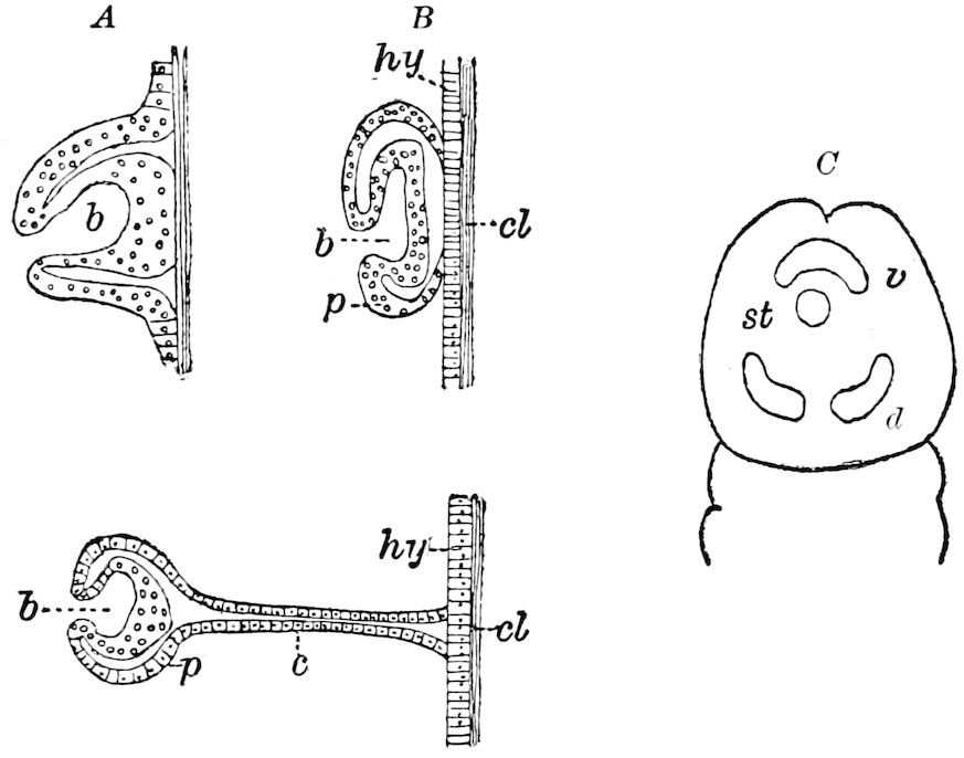
Fig. 636.—Imaginal buds in Musca,—A, in Corethra,—B, in Melophagus,—C, in embryo of Melophagus: dorsal view of head; b, bud; p, peripodal membrane; c, cord; hy, hypodermis; cl, cuticula; st, stomodæum; v, ventral cephalic bud, behind are the two dorsal cephalic buds (d).—After Pratt.
The thoracic buds, which arise as hypodermic thickenings, do not appear until late in embryonic life, until the time of the involution of the head.
Pratt did not observe in the embryo the buds of the internal organs and of the abdominal hypodermis, and thinks it probable that they appear first in the larva.
c. General summary
We have seen that in Coleoptera, Lepidoptera, Diptera, and Hymenoptera, and with little doubt in all the holometabolous insects, the parts of the imago originate in single formative cellular masses (imaginal buds) already present in the larva, and often even in the later embryonic stages. There are such imaginal buds for each part of the body,—for the appendages of the head, for the legs and wings, for the ovipositor, and probably for the cercopods, for the hypodermis of the abdomen, and for the different sections of the digestive 688canal. We have seen, as Korschelt and Heider state, that the formation of the mesodermal organs of the imago (muscles, connective tissue, fat-body) begins in the mesodermal part of the imaginal buds, whose first origin is still obscure. Simultaneously with the formation of the imaginal organs, there goes on under the influence of the leucocytes the destruction of the larval organs. Both processes (destruction and regeneration) therefore go on hand in hand, so that the continuity of the organs in question in most cases remains perfect, inasmuch as the complete destruction only ensues after the formation of the final organs. The only exceptions are most of the muscles of the larva, which are destroyed at a very early period.
Moreover, it is evident that the sharp division into larval, pupal, and imaginal stages only applies to the external surface of the body, since they follow one another after successive moults. The processes of the internal development, on the other hand, form an entirely continuous series of transformations between which is no sharp line of demarcation. Yet as a whole the form of the larva, pupa, and imago are kept distinct in adaptation to their separate environments and habits.
Finally, as Pratt very truly remarks, the epigenetic period in insects, when new organs are forming, does not end with the birth of the larva from the egg, but extends through the larval, and even through the pupal period. “The principal significance of the pupal period and the metamorphosis is that it is the time when the larval characters which were adapted for use during a period of free life in the midst of the development, and which would be valueless to the imago, are corrected or abandoned.”
HYPERMETAMORPHISM
When an insect passes through more than the three normal stages of metamorphosis, i.e. the larval, pupal, and imaginal, it is said to undergo a hypermetamorphosis. The best-known examples are the supernumerary stages of Meloë, Stylops, etc.
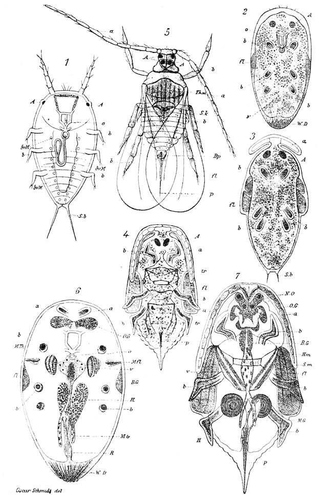
Fig. 637.—Hypermetamorphosis of male of Aspidiotus nerii: 1, freshly hatched larva; 2, larva shortly before pupating; b, rudiments of the legs; fl, of the wings; 3, pupa before moulting; 4, the same after moulting; 6, larva farther advanced than in 2; a, antennal rudiments; b, rudiments of legs; v, stomach; OG, brain; M. Fl, rudiments of the elevator and depressor muscles of the wing; M. Th, rudiments of the dorsal muscles; H, rudiments of the testes; 7, pupa shortly before entering upon the imago state (5); A, eyes; a, antenna; o, mouth; WD, wax-glands; BG, ventral nervous cord; Sb, caudal setæ; tr, tracheæ; p, genital armature.—After Schmidt.
690As has already been observed, Schmidt has shown that in the male of the Coccidæ, there is a true hypermetamorphosis, as shown by Fig. 637. In Aspidiotus nerii there are five stages, there being two larval (1, 2) and two pupal stages (3, 4, 7). Stage 3 (Fig. 637, 2) may be compared with the pro-pupa stage of Riley (Fig. 581).
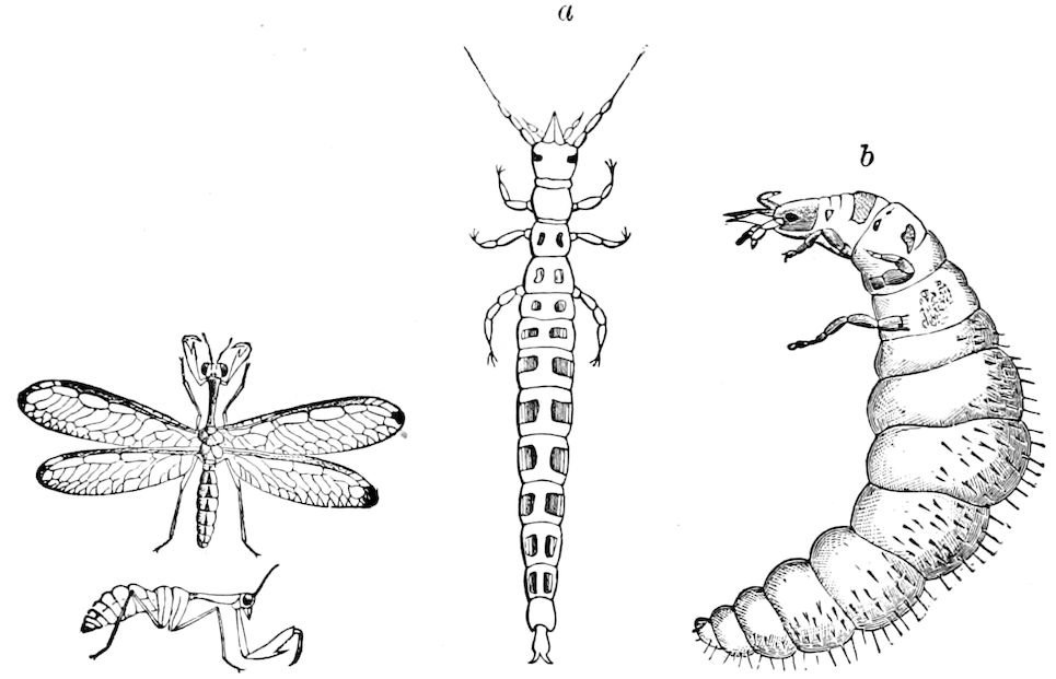
Fig. 638.—Mantispa interrupta, and side view of the same without wings: natural size.—Emerton del. a, freshly-hatched campodeoid larva of Mantispa styriaca, enlarged; b, the same, but older, before the first moult; enlarged.—Brauer.
We have already, on page 602, described the hypermetamorphosis of the neuropterous insect Mantispa (Fig. 638).
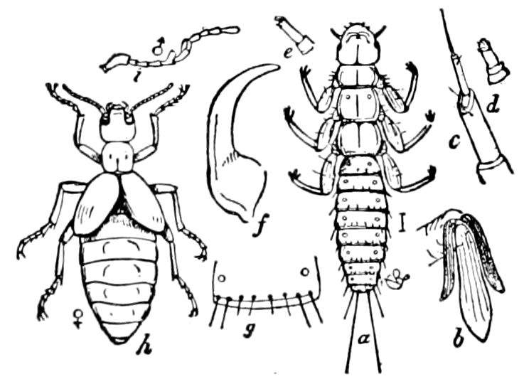
Fig. 639.—Triungulin (a) of a Californian Meloë: b, the three triungulin claws; c, antenna; d, maxillary palpus; e, labial palpus; f, mandible; g, an abdominal joint; h, imago, ♀; i, antenna of ♂.—After Riley.
In Meloë the freshly hatched larva, or “triungulin” (Fig. 639, a), is an active Campodea-like larva, which runs about and climbs up flowers, from which it creeps upon the bodies of bees, such as Anthophora and Andrena, who carry it to their cells, wherein their eggs are situated. The triungulin feeds upon and destroys the eggs of its hostess. Meanwhile its inactive life in the bee’s cell reacts upon the organism; after moulting, the-second larval form (Fig. 640, b) is attained, and now the body is thick, cylindrical, soft, and fleshy, and it resembles a lamellicorn larva, with three pairs of rather long thoracic legs. This is Riley’s carabidoid stage. This second larva feeds upon the honey stored up for the young or larval bees. After 691another moult, there is another entire change in the body; it is motionless, the head is mask-like without movable appendages, and the feet are represented by six tubercles. This is called the semipupa or pseudo-pupal stage. This form moults, and changes to a third larval form (c), when apparently, as the result of its rich, concentrated food, it is overgrown, thick-bodied, without legs, and resembles a larval bee.
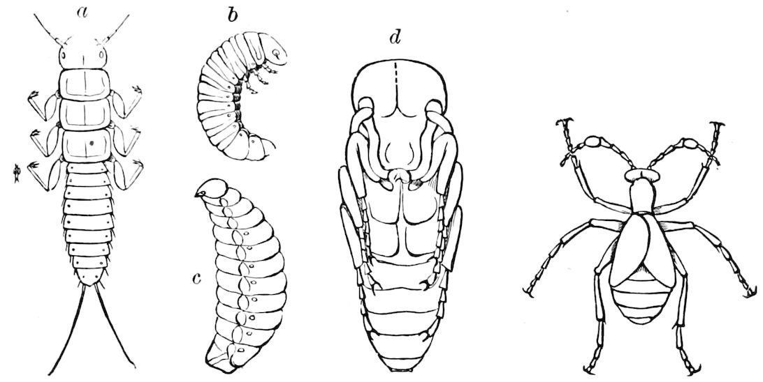
Fig. 640.—Oil-beetle: a, first larva; b, second larva; c, third larva; d, pupa.
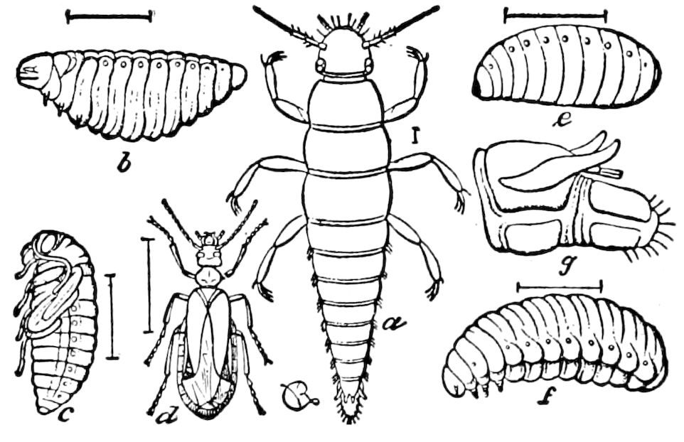
Fig. 641.—History of Sitaris: a, triungulin or 1st larva; g, anal spinnerets and claspers of same; b, 2d larva; e, pseudo-pupa; f, 3d larva; c, true pupa; d, imago, ♀.—After V. Mayet, from Riley.
After thus passing through three larval stages, each remarkably different in structure and in the manner of taking food, it transforms into a pupa of the ordinary coleopterous shape (d).
The history of Sitaris, as worked out by Fabre and more recently by Valery-Mayet, is a similar story of two strikingly different adaptational larval forms succeeding the triungulin or primitive larval stage. The first larva (Fig. 641, a) is in general like that of Meloë, the second (b) is thick, oval, fleshy, soft-bodied, and with minute legs, evidently of no use, the larva feeding on the honey stored by its host. The pseudo-pupal stage is still more maggot-like than in the corresponding stage of 692Meloë, and the third larva (f) is thick-bodied, with short thoracic legs.
In the complicated life-history of another cantharid, Epicauta vittata, as worked out by Riley (Fig. 642), we have the same acquisition of new habits and forms after the first larval stage, which evidently were at the outset the result of an adaptation to a change of food and surroundings. The female Epicauta lays its eggs in the same warm, sunny situation as that chosen by locusts (Caloptenus) for depositing their eggs. On hatching, the active minute carnivorous triungulin, ever on the search for eggs, on happening upon a locust egg gnaws into it, and then sucks the contents. A second egg is attacked and its contents exhausted, when, owing to its comparatively inactive habits and rich nourishing food after a period of inactivity and rest, the skin splits along its back, and at about the eighth day from beginning to take food the second larva appears, with much smaller and shorter legs, a much smaller head, and with reduced mouth-parts. This is the carabidoid stage of Riley. After feeding for about a week in the egg a second moult occurs, and the change of form is slight, though the mouth-parts and legs are still more rudimentary, and the body assumes “the clumsy aspect of the typical lamellicorn larva.” This Riley denominates the scarabæidoid stage of the second larva.
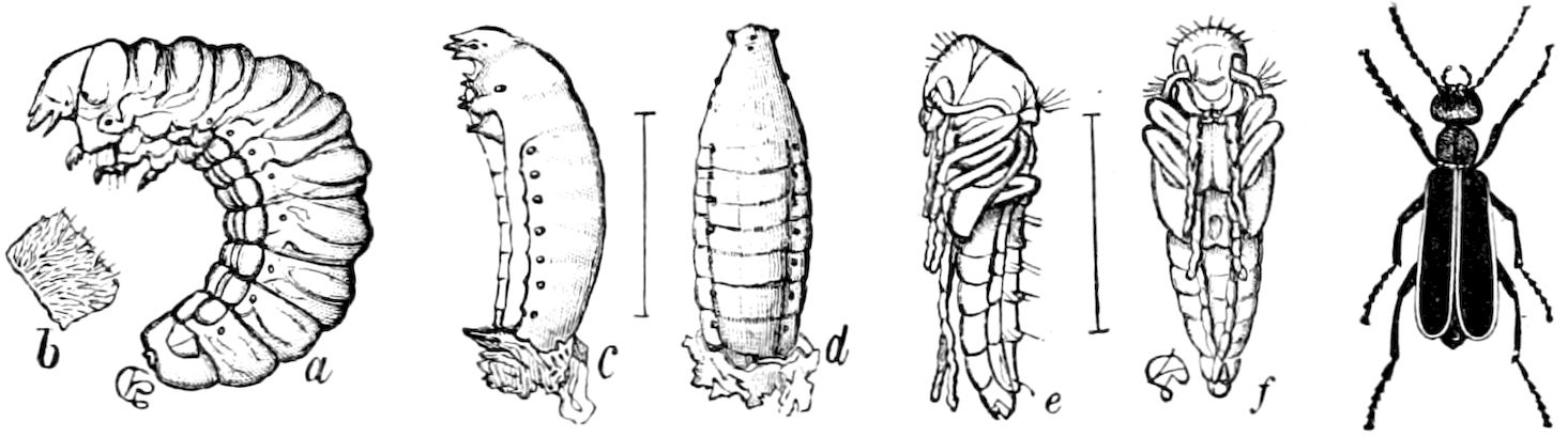
Fig. 642.—Epicauta cinerea: a, end of 2d larval stage; b, portion of dorsal skin; c, d, coarctate larva; e, f, pupa.—After Riley.
After six or seven days there is another transformation, the skin being cast, and the insect passes into another stage, “the ultimate stage of the second larva.” The larva, immersed in its rich nutritious food, grows rapidly, and after about a week leaves the now addled and decaying locust eggs, and burrows into the clear sand, where it lies on its side in a smooth cell or cavity, and where it undergoes an incomplete ecdysis, the skin not being completely shed, and assumes the semipupa stage, or coarctate larval stage of Riley.
In the spring the partly loose skin is rent on the top of the head 693and thorax, and then crawls out of it the “third larva,” which only differs from the ultimate stage of the second larva “in the somewhat reduced size and greater whiteness.” The insect in this stage is said to be rather active, and burrows about in the ground, but food is not essential, and in a few days it transforms into the true pupa state.
These habits and the corresponding hypermetamorphosis are probably common to all the Meloidæ, though the life-history of the other species has yet to be traced.
In the genus Hornia described by Riley, the wings of the imago are more reduced than in any other of the family, both sexes having the elytra as rudimentary as in the European female glow-worm (Lampyris noctiluca). These, with the simple tarsal claws and the enlarged heavy abdomen, as Riley remarks, “show it to be a degradational form.”
Its host is Anthophora, and the beetle itself lives permanently in the sealed cells of the bee, and Riley thinks it is subterranean, seldom if ever leaving the bee gallery. The triungulin is unknown, but the ultimate stage of the second larva, as well as the coarctate larva, is like those of the family in general, the final transformations taking place within the two unrent skins, in this respect the insect (Fig. 643) approaching Sitaris.
It appears, then, that as the result of its semi-parasitic mode of life the Campodea-form or triungulin larva of these insects, which has free-biting mouth-parts like the larvæ of Carabidæ and other carnivorous beetles, instead of continuing to lead an active life and feeding on other insects; living or dead, and then like other beetles directly transforming into the normal pupa, moults as many as five times, there being six distinct stages before the true pupa stage is entered upon. So that there are in all eight stages including the imaginal or last stage.
One cannot avoid drawing the very obvious conclusion that the five extra stages constituting this hypermetamorphosis, as it is so well styled, are structural episodes, so to speak, due to the peculiar parasitic mode of life, and were evidently in adaptation to the remarkable changes of environment, so unlike those to which the members of other families of Coleoptera, the Stylopidæ excepted, have been subjected. The fat overgrown body and the atrophied limbs and mouth-parts are with little doubt due to the abundant supply of rich food, the protoplasm of the egg of its host, in which the insect during the feeding time of its life is immersed. Since it is well known that parthenogenesis is due to over, or at least to abundant nutrition, or to a generous diet and favoring temperature, there is little reason to doubt that the greatly altered and abnormally fat or bloated body of the insect in these supernumerary stages is the result of a continuous supply of rich pabulum, which the insect can imbibe with little or no effort.
695
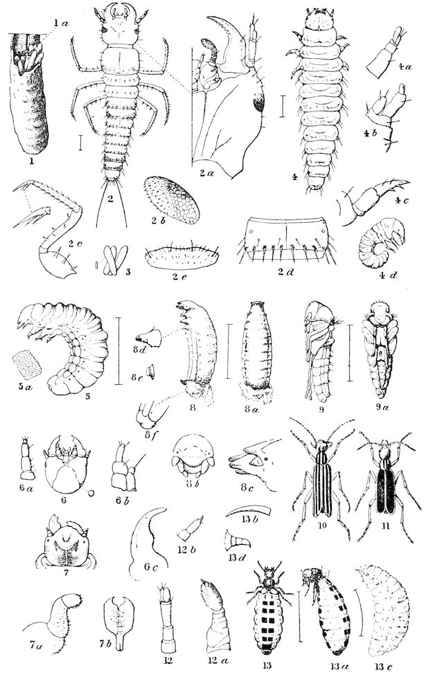
Fig. 643.—1, Egg-pod of Caloptenus differentialis with the mouth torn open, exposing the newly hatched larva of Epicauta vittata (1 a) eating into an egg and the passage which it made through the mucous covering; natural size. 2, dorsal view of the 1st larva, or triungulin, of E. vittata; 2 a, one side of the head of same from beneath, greatly enlarged so as to show the mouth-parts; 2 b, terminal joint of maxillary palpus, showing imbrications and flattened inner surface armed with stout points; 2 c, leg, showing more plainly the tarsal spines; 2 e, labrum; 2 d, one of the abdominal joints from above, showing stout points, stigmata, and arrangement of spinous hairs. 3, eggs of E. vittata, the natural size indicated at side. 4, dorsal view of the carabidoid stage of the 2d larva of E. vittata: 4 a, its antenna; 4 b, its right maxilla; 4 c, its leg; 4 d, side view of same, showing its natural position within the locust-egg mass. 5, lateral view of the ultimate or full-grown stage of the 2d larva of E. vittata; 5 a, portion of the dorsal skin, showing short setaceous hairs. 6, third head, or that from the scarabæidoid stage of the 2d larva of E. vittata from beneath, showing the reduction of mouth-parts as compared with the first head (2 a); 6 a, antenna of same; 6 b, maxilla of same; 6 c, mandible of same. 7, fourth head, or that of the full-grown larva of E. vittata, from above; 7 a, leg of same; 7 b, the breastplate or prosternal corneous piece. 8, lateral view of the pseudo-pupa or coarctate larva of E. vittata, with the partially shed skin adhering behind: 8 a, dorsal view of same; 8 b, its head, from the front; 8 c, same from side; 8 d, tuberculous leg; 8 e, raised spiracle; 8 f, anal part of same, 9. lateral view of the true pupa of Epicauta cinerea Forst: 9 a, ventral view of same. 10, Epicauta vittata (lemniscata or trivittate var.). 11, Epicauta cinerea Forst. (= marginata Fabr.). 12, antenna of the triungulin of Epicauta pennsylvanica: 12 a, maxilla of same; 12 b, labial palpus of same. 13. ♂ Hornia minutipennis, dorsal view; 13 a, lateral view of same; 13 b, simple claw of same; 13 c, coarctate larva; 13 d, leg of ultimate stage of 2d larva.—After Riley.
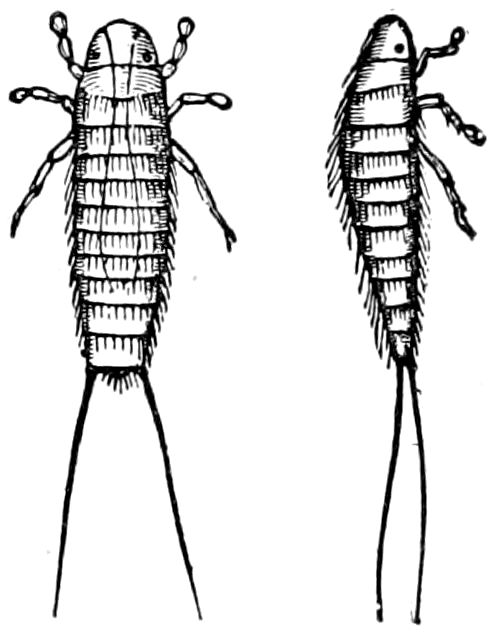
Fig. 644.—Triungulin stage of Stylops childreni.
The life-history of the Stylopidæ is after the same general fashion, though we do not as yet know many of the most important details. The females are viviparous, the young hatching within the body of the parent, as we once found as many as 300 of the very minute triungulin larvæ issuing in every direction from the body of what we have regarded as the female of Stylops childreni in a stylopized Andrena caught in the last of April. The larvæ differ notably from those of the Meloidæ in the feet being bulbous and without claws, yet it is in general Campodea-like and in essential features a triungulin (Fig. 644). The intestine ends in a blind sac, as in the larvæ of bees, and this would indicate that its food is honey. The complete life-history of no Stylopid is completely known. It is probable that, hatched in June from eggs fertilized in April, the larvæ crawl upon the bodies of bees and wasps; finally, after a series of larval stages as yet unknown,[119] penetrating within the abdomen of its host before the latter hibernates, and living there through the winter. 696The females, owing to their parasitic life, retain the larval form, while the free males are winged, not leading in the adult stage a parasitic life, though passing their larval and pupal stages in the body of their host, and are so unlike ordinary beetles as to be referred by good authorities to a distinct order (Strepsiptera).
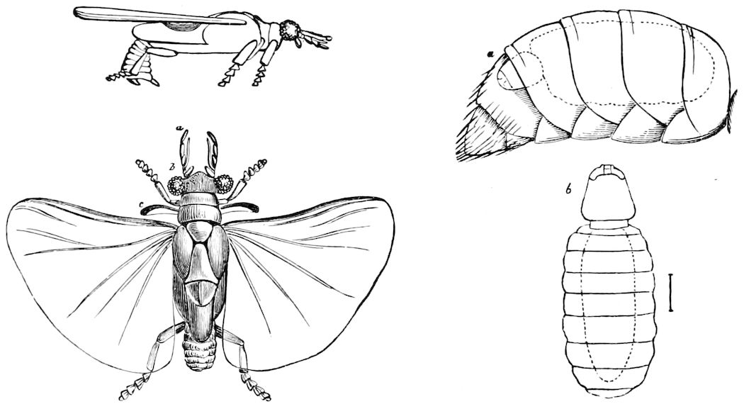
Fig. 645.—Stylops childreni, ♂: a, abdomen of Andrena with ♀ Stylops (b).
The triungulin stage of these insects corresponds in general to the form of the larval Staphylinidæ and allied families, such as the Tenebrionidæ, which are active in their habits, running about and obtaining their food in a haphazard way, often necessarily suffering long fasts. In the external-feeding, less active coleopterous larvæ, like the phytophagous species, which have an uninterrupted supply of nutritious food, we see that the body is thick and fleshy. So also in the larvæ of the Scarabæidæ, Ptinidæ, and the wood-boring groups. In internal feeders, like the larval weevils and Scolytidæ, which live nearly motionless in seeds, fruits, and the sap-wood of plants and trees, with a constant supply of nourishing, often rich food, the eruciform body is soft, thick, and more or less oval-cylindrical. So it is with the larvæ of Hymenoptera, especially in the parasitic forms, and in the ants, wasps, and bees, which are nearly if not quite motionless, at least not walking about after their food.
Now the change from the active triungulin stage to the series of secondary, nearly legless, sedentary, inactive stages is plainly enough due to the change of station and to the change of food. From being an independent, active, roving triungulin, the young insect becomes a lodger or boarder, fed at the expense of its host, and the lack of bodily exertion, coupled with the presence of more liquid food than 697is actually needed for its bare existence, at once induces rotundity of body and a loss of power in the limbs, followed by their partial or total atrophy.
That this process of degeneration may even occur in one and the same stage of larval existence is very well illustrated by what we know of the life-history of the wasp-parasite of Europe, Rhipiphorus paradoxus. Thanks to the very careful and patient observations of Dr. T. A. Chapman, we have a nearly complete life-history of this beetle, the representative of a family in many respects connecting the Meloidæ and Stylopidæ.[120] Where Rhipiphorus lays her eggs is unknown. Dr. Chapman, however, found a solitary specimen of the young larva in the triungulin stage. He describes it as “a little black hexapod, about 1 50 inch (.5 mm.) in length, and 1 120 inch in breadth, broadest about the fourth segment, and tapering to a point at the tail; a triangular head with a pair of three-jointed antennæ nearly as long as the width of the head, with legs very like those of Meloë larvæ; the tibiæ ending in two or three claws, which are supported and even obscured by a large transparent pulvillus or sucker of about twice their length; this was marked by faint striæ radiating from the extremity of the tibiæ, giving it much the aspect of a lobe of a fly’s proboscis. Each abdominal segment had a very short lateral spine pointing backwards; the last segment terminated by a large double sucker similar to those of the legs; and the little animal frequently stood up on this, and pawed the air with its feet, as if in search of some fresh object to lay hold of.”
This almost microscopic larva finds a wasp grub and bores into its body, probably entering at a point near the back of the first or second segment behind the head. Dr. Chapman succeeded in finding the larva of the beetle within that of the wasp, before the latter had spun up. “Assuming that the wasp larva lives six days in its last skin before spinning up, I should guess that the youngest of these had still two or three days’ feeding to do. The Rhipiphorus larvæ were but a little way beneath the skin of the back, about the fourth and fifth segments [counting the head as the first], and indifferently on either side. The smallest of these was 1 16 inch in length, and, except its smaller size, was precisely like the larger ones I am about to refer to, having the same head, legs, plates, etc. These were of the same size as those of the larger larvæ, the difference in size of the latter being due to the expansion of the intermediate colorless integument.”
698After the wasp grub has spun the silken covering of its cell the larva of Rhipiphorus may still be detected in some of them, being rendered visible by its black legs and dark dorsal and ventral plates. “On extracting this larva, it bears a general resemblance in size and outline to the youngest larva of Rhipiphorus that I had found feeding externally on the wasp grub, but with the very notable exception of the already mentioned black marks. These are, in fact, a corneous head, six-jointed legs, and a dorsal and ventral series of plates. I immediately recognized the head and legs as identical with those of the little black mite already described, but presenting a ludicrous appearance in being widely separated from each other by the white skin of the larva. I have no doubt that the dorsal and ventral series of black marks are the corresponding plates of the mite-like larva floated away from each other by the expansion of the intervening membrane. By measurement also they agree exactly in size, although the larva extracted from the wasp grub is ten times the length and six times the width of the little Meloë-like larva. In length it is ⅙ inch (4.5 mm.), and 1 28 inch in breadth.”
The remarkable changes thus described in the larva of this beetle after it has begun its parasitic life within the body of its host are especially noteworthy because the great increase in size and difference in shape, as well as in habits, all take place before the insect has moulted. The rapid development in size, and consequent distension of the body and the separation of the sclerites of the segments behind the head, are paralleled, as Chapman says, by the greatly swollen abdominal region of the body in Sarcopsylla penetrans and in the female of the Termitidæ. In those insects this distension is due to the enlargement of the ovaries and of the eggs contained within them, but in the Rhipiphorus it is due to the comparative inactivity of the larva, and to its being gorged with an unending supply of rich food, the blood and fat of its host. It follows, then, that if a sedentary life and over, or at least abundant, nutrition will have this effect within the short period covered by the single first larval stage of the Rhipiphorus, it is reasonable to infer that the hypermetamorphosis is also due to the same factors.
Chapman then goes on to say that finally, within six hours of the time of spinning up of the wasp grub, the Rhipiphorus larva at the end of Stage 1., which is “usually in motion, and for its situation might be called tolerably active, is seen to lay hold of the interior of the skin with its anterior legs, and keeps biting and scratching with its strong and sharp jaws until it is able to thrust through its head, when, in less than a quarter of an hour, it completely emerges by a 699vermiform movement; and at the same time it casts a skin, together with the black head, legs, plates, etc.”
The larva, now in its second stage, passes forward and seizes hold of the upper or lateral aspect of the prothoracic segment of the wasp grub. On emerging it becomes shorter and thicker, “and very soon loses length by that curving forward of its head which is so marked in the full-grown larva, and which does not exist before its emergence.” The larva is now found “lying like a collar immediately under the head of the wasp grub, and is attached to it by the head, though not very firmly.” At this stage the feeding of the young Rhipiphorus is rather sucking than eating.
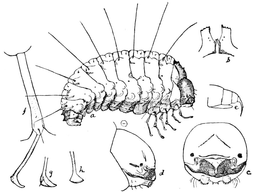
Fig. 646.—First larva (a) of Bruchus fabæ, greatly enlarged; b, thoracic processes; c, head, from front; d, from side; e, antenna; f, thoracic leg; g, rear view of tarsus; h, same, front view.—After Riley.
When about 6 mm. in length it moults a second time, and the full-grown larva closely though superficially resembles a Crabro or Pemphredon larva, the small head being bent over forwards. By the time it is ready to pupate it has wholly eaten the wasp larva, and the temperature of the cell being high, a larva 5 mm. long grows large enough in two days to fill the top of the cell of its host, and the larva is ready to pupate about a week after hatching, so that its development is very rapid. The beetles themselves do not live in the cells. Chapman thinks they hibernate, and that the eggs are laid in the spring or summer.
We thus have in this insect three larval stages, the triungulin, and 700two later stages, the great differences between the first and the last two being apparently due to their parasitic mode of life, the larva spending its second stage within its host, involving an existence in a cell with a high temperature, an uninterrupted supply of rich, stimulating food, and a comparatively sedentary mode of life compared with that of the triungulin at the beginning of its existence. It is quite obvious that the hypermetamorphosis is primarily due to a great change in its surroundings, i.e. the parasitic mode of life of the beetle, habits of very rare occurrence in the Coleoptera, numerous in species as they are.
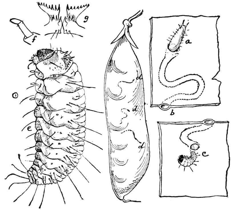
Fig. 647.—First larval stage of Bruchus pisi: a, egg in pea-pod; b, cross-section of opening of mine; c, young larva and opening on inside of pod by which it has entered, enlarged; d, d, d, eggs, natural size; e, 1st larval stage; f, a leg of same; g, prothoracic spinous processes.—After Riley.
In this connection attention may be drawn to a supernumerary larval stage observed by Riley in the pea- and bean-weevils (Figs. 646 and 647). The larva on hatching has long slender legs, though differing from those of an ordinary coleopterous larva in having but three joints (j, g, h). This stage is very short, and the legs temporary, as, after entering the bean or pea, it casts its skin, losing its legs, and assuming the vermiform shape of the second larval stage. In this case the change from a pedate to an apodous larva is plainly enough due to the change from an external feeder, like a chrysomelid larva, to a larva leading a boring, internal, almost quiescent life.
701Certain ichneumons also appear to have two distinct larval stages, as Ratzeburg inferred that in Anomalon there are four larval stages (Fig. 648).
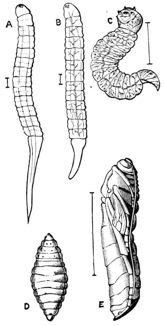
Fig. 648.—History of Anomalon circumflexum: A, 1st instar or stage. B, 2d instar. C, larva in the 3d or encysted stage removed from its cyst. D, mature larva. E, pupa.—After Ratzeburg, from Sharp.
In another ichneumon, Klapálek detected what he calls the “sub-nymph.” The insect pupates within the case of a caddis-fly, Silo (Fig. 649).
In the Proctotrypidæ there is also a hypermetamorphosis, though the remarkable precocious stages they pass through are rather embryonic than larval.
In a species of Platygaster which is parasitic in the larva of Cecidomia, the first larva (Cyclops stage) is of a remarkable shape, not like an insect, but rudely resembling a parasitic Copepod crustacean. In this condition it clings to the inside of its host by means of its hook-like jaws, moving about, as Ganin says, like a Cestodes embryo with its well-known six hooks. In this stage it has no nervous, vascular, or respiratory system, and the digestive canal is a blind one (Fig. 651).
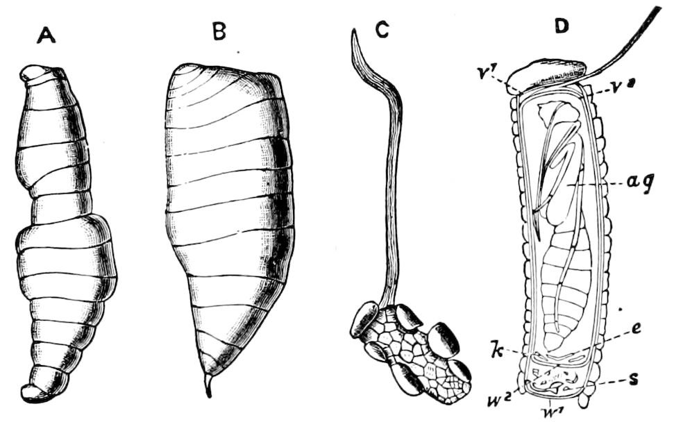
Fig. 649.—Metamorphosis of Agriotypus: A, larva. B, “sub-nymph.” C, case of the Silo, with the string of attachment formed by Agriotypus. D, section of the case: v1, operculum of case; v2, cocoon; ag, pupa of Agriotypus; e, exuvia of same; w2, wall of cocoon; s, remains of Silo; w1, closure of case.—After Klapálek, from Sharp.
After moulting, the insect entirely changes its form; it is thick oval-cylindrical, 702nearly motionless, with no appendages, but with a digestive canal and a nervous and vascular system (Fig. 652).
After a second moult the third and last larval stage is attained, and the insect is of the ordinary appearance of ichneumon larvæ.
Not less striking is the life-history of Polynema, which lays its eggs in those of a small dragon-fly (Agrion virgo). The first larval stage is most remarkable. It hatches as a microscopic immovable being, entirely unlike any insect, with scarcely a trace of organization, being merely a flask-shaped sac of cells. After remaining in this state five or six days it moults.
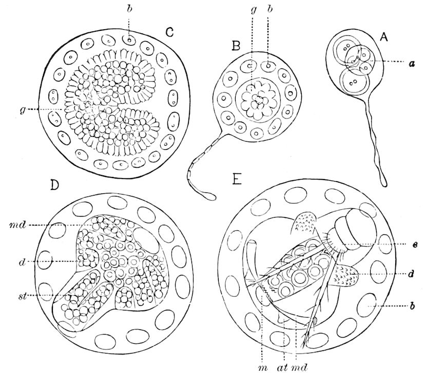
Fig. 650.—Development of Platygaster: A, stalked egg: a central cell giving origin to the embryo. B, g, germ; b, blastoderm cells. C, the same, farther advanced. D, cyclops-like embryo: md, rudiments of mandibles; d, rudimentary pad-like organs, seen more developed in E; st, bilobed tail.
The second stage, or Histriobdella-like form, as Ganin names it, is more like that leech-like worm than an insect.
The third larval form is very bizarre, though more as in insects, having rudimentary antennæ, mouth-parts, legs, and ovipositor. In this condition it lives from six to seven days before pupating (Fig. 653).
The strange history of another egg-parasite (Ophioneurus) agrees in some respects with that of the foregoing forms. It is when hatched of an oval shape, with scarcely any organs, and differs from the genera already mentioned in remaining within its egg-membrane, and not assuming their strange shapes. From the cylindrical sac-like 703non-segmented larva resembling the second larva of Platygaster it passes directly into the pupa state.
A fourth form, Teleas (Fig. 654, A-D), is an egg-parasite of Gerris, and in America one species oviposits in the eggs of Œcanthus.
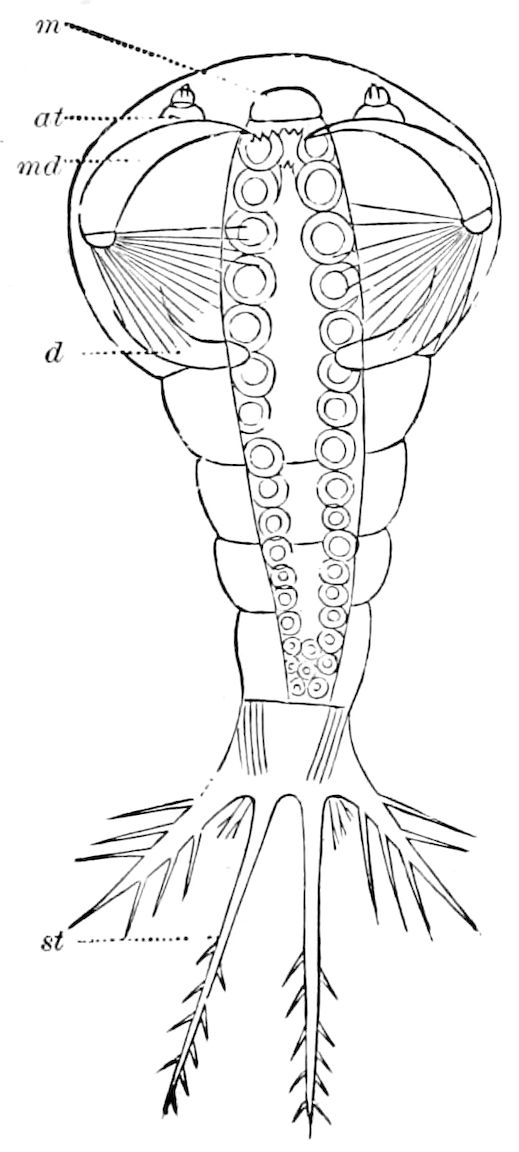
Fig. 651.—First larva of Platygaster: m, mouth; at, rudimentary antenna; md, mandibles; d, tongue-like appendages.
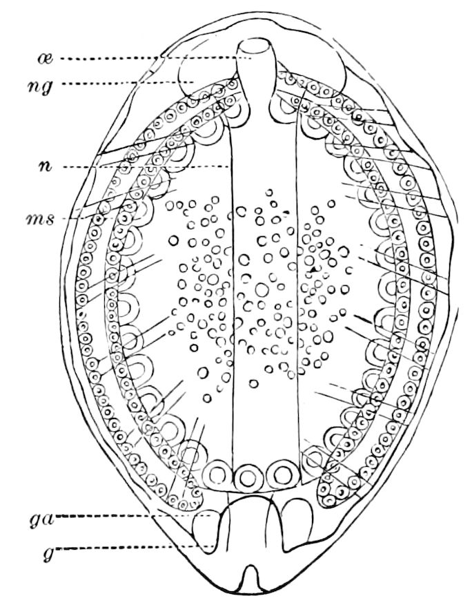
Fig. 652.—Second larva of Platygaster: œ, œsophagus; ng, brain; n, nervous cord; ga and g, genital organs; ms, muscular band.
The spindle-shaped larva in its first stage roughly resembles a trochosphere of a worm rather than the larva of an insect so high in the scale as a Hymenopter. It is active, but after moulting the second larva is oval, still without segments. Dr. Ayers gives a profusion of details and figures of the first and second stages of our Teleas, the second strongly resembling the Cyclops stage of Ganin. He describes three stages, and though he did not complete the life-history of the insect, he thinks it changes to an ovoid flattened form which succeeds the Cyclops stage in other Pteromalidæ, and that there are at least four ecdyses.
It is difficult to account for these strange larval forms, unless we suppose that the embryos, by their rich, abundant food, have undergone a premature development, the growth of the body-walls being greatly accelerated, the insects so to speak having been, under the stimulus of over-nutrition and their unusual environment, and perhaps also the high temperature of the egg, hurried into vermian 704existence on a plane scarcely higher than that of an active ciliated gastrula.
Further observations, difficult though they will be, are needed to enable us to account for the singular prematurity of the embryo of these parasites. That these stages are reversional and a direct inheritance from the vermian ancestors of these insects is not probable, but the forms are evidently the result of adaptation in response to a series of stimuli whose nature is in part appreciable but mostly unknown.
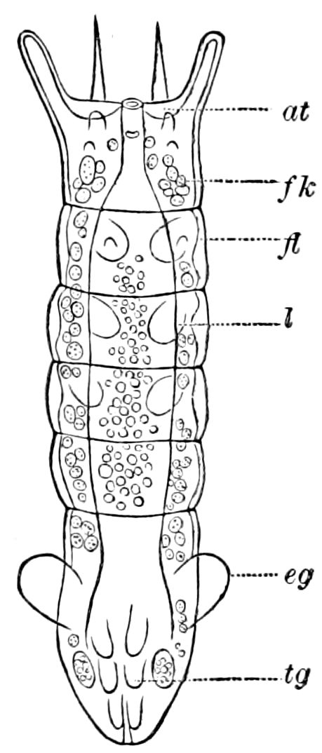
Fig. 653.—Third larva of Polynema: at, antenna; fl, imaginal bud of wing; l, rudimentary legs; tg, buds of one of the three pairs of styles of the ovipositor; fk, fat-body; eg, ear-like process.
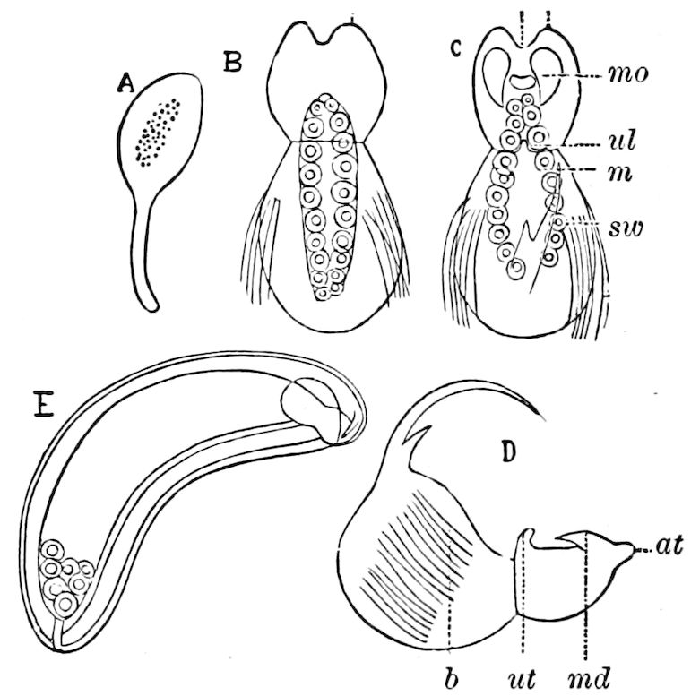
Fig. 654.—A-D, development of Teleas; A, stalked egg; B, C, D, the 1st larval stage: at, antenna; md, hook-like mandibles; mo, mouth; b, bristles; m, intestine; sw, the tail; ul, under lip or labium. E, larva of another parasite, Ophioneurus.—This and Figs. 650–653 after Ganin.
It may be noted, however, that the appearance of a primitive band in the second larval stage suggests the origin of these forms, as well as that of insects in general, from a Peripatus-like, and again from an earlier leech-like Annelid ancestor. Hence the first larval or Cyclops stage is due to a precocious development caused by the unusual environment, and is simply adaptational, and not of phylogenetic significance.
705
SUMMARY OF THE FACTS AND SUGGESTIONS AS TO THE CAUSES OF METAMORPHISM
An explanation of the causes of metamorphosis is one of the most difficult undertakings in biology, and the phenomenon has been considered as one of the chief difficulties in the way of the acceptance of the theory of descent.
A review, however, of the facts of hypermetamorphism, particularly the life-history of Mantispa, throws much light on the subject, since it is very probable that the supernumerary stages and marked changes of form characterizing them are due to changes of environment, of habits, and of food, causes which have exerted such a profound influence on organic beings throughout all time. Besides these, as the result of changes in the environment and nature of the food, we have the results brought about by the use or disuse of structures brought into existence by the action of stimuli from without, the class of insects abounding in examples of temporary structures which perform a certain function, and then disappear.
Again, if the origin of a hypermetamorphosis can thus be explained, it follows that normal metamorphosis is most probably due to changes of habitat, of seasons, of food, and to accelerated growth resulting from the approach of sexual maturity.
The following facts and conclusions appear to be well established:—
1. The apterous insects (Synaptera) are ametabolous, only the winged insects undergoing a metamorphosis.
2. The complete metamorphosis was not inherited from the primitive ancestor of all insects, but acquired at a later period (F. Müller). The eruciform type is a secondary, adaptive form, derived from the earlier, campodeoid type of larva.
3. The earliest, most primitive pterygote insects passed through only a slight metamorphosis. In other words, as soon as the wings were evolved and insects became adapted to live or take refuge in a new medium, the air, at the approach of the period of adult life, with the ripening or perfection of the reproductive organs, a metamorphosis began to take place, and the number of species greatly multiplied. On the other hand, the Arachnida and Myriopoda, in which as a rule there is no metamorphosis, being confined to a creeping life, with no change of medium, remained poor in number of species.
4. At first the nymphs mainly differed from the adults in lacking 706wings, though having the same habits; in holometabolous insects, the larva became adapted to entirely different habits and environments, so that in Hymenoptera, and especially Diptera, the larva became remarkably unlike the imago.
5. Until the Mesozoic age, or late in the Carboniferous period, there were, so far as we now know, only ametabolous and heterometabolous insects, and these orders (Orthoptera, Dermaptera, Hemiptera, Plectoptera, Odonata, and Neuroptera) were not numerically rich in genera and species, while since early Mesozoic times geological extinction has reduced their numbers.
6. During the Mesozoic age, and since then, the number of species, genera, families, and orders has greatly increased, and insects have become more and more holometabolous. The orders of Coleoptera, Lepidoptera, Hymenoptera, and Diptera are many fold greater in number of species and variety of form than the heterometabolous orders.
The rapid increase in the number and variety of types of insects evidently is correlated with the profound geological changes which took place at the end of the Paleozoic age, involving the appearance of larger continental masses, or a greater land area, thus opening new regions for settlement. Also the origin of flowering plants at about this time undoubtedly had much to do with the genesis of new adaptive structures, such as the changes in the mouth-parts and wings.
7. The process of metamorphosis, at least in the subtropical, temperate, and polar regions, is largely dependent on the change from summer to winter, and, in the tropics, from the rainy to the dry season.
As regards the organization of larval (nepionic) as compared with imaginal forms, the nymphs and larvæ of insects are, with the exception of many Diptera, nearly as perfectly developed as the adult. In this respect the immature insect differs fundamentally from the larvæ of certain worms (for example, the pilidium of Nemerteans) and from the pluteus and brachiolaria stages of echinoderms, which possess only digestive and water-vascular organs.
Insect nymphs and larvæ also differ from the nauplius and zoëa of Crustacea in having at birth all the most important systems of organs (digestive, circulatory, respiratory, nervous, muscular, with sometimes a nearly perfected reproductive system) of the imago, also the same number of cephalic, thoracic, and abdominal segments and appendages. Metamorphism in insects involves (except in the Diptera) rather modifications in the form and functions of organs and appendages already present than the formation of new ones. In 707larval Crustacea, the thoracic and abdominal appendages do not arise until some time after hatching from the egg.
8. While cases of the suppression or abbreviation of larval characters and direct development are not uncommon in echinoderms and crustaceans, in insects this phenomenon occurs only so far as yet known in the Diptera. In these insects the polypody in the embryo is outgrown, or lost, the embryos and larvæ not having even the temporary rudiments of abdominal appendages. The campodeoid characters also are entirely suppressed, dropped, or lost in the more specialized holometabolous orders, Lepidoptera, Hymenoptera, and Diptera, though retained in the more primitive and generalized Coleoptera. (This proves that the Coleoptera are lower or more primitive and generalized than the other orders mentioned.) This abbreviation or loss of organs is, as Hyatt and Arms claim, due to the prepotency of acquired characters in phylogeny, and are also the result of homochronous heredity.
“The Insecta of the more specialized orders, x.-xvi., afford, next to some parasites, the most notable examples of this mode of evolution. Their larval or nepionic, and pupal or neanic, stages are prolonged at the expense of the ephebic, winged stage, and the reasons for this prolongation are found in the great number of new features introduced into these stages of development in these orders as contrasted with those of the more primitive, and, in large part, more ancient orders, i.-ix. The law of tachygenesis has been at work here, as in the former cases alluded to above, and it is shown in the encroachments of the adaptive characteristics of the caterpillar, grub, and maggot upon the inherited characteristics of the Thysanuran stage, which loses its ancestral characteristics, until in most cases they are either obsolete or recognizable with difficulty.” (Hyatt and Arms, Natural Science, 1896, p. 400.)
9. In the holometabolous insects there is a resting, quiescent stage during the pupal period, when the insect takes no food. In this respect the more specialized insects differ from other metamorphic animals. The larva has an abundant supply of fat lasting through pupal life, while in the quiescent pupa, respiration and circulation is much lessened, the animal being as a rule motionless. This resting stage is also necessary for the histolysis and formation of the adult body from the imaginal buds present in the larva.
10. The hypermetamorphosis of Mantispa, Meloë, Stylops, etc., indicate very plainly that the eruciform type of larva is derived from the campodeoid, since one and the same insect passes through these stages before reaching sexual maturity.
11. As observed by Miall, the larva of insects differs from that of other invertebrate animals in being larger than the adult.
12. The metamorphoses of insects are in some important respects 708paralleled by those of the Amphibia. The case of pædogenesis of Chironomus affords a parallel with that of the Siredon, or larva of Amblystoma. Also the organs and appendages of the insects, such as caterpillars, are present, just as the skeleton and other organs of the tadpole are the homologues of those of the adult, although these parts undergo a profound modification, and new structures are added. (See the discussion of this point by Miall, and by Hyatt and Arms.)
Theoretical conclusions; Causes of metamorphosis.—It results from a review of the known facts, together with reasonable inductions from such facts, that so far from opposing the theory of descent, the facts of metamorphosis, and particularly of hypermetamorphosis, seem to afford solid foundation for the theory. While natural selection was not the initiative cause, it plays a part as one of several factors; but the fundamental causes are the same as those which have controlled the origin of species and of the larger groups of animals in general. Owing to the struggle for existence, due to overcrowding, the early insects were forced to take to the air, acquiring wings to enable them to avoid the attacks of creeping and running insects. In the end the insects became, owing to this acquisition of wings, and afterwards to the establishment of a complicated metamorphosis, numerically the most successful type of life in existence, the number of species, extinct and living, mounting into the millions.
All aquatic insects are evidently the descendants of terrestrial forms, and the numberless contrivances and temporary larval organs, particularly of dipterous larvæ, are evidently adaptations to the needs of the insect during its aquatic life, and which are cast aside when the creature passes to a different medium. The sudden or tachygenic appearance of temporary structures, such as hatching spines, various setæ, spines, respiratory organs, so characteristic of dipterous larvæ, and of the protective colors and markings of caterpillars, and which are discarded at pupation, or imagination, are evidently due to the action of stimuli from without, to the primary neolamarckian factors, the characters proper to each larval stadium, and to the pupal and imaginal stadia,—characters probably acquired during the lifetime of the individual,—becoming finally fixed by homochronous heredity.
709
LITERATURE ON POSTEMBRYONIC DEVELOPMENT AND METAMORPHOSES
Herold, Moritz Johann David. Entwicklungsgeschichte der Schmetterlinge anatomisch und physiologisch bearbeitet. (Cassel, u. Marburg, 1815. 33 Taf., 4º, pp. 1–118, i-xxxiv.)
Ratzeburg, F. T. C. Ueber Entwickelung des fusslosen Hymenopteren-larven, mit besonderer Rücksigt auf die Gattung Formica. (Nova Acta Natur. Curios., xvi, 1832, pp. 145–176.)
Agassiz, Louis. The classification of insects from embryological data. (Smithsonian Contr., ii, Washington, 1851, pp. 28, 1 Pl.)
Ganin, M. Beiträge zur Erkenntniss der Entwicklungsgeschichte bei den Insecten. (Zeitschr. f. wiss. Zool., xix, 1869, pp. 381–451, 4 Taf.)
—— Ueber die Embryonalhülle der Hymenopteren- und Lepidopteren-embryonen. (Mém. Acad. St. Petersbourg (7), xiv, 1869, pp. 18, 1 Pl.)
—— Materialien zur Kenntniss der post-embryonalen Entwicklungsgeschichte der Insecten. (Russian.) Warschau, 1876. (Abdruck aus den Arbeiten der V. Versammlung russischer Naturf. und Aerzte in Warschau, 1876. Abstract by Hoyer in Jahresber. der Anat. und Phys. von Hoffmann und Schwalbe, v, 1876, and in Zeitschr. f. wiss. Zool., xxviii, 1877, pp. 386–389.)
Weismann, A. Die nachembryonale Entwicklung der Musciden nach Beobachtungen an Musca vomitoria und Sarcophaga carnaria. (Zeitschr. f. wiss. Zool., xiv, 1864, pp. 101–263, Taf. 8–14.)
—— Die Metamorphose von Corethra plumicornis. (Zeitschr. f. wiss. Zool., xvi, 1866, pp. 1–83, 5 Taf.)
Packard, A. S. Observations on the development and position of the Hymenoptera, with notes on the morphology of insects. (Proceedings Boston Society of Natural History, 1866, pp. 279–295.)
Künckel d’Herculais, J. Recherches sur l’organisation et le développement des Volucelles. Paris, 1875, Pt. I, pp. 208, 12 Pis.; II, 1881. Atlas of 15 Pls.
Dewitz, H. Beiträge zur Kenntniss der post-embryonalen Gliedmaassenbildung bei den Insecten. (Zeitschr. f. wiss. Zool., xxx, Suppl., 1878, pp. 78–105, 1 Taf.; Nachtrag, Ibid., pp. 25–28.)
—— Ueber die Flügelbildung bei Phryganiden und Lepidopteren. (Berl. Ent. Zeitschr., xxv, 1881, pp. 53–66, 2 Taf.)
Lowne, B. Th. Anatomy, physiology, morphology, and development of the blow-fly. London, Part I, 1880; Part II, 1891.
Viallanes, H. Recherches sur l’histologie des Insectes et sur les phénomènes histologiques qui accompagnent le développement post-embryonnaire de ces animaux. (Ann. Sc. Nat. (6), xiv, 1882.)
Metschnikoff, E. Untersuchungen über intracelluläre Verdauung bei wirbellosen Thieren. (Arb. a. d. zoolog. Inst. zu Wien., v, 1883.)
—— Untersuchungen über die mesodermalen Phagocyten einiger Wirbelthiere. (Biol. Centralbl., iii, 1883.)
Wielowiejsky, H. v. Ueber den Fettkörper von Corethra plumicornis und seine Entwicklung. (Zool. Anzeiger, vi Jahrg., 1883, pp. 318–322.)
Pancritius, P. Beiträge zur Kenntnis der Flügelentwicklung bei den Insecten. In.-Diss. Königsberg, 1884.
Rees, J. van. Over intra-cellulaire spijsverteering en over de beteekenis der witte bloedlichampjes. (Maandblad voor Natuurwetenschappen, xi Jaarg., 1884, pp. 28.)
—— Over de post-embryonale ontwikkeling von Musca vomitoria. (Maandblad voor Natuurwetenschappen, Juli, 1885.)
710—— Beiträge zur Kenntniss der inneren Metamorphose von Musca vomitoria. (Zool. Jahrb. Abth. f. Anat. u. Ontog., iii, 1888, pp. 1–134, 2 Taf., 14 Figs.)
Frenzel, J. Einiges über den Mitteldarm der Insecten, sowie über Epithel-regeneration. (Arch. Micr. Anat., xxvi, 1885.)
Kowalevsky, A. Beiträge zur nachembryonalen Entwicklung der Musciden. (Zool. Anzeiger, viii, 1885, pp. 98, 123, 153.)
—— Beiträge zur Kenntniss der nachembryonalen Entwicklung der Musciden. (I. Theil., Zeitschr. f. wiss. Zool., xlv, 1887, pp. 542–594, 5 Taf.)
Schneider, Ant. Ueber die Anlage der Geschlechtsorgane und die Metamorphose des Herzens bei den Insecten. (Zool. Beiträge, 1885, i, pp. 140–143, 1 Taf.)
Rehberg, A. Ueber die Entwickelung des Insectenflügels (an Blatta germanica). (Marienwerder, 1886, pp. 12, 1 Taf.)
Schäeffer, C. Beiträge zur Histologie der Insecten. (Spengel’s Zool. Jahrb., iii, Abth. f. Anat., 1889, pp. 611–652, 2 Taf.)
Hurst, H. The post-embryonic development of a gnat (Culex). Manchester, 1890, pp. 26, 1 Pl.
Verson, E. Der Schmetterlingsflügel und die sog. Imaginalscheiben desselben. (Zool. Anzeiger, xiii, 1890, pp. 116, 117.)
Bugnion, Edouard. Recherches sur le développement post-embryonnaire, l’anatomie, et les mœurs de l’Encyrtus fuscicollis. (Recueil zool. Suisse, v, 1891, pp. 435–534, 6 Pls.)
Petersen, Wilhelm. Die Entwicklung des Schmetterlings nach dem Verlassen der Puppenhülle. (Deutsch. Ent. Zeitschr., 1891, 2 lepid. Hft., pp. 199–214, 5 Figs.)
Kulagin, Nicolas. Notice pour servir à l’histoire de développement des hyménoptères parasites. (Congrès international de Zoologie, 2e Session, à Moscou, 1892, pp. 253–277. Also in Zool. Anzeiger, xv, 1892, pp. 85–87.)
—— On the development of Platygaster. (Journ. of Friends of Nat. Sc. Moscow. Zool., 1890.) (In Russian.)
—— Beiträge zur Kenntniss der Entwicklungsgeschichte von Platygaster. (Zeitschr. f. wiss. Zool., lxiii, 1897, pp. 195–235, 2 Taf.)
Miall, L. C., and Hammond, A. R. The development of the head of Chironomus. (Trans. Linn. Soc. London, 2d Ser., v, 1892, pp. 265–279, 4 Pls.)
Pratt, Henry S. Beiträge zur Kenntniss der Pupiparen. In.-Diss. Berlin, 1893, pp. 53 (Archiv f. Naturgesch., 1893), 1 Taf.
—— Imaginal discs in insects. (Psyche, viii, 1897, pp. 15–30, 11 Figs.)
Gonin, J. Recherches sur la métamorphose des lépidoptères. De la formation des appendices imaginaux dans la chenille du Pieris brassicæ. (Bull. Soc. Vaud. sc. nat., xxx, 1894, pp. 1–52, 5 Pls.)
Heymons, R. Ueber Flügelbildung bei der Larve von Tenebrio molitor. (Sitz. Ber. Gesell. Natf. Freunde. Berlin, Jahrg. 1896, pp. 142–144, 1 Fig.)
Also the writings of Malpighi, Swammerdam, De Geer, Lyonet, Bonnet, Newport, Brauer, Chapman, Fabre, Valery-Mayet, Riley, Chobaut, Nassonow, Miall (Nature, 1895, pp. 152–158), Hyatt and Arms (Natural Science, 1896, pp. 395–403).
END OF PART III.
711