PART I.—MORPHOLOGY AND PHYSIOLOGY (continued 6)
Reading edition
The cells of the mushroom bodies, observes Kenyon, “stand out in sharp contrast to all other nerve cells known, though they recall to some extent the cells of Purkinje in the higher mammals. Each of the cells contained within the fibrillar cup sends a nerve-process into the latter, where it breaks up into a profusely arborescent system of branchlets, which often appear with fine, short, lateral processes, such as are characteristic of the dendrites of some mammalian nerve-cells.” Just before entering the fibrillar substance, a fine branch is given off that travels along the inner surface of the cup along with others of the same nature, forming a small bundle to the stalk of the mushroom body, down which it continues until it reaches the origin 235of the anterior and the inner roots above mentioned. “Here it branches, one branch continuing straight on to the end of the anterior root, while the other passes to the end of the inner root. Throughout its whole course the fibre and its two branches are very fine. Nearly the whole stalk and nearly the whole of each root is made up of these straight, parallel fibres coming from the cells within the cup of the mushroom bodies. What other fibres there are enter these bodies from the side, and branch between the straight fibres very much as the dendrites of the cells of Purkinje branch among the parallel fine fibres from the cells of the granular layer in the mammalian cerebellum. These fibres are of the nature of association fibres.”
Viallanes showed that from the olfactory or antennal lobes, as well as from the optic ganglia, there are tracts of fibres which finally enter the cups of the mushroom bodies, and Kenyon has confirmed this observation. Kenyon has also, by the Golgi method, detected another tract, before unknown, “passing down the hinder side of the brain, from the cups to the region above the œsophagus, where it bends forward and comes in contact with fibres from the ventral cord, which exists, although Binet was unable to discover any growth of fibres connecting the cord with the brain.
“The fibres entering the cups from the antennal lobe, the optic ganglia, and the ventral region, spread out and branch among the arborescent endings of the mushroom-body cells. The fibres branching among the parallel fibres of the roots and the stalk lead off to lower parts of the brain, connecting with efferent or motor-fibres, or with secondary association fibres, that in their turn make such connections. This portion of the circuit has not been perfectly made out, though there seems to be sufficient data to warrant the assumption just made.
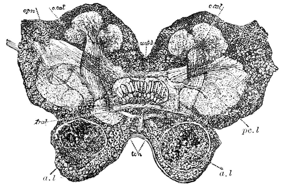
Fig. 252.—Section 17, showing the central body (centr. b) and mushroom body, optic and antennal lobes (a. l), and procerebral lobes (pc. l); o. cal, outer division of the calyx; op. n, optic nerve; trab, trabeculum; tc. n, transverse nerve.
“Such fibres existing as described, there is then a complete circuit for sensory stimuli from the various parts of the body to the cells of the mushroom bodies. The dendritic or arborescent branches of these cells take them up and pass them on out along the parallel fibres or neurites in the roots of the mushroom bodies as motor or other efferent impulses.
“This, however, is not all. For there are numerous fibres evident in my preparations, the full courses of which I have not been thus far able to determine, but which are so situated as to warrant the inference that they may act as 236association fibres between the afferent fibres from the antennæ, optic ganglia, and ventral system, and the efferent fibres. There is then a possibility of a stimulus entering the brain and passing out as a motor impulse without going into the circuit of the fibres of the mushroom bodies; or, in other words, a possibility of what may be compared to reflex action in higher animals.”
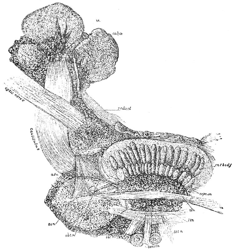
Fig. 253.—Enlarged view of the trabeculum (the dotted lines tcn and obt. n pass through it) and its nerves, of the mushroom body,—its calices and stalk, and the origin of the optic nerve × 225 diameters: atn, ascending trabecular nerve; obt. n, oblique trabecular nerve; tcn, transverse nerve; lat. n, lateral nerve; cent. n, central nerve.
The mushroom bodies have not yet been found to be present in the Synaptera, but occur in the larvæ, at least of those of most metamorphic insects (Lepidoptera and Hymenoptera), though not yet found in the larvæ of Diptera. The writer has found these bodies in the nymphs of the locust (Melanoplus spretus), but not in the embryo just before hatching. They occur in the third larval or nymph stage of this insect. It is evident that by the end of the first larval stage the brain attains the development seen in the third larval state of the two-banded species (C. bivittatus).
237
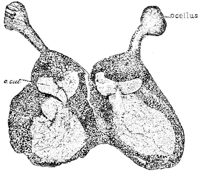
Fig. 254.—Section through the brain of Caloptenus bivittatus in the third larval stage, showing the two hemispheres or sides of the brain, and the ocelli and ocellar nerves, which are seen to arise from the top of the hemispheres directly over the calices (compare Fig. 251): o. cal, outer division of calyx of left mushroom body.
The result of our studies on the brain of the embryo locust was that from the embryonic cerebral lobes are eventually developed the central body and the two mushroom bodies. Fig. 254 shows the early condition of the mushroom bodies and their undoubted origin from the cerebral ganglia. Hence these bodies appear to be differentiations of the cerebral ganglia or lobes, having no connection with the optic or antennal lobes.
The central body (Fig. 252, centr. b).—This is the only single or unpaired organ in the brain. Dietl characterizes it as a median commissural system. Viallanes describes it as formed entirely of a very fine and close fibrillar web, like a thick hemispherical skull-cap, situated on the median line and united with the cerebral lobes. “It is like a central post towards which converge fibres passing from all points of the brain; being bound to the cerebral lobes, to the stalked bodies, to the optic ganglia, and to the olfactory lobes by distinct fibrous bundles.”
The antennal or olfactory lobes (Deutocerebrum).—This portion of the brain consists of two hemispherical lobes, highly differentiated for special sensorial perceptions, and connected by a slightly differentiated medullary mass, the dorsal lobe (Figs. 248, 249 lo), from which arise the motor fibres and those of general sensibility. The antennal lobes are in part attached to the optic ganglia, and partly to the stalked body on the same side, by the optic olfactory chiasma (Fig. 250 fch, choo), a system of fibres partially intercrossed on the median line.
The œsophageal lobes (Tritocerebrum) (Figs. 249, 250).—From this region the labrum and viscera are innervated, the nerves to the latter being called the visceral, sympathetic, or stomatogastric system. As Viallanes remarks, though plainly situated in front of the mouth, they are in fact post-œsophageal centres. The two lobes are situated far apart, and are connected by a bundle of fibres passing behind the œsophagus, called the transverse commissure of the œsophageal ring (Lienard). The œsophageal ganglia, besides giving 238rise to the labral nerves, also give origin to the root of the frontal ganglion.
c. Histological elements of the brain
The brain and other ganglia are composed of two kinds of tissue.
1. The outer slightly darker, usually pale grayish white portion consists of cortical or ganglion-cells differing in size. This portion is stained red by carmine, the cells composing it readily taking the stain.
The large ganglion cells (represented in Figs. 252 and 253) are oval, and send off usually a single nerve-fibre; they have a thin fibrous cell-wall, and the contents are finely granular. The nucleus is very large, often one-half the diameter of the entire cell, and is composed of large round refractive granules, usually concealing the nucleolus.
2. The medullary or inner part of the brain consists of matter which remains white or unstained after the preparation has remained thoroughly exposed to the action of the carmine. It consists of minute granules and interlacing fibres. The latter often forms a fine irregular network inclosing masses of finely granulated nerve matter.
This is called by Dietl “marksubstanz.” Leydig, in his Vom Bau des thierischen Körpers, p. 89, thus refers to it:—
“In the brain and ventral ganglia of the leech, of insects, and in the brain of the gastropods (Schnecken) I observe that the stalks (stiele) of the ganglion-cells in nowise immediately arise as nerve-fibres, but are planted in a molecular mass or punktsubstanz, situated in the centre of the ganglion, and merged with this substance. It follows, from what I have seen, that there is no doubt that the origin of the nerve-fibres first takes place from this central punktsubstanz.”
“This relation is the rule. But there also occur in the nerve-centres of the invertebrates single, definitely situated ganglion-cells, whose continuations become nerve-fibres without the intervention of a superadded punktsubstanz.” We may, with Kenyon, call it the fibrillar substance.
Leydig subsequently (p. 91) further describes this fibrillar substance, stating that the granules composing it form a reticulated mass of fibrillæ, or, in other words, a tangled web of very fine fibres:—
“We at present consider that by the passage of the continuation of the ganglion-cells into the punktsubstanz this continuation becomes lost in the fine threads, and on the other side of the punktsubstanz the similar fibrillar substance forms the origin of the axis-cylinders arranged parallel to one another; so it is quite certain that the single axis-cylinder derives its fibrillar substance as a mixture from the most diverse ganglion-cells.”
d. The visceral (sympathetic or stomatogastric) system
This system in insects is composed (1) of a series of three unpaired ganglia (Fig. 249, gv1, gv2, gv3), situated over the dorso-median line of the œsophagus, and connected by a median nervous cord or recurrent 239nerve (nr, vagus of Newport). The first of these ganglia is the frontal ganglion, which is connected with the œsophageal ganglia by a pair of roots (rvt), which have an origin primitively common with that of the labral nerves (Fig. 248, fg and lbr).
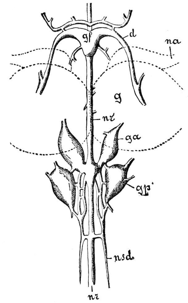
Fig. 255.—Anterior portion of the paired and unpaired visceral nervous system of Blatta orientalis seen from above. The outlines of the brain (g) and the roots of the antennal nerve (na), which cover a portion of the sympathetic nervous system, are given by dotted lines. Lettering as in Fig. 247. nsd, nerve to salivary gland. The nervus recurrens (nr) enters an unpaired stomach ganglion farther back.—After Hofer, from Lang.
2. Of two pairs of lateral ganglia (Fig. 255, ga, gp) situated two on each side of the œsophagus. They are connected both with the antennal lobes by a nerve (rvd), and to the chain of unpaired ganglia by a special connective. The first pair of these ganglia sends nerves to the heart and aorta; the second pair to the tracheæ of the head.
The unpaired median or recurrent nerve (nr) extends back from under the brain along the upper side of the œsophagus, and (in Blatta), behind the origin of the nerves to the salivary glands, enters an unpaired ganglion, called the stomachic ganglion (ganglion ventriculare), situated in front of the proventriculus. The number of these stomachic ganglia varies in different orders of insects.
In Blatta, Küpffer and also Hofer have shown (Fig. 255) (Müller, Brandt, ex Kolbe) that the nerve to each salivary gland arises from three different centres: the anterior end situated under the œsophagus is innervated by the paired visceral nerves from the hinder paired ganglia; the remaining part by nerves arising from each side of the recurrent nerve; and thirdly by a pair of nerves arising from the subœsophageal ganglion which accompanies the common salivary duct, and ends in branches which partly innervate the salivary glands and in part their muscles.
Hofer considers that the function of this complex system of paired and unpaired ganglia, with their nerves, is a double one, viz. serving both as a centre for the peristaltic action of the œsophagus, and as innervating the salivary glands.
Besides these a second portion of the visceral system arises from the thoracic and abdominal ventral cord. It may be seen in the 240simplest condition yet known in the nervous system of Machilis (Fig. 239 s). It consists of a fine, slender nerve, which extends along the surface of the ventral chain of ganglia, and sending off a pair of branches (accessory transverse nerves) in front of each ganglion. These accessory nerves receive nerve-twigs from the upper cord of the ventral chain, dilating near their origins into a minute elongated ganglion, and then passing partly outwards to the branches of the tracheæ and the muscles of the spiracles, uniting in the middle line of each segment of the body behind the head, i.e. of those segments containing a pair of ganglia.
e. The supraspinal cord
In the adult Lepidoptera has been detected, continuous with and on the upper side of the abdominal portions of the ventral cord, a longitudinal cord of connective tissue forming a white or yellowish band, and which seems to be an outgrowth of the dorsal portion of the neurilemma of the ventral cord. Muscles pass from it to the neighboring ventral portions of the integument. Its use is unknown, and attention was first called to it by Treviranus, who called it “an unknown ventral vessel” (Bauchgefäss). Afterwards it was re-discovered by Newport, who described it as “a distinct vascular canal.” But Burger has proved by cross-sections that it is not tubular, but a comparatively solid cord composed, however, of loose connective tissue. Newport found it in the larva of Sphinx ligustri, but Cattie states that it is not present in that of Acherontia atropos. It has not yet been observed in insects of other orders, but its homologue exists in the scorpion and in the centipede, and it may prove to correspond with the far more complete arterial coat which, with the exception of the brain, envelops the nervous system of Limulus.
f. Modifications of the brain in different orders of insects
There are different grades of cerebral development in insects, and Viallanes claimed that it was no exaggeration to say that the brain of the locust (Melanoplus) differs as much from that of the wasp as that of the frog differs from that of man. He insists that the physiological conditions which determine the anatomical modifications of the brain are correlated with 1, the food; 2, the perfection of the senses; and 3, with the perfection of the psychic faculties. For example, in those which feed on solid food and whose œsophagus is large (Orthoptera and Coleoptera), the connectives are elongated, the subœsophageal commissure free in all 241its extent, and the tritocerebrum is situated quite far from the preceding segment of the brain.
On the other hand, in insects which feed on fluid food (Hymenoptera, Lepidoptera, Diptera, Hemiptera), the œsophagus is slender and the nervous centres which surround them are very much condensed; the connectives are short, and the tritocerebrum is closely fused, partly to a portion of the antennal lobes (deutocerebrum) and partly to the mandibular ganglion.
As regards the perfection of the senses, where, as in dragon-flies, the eyes are very large, the optic ganglia are correspondingly so, and in the same insects the antennæ being very small, the antennal lobes are almost rudimentary. The ants exhibit inverse conditions; in their brain the antennal lobes are well developed, while the optic ganglia are reduced, and where, as in Typhlopone, the eyes are wanting, they are completely atrophied.
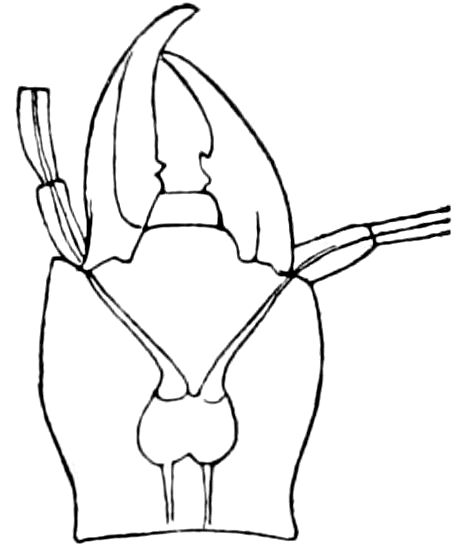
Fig. 256.—Head of Anophthalmus tellkampfii, showing the brain,—the optic ganglia, nerves, and eyes totally atrophied.
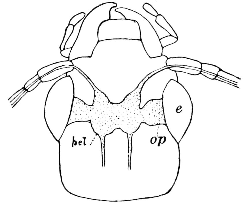
Fig. 257.—Head of another Carabid, with the brain and eyes normal: op, optic ganglion; pcl, brain.
In certain cave insects where the eyes are wanting, the optic ganglia are also absent. In the eyeless cave species of Anophthalmus the optic ganglia and nerves are entirely atrophied, as they are in Adelops, which, however, has vestiges of the facets (ommatidia). Fig. 257 represents the brain of Chlænius pennsylvanicus, a Carabid beetle, with its eyes and optic ganglia (op) which may be compared with Anopthalmus, in which these parts are totally atrophied.
Dujardin claimed that the degree of complication of the stalked body of the Hymenoptera was in direct relation with their mental powers. This has been proved by Forel, who has shown that in the honey bee and ants the mushroom bodies are much more developed in the workers than in the males or females and Viallanes adds that these bodies are almost rudimentary in the dragon-flies, whose eyes are so large; while on the contrary in the blind ants (Typhlopone), these bodies are as perfect and voluminous as in the ants with eyes.
242
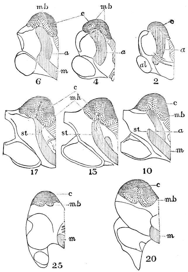
Fig. 258.—Diagrammatic outlines of sections of the upper part of the brain of a cockroach. Only one side of the brain is here represented. The numbers indicate the position in the series of 34 sections into which this brain was cut. mb, mushroom bodies, with their cellular covering (c) and their stems (st); a, anterior nervous mass; m, median nervous mass.—After Newton.
243Within the limits of the same order the stalked bodies are most perfect in the most intelligent forms. Thus in the Orthoptera, says Viallanes, the Blattæ, Forficulæ, and the crickets, the mushroom bodies are more perfect than in the locusts, which have simpler herbivorous habits. This perfection of the mushroom bodies is seen not only in the increase in size, but also in the complication of its structures. Thus in the groups with lower instincts (Tabanus, Æschna) the stalk does not end in a calyx projecting from the surface of the brain, but its end, simply truncated, is indicated externally only by an accumulation of the ganglionic nuclei which cover it.[43]
In types which Viallanes regards as more advanced, i.e. Œdipoda and Melanoplus, the end of the stalk projects and is folded into a calyx.
The brain of the cockroach (Periplaneta, Fig. 258) is a step higher than that of the locusts, each calyx being divided into two adjacent calices, although the cockroaches are an older and more generalized type than locusts.
The stalked bodies of cockroaches are thus complex, like those of the higher Hymenoptera, the calices in Xylocopa, Bombus, and Apis being double and so large as to cover almost the entire surface of the brain.
Finally, in what Viallanes regards as the most perfect type (Vespa), the sides of the calices are folded and become sinuous, so as to increase the surface, thus assuming an appearance which, he claims, strongly recalls that of the convolutions of the brain of the mammals.
Cheshire also calls attention to a progression in the size of these appendages, as well as in mental powers as we rise from the cockchafer (Melolontha vulgaris) to the cricket, up to the ichneumon, then to the carpenter bee, and finally to the social hive bee, “where the pedunculated bodies form the ⅕ part of the volume of the cerebral mass, and the 1 870 of the volume of the entire creature, while in the cockchafer they are less than 1 2300 the part. The size of the brain is also a gauge of intelligence. In the worker bee the brain is 1 174 of the body; in the red ant, 1 296; in the Melolontha, 1 3500; in the Dyticus beetle, 1 4400.” (Bees and bee-keeping, p. 54.)
g. Functions of the nerve-centres and nerves
As we have seen, the central seat of the functions of the nervous system is not the brain alone (supraœsophageal ganglion), but each ganglion is more or less the seat of vital movements, those of the 244abdomen being each a distinct motor and respiratory centre. The two halves of a ganglion are independent of each other.
According to Faivre, the brain is the seat of the will and of the power of coördinating the movements of the body, while the infraœsophageal ganglion is the seat of the motive power and also of the will.
The physiological experiments of Binet, which are in the line of those of Faivre, but more thorough, demonstrate that an insect may live for months without a brain, if the subœsophageal ganglion is left intact, just as a vertebrate may exist without its cerebrum. As Kenyon says: “Faivre long ago showed that the subœsophageal ganglion is the seat of the power of coördination of the muscular movements of the body. Binet has shown that the brain is the seat of the power directing these movements. ‘A debrained hexapod will eat when food is placed beneath its palpi, but it cannot go to its food even though the latter be but a very small space removed from its course or position. Whether the insect would be able to do so if the mushroom bodies only were destroyed, and the antennal lobes, optic lobes, and the rest of the brain were left intact, is a question that yet remains to be answered’” (Kenyon).
In insects which are beheaded, however readily they respond to stimulation of the nerves, they are almost completely wanting in will power. Yet insects which have been decapitated can still walk and fly. Hymenoptera will live one or two days after decapitation, beetles from one to three days, and moths (Agrotis) will show signs of life five days after the loss of their head.
That the loss of will power is gradual was proved by decapitating Polistes pallipes. A day after the operation she was standing on her legs and opening and closing her wings; 41 hours after the operation she was still alive, moving her legs, and thrusting out her sting when irritated. Ichneumon otiosus, after the removal of its head, remained very lively, and cleaned its wings and legs, the power of coördination in its wings and legs remaining. A horse-fly, a day after decapitation, was lively and flew about in a natural manner.[44]
When the abdomen is cut off, respiration in that region is not at first interrupted. The seat of respiratory movements was referred by Faivre to the hinder thoracic ganglion, but Plateau says that this view must be entirely abandoned, remarking: “All carefully performed experiments on the nervous system of Arthropoda have shown that each ganglion of the ventral chain is a motor centre, and 245in insects a respiratory centre, for the somite to which it belongs” (Miall and Denny’s The Cockroach, p. 164).
The last pair of abdominal ganglia serve as the nervous centre of the nerves sent to the genital organs.
The recurrent or stomatogastric nerve, which, through the medium of the frontal ganglion, regulates digestion, has only a slight degree of sensibility; the insect remains quiet even when a powerful allurement is presented to the digestive tract (Kolbe).
Faivre states that the destruction of the frontal ganglion, or a section of the commissures connecting it with the brain, puts an end to swallowing movements; on the other hand, stimulation results in energetic movements of this nature.
Yersin, by cutting through the commissure in different places, and thus isolating the ganglia of the nervous cord of Gryllus campestris, arrived at the following results:—
1. The section of a nerve near its origin rendered the organ supplied by this nerve incapable of performing its functions.
2. If the connectives between two ganglia, i.e. the second and third thoracic ganglia, are cut through, the fore as well as hinder parts of the body retain their power of motion and sensation; but a stimulus applied to the anterior part of the body does not pass to the hinder portion.
3. Insects with an incomplete metamorphosis after section of the connectives are not in every case unable to moult and to farther develop.
4. If only one of the two connectives be cut through, the appendages of the side cut through which take their origin between the place injured and the hinder end of the body, often lose sensation and freedom of motion, or the power of coördination of movements becomes irregular. Sometimes this is shown by an unsteadiness in the gait, so that the insect walks around in a circle; after a while these irregularities cease, and the movements of the limbs on the injured side are only slightly restrained. By a section of both connectives in any one place the power of coördination of movements is not injured.
5. The section of the connectives appear to have no influence on nutrition, but affects reproduction, the attempt at fertilization on the part of the male producing no result, and the impregnated female laying no eggs.
6. Injury to the brain, or to the subœsophageal, or one of the thoracic ganglia, is followed by a momentary enfeeblement of the ganglion affected. Afterwards there results a convulsive trembling, 246which either pervades the whole body or only the appendages innervated by the injured ganglion.
7. As a result of an injury to the brain there is such a lack of steadiness in the movements that the insect walks or flies in a circle; for instance, a fly or dragon-fly thus injured in flying describes a circle or spiral. Steiner, in making this experiment, observed that the insect circled on its uninjured side. The brain is thus a motor centre.
8. By injuring a thoracic ganglion, one or all the organs which receive nerves from the ganglion are momentarily weakened. Afterwards the functions become restored. Sometimes, however, the insect walks in a circle. Faivre observed that after the destruction of the metathoracic ganglion of Dyticus marginalis the hind wings and hind legs were partially paralyzed (Kolbe, ex Yersin).
LITERATURE ON THE NERVOUS SYSTEM
a. General
Newport, George. On the nervous system of the Sphinx ligustri L., and on the changes which it undergoes during a part of the metamorphoses of the insect. (Phil. Trans. Roy. Soc., London, 1832, pp. 383–398; 1834, pp. 389–423, Pls.)
Helmholtz, H. L. F. De fabrica systematis nervosi evertebratorum. Diss. in aug. Berolini, 1842.
Blanchard, E. Recherches anatomiques et zoologiques sur le système nerveux des animaux sans vertèbres. Du système nerveux des insectes. (Annales des Sciences nat., Sér. 3, v, 1846, pp. 273–379, 8 Pls.)
—— Du système nerveux chez les invertèbres dans ses rapports avec la classification de ces animaux. Paris, 1849.
—— in Cuvier’s Règne animal. (Edition accompagnée de planches gravées. Insectes. Pl. 3, 3a, and 4.)
Leidy, Joseph. History and anatomy of the hemipterous genus Belostoma. (Memoirs Amer. Acad. Arts and Sc., N. S. iv, 1849, pp. 57–67, 1 Pl.)
Scheiber, S. H. Vergleichende Anatomie und Physiologie der Œstridenlarven. (Sitzungsb. k. Akad. wiss. Wien. Math.-Naturwiss. Cl., xli, 1860, pp. 439–496; xlv, 1862, pp. 7–68; 5 Taf.)
Tullberg, Tycho. Sveriges Podurider. (K. Svenska vet. Akad. Handl. x, 1872, pp. 1–70, 12 Taf.)
Berlese, A. Osservazione sulla anatomia descrittiva del Gryllus campestris L. (Atti della soc. Veneto-Trentina, 1880, vii, pp. 200–299.)
Baudelot, E. Contributions à la physiologie der système nerveux des insectes. (Revue d. sc. nat., i, pp. 269–280, 1872.)
Studer, Th. Ueber Nervenendigung bei Insekten. Kleine Beiträge zur Histologie der Insekten. (Mitt. Naturf. Ges., Bern, 1874, pp. 97–104, 1 Taf.)
Brandt, E. Recherches anatomiques et morphologiques sur le système nerveux des insectes Hyménoptères. (Compt. rendus de l’Acad. Sc., Paris, 1875.)
—— Ueber das Nervensystem der Apiden. (Sitzungsb. d. naturf. Ges., in Petersbourg, vii, 1876.)
—— Ueber das Nervensystem der Schmetterlingsraupen. (Verhandl. der Russ. 247Ent. Gesellsch., x, 1877. Also 16 other articles with plates, in Horæ Soc. Ent. Ross., 1878–1882.)
Mark, E. L. The nervous system of Phylloxera. (Psyche, ii, pp. 201–207, 1879.)
Riley, Charles Valentine. The nervous system and salivary glands of Phylloxera. (Psyche, ii, pp. 225, 226, 1879.)
Cholodkowsky, N. Zur Frage über den Baue und über die Innervation der Speicheldrüsen der Blattiden. (Horæ Soc. Ent. Ross., 1881, xvi, pp. 6–9, 2 Taf.)
Liénard, V. Constitution de l’anneau œsophagien. (Archives de Biologie, i, pp. 381–391, 1880, 1 Taf.)
Michaels, H. Nervensystem von Oryctes nasicornis im Larven-, Puppen-, und Käferzustande. (Zeits. f. wissens. Zool., xxxiv, 1880, pp. 641–702, 4 Taf.)
Rossi, A. Sul modo di terminare dei nervi nei muscoli dell’ organo sonoro della Cicala commune (Cicada plebeja). (Mem. accad. sc. Bologna, 1880, 4 Ser., i, pp. 661–665.)
Foettinger, A. Sur le termination des nerfs dans les muscles des insectes. (Archiv de Biologie, i, 1880.)
Binet. Contribution à l’étude der system nerveux sous intestinal des insectes. (Journ. l’anat. et phys., xxx, pp. 449–580, 1894.)
Paulowa. Zum Bau des Eingeweide Nervensystems der Insekten. (Zool. Anzeiger., xviii, Feb. 25, 1895, pp. 85–87.)
Also the writings of Lyonet, Cuvier, Rolando, Straus-Durckheim, Leydig, Newport, Graber, Viallanes, Grassi, Oudemans.
b. The brain
Dujardin, F. Mémoires sur le système nerveux des insectes. (Annales des Sciences nat, Sér. 3, 1850, xiv, pp. 195–206, Pl. 1, 1850.)
Rabl-Rückhard. Studien über Insectengehirne. (Archiv für Anatomie, Physiologie, etc., herausg. von Reichert u. R. du Bois-Raymond, 1876, p. 480, Taf. i.)
Dietl, M. J. Die Organization des Arthropodengehirns. (Zeitschr. wissens. Zool., xxvii, 1876, p. 488, Taf. xxxvi.-xxxviii.)
Flogel, T. H. L. Ueber den einheitlichen Bau des Gehirns in den verschiedenen Insectenordnungen. (Zeitschr. wissens. Zool., xxx, Suppl., 1878, p. 556, Taf. xxiii, xxiv.)
Newton, E. T. On a new method of constructing models of the brains of insects, etc. (Journ. Quekett Microscopical Club, pp. 150–158, 1879.)
—— On the brain of the cockroach, Blatta orientalis. (Quart. Journ. Microscopical Science, July, 1879, p. 340, Pl. xv, xvi.)
Packard, A. S. The brain of the locust. (Chapter xi, Second Report of the U. S. Entomological Commission, pp. 223–242, Pls. ix-xv, 1880.)
Cuccati, Giovanni. Sulla stuttura del ganglio sopraesofageo di alcuni ortotteri. (Acrydium lineola, Locusta viridissima, Locusta (species?), Gryllotalpa vulgaris, Bologna, 1887, 4º, pp. 1–27, Pl. i-iv.)
—— Intorno alla struttura del cervello della Sonomya erythrocephala, nota preventiva. Bologna, 1887.
—— Ueber die Organization des Gehirns des Sonomya erythrocephala. (Zeitschr. f. wissens. Zool., 1888, xlvi, pp. 240–269, 2 Taf.)
Viallanes, H. Études histologiques et organologiques sur les centres nerveux et les organes des sens des animaux articulés.
1. Mémoire. Le ganglion optique de la langouste (Palinurus vulgaris). (Annal. d. Sc. Nat. Zool., 1884, 6e Sér., xvii, Art. 3, pp. 1–74, 5 Pls.)
2482. Mémoire. Le ganglion optique de la Libellule (Æschna maculatissima). (Ibid., 1885, 6e Sér., xviii, Art. 4, pp. 1–34, 3 Pls.)
3. Mémoire. Le ganglion optique de quelques larves de Diptères (Musca, Eristalis, Stratiomys). (Ibid., 1886, 6e Sér., xix, Art. M. 4, pp. 34, 2 Pls.)
4. Mémoire. Le cerveau de la guêpe (Vespa crabro et vulgaris). (Ibid., 1887, 7e Sér., ii, pp. 5–100, 6 Pls.)
5. Mémoire. 1. Le cerveau du criquet (Œdipoda cœrulescens et Caloptenus italicus). 2. Comparaison du cerveau des Crustacés et des Insectes. 3. Le cerveau et la morphologie du squelette céphalique. (Ibid., 1888, 7e Sér., iv, pp. 1–120, 6 Pls.)
—— Sur la structure interne du ganglion optique de quelques larves de Diptères. (Bull. Soc. Phil., Paris, 1885, 7e Sér., ix, pp. 75–78.)
—— La structure du cerveau des Hyménoptères. (Bull. Soc. Philomat., Paris, 1886, 7e Sér., x, pp. 82, 83.)
—— La structure du cerveau des Orthoptères. (Bull. Soc. Philomat., Paris, 1886, 7e Sér., xi, pp. 119–126.)
—— Sur la morphologie comparée du cerveau des Insectes et des Crustacés. (Compt. rend. Acad. Sc. Paris, 1887, civ, pp. 444–447.)
Kenyon, F. C. The meaning and structure of the so-called “mushroom bodies” of the hexapod brain. (Amer. Naturalist, xxx, 1896, pp. 643–650, 1 fig.)
—— The brain of the bee. (Journ. Comp. Neurology, vi, fasc. 3, 1896, pp. 133–210.)
—— The optic lobes of the bee’s brain in the light of recent neurological methods. (Amer. Nat., xxxi, 1897, pp. 369–376, 1 Pl.)
With the embryological works of Graber, Heider, Korscheldt, Patten, Wheeler, etc.
c. Histology of the nervous System
Helmholtz. De fabrica systematis nervosi evertebratorum. Diss. Berolini, 1842.
Remak. Ueber d. Inhalt d. Nervenprimitivröhren. (Archiv f. Anat. u. Phys., 1843.)
Leydig. Lehrbuch der Histologie der Menschen und der Thiere. 1857.
—— Vom Bau des thierischen Körpers. i. 1864.
—— Tafeln zur vergleichenden Anatomie. i. Tübingen, 1864.
—— Zelle und Gewebe, neue Beiträge zur Histologie des Tier-Körpers. Bonn, 1885, pp. 219, 6 Taf.
Walter. Mikroscopische Studien über das Centralnervensystem wirbelloser Thiere. 1863.
Dietl, M. J. Die Gewebselemente des Centralnervensystems bei wirbellosen Thieren. (Aus den Berichten des naturw.-medic. Vereins in Innsbruck.) Innsbruck, 1878.
Berger. Untersuchungen über den Bau des Gehirns und der Retina der Arthropoden. (Arbeiten des zool. Instituts zu Wien, Heft 2, p. 173, 1878.)
—— Nachtrag zu den Untersuchungen über den Bau des Gehirns und der Retina der Arthropoden. (Ibid., Heft 3.)
Viallanes, H. Recherches sur l’histologie des insectes, etc. Paris, 1882. (Annales des Sciences nat., pp. 1–348, Pls. 1–18.)
—— Sur la structure de la substance ponctuée des insectes. Paris, 1885.
Haller, B. Ueber die sogenannte Leydig’sche Punktsubstantz im Centralnervensystem. (Morp. Jahrb., xi, 1886.)
Nansen, F. The structure and combination of the histological elements of the central nervous system. (Bergen’s Museum Aarsberetning for 1886. Bergen, 1887.)
Also the writings of Benedicenti, Holmgren.
249
THE SENSORY ORGANS
a. The eyes and insect vision
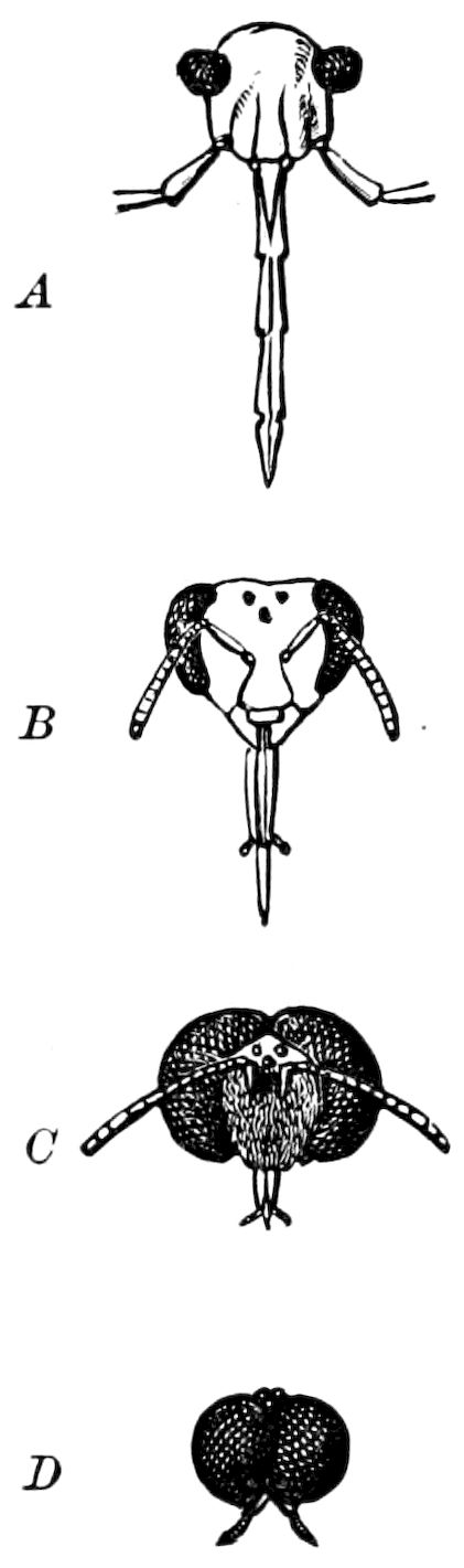
Fig. 259.—Different forms of compound eyes. A, a bug (Pyrrhocoris). B, worker bee. C, drone. D, male Bibio, a holoptic insect.—From Judeich and Nitsche.
Of the eyes of insects there are two kinds, the simple and the compound. Of the former there are usually three, arranged in a triangle near the top of the head, between the compound eyes (Fig. 259, B). The compound or facetted eyes, which are usually round and prominent, differ much in size and in the number of facets.
The number of facets varies from 12 in Lepisma,—though in a Brazilian beetle (Lathridius) there are only seven unequal facets,—to 50 in the ant, and up to 4000 in the house-fly, 12,000 in Acherontia atropos, 17,000 in Papilio, 20,000 in the dragon-fly (Æschna), 25,000 in a beetle (Mordella), while in Sphinx convolvuli, the number reaches 27,000. The size of the facets seems to bear some relation to that of the insect, but even in the smallest species none have been observed less than 1 2000 of an inch in diameter. Day-flying Lepidoptera have smaller facets than moths (Lubbock).
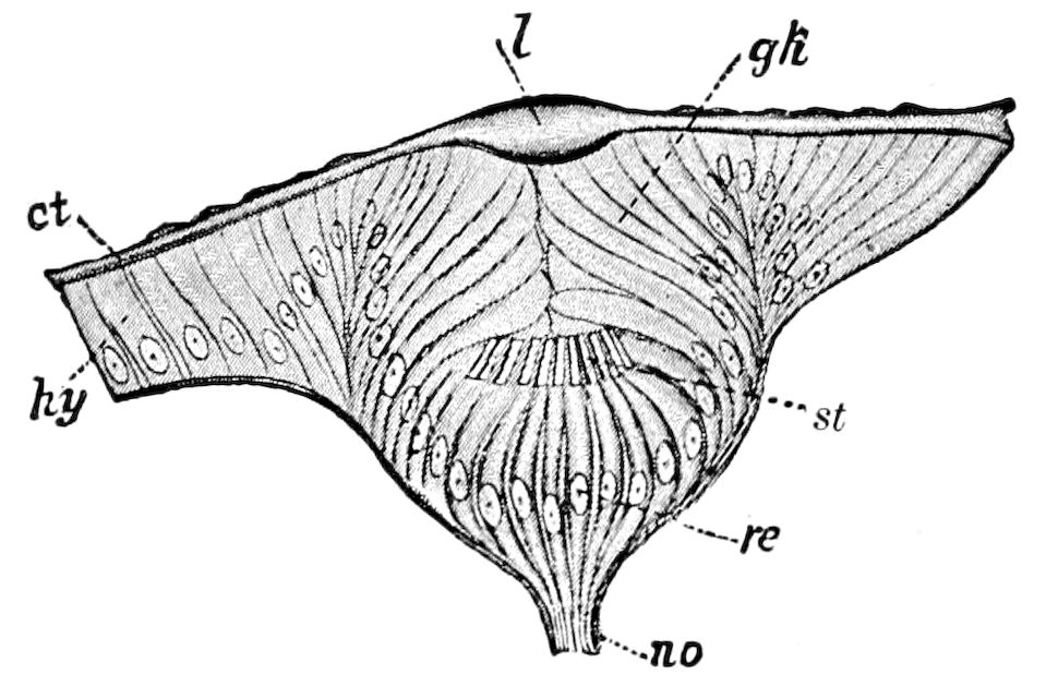
Fig. 260.—Section through the ocellus of a young Dyticus larva: ct, cuticula; l, corneal lens; gh, cells of the vitreous body, being modified hypodermal cells (hy); st, rods; re, retinal cells; no, optic nerve.—After Grenacher, from Lang.
The simple, or single-lensed eye (ocellus).—Morphologically the simple eye is a modified portion of the ectoderm, the pigment enclosing the retinal cells arising from specialized hypodermal cells, and covered by a specialized transparent portion of the cuticula, forming the corneal lens. The apparatus is supplied with a nerve, the fibres of which end in a rod or solid nerve-ending, as in other sensory organs.
As seen in the ocellus of Dyticus (Fig. 260), under the corneal lens the hypodermis forms a sort of pit, and the cells are modified 250to form the vitreous body (vitrella) and retina. Each retinal cell (re) is connected with a fibre from the optic nerve, contains pigment, and ends in a rod directed outwards towards the lens. The cells at the end of the pit or depression are, next to the lens, without pigment, and, growing in between the retina and the lens, fill it up, and thus form a sort of vitreous body.
The ocellus appears to be a direct heirloom from the eyes of worms, while the many-facetted compound eye of the crustaceans and of insects is peculiar to these classes. The compound eye of the myriopod Scutigera differs structurally in many respects from the compound eye of insects, and that of Limulus still more so.
It should be observed that in the young nymph of Ephemera, as well as in the semipupa of Bombus, each of the three ocelli are situated on separate sclerites. In Bombus the anterior ocellus has a double shape, being broad, transversely ovate, and not round like the two others, as if resulting from the fusion of what were originally two distinct ocelli.
The ocelli are not infrequently wanting, as in adult Dermaptera, in the Locustidæ, and in certain Hemiptera (Hydrocora). In Lepidoptera there are but two ocelli; in geometrid moths they are often atrophied, and they are absent in butterflies (except Pamphila).
The compound or facetted eye (ommateum).—The facetted arthropod eye is wonderfully complex and most delicately organized, being far more so than that of vertebrates or molluscs. The simplest or most primitive facetted eye appears to be that of Lepisma. As stated by Watase, the compound eye of arthropods is morphologically “a collection of ectodermic pits whose outer open ends face towards the sources of light, and whose inner ends are connected with the central nervous system by the optic nerve fibres.”
The facetted eye is composed of numerous simple eyes called ommatidia, each of which is complicated in structure. The elements which make up an ommatidium are the following: (1) The facet or cornea, which is a specialized portion of the cuticula; and (2), the crystalline lens or cone; (3), the nerve-ending or retinula, which is formed out of the retinula cells and the rhabdom or rod lying in its axis; and (4) of the pigment enclosing the lens and rod; the last three elements are derived from the hypodermis. The single eyes are separated from each other by pigment cells.
The facet or cornea.—This is biconvex, clear, transparent, usually hexagonal in outline, and refracts the light. The corneal lenses are cast in moulting.
The corneal lenses are circular in most cases where they are very convex, as in Lathridius and Batocera. The hexagonal ones are very irregular. When they are very convex the eye has a granular appearance, but when not greater than the convexity of the eye itself, the eye appears perfectly smooth (Bolbocerus, 251etc.). The facets in the lower part of the eye of Dineutes are a trifle larger than in the upper part (about nine to ten). In many insects the reverse is the case, the upper facets being larger than the lower, a notable instance being Anax. The intervening lines between the facets are often beset with hairs, sometimes very long and dense, as in the drone bee and Trichophthalmus; and the modifications of the hairs into scales which takes place on the body occurs on the eyes also, the scales on the eyes of some beetles of the family Colydiidæ being very large, arranged in lines over the eyes like tombstones (Trachypholis).[45]
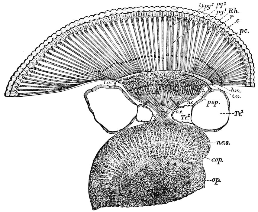
Fig. 261.—Section through the eye of a fly (Musca vomitoria): c, cornea, or facet; pc, pseudocone; r, retinula; Rh, rhabdom; pg1, pg2, pg3, pigment cells; b.m, basilar membrane; T, Tt1, Tt2, trachea; tv, tracheal vesicle; t.a, terminal anastomosis; op, opticon; c.op, epiopticon; p.op, periopticon; n.c, nuclei; n.c.s, nerve-cell sheath; N.f, decussating nerve-fibres.—After Hickson, from Lubbock.
The crystalline lens or cone.—Behind or within the facets is a layer composed of the cones, behind which are the layers of retinulæ and rhabdoms, and which correspond to the layer of rods and cones, but not the retina as a whole, of vertebrate animals.
The crystalline lens is, when present, usually more or less conical, and consists of four or more hypodermis-cells.
The cones are of various shapes and sizes in insects of different groups, or are entirely wanting, and Grenacher has divided the eyes of insects into eucone, pseudocone, and acone. As the pseudocone seems, however, to be rather a modification of the eucone eye, the following division may be made:—
1. Eucone eyes, comprising those with a well-developed cone. They occur in Lepisma, Blatta (Fig. 262), and other Orthoptera, in Neuroptera, in Cicadidæ, in those Coleoptera with five tarsal 252joints, in the dipterous genus Corethra, and in the Lepidoptera and Hymenoptera (Fig. 263).
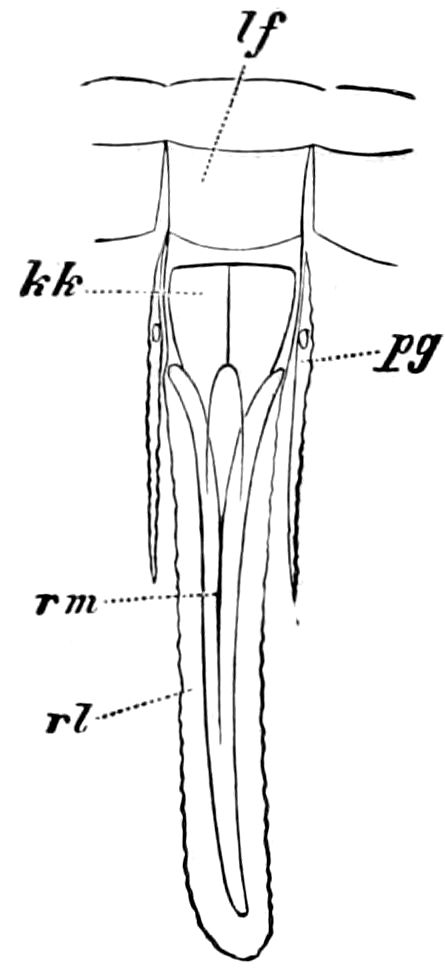
Fig. 262.—Ommatidium of cockroach (Periplaneta): lf, cornea; kk, crystalline cone; pg′ pigment cell; rl, retinula; rm, rhabdom.—After Grenacher, from Lubbock.
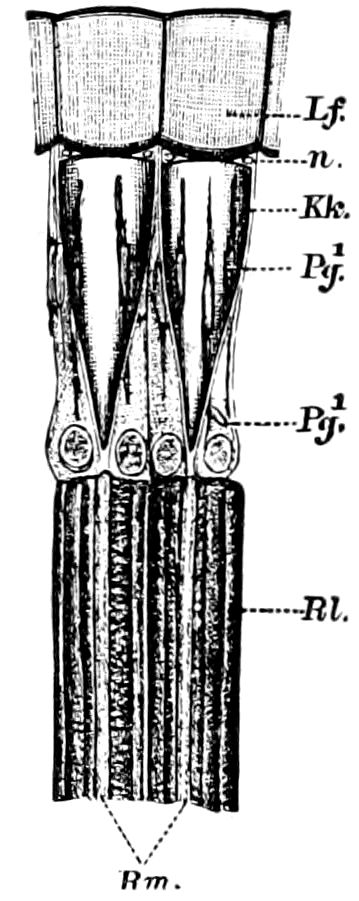
Fig. 263.—Two separate elements of the eucone eye of a bee; Lf, cornea; n, nucleus of Semper; Kk, crystalline cone; Pg, pigment cells; Rl, retinula; Rm, rhabdom.—After Grenacher, from Lubbock.
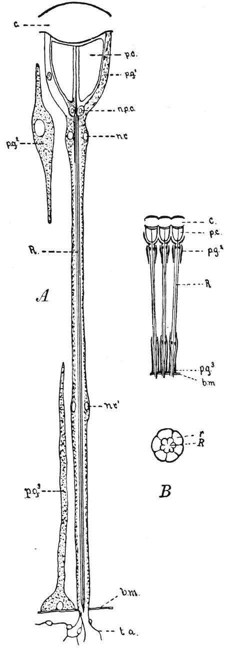
Fig. 264.—Three ommatidia of a pseudocone eye, diagrammatic: A, a separate ommatidium of Musca vomitoria, semi-diagrammatic: c, cornea; p.c, pseudocone; pg′, pigmented cells surrounding the pseudocone; p.g2, additional pigment cells; p.g3, basal pigment cells; n.p.c, nuclei of pseudocone; r, retinulæ; n.r, n.r′, nucleus of retinulæ; R, rhabdom; b.m, basal membrane; t.a, terminal anastomosis sending nerve-fibrils to the retinulæ. B, section through a retinula and rhabdom near the basal membrane, the six retinulæ (r) fused into a tube ensheathing the rhabdom (R).—After Hickson.
a. Pseudocone eyes; in which, instead of the crystalline lens or cone, there are four cells filled with a transparent fluid medium, and a smaller protoplasmic portion containing a nucleus (Muscidæ, Fig. 264, pc). Hickson states that the difference between the eucone and pseudocone eyes lies in the fact that in the pseudocone eye “the refracting body formed by the cone-cell lies behind the nuclei,” and in the eucone eye in front of it.
2. Acone eyes, where the cone or refracting body is wanting, but is represented by the four primitive cone-cells. Acone eyes occur in Forficulidæ, Hemiptera (except Cicadidæ), the nematocerous Diptera (Tipula, etc.), and those Coleoptera which have less than five tarsal joints.
253The retinula and rod.—The retinula is morphologically a nerve-end cell, situated at the end of a nerve-fibril arising from the optic nerve. The elements of the retinula of Musca are six in number and surround the rhabdom (Fig. 264), which consists of a bundle of six long, delicate chitinous rods, more or less firmly united together (Fig. 264, R).
The six elements of the retinula of Musca are in their outer or distal portion free from one another, but towards their base are fused into a sheath (Fig. 264, r). They are true nerve-end cells, as shown by Müller and by Max Schultze, their views having been confirmed by Grenacher and by Hickson. The relations of the nerves to the rods after passing through the basal membrane is seen in Fig. 266.
The pigment.—The cones or pseudocones are mostly buried in pigment, as well as the rods; and the pigment forms two layers. The outer of the two layers is called the iris pigment (Fig. 265, e, iris tapetum), and the inner (f) the retinal pigment.
Between the ommatidia internally there occur, according to Hickson, pigment cells (Fig. 264, p.g3), each of which stands on the basilar membrane and sends a fine process outwards towards the internal process of the external pigment-cell (p.g2). A long, slender tracheal vesicle also passes in between the retinulæ.
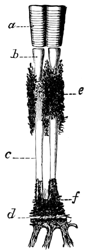
Fig. 265.—Two ommatidia from the eye of Colymbetes fuscus, × 160: a, cornea; b, cone; c, rhabdom; d, basal membrane, with nerve filaments below it: e, iris pigment; f, retina pigment.—After Exner, from Sharp.
The basilar membrane.—This is a thin fenestrate membrane (Fig. 261) separating the cones and rods from the optic tract (Fig. 264, b.m). It is perforated for the passage of tracheal diverticula and of the optic nerve fibrils. It separates the dioptric or instrumental portion of the eye from the percipient portion, i.e. the optic tract.
The optic tract.—This is the optic ganglion of earlier writers, and appears to be the percipient portion of the eye, as opposed to the dioptric portion. If the reader will examine Figs. 249 and 261, he will see that it consists of three distinct ganglionic swellings, i.e. the opticon, epiopticon, and periopticon, whose structure is very complicated. In Musca (Fig. 261) the first ganglionic swelling (opticon) is separated from the brain by a slight constriction, which Berger regards as the homologue of the optic nerve of the other arthropods. It consists of a very fine granular matrix traversed throughout by a fine meshwork 254of minute fibrillæ, the neurospongium of Hickson. In the young cockroach (Periplaneta) the optic nerve separating the cerebral ganglion from the opticon is much longer in proportion than it is in the adult blow-fly.
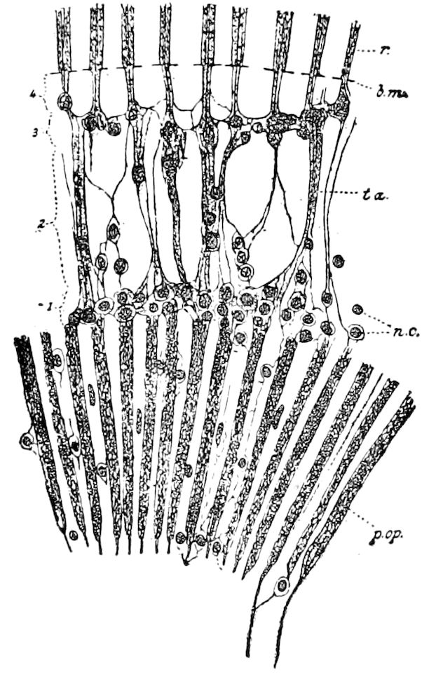
Fig. 266.—Periopticon and terminal anastomosis of Agrion, showing the character of the elements of the periopticon (p.op) and the structure of the terminal anastomosis (t.a). 1. The first layer of the terminal anastomosis, consisting of a plexus of fibrils and nerve-cells (n.c). 2. The second layer, in which the fibrils are collected together in bundles. 3. The final optic plexus and nerve-cells. 4. The layer in which the optic fibrils are collected in bundles to be distributed to the retinulæ (r); b.m, basal membrane.—After Hickson.
The second ganglionic swelling (epiopticon, Fig. 261, c.op) is separated from the opticon by a tract of fine nerve-fibrils, which partially decussate; at the decussation two or three larger nerve-cells may be seen. It also contains a few scattered nerve-cells (n.c). The third ganglionic swelling (periopticon, p.op) is separated from the others by a bundle of long optic nerve-fibrils, which cross one another. It is composed of a number of cylindrical masses of neurospongium arranged side by side (Fig. 261, p.op). Between these elements of the periopticon, which do not seem to bear any relation to the number of ommatidia, a single nerve-cell is very frequently seen. The periopticon does not occur in Periplaneta and Nepa (Hickson). The three optic ganglia thus described, together with the cerebral ganglia, are surrounded by a sheath of densely packed nerve-cells.
Bearing in mind the fact that the retinulæ are the nerve-end cells of the fibres passing through the periopticon, it will be well to read the following account, by Hickson, of the terminal anastomosis of the optic fibrils in the periopticon of Agrion bifurcatum, and to examine his sketch (Fig. 266):
“The terminal anastomosis of Agrion may be conveniently divided into four regions. First the region (1) lying nearest to the periopticon in which the nerve-cells are numerous, and the fibrils leaving the periopticon form a complicated plexus; the region (2) next to this, in which the fibrils have collected into bundles separated by spaces occupied by very thin-walled tracheæ in which there are no spiral markings, and lymph-spaces; next, the region (3) in which the fibrils form a final plexus, and in which there are again a considerable number of nerve-cells; and, lastly, the region (4) in which the fibrils are again collected into bundles, separated by spaces containing tracheæ, which perforate the basement membrane to supply the retinulæ.”
It would seem as if the decussation of the optic nerve-fibrils were a matter of 255primary importance, as it so generally occurs, but in the young of that most generalized of all pterygote insects, the cockroach (Periplaneta), Hickson states that the optic nerve-fibrils which leave the periopticon pass without decussating to the ommateum, and in the adult there is only a partial decussation. In Nepa there is no decussation, but the anastomosis is complicated by the presence of looped and transverse anastomoses.
Looking at the eye as a whole, Hickson regards all the nerve structure of the eye lying between the crystalline cone-layer and the true optic nerve to be analogous with the retina of other animals. With Ciaccio, Berger, and others, he does not regard the layer composed of the retinulæ and rhabdoms as the equivalent of the retina of vertebrates, etc.
Origin of the facetted eye.—The two kinds of eye, the simple and the compound, are supposed to have been derived from a primitive type, resembling the single eye (ommatidium) of the acone eye of Tipula. As stated by Lang, “an increase of the elements of this primitive eye led to the formation of the ocellus; an increase in number of the primitive eyes, and their approximation, led to the formation of the compound facet eye.” This view is suggested, he says, by the groups of closely contiguous single eyes of the myriopods, considered in connection with the compound eye of Scutigera. Grenacher looks upon simple (ocelli) and compound eyes as “sisters,” not derived from one another, but from a common parentage.
Immature insects rarely possess compound eyes; they are only known to occur in the nymphs of Odonata and Ephemeridæ, and in the larvæ and pupa of Corethra.
Mode of vision by single eyes or ocelli.—In their simplest condition, the eyes of worms and other of the lower invertebrates, probably only enable those animals to distinguish light from darkness. The ocelli of spiders and of many insects, however, probably enable them, as Lubbock remarks, to see as our eyes do. The simple lens throws on the retina an image, which is perceived by the fine terminations of the optic nerve. The ocelli of different arthropods differ, however, very much in degree of complexity.
Müller considered that the power of vision of ocelli “is probably confined to the perception of very near objects.”
“This may be inferred,” Müller states, “partly from their existing principally in larvæ and apterous insects, and partly from several observations which I have made relative to the position of these simple eyes. In the genus Empusa the head is so prolonged over the middle inferior eye that, in the locomotion of the animal, the nearest objects can only come within the range. In Locusta cornuta, also, the same eye lies beneath the prolongation of the head.... In 256the Orthoptera generally, also, the simple eyes are, in consequence of the depressed position of the head, directed downwards towards the surface upon which the insects are moving.”[46] Lowne considers that in the ocellus of Eristalis, the great convexity of the lens must give it a very short focus, and the comparatively small number of rods render the picture of even very near objects quite imperfect and practically useless for purposes of vision, and that the function of the ocelli is “the perception of the intensity and the direction of light, rather than of vision, in the ordinary acceptation of the term.”
Réaumur, Marcel de Serres, Dugès, and Forel have shown by experiment, that in insects which possess both ocelli and compound eyes, the former may be covered over without materially affecting the movements of the animals, while if the facetted eyes are covered, they act as if in the dark (Lubbock).
While Plateau regards the ocelli as of scarcely any use to the insect, and Forel claims that wasps, humble bees, ants, etc., walk or fly almost equally well without as with the aid of their ocelli, Lubbock demurs to this view, and says the same experiments of Forel’s might almost be quoted to prove the same with reference to the compound eyes. Indeed, the writer has observed that in caves, eyeless beetles apparently run about as freely and with as much purpose, as their eyed relatives in the open air.
Plateau has recently shown that caterpillars which have ocelli alone are very short-sighted, not seeing objects at a distance beyond one or two centimetres, and it has been fully proved by Plateau and others, that spiders, with their well-formed ocelli, are myopic, and have little power of making out distinctly the shape of the objects they see.
On the whole, we are rather inclined to agree with Lubbock and Forel, that the ocelli are useful in dark places and for near vision. They are, as Lubbock states, especially developed in insects, such as ants, bees, and wasps, which live partly in the open light and partly in the dark recesses of nests. Moreover, the night-flying moths nearly all possess ocelli, while with one known exception (Pamphila) they are wanting in butterflies.
Finally, remarks Lubbock, “Whatever the special function of ocelli may be, it seems clear that they must see in the same manner as our eyes do—that is to say, the image must be reversed. On the other hand, in the case of compound eyes, it seems probable that the vision is direct, and the difficulty of accounting for the existence in the same animal of two such different kinds of eyes is certainly enhanced by the fact that, as it would seem, the image given by the medial eyes is reversed, while that of the lateral ones is direct” (p. 181).
Mode of vision by facetted eyes.—The complexity of the facetted eyes of insects is amazing, and difficult to account for unless we accept the mosaic theory of Müller, who maintained that the distinctness of the image formed by such an eye will be greater in proportion to the number of separate cones. His famous theory is thus stated: “An image formed by several thousand separate points, of which each corresponds to a distinct field of vision in the external world, will resemble a piece of mosaic work, and a better idea cannot be conceived of the image of external objects which will be depicted on the retina of beings endowed with such organs of vision, than by comparing it with perfect work of that kind.”
257
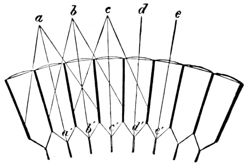
Fig. 267.—From Lubbock.
How vision is effected by a many-facetted eye is thus explained by Lubbock: “Let a number of transparent tubes, or cones with opaque walls, be ranged side by side in front of the retina, and separated from one another by black pigment. In this case the only light which can reach the optic nerve will be that which falls on any given tube in the direction of its axis.” For instance, in Fig. 267, the light from a will pass to a′, that from b to b′, that from c to c′, and so on. The light from c, which falls on the other tubes, will not reach the nerve, but will impinge on the sides and be absorbed by the pigment. Thus, though the light from c will illuminate the whole surface of the eye, it will only affect the nerve at c′.
According to this view those rays of light only which pass directly through the crystalline cones, or are reflected from their sides, can reach the corresponding nerve-fibres. The others fall on, and are absorbed by, the pigment which separates the different facets. Hence each cone receives light only from a very small portion of the field of vision, and the rays so received are collected into one spot of light.
It follows from this theory that the larger and more convex the eye, the wider will be its field of vision, while the smaller and more numerous are the facets, the more distinct will be the vision (Lubbock).
The theory is certainly supported by the shape and size and the immense number of facets of the eye of the dragon-fly, which all concede to see better, and at a longer range, than probably any other insect.
Müller’s mosaic theory was generally received, until doubted and criticised by Gottsche (1852), Dor (1861), Plateau, and others. As Lubbock in his excellent summary states, Gottsche’s observation (previously made by Leeuwenhoek) that each separate cornea gives a separate and distinct image, was made on the eye of the blow-fly, which does not possess a true crystalline cone. Plateau’s objection loses its force, since he seems to have had in his mind, as Lubbock states, Gottsche’s, rather than Müller’s, theory.
Müller’s theory is supported by Boll, Grenacher, Lubbock, Watase, and especially by Exner, who has given much attention to the subject of the vision of insects, and is the weightiest authority on the subject.
Gottsche’s view that each of the facetted eyes makes a distinct image which partially overlaps and is combined with all the images made by the other facets, 258was shown by Grenacher to be untenable, after repeating Gottsche’s experiments with the eyes of moths, in which the crystalline cones are firm and attached to the cornea. He was thus able to remove the soft parts, and to look through the cones and the cornea. When the microscope was focussed at the inner end of the cone, a spot of light was visible, but no image. As the object-glass was moved forward, the image gradually came into view, and then disappeared again. Here, then, the image is formed in the interior of the cone itself.
Exner attempted to make this experiment with the eye of Hydrophilus, but in that insect the crystalline cones always came away from the cornea. “He, however, calculated the focal length, refraction, etc., of the cornea, and concluded that, even if, in spite of the crystalline cone, an image could be formed, it would fall much behind the retinula.”
“In these cases, then,” adds Lubbock, “an image is out of the question. Moreover, as the cone tapers to a point, there would, in fact, be no room for an image, which must be received on an appropriate surface. In many insect eyes, indeed, as in those of the cockchafer, the crystalline cone is drawn out into a thread, which expands again before reaching the retinula. Such an arrangement seems fatal to any idea of an image.”
Lubbock thus sums up the reasons which seem to favor Müller’s theory of mosaic vision, and to oppose Gottsche’s view: “(1) In certain cases, as in Hyperia, there are no lenses, and consequently there can be no image; (2) the image would generally be destroyed by the crystalline cone; (3) in some cases it would seem that the image would be formed completely behind the eye, while in others, again, it would be too near the cornea; (4) a pointed retina seems incompatible with a clear image; (5) any true projection of an image would in certain species be precluded by the presence of impenetrable pigment, which only leaves a minute central passage for the light-rays; (6) even the clearest image would be useless, from the absence of a suitable receptive surface, since both the small number and mode of combination of the elements composing that surface seem to preclude it from receiving more than a single impression; (7) no system of accommodation has yet been discovered; finally (8), a combination of many thousand relatively complete eyes seems quite useless and incomprehensible.”
In his most recent work (1890) on the eyes of crustacea and insects, Exner states that the numerous simple eyes which make up the compound eye have each a cornea, but it is more or less flat, and the crystalline part of the eye has not the shape of a lens, but of a “lens cylinder,” that is, of a cylinder which is composed of sheets of transparent tissue, the refracting powers of which decrease toward the periphery of the cylinder. If an eye of this kind is removed and freed of the pigment which surrounds it, objects may be looked at through it from behind; but its field of vision is very small, and the direct images received from each separate eye are either produced close to one another on the retina (or rather the retinulæ of all the eyes) or superposed. In this last case no less than thirty separate images may be superposed, which is supposed to be of great use to night-flying insects. Exner claims that many other advantages result from the compound nature of an insect’s eye. Thus the mobile pigment, which corresponds to our iris, can take different positions, either between the separate eyes or behind the lens cylinders, in which case it acts as so many screens to intercept the over-abundance of light. Exner finds that with its compound eyes the common glow-worm (Lampyris) is capable of distinguishing large signboard letters at a distance of ten or more feet, as well as extremely fine lines engraved one-hundredth of an inch apart, if they are at a distance of less than half an inch from the eye. Exner substantiates the truth of the results of Plateau’s 259experiments, and claims that while the compound eye is inferior to the vertebrate eye for making out the forms of objects, it is superior to the latter in distinguishing the smallest movements of objects in the total field of vision.
More recently Mallock has given some optical reasons to show that Müller’s view is the true one. He concludes, and thus agrees with Plateau, that insects do not see well, at any rate as regards their power of defining distant objects, and their behavior certainly favors this view. It might be asked, What advantage, then, have insects with compound eyes over those with simple eyes? Mallock answers, that the advantage over simple-eyed animals lies in the fact that there is hardly any practical limit to the nearness of the objects they can examine. “With the composite eye, indeed, the closer the object the better the sight, for the greater will be the number of lenses employed to produce the impression; whereas, in the simple eye the focal length of the lens limits the distance at which a distinct view can be obtained.” He gives a table containing measures of the diameters and angles between the axes of the lenses of various insect eyes, and states that the best of the eyes would give a picture about as good as if executed in rather coarse woodwork and viewed at a distance of a foot, “and although a distant landscape could only be indifferently represented on such a coarse-grained structure, it would do very well for things near enough to occupy a considerable part of the field of view.”
The principal use of the facetted eye to perceive the movements of animals.—Plateau adopts Exner’s views as to the use of the facetted eye in perceiving the movements of other animals. He therefore concludes that insects and other arthropods with compound eyes do not distinguish the form of objects; but with Exner he believes that their vision consists mainly in the perception of moving bodies.
Most animals seem but little impressed by the form of their enemies or of their victims, though their attention is immediately excited by the slightest displacement. Hunters, fishermen, and entomologists have made in confirmation of this view numerous and demonstrative observations.
Though the production of an image in the facetted eye of the insect seems impossible, we can easily conceive, says Plateau, how it can ascertain the existence of a movement. Indeed, if a luminous object is placed before a compound eye, it will illuminate a whole group of simple eyes or facets; moreover, the centre of this group will be clearer than the rest. Every movement of the luminous body will displace the centre of clearness; some of the facets not illuminated will first receive the light, and others will reënter into the shade; some nervous terminations will be excited anew, while those which were so formerly will cease to be. Hence the facetted eyes are not complete visual organs, but mainly organs of orientation.
Plateau experimented in the following way: In a darkened room, with two differently shaped but nearly equal light-openings, one square and open, the other subdivided into a number of small holes, and therefore of more difficult egress, he observed the choices of opening made by insects flying from the other end of the room. Careful practical provisions were made to eliminate error; the light-intensity of the two openings was as far as possible equalized or else noted, and no trees or other external objects were in view. The room was not darkened beyond the limit at which ordinary type ceases to be readable, otherwise the insects refused to fly (it is well known that during the passage of a thick cloud insects usually cease to fly). These observations were made on insects 260both with or without ocelli, in addition to the compound eyes, and with the same results.
From repeated experiments on flies, bees, etc., butterflies and moths, dragon-flies and beetles, Plateau concludes that insects with compound eyes do not notice differences in form of openings in a half-darkened room, but fly with equal readiness to the apparently easy and apparently difficult way of escape; that they are attracted to the more intensely lighted opening, or to one with apparently greater surface; hence he concludes that they cannot distinguish the form of objects, at least only to a very slight extent, though they readily perceive objects in motion.
One result of his experiments is that insects only utilize their eyes to choose between a white luminous orifice in a dark chamber, or another orifice, or group of orifices, equally white. They are guided neither by odorous emanations nor by differences of color. He thinks that bees have as bad sight and act almost exactly as flies.
From numerous experiments on Odonata, Coleoptera, Lepidoptera, Diptera, and Hymenoptera Plateau arrives provisionally at the following conclusions:
1. Diurnal insects have need of a quick strong light, and cannot direct their movements in partial obscurity.
2. Insects with compound eyes do not notice differences of form existing between two light orifices, and are deceived by an excess of luminous intensity as well as by the apparent excess of surface. In short, they do not distinguish the form of objects, or if they do, distinguish them very badly.
Lubbock, however, does not fully accept Plateau’s experiments with the windows, and thinks they discern the form of bodies better than Plateau supposes.
How far can insects see?—It is now supposed that no insects can perceive objects at a greater distance than about six feet. On an average Lepidoptera can see the movements of rather large bodies 1.50 meters, but Hymenoptera only 58 cm., and Diptera 68 cm.; while the firefly (Lampyris) can see tolerably well the form of large objects at a distance of over two meters.
Until further experiments are made, it seems probable, then, that few if any insects have acute sight, that they see objects best when moving, and on the whole—except dragon-flies and other predaceous, swiftly flying insects, such as certain flies, wasps, and bees, which have very large rounded eyes—insects are guided mainly rather by the sense of smell than of sight.
Relation of sight to the color of eyes.—It appears from the observations of Girschner that those Diptera with eyes of a uniform color see better than those with brightly banded or spotted eyes. Thus those flies (Asilidæ, Empidæ, Leptidæ, Dolichopidæ) whose predaceous habits requires good or quick sight have uniformly dark eyes, as have also such flies as live constantly on the wing, i.e., the holoptic Bombyliidæ, Syrphidæ, Pipunculidæ, etc., whose eyes are also very large.
Those flies whose larvæ are parasitic on other animals have eyes of a uniform color that they may readily detect the most suitable host for their young; such are the Bombyliidæ, Conopidæ, Pipunculidæ, and Tachinidæ.
Certain flies which live in the clear sunlight, as many Dolichopidæ, some Bombyliidæ, and certain Tabanidæ (Tabanus, Chrysops, Hæmatopota), and which are often easily caught with the hand, have eyes spotted or banded with bright or metallic colors. This is also a sexual trait, as the males of some horse-flies visiting flowers have eyes of a single color, the spots and bands surviving only on the lower and hinder parts of the eye, while their voracious blood-sucking females have the entire eye spotted or banded (Kolbe).
The color-sense of insects.—Insects, as Spengel first suggested, appear to be 261able to distinguish the color of objects. Lubbock has experimentally proved that bees, wasps, and ants have this power, blue being the favorite color of the honey-bee, and violet of ants, which are sensitive to ultra-violet rays.
It is well known that butterflies will descend from a position high in the air, mistaking white bits of paper for white flowers; while, as we have observed, white butterflies (Pieris) prefer white flowers, and yellow butterflies (Colias) appear to alight on yellow flowers in preference to white ones.
The late Mr. S. L. Elliott once informed us that on a red barn with white trimmings he observed that white moths (Spilosoma, Hyphantria, and Acronycta oblinita) rested on the white parts, while on the darker, reddish portions sat Catocalæ and other dark or reddish moths. Gross observed that house-flies would frequent a bluish green ring on the ceiling of his chamber; but if it were covered by white paper, the flies would leave the spot, though they would return as soon as the paper ring was removed (Kolbe). We have observed that house-flies prefer green paper to the yellowish wall of a kitchen, but were not attracted to sheets of a Prussian blue paper, attached to the same wall and ceiling.
It is generally supposed that the shape and high colors of flowers attract insects; but Plateau has made a number of ingenious experiments which tend to disprove this view. He used in his investigations the dahlia, with its central head of flowerets, which contrast so strongly with the corolla. He finds (1) that insects frequent flowers which have not undergone any mutilation, but whose form and colors are hidden by green leaves. (2) Neither the shape nor lively colors of the central head (capitulum) seem to attract them. (3) The gayly colored peripheral flowerets of simple dahlias and, consequently, of the heads of other composite flowers, do not play the rôle of signals, such as has been attributed to them. (4) The insects are evidently guided by another sense than that of sight, and this sense is probably that of smell.
LITERATURE ON THE EYES AND VISION
a. General
Serres, Marcel de. Mémoires sur les yeux composés et les yeux lisses des insectes. Montpellier, 1813.
Müller, Johannes. Zur vergleichenden Physiologie des Gesichtssinnes der Menschen und der Tiere. 8 Taf. Leipzig, 1826.
—— Ueber die Augen des Maikäfers. (Meckel’s Archiv f. Anat. u. Phys., 1829, pp. 177–181; Ann. d. Sc. nat., 1829, sér. 1, xviii, pp. 108–112.)
Dujardin, F. Sur les yeux simples ou stemmates des animaux articulés. (C. R. Acad. Sci., Paris, 1847, xxv, pp. 711–714.)
Gottsche, C. M. Beitrag zur Anatomie und Physiologie des Auges der Krebse und Fliegen. (Müller’s Archiv für Anat. u. Phys., 1852, pp. 483–492. Figs.)
Murray, Andrew. On insect vision and blind insects. (Edinburgh New Phil. Jour., new ser. vi, 1857, pp. 120–138.)
Claparède, Édouard. Zur Morphologie der zusammengesetzten Augen bei den Arthropoden. (Zeitschr. f. wissensch. Zool., 1859, x, pp. 191–214, 3 Taf.)
Dor, H. De la vision chez les Arthropodes. (Archives Sci. Phys, et Nat., 1861, xii, p. 22, 1 Pl.)
Landois, H. Die Raupenaugen (Ocelli compositi mihi). (Zeitschr. f. wissensch. Zool., xvi, 1866, pp. 27–44, 1 Taf.)
—— und W. Thelen. Zur Entwicklungsgeschichte der fasettierten Augen von Tenebrio molitor L. (Zeitschr. f. wissensch. Zool., xvii, 1867, pp. 34–43, 1 Taf.)
262Schultze, Max. Untersuchungen über die zusammengesetzten Augen der Krebsen und Insecten. Bonn, 1868.
Schmidt, Oscar. Die Form der Krystallkegel in Arthropodenauge. (Zeitschr. f. wissensch. Zool., xxx, Suppl., 1878, pp. 1–12, 1 Taf.)
Grenacher, H. Untersuchungen ueber das Sehorgan der Arthropoden, insbesondere Spinnen, Insecten und Crustaceen. (Göttingen, 1879, 4º, pp. 1–188, 11 Taf.)
Reichenbach, H. Wie die Insekten sehen. Fig. (Daheim, xvi Jahrg., 1880, pp. 284–286.)
Poletajew, N. Ueber die Ozellen und ihr Sehvermögen bei den Phryganiden. (Horæ Soc. Ent. Ross., 1884, xviii, p. 23, 1 Taf. In Russian.)
Hickson, S. J. The eye and optic tract of insects. (Quart. Journ. Micr. Sc., ser. 2, xxv, 1885, pp. 215–221, 3 Pls.)
Notthaft, Jul. Ueber die Gesichtswahrnehmungen vermittelst des Fazettenauges. (Abhandl. Senckenberg. naturf. Ges., xii., 1880, pp. 35–124, 5 Taf.)
—— Die physiologische Bedeutung des fazettierten Insektenauges. (Kosmos, 1886, xviii, pp. 442–450, Fig.)
Mark, E. L. Simple eyes in arthropods. (Bull. Mus. Comp. Zool., 1887, xiii, pp. 49–105, 5 Pls.)
Girschner, E. Einiges über die Färbung der Dipterenaugen. (Berlin. Ent. Zeitschr., 1888, xxxi, pp. 155–162, 1 Taf.)
Graber, V. Das unicorneale Tracheatenauge. (Archiv f. Mikroskop. Anat., xvii, 1879, pp. 58–93, 3 Taf.; Nachtrag, p. 94.)
—— Fundamentalversuche über die Helligkeits- und Farbenempfindlichkeit augenloser und geblendeter Tiere. (Sitzgs.-Ber. Akad. Wissensch., Wien, 1883, lxxxvii, pp. 201–236.)
Dahl, Fr. Die Insekten können Formen unterscheiden. (Zool. Anz., xii, 1889, pp. 243–247.)
Ciaccio, G. V. Figure dichiarative della minuta fabbrica degli occhi de’ Ditteri. Bologna, 1884, 12 Taf., 30 pp.
—— Della minuta fabbrica degli occhi de’ Ditteri. (Mem. Accad. Bologna, 1886, ser. 4, vi, pp. 605–660.)
—— Sur la forme et la structure des facettes de la cornée et sur les milieux refringents des yeux composés des Muscidés. (Journ. Micr., Paris, 1889, xiii Année, pp. 80–84.)
Carrière, J. On the eyes of some invertebrata. (Quart. Journ. Micr. Sc. 1884, ser. 2, xxiv, pp. 673–681, 1 Pl.)
—— Ueber die Arbeiten von Viallanes, Ciaccio und Hickson. (Biolog. Centralblatt, v, 1885, pp. 589–597.)
—— Die Sehorgane der Tiere vergleichend anatomisch dargestellt. München u. Leipzig, 1885, 205 pp., 147 Figs., 1 Taf.
—— Kurze Mitteilungen aus fortgesetzten Untersuchungen über die Sehorgane. (Zool. Anz., ix Jarhg., 1886, pp. 141–147, 479–481, 496–500.)
Forel, A. Les fourmis de la Suisse. (Neue Denkschriften der schweiz. naturforsch. Gesellsch. xxvi. 1874, pp. 480, 2 Pls.) Separate. pp. iv u. 457. Genève.
—— Beitrag zur Kenntnis der Sinnesempfindungen der Insekten. (Mitteil. d. Münchener Ent. Vereins, ii Jahrg., 1878, pp. 1–21.)
—— Sensations des insectes. (Recueil Zool. Suisse, iv, 1886 et 1887.)
Plateau, F. L’instinct chez les insectes mis en défaut par les fleurs artificielles? (Assoc. française avancement des sciences. Congrès de Clermont. Ferrand, 1876.)
263Plateau, F. Recherches expérimentales sur la vision chez les insectes. Les insectes distinguent-ils la forme des objets? (Bull. Acad. Belg. 3 Sér. x, 1885, pp. 231–250.)
—— Recherches expérimentales sur la vision chez les insectes.
1. Part, a. Résumé des travaux effectués jusqu’en 1887 sur la structure et le fonctionnement des yeux simples. b. Vision chez les Myriapodes. (Ibid. Sér. 3, xiv, 1887, pp. 407–448, 1 Pl.)
3. Part, a. Vision chez les chenilles, b. Rôle des ocelles frontaux chez les insectes parfaits. (Ibid. Sér. 3, xv, 1888, pp. 28–91.)
4. Part. Vision à l’aide des yeux composés. a. Résumé anatomo-physiologique. b. Expériences comparatives sur les insectes et sur les vertébrés. (Mém. cour. et autres Mém. Acad. Belg. 1888, xliii, pp. 1–91, 2 Pls.)
5. Part, a. Perception des mouvements chez les insectes. b. Addition aux recherches sur le vol des insectes avenglés. c. Résumé général. (Bull. Acad. Belg. 1888, sér. 3, xvi, pp. 395–457, 1 Pl.)
—— Recherches expérimentales sur la vision chez les Arthropodes, 2 Pls. (Mém. couronn. et autres Mém. publ. p. l’Acad. Roy. d. Sciences, etc., de Belgique, xliii, Bruxelles, 1889.)
Watase, S. On the morphology of the compound eyes in the Arthropoda. (Studies from biol. laborat. Johns-Hopkins Univ., 1890, pp. 287–334, 4 Pls.)
Stefanowska, M. La disposition histologique du pigment dans les yeux des Arthropodes. (Recueil Zool. Suisse, 1890, pp. 151–200, 2 Pls.)
Pankrath, O. Das Auge der Raupen und Phryganiden larven. (Zeitschr. f. wissensch. Zool., 1890, xlix, pp. 690–708, 2 Taf.)
Lowne, B. Th. On the modifications of the simple and compound eyes of insects. (Philos. Trans. Roy. Soc., London, clxix, 1878, pp. 577–602, 3 Pls.)
—— On the structure and functions of the eyes of Arthropoda. (Proc. Roy. Soc., London, 1883, xxxv, pp. 140–145.)
—— On the compound vision and the morphology of the eye in insects. (Trans. Linn. Soc., London, 1884, ii, pp. 389–420, 4 Pls.)
—— On the structure of the retina of the blow-fly (Calliphora erythrocephala). (Jour. Linn. Soc., London, 1890, xx, pp. 406–417, 1 Pl.)
Patten, W. Eyes of molluscs and arthropods. (Journal of Morphol., Boston, 1887, i, pp. 67–92, 1 Pl.; Mitteil. Zool. Stat. Neapel, vi, 1886, pp. 542–756, 5 Taf.)
—— Studies on the eyes of arthropods.—1. Development of the eyes of Vespa, with observations on the ocelli of some insects. (Ibid., pp. 193–226, 1 Pl.)—2. Eyes of Acilius. (Ibid., 1888, ii., pp. 190–97, 7 Pls.)
—— On the eyes of molluscs and arthropods. (Zool. Anzeiger, 1887, x Jahrg., pp. 256–261.)
—— Is the ommatidium a hair-bearing sense-bud? (Anatom. Anzeiger, 1890, v, pp. 353–359, 4 Figs.)
Exner, S. Ueber das Sehen von Bewegungen und die Theorie des zusammengesetzten Auges. (Sitzgsber. d. math. naturwiss. Cl. kais. Akad. d. Wissens. Wien, lxxii Jahrg., 1875, 3 Abt. Physiologie, pp. 156–190, 1 Taf.)
—— Die Frage von der Funktionsweise der Fazettenauges. (Biolog. Centralblatt, i, 1881, pp. 272–281.)
—— Das Netzhautbild des Insektenauges. (Sitzgsber. kais. Akad. d. Wissensch. Wien, 1889, xcviii, 3 Abt., pp. 13–65, 2 Taf. u. 7 Figs.)
—— Durch Licht bedingte Verschiebungen des Pigmentes im Insektenauge und deren physiologische Bedeutung. (Ibid., pp. 143–151, 1 Taf.)
264Exner, S. Die Physiologie der fazettierten Augen von Krebsen und Insekten, 7 Taf., 1, Lichtdruck u. 23 Holzschn. pp. 206. Wien, F. Deuticke, 1891.
Lubbock, John. On the senses, instincts, and intelligence of animals, with special reference to insects. London, 1888, pp. 292.
Mallock, A. Insect sight and the defining power of composite eyes. (Proc. Roy. Soc., London, 1894, lv, pp. 85–90, 3 Figs.)
b. The color-sense
Nussli, J. Ueber den Farbensinn der Bienen. (Schweiz. Bienenzeitung, N. F., ii Jahrg., 1879, pp. 238–240.)
Kramer. Der Farbensinn der Bienen. (Ibid., iii Jahrg., 1880, pp. 179–198.)
Gross, Wilhelm. Ueber den Farbensinn der Tiere, insbesondere der Insekten. (Isis v. Russ., v Jahrg., 1880, pp. 292–294, 300–302, 308–309.)
Lubbock, John. Ants, bees, and wasps. London, 1882, pp. 448. Also On the senses, etc., of animals, 1889.
Graber, Vitus. Grundlinien zur Erforschung des Helligkeits und Farbensinnes der Tiere. Prag u. Leipzig, 1884, pp. 322. (See also p. 262.)
Forel, Auguste. Les Fourmis perçoisent-elles l’ultra-violet avec leurs yeux ou avec leur peau? (Arch. Sci. Phys. Nat. Genève, 1886, 3 sér., xvi, pp. 346–350.)
Also the works of Darwin, Wallace, F. Müller, Grant Allen’s The Color Sense (1879), Beddard’s Animal Coloration, etc.
b. The organs of smell
The seat of the organs of smell is mainly in the antennæ, and they may be regarded as the principal olfactory organs. For our present knowledge of the anatomy and physiology of the olfactory organs of insects we are mainly indebted to the recent investigations of Hauser and of Kraepelin. The following historical and critical remarks are translated from Kraepelin’s able treatise:
Historical sketch of our knowledge of the organs of smell.—In the first half of the last century began the inquiries as to the seat of the sense of smell in the arthropods. Thus Réaumur, in his Mémoires (i, p. 283; ii, 224), expressed the view that in the antennæ was situated a special organ which might be an organ of smell.
Lesser, Roesel, Lyonet, Bonnet, and others expressed the same opinion. Before this Sulzer suggested that an “unknown sense” might exist in the antennæ; others regarded the stigmata as organs of smell, as these were considered the natural passages for the olfactory currents. Duméril, in two special treatises as well as in his Considérations générales, sought to prove the theory as to the seat of the organs of smell in the stigmata.
Against both of these leading views as to the seat of the sense of smell were expressed, in the last century, different opinions. Thus Comparetti thought that the sense of smell might be localized in very different points of the head, in the antennal club of lamellicorns, in the sucking-tube of Lepidoptera, in 265special frontal holes of flies and Orthoptera, etc., while Bonsdorf considered the palpi as organs of smell.
Thus four different views, confused, were held at the opening of this century; the Hamburg zoölogist, M. C. S. Lehrman, in three different treatises, brought together all the hitherto known observations and arguments, treated them critically, and completed them by his own extended studies. Lehrman adopted the opinions of Reimarus, Baster, Duméril, and Schelver, that the stigmata presented the most convenient place for the site of the organs of smell. Cuvier followed throughout the lead of Lehrman, but Latreille returned to the view of the perception of smell by the antennæ, while Treviranus considered the mouth of arthropods as the probable site of the sense of smell, an opinion which, before his time, Huber, in his experiments on bees, had thought to be correct. Marcel de Serres (1811) returned again to the palpi, and asserted—at least in the Orthoptera—their functions to be olfactory, while Blainville, ten years later, again expressed anew the old opinion that the antennæ, or at least their terminations, were organs of smell. Up to that date there was an uncertainty as to the seat of the organs both of smell and hearing. Fabricius, indeed, had already, in 1783, thought he had found an organ of hearing at the base of the outer antenna. In 1826 J. Müller mentioned an already well-known organ in the abdomen of crickets as an organ of hearing. Müller, however, was doubtful, from the fact that the nerve passing to this organ arose, not from the brain, but from the third thoracic ganglion; but, notwithstanding, he remarks: “Perhaps we have not found the organ of hearing in insects because we sought for it in the head.” This discovery was afterwards considerably broadened and extended by Siebold’s work, for the views of these naturalists on the seat of both organs had a definite influence, especially in Germany. For awhile, indeed, Müller’s hypothesis stood in complete contradiction, so that during the following decennial was presented anew the picture of opposing observations and opinions as to the nature of the organs of smell. While Robineau-Desvoidy, at the end of the twentieth year, and also later, in different writings, strove energetically for the olfactory nature of the antennæ, Straus-Dürckheim held fast to the view that the tracheæ possessed the function under discussion. At the same period Kirby and Spence, in their valuable Introduction to Entomology, maintained that “two white cushions on the under side of the upper lip” in the mouth of biting insects formed a nose or “rhinarium” peculiar to insects. This opinion was afterwards adopted by Lacordaire (Introduction à Entomologie), and also by Oken in his Lehrbuch der Naturphilosophie, while Burmeister, rejecting all the views previously held, believed that insects might perhaps smell “with the inner upper surface of the skin.” Müller’s locust’s ear he regarded as a vocal organ.
Besides these occasional expressions of opinion, the French literature of the thirtieth and fortieth years of this century recorded a long series of special works, with weighty experimental and physiological contents, on this subject. Thus Lefebre, in 1838, described the experiments which he made on bees, and which seemed to assign the seat of the sense of smell to the antennæ. Dugès reported similar researches on the Scolopendræ, and Pierret thought that the great development of the antennæ in the male Bombycidæ might be similarly interpreted. Driesch sought to give currency to the views of Bonsdorf, Lamarck, and Marcel de Serres, that the sense of smell was localized in the palpi, though Duponchel went back to the old assertion of æroscepsis of Lehrman, i.e. of the air-test through the antennæ, and Goureau again referred the seat of the sense of smell to the mouth. In England, Newport at this period put forth a work in which he considered the antennæ as organs of touch and hearing, and the palpi 266as organs of smell—a view which, as regards the antennæ, was opposed by Newman.
Thus the contention as to the use of the antennæ and the seat of the organs of smell and hearing fluctuated from one side to the other, and when in 1844 Küster, by reason of his experiments on numerous insects, again claimed that “the antennæ are the smelling organs of insects,” he argued on a scientific basis; yet v. Siebold and Stannius (1848), in their valuable Lehrbuch der vergleichenden Anatomie (p. 581), remarked that “organs of smell have not yet with certainty been discovered in these animals.”
The following decennial was of marked importance in the judgment of many disputed questions. Almost contemporaneously with Siebold and Stannius’ Lehrbuch appeared an opportune treatise by Erichson, in which this naturalist first brought forward certain anatomical data as to the structure of the antennæ of insects. In a great number of insects Erichson described on the upper surface of the antennæ peculiar minute pits, “pori,” which, according to him, were covered by a thin membrane, and to which he ascribed the perception of smell. A still more thorough work on this subject was published in the following year by Burmeister, who recognized in the pits of lamellicorns many small tubercles and hairs; and about the same time Slater, as also Pierret and Erichson before him had done, out of the differences of the antennal development in the males and females in flesh and plant-eating insects, brought together the proof of the olfactory function of the antennæ. But the most valuable work of this period is that of Perris, who, after a review of previous opinions, by exact observations and experiments, a model of their kind, sought to discover the seat of the sense of smell. He comes to the conclusion that the antennæ, and perhaps also the palpi, may claim this sense, and finds full confirmation of Dufour’s views, and adopts as new the physiological possibility expressed by Hill and Bonnet, that the antennæ might be the seat of both senses—those of smell and hearing.
The beautiful works of Erichson, Burmeister, and Perris could not remain long unnoticed. In 1857 Hicks published complete researches on the peculiar nerve-endings which he had found in the antennæ, also in the halteres of flies and the wings of all the other groups of insects, and which he judged to be for the perception of smell. But Erichson’s and Burmeister’s “pori” were by Lespès, in 1858, explained to be so many auditory vesicles with otoliths. This view was refuted by Claparède and Claus without their deciding on any definite sense. Leydig first made a decided step in advance. In different writings this naturalist had busied himself with the integumental structures of arthropods, and declared Erichson’s view as to the olfactory nature of the antennal pits as the truest, before he, in his careful work on the olfactory and auditory organs of crabs and insects, had given excellent representations of the numerous anatomical details which he had selected from his extensive researches in all groups of arthropods. Besides the pits which were found to exist in Crustacea, Scolopendræ, beetles, Hymenoptera, Diptera, Orthoptera, Neuroptera, and Hemiptera, and which had only thus far been regarded as sense-organs, Leydig first calls attention to the widely distributed pegs and teeth, also considering them as sense-organs. “Olfactory teeth,” occurring as pale rods, perforated at the end, on the surface of the antennæ of Crustacea, Myriopoda, Hymenoptera, Lepidoptera, Coleoptera, are easily distinguished, and besides the “olfactory pegs” of the palpi, may be claimed as organs of smell. The nerve-end apparatus first discovered by Hicks in the halteres and wings, Leydig thinks should be ranked as organs of hearing.
There was still some opposition to Leydig’s opinion that in the insects the 267sense of smell is localized in the antennæ (teeth and pits), and here the work of Hensen might be mentioned, which in 1860 had a decided influence upon the conclusion of some inquiries.
Thus Landois denied that the antennæ had the sense of smell, and declared that the pits in the antennæ of the stag beetle were auditory organs. So, also, Paasch rejected Leydig’s conclusion, while he sought to again reinstate the old opinion of Rosenthal as to the olfactory nature of the frontal cavity of the Diptera. In spite of the exact observations and interesting anatomical discoveries of Forel in ants, made in 1874, there appeared the great work of Wolff on the olfactory organs of bees, in which this observer, with much skill and acuteness, sought to give a basis for the hypothesis of Kirby and Spence that the seat of the sense of smell lay in the soft palatine skin of the labrum within the mouth (i.e. the epipharynx). Joseph, two years later, drew attention to the stigmata as olfactory organs, referring to the olfactory girdle, and Forel sought by an occasional criticism of Wolff’s conclusions to prove experimentally the olfactory function of the antenna; but Graber, in his widely read book on insects, defended the Wolffian “nose” in the most determined way, and denied to the antennæ their so often indicated faculty of smell. In 1879 Berté thought he had observed in the antenna of the flea a distinct auditory organ, and Lubbock considered the organs of Forel in the antennæ of ants as a “microscopic stethoscope.” In 1879 Graber described a new otocyst-like sense-organ in the antennæ of flies, which was accompanied by a complete list of all the conceivable forms of auditory organs in arthropods. In this work Graber described in Musca and other Diptera closed otocysts with otoliths and auditory hairs, as Lespès had previously done. But Paul Mayer, in two essays, refuted this view in a criticism of the opinion of Berté, referring the “otocysts with otoliths” to the well-known antennal pits into which tracheæ might pass. Mayer did not decide on the function of the hairs which extend to the bottom of the pits; while in the most recent research, that of Hauser, the author again energetically contended for the olfactory function of the antennæ. Both through physiological experiments and detailed anatomical investigations Hauser sought to prove his hypothesis, as Pierrot, Erichson, Slater, and others had done before him, besides working from an evolutional point of view. In a purely anatomical aspect, especially prominent are his discovery of the singularly formed nerve-rods in the pits and peg-like teeth of the Hymenoptera and their development, as well as the assertion that numerous hairs in the pits described by Leydig, Meyer, etc., should be considered as direct terminations of nervous fibres passing into the pits. In the pits he farther, with Erichson, notices a serous fluid, which may serve as a medium for the perception of smells. Among the latest articles on this subject are those of Künckel and Gazagnaire, which are entirely anatomical, while the latest treatise of Graber on the organs of hearing in insects opposes Hicks’s theory of the olfactory function of the nerve-end apparatus in the halteres, wings, etc., and argues for the auditory nature of these structures. Finally, according to Voges, the sense of smell is not localized, but spread over the whole body.
My own observations on different groups of insects agree, in general, with those of Perris, Forel, and Hauser, without being in a position to confirm or deny the varying relations of the Hemiptera. That irritating odorous substances (chloroform, acetic acid) cause the limbs to move in sympathy with the stimulus, I have seen several times in Acanthosoma; still it may be a gustatory rather than olfactory stimulus.
Turning now from speculation and simple observation to exact anatomical and histological data, the nerve-end apparatus seems to have a distinct reference to 268the perception of odors. It comprises a structure composed of nervous substances which are enclosed in a chitinous tube, and either only stand in relation to the surrounding bodies by the perforated point, or pass to the surface as free nerve-fibrillæ.
In insects there is a remarkable and fundamental difference in the structures of the parts supposed to be the organs of smell. Erichson was acquainted only with the “pori” covered by a thin membrane; but Burmeister, in his careful work on the antennæ of the lamellicorns, distinguished pits at the bottom of which hairs rise from a cup-like tubercle, from those which were free from hairs. Leydig afterwards was the first to regard as olfactory organs the so-called pegs (kegel), a short, thick, hair-like structure distinctly perforated at the tip, which had already, by Lespès in Cercopis, etc., been described as a kind of tactile papilla. Other very peculiar olfactory organs of different form, Forel (Fourmis de la Suisse) discovered in the antennæ of ants, which Lubbock incorrectly associated with the nerve-end apparatus found by Hicks in other insects.
As the final result of his researches Kraepelin states that the great variety of antennal structures previously described may be referred to a single common fundamental type of a more or less developed free or sunken hair-like body which stands in connection by means of a wide pore-canal with a many-nucleated ganglion-cell. The latter sends only a relatively slender nerve-fibre (axial cord) through the pore-canal into the hair; but the same is enclosed by epithelial cells which surround the pore-canal.
Hauser’s researches on the organs of smell in insects were so carefully made and conclusive that our readers will, we feel sure, be glad to have laid before them in detail the facts which prove so satisfactorily that the antennæ of most insects are olfactory rather than auditory in their functions.
Physiological experiments.—First of all one should observe as exactly as possible the normal animal in its relation to certain odorous substances, whose fumes possess no corrosive power or peculiarities interfering with respiration; then remove the antennæ and try after several days to ascertain what changes have taken place in the relation of the animal to the substance. In order to come to no false results it is often necessary to let the insects operated upon rest one or two days, for immediately after the operation they are generally so restless that a careful experiment is impossible.
The extirpation of the antennæ is borne by different insects in different ways; many bear it very easily, and can live for months after the operation, while others die in the course of a few days after the loss of these appendages. The animals seem to be least injured if the operation is performed at a time when they are hibernating. Pyrrhocoris apterus, and many other insects, afforded a very striking proof of this relation.
269Experiments made by placing the antennæ in liquid paraffine so as to cover them with a layer of paraffine, thus excluding the air, gave the same result as if the antennæ had been removed.
The experiments may be divided, according to their object, into three groups. Experiments of the first kind were made on insects in their relation to strong-smelling substances, as turpentine, carbolic acid, etc., before and after extirpation of the antennæ. The second group embraces experiments on the relation of animals as regards their search for food; and finally the third group embraces experiments on the relation of the sexes relative to reproduction before and after the extirpation of the antennæ.
Relation of insects to smelling substances before and after the loss of their antennæ.—Taking a glass rod dipped in carbolic acid and holding it within 10 cm. of Philonthus œneus, found under stones at the end of February, it was seen to raise its head, turn it in different directions, and to make lively movements with its antennæ. But scarcely had Hauser placed the rod close to it when it started back as if frightened, made a sudden turn, and rushed, extremely disturbed, in the opposite direction. When he removed the glass rod, the creature busied itself for some time with its antennæ, while it drew them, with the aid of its fore limbs, through its mouth, although they had not come into direct contact with the carbolic acid. There was the same reaction against oil of turpentine, and it was still more violent against acetic acid.
After having many times carefully tested the relations of the normal animal to the substances mentioned, the antennæ were removed from the socket-cavity.
On the second day after Hauser experimented with the insects, they exhibited no reaction either against the carbolic acid, the oil of turpentine, or even against the acetic acid, although he held the glass rod which had been dipped into it for one or two minutes before and over the head. The creatures remained completely quiet and immovable, at the most slightly moving the palpi. They showed otherwise no change in their mode of life and their demeanor; they ate with great eagerness flesh which had been placed before them, or dead insects, and some were as active as usual as late as May. These beetles had, as proved by the experiments, lost the sense of smell alone; how far the sense of touch was lost Hauser could not experimentally decide.
The same results followed experiments with species of the genus Ptinus, Tenebrio, Ichneumon, Formica, Vespa, Tenthredo, Saturnia, Vanessa, and Smerinthus; also many species of Diptera and Orthoptera, 270besides Julus and Lithobius, while many larvæ reacted in the same manner.
Less satisfactory were the experiments with Carabus, Melolontha, and Silpha; there is no doubt that the species of these genera, through the extirpation of their antennæ, become more or less injured as to the acuteness of their powers of smelling; but they never show themselves wholly unable to perceive strong-smelling substances.