Chapter VIII
Reading edition
THE SKELETON
All Fishes possess an internal skeleton which, in order that it may be distinguished from the more superficial scaly exoskeleton described in Chapter VII., is termed the endoskeleton. The latter consists (i.) of an axial part, including the vertebral column and the skull; and (ii.) of an appendicular portion, consisting of the skeleton of the limbs and their supporting pectoral and pelvic girdles.
The Vertebral Column.[174]—The individual segments or vertebrae which, arranged in a linear series, collectively form the vertebral column, are highly complex structures, each being formed by a number of vertebral elements, the sum total of which constitutes a vertebra. Perhaps the best conception of the nature of vertebral elements is to be gleaned from the study of such primitive Fishes as the Elasmobranchs, in which not only are all the vertebral components present, but they are less modified by suppression and fusion than in most other Fishes, and on this account they afford a convenient introduction to the study of the puzzling eccentricities of vertebral structure in other groups. Selecting any common Dog-Fish, such as Scyllium canicula, and starting with an early embryonic stage, it may be stated that the first indication of a vertebral column is the formation of the notochord, which, invested by its chordal sheath, extends from the tip of the tail to a point on the under surface of the brain just behind the hypophysis or pituitary body.
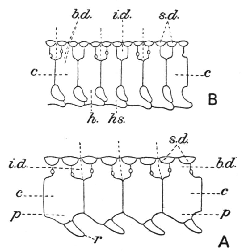
Fig. 111.—A, side view of precaudal vertebrae of Scyllium canicula; B, similar view of caudal vertebrae. b.d, Basi-dorsal; c, centrum; h, basi-ventral; h.s, haemal spine; i.d, inter-dorsal; p, parapophysis; r, rib; s.d, supra-dorsals. The vertical dotted lines indicate the limits of neuromeres and myotomes. The small circles represent the exits of the dorsal and ventral roots of spinal nerves. (After Ridewood.)
Subsequently, a number of cartilaginous pieces are developed in connexion with the dorsal and ventral surfaces of the notochord, which, as they form portions of a system of dorsal and ventral arches, are termed "arcualia" (Fig. 111). On the dorsal side there are: (i.) a series of paired basi-dorsal cartilages (neurapophyses or neural arches), the two elements of each pair contributing to form the side walls of the neural canal in which the spinal cord is lodged (Fig. 112, A); (ii.) a series of inter-dorsal cartilages (intercalary neural arches), regularly alternating with the preceding, and completing the walls of the neural canal by filling up the intervals between the basi-dorsals; and (iii.) a series of supra-dorsal elements, typically also in pairs, but in the Dog-Fish fused to form single median cartilages. Of the latter there are two sets—one the supra-basi-dorsals, or neural spines, are situated over the basi-dorsals; and the other, supra-inter-dorsals, alternating with the former, lie over the inter-dorsals, the two series forming the keystones of the dorsal arches, and thus completing the roof of the neural canal. On the ventral side of the notochord this arrangement is substantially repeated by a series of ventral arcualia, which, however, are somewhat differently arranged in the trunk and tail. Thus, in the trunk there are: (i.) a series of basi-ventral or haemal cartilages, corresponding with the basi-dorsals above, which grow out laterally into short processes, the parapophyses or transverse processes, and terminate in (ii.) short, slender cartilages—the costal elements or ribs—which may perhaps be regarded as the ventral equivalents of supra-basi-dorsals. The ribs project outwards into the dorsal wall of the coelom and end in the myocommata separating the myotomes of the body-wall. In the tail the basi-ventrals lose their ribs and, growing downwards into ventral prolongations, they unite in pairs beneath the caudal artery and vein, and so form a series of inverted arches (haemal arches) enclosing a haemal canal (Fig. 112, B). The apex of each arch is prolonged into a median process or haemal spine. Although not recognisable in the Dog-Fish, paired inter-ventral cartilages, corresponding with the inter-dorsals above, are present in some Elasmobranchs and alternate with the basi-ventrals. In the caudal region of others (e.g. Skates and Rays) ventral counterparts of the supra-interdorsals are present, and are termed infra-ventral cartilages. Much in the same way that their dorsal equivalents enclose a neural canal, so the ventral arcualia partially surround the viscera-containing coelom in the trunk; and in the tail, but more completely, the vestigial coelom of that region or the haemal canal.
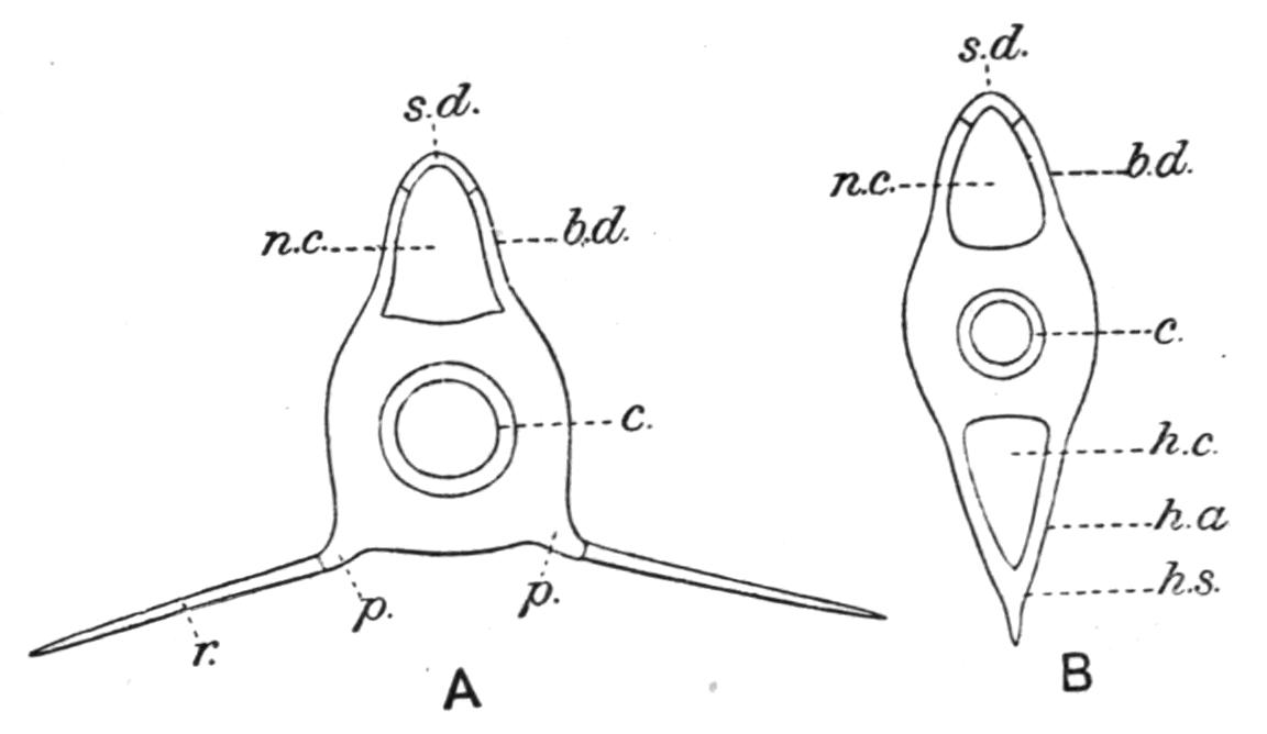
Fig. 112.—A, transverse section of a precaudal vertebra; B, similar section of a caudal vertebra. h.a, Haemal arch (basi-ventrals); h.c, haemal canal; h.s, haemal spine; n.c, neural canal. Other reference letters as in Fig. 111.
The different vertebral components are by no means of equal morphological value. The basi-dorsals and basi-ventrals, and the inter-dorsals and inter-ventrals, are the primary elements and the most important. The supra-dorsals are merely cartilages segmented off from the basi-dorsals and inter-dorsals, while the ribs and the infra-ventrals are similarly derived from the basi-ventrals and inter-ventrals respectively. As to the vertebral elements which collectively form a vertebra in the Dog-Fish, it would seem from evidence afforded by the neuromeres,[175] and more especially by the facts of development, that each complete skeletal segment or vertebra consists of a pair of basi-dorsals with the preceding pair of inter-dorsals, and of a pair of basi-ventrals with the next succeeding pair of inter-ventrals. It must be emphasised, however, that, considered as a joint or segment in a flexible back-bone, a vertebra is a physiological unit, the morphological value of which may differ widely in different Fishes. Hence, in other Fishes, the grouping of vertebral components to form individual vertebrae may be quite different to that which takes place in the Dog-Fish, and may even be accompanied by their more or less complete fusion.
In the more primitive types of vertebral column, such as are characteristic of many fossil and not a few existing Fishes, arcualia alone are present, and remain associated with a persistent notochord which has grown with the growth of the animal. In the more specialised Fishes, on the contrary, the need of an axial support for the body, which, while retaining the necessary flexibility, must possess greater strength, has resulted in the development of a series of solid cartilaginous, calcified or bony, discoidal joints or segments, the centra, which surround and more or less completely replace the notochord, and, while supporting, form also a bond of connexion between the dorsal and ventral arches. Notwithstanding their superficial resemblance, an important developmental distinction is to be noted in the mode of formation of centra in different Fishes, which enables one kind to be distinguished as "chorda-centra," and another as "arch-centra."[176] Chorda-centra are centra formed by the conversion of the chordal sheath into a series of ring-like cartilaginous segments, which subsequently, by a process of inward thickening, become biconcave, disc-like structures, and more or less completely replace the notochord, except in the spaces between them. Arch-centra, on the other hand, owe their formation to the growth of the bases of the primary arcualia round the notochord, external to the chordal sheath, and their subsequent fusion to form annular segments, which, later, become biconcave centra. Of Fishes which possess vertebral centra the Elasmobranchs alone have chorda-centra; the Holostei and the Teleostei, and very probably the Crossopterygii also, having arch-centra. The Dipnoi and the Holocephali, and the Chondrostean Teleostomi are acentrous—that is, they are devoid of vertebral centra and possess a persistent notochord. Neither in their embryonic development nor in their evolution in time are the different vertebral components synchronous in their appearance. Developmentally, the arcualia are the first to be formed, and of these those on the dorsal aspect of the notochord appear earlier than their representatives on the ventral side, while the centra are the last of all; and in a general way the palaeontological sequence agrees with the embryological.
The independent evolution of a more specialised vertebral column from a more primitive one may often be traced within the limits of the same group of Fishes when the more ancient genera are compared with the more recent. In the Elasmobranchs and the Crossopterygii, for example, the oldest known types were acentrous, while the more recent have acquired calcified or bony centra, and altogether they have reached a more advanced stage of vertebral evolution. Some Fishes, like the Chondrostei and the Dipnoi, seem, however, to exhibit comparatively little advance in vertebral structure, since both the Palaeozoic and the living representatives of these groups agree in being acentrous.
Some of the more notable features in the structure of the vertebral column in the Cyclostomata and Fishes will now be briefly considered.
In the Cyclostomata the acentrous vertebral column is more primitive than in any other Craniates, and in the Lamprey it consists of a persistent notochord, supporting a series of isolated cartilages on each side of the spinal cord.[177] As two pairs of these cartilages are included in each neuromere it is possible that they represent alternating basi-dorsals and inter-dorsals. There are no ventral arcualia in the trunk and no ribs. In the Hag-Fish (Myxine) the dorsal cartilages are restricted to the tail.
The description of the vertebral column of the Dog-Fish may be taken as fairly applicable to Elasmobranchs in general, and hence only certain notable features in some other members of the group need be referred to here. The most primitive Elasmobranchs, the Palaeozoic genera Cladoselache and Pleuracanthus were acentrous, although calcified rings have been observed in a Permian species of the latter genus and scattered calcifications in others. Some of the earlier Mesozoic genera (e.g. Hybodus) were also devoid of centra, at least in the trunk-region. The first indication of complete centra occurs in the Lower Lias Cestraciont, Palaeospinax.[178] All the later extinct, as well as all existing forms, have more or less well-developed centra, hardened by the deposit of lime salts in their primitively cartilaginous substance, but never in the form of true bone.
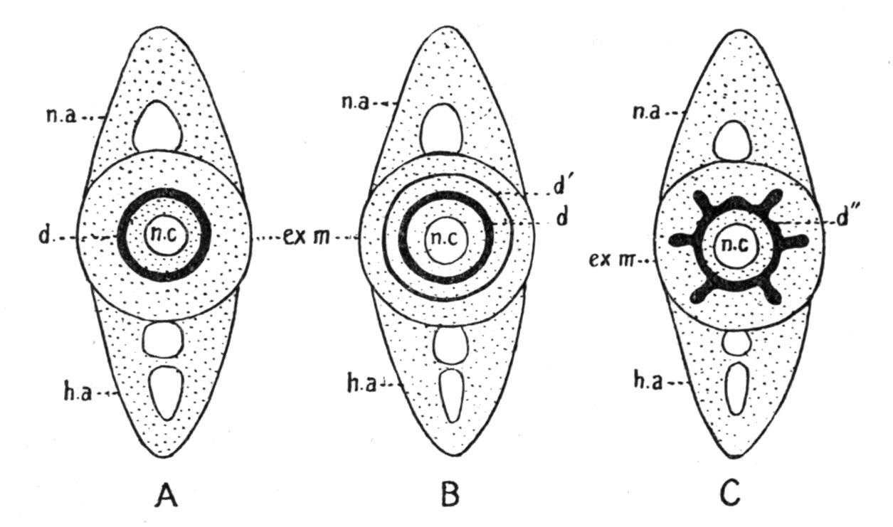
Fig. 113.—Schematic transverse section through the middle of a Cyclospondylic (A), a Tectospondylic (B), and an Asterospondylic vertebra (C). d, Middle portion of the calcified double cone; d′, additional concentric calcified layers; d″, double cone with radiating calcified layers; ex.m, external elastic membrane; h.a, haemal arch; n.a, neural arch; n.c, notochordal cavity. (From Zittel, after Hasse.)
The mode in which the lime is deposited is marked by certain peculiarities which are characteristic of particular families[179] (Fig. 113). In some genera, as in the extinct Palaeospinax and the living Acanthias and Scymnus, the calcified portion of each centrum takes the form of a cylinder constricted across the middle, like two cones joined apex to apex (cyclospondylic). This condition is probably the most primitive, but it may be modified in other genera by the further addition of calcic salts in two different ways. Thus, the deposit may take place by the simple addition of concentric layers to the original constricted cylinder (tectospondylic), as in the Skates and Rays; or it may take the form of a series of longitudinal plates radiating outwards from the cylinder, and giving rise to a star-like pattern in cross-section (asterospondylic), as in Scyllium and Lamna. In most living Elasmobranchs (e.g. Scyllium), but not in such genera as Notidanus, Heterodontus, and Squatina, the bases of the dorsal and ventral arches grow round the centra and meet, or even fuse, so that the latter become surrounded by rings of cartilage which, after a fashion, suggest incipient arch-centra (Fig. 112, A). The caudal portion of the vertebral column is often described as "diplospondylic," that is, there are two centra, two pairs of basi-dorsals, two pairs of inter-dorsals, and two pairs of basi-ventrals, or in other words, two vertebrae to each neuromere[180] (Fig. 111, B).
The Holocephali have a vertebral column essentially similar to that of other Elasmobranchs, but of a more primitive type (Fig. 114). The notochord is persistent and there are no centra; but ring-like calcifications, four or five to each neuromere, occur in the chordal sheath in Chimaera, although not in Callorhynchus. Ribs are absent. In the whip-like terminal portion of the tail the arcualia and the notochord become replaced by a slender continuous filament of cartilage.
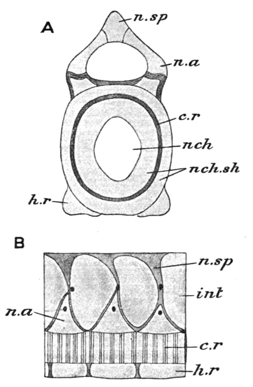
Fig. 114.—A, transverse section of the vertebral column of Chimaera monstrosa; B, lateral view, c.r, Calcified ring; h.r, basi-ventral; int, inter-dorsal; n.a, neural arch (basi-dorsal); nch, notochord; nch.sh, chordal sheath; n.sp, neural spine (supra-dorsal). (From Parker and Haswell, after Hasse.)
In the more obvious features of vertebral structure the Dipnoi[181] have much in common with the Elasmobranchs, especially with certain of the acentrous Palaeozoic representatives of that group. The notochord is persistent, centra are wanting, and the different vertebral components continue to retain their primitive distinctness. On the other hand, the basi-dorsals are much better developed than the inter-dorsals, which are either vestigial or absent. The basi-dorsals unite in pairs over the spinal cord to form complete neural arches, and each arch supports dorsally the legs of a Λ-shaped, gable-like element or neural spine, which probably represents a pair of fused supra-basidorsals. Ventrally, there are basi-ventral cartilages, fused in pairs beneath the notochord, and supporting well-developed, bone-ensheathed ribs. Inter-ventrals appear to be absent. Each neuromere corresponds with a pair of basi-ventrals, of basi-dorsals and of inter-dorsals. The haemal arches and spines are formed partly by the basi-ventrals, but mainly by the ventral union of the successive pairs of ribs. As in the Holocephali, the terminal arcualia of the tail become fused into a straight axial cartilaginous filament, transversely divided into segments, which replaces the notochord. Each segment supports a variable number of dorsal and ventral gable-pieces, or neural and haemal spines. Certain of the vertebral components, such as the ribs, and the neural and haemal spines, are ensheathed by membrane bone.
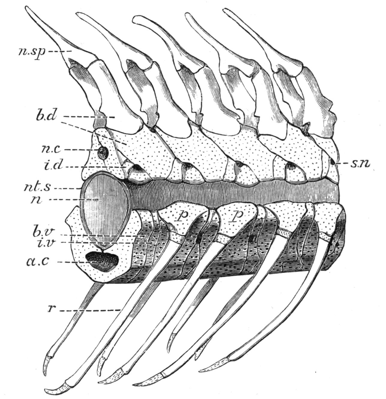
Fig. 115.—Side view of the precaudal vertebrae of a Sturgeon (Acipenser sturio). a.c, Aortic canal, formed by the median union of ingrowths from the basi-ventrals and inter-ventrals of opposite sides; b.d, basi-dorsals; b.v, basi-ventral; i.d, inter-dorsal; i.v, inter-ventral; n, notochord; n.c, neural canal; n.sp, neural spine; nt.s, cuticular sheath of the notochord; p, parapophysis; r, rib; s.n, aperture for the root of a spinal nerve.
With certain modifications in details the preceding description will also apply to the vertebral column of the Chondrostei (Fig. 115). It will be noted, however, that the inter-dorsals are much better developed than in the Dipnoi, although when compared with the basi-dorsals they take but a small share in forming the walls of the neural canal. Well-developed but somewhat fragmentary inter-ventrals are present. The haemal arches and spines are formed by the downgrowth and ventral union of the basi-ventrals as in the Dog-Fish, and apparently without the aid of costal elements. In Polyodon the ribs are vestigial,[182] but in Acipenser they are well developed. The neural arches and spines, and their haemal representatives in the tail, and also the ribs, are partially ossified, or ensheathed by bone.
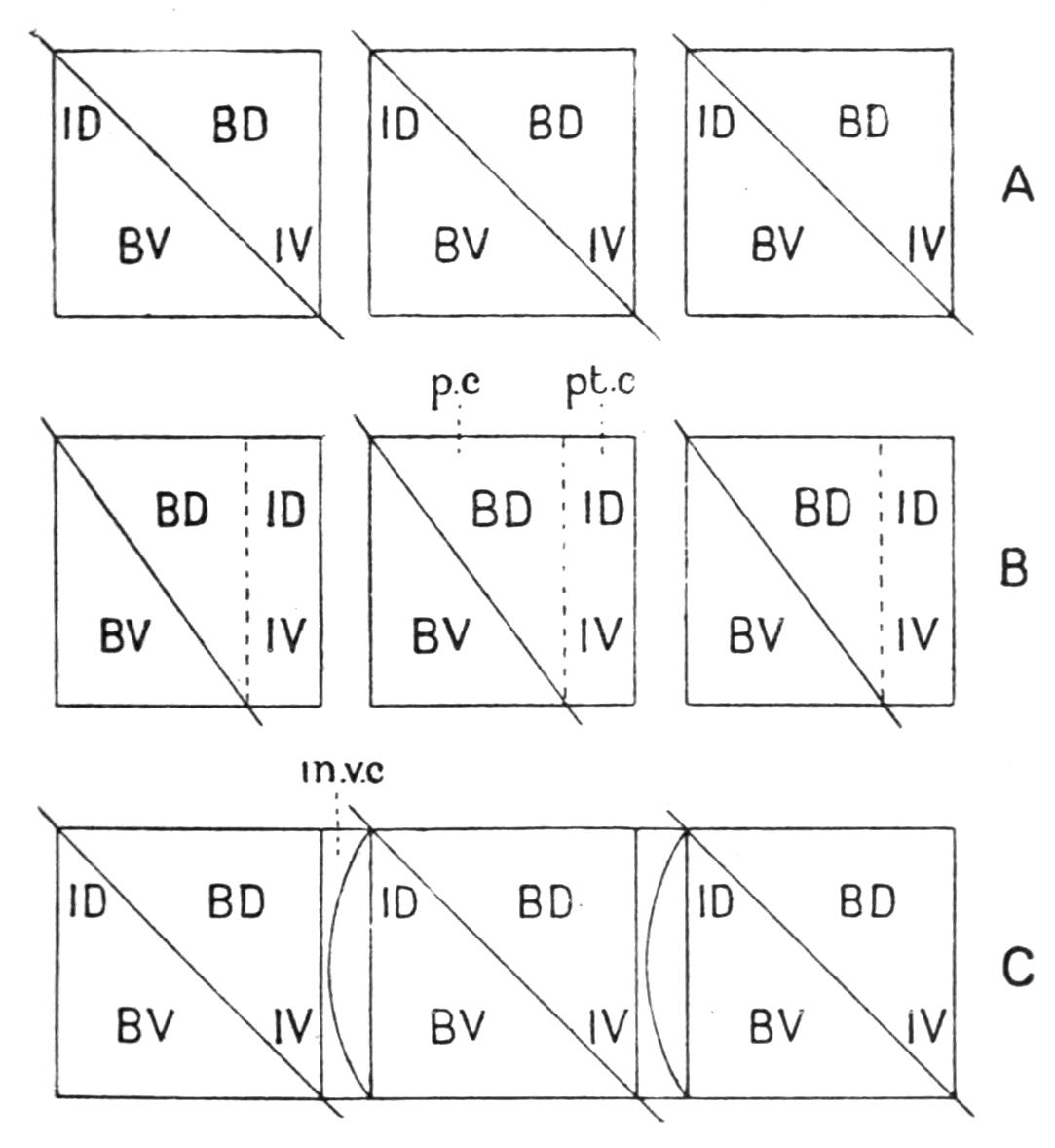
Fig. 116.—Diagram to illustrate the grouping of vertebral elements to form vertebrae, A, in an Elasmobranch, B, in Amia, and C, in Lepidosteus. B.D, Basi-dorsals; B.V, basi-ventrals; I.D, inter-dorsals; I.V, inter-ventrals; in.v.c, inter-vertebral cartilage divided by a concavo-convex cleft; p.c, precentrum; pt.c, postcentrum. The square blocks represent individual vertebrae, and the oblique lines, the attachments of the myocommata.
In the existing Crossopterygii, Holostei, and Teleostei, popularly known as the "bony Fishes," the vertebral column assumes a more familiar character, and at the same time we meet with interesting illustrations of the different methods by which the separate component vertebral elements of the more primitive types of "backbone" are concentrated together in groups, and fused to form that complex physiological product, the complete bony vertebra.[183] In most of these Fishes each vertebra is formed by the aggregation and fusion of a pair of basi-dorsals and a pair of basi-ventrals, and includes, in addition, a pair of inter-dorsals, which may either be the pair in front of the basi-dorsals or the pair behind, and also a pair of inter-ventrals, which, similarly, may be the pair in front or behind the basi-ventrals (Fig. 116). The product of this fusion is a series of bony vertebrae, each consisting of a biconcave arch-centrum, which includes the fused basal portions of a pair of basi-dorsals and a pair of basi-ventrals. The distal portions of the basi-dorsals form the neural arch, while the rib-bearing parapophyses are lateral outgrowths from the basi-ventrals which otherwise have become merged in the centrum. Finally, the centrum is completed by its fusion with a pair of inter-dorsals and a pair of inter-ventrals. Supra-dorsal elements may also be included as minor contributory factors. The supra-basi-dorsals co-ossify with their basi-dorsals and then unite to form the ordinary unpaired neural spine of most bony Fishes, or, as in Amia, they remain distinct from each other, and are obvious as a double spine. In Lepidosteus these elements co-ossify with the neural arches and form the post-zygapophyses. Supra-inter-dorsals have been identified in the embryo as distinct elements, but their eventual fate is not always known. In Lepidosteus they persist as distinct cartilages in the adult (Fig. 118, A). Well-developed bony ribs are usually present. The haemal arches of the tail are formed by the downgrowth of the parapophyses and their ribs, or by the latter alone, and by their ventral union to form haemal spines; consequently, each arch always includes a pair of costal elements. With such general features in common there are certain notable variations in some of these Fishes, to which brief reference may be made.
Little is at present known of the development of the vertebral column in either of the only two existing genera of Crossopterygii, Polypterus[184] and Calamichthys, and hence the precise mode of grouping of their vertebral components to form vertebrae is unknown. The condition of the vertebral column in the fossil forms varies greatly in different families, but in none is it so specialised as in the surviving members of the group. In the Devonian Holoptychidae, and even in genera so comparatively recent as the Upper Cretaceous Coelacanth Macropoma, the persistence of the notochord and the absence of centra indicate a very primitive grade of vertebral evolution. The Devonian and Carboniferous Rhizodontidae (e.g. Eusthenopteron and Rhizodus), on the contrary, seem to have had well-ossified ring-like vertebrae.
In the caudal region of Amia the basi-dorsals and basi-ventrals, and the inter-dorsals and inter-ventrals, form separate arch-centra which remain distinct; hence each vertebra is double, and there is a regular alternation of arch-bearing "pre-centra" and arch-less "post-centra" (Fig. 117, D). In the trunk-region the pre- and post-centra have fused, and in this region the vertebrae are single.
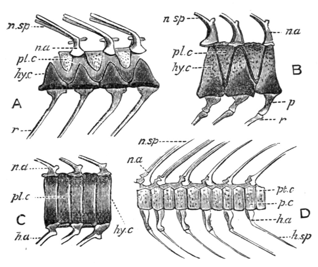
Fig. 117.—A, precaudal vertebrae of Caturus furcatus; B, similar vertebrae of Eurycormus speciosus; C, caudal vertebrae of the latter species; D, caudal vertebrae of Amia calva. h.a, Haemal arch; h.sp, haemal spine; hy.c, hypo-centrum; n.a, neural arch; n.sp, neural spine; p, parapophysis; p.c, pre-centrum; pl.c, pleuro-centrum; pt.c, post-centrum; r, rib. (After Zittel.)
A very primitive type of vertebral column occurs in some of the Jurassic allies of Amia, in which certain of the vertebral components, confluent in the adult Amia, retain some measure of their primitive distinctness.[185] Thus, in the precaudal region of Eurycormus (Fig. 117, B) there is a series of alternating dorsal and ventral half-rings of bone, which between them completely invest the persistent notochord. Each ventral half-ring or "hypocentrum" represents a pair of fused and ossified basi-ventrals, and possibly also a pair of included inter-ventrals, and supports dorsally a pair of basi-dorsals, forming a neural arch, and laterally a pair of ribs. The dorsal semi-rings, or "pleuro-centra," similarly represent fused and ossified pairs of inter-dorsals. In the tail, modifications approximating to what is seen in the caudal region of Amia are to be noticed (C). By the upgrowth of the ventral arch-bearing semi-rings, and their conversion into complete rings encircling the notochord, incipient pre-centra are formed, and by a similar modification of the down-growing, archless, dorsal half-rings, structures comparable to post-centra are produced. In brief, Eurycormus, as well as such other extinct Amioid genera as Caturus (Fig. 117, A), Callopterus, and Euthynotus, retain in the adult a stage of vertebral evolution which is closely paralleled by transitory stages in the embryonic and young forms of Amia.
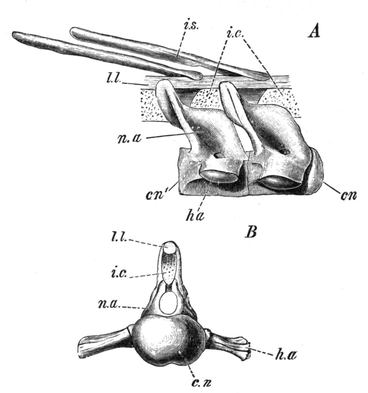
Fig. 118.—A, two vertebrae from the trunk-region of Lepidosteus; B, anterior face of a vertebra. c.n, Anterior convex face of the centrum; c.n′, posterior concave face; h.a, parapophysis, with its articular facet for a rib; i.c, median cartilage, representing a pair of fused supra-interdorsals; i.s, radial element of the dorsal fin; l.l, superior longitudinal ligament; n.a, neural arch. (From Wiedersheim, after Balfour and W. N. Parker.)
Lepidosteus[186] is unique amongst existing Fishes in having opisthocoelous vertebrae; that is, the centra are convex in front and concave behind, and therefore articulate with one another by ball-and-socket joints (Fig. 118). This condition is due to the development of a series of intervertebral rings of cartilage round the notochord. The subsequent inward growth of each of these rings leads to the constriction, and ultimately to the complete obliteration, of the notochord, much in the same way as by the growth of ordinary centra. Later, this solid mass of cartilage becomes transversely divided by a cleft which is convex anteriorly and concave behind (Fig. 116, C), and of the two portions one fuses and co-ossifies with the centrum of the vertebra in front, and the other with the one pertaining to the vertebra behind. Reference to Fig. 116 will show that the grouping of the vertebral elements to form the individual vertebrae is not the same as in Amia.
In the dominant group of existing Fishes, the Teleostei, the centra are almost invariably biconcave, although in the Eels they may be flat or even slightly convex in front. Ribs are absent in the Syngnathidae and in the Plectognathi. In addition to the usual articulation between the centra, the vertebrae often articulate with one another by means of paired processes arising from the anterior margin of each neural arch, or from the centrum at the base of the arch (pre-zygapophyses), and meeting similar processes which project either from the hinder margin of the arch of the vertebra in front, or from the adjacent portion of its centrum (post-zygapophyses). The haemal arches may have similar processes (Fig. 119). One, two, or in some Teleosts, three pairs of slender intermuscular bones radiate outwards from the centra into the myocommata (epicentrals), or from the neural arch (epineurals), or from the ribs (epipleurals).
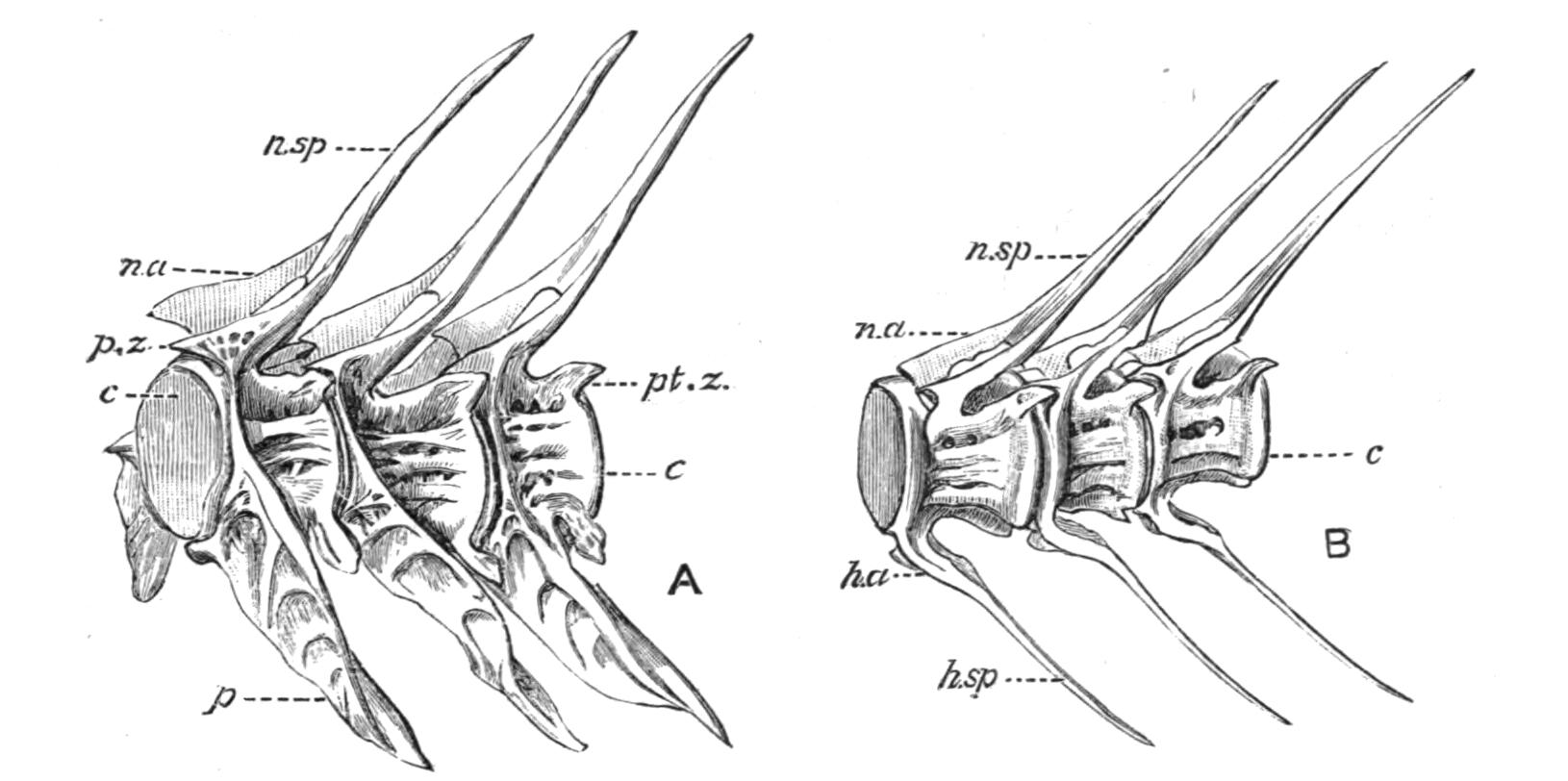
Fig. 119.—A, side view of precaudal vertebrae of a Cod (Gadus morrhua) without the ribs; B, similar view of caudal vertebrae of the same Teleost. c, Centrum; h.a, haemal arch; h.sp, haemal spine; n.a, neural arch; n.sp, neural spine; p, parapophysis; p.z, pre-zygapophysis; pt.z, post-zygapophysis.
The Ribs.—It is doubtful if the structures termed "ribs" are homologous in the different groups of Fishes. There appear to be two kinds, distinguishable as dorsal and ventral ribs (Fig. 156). Dorsal ribs are situated in the fibrous tissue separating the epiaxial from the hypaxial muscles of the body wall, and they take no part in forming the haemal arches of the caudal region. Ventral ribs, on the other hand, always lie internal to the hypaxial muscles, and directly external to the peritoneal lining of the coelom, and they usually contribute to the formation of the haemal arches. To the former belong the ribs of the Elasmobranchs, and to the latter the ribs of the Teleostomi and Dipnoi. Polypterus alone has both kinds of ribs.
The Skull.
The skull is a highly complex structure, the various components of which are as different physiologically as they are morphologically. It consists (i.) of the cranium, for the enclosure and protection of the brain; (ii.) of sense capsules, which fulfil a like function for the auditory, visual, and olfactory organs; (iii.) of certain vertebrae or vertebral elements fused with the hinder part of the cranium; (iv.) of a series of visceral arches; and (v.) of a series of paired or median cartilages developed in relation with the mouth and nostrils, which may be collectively spoken of as "labial" cartilages.
The cranium is formed in the embryo from two pairs of cartilaginous rods or plates, developed in the mesoblast of the head. Of these the posterior pair, or parachordals, underlie the hinder part of the brain, and are situated one on each side of the cranial portion of the notochord. The anterior pair or trabeculae are pre-notochordal, and lie beneath the anterior portion of the brain.[187] Between their hinder extremities, and in front of the anterior termination of the notochord, is the pituitary body. As development proceeds the parachordals blend with each other and with the trabeculae, while the latter fuse in front to form a median plate—the mesethmoid cartilage. The hinder portions of the two trabeculae remain distinct for some time, and enclose between them the pituitary fontanelle, but later they fuse beneath the pituitary body, leaving, however, a pit for its reception—the pituitary fossa. Cartilaginous capsules are formed round the cranial sense organs. The auditory or periotic capsules fuse on each side with the parachordals. The optic capsules, either fibrous or cartilaginous, remain free, and do not fuse with the adjacent trabecular region. The olfactory capsules alone are not developed independently, but are formed as lateral outgrowths from the mesethmoid plate. Later, the parachordals and trabeculae grow upwards on each side round the brain, and to a greater or less extent they meet and fuse on its dorsal surface, thus enclosing the latter organ in a cranial cavity, leaving, nevertheless, a large foramen behind (foramen magnum) through which the brain is continuous with the spinal cord. In this condition the primitive cartilaginous cranium, with its included sense-capsules, has reached a stage which is permanently retained in such Fishes as the Elasmobranchs.
The visceral arches consist of a number of pairs of curved rods of cartilage, at first simple, but subsequently segmented, and developed in the splanchnic mesoblastic walls of the oral cavity and pharynx. Each rod is connected with its fellow by a median cartilage in the floor of the pharynx, so that the whole form a series of dorsally incomplete hoops encircling the anterior portion of the alimentary canal. No doubt all the visceral arches were originally branchial arches, and were so disposed between the successive gill-clefts as to support their walls and the vascular folds or gill-lamellae to which they gave rise. In Fishes most of the arches still retain their primitive gill-supporting function, but the first or mandibular arch has become modified to form upper and lower jaws, although in the Sharks and Dog-Fishes it may lie in front of a gill-cleft and still be associated with vestigial gills. The second or hyoid arch is less removed from the condition of a branchial arch, and generally supports either a functional or a vestigial gill, but in most Fishes it has acquired the secondary function of forming a suspensorium for the attachment of the jaws to the cranium.
The skull of the common Dog-Fish, Scyllium canicula (Fig. 120),[188] may be studied as a type which in the adult remains cartilaginous, and has no secondary addition of cartilage- or membrane-bones. In this Fish the chondrocranium, or primary cartilaginous cranium, presents the appearance of a somewhat depressed oblong box, which has a complete roof, side-walls, and floor, but is open in front (anterior cranial fontanelle) and also behind (foramen magnum). The hinder, or parachordal portion of the cranium surrounds the foramen magnum, and there forms the occipital region. At the ventral margin of the foramen there are two prominences, or occipital condyles, for articulation with the first vertebra, and between them the remains of the notochord are traceable into the cranial floor.
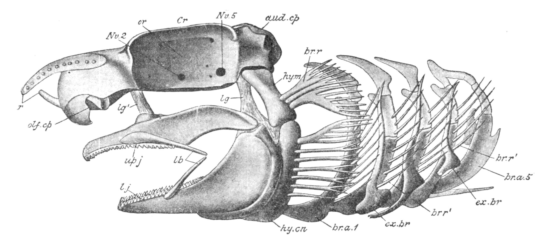
Fig. 120.—Side view of the skull of the common Dog-Fish (Scyllium canicula). aud.cp, Auditory capsule; br.a 1, 5, branchial arches; br.r, br.r′, cartilaginous rays attached to the hyoid arch and the first four branchial arches; Cr, cranium; ex.br, extra-branchial cartilages; hy.cn, cerato-hyal; hy.m, hyomandibular; lb, labial cartilages; lg, ligaments passing from the jaws to the cranium and to the distal end of the hyomandibular; lg′, ethmo-palatine ligament; l.j, lower jaw or Meckel's cartilage; Nv. 2, optic foramen; Nv. 5, foramen for the Vth and part of the VIIth cranial nerves; olf.cp, olfactory capsule; or, orbit; up.j, upper jaw or palato-quadrate cartilage. (From Wiedersheim, after W. K. Parker.)
In front of the occipital region two lateral bulgings indicate the periotic capsules, and more anteriorly still, in the trabecular region, the sides of the cranium are modified to form two spacious lateral recesses, the orbits, each of which is bounded above and below by supra-orbital and infra-orbital ridges respectively, behind by an outgrowth from the periotic capsule (post-orbital process), and in front by a similar projection from the hinder wall of the olfactory capsule (lateral ethmoidal process). In front of the cranial cavity and the orbits may be seen the laterally-placed dome-like olfactory capsules, which are open below, where the nasal sacs communicate with the exterior. Between the two capsules an anterior extension of the cranial floor forms a flattened mesethmoidal plate, behind which is the large, membrane-closed, anterior cranial fontanelle. The lateral walls of the cranium are perforated by numerous apertures, some of which serve for the entrance or exit of blood-vessels, and others, mostly pertaining to the inner walls of the orbits, for the transmission of the different cranial nerves from the brain to various parts of the head. In many Elasmobranchs the roots of certain of the anterior spinal nerves perforate the side-walls of the occipital region, and indicate the fusion of vertebral components with the cranium. In the cranial roof between the two periotic capsules there are two small apertures at the bottom of a common median depression: through each aperture the ductus endolymphaticus (aqueductus vestibuli) passes from the vestibular part of the auditory organ to the exterior of the skull.
Three cartilaginous rods, one from the roof of each olfactory capsule, and one, the prenasal or rostral process, from the ethmoid cartilage, converge and meet, or nearly meet, in front to form the rostrum or support for the preoral or "cut-water" portion of the head.
The visceral arches are seven in number. The first or mandibular arch consists on each side of an upper portion, the palato-pterygo-quadrate or palato-quadrate cartilage, which passes forwards in the side-wall of the oral cavity, along the upper margin of the mouth, its anterior or palatine part curving inwards to a ligamentous connexion with its fellow beneath the cranial floor. Each cartilage has an upwardly directed process (ethmo-palatine process) which is connected by a suspensory ethmo-palatine ligament with the lateral wall of the cranium behind the lateral ethmoid process. The lower or ventral half of the mandibular arch (Meckel's cartilage) is similar in shape to the upper; it articulates behind with the quadrate portion of the latter by a movable joint, and is thence prolonged forwards and downwards in relation with the lower margin of the mouth to a median ligamentous union with its fellow of the opposite side. The palato-pterygo-quadrate and Meckel's cartilages together form the primitive upper and lower jaws, and support the teeth. The hyoid arch also consists of a dorsal and a ventral half on each side. The dorsal half or hyomandibular element articulates above with the periotic capsule. The ventral portion, or cerato-hyal, passes downwards and is connected with its fellow by a median copula or basi-hyal cartilage situated in the floor of the oral cavity. A series of simple cartilaginous rays (branchial rays) are attached to the hinder margins of the hyomandibular and cerato-hyal elements. The distal end of the hyomandibular is connected by strong ligaments with the hinder portions of both the palato-pterygo-quadrate cartilage and Meckel's cartilage; in fact, the hyomandibular is the effective suspensorium by which the upper and lower jaws are connected with the skull, and all Fishes in which this arrangement exists are said to be hyostylic.[189] Behind the hyoid arch follow five branchial arches. Each of these is segmented into a dorsal or pharyngo-branchial element, followed by an epi-, a cerato-, and a hypo-branchial piece, but the later element is absent in the fifth arch. The lateral halves of the last three arches are connected ventrally by a large median basi-branchial cartilage, but in the first and second arches by the median apposition of their respective hypo-branchial elements. Like the hyomandibular and cerato-hyal segments of the hyoid arch, the epi- and cerato-branchial elements of all the branchial arches except the fifth are fringed along their outer convex margins by a series of branchial rays, and, in addition, there are three pairs of slender, curved, cartilaginous rods, or extra-branchials, in relation with the distal extremities of the branchial rays of the second, third, and fourth branchial arches. The function of the branchial arches, and their branchial rays, and extra-branchial cartilages, is to support the inter-branchial septa which separate the gill-clefts and carry the vascular gill lamellae. All the arches lie near the inner margins of the septa, close to the hypoblastic epithelium of the pharynx, while the outer portions of the septa are supported by the branchial rays and the extra-branchials, the latter lying directly beneath the external skin. The segments of the arches are movably connected with one another by ligaments; and by the contraction of the branchial muscles the arches may be separated or approximated so as to enlarge or diminish the size of the intervening clefts.
The labial cartilages are represented by a pair of slender rods in relation with the outer surfaces of the palato-pterygo-quadrate cartilages, and a similar pair in connexion with the Meckelian cartilages. There is also a pair of small cartilages in relation with the nostrils. It is probable that the rods which constitute the lateral elements of the rostrum belong to the same category.
In the Cyclostomes and the Elasmobranchs the skull is entirely cartilaginous, although it may often be superficially calcified in Elasmobranchs, and although there may even be definitely and symmetrically arranged calcified plates in Pleuracanthus, true bone is never present. In many Fishes, and notably in the Teleostomi, the embryonic cartilaginous cranium becomes complicated by the addition of an extensive series of investing membrane bones, formed by the ossification of the connective tissue external to the cartilage, so that a secondary bony cranium is formed external to the primary cranium much in the same way that a secondary pectoral girdle is formed in connexion with the primary girdle. Such bones probably owe their primary origin to the fusion and insinking of exoskeletal structures (scales or dermal spines). To these investing bones there may also be added a series of bones formed by the actual conversion of the cranial cartilage into osseous tissue (cartilage bones), which to a greater or less extent in different Fishes replaces the original cartilage. The bones of the skull may conveniently be classified as follows:—(i.) Dermal or membrane bones. Under this head are included—(a) the ordinary investing bones of the skull. (b) Tooth-bones, that is, bones formed by the fusion of the bases of teeth and developed in relation with the walls of the oral cavity. Probably all tooth-bearing bones are of this nature. (c) Sensory canal bones, that is, tubular bones developed round the sensory canals of the head. Certain of these bones may secondarily acquire the shape and character of investing bones while still retaining protective relations to their sensory canals. (ii.) Cartilage bones.
As an easily obtainable example of a skull which has acquired a fairly complete series of both cartilage- and membrane-bones, while retaining a well-developed primary cranium, the skull of the Salmon (Salmo salar) may be described.[190] At an early stage of development, even so late as the second week of hatching, the primary cranium is still entirely cartilaginous, and in this condition the Salmon's skull is comparable with that of an adult Dog-Fish. As development proceeds the primary cranium becomes supplemented by the addition of numerous investing dermal bones which form the secondary cranium, and later cartilage bones appear and, to a considerable extent, replace the original cartilage. The Salmon's skull is interesting in this respect, that the primary cranium grows with the growth of the Fish, so that in the adult the nasal, ethmoidal, and prenasal regions are entirely cartilaginous, and in the hinder part of the cranium cartilage is largely persistent between the cartilage bones.
Dealing first with the cartilage bones of the primary cranium, it may be stated that there are formed in that part of the parachordal cartilage surrounding the foramen magnum a median basioccipital below, which is concave behind where it articulates with the centrum of the first vertebra, a supraoccipital above, and two laterally-placed exoccipital bones (Figs. 121, 122). Each periotic capsule is ossified by the formation of five bones in the primitively cartilaginous mass, the prootic, sphenotic, opisthotic, epiotic, and the pterotic. The inner walls of the capsules have atrophied in the adult, and hence the cavities which contain the auditory organs appear as open lateral recesses of the cranial cavity. In front of the periotic capsules there are various bones which are formed in the cartilage of the trabecular part of the cranium. Thus, in front of the basi-occipital, and developed in the cartilage of the cranial floor, there is a median Y-shaped basisphenoid, and, at some distance above it on each side, an alisphenoid helps to form the lateral wall of the cranial cavity. Between the eyes the side walls of the cranium fuse to form a vertical inter-orbital septum, and, in consequence, two orbito-sphenoid bones, which normally form the lateral cranial walls in this region, become partially confluent in the median line and close the cranial cavity in front. The only cartilage bones found in the massive persistent portion of the primary cranium which forms the pre-orbital region are the projecting lateral ethmoids, forming the posterior boundaries of the recesses for the olfactory organs, and separating the latter from the orbits.
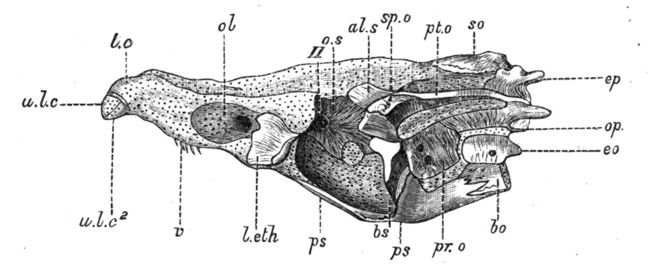
Fig. 121.—Side view of the cranium of a Salmon (Salmo salar). Most of the membrane bones and the jaws have been removed. The cartilage is dotted. al.s, Alisphenoid; bo, basioccipital; bs, basisphenoid; eo, exoccipital; ep, epiotic; l.eth, lateral ethmoid; ol, olfactory capsula; op, opisthotic; o.s, orbito-sphenoid; pr.o, prootic; ps, parasphenoid; pt.o, pterotic; so, supraoccipital; sp.o, sphenotic; t.c, trabecular cornu; u.l.c, u.l.c2, first and second upper labial cartilages; v, vomer; II, foramen for the optic nerve. (From W. K. Parker.)
The roof and floor of the primary cranium is completed by certain investing dermal bones (Fig. 123, A). A pair of large frontal bones form the cranial roof, and also help to roof in the orbital cavities. Behind the frontals, and separated from each other by the supraoccipital, there is a pair of small parietals, and anterior to the frontals a median dermal mesethmoid. A small nasal bone overlies each olfactory recess. Ventrally, the base of the cranium, from the basi-occipital to the prenasal region, is strengthened by a large parasphenoid behind, and a much smaller vomer in front, both of which lie in the roof of the mouth. The vomer is a tooth-bone, and probably the parasphenoid also.
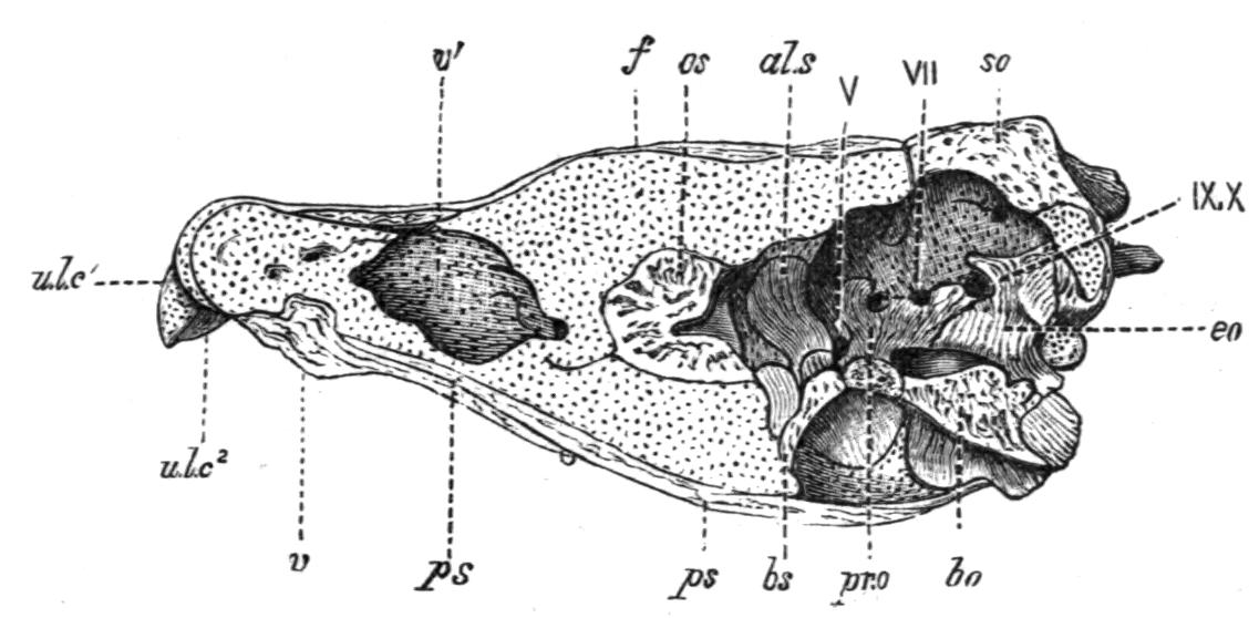
Fig. 122.—Vertical and longitudinal section of the cranium of Salmo salar, showing the right half of the cranial cavity. Cartilage is dotted. f, Frontal; v′, fat-containing cavity in the mesethmoid cartilage; V, VII, IX, X, foramina for the fifth, seventh, ninth, and tenth cranial nerves. Remaining reference-letters as in Fig. 121. (From W. K. Parker.)
The mandibular arch (Fig. 123, B) is more modified than that of the Dog-Fish. The palato-pterygo-quadrate bars, or primitive upper jaw, no longer meet in front beneath the cranial floor, but each separately articulates in front with the lateral ethmoid of its side. Although still partly cartilaginous each bar is largely replaced either by cartilage bones, or by bones which begin as membrane bones or as tooth-bones and complete their growth by invading the cartilage and becoming in part cartilage bones. Its anterior portion is formed by a palatine bone which articulates with the lateral ethmoid, and the middle portion by a pterygoid and a mesopterygoid bone, while the hinder part is ossified above as a metapterygoid and below as a quadrate. The latter articulates with the lower jaw. Functionally, however, the primitive upper jaw is now replaced by a secondary upper jaw, formed on each side by a series of tooth-bones, situated external to the former, and meeting in front of the prenasal region of the primary cranium (Fig. 123, A). The series includes a dentigerous premaxilla and maxilla, and a small toothless, scale-like jugal bone. Each half of the lower jaw (Fig. 123, A, B) consists of a rod-like Meckel's cartilage or primary lower jaw.
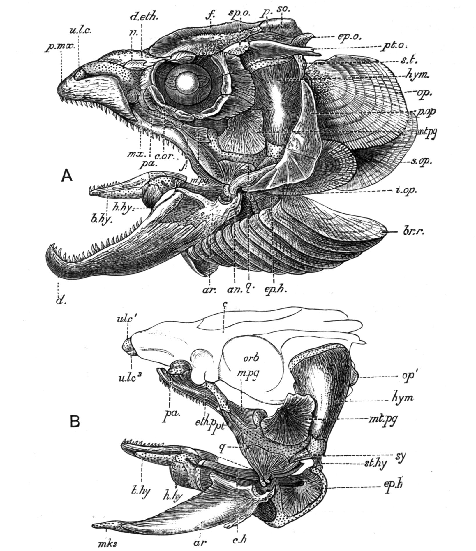
Fig. 123.—A, view of the left side of the skull of a Salmon; B, the left half of the primary upper and lower jaws, and the hyoid arch. The cartilage is dotted. an, Angular; ar, articular; b.hy, basi-hyal; br.r, branchiostegal rays; c, cranium; c.h, cerato-hyal; c.or, circum-orbital bones; d, dentary; d.eth, dermal mesethmoid; ep.h, epihyal; ep.o, epiotic; eth.p, ethmo-palatine process; f, frontal; h.hy, hypo-hyal; hym, hyomandibular; i.op, inter-operculum; j, jugal; mks, Meckel's cartilage; mpg, mesopterygoid; mt.pg, metapterygoid; mx, maxilla; n, nasal; op, operculum; op′, condyle on the hyomandibular for the operculum; orb, orbit; p, parietal; pa, palatine; p.mx, premaxilla; p.op, pre-operculum; pt, pterygoid; pt.o, pterotic; q, quadrate; so, supra-occipital; s.op, suboperculum; sp.o, sphenotic; s.t, supra-temporal (or squamosal); st.hy, stylo-hyal; sy, symplectic; u.l.c, u.l.c′, upper labial cartilages; u.l.c2, second upper labial. (From W. K. Parker.)
The hinder part of this is ossified to form the articular, which has a deeply concave surface for articulation with the quadrate; and below this there is a small membrane bone, the angular. The rest of the cartilage is partially ensheathed on its outer side by a large tooth-bone, the dentigerous dentary. The hyoid arch is similar to that of the Dog-Fish, except that its primitively cartilaginous segments are almost completely ossified (Fig. 123, B). The large upper segment or hyomandibular bone articulates mainly with the pterotic, but partly also with the sphenotic element of the periotic capsule; below it is connected with a slender symplectic bone, and from the cartilage connecting the two depends the rest of the hyoid arch, consisting in succession of stylo-hyal, epi-hyal, cerato-hyal, and hypo-hyal bones, with a median teeth-bearing basi-hyal. The palato-pterygo-quadrate bar has no direct connexion with the skull, except anteriorly where its palatine element articulates with the lateral ethmoid. The real suspensorium is formed by the hyomandibular and symplectic bones, to which the hinder margins of the quadrate and metapterygoid bones are rigidly attached by suture, hence, as in the Dog-Fish, the skull is hyostylic. Behind the hyoid arch there are five branchial arches, which generally resemble those of the Dog-Fish, except that their component segments are ossified as cartilage bones.
Connected with the hyomandibular and cerato-hyal elements of the hyoid arch there is, on each side, a series of membrane bones for the support of the movable operculum or gill-cover. These consist of an operculum above, which articulates with a backwardly projecting process from the hyomandibular, followed in succession below by a sub-operculum and an inter-operculum, the latter being connected by ligament with the angle of the lower jaw. The series is completed by ten sabre-shaped branchio-stegal rays, which are attached to the cerato-hyal and support the lower margin of the gill-cover.
Sensory canal bones are represented in the Salmon by a ring of small bony plates which encircle the orbit (Fig. 123, A), and by one or two small bones situated above and on the outer side of each periotic capsule (squamosals). To these may be added the pre-operculum situated external to the hinder margins of the hyomandibular and quadrate bones, firmly clamping these bones together, and also the post-temporals, by which the secondary pectoral girdle is attached to the skull. The nasal bones may also be regarded as pertaining to the same series.
In other Fishes with a more or less complete bony skull there are certain additional cartilage- and membrane-bones which are not present in the Salmon. There is usually a median ossification of the ethmoid cartilage, the mesethmoid. An entopterygoid is sometimes added to the palato-pterygo-quadrate series of bones. An ossification of the anterior extremity of each Meckelian cartilage may form a mento-Meckelian bone. Certain additional membrane bones are sometimes developed in relation with the lower jaw, such as splenial and coronary bones on the inner side, and a supra-angular bone at the angle of the jaw, above the angular element. To these there may be added the singular series of infra-dentaries, which in some fossil Crossopterygii (e.g. Rhizodopsis) fringe the outer margin of the jaw beneath the true dentary (Fig. 274, A). A system of jugular plates may also form a characteristic armature for the throat between the lateral halves of the lower jaw (Fig. 274, C). Besides those already mentioned, additional sensory canal bones are present in some Fishes. A transverse row of plates (supra-temporals) sometimes crosses the occipital region behind the parietals. There are also other canal-ossicles which lose their identity by fusing with certain cranial or periotic bones. Thus, each of the pterotic and sphenotic bones often includes a superficial dermal bone transmitting a section of a sensory canal, which has fused with it; and as the frontal bone is often similarly perforated, it may be taken that it also includes a canal-ossicle; and the same can often be said of the articular and dentary bones of the lower jaw.[191]
Having now considered the general structure of a primitive cartilaginous type of skull, and the nature, disposition, and terminology of the various membrane- and cartilage-bones which may be added to, or more or less completely replace the former, reference will now be made to the more important features in the structure of the skull in the Cyclostomata and the Fishes.
In the Cyclostomata the skull presents a remarkable combination of characters, in some of which it is more primitive than in any other Craniates, while in others it has evidently attained a very high degree of specialisation on lines peculiar to the group, but differing in the two subdivisions.
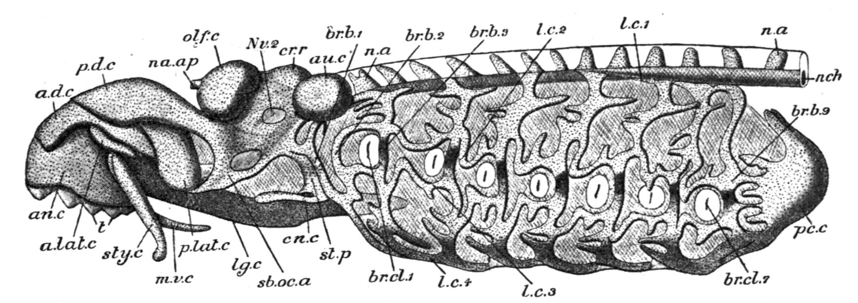
Fig. 124.—Skull, with branchial basket and anterior part of the vertebral column, of Petromyzon marinus. a.d.c, Anterior dorsal cartilage; a.lat.c, anterior lateral cartilage; an.c, annular cartilage; au.c, auditory capsule; br.b.1-9, vertical bars of the branchial basket; br.cl.1-7, external branchial clefts; cn.c, cornual cartilage; cr.r, cranial roof; l.c.1-4, longitudinal bars of branchial basket; lg.c, lingual cartilage; m.v.c, median ventral cartilage; n.a, neural arches; na.ap, nasal aperture; n.ch, notochord; Nv2, foramen for optic nerve; olf.c, olfactory capsule; pc.c, cartilage surrounding pericardial cavity; p.d.c, posterior dorsal cartilage; p.lat.c, posterior lateral cartilage; sb.oc.a, subocular arch; st.p, styloid process; sty.c, styliform cartilage; t, teeth. (From Parker and Haswell, after W. K. Parker.)
In the Lamprey[192] (Fig. 124) the paired parachordals and trabeculae together form a trough-like chondrocranium, which has only a fibrous roof, except where a slender synotic band of cartilage extends between the two periotic capsules. The floor is also incomplete, a large pituitary fontanelle remaining to indicate the original separation of the trabeculae while transmitting the hypophysial or pituitary caecum. The notochord traverses the floor of the parachordal portion of the cranium as far as the pituitary fontanelle, and from the sides of the synotic ring the auditory capsules project in the shape of conspicuous lateral prominences. In front the otherwise open end of the cranial cavity is closed by the dorsally-placed and unpaired olfactory capsule, which is perforated behind by two apertures for the olfactory nerves, and has only a fibrous connexion with the cranial walls. Anteriorly to the olfactory capsule the cranial floor is prolonged forwards over the roof of the mouth as a large laterally-expanded plate, formed by the united anterior portions of the trabeculae, and no doubt representing the mesethmoid cartilage of the Dog-Fish. So far the cranium presents no special difficulty, and in its general features may be readily compared with that of an embryonic Elasmobranch. As for the rest of the skull, it is obvious that it has been greatly modified, partly to form and to support the skeletal framework of the remarkable suctorial buccal funnel, and partly to form the singular rasping lingual apparatus. Hence it is always difficult and sometimes impossible to identify with certainty the component parts as being represented in other Craniates. On each side of the cranium, beneath the eye, there is a characteristic V-shaped subocular arch. Of its two legs the hinder one is continuous above with the periotic region of the cranium, and the other with the anterior trabecular region, while the pointed apex is directed obliquely downward and forward. From the hinder margin of the posterior limb a slender styloid process passes downward in the side wall of the pharynx, and terminates below in a forwardly directed cornual cartilage. A velum, fringed along its free margin with a series of tentacles, projects forwards into the oral cavity from between the oral apertures of the oesophagus and the branchial canal, and probably serves to prevent the entrance of foreign particles to the gill-sacs. This valve-like velum is supported by a velar skeleton, consisting of two lateral cartilages which are prolonged into the tentacles, and extend transversely between the inner surfaces of the two styloid processes. The apex of each subocular arch is connected with a small and somewhat triangular cartilage (postero-lateral cartilage), which is directed upward and forward, and lies in the side wall of the oral cavity. With some degree of probability the subocular arch may be compared to the palato-quadrate cartilage of a skull which has become "autostylic" in order to form a rigid support for the skeleton of the buccal funnel; the styloid processes and cornual cartilages to the hyoid arch; while the relations of the posterior lateral cartilages to the subocular arches suggest that they may possibly be regarded as Meckelian cartilages which have lost their primitive function of forming biting jaws. In the median line below, and projecting backward for some distance beneath the branchial canal, there is a long and stout lingual cartilage, carrying a small median and a still smaller pair of lateral cartilages at its anterior extremity, where it supports the lingual teeth and projects into the buccal funnel beneath the mouth. In front of the lingual cartilage, and connected by fibrous tissue with the inferior and hinder margin of the annular cartilage, there is a median T-shaped element, the median ventral cartilage. It has been conjectured that the lingual cartilage is a free basi-hyal element, and the median ventral cartilage the equivalent, elsewhere unknown, of the corresponding element of the mandibular arch.[193]
The remaining anterior skull elements are principally skeletal supports for the roof and walls of the buccal funnel. The roof is supported by an extended anterior dorsal cartilage, which is overlapped behind by the ethmoid cartilage, while the circular margin of the funnel is strengthened by a large ring-like annular cartilage. On each side of the latter there is a slender, rod-like, styloid cartilage, and above the latter a small anterior lateral cartilage. All these cartilages are usually termed labial cartilages, and it is at least possible that they possess representatives in the similarly named structures of the Dog-Fish and the larvae of some of the tailless Amphibia. It must not be forgotten, however, that the annular cartilage bears some resemblance to the ring of cartilage which encircles the lips of the buccal cavity in Amphioxus.
The complex supporting skeleton of the gill-sacs forms a basket-like structure. It consists on each side of nine unsegmented, irregularly curved, and slightly branched cartilaginous rods, situated in the outer margins of the inter-branchial septa, directly internal to the skin. The first lies directly behind the styloid process (hyoid arch), the second and third in front of and behind the first gill-sac, and of the remainder one lies just behind each of the six succeeding gill-openings; above and below each gill-aperture the rods are connected by longitudinal bars, and also in the median ventral line by a pair of similar partially united bars. The dorsal ends of the rods are also connected on each side by another longitudinal bar, which runs alongside the notochord and in front blends with the chondrocranium. The rods forming the last pair are continuous with a cup-like cartilage, supporting the lateral and hinder walls of the pericardium.
This singular branchial basket undoubtedly bears a superficial resemblance to the branchial arches of Fishes, but in any comparison of the two structures it is well to bear in mind that the branchial rods of the Lamprey are situated along the outer edges of the inter-branchial septa, and are therefore external to the gill-sacs and branchial arteries, and further, that they are developed in the somatic mesoblast of the embryonic protovertebrae, whereas true branchial arches are situated at the inner margins of the septa, internal to the gill-clefts and branchial arteries, and have their origin from the splanchnic layer of the mesoblast. So far as their position is concerned, the rods agree rather with the extra-branchial cartilages of an Elasmobranch than with the more deeply-seated branchial arches.
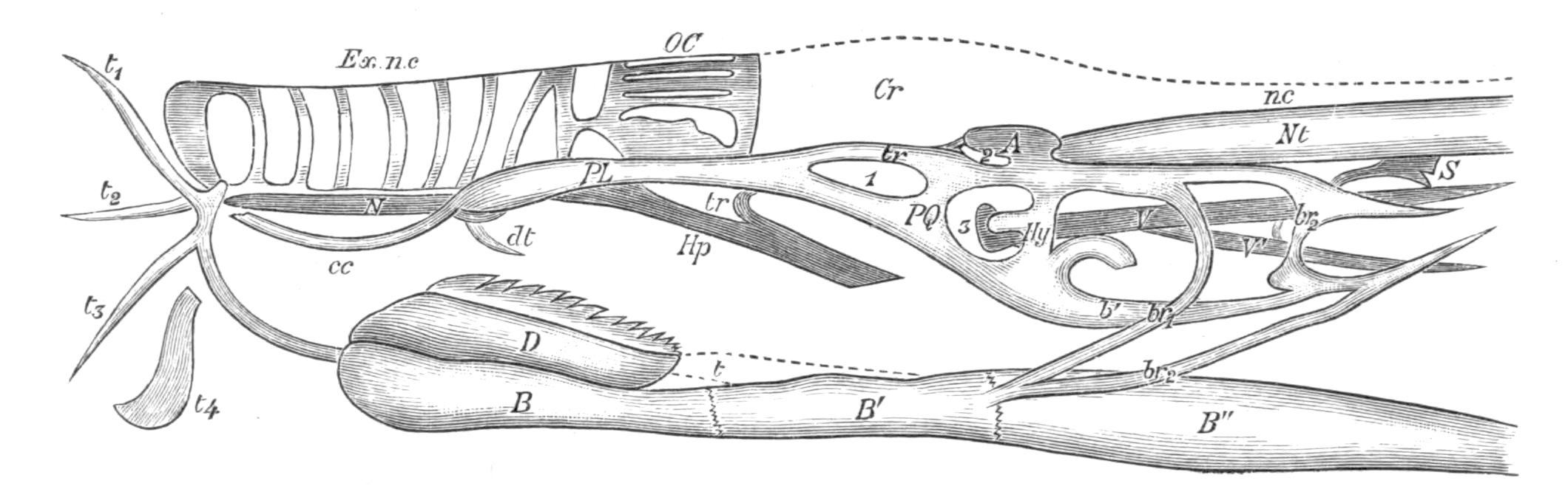
Fig. 125.—Side view of the skull of Bdellostoma; the gill-apertures and their cartilages have been omitted. A, Auditory capsule; B, B′, B″, the anterior, middle, and posterior segments of the lingual bar; b1, cartilage connecting the hyoid arch with the second branchial arch; br1, br2, first and second branchial arches; c.c, coronal cartilage; Cr, cranium; D, dental plate; dt, median dorsal tooth; Ex.n.c, external part of the naso-pituitary canal; Hp, hypophysial plate; Hy, hyoid arch; N, subnasal cartilage; nc, neural canal; Nt, notochord; OC, olfactory capsule; PL, palatine portion of the palato-quadrate cartilage PQ; S, supra-pharyngeal plate supporting the velum; t, tendon of the retractor mandibuli muscle; t1, t2, t3, tentacular cartilages; t4, cartilage supporting mouth lobe; tr, trabecula; V1, rod connecting S with the inner surface of the hyoid arch of its side; V, outer lateral rod which joins V1; 1, 2, 3, fenestrae. (Modified from Ayers and Jackson.)
While the skull of the Myxinoid Cyclostomes[194] is constructed on the same general lines as that of the Lamprey, it is in some respects more primitive. It is also clear that in other features the skull has undergone marked specialisation on lines of its own, and in some points again it seems to deviate less from the more normal Craniate type. Of the more obvious differences, as illustrated by the skull of Bdellostoma (Figs. 125-127), it will be sufficient here to mention the following: (i.) The more primitive condition of the chondrocranium, the roof and side walls of the cranial cavity being entirely membranous. (ii.) The non-development of a suctorial buccal funnel and the presence of oral tentacles, associated with the absence of the complex system of labial cartilages and the substitution of a special tentacular skeleton. (iii.) The special modifications induced by the length and physiological importance of the naso-pituitary canal and by its communication with the pharynx after perforating the pituitary fontanelle in the cranial floor.
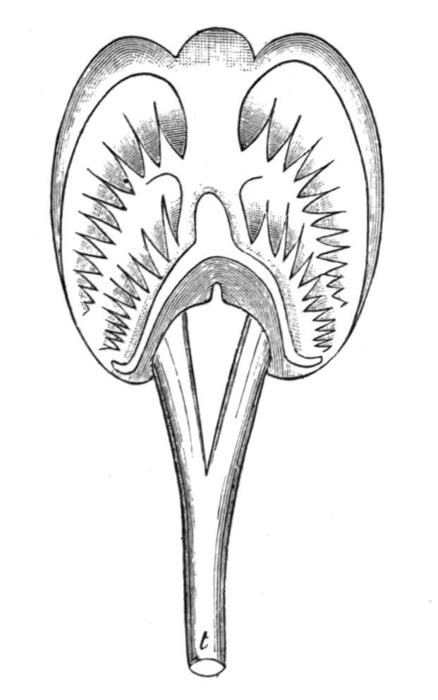
Fig. 126.—View of the upper surface of the dental plate of Bdellostoma. t, Tendon of retractor muscle. (From Ayers and Jackson.)
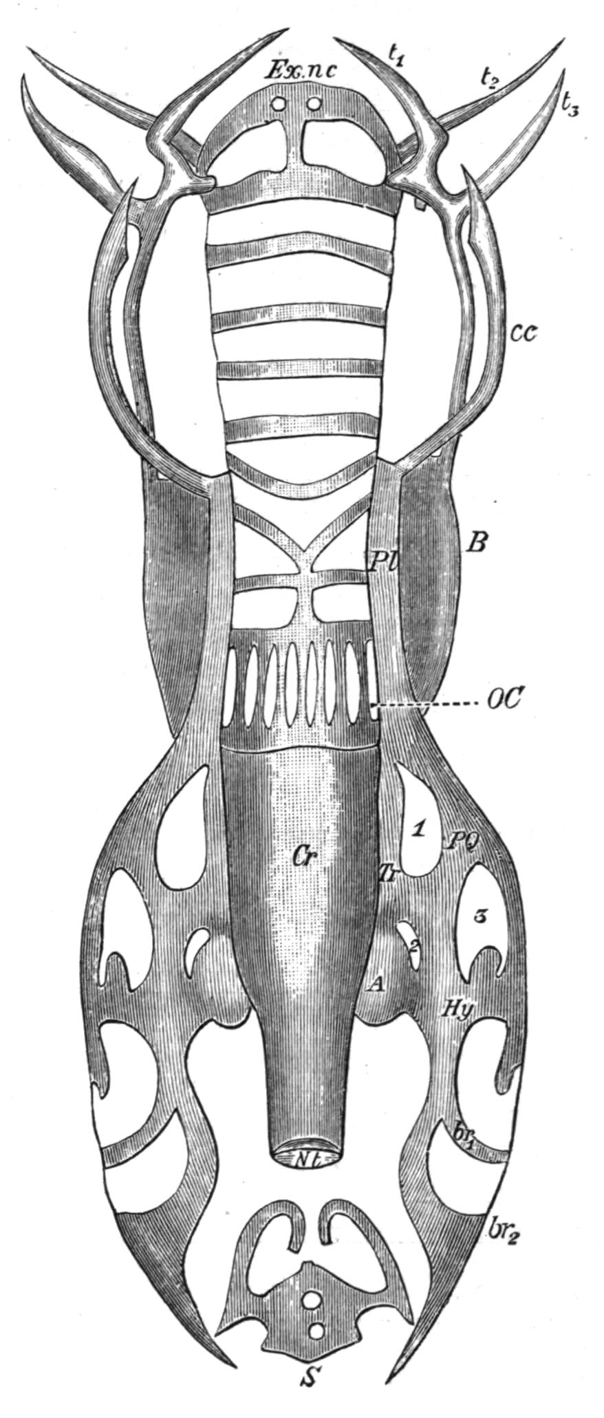
Fig. 127.—Dorsal view of the skull of Bdellostoma. Reference letters as in Fig. 125. (After Ayers and Jackson.)
Under this head may be included the depression of the mesethmoid or hypophysial plate for the support of the naso-pituitary canal, the forward prolongation and median union of the palato-quadrate cartilages of opposite sides beneath the external portion of the canal, apparently for the support of the latter, and the encircling of the canal by supporting annular rings of cartilage. (iv.) The presence of two branchial arches, connected, as in Fishes, with a median basi-branchial segment which forms the middle one of the three divisions of the lingual apparatus. (v.) The reduction of the complicated extra-branchial basket to small isolated cartilages in relation with the gill-apertures and the œsophago-cutaneous duct. (vi.) The extraordinary development of the lingual apparatus, of which it has been remarked that it "dominates the whole body, everything else yields to it." Meckel's cartilages are represented either by the cornual cartilages, as seems most probable, or by the dental plate (Fig. 125, c.c. and D).
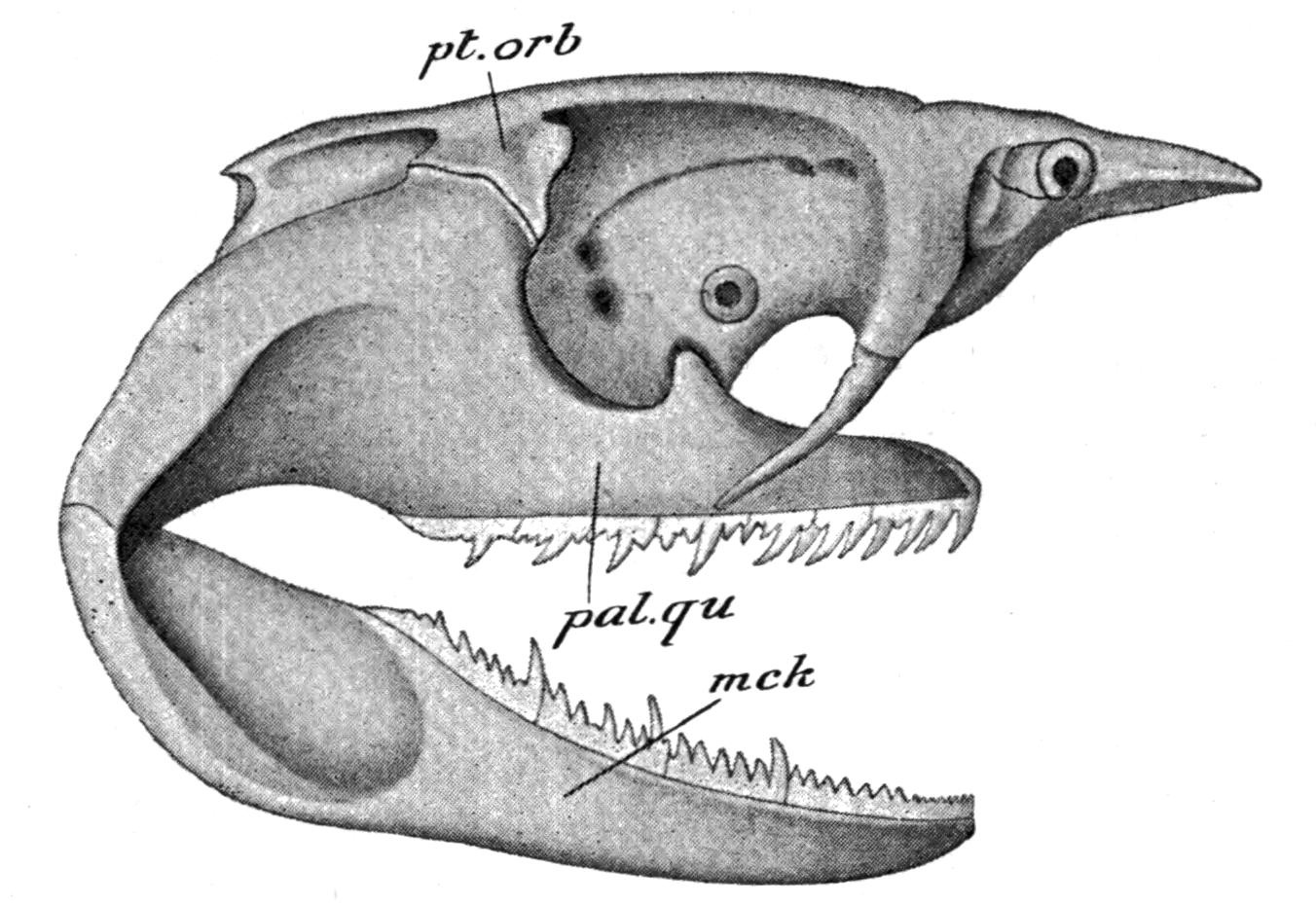
Fig. 128.—Lateral view of the skull of Notidanus (Heptanchus) cinereus; mck, Meckel's cartilage, or primitive lower jaw; pal.qu, palato-quadrate cartilage or primitive upper jaw; pt.orb, post-orbital process of the cranium with which the post-orbital process of the palato-quadrate articulates. (From Parker and Haswell, after Gegenbaur.)
In the generality of Elasmobranchs the skull resembles that of the Dog-Fish in essential structure. The more important modifications within the limits of the group relate to differences in the mode of attachment of the primitive upper jaw to the skull, and the number of branchial arches. In most Elasmobranchs the skull is hyostylic, as in Scyllium, but there are two genera which, in different ways, are exceptions to this rule. In Notidanus the hinder part of each palato-quadrate cartilage grows upwards into a strong post-orbital process, which articulates with the suitably modified post-orbital process of the periotic capsule (Fig. 128); hence the primitive upper jaw acquires a direct dorsal connexion with the cranium, and, as the hyoid arch is now relieved from taking any part in its support, the hyomandibular is reduced to the condition of a relatively slender rod of cartilage. By this arrangement both the mandibular and hyoid arches have their own separate and independent connexions with the cranium, and the skull is said to be amphistylic.[195] The Port Jackson Shark (Heterodontus) exhibits another and quite different modification. In this Fish the dorsal border of each palato-quadrate cartilage fits into a deep groove along the infero-lateral surface of the cranium, and is firmly attached thereto by ligament. Thus the first step is taken towards that more complete fusion of the two structures which is so characteristic a feature in the more typically autostylic Fishes like the Holocephali and the Dipnoi. Autostylism, whether incipient, as in Heterodontus, or complete, is to be regarded as a secondary modification, which may be independently acquired in widely different groups of Fishes, and is usually associated with the need of a firm and rigid support for an exceptionally massive dentition.[196]
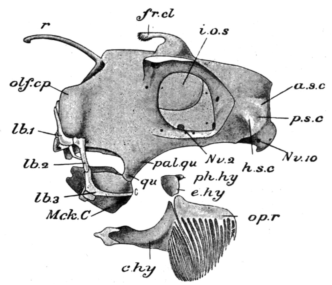
Fig. 129.—Lateral view of skull of Chimaera monstrosa. a.s.c, Position of anterior semicircular canal; c.hy, cerato-hyal; e.hy, epi-hyal; fr.cl, frontal clasper; h.s.c, position of horizontal semicircular canal; i.o.s, inter-orbital septum; lb.1, lb.2, lb.3, labial cartilages; Mck.C, mandible; Nv.2, optic foramen; Nv.10, vagus foramen; olf.cp, olfactory capsule; op.r, opercular rays; pal.qu, palato-quadrate; ph.hy, pharyngo-hyal, or hyomandibular; p.s.c, position of posterior semicircular canal; qu, quadrate region; r, rostrum. (From Parker and Haswell, after Hubrecht.)
In the Holocephali (e.g. Chimaera[197]) the cranium retains its primitively cartilaginous condition, and assumes a somewhat peculiar appearance owing to the lateral compression and vertical growth of its inter-orbital and nasal regions (Fig. 129). There is a complicated series of labial cartilages in relation with the ventrally-placed nostrils and the upper and lower jaws. In the males of Chimaera and Callorhynchus, but not in Harriotta, a movable cartilage is attached to the cranial roof, and supports the frontal clasper. The skull is typically autostylic. Along the whole length of its dorsal border the palato-quadrate cartilage is fused with the inferior lateral margin of the cranium from the periotic to the olfactory region, thus forming a triangular plate of cartilage, the base of which is continuous with the cranium, while the downwardly directed apex provides an articular surface for the lower jaw. The hyoid arch is little better developed than the succeeding branchial arches, and includes a vestigial hyomandibular, an epi-hyal, and a cerato-hyal. As in other autostylic skulls the hyomandibular element is attached by ligament to the hinder margin of the palato-quadrate, instead of being directly connected with the periotic capsule, and obviously takes no part in supporting the jaws. Branchial rays for the support of the operculum are attached to the cerato-hyal, and some of them have their bases fused together. The five branchial arches resemble those of the Dog-Fish, except that they tend to become concentrated beneath the skull.
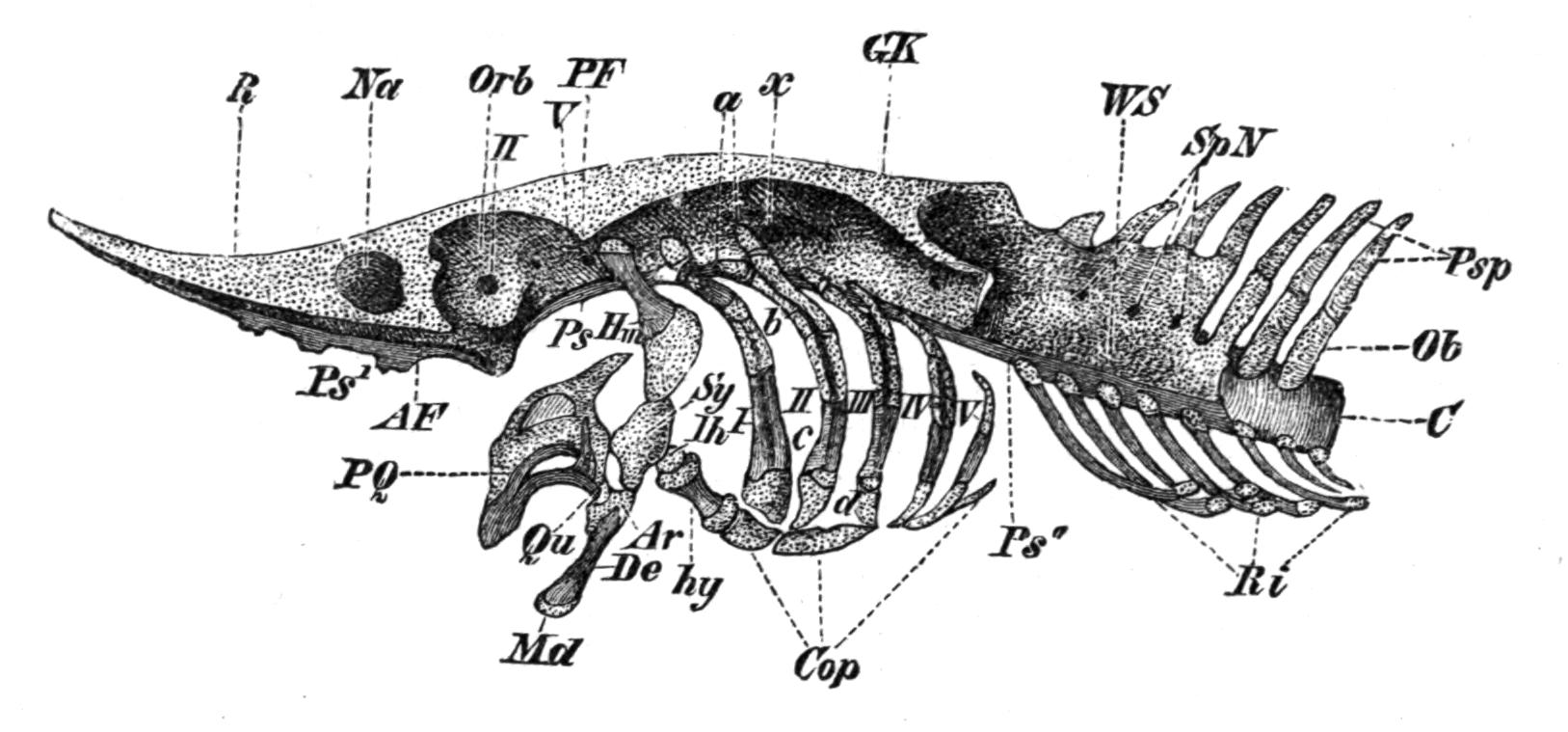
Fig. 130.—Side view of the skull of a Sturgeon, with the investing membrane bones removed. a, Pharyngo-branchial; AF, antorbital or lateral ethmoid cartilage; AR, articular; b, epi-branchial; c, cerato-branchial; C, notochord; Cop, basi-branchials; d, hypo-branchial; De, dentary; GK, auditory capsule; Hm, hyomandibular; hy, cerato-hyal; Ih, inter-hyal; Md, lower jaw; Na, nasal capsule; Ob, neural arches; Orb, Orbit; PF, post-orbital process; PQ, palato-quadrate; Ps, Ps′, Ps″, parasphenoid; Psp, neural spines; Qu, quadrate; R, rostrum; Ri, ribs; Sp.N, foramina for spinal nerves; Sy, symplectic; WS, vertebral column; x, foramen for the vagus nerve; I-V, branchial arches; II-V, foramina for the optic and the fifth cranial nerves. (From Parker and Haswell, after Wiedersheim.)
The existing Chondrostei,[198] and especially the Sturgeon, are remarkable for the persistence and continuous growth of the chondrocranium, and the absence of true cartilage bones.
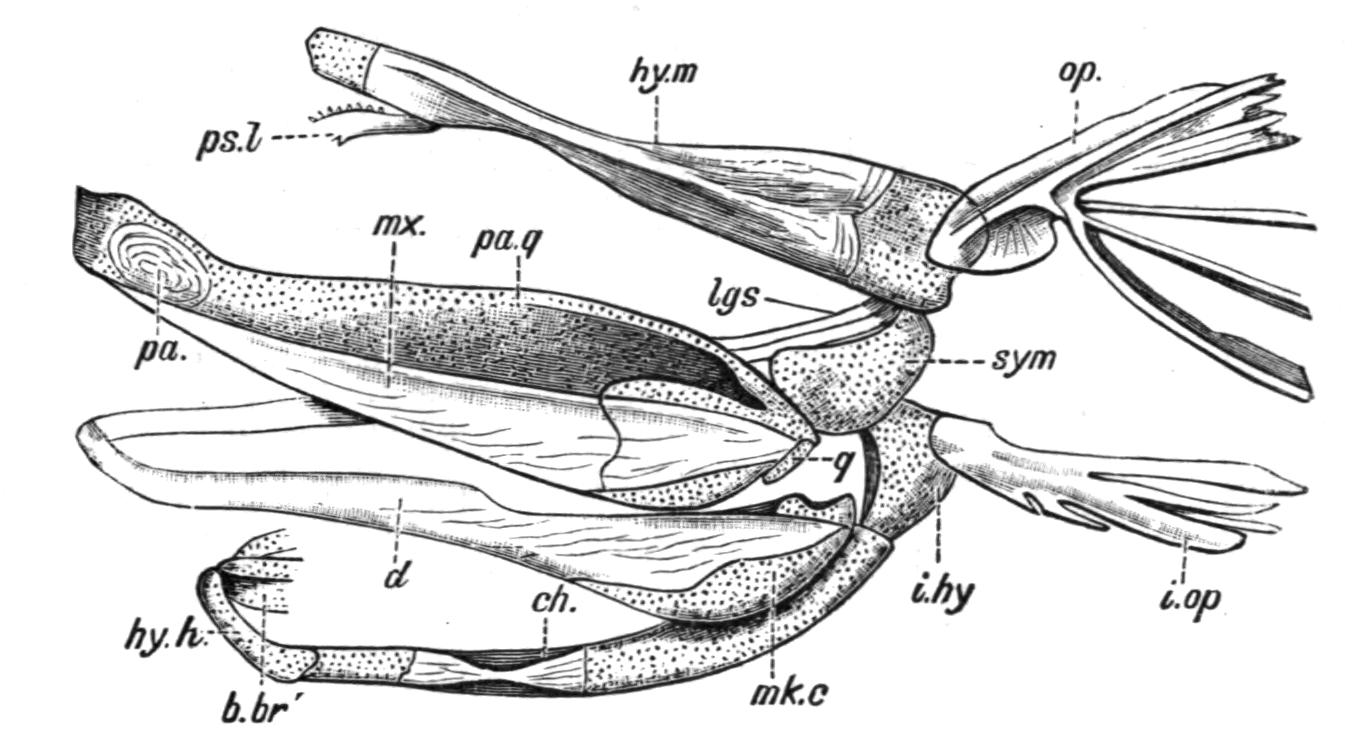
Fig. 131.—Lateral view of the primary and secondary upper and lower jaws of Polyodon. b.br′, First basi-branchial; ch, cerato-hyal; d, dentary; hy.h, hypo-hyal; hy.m, hyomandibular; i.hy, inter-hyal; i.op, inter-operculum; lgs, ligaments connecting the palato-quadrate cartilage with the hyomandibular; mk.c, Meckel's cartilage; mx, maxilla; op, operculum; pa, palatine; pa.q, palato-quadrate; ps.l, pre-spiracular ligament; q, quadrate cartilage; sym, symplectic. (From Bridge.)
Numerous dermal bones invest the dorsal surface of the chondrocranium, and only to a limited extent correspond with the less numerous membrane bones of the Salmon. To these are added a series of circum-orbital bones and a large parasphenoid. Undoubtedly the most striking feature in these Fishes is the primitive character of the upper jaw. In Polyodon (Fig. 131) the palato-quadrates are wholly cartilaginous, and, as in the Dog-Fish, they meet in front beneath the basis cranii, where the two are connected by ligament. The secondary upper jaw is but feebly developed, and is represented on each side by a thin splint-like maxilla in relation with the outer surface of each palato-quadrate cartilage, which meets its fellow in front. There are no premaxillae. The lower jaw is also very primitive. Meckel's cartilages are persistent, and except for a mento-Meckelian bone on each side, they are unossified, although membrane bones representing dentary and splenial elements are present. The skull is hyostylic. The hyoid and branchial arches are only partially ossified. Each opercular fold is supported by an operculum and an interoperculum, and both of these retain somewhat the shape of the cartilaginous hyoidean rays which they have replaced. In the Sturgeon (Fig. 130) the upper jaw is greatly modified in relation with the singular mouth of this Fish. The palato-quadrate cartilages meet not only in front, but also along their dorsal margins, and, with the help of the similarly opposed and somewhat fragmentary metapterygoid cartilages, they form a complete concave roof for the protrusible spout-like mouth. Palatine, mesopterygoid, and pterygoid bones invest, and in some measure replace these cartilages. In brief, the skull of the Chondrostei occupies an interesting intermediate position between the purely cartilaginous and mainly bony types. While retaining a well-developed and unossified primary cranium, it has acquired a complete secondary cranium of dermal bones. Equally notable is the condition of the jaws. Unique among the Teleostomi in possessing the typical Elasmobranch union of the palato-quadrate cartilages beneath the basis cranii, the Chondrostei are so far specialised that they have acquired certain of the membrane bones which constitute the secondary jaws of the more typical bony Fishes.
As regards the general structure of the skull and the nature and disposition of its cartilage- and membrane-bones, the remaining living Teleostomi have much in common with the Salmon. In all the skull is hyostylic, and, unlike the Chondrostei, each half of the primitive upper jaw remains distinct from its fellow, and is separately articulated in front with the lateral ethmoid of the same side by its palatine element. The palato-quadrate cartilage is always more or less completely replaced by bones similar to those of the Salmon, and although they often carry teeth, as a rule they do little more than constitute a rigid buttress for the fixation of the quadrate condyle for the lower jaw. The secondary upper jaw is nearly always well developed, and includes a premaxilla as well as a maxilla on each side. There are, however, certain features in each of the minor groups which are either distinctive or highly characteristic.
In the surviving Crossopterygii (e.g. Polypterus[199]) the chondro-cranium is complete in the ethmoidal and post-orbital regions, except where it has been partially replaced by cartilage bones, but in the inter-orbital region the continuity of the roof is interrupted by a large fontanelle, which is only closed by the investing frontal bones (Fig. 132, C). There is also a large basi-cranial fontanelle in the sphenethmoid, closed, however, by the underlying parasphenoid. A large "occipital" bone continuously ossifies in the occipital cartilage and completely surrounds the foramen magnum.
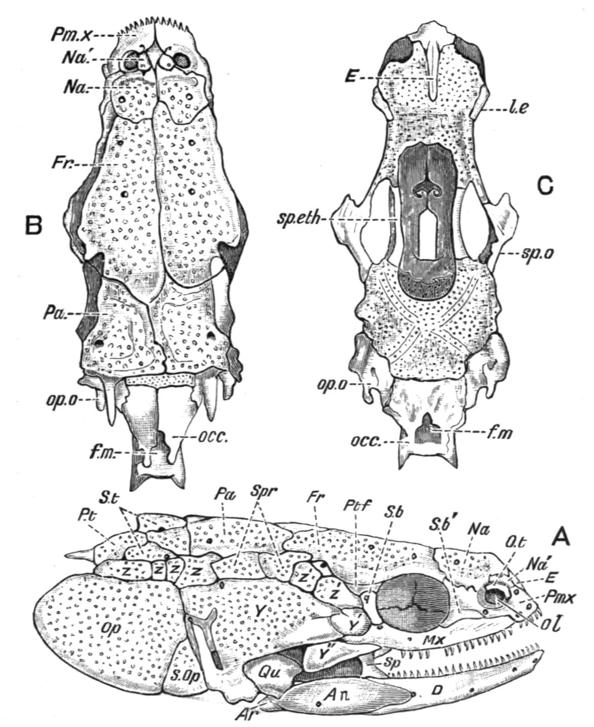
Fig. 132.—A, side view of the skull of Polypterus; B, dorsal view, showing the chief dermal bones; C, similar view of the chondro-cranium after the removal of the dermal bones. An, Angular; Ar, articular; D, dentary; E, mesethmoid; f.m, foramen magnum; Fr, frontal; l.e, lateral ethmoid; Mx, maxilla; Na, Na′, nasal and accessory nasal bones; occ, occipital; ol, nasal aperture; Op, operculum; op.o, opisthotic; O.t, os terminate; Pa, parietal; Pm.x, premaxilla; P.t, post-temporal; Ptf, post-frontal; Qu, quadrate; S.b, S.b′, circum-orbital ossicles; S.Op, sub-operculum; Sp, splenial; sp.eth, sphenethmoid; sp.o, sphenotic; Spr, spiracular ossicles, between which is the spiracle; S.t, supra-temporals; Y, cheek-plate (pre-operculum); Y′, Y″, smaller cheek-plates; z, z, z, z, post-spiracular ossicles; z′, z′, prespiracular ossicles. In C the cartilage is dotted. (From Traquair.)
Prootics and pterotics are absent, and the opisthotics seem to be confluent with their respective epiotics. The floor and side walls of the inter-orbital section of the cranium are formed by a remarkable "sphenethmoid" bone which occupies the position of the paired ali- and orbito-sphenoids in other bony Fishes; and in one species, P. lapradei,[200] it forms in front distinct tubular investments round the olfactory nerves. In many respects this bone is singularly like the sphenethmoid bone of the Frog and other tailless Amphibia. A median ethmoid as well as lateral ethmoids are present. In addition to the ordinary dermal bones which invest the cranial roof there is a transverse row of supra-temporal plates crossing the cranial roof behind the paired parietals (Fig. 132, A). Fringing the outer margins of the frontals and parietals a row of pre- and post-spiracular ossicles extends nearly to the orbits, and between two of them, which form a valve, is the spiracular aperture itself. There is a dentigerous splenial on the inner surface of the lower jaw. The hyoid arch has no separate symplectic bone. An operculum and a suboperculum are present, but no inter-operculum; and unless the hinder part of the large cheek-plate, which is traversed by the mandibulo-hyoid sensory canal, represents a pre-operculum, the latter is wanting. Branchiostegal rays are absent, but there is a single pair of large jugular plates.
Very little is certainly known about the cranial cartilage-bones in the fossil members of the group, but the investing dermal bones, which bear a general resemblance to those of Polypterus, are often somewhat more numerous, and they form a very complete dermal armature for the entire head. There is a very complete ring of circum-orbital bones, and very often a ring of sclerotic plates. Two large cheek-plates are often present. Nothing comparable to pre- and post-spiracular ossicles is known, but squamosal and supra-temporals can often be identified. To the ordinary bones of the lower jaw there may be added a series of infra-dentary plates, and besides the paired principal jugular plates there may also be present a small anterior median plate and a series of small lateral jugular plates on each side, as in the Carboniferous Rhizodopsis (Fig. 274). Most of the superficial dermal bones, both in the living and extinct Crossopterygii, are invested externally by a granulated or rugose layer of enamel-like ganoin.
In the Holostei, and especially in Amia, the skull approximates more closely to the normal Teleostean type as represented by the Salmon's skull. In Amia[201] all the occipital cartilage-bones are present—a basi-occipital, two exoccipitals, and a supra-occipital; and, except for the absence of a pterotic, the periotic series of bones is also complete. Paired ali- and orbito-sphenoids form the lateral walls of the inter-orbital portion of the cranial cavity. Above, the complete cartilaginous roof of the cranial cavity is invested by a shield of suturally united and ganoin-covered dermal plates. The hyomandibular element has a symplectic bone at its distal extremity. There is a complete series of opercular bones, and the branchiostegal rays are numerous. A single median jugular plate is present. The lower jaw has on each side five dentigerous splenial bones in addition to dentary and angular bones, while cartilage-bones are represented by articular and mento-Meckelian elements. In its essential structure the skull of Lepidosteus[202] resembles that of Amia, but it has obviously undergone much specialisation. In some species (e.g. L. osseus) its appearance is greatly modified by the exceptional length and tapering shape of the beak, due to the elongation of that part of the skull which lies between the orbital and nasal regions; but in L. platycephalus the reduced length and greater width of the beak, combined with its somewhat flattened condition, impart an almost Crocodilian aspect to the head. Amongst other points of difference it may be mentioned that in Lepidosteus the continuity of the chondro-cranial roof is interrupted by a large superior fontanelle. There is no supra-occipital, and there are no lateral ethmoids, at all events in the usual position. The inter-orbital portion of the cranial cavity is largely obliterated by the formation of an inter-orbital septum, consisting of a thin vertical plate of bone, which either represents a pair of fused orbito-sphenoids or a pair of similarly modified lateral ethmoids. In addition to the ordinary investing dermal bones, including circum-orbitals, squamosal, and supra-temporals, there are numerous scale-like ossicles which take the place of the cheek-plates of Polypterus. The maxillae are segmented into numerous dentigerous bones fringing the margins of the upper jaw. The lower jaw has no mento-Meckelian bones, but there is a very complete series of dermal elements, including dentary, coronary, splenial, angular, and supra-angular bones in addition to an articular cartilage-bone. One of the most remarkable features in the skull of Lepidosteus is the existence of a secondary articulation between the metapterygoid bones and a pair of transversely elongated condyles formed on each side by a lateral outgrowth from the parasphenoid and alisphenoid bones. By a horizontal sliding movement of the former on the latter, provision is made for the lateral expansion and contraction of the walls of the oral cavity and the separation and approximation of the lateral halves of the upper jaw.[203]
The generality of Teleosts[204] more or less closely agree with Amia in the main features of their cranial structure. There are, however, certain minor features which are characteristic if not always distinctive of the group. As a rule, to which, nevertheless, there are notable exceptions, there is little of the primary cartilaginous cranium in the adult, nearly the whole of it having become absorbed or converted into cartilage-bones. A supraoccipital is invariably present, and usually a mesethmoid and a basisphenoid. An additional bone is added to the periotic series, viz. a pterotic. Supra-temporal bones and jugular plates are always absent, and it may be doubted if mento-Meckelian bones and dentigerous splenials are ever developed in the lower jaw. Within the group itself the skull exhibits many notable modifications, of which only a few can here be mentioned. The shape, size, and character of the mouth and jaws, the extent to which they can be protruded and retracted, and the nature of the dentition, are the source of many characteristic modifications in the structure and appearance of the fore-part of the skull, and these again largely depend upon differences of habit and food. A protrusible mouth, or a mouth which is projected forwards, is usually associated with a suspensorium (hyomandibular) of considerable length, and so greatly inclined forwards as to make a more or less acute angle with the forepart of the cranium.
The presence or absence of an inter-orbital septum is also a feature in which considerable variation occurs. In some Teleosts there is no septum, and the cranial cavity is prolonged forwards between the orbits, where its lateral walls are formed by well-developed, paired ali- and orbito-sphenoid bones, as, for example, in the Carp and other Cyprinidae. In others the fusion of the cranial walls is accompanied by the median union of the orbito-sphenoids, so that a partly bony and partly cartilaginous inter-orbital septum is found, and the cranial cavity becomes largely obliterated in this region, as in the Salmon; or the orbito-sphenoids may be non-existent, the cartilage may undergo absorption, and the inter-orbital septum may become reduced to a vertical fibrous sheath extending between the frontals above and the parasphenoid below, as is the case in the Cod (Gadus).
An interesting modification of certain of the bones of the primary and secondary upper jaw occurs in the Siluridae. In these Fishes the maxillae are very small and edentulous, and serve no other purpose than forming basal supports for the maxillary barbels, while the rod-like palatine bone, losing its connexion with the pterygoid portion of the primitive upper jaw, but retaining its articulation with the lateral ethmoid, serves to support the maxilla, and at the same time receives the insertion of the muscles by which the barbel is moved in various directions.
In the Plectognathi the premaxillae are co-ossified with the maxillae. Many other interesting cranial modifications occur in Teleosts, and to some of them reference is made in subsequent chapters.
In some respects the skull of Dipnoi[205] is remarkably like that of the Holocephali, especially in its typical autostylism; but in possessing both cartilage- and membrane-bones it in some measure approaches the Teleostome skull. The investing dermal bones are not always easy to identify with those of other Fishes. In Neoceratodus an anterior median membrane-bone or dermal mesethmoid covers the ethmo-nasal region, and, on each side of it, forming the anterior boundary of the orbit, there is situated a pre-orbital or dermal lateral ethmoid. Behind the mesethmoid there is a much larger posterior median bone, and on each side a singular backward prolongation of the dermal lateral ethmoid separates it from a squamosal element. The latter bone descends on the outer surface of the quadrate portion of the palato-quadrate cartilage as far as the condyle for the lower jaw. Collectively, these bones form a fairly complete investment to the upper surface of the cranium, but the posterior median bone and the adjacent portions of the dermal lateral ethmoid and the squamosal are widely separated from the underlying chondrocranium by the powerful jaw muscles, and in this respect they differ from the ordinary roofing bones of other Fishes.
In Protopterus (Fig. 133) and Lepidosiren (Fig. 134) the posterior median bone is non-existent, and its place is taken by a large fronto-parietal, which forms the greater part of the cranial roof, internal to the jaw muscles, and is much larger in the latter Dipnoid than in the former. Circum-orbital bones are present only in Neoceratodus. A large parasphenoid supports the cranial floor. Vomers are absent, although there are two small vomerine teeth.
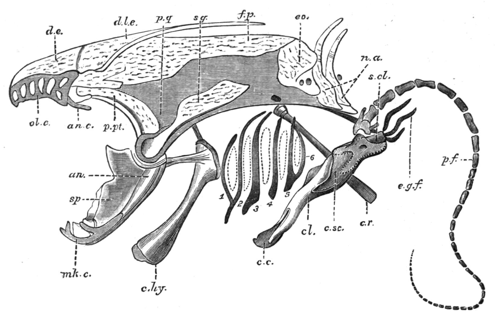
Fig. 133.—Side view of the skull of Protopterus, with the pectoral girdle and fin. an, Angular; an.c, antorbital cartilage; c.c, coracoid cartilage (epi-coracoid); c.hy, cerato-hyal; cl, clavicle; c.r, cranial rib; c.sc, coraco-scapular cartilage; d.e, dermal ethmoid; d.l.e, dermal lateral ethmoid; e.g.f, external gills; eo, exoccipital; f.p, fronto-parietal; mk.c, Meckel's cartilage; n.a, neural arches; ol.c, fenestrated roof of the olfactory capsule; p.f, skeleton of the pectoral fin; p.pt, palato-pterygoid bone; p.q, palato-quadrate cartilage; s.cl, supra-clavicle; sp, splenial; sq, squamosal; 1-6, the branchial arches; the segmentation of the second and third arches is not shown. (From Wiedersheim.)
Relatively small opercular and inter-opercular bones are present, and on the inner surface of each may be found vestigial remains of cartilaginous hyoidean rays. The chondrocranium is complete in Neoceratodus, but in the remaining genera it has undergone considerable absorption in the inter-orbital region, so that the roof and floor, and, in part, even the side walls of the cranial cavity, are formed by the fronto-parietal and parasphenoid bones. Two exoccipitals are present in all Dipnoi. There are small labial cartilages in relation with the ventrally-placed nostrils, and large lateral outgrowths from the ethmoid cartilage furnish the olfactory organs with conspicuous lattice-like roofs. A pair of strong palato-pterygoid bones fringe the lower margins of the palato-quadrate cartilage, and meeting in front beneath the ethmoid region their symphysial extremities support the large palatal teeth. The Meckelian cartilages are persistent in all Dipnoi. In Neoceratodus each is flanked by a dentary and an angular externally, and internally by a splenial; but in Protopterus and Lepidosiren distinct dentary bones are wanting. The hyoid arch is best developed in Neoceratodus,[206] and includes a small hyomandibular cartilage, a partially bony cerato-hyal and cartilaginous hypo-hyal and basi-hyal element. In the other genera (Fig. 133) only a cerato-hyal is retained. The branchial arches are but feebly developed in the Dipnoi. Neoceratodus has five, of which the first four are divided into epi-branchial and cerato-branchial segments, while the fifth is undivided. Protopterus has six, but only the second and third are segmented as in Neoceratodus.[207] In Lepidosiren all the arches are simple undivided rods.
In all three genera the skull conforms to the same general type of structure, but it is much more primitive in Neoceratodus than in the other two genera.
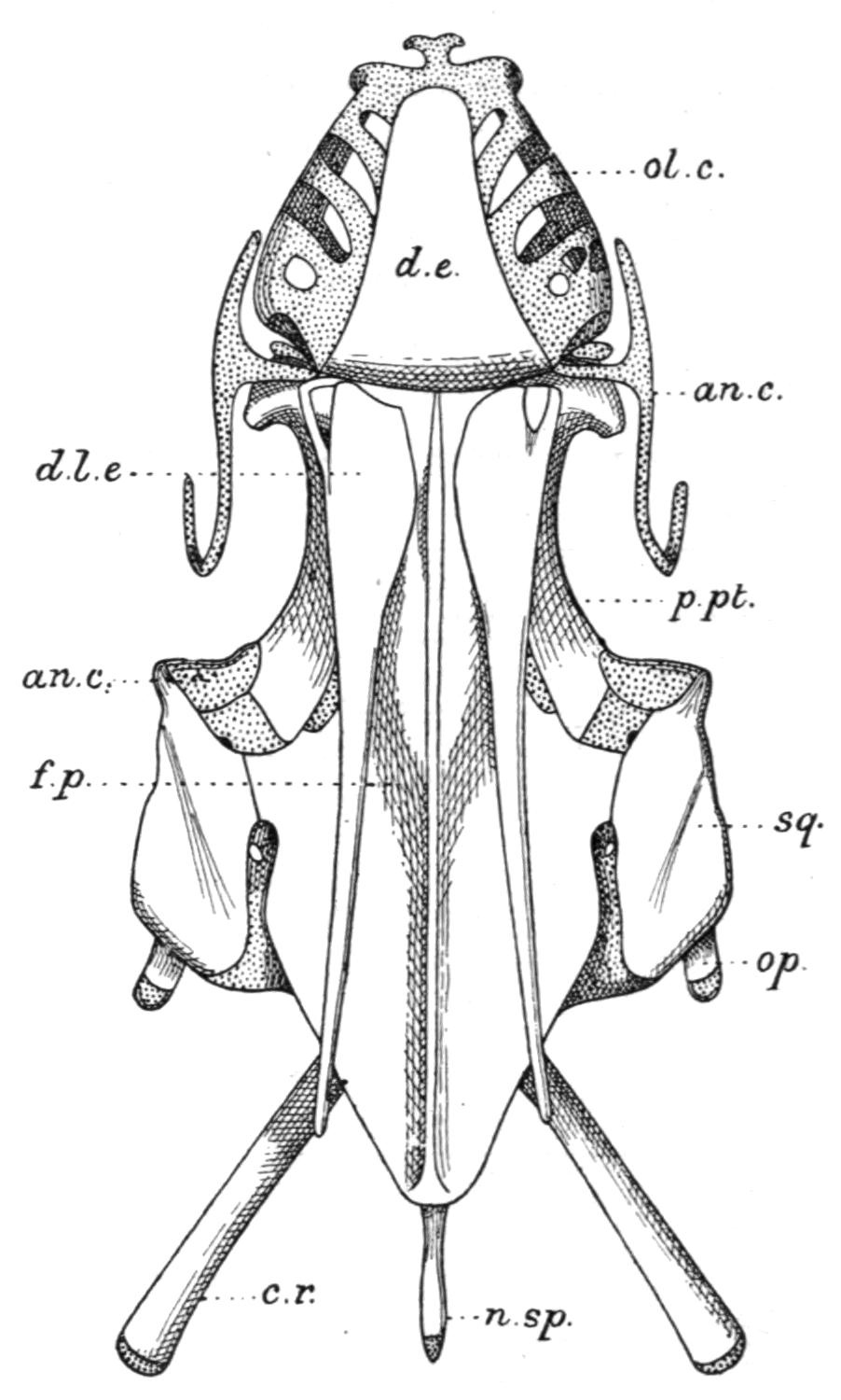
Fig. 134.—Dorsal view of the skull of Lepidosiren. an.c, Condyle on the quadrate cartilage for the lower jaw; n.sp, neural spine; op, operculum. For other reference letters see Fig. 133. (From Bridge.)
With reference to the fossil Dipnoi, it may be stated that, so far as they are known, the cranial roofing bones are more numerous than in the existing genera, and they cannot readily be compared with those of the latter, or with the numerically reduced and more definitely arranged bones of most Teleostomi. There is also evidence that in some fossil Dipnoi (e.g. Dipterus) the chondrocranium and the mandibular suspensorium (palato-quadrate) must have been replaced by cartilage bones to an extent which has no parallel in any of the surviving types.[208] Jugular bones were present in Dipterus and Phaneropleuron.
Median Fins and Appendicular Skeleton
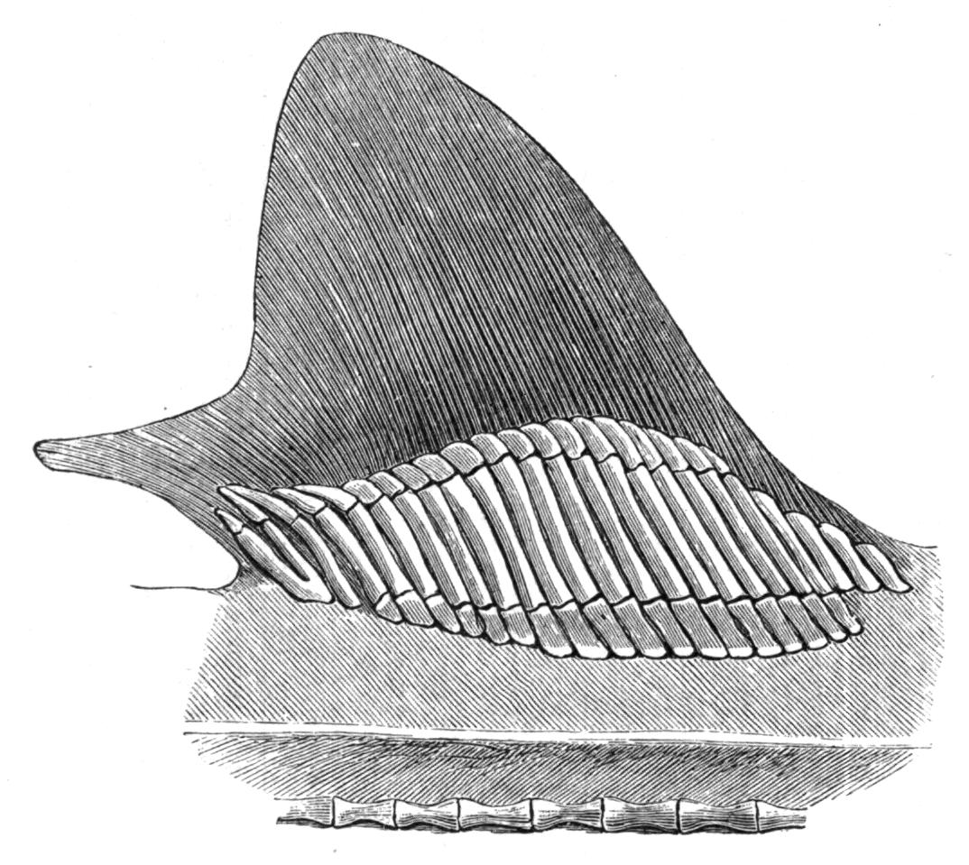
Fig. 135.—The cartilaginous radialia of the first dorsal fin of Mustelus antarcticus. (From Mivart.)
The Median Fins.—Whether existing in the form of a continuous fin, or as discontinuous isolated fins, the median fins are provided with skeletal supports, and also with muscles, primitively formed from intrusive clusters of cells derived from a variable number of the neighbouring myotomes, for their varied movements. The skeletal structures of the dorsal and anal fins consist of a series of bony or cartilaginous, rod-like, and typically tri-segmented radial elements or pterygiophores,[209] supporting distally a series of dermal structures in the shape of numerous slender horny fibres or ceratotrichia, as in the Elasmobranchii and Holocephali, or a smaller number of bony dermal fin-rays, which are probably modified scales or lepidotrichia,[210] as in the Teleostomi. The typical tri-segmented character of the radialia is often retained in many existing Elasmobranchs (Fig. 135) and in Pleuracanthus, in Neoceratodus amongst the Dipnoi, in the Chondrostei, in existing Holostei (Fig. 136), and to a greater or less extent in several families of Teleosts (e.g. Salmonidae, Esocidae, Cyprinidae, and some Acanthopterygii); but in the latter group the radialia are greatly prone to reduction, and hence they are more generally bi-segmented, and sometimes consist of a single proximal segment only (e.g. Gymnotus). In all these Fishes the proximal segments are the longest and the most persistent, and when reduction occurs it is at the expense of the middle and distal segments.
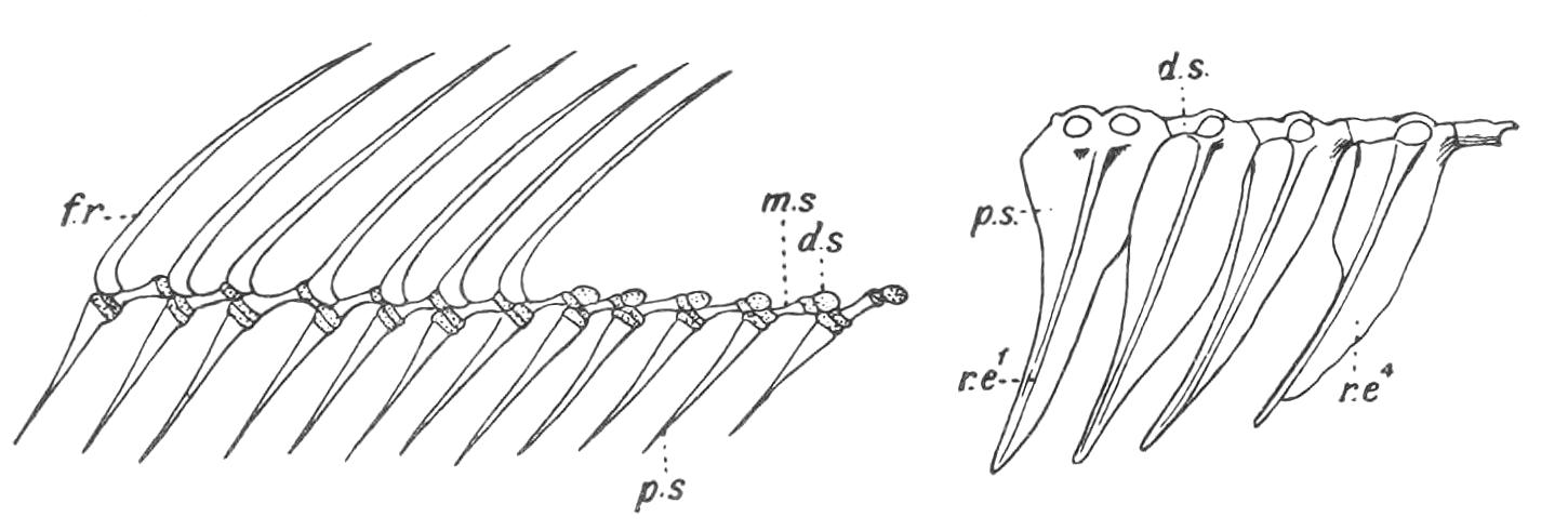
Fig. 136.—The tri-segmented radialia and the fin-rays of part of the dorsal fin of Amia calva. p.s, m.s, and d.s, The proximal, middle, and distal segments of a radial; f.r, fin-rays. (From Bridge.)
Fig. 137.—The first four radialia of the dorsal fin of Mesoprion gembra, showing the chain-links for the ring-like bases of the fin-rays. r.e1, r.e4, First and fourth proximal radialia.
The cause of this reduction is often, but not always, to be found in the fact that, whenever the dermal fin-rays take the form of stout spines, as in the anterior dorsal fin in many Acanthopterygian Teleostei, the segmentation of their radialia would obviously detract from their value as skeletal supports, and hence they rarely consist of more than their proximal segments, although the radialia which in the same Fish support soft rays may be bi-segmented or tri-segmented. The radialia are, however, unsegmented, even slightly branched, cartilaginous rods in the Cyclostomata; short simple rods in the Holocephali; and equally simple bony rods in the dorsal fin of Polypterus, where they support the strong spines of the numerous finlets; but they are bi-segmented in the soft-rayed anal fin. As previously mentioned, the proportional share taken by the radialia and the horny fibres or the dermal fin-rays in the support of the fins differs greatly in different Fishes. In the Cyclostomata radialia are the sole, and in Elasmobranchs the main supports, and they may extend nearly to the free margin of the fin. In the more specialised Fishes, as in most Teleostomi, the reverse is the case. The radialia sink into the muscles of the body-wall and leave the strongly developed fin-rays as the sole support of the visible portions of the fins. In not a few Fishes there is an obvious segmental correspondence between the radialia and the vertebral neural or haemal spines, to the extent that the former equal the latter in number and articulate with their distal extremities, as, for example, in the caudal region of Pleuracanthus and in existing Dipnoi. In others again, as in most Teleostomi, there is no such segmental relation, and the radialia are more numerous than the vertebrae whenever the two are co-extensive. The exoskeletal fin-supports exhibit similar relations to their radialia, but in inverse order. Much more numerous than the radialia in the Elasmobranchs, Holocephali, and the Dipnoi, the former become gradually reduced in the Teleostomi, until in the Holostei and Teleostei they correspond in number with the supporting radialia. Complete numerical correspondence between the neural and haemal spines and the radialia and fin-rays is very rare, and has only been observed in the caudal region of certain Crossopterygii (e.g. the Coelacanthidae).[211]
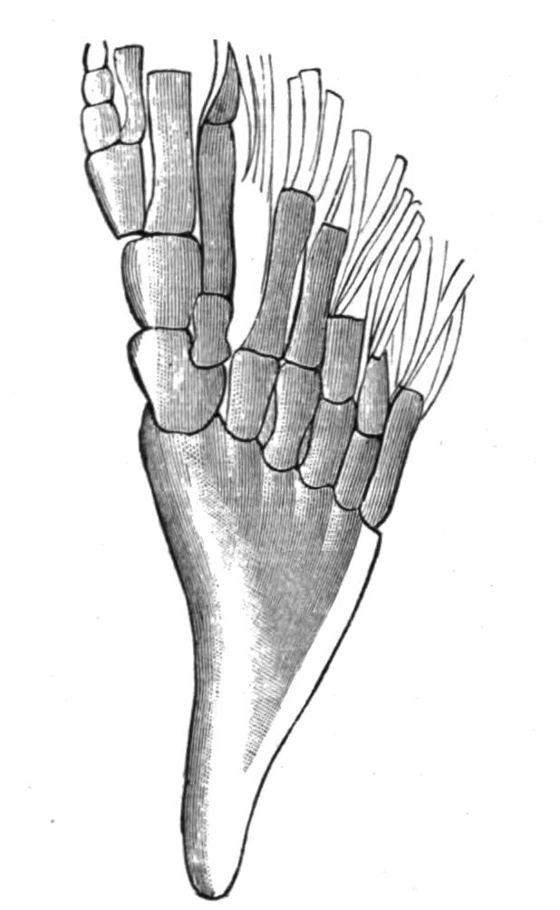
Fig. 138.—The posterior dorsal fin of Holoptychius leptopterus from the old Red Sandstone of Nairnshire. Traces of dermal fin-rays may be seen at the distal margin of the fin. (After Smith Woodward.)
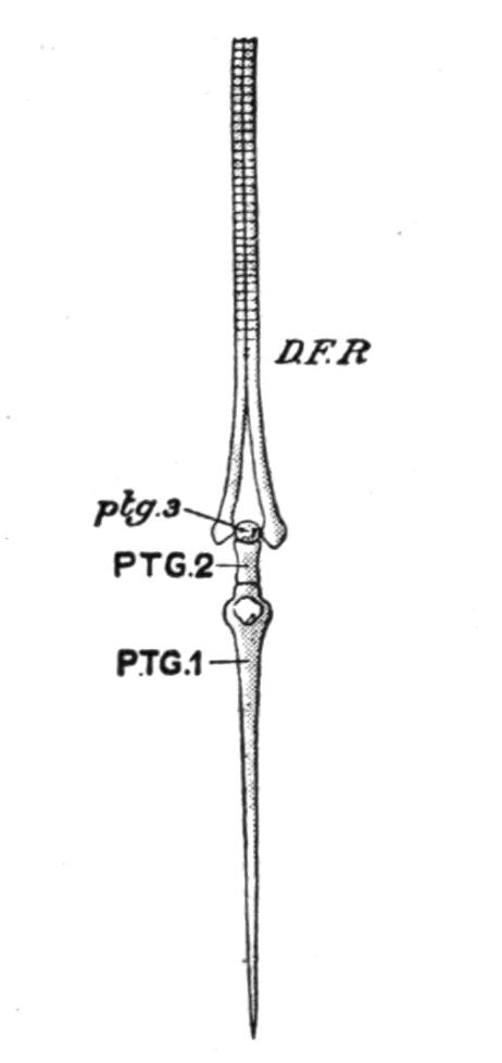
Fig. 139.—A dermal fin-ray and its supporting radial or pterygiophore in the Trout (Salmo fario). D.F.R, Dermal fin-ray; PTG.1, PTG.2, ptg.3, the proximal, middle, and distal segments of which the tri-segmented radial consists; ptg.3 is cartilaginous; the other two are bony. (From Parker and Haswell.)
In not a few Fishes the radialia of the median fins undergo modifications which offer an interesting parallel to an early stage in the evolution of the paired fins from primitively continuous lateral fins. The concentration of radialia which occurs in isolated median fins often results, through growth pressure, in the complete fusion of the proximal segments of more or fewer of the radialia into two or three basal supports, or even into a single basal piece. Examples of such basal fusion are frequent in the dorsal fins of Elasmobranchs, and the same modification may also be seen in the anal fin of Pleuracanthus, and especially in the dorsal fin of the Devonian Crossopterygian, Holoptychius[212] (Fig. 138), where several radialia, which are free distally, have their bases united into a single basal piece, or basipterygium. In most Teleostomi elevator and depressor muscles arise from the radialia, and are inserted into different points on the bases of the fin-rays, and by their contraction the latter may either be elevated into an erect position, or folded back like a fan along the middle line of the body, where, as in some Teleosts, there is a groove for their reception. When fin-rays are only capable of simple elevation or depression, the connexion between a radial element and its fin-ray is usually by some form of a hinge-joint, the cleft base of the ray clipping the distal segment of the radial (Fig. 139). In some Teleosts the articulation of the two is by means of a kind of chain-link (Fig. 137). In those Fishes in which the median fins are capable of lateral undulatory movements the articulation is of a more mobile character.
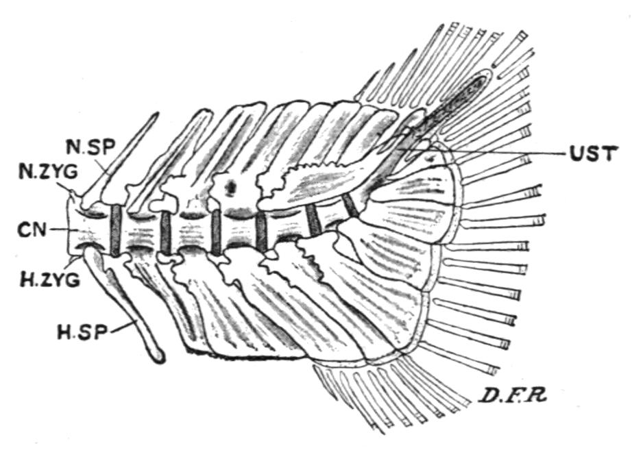
Fig. 140.—Caudal end of the vertebral column of a Trout (Salmo fario). CN, Centrum; D.F.R, dermal fin-rays; H.SP, haemal spine; H.ZYG, haemal zygapophysis; N.SP, neural spine; N.ZYG, neural zygapophysis; UST, the up-tilted, partly ossified, and unsegmented terminal portion of the notochord, or urostyle. (From Parker and Haswell.)
In the different types of caudal fin, diphycercal, heterocercal, and homocercal, the supporting elements of the ventral lobe are formed by the haemal spines of the terminal caudal vertebrae which are inclined backwards, and are often greatly expanded for the purpose (Fig. 140). The dorsal lobe may be supported either by the adjacent neural spines, or by radialia, or by both.
The Appendicular Skeleton.[213]—It is probable that the skeleton of the paired fins and the pectoral and pelvic girdles have been formed from the supporting radialia of the isolated and enlarged anterior and posterior portions of primitively continuous lateral fins, by a sequence of structural modifications in the same direction as in the median fins. The initial stage was probably marked by the fusion of the proximal portions of the radialia to form a basal support or basipterygium for the free distal portions. Subsequently, it may be, a rudiment of the future limb-girdle became segmented off from the inner extremity of the basipterygium, and by its dorsal and ventral growth in the body-wall the lateral half of a girdle was developed. The subsequent union of the two halves across the mid-ventral line resulted in the evolution of the dorsally incomplete hoop of cartilage which is the primary form of the complete limb-girdle in Craniates. The primitive fin skeleton or "archipterygium" was formed from the residue of the basipterygium in conjunction with the free distal radialia which it carried. The precise structure of the archipterygium is purely hypothetical. Possibly it was a biserial fin of the Pleuracanthus or Neoceratodus type, consisting of a cartilaginous segmented axis, fringed along its anterior and posterior, or pre-axial and post-axial margins, by a series of slender, simple, or jointed radialia (Fig. 147); or it may have been a uniserial structure, somewhat resembling the pelvic fin of Pleuracanthus, or the pectoral and pelvic fins of existing Elasmobranchs (Figs. 250, 141), in which an axis formed by the residue of the basipterygium or metapterygium had a fringe of radialia on its anterior or preaxial side only. If the archipterygium was biserial then the uniserial fin was probably derived from it by the subsequent suppression of all the post-axial radialia; or, if uniserial, the biserial fin was evolved by a later extension of radialia on to the post-axial margin. The evidence of comparative anatomy is not conclusive as to the nature of the archipterygium, and palaeontology seems to support either view with puzzling impartiality.[214] It may be admitted that the lateral fin theory offers the best solution of the problem of the origin of the paired fins, but it must be borne in mind that no Fish, living or fossil, is known to possess fins of this nature, unless the singular lateral lobes of some Ostracodermi (e.g. the Coelolepidae) are kindred organs[215]; neither do continuous lateral fins ever exist as vestiges, unless, indeed, the bilateral series of spines, which extend between the pectoral and pelvic fins, in some of the Lower Devonian Acanthodei (e.g. Climatius), may be regarded in that light.
The Pectoral and Pelvic Girdles.—The pectoral girdle is more primitive in Cladoselache and Pleuracanthus than in any other Elasmobranch. In the former (Fig. 145, A) it may be doubted if the girdle has passed beyond the basipterygial stage, and although a definite girdle is present in the latter genus (Fig. 250) its lateral halves retain their primitive distinctness. Existing Elasmobranchs, including the Holocephali, have a pectoral girdle in the form of a dorsally incomplete hoop of cartilage imbedded in the muscles of the body-wall, close behind the last branchial arch (Fig. 141). The upper or dorsal portion of each half is the scapula, and the ventral is the coracoid. Between these two portions of the girdle, and defining their limits, there are articular surfaces for the basal cartilages of the pectoral fin.
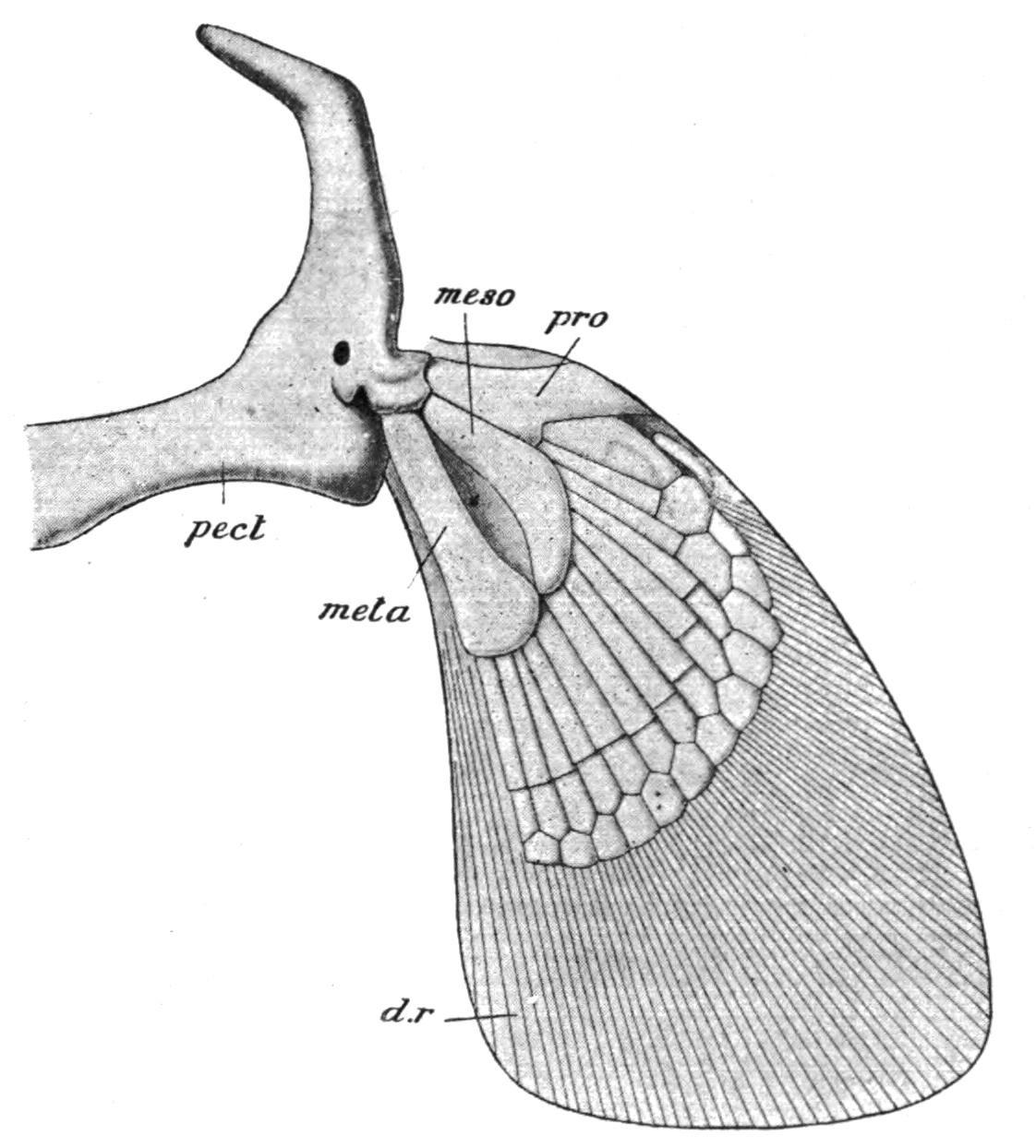
Fig. 141.—The right half of the pectoral girdle and the fin of an Elasmobranch (Chiloscyllium). d.r, Dermal horny fibres; meso, mesopterygium; meta, metapterygium; pect, pectoral girdle; pro, propterygium. (From Parker and Haswell.)
Cladoselache (Fig. 145, B) had no pelvic girdle, nor does it appear that this primitive Elasmobranch had acquired even a basipterygium. Pleuracanthus, on the contrary, had a pair of pelvic rudiments distinct from well-developed basipterygia. In other Elasmobranchs there is a distinct girdle, formed by the median union of primitively distinct lateral rudiments, consisting of a simple transverse bar of cartilage, imbedded in the ventral abdominal wall, just in front of the cloacal aperture, and having articulated to each of its outer extremities the basal cartilage (metapterygium) of the pelvic fin.
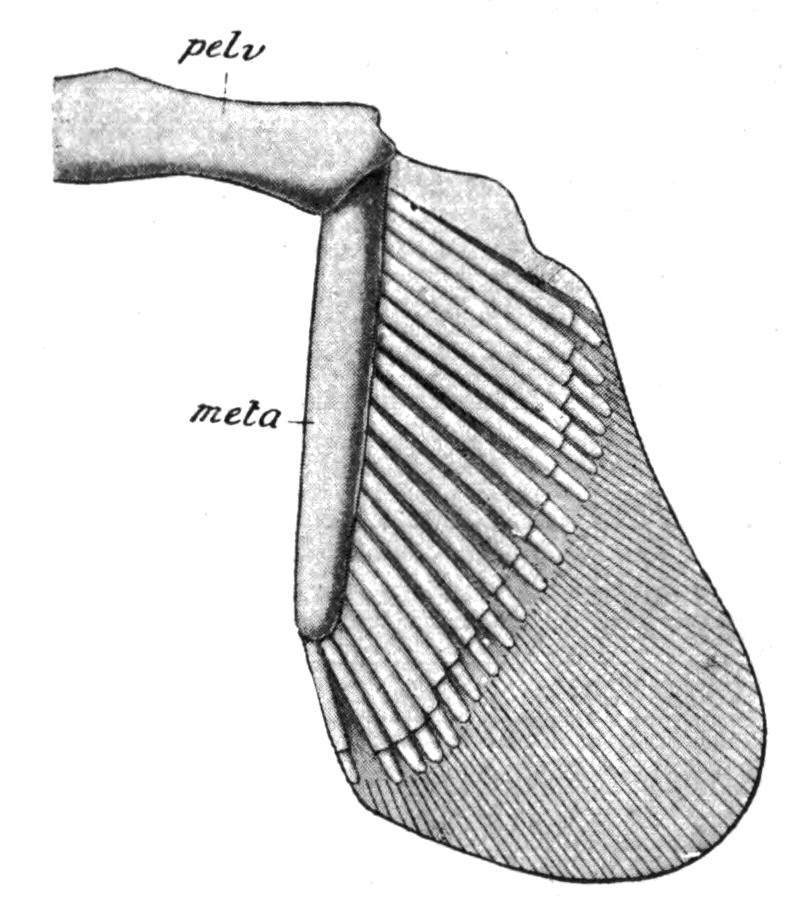
Fig. 142.—The left half of the pelvic girdle and the right pelvic fin of Chiloscyllum. meta, Metapterygium; pelv, pelvic girdle. (From Parker and Haswell.)
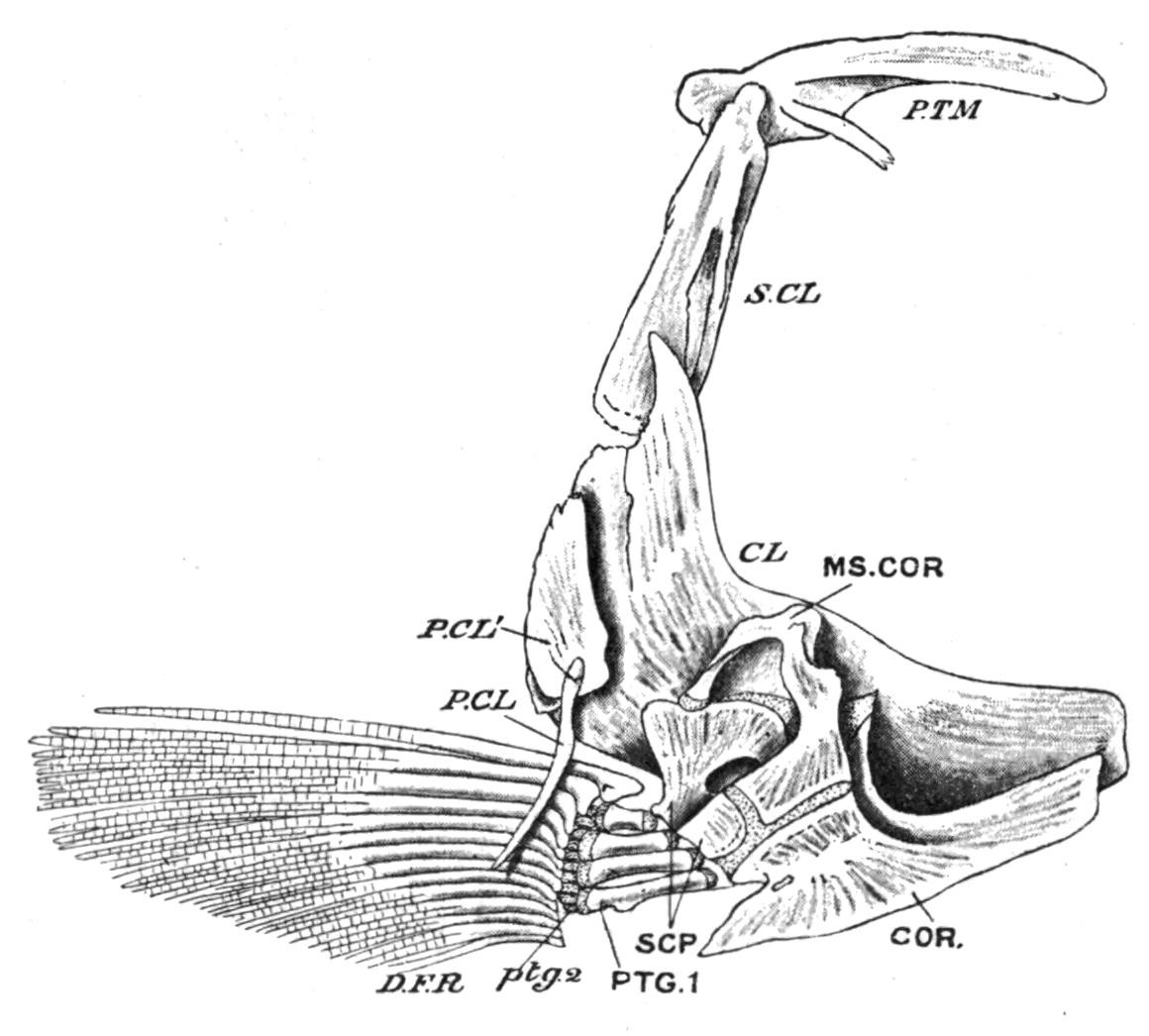
Fig. 143.—Left half of the pectoral girdle of a Trout (Salmo fario), seen from the inner surface. CL, Clavicle (cleithrum); COR, coracoid; D.F.R, dermal fin-rays; MS.COR, meso-coracoid; P.CL, P.CL′, post-clavicles; PTG.1, proximal; ptg.2, distal pterygiophores; P.TM, post-temporal; S.CL, supra-clavicle; SCP, scapula. (From Parker and Haswell.)
Sometimes there is a rudiment of a dorsally-directed "iliac" process at each extremity of the girdle, but in no Fish do these processes ever acquire a dorsal connexion with the vertebral column. In the Holocephali the iliac processes are better developed than in any other Fishes, but ventrally the lateral halves of the girdle are united by ligament alone. In the Teleostomi important differences are observable in both girdles. The primary cartilaginous pectoral girdle now consists of distinct lateral halves which have no ventral connexion with each other. In addition, there is developed on the outer surface of each half a series of membrane bones, which form a secondary girdle (Fig. 143). From above downward the series includes a supraclavicle and a cleithrum (clavicle of Teleosts) which are always present, and to these may be added in the Crossopterygii and Chondrostei an infraclavicle or clavicle proper, while one or two "post-clavicles" may be present in relation with the hinder margin of the cleithrum. The infraclavicles, or in their absence the cleithra (e.g. Holostei and most Teleostei), usually meet in a median ventral symphysis, so that the secondary girdle tends to acquire the characteristic hoop-like arrangement of its parts which has been lost in the primary girdle. With the development of a bony secondary girdle, the primary girdle (scapula and coracoid) becomes much reduced, and, as a rule, does little more than connect the fins with the cleithra. The secondary girdle acquires a dorsal connexion with the skull on each side by means of the post-temporal bone, which is attached below to the supra-clavicle and above to the periotic capsule. In the Chondrostei and the Dipnoi the primary girdle retains its primitive cartilaginous condition, but in the Crossopterygii, Holostei, and in all Teleosts it is ossified as distinct scapulae and coracoids. To these may be added in some Teleosts a mesocoracoid formed by a separate ossification of the coracoid cartilage (Fig. 143).[216]
Fig. 144.—Ventral view of the pelvic girdle of Protopterus. a, Prepubic process; b, lateral process for the fin; c, epipubic process; Gr, ridge for the origin of the fin muscles; HE, skeleton of the fin; M, myotomes; M′, myocommata. (From Wiedersheim.)
With the possible exception of small paired or median cartilages inserted between the inner extremities of the basipterygia in Polypterus and a few other Teleostomi, the pelvic girdle is absent in all the existing members of this group, having either become completely suppressed, or remaining unseparated from the basipterygia of the pelvic fins.[217] In the Dipnoi (Fig. 144) there is a true pelvic girdle which has some points of resemblance to that of certain of the caudate Amphibia. It is represented by a median, lozenge-shape, cartilaginous plate, produced in front into a long tapering epipubic process, and on each side of this into a forwardly inclined prepubic process. The hinder part of the plate bears two short processes for the basal cartilages of the pelvic fins. There is no trace, however, of iliac processes.
The Pectoral Fins.—The skeleton of the pectoral fins exhibits remarkable structural variations in different Elasmobranchs. In the existing members of the group two large basal cartilages, the propterygium and the mesopterygium, are formed by the concentration and fusion of the proximal portions of certain of the preaxial radialia, and they, with the metapterygium, articulate with the pectoral girdle; hence the fin is tribasal as well as uniserial (Figs. 141 and 146, A, B). In striking contrast to all other Elasmobranchs the pectoral fin of Cladoselache (Fig. 145, A) is far more primitive than in any other Fish. Each fin is supported by a distal series of slender, more or less parallel, unjointed, cartilaginous radialia, and basally by a similar series of shorter, stouter, and less numerous cartilages, which apparently were imbedded in the body-wall, the entire fin skeleton presenting a striking resemblance to an isolated median fin in which the supporting radialia have concentrated by growth pressure, and their proximal portions have been reduced in number by partial fusion.[218] Pleuracanthus, on the other hand, had a biserial fin, the preaxial and postaxial radialia supporting fan-like clusters of horny fibres at their distal ends (Fig. 250).
Fig. 145.—A, Pectoral fin, and B, pelvic fin of Cladoselache. (From Bashford Dean.)
Fig. 146.—Pectoral fins of various Fishes. A, Acanthias vulgaris; B, Raia sp.; C, Chimaera monstrosa; D, Acipenser rhynchaeus; E, Amia calva; F, Lepidosteus platyrhynchus; G, Polypterus bichir; H, Salmo salvelinus. The preaxial side of each fin is to the left and the postaxial to the right. f.r, Dermal fin-ray; ms, mesopterygium; mt, metapterygium; p, propterygium; r, free radialia; 1, 5, the preaxial and postaxial basal elements in a Teleost, which may be mesopterygial and metapterygial pieces respectively, the three remaining basal pieces probably being intrusive metapterygial radialia directly articulating with the pectoral girdle. In B, D, E, and F, similar intrusive radialia are shown. (From Gegenbaur.)
The broadly lobate pectoral fin of the existing Crossopterygii (Fig. 146, G) is uniserial, closely resembling that of the more typical Elasmobranchs.[219] There are three basal elements, a propterygium, a mesopterygium, and a metapterygium, each of which supports a series of partially ossified radialia. Little is known of the endoskeletal elements of the broadly or acutely lobate fins of the fossil Crossopterygii, but it seems probable that their disposition was uniserial and abbreviate in obtusely lobate fins and biserial in acutely lobate fins. In the remaining Teleostomi (Actinopterygii) the endoskeletal elements become gradually reduced in number and importance, their place as fin-supports being usurped by the dermal fin-rays. In addition, more than three, usually several, basal elements articulate directly with the pectoral girdle, and hence the fins become multi-basal. In the Chondrostei and the Holostei a metapterygium is always recognisable, supporting several radialia along its preaxial border, as in Acipenser (Fig. 146, D) and Amia (Fig. 146, E), or only a single one, as in Lepidosteus (Fig. 146, F). The anterior part of the fin is supported by a variable number of cartilaginous or bony radialia, which, with the metapterygium, articulate with the limb-girdle. In Teleosts the process of reduction reaches its maximum. Usually there is but a single row of short, hour-glass-shaped ossicles, of which the postaxial one may represent a vestigial metapterygium, and sometimes there is also a distal row of small cartilages or ossicles, partially hidden in the cleft bases of the dermal fin-rays (Fig. 146, H). In all these Fishes the fin is a much reduced uniserial fin, in which more or fewer of the preaxial radialia have acquired a direct secondary connexion with the pectoral girdle. Of living Dipnoids Neoceratodus has a nearly typical biserial fin, but, as seems to be the case in all fins of this type at present known, there is a marked absence of symmetry in the number and disposition of the radialia on the two sides of the axis. There is also much individual variation. No two fins are precisely alike, and the radialia may sometimes divide. In the very acutely lobate fins of the remaining Dipnoids it is evident that great reduction has taken place. Protopterus has lost all trace of postaxial radialia, and in Lepidosiren even the preaxial have atrophied, leaving only the long jointed axis to represent the originally biserial fin.