Chapter XII
Reading edition
THE VASCULAR SYSTEM, THE LYMPHATICS, AND THE BLOOD-GLANDS
The Cyclostomata and Fishes possess a closed vascular system, consisting of a heart, arteries, capillaries, and veins, the whole forming a continuous series of blood-containing channels provided with definite limiting walls, through which the blood is propelled in a constant direction by the rhythmical contractions of the heart. In the course of the circulation the blood flows from the heart through a single large trunk, the ventral aorta, to the capillaries of the gills. From the gills the arterialised blood is collected into a large dorsally-situated vessel, the dorsal aorta, and thence is distributed through a system of arteries to the capillaries of the various organs of the body. Finally, the blood is collected from the capillaries and returned to the heart by the veins.
Although in most instances the organs of the body are supplied with arterialised blood conveyed to them by arteries, there are nevertheless cases in which an organ may receive venous blood by a vein in addition to arterial blood supplied by an artery. For example, the capillaries of the liver not only receive blood from the hepatic artery, but also venous blood by a large vein (hepatic portal vein), formed by the union of a number of smaller veins by which venous blood is collected from the capillaries of the stomach, intestine, spleen, and pancreas. In this and similar instances, where a vein formed by the union of the capillaries of an organ, or of a series of organs, instead of uniting with other veins and proceeding towards the heart, becomes continuous with a second set of capillaries in some other organ, a "portal" system is said to be formed, and in the particular example of the liver it is termed the "hepatic portal" system. A similar, or "renal portal," system also exists in connexion with the kidneys in the majority of Fishes.
There is little doubt that, primarily, the vascular system of Vertebrate animals consisted of a dorsal artery (dorsal aorta), running along the median dorsal line of the alimentary canal, and a ventral or subintestinal vein similarly related to the ventral surface of the digestive tube. The two vessels were connected by a series of pairs of lateral branches, which had their origins from the dorsal vessel, and, by their subdivision, formed a capillary network in the walls of the alimentary canal. From these networks paired veins issued and opened into the subintestinal vein. The simplicity of this primitive arrangement was somewhat disturbed in the region of the pharynx by the development of gill-clefts, in the walls of which the blood circulated for respiratory purposes from the ventral to the dorsal vessel; and also by the development of a hepatic portal circulation in connexion with the liver. In the latter instance the subintestinal vein entered the liver and subdivided into capillaries in the substance of that organ, the corresponding efferent vessel, or hepatic vein, becoming continuous with the anterior or pharyngeal section of the subintestinal vein, or, as it is usually termed, the ventral aorta. In this low grade of vascular system, which is perhaps most completely retained in Amphioxus, the circulation of the blood was probably effected by the wave-like contractions of more or fewer of the larger vessels; but subsequently a definite chambered heart was developed at the origin of the ventral aorta.
Of Fishes in general it may be said that the primitive dorsal and ventral vessels are present in the embryo, and for a time retain their original relations and physiological importance. To a very unequal extent they may also be retained in the adult, where, however, they co-exist with numerous other vessels, which the increasing differentiation of the body has called into existence. Thus, at a later period of embryonic life, the subintestinal vein becomes somewhat fragmentary. Its caudal section (caudal vein) ceases to be continuous with the precaudal portion, and the blood collected from the muscles and other structures of the tail is conveyed to the heart by a pair of posterior cardinal veins, which are either directly continuous with the caudal vein, or indirectly through the intervention of a renal portal system in the kidneys. The precaudal portion of the subintestinal vein is represented by a vein which runs forwards in the intestinal wall, and is one of the minor affluents of the hepatic portal vein, while its prehepatic section is represented in succession by the hepatic vein, the heart, and the ventral aorta. Of the additional veins which supplement these remnants of a primitively continuous subintestinal vein, the largest and most constant are (a) the posterior cardinal veins which, commencing in the kidneys and receiving the blood from those organs, pass forwards to the heart; (b) a pair of anterior cardinal veins, formed by the union of smaller veins from the head, including the brain, and passing backwards towards the heart. At the level of the latter organ each anterior cardinal vein joins the posterior cardinal of the same side of the body to form a short but wide transverse vessel, the Cuvierian duct or precaval vein, which opens into the hindermost of the cavities of the heart, viz. the sinus venosus; (c) a pair of inferior jugular veins by which the nutrient blood of the branchial apparatus is returned to the right and left Cuvierian ducts. In addition to these principal veins there may also be a pair of lateral veins collecting the blood from the lateral walls of the trunk, and also opening into the Cuvierian ducts; and subclavian and femoral veins from the pectoral and pelvic fins.
On the other hand, the primitive dorsal vessel (dorsal aorta), retains not only its original position and relations, but also its primary function as the main channel for the distribution of arterialised blood to all parts of the body. The system of lateral and probably segmentally arranged vessels, by which the dorsal and subintestinal vessels were connected in the primitive Vertebrata, have undergone considerable modification in all existing Fishes, but nevertheless retain much of their original disposition and relations in the pharyngeal region of the alimentary canal, where they are represented by the afferent and efferent vessels of the gills.
A more detailed account of the condition of the vascular system in the Cyclostomata and Fishes will now be given.
The Venous System.—The Cyclostomata,[359] as might be expected, exhibit a more primitive condition of the venous system in certain features than is the case in any other group. The precaudal portion of the subintestinal vein retains much of its original importance and runs in the rudimentary intestinal spiral valve as far as the liver, where it becomes the hepatic portal vein. From the liver the blood is collected into a single hepatic vein, and by it is conveyed to the sinus venosus. The caudal section of the subintestinal vein, now known as the caudal vein, bifurcates near the anus, and its two branches become directly continuous with the right and left posterior cardinals, without forming a renal portal system. In their forward course to the heart the posterior cardinals are situated directly beneath the notochord, and after receiving the blood from the kidneys and gonads, and from the numerous pairs of segmental veins of the body-wall, join the corresponding anterior cardinal veins, and form on each side a short transverse Cuvierian duct which opens into the sinus venosus. There is also a pair of inferior jugular veins which, however, unite opposite the fifth pair of gill-sacs to form a single trunk; this vessel is continued backwards, externally to the medio-ventral cartilage of the branchial basket, and finally opens directly into the sinus venosus.
In Elasmobranchs (e.g. Mustelus antarcticus)[360] the caudal vein (Fig. 186) lies in the haemal canal of the caudal portion of the vertebral column. On reaching the kidneys the vein divides into two renal portal veins, which, however, are not directly continuous with the posterior cardinal veins as in the Cyclostomata, but, on the contrary, after receiving the posterior segmental and oviducal veins, become continuous with the capillaries of the kidneys.
From the latter organs the blood is collected by a series of renal veins, and by them conveyed to the posterior cardinals, and thence to the Cuvierian ducts. In the adult, therefore, there is a well-developed renal portal system, but it is worthy of note, nevertheless, that this system is developed comparatively late in embryonic life, and that at an earlier stage the caudal vein is directly continuous with the two posterior cardinals, precisely as is the case in the Cyclostomata throughout life. The posterior cardinal veins are situated in the dorsal wall of the coelom (Fig. 187), beneath the vertebral column. For the hinder portion of their extent they are embedded in the kidneys (Fig. 186); and in this region the two veins are in close relation in the median line, and here and there freely communicate with each other. More anteriorly, they enlarge so much that they present the appearance of cavernous sinuses. In addition to the anterior segmental and oviducal veins, the posterior cardinals receive the spermatic or the ovarian vein from the male or female gonad.
The precaudal section of the primitive subintestinal vein, now termed the internal intestinal vein (Figs. 186 and 187), traverses the spiral valve as it passes forwards to the liver, but from a physiological point of view is now merely one of the factors of the great hepatic portal vein, the principal tributaries of which are the veins from the stomach and intestine, including the rectal gland, and the pancreas and spleen. On entering the liver the hepatic portal vein divides into two principal branches for the right and left halves of the gland. From the liver the blood is conveyed by two hepatic veins to the sinus venosus.
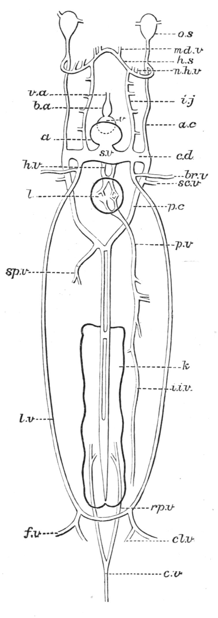
Fig. 186.—Venous system of Mustelus antarcticus. a, Auricle; a.c, anterior cardinal; b.a, conus arteriosus; br.v, brachial vein; c.d, Cuvierian duct or precaval vein; c.v, caudal vein; cl.v, cloacal vein; f.v, femoral vein; h.s, hyoidean sinus; h.v, hepatic vein; i.i.v, internal intestinal vein; i.j, inferior jugular; k, kidney; l, liver; l.v, lateral vein; md.v, mandibular vein; n.h.v, nutrient hyoidean veins; o.s, orbital sinus; p.c, posterior cardinal; p.v, hepatic portal vein; rp.v, renal portal vein; sc.v, subscapular vein; sp.v, spermatic vein; s.v, sinus venosus; v, ventricle; v.a, ventral aorta. (After T. J. Parker.)
The lateral veins (Fig. 186) are situated in the lateral walls of the abdomen, immediately external to the peritoneum (Fig. 187). Each vein begins near the pelvic fin, where it is connected with its fellow across the dorsal face of the ischio-pubic cartilage, and thence runs forward towards the pectoral fin. At its origin the lateral vein receives a femoral vein from the pelvic fin and a cloacal vein, and also, near its anterior end, a brachial vein from the pectoral fin, finally joining the Cuvierian duct of its side.[361]
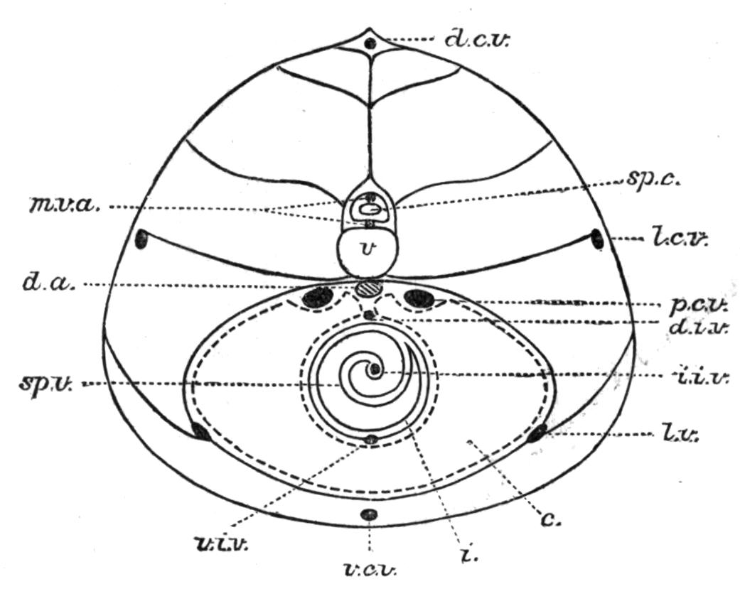
Fig. 187.—Diagrammatic transverse section of an Elasmobranch, showing the position of the principal longitudinal blood-vessels. c, Coelom; d.a, dorsal aorta; d.c.v, dorsal cutaneous vein; d.i.v, dorsal intestinal vein; i, intestine; i.i.v, internal intestinal vein; l.c.v, lateral cutaneous vein; l.v, lateral vein; m.v.a, myelonic vein and artery; p.c.v, posterior cardinal vein; sp.c, spinal cord; sp.v, spiral valve; v, vertebral centrum; v.c.v, ventral cutaneous vein; v.i.v, ventral intestinal vein. (From T. J. Parker.)
The anterior cardinal vein is situated directly above the gill-arches of its side of the head, and extends forwards from its junction behind with the Cuvierian duct to the outer side of the auditory capsule, where it communicates by a valvular orifice with a large sinus surrounding the eye-muscles (orbital sinus), and ventrally, by means of a similar aperture, with another large sinus, the hyoidean sinus, which lies on the outer face of the corresponding hyoid arch, and is continuous ventrally with its fellow of the opposite side. Into the orbital sinus open the anterior facial vein from the anterior and external regions of the head, and the anterior cerebral vein from the lateral half of the brain, and, into the hyoidean sinus, the nutrient veins from the hyoidean hemibranch.
The inferior jugular veins are situated beneath the branchial apparatus. Each vein begins anteriorly by communicating with the hyoidean sinus of its side, and, after receiving the nutrient veins from the holobranchs of the first four branchial arches, opens into the corresponding Cuvierian duct.
The venous blood from the heart itself is collected into two coronary veins, which open into the sinus venosus.
In addition to the more important veins already described, there is also a series of median and lateral cutaneous veins communicating at different points with certain of the more deeply seated veins (Fig. 187).
Characteristic features in the venous system of Mustelus, as also of Elasmobranchs in general, are the development of transverse connexions between certain of the principal paired veins, and the tendency of many of the main veins to enlarge into more or less irregularly-shaped sinuses.
In its broad outlines the venous system of the Teleostomi agrees with that of Elasmobranchs, but is nevertheless characterised by several more or less important modifications, while at the same time exhibiting many differences in minor details.
A renal portal system is usually present, but is singularly variable in the source of its tributary veins, even in closely allied forms.[362] In the Sturgeon (Acipenser) and in some Teleosts, as in the Siluroid, Amiurus catus, it resembles that of Elasmobranchs. In other Teleosts, on the contrary, the renal portal system presents various grades of degeneration, or, possibly, of imperfect evolution, as will be seen from the following illustrations of its condition in different genera.
In Amiurus the caudal vein, after giving off right and left renal portal veins to the renal capillaries, emerges from the ventral surface of the kidneys, and is then continued forwards between the gonads, the veins from which it receives, as the radicle of the hepatic portal vein.
In the Eel (Anguilla vulgaris) the caudal vein (Fig. 188) traverses the fused hinder portions of the kidneys, receiving several segmental veins from the body-wall and also giving off from each side numerous renal portal branches. More anteriorly, where the two kidneys become distinct, the caudal vein also divides into two renal portal veins and, as each vein extends forwards along the outer border of the kidney of its side, it receives a number of segmental veins, and, at the same time, gives off branches to the renal capillaries. In addition, each renal portal vein is connected with the hepatic portal vein by a series of singular arch-like vessels into which the ovarian or spermatic veins open.
It is obvious, therefore, that in both Amiurus and Anguilla the primitive direct continuity of the caudal and posterior cardinal veins has been interrupted by the formation of a well-developed renal portal system, and further, that the residue of the caudal venous blood finds its way to the liver through the hepatic portal vein; hence it follows that, as in so many of the lower air-breathing Vertebrates, the whole of the venous blood from the tail is distributed either to the kidneys or liver in the course of its return journey to the heart.
The Tench (Tinca vulgaris) exhibits the interesting anomaly of possessing two caudal veins, a dorsal and a ventral (Fig. 189). The dorsal vein is directly continuous with the right posterior cardinal, while the ventral one divides into three branches, two forming right and left renal portal veins and receiving numerous segmental veins, and the third becoming one of the affluents of the hepatic portal vein. In this Teleost it is clear that a portion of the caudal blood passes directly to the heart through the right posterior cardinal without traversing either the renal portal or hepatic portal system.
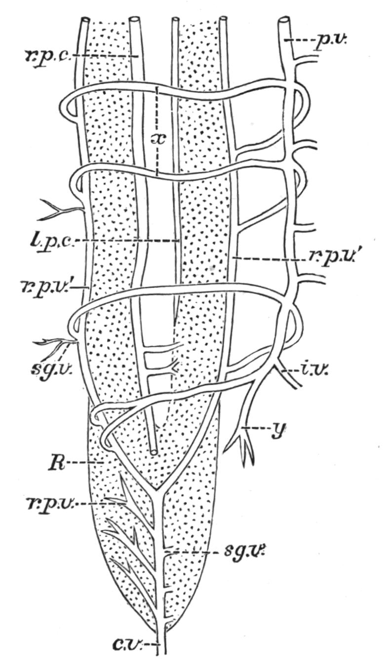
Fig. 188.—Renal portal circulation in the Eel (Anguilla vulgaris). c.v, Caudal vein; i.v, intestinal vein; l.p.c, r.p.c, left and right posterior cardinal veins; p.v, hepatic portal vein; R, kidney; r.p.v, r.p.v′, renal portal veins; sg.v, segmental veins; x, arch-like anastomoses between the renal portal and hepatic portal veins; y, vein from the urinary bladder. (From Jourdain.)
In the Cod (Gadus morrhua) the caudal vein divides into two branches. The larger right vein retains its direct continuity with the corresponding posterior cardinal; the left, on the contrary, has ceased to be continuous with the greatly reduced left posterior cardinal and forms a renal portal vein, the distribution of which is, however, restricted to the hinder portion of the left kidney (Fig. 190). As in Amiurus, a branch of the caudal vein forms one of the tributaries of the hepatic portal vein. In the Cod it would therefore seem that only a relatively small proportion of the caudal blood flows through the imperfectly developed renal portal system, the bulk of it traversing the right posterior cardinal and passing directly to the heart, leaving, nevertheless, a modicum for transmission to the liver. Finally, it may be mentioned that in some Teleosts the caudal vein retains its embryonic continuity with one, usually the right, posterior cardinal, without giving off a renal portal affluent, as in the Perch (Perca fluviatilis); or, after division, with both posterior cardinals, as in the Lump-sucker (Cyclopterus lumpus). In such instances as these no portion of the caudal blood traverses the kidneys, and if a renal portal system exists at all, the only true renal portal veins are the adjacent segmental veins, which transmit venous blood directly to the kidneys, instead of first uniting with renal portal branches of the caudal vein as in the Tench and the Eel.
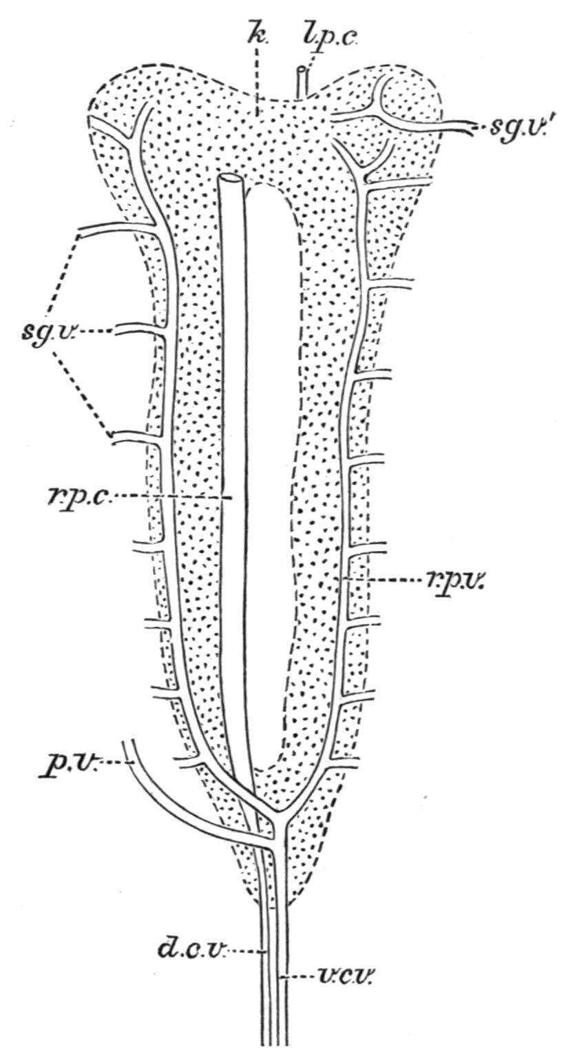
Fig. 189.—Renal portal system in the Tench (Tinca vulgaris). d.c.v, v.c.v, Dorsal and ventral caudal veins; k, kidney; l.p.c, r.p.c, left and right posterior cardinal veins; p.v, hepatic portal vein; r.p.v, renal portal vein; sg.v, sg.v′, segmental veins. (From Jourdain.)
Whatever may be the condition of the renal portal system, all the renal blood is eventually collected by renal veins and conveyed to the posterior cardinals, which are often connected by one or by several transverse anastomoses (Fig. 190). In the region of the heart each posterior cardinal joins the corresponding anterior cardinal to form a short but wide Cuvierian duct, which finally opens into the sinus venosus.
A subintestinal vein is present in the embryo (e.g. Lepidosteus, Acipenser, and some Teleosts),[363] but in the adult Teleostome its precaudal section is usually absorbed, or at all events ceases to be recognisable except, perhaps, as one of the minor tributaries of the hepatic portal vein.[364]
The hepatic portal vein is formed as in Elasmobranchs, but in different Teleostomi it may also receive the veins from the pyloric caeca, from a portion of the air-bladder, the gonads, and, as previously mentioned, a tributary from the caudal vein. There are usually two hepatic veins opening into the sinus venosus, and generally of equal size (Fig. 190).
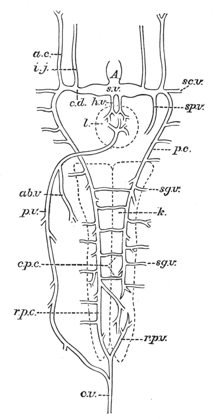
Fig. 190.—Venous system of a Teleost (diagrammatic). A, Auricle; ab.v, vein from the air-bladder; a.c, anterior cardinal; c.d, Cuvierian duct; c.p.c, transverse anastomoses between the two posterior cardinals; c.v, caudal vein; h.v, hepatic vein; i.j, inferior jugular; k, kidney; l, liver; p.c, left posterior cardinal; p.v, hepatic portal vein; r.p.c, right posterior cardinal; r.p.v, renal portal vein; sc.v, subclavian vein; sg.v, segmental vein; sp.v, spermatic vein; s.v, sinus venosus.
Most of the veins from the air-bladder join the hepatic portal vein, as already mentioned (Fig. 190), but more or fewer of them, especially those from the dorsal wall of the organ, open into the posterior cardinals. They may, as in Polypterus, even join the hepatic veins.[365]
The veins from the gonads are very variable in their destination, sometimes joining the posterior cardinals, as in the Salmon (Salmo salar); or the hepatic portal vein, as in Amiurus; or, as in the Perch (Perca fluviatilis), forming by their union a single trunk, which communicates directly with the left Cuvierian duct.
Representatives of the great lateral veins of Elasmobranchs appear to be absent in the Teleostomi, the veins from the pectoral and pelvic limbs joining the Cuvierian duct and the posterior cardinal veins respectively.
The two large anterior cardinal veins, which collect the blood from the head and brain, occupy their usual position directly above the branchial apparatus, and are sometimes connected by transverse anastomoses as they pass backwards to join the Cuvierian ducts. The inferior jugular vein is either single (e.g. Gadus); or paired, as in Perca (Fig. 190).
In the Dipnoi the venous system is distinguished by an interesting combination of characters, some of which are either primitive or peculiar to the group, while others exhibit a distinct transition to the embryonic or the adult condition of the lower air-breathing Vertebrates.
In Neoceratodus[366] (Fig. 191) the renal portal system is unusually complex, the veins distributing venous blood to the kidneys being derived from several sources, as follows: (1) from each of the two branches into which the caudal vein divides on its exit from the haemal canal (af.r.v); (2) from a common trunk (pt.v) which, on each side, is formed by the union of segmental veins from certain of the post-cloacal myotomes and is united with its fellow by a transverse anastomosis; (3) from more anteriorly situated intercostal or segmental veins (i.c.v) which enter each kidney directly; and (4) from a vein on each side corresponding to the renal portal vein of Amphibia. The latter vein (rp.v) is formed by one of the two branches of the iliac or femoral vein, and joins the corresponding vein from the caudal myotomes; from the common trunk numerous branches enter the kidney.
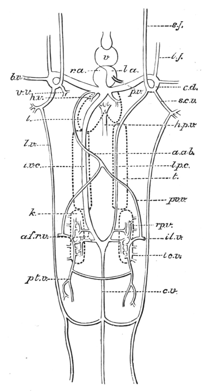
Fig. 191.—Venous system of Neoceratodus. a.ab, Anterior abdominal; af.r.v, afferent renal veins; b.v, brachial; c.d, Cuvierian duct; c.v, caudal; h.p.v, hepatic portal; h.v, hepatic; i.c.v, intercostal veins; i.j, inferior jugular; il.v, iliac; i.v.c, inferior vena cava or postcaval; k, kidney; l, liver; l.a, left auricle; l.p.c, left posterior cardinal; l.v, lateral cutaneous vein; pt.v, vein from postcloacal myotomes; p.v, pulmonary vein; pv.v, pelvic; r.a, right auricle; rp.v, renal portal; s.c.v, subscapular; s.j, superior jugular or anterior cardinal; t, testis; v, ventricle; v.v, right vertebral vein. (After Baldwin Spencer.)
In the derivation of renal portal veins from each of the two veins into which the caudal vein divides, Neoceratodus approaches the Elasmobranchs. On the other hand, the utilisation of ordinary segmental veins from the caudal and pre-caudal myotomes, some of which directly enter the kidney, is a feature which has already been remarked in some Teleosts; while the formation of a renal portal affluent by a branch of the femoral vein is an even more striking Amphibian characteristic.
The efferent renal veins[367] join the root of the left posterior cardinal and the adjacent portion of the caudal vein.
Of the two great venous trunks into which the caudal vein divides, the right is much the larger and behaves somewhat differently to the left. The former (i.v.c) passes forwards in relation with the right kidney, receiving in its course the spermatic or ovarian veins from the gonad of its side, and then traverses the liver, finally opening into the median portion of the sinus venosus, between the orifices of the two hepatic veins. The left branch of the caudal vein (l.p.c) also passes forwards in relation with the left kidney and receives veins from the corresponding gonad; but, instead of traversing the liver, it passes above that organ, and finally opens into the left Cuvierian duct. The course of the left vein, and the relations of the vessel to the caudal vein and the left Cuvierian duct, point to the conclusion that it represents the left posterior cardinal of other Fishes. From its continuity with the caudal vein it is also obvious that the hinder or renal portion of the right trunk is a remnant of the right posterior cardinal; but the more anterior section so closely resembles the postcaval vein, or inferior vena cava of the higher Vertebrates, in its relations to the liver, the hepatic veins, and the sinus venosus, that its identity as such seems beyond doubt, and this interpretation is supported by well-known observations[368] on the mode of origin of the inferior vena cava in Amphibia, and especially the union of the independently formed inferior vena cava with the posterior or inter-renal portion of the embryonic right posterior cardinal vein, combined with the atrophy of the anterior portion of the latter vein.[369] The singular connexions and relations of these two great veins afford an additional illustration of the significant transitional condition of the venous system in the Dipnoi. On the other hand, the direct continuity of the caudal vein with vessels which, wholly or in part, represent the two posterior cardinals, is a feature alike characteristic of the adult Cyclostome and the embryonic Elasmobranch, Teleost, and Amphibian.
As in the Cyclostomes and Elasmobranchs, the precaudal section of the embryonic subintestinal vein is represented in the adult by an intra-intestinal vein which traverses the spiral valve near its free edge and is a tributary of the hepatic portal vein.
The two veins from the undivided air-bladder unite to form a single vessel, which, instead of joining the hepatic portal or posterior cardinal veins as in other Fishes, opens into the left auricle, like the pulmonary veins of the Amphibia.
A further resemblance to the Amphibia is to be found in the presence of an anterior abdominal vein. After leaving the pelvic limb each femoral vein divides into two branches; one of these forms a renal portal vein as previously described; the other, which may rightly be termed a pelvic vein (pv.v), unites with its fellow to form a median anterior abdominal vein (a.ab). Pursuing its course forwards in the ventral abdominal wall, the vein eventually reaches the heart and opens into the sinus venosus. The direct connexion of the anterior abdominal vein with the heart is yet another example of the retention in the adult Neoceratodus of a transitory embryonic feature in the developing Amphibian.[370]
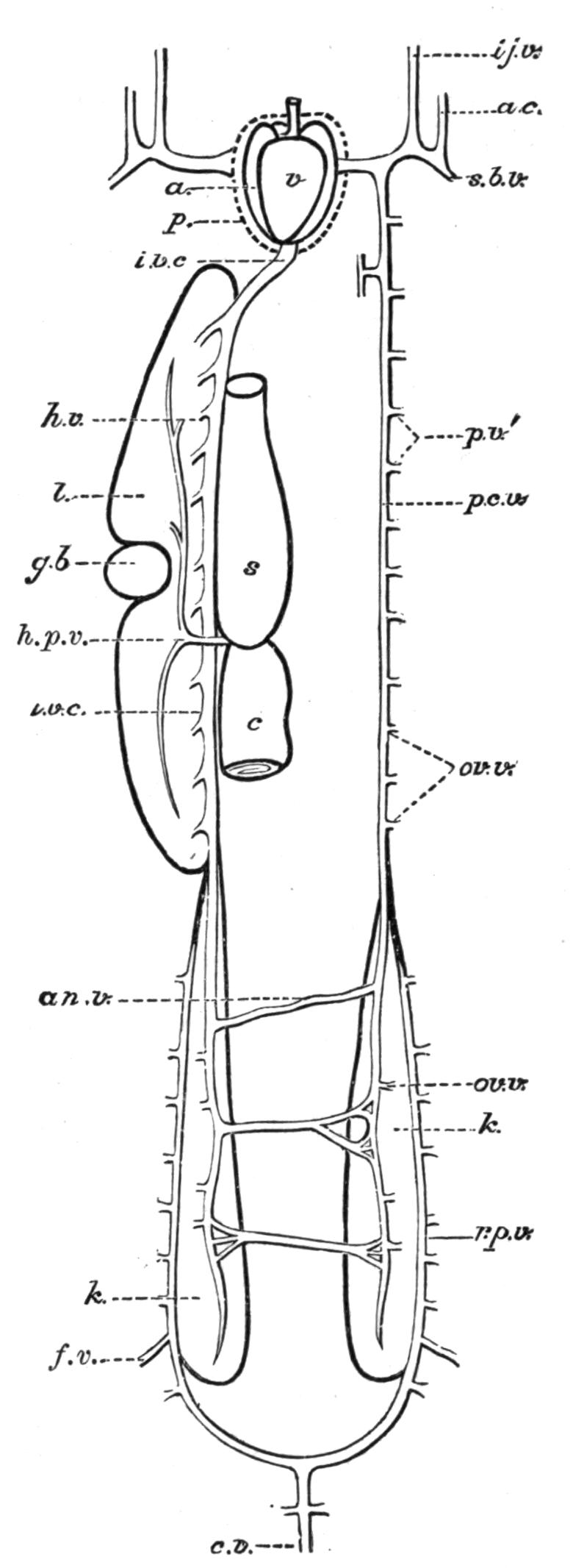
Fig. 192.—Venous system of Protopterus. a, Auricle; a.c, anterior cardinal; an.v, anastomotic veins; c, intestine; f.v, femoral or iliac vein; g.b, gall-bladder; h.p.v, hepatic portal vein; i.j.v, inferior jugular; ov.v, ovarian veins; p, pericardium; p.c.v, left posterior cardinal; p.v′, parietal or segmental veins; s, stomach; sb.v, subclavian. Other reference letters as in Fig. 191. (From Newton Parker.)
As in other Fishes, the blood from the head is conveyed to the Cuvierian ducts by an anterior cardinal and an inferior jugular on each side. There are no lateral veins, the blood from the pelvic fins flowing into the renal portal system or into the anterior abdominal vein, and that from the pectoral fin through subscapular and brachial veins into the Cuvierian ducts. Lateral cutaneous veins are, however, present; and, as in Elasmobranchs (e.g. Mustelus antarcticus), anastomose anteriorly with the subscapular vein and behind with the caudal vein.
Less is known of the venous system of Protopterus,[371] but it is certain, nevertheless, that it presents a more advanced grade of evolution than in Neoceratodus, and, except for the doubt as to the existence of an anterior abdominal vein, it is essentially similar to that of a Urodele Amphibian in which the right posterior cardinal vein has aborted.
The caudal vein (Fig. 192) divides into right and left renal portal branches, neither of which, however, is directly continuous with the inferior vena cava or the left posterior cardinal; on the contrary, each renal portal vein is joined by the corresponding iliac or femoral vein, and also by numerous segmental veins, and then distributes the whole of its venous blood to the kidney. The radicles of the inferior vena cava and the left posterior cardinal are formed by the renal veins from the two kidneys, and in their forward course to the heart both veins receive in addition genital and segmental veins. In its course through the liver the inferior vena cava receives several hepatic veins, and finally opens into the sinus venosus, while the left posterior cardinal vein joins the corresponding Cuvierian duct, which also receives anterior cardinal, inferior jugular, and subclavian veins. There is an intra-intestinal vein as in Neoceratodus, but an anterior abdominal vein has yet to be discovered. The two pulmonary veins from the double air-bladder form a single trunk before communicating with the left auricle.
With the exception of certain doubtful details which need further investigation, the venous system of Lepidosiren[372] seems to resemble that of Protopterus.
The Heart.—The heart is more anteriorly placed than in other Vertebrates, being situated directly behind and beneath the last pair of branchial clefts and internal to the ventral portion of the pectoral girdle. The organ is enclosed in a pericardial cavity, which, in the adult, is separated from the abdominal portion of the coelom by a transverse pericardio-peritoneal septum, and in the Lamprey (Petromyzon) is partially enclosed within a cartilaginous, cup-like modification of the hinder part of the branchial basket. In the Ammocoetes-stage of the Lamprey the pericardium is in communication behind with the general coelom, but the connexion is lost in the adult. In Elasmobranchs the two cavities are connected by a single pericardio-peritoneal canal, or by two such canals; and in Chimaera, and in the Sturgeon (Acipenser) and Polyodon, by a single canal.
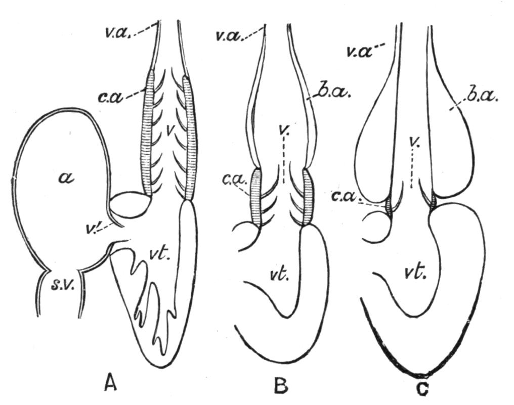
Fig. 193.—Diagram of the structure of the heart in different Fishes. A, In an Elasmobranch; B, in Amia; and C, in a Teleost. a, Auricle; b.a, bulbus aortae; c.a, conus arteriosus; s.v, sinus venosus; v, semi-lunar valves; v′, auriculo-ventricular valve; v.a, ventral aorta; vt, ventricle. (From Boas.)
The heart consists of at least three chambers, a sinus venosus which receives the venous blood from the body, an auricle and a ventricle, to which is added a conus arteriosus in the Elasmobranchs, certain Teleostomi (Crossopterygii, Chondrostei, and Holostei), and in the Dipnoi. Through these cardiac chambers the blood is forced in the order mentioned. In the Dipnoi the auricle is subdivided by a more or less complete interauricular septum into a right and left auricle,[373] the former receiving the venous blood from the sinus venosus, and the latter the aerated blood from the lung-like air-bladder.
The sinus venosus and the auricle have very thin walls; the ventricular walls, on the contrary, are very thick and in great measure are composed of a sponge-like network of muscular bundles which generally encroaches considerably on the ventricular cavity. Membranous valves, the sinu-auricular, and the auriculo-ventricular valves, are developed at the junctions of the sinus venosus with the auricle, and the auricle with the ventricle respectively. The conus arteriosus is muscular and contractile, and is interposed between the ventricle and the root of the ventral aorta. Internally, the conus is provided with several transverse rows of pocket-shaped or semilunar valves. In Teleosts the conus is non-muscular and vestigial, and has but a single row of valves, corresponding to the most anterior of the multiple rows of valves in the Elasmobranchs. In these Fishes the vestigial conus is succeeded by a non-contractile, bulb-like dilatation, or bulbus aortae, of the root of the ventral aorta. In only a single Teleost, viz. Albula, one of the Albulidae, is the vestigial conus muscular, and at the same time provided with two rows of valves.[374] In the Cyclostomata there is a bulbus with a single row of two valves, but no true conus.
In the Dipnoi (e.g. Protopterus) the heart, like the rest of the vascular system, exhibits certain interesting resemblances to the Amphibian heart. In addition to a more or less complete interauricular septum separating right and left auricles, there is a median longitudinal ridge, partly muscular and partly fibrous, which incompletely subdivides the cavity of the ventricle. The spirally-twisted conus arteriosus is furnished with several transverse rows of valves, certain of which coalesce longitudinally to form a complete septum dividing the cavity of the conus into two distinct lateral channels: with this septum there coalesces another septum, which cuts off the origins of the anterior two pairs from the remaining afferent branchial arteries. The formation of these septa has the physiological effect of subdividing the series of cardiac cavities into two parallel channels, of which one has its origin behind in the sinus venosus and transmits venous blood to the posterior afferent branchial vessels; while the other, commencing with the left auricle, conveys arterial blood to the first two pairs of afferent branchial arteries.[375] In Neoceratodus, however, the longitudinal septum in the conus is incomplete, and hence the blood which is sent to the anterior afferent vessels is mixed.[376]
The Arterial System.—The ventral aorta is a median artery situated beneath the floor of the pharynx, and having its origin, behind, either directly from the ventricle or from the conus arteriosus.
In the Cyclostomata[377] (e.g. Petromyzon) the ventral aorta (Fig. 194) is continued forwards from the heart as a single vessel to the fourth pair of gill-sacs, where it divides into right and left branches which extend as far as the anterior walls of the first pair of gill-sacs. Eight pairs of afferent branchial arteries arise from the ventral aorta and its two branches, of which the first and last supply the anterior walls of the first pair of sacs and the posterior walls of the last pair respectively. Each of the remaining afferent vessels extends into an interbranchial septum, and supplies the gill-lamellae of the posterior wall of one sac and those of the anterior wall of the next sac behind. The corresponding efferent branchial vessels have a similar distribution, and unite dorsally to form a median dorsal aorta. Beneath the base of the skull the latter vessel divides into two branches which, after receiving the first pair of efferent branchial vessels, pursue a divergent course forwards, but subsequently converge and unite to form a "circulus cephalicus," as in Teleosts. From the cephalic circle are given off on each side (1) an "internal carotid" artery for the brain and eye; (2) an "external carotid" for the lateral and ventral walls of the head; and (3) a large ventral branch which supplies the lingual apparatus; while from the abdominal portion of the dorsal aorta are derived, first, a coeliaco-mesenteric artery for the liver and alimentary canal, and subsequently branches for the myotomes, kidneys, and the gonad. The terminal portion of the aorta then enters the tail and forms the caudal artery.
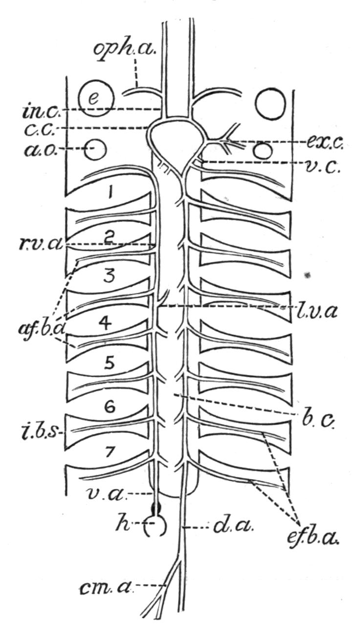
Fig. 194.—Branchial arterial system of the Lamprey (Petromyzon fluviatilis). The ventral aorta and the afferent branchial arteries are seen on the left side, and the efferent branchial and dorsal aorta on the right (diagrammatic). af.b.a, ef.b.a, Afferent and efferent branchial arteries; a.o, auditory organ; b.c, branchial canal; c.c, cephalic circle; cm.a, coeliaco-mesenteric artery; d.a, dorsal aorta; e, eye; ex.c, "external carotid"; h, heart; i.b.s, interbranchial septum; in.c, internal carotid; oph.a, ophthalmic artery; r.v.a, l.v.a, right and left ventral aorta; v.a, median ventral aorta; v.c, "ventral carotid"; 1-7, gill-sacs. (Modified from Vogt and Yung.)
In Elasmobranchs[378] (e.g. Mustelus antarcticus) the undivided ventral aorta gives off five pairs of afferent branchial arteries which, on each side, ascend in succession the outer convex sides of the hyoid and first four branchial arches (Fig. 195).
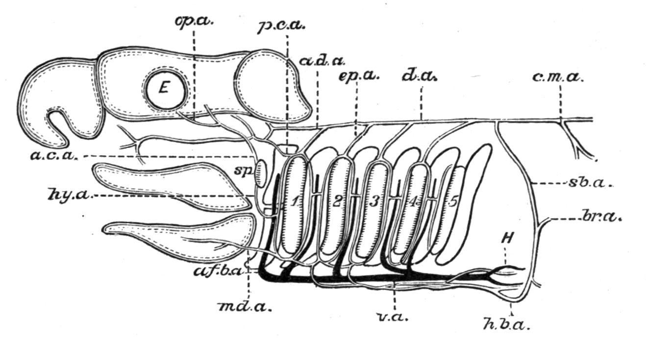
Fig. 195.—The branchial arterial system of Mustelus antarcticus. Left lateral view. The ventral aorta and afferent branchial vessels are in solid black, the efferent arteries and their branches have double contours. The branchial clefts have fringed borders to indicate their hemibranchs, and the arches are in simple outline. a.c.a, Anterior carotid; a.d.a, anterior dorsal aorta; af.b.a, afferent branchial artery; br.a, brachial artery; c.m.a, coeliaco-mesenteric; d.a, dorsal aorta; E, eye; ep.a, epibranchial artery; H, heart; h.b.a, hypobranchial artery; hy.a, afferent pseudobranchial or hyoidean artery; md.a, mandibular artery; op.a, ophthalmic artery; p.c.a, posterior carotid; sb.a, subclavian; sp, spiracle; v.a, ventral aorta; 1-5, the hyobranchial and four succeeding branchial clefts. The hypobranchial artery is seen immediately beneath the ventral aorta. (After T. Jeffery Parker, diagrammatic.)
The first or most anterior of these arteries supplies the hyoidean hemibranch, while the succeeding four supply the holobranchs of the four branchial arches. The blood is collected from the capillaries of the branchial lamellae by a series of efferent branchial vessels, a pair for the two hemibranchs of each branchial arch and a single vessel for the hyoidean hemibranch, which unite with one another in a somewhat singular fashion. The efferent arteries from the anterior and posterior hemibranchs of each branchial cleft unite above and below each cleft in such a way as to form a series of complete vascular loops round the hyoidean cleft and the three succeeding branchial clefts, which are connected by short longitudinal trunks in each arch and also by a longitudinal commissural vessel between their ventral extremities. As the fifth arch is gill-less, there is no complete loop round the fifth cleft, the blood collected by the efferent vessel of the posterior hemibranch of the fourth arch being conveyed to the corresponding vessel of the anterior hemibranch of the same arch by one of the short longitudinal vessels above mentioned. Dorsally, each arterial loop is continuous with an epibranchial artery; and by the dorsal union of the four epibranchial arteries of the two sides the dorsal aorta is formed. It may be pointed out that the anterior efferent vessel of each arch, which is usually larger than the posterior one, is to be regarded as the primary efferent artery of the corresponding holobranch, and as such is directly continuous with an epibranchial artery, the posterior efferent artery being a secondary vessel which opens not into the primary trunk of its own branchial arch, but into that of the succeeding arch.[379] The principal arteries which supply the various parts of the head with blood are derived from the first efferent branchial vessel. From the ventral end of this artery a mandibular artery is given off, which subdivides into branches for the muscles of the lower jaw as well as into nutrient vessels for the hyoidean hemibranch. At about the middle of its length the same artery gives off an afferent pseudobranchial or hyoidean artery, to the spiracular or mandibular pseudobranch. From the latter organ the blood is collected by an anterior carotid artery which, after giving off an ophthalmic branch to the eye, perforates the orbital wall and enters the cranial cavity, where it is joined by an anastomotic trunk from the posterior carotid of the opposite side; finally, the anterior carotid divides into anterior and posterior cerebral arteries for the brain. The third and last of the cephalic arteries is the posterior carotid; this artery arises from the dorsal extremity of the first efferent branchial vessel, and, on entering the orbit, gives off the anastomotic trunk previously mentioned. The latter vessel enters the cranial cavity, and, after crossing its fellow, joins the anterior carotid of the opposite side, as described above. The main trunk is then continued forwards in the orbit, and its various branches eventually supply the eye-muscles, the mandibular adductor muscle, and some other parts of the head.
It is worthy of note that the median dorsal aorta is prolonged forwards in front of the first pair of epibranchial arteries as a slender median vessel (a.d.a), which ultimately divides into two branches, each branch uniting with the posterior carotid of its side.
A remarkable system of arteries for the supply of nutrient blood to the gills and heart has its origin in the following manner. On each side, the longitudinal commissural vessel, which connects the ventral ends of the arterial loops surrounding the different gill-clefts, gives origin to a series of pairs of short transverse vessels, and by their union these combine to form a median longitudinal hypobranchial artery which lies beneath the ventral aorta. From the hypobranchial artery are derived the coronary arteries for the heart; and from the same artery, or from its lateral connexions with the longitudinal commissural artery, and, in the case of the hyoidean hemibranch, from the mandibular artery, are derived the various nutrient vessels for the gills.
The arteries for the trunk, and for the pectoral and pelvic limbs, arise in succession from the dorsal aorta. The first of the series is the subclavian artery, which has its origin from the aorta close to the dorsal extremities of the fourth pair of epibranchial arteries. Each subclavian artery gives off a brachial artery to the pectoral fin, and is then continued forwards as a lateral hypobranchial artery, which, with its fellow of the opposite side, eventually becomes continuous with the hinder end of the median hypobranchial artery. Behind the subclavian artery there is a median coeliaco-mesenteric artery, the various branches of which are distributed to the liver, stomach, and intestine. A lieno-gastric artery supplies the pancreas and spleen, and also sends branches to the stomach. In addition, there are also arteries for the gonads, numerous segmental arteries for the myotomes, and renal arteries for the kidneys. Finally, the aorta gives off a pair of iliac arteries for the pelvic fins, and then enters the haemal canal as the caudal artery.
The more important differences in the arterial system of the Holocephali and the Teleostomi relate to (1) the absence of the posterior efferent branchial artery in each branchial arch; (2) modifications dependent on the condition of the spiracular and hyoidean hemibranchs, and the mode of origin and the course of their afferent and efferent vessels; and (3) the source from whence the air-bladder derives its blood when that organ is present.
(1) The branchial arterial system is somewhat more primitive than in the generality of Elasmobranchs.[380] There are no complete vascular loops round the gill-clefts, and the blood from the two hemibranchs of each branchial arch is conveyed to the dorsal aorta by a single efferent vessel which corresponds to the more anterior of the two in Mustelus antarcticus.[381]
(2) In Callorhynchus[382] among the Holocephali, where the spiracle is absent but the hyoidean hemibranchi is still a true gill, the latter organ is supplied with venous blood by a branch from the ventral aorta, the corresponding efferent vessel joining the dorsal aorta (Fig. 196). In the absence of a spiracular pseudobranch the anterior carotid may be regarded as continuous with the hyoidean artery,[383] and as having its origin directly from the efferent artery of the hyoidean hemibranch (Fig. 196). At its origin the anterior carotid anastomoses with the mandibular artery.
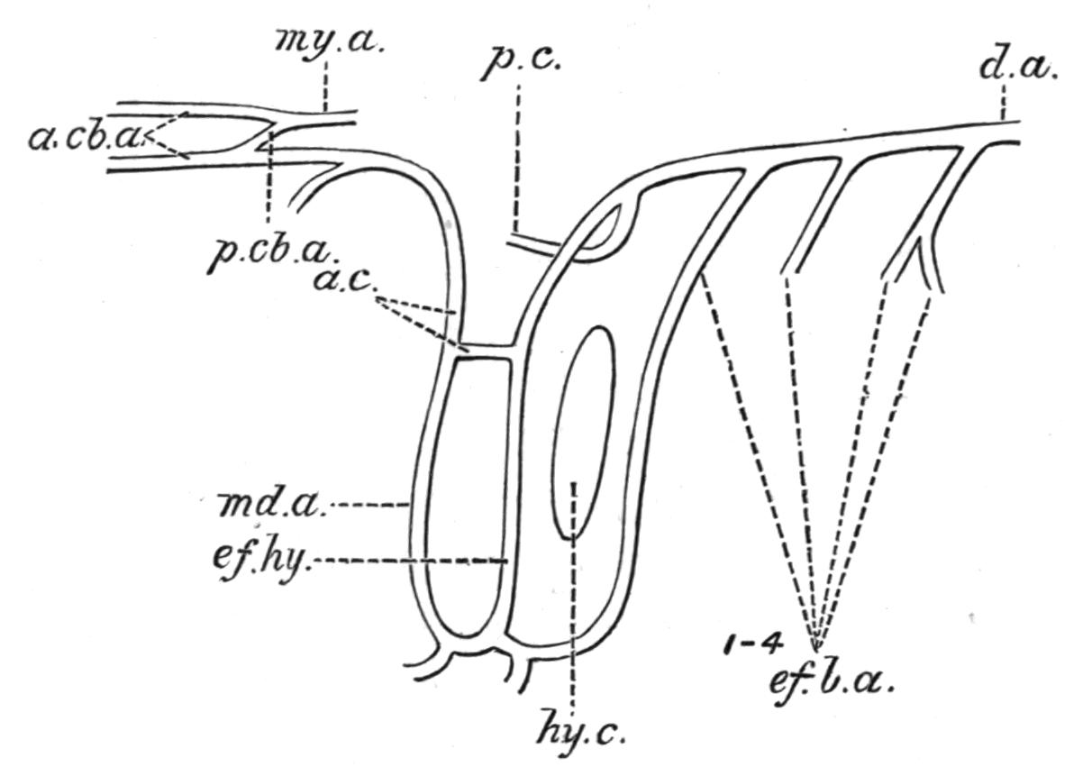
Fig. 196.—Portion of the efferent branchial system of Callorhynchus. a.c, Anterior carotid; a.cb.a, anterior cerebral arteries; d.a, dorsal aorta; ef.b.a, 1-4, efferent branchial arteries; ef.hy, efferent artery from the hyoidean hemibranch; hy.c, hyobranchial cleft; md.a, mandibular artery; my.a, myelonal artery; p.c, posterior carotid; p.cb.a, posterior cerebral artery. (From T. Jeffery Parker.)
The Sturgeon more closely resembles the Elasmobranchs. The hyoidean gill is supplied by an afferent branchial artery from the ventral aorta, and its efferent vessel joins the corresponding trunk from the holobranch of the first branchial arch. A hyoidean artery supplies the spiracular pseudobranch, the efferent vessel of which contributes to the blood-supply of the brain and the eye, and probably represents an anterior carotid.
Lepidosteus[384] offers a singularly interesting transition from the Elasmobranch to the Teleost. As indicated in the preceding chapter, this Fish possesses both a hyoidean gill and a spiracular pseudobranch (Figs. 197 and 198). The hyoidean gill is supplied by an afferent artery direct from the ventral aorta, but the proper efferent vessel of the gill, which primitively joined the dorsal aorta, is suppressed, and the blood is collected into a vessel, which, like the hyoidean artery in Elasmobranchs, becomes the afferent artery of the spiracular pseudobranch. The latter artery unites, however, with a second hyoidean artery derived from the efferent branchial vessel of the first branchial arch, and represents the artery termed "hyoidean" in Teleosts. The efferent vessel from the spiracular pseudobranch joins an internal branch from the carotid artery, and then distributes its blood both to the eye and the brain.
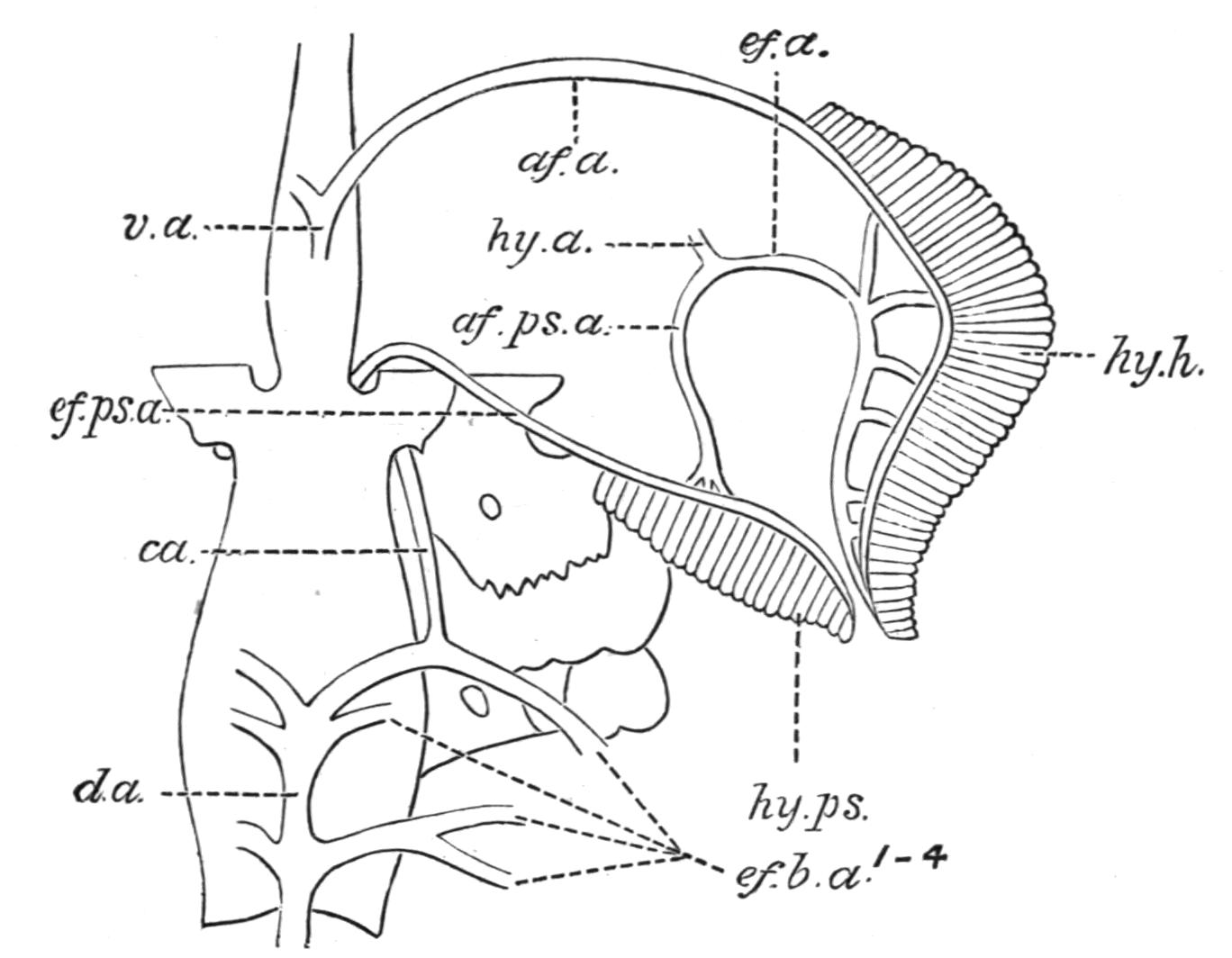
Fig. 197.—Blood-vessels of the spiracular pseudobranch and the hyoidean gill in Lepidosteus. af.a, ef.a, Afferent and efferent vessels of the hyoidean gill; af.ps.a, ef.ps.a, afferent and efferent vessels of the spiracular pseudobranch; ca, carotid (posterior); d.a, dorsal aorta; ef.b.a1-4, efferent branchial vessels; hy.a, hyoidean artery; hy.h, hyoidean gill; hy.ps, spiracular pseudobranch; v.a, ventral aorta. (From F. W. Müller, after Johannes Müller.)
In Teleosts, as already mentioned in a preceding chapter, it is probable that the hyoidean hemibranch is suppressed, the so-called hyoidean pseudobranch being a spiracular pseudobranch. The latter is now supplied by a "hyoidean" artery, which has its origin from the ventral end of the efferent branchial artery of the first branchial arch, the corresponding efferent trunk forming an ophthalmic artery, and passing to the choroid gland of the eye (Fig. 199). Both the proper afferent and efferent arteries of the hyoidean hemibranch either disappear or, as in the Cod (Gadus morrhua), the efferent artery may be represented on each side by an anastomosis between the hyoidean artery and the cephalic circle. Hence, the "hyoidean" artery of Teleosts corresponds to the one which has a similar origin in Lepidosteus.
A brief description of the remaining efferent branchial arteries and their derivatives in the Cod (Gadus morrhua) will illustrate the condition of these structures in a well-known Teleost.
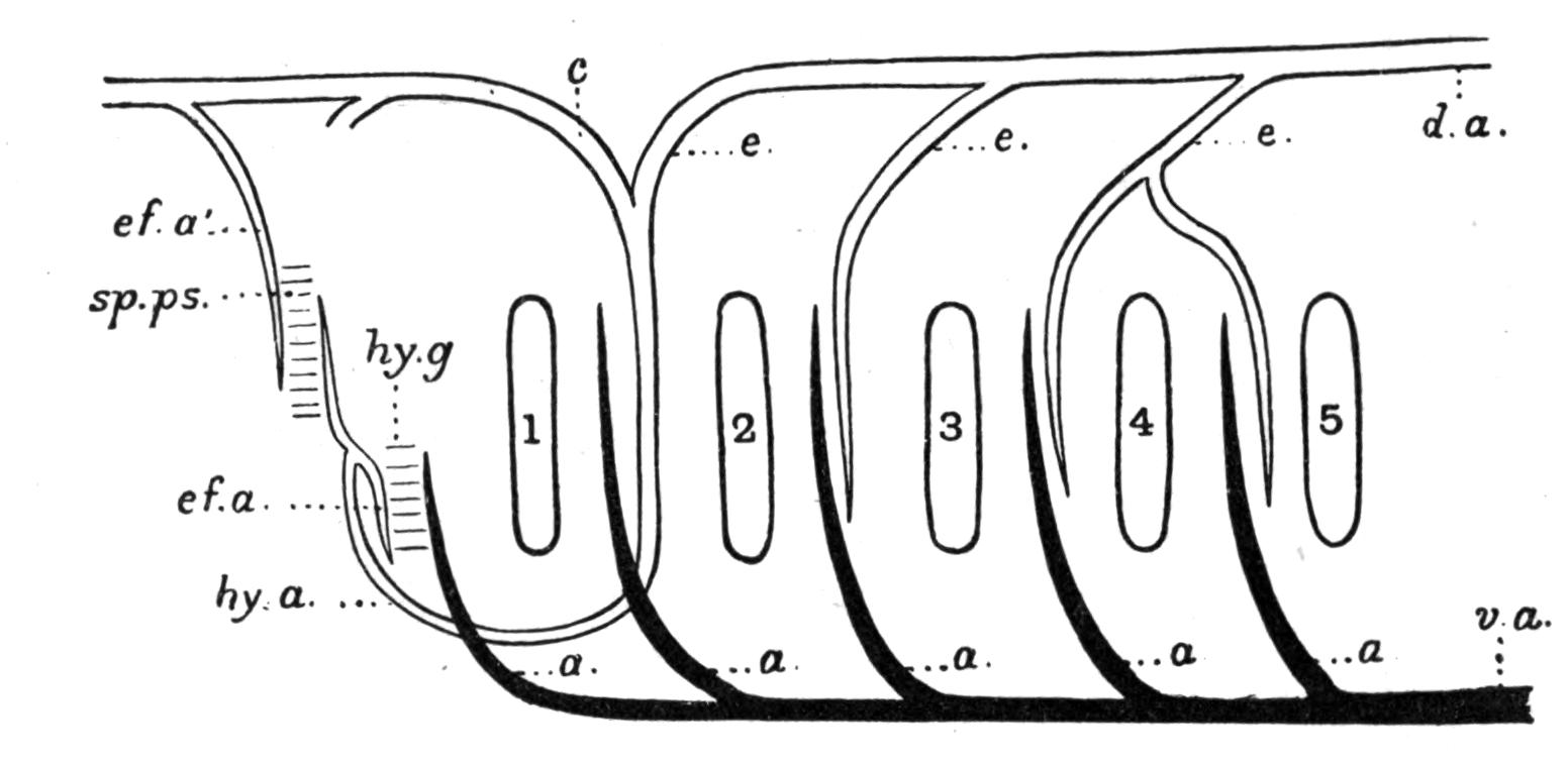
Fig. 198.—The branchial circulation in Lepidosteus (diagrammatic). a, a, Afferent branchial arteries; c, carotid; d.a, dorsal aorta; e, e, efferent branchial arteries; ef.a, efferent vessel from the hyoidean gill which, after its union with the hyoidean artery, becomes the afferent vessel of the spiracular pseudobranch; ef.a′, efferent vessel of the spiracular pseudobranch; hy.a, hyoidean artery; hy.g, hyoidean gill; sp.ps, spiracular pseudobranch; v.a, ventral aorta; 1-5, the hyo-branchial and succeeding gill-clefts. (After F. W. Müller and Ramsay Wright.)
In this Fish the efferent branchial vessels open dorsally into right and left suprabranchial arteries,[385] which unite behind to form a median dorsal aorta (Fig. 199). Anteriorly, the paired suprabranchial arteries extend towards the base of the skull as the so-called "carotid" arteries. The two carotids enter the cranial cavity, and there unite in the median line, as in the Cyclostomes. By the union of these arteries in front, and of the right and left suprabranchial arteries behind, the characteristic "circulus cephalicus" of Teleosts is completed.[386] From the anterior part of the cephalic circle are derived two internal carotid arteries[387] for the brain, and also a pair of orbito-nasal arteries for the eye-muscles and the nasal sacs, while more posteriorly an external carotid has its origin from each suprabranchial artery.
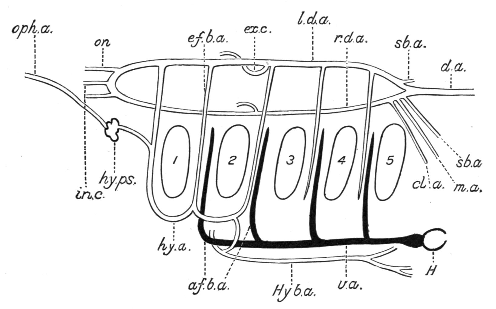
Fig. 199.—Branchial arterial system of the Cod (Gadus morrhua). Lateral view. af.b.a, First afferent branchial artery; cl.a, coeliac artery; d.a, median dorsal aorta; ef.b.a, first efferent branchial artery; ex.c, external carotid; H, heart; hy.a, hyoidean artery; Hy.b.a, hypobranchial artery for the heart and pelvic fins; hy.ps, spiracular pseudobranch; in.c, internal carotid;[387] l.d.a, left suprabranchial artery; m.a, mesenteric artery; on, orbito-nasal artery; oph.a, ophthalmic artery; r.d.a, right suprabranchial artery; sb.a, subclavian artery; v.a, ventral aorta; 1-5, hyobranchial and succeeding gill-clefts. (Altered from T. Jeffery Parker.)
(3) In most Teleostomi the air-bladder is supplied with blood by branches of the coeliac artery, with the addition of small branches arising directly from the dorsal aorta. Polypterus and Amia[388] are, however, exceptional, inasmuch as the arteries for the air-bladder are derived from the last or fourth pair of efferent branchial vessels, and in this respect, but not in the destination of the corresponding veins, the two genera exhibit a significant resemblance to the Dipnoi.
In the Dipnoi the ventral aorta is so short that the afferent branchial arteries arise almost directly from the conus arteriosus with their roots in close contiguity to one another (Fig. 200).
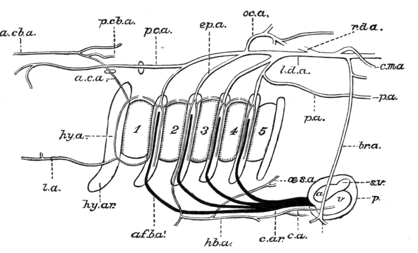
Fig. 200.—Branchial arterial system of Neoceratodus. Lateral view. The conus arteriosus and the afferent branchial vessels are represented in solid black, the efferent vessels and their derivatives with double contours. a, Auricle; a.c.a, anterior carotid; a.cb.a, anterior cerebral artery; af.b.a′, first afferent branchial artery; br.a, brachial artery; c.a, coronary artery; c.ar, conus arteriosus; c.m.a, coeliaco-mesenteric; ep.a, epibranchial artery; hb.a, hypobranchial artery; hy.a, hyoid artery; hy.ar, hyoidean arch; l.a, lingual; l.d.a, r.d.a, left and right dorsal aortae; oc.a, occipital artery; oes.a, oesophageal artery; p, pericardium; p.a, pulmonary artery; p.c.a, posterior carotid; p.cb.a, posterior cerebral artery; s.v, sinus venosus; v, ventricle; 1, hyobranchial cleft; 2-5, branchial clefts. (After Baldwin Spencer, diagrammatic.)
In Neoceratodus (Fig. 200),[389] there are two efferent vessels to each gill-bearing branchial arch, which unite above to form an epibranchial artery, and by the successive union of the four epibranchial arteries a short common trunk is formed on each side. Posteriorly, the two trunks unite to form a median dorsal aorta. Immediately above the gill-clefts each efferent vessel gives off a branch which, passing either forwards or backwards, unites with the corresponding branch of the efferent vessel in front or behind as the case may be. A hyoidean artery arises from the ventral extremity of the anterior efferent artery of the first branchial arch, and, after giving off a lingual artery, ascends the hyoid arch and supplies the hyoidean pseudobranch. The efferent vessel of the pseudobranch (a.c.a) or anterior carotid artery, eventually enters the cranial cavity and subdivides into anterior and posterior cerebral arteries for the brain, also giving off a branch which unites with its fellow of the opposite side directly behind the infundibulum. A posterior carotid springs from the epibranchial of the first branchial arch and divides into palatine, orbital, and ocular branches; and from the ventral end of the anterior efferent vessel of the second branchial arch is derived a hypobranchial artery for the heart and pericardium. The pulmonary arteries for the lung-like air-bladder have their origin from the fourth pair of epibranchial arteries.
As in so many other details of its anatomy, Neoceratodus exhibits in its arterial system abundant evidence of the wide-spreading affinities of the group to which it belongs. In its branchial arterial system Neoceratodus presents a singular combination of features which, individually, are characteristic of Amphibia and Elasmobranchs. Special Amphibian features may be noted in the origin of the afferent branchial arteries almost simultaneously from the anterior end of the conus arteriosus; in the mode of union of the epibranchial arteries to form the dorsal aortae; in the origin of a lingual artery from the efferent vessel of the first branchial arch; and in the derivation on either side of a pulmonary artery from the fourth epibranchial artery. Agreement with Elasmobranchs is to be found in the presence of two efferent branchial vessels in each branchial arch, although the relations of these arteries are more primitive than in most adult Elasmobranchs, inasmuch as the two efferent vessels of the same arch unite to form an epibranchial artery; and also in the origin and distribution of the anterior and posterior carotids. Lastly may be mentioned the fact that Neoceratodus agrees not only with the Amphibia but also with those generalised Teleostomi, Polypterus and Amia, in the mode of origin of the great arteries for the air-bladder.
Of the two remaining Dipnoi, the arterial system of Protopterus[390] is better known than that of Lepidosiren, but in both cases further research is needed before a satisfactory comparison can be made with Neoceratodus and other Vertebrates. It is evident, nevertheless, that both genera differ from Neoceratodus in approximating more closely to the Amphibia than to the lower Fishes, in so far as the branchial part of the arterial system is concerned.
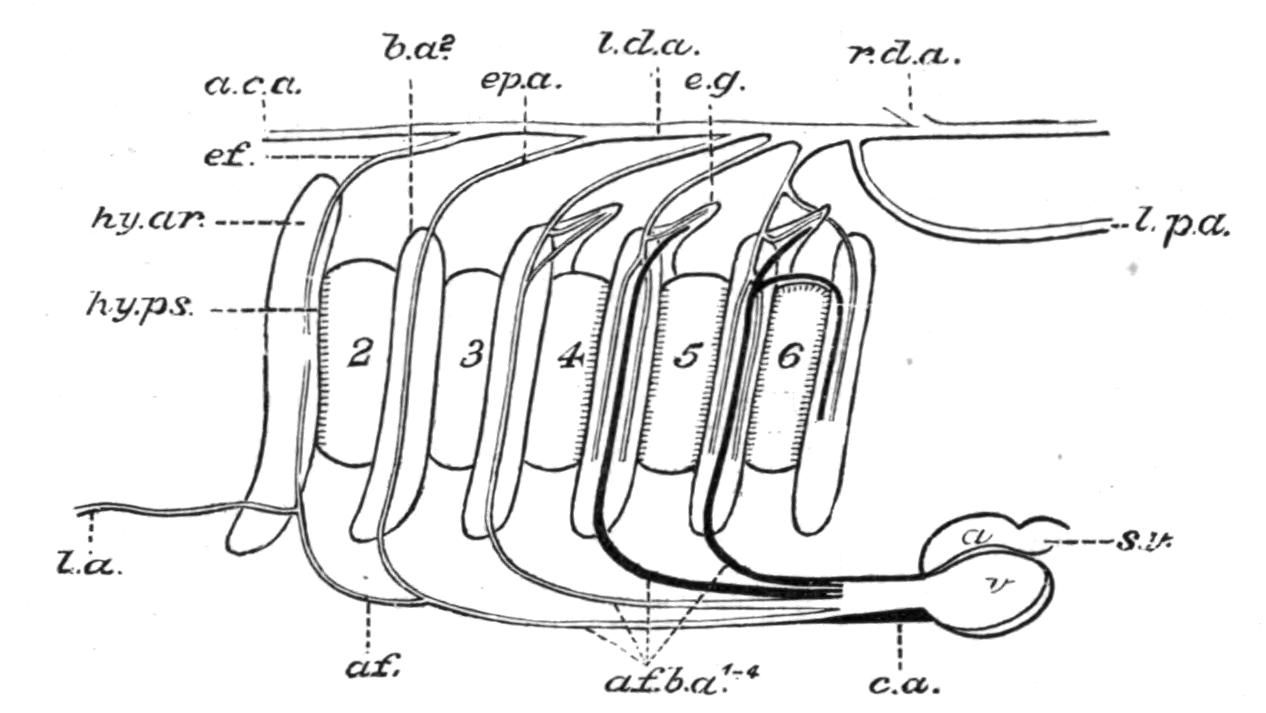
Fig. 201.—Branchial arterial system of Protopterus (diagrammatic). a, Auricle; a.c.a, carotid artery; af.b.a1-4, afferent branchial arteries; af, ef, afferent and efferent vessels of the hyoidean pseudobranch; b.a2, second branchial arch, the vestigial first arch being omitted; c.a, conus arteriosus; e.g, external or cutaneous gill; ep.a, epibranchial artery; hy.ar, hyoid arch; hy.ps, hyoidean pseudobranch; l.a, lingual artery; l.d.a and r.d.a, right and left dorsal aortae; l.p.a, left pulmonary artery; s.v, sinosus venosus; v, ventricle; 2-6, the second branchial and succeeding clefts, the hyobranchial cleft being closed. The vestigial first branchial arch is not shown. The epibranchial arteries unite to form the right or left dorsal aorta at the same point and not in succession as in the figure. (Altered from Newton Parker.)
In their origin from the conus the four afferent branchial arteries of Protopterus resemble those of Neoceratodus, but their relations to the branchial clefts are somewhat different (Fig. 201). The first or hyoidean cleft is closed, and the first afferent vessel lies between the second cleft and the third, and is therefore in relation with the second branchial arch. The remaining afferent arteries are disposed between the succeeding clefts and are related to the corresponding arches. As the second and third arches, like the vestigial first arch, bear no gill-lamellae, their afferent arteries are directly continuous with the corresponding efferent vessels, as in those Teleosts in which certain arches are gill-less, as well as in the Tadpole-stage of the tailless Amphibia when the internal gills begin to degenerate; and they apparently transmit arterial blood directly to the dorsal aorta.[391] The third and fourth afferent arteries, on the contrary, supply venous blood to the two hemibranchs which are borne by each of the two corresponding arches, viz.: the fourth and fifth, and from each pair of hemibranchs the blood is collected into two efferent vessels which unite dorsally to form an epibranchial artery. From the dorsal end of the fourth afferent artery there arises a recurrent branch which curves round the upper margin of the sixth cleft and supplies the gill-lamellae on the posterior margin of that cleft, a fact which lends support to the view that these lamellae are "emigrants" from the anterior margin of the cleft; the efferent vessel from the "emigrant" lamellae joins the fourth epibranchial artery. The blood-supply of the external or cutaneous gills is derived from the dorsal extremities of the second, third, and fourth afferent arteries, while the efferent vessels from these organs join the corresponding epibranchial arteries; in this respect there is a close resemblance between Protopterus and those larval Amphibians which possess similar cutaneous gills. All four epibranchial arteries unite together at about the same point to form a short common trunk, the right or left dorsal aorta, which subsequently unites with its fellow to form the median dorsal aorta.
There is a so-called "hyoidean" artery, which, however, has its origin, not from an anterior efferent branchial vessel as in Neoceratodus, but from the first afferent branchial artery. After giving off a submaxillary or lingual artery, the "hyoidean" artery (af) becomes the afferent vessel for the "opercular gill" or "hyoidean pseudobranch,"[392] and supplies the latter with arterial blood. The efferent vessel (ef) from the pseudobranch unites with the four epibranchial arteries in forming the right or left dorsal aorta. A "carotid" artery arises from the efferent vessel of the "hyoidean pseudobranch," and a pulmonary artery has its origin from the root of the dorsal aorta of its side, and not from the fourth epibranchial artery as in Neoceratodus.
The Blood.—The blood consists of a nutritive fluid plasma in which float red corpuscles and leucocytes. In the Cyclostomata (e.g. Petromyzon) the red corpuscles are circular, but in Myxine they have the usual oval shape. In Fishes the red corpuscles are almost invariably flat, oval, biconvex, and nucleated, and owe their colour to the presence of the characteristic oxygen-absorbing, iron-containing pigment, haemoglobin. They are unusually large in the Dipnoi and are only exceeded in size by those of certain Urodele Amphibians. The leucocytes are much less numerous than the red corpuscles, although their relative proportions are very variable, even in the same species under different conditions. They appear to be more numerous in the Dipnoi (e.g. Protopterus) than in any other Vertebrates, except under pathological conditions.[393]
The Lymphatic System.—In addition to blood-vessels, Fishes possess a lymphatic system, consisting of smaller vessels, lymph-capillaries or lymph-spaces, distributed in the connective tissue of different parts of the body, and by their union ultimately forming larger lymph-vessels or sinuses which communicate with certain of the principal veins, the whole forming a series of channels for the collection of the blood-plasma which has exuded from the blood-capillaries for the nutrition of the tissues, and for its conveyance to the general venous system. The fluid in the lymphatics, or lymph, consists of dilute blood-plasma containing leucocytes but devoid of red corpuscles. At the points where the larger lymphatics open into the veins, lymph-hearts may be developed. In the Eel (Anguilla vulgaris) there is a lymph-heart in the tail, which communicates by a valvular orifice with the smaller of the two caudal veins, and by its rhythmical pulsations propels the lymph into the vein. In Silurus there are two caudal lymph-hearts. Apart from the lymphoid tissue, which is so abundantly present in certain parts of the body, Fishes appear to be devoid of the special "lymphatic glands" of the higher Vertebrates.
The Ductless or Blood-Glands.—All the important blood-glands of other Vertebrates have their representatives in Fishes. Nothing is certainly known of the function of these organs in Fishes, but from the general structural resemblance which they present to their equivalents in the higher Vertebrates, it is perhaps not unreasonable to infer that they are similar in function. If this be so, the blood-glands of Fishes are organs for leucocyte-formation and phagocytosis, involving the destruction and removal of effete red blood-corpuscles; in addition, they may also be concerned with certain obscure chemical changes in the composition of the blood, which have an important relation to general or local nutrition.
The Spleen.—This lymphoid organ is the largest of all the blood-glands, and, in the form of a compact or more or less lobulated body, is present in all Fishes, and possibly in Cyclostomes. In position the spleen is usually in close proximity to the stomach, to which it is attached by an extension round it of the peritoneal investment of that organ. Thus, in the Dog-Fish (Scyllium), the spleen is a large reddish body attached to the convexity of the U-shaped stomach, and, in addition, sends a long narrow lobe between the distal limb and the valvate portion of the intestine (Fig. 153, spl). In the Sturgeon (Acipenser), the organ is also large, but is attached to the left side of the commencement of the intestine. In the Cod (Gadus) among Teleosts the spleen is much elongated and is situated on the dorsal side of the stomach. In the Dipnoi (e.g. Protopterus)[394] the organ is probably represented by a large compact lymphoid mass, closely connected with the dorsal and lateral walls of the stomach (Fig. 154, A, s).
The Thyroid Gland.—This organ[395] usually arises in the form of a small median evagination of the hypoblastic epithelium of the ventral wall of the pharynx, in the region of the second visceral arch. Later it becomes detached from the place of origin and converted into a solid spherical body. Eventually the component cells form the limiting epithelium of a series of follicles or vesicles embedded in a matrix of connective tissue and blood-vessels, and the characteristic adult structure is attained.
Among the Cyclostomata the evagination is relatively large in the young Lamprey (Petromyzon fluviatilis), as also is the orifice of communication with the pharynx (Fig. 202, th).[396] The aperture soon becomes reduced to a mere pore, and finally disappears. During the larval or Ammocoetes-stage the organ consists of a median ciliated portion, communicating with a pair of laterally placed glandular sacs, but in the adult it is much smaller, and acquires the usual follicular structure. In adult Elasmobranchs the thyroid is represented by a moderately large compact organ, situated near the anterior end of the ventral aorta. In Teleostomi the organ may be paired, or, as in the Perch (Perca), more diffuse, consisting of masses of reddish lobules lying beneath the aorta, and also scattered for a variable distance along the course of the afferent branchial arteries.
In the Dipnoi (e.g. Protopterus)[397] the thyroid is small, consisting of two lateral lobes connected by a constricted median portion, and situated beneath the epithelium of the tongue, immediately above the hyoidean symphysis. A similar structure has been described by Bischoff[398] in Lepidosiren, and was regarded by him as a salivary gland.
As in Reptiles, Birds, and Mammals, paired or accessory thyroid bodies ("supra-pericardial organs")[399] are present in many Fishes, and appear to be similar in structure to the median thyroid. In Elasmobranchs these bodies originate as a pair of outgrowths from the epithelium of the pharynx behind the last pair of branchial arches (Fig. 202, B, a.th). Subsequently they become detached from the pharynx, and in the adult are situated on the dorsal side of the pericardium, remote from the median thyroid.
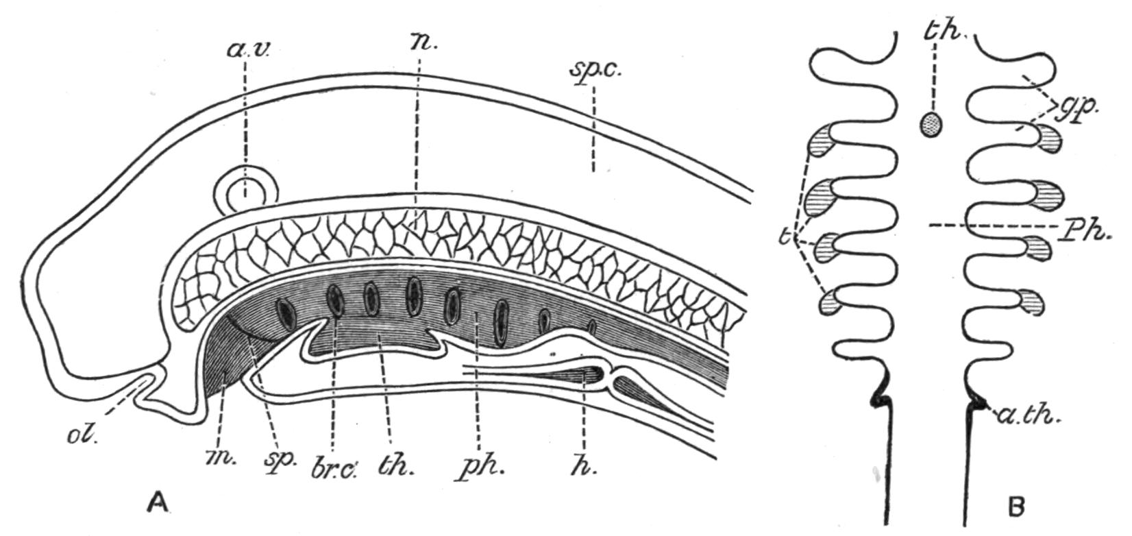
Fig. 202.—A, a vertical section through a just-hatched larva of Petromyzon. a.v, Auditory vesicle; br.c, branchial cleft; h, heart; m, mouth; n, notochord; ol, olfactory pit; ph, pharynx; sp, septum or velum between the stomodaeum and the mesenteron; sp.c, spinal cord; th, thyroid outgrowth from the floor of the pharynx. (From Gegenbaur, after Calberla.) B, diagram illustrating the development of the thyroid, the thymus, and the accessory thyroids, and their relations to the branchial clefts. a.th, Accessory thyroids; g.p, gill-pouches; Ph, pharynx; t, thymus; th, median thyroid. (From Hertwig, after de Meuron.)
According to Dohrn the median thyroid is to be regarded as the vestige of a gill-cleft which primitively existed between the hyomandibular cartilage and the hyoidean arch. This conclusion seems, however, to be less in harmony with the facts of development than the view[400] that the organ is derived from the characteristic hypobranchial groove or "endostyle" of Ascidians and Amphioxus, which has undergone a change of function from a mucus-conveying groove to a blood-gland. On the other hand, the mode of origin of the paired thyroids certainly favours the suggestion that they represent a posterior pair of vestigial gill-clefts, a view which derives some support from the fact that in Notidanus, where additional branchial arches and clefts are present, the paired thyroids are absent.
The Thymus.—In the embryo Elasmobranch and Teleost[401] the thymus has a multiple origin, being derived from a series of distinct epithelial thickenings, one of which is developed at the dorsal extremity of each of the gill-clefts except of the spiracle. These rudiments subsequently detach themselves from the epithelial surface and sink inwards, eventually fusing together on each side to form a single independent structure. Later, the epithelial mass thus formed becomes invaded by connective tissue, and by leucocytes which form lymph follicles, and the thymus gradually assumes the structure of a lymphoid organ. From its mode of development it has been suggested that the thymus owes its evolution to the metamorphosis and ingrowth of branchial filaments,[402] but it is also noteworthy that each embryonic rudiment of the organ closely resembles, both in position and origin, one of the developing branchial tongue-bars of Amphioxus.[403] The abundance of leucocytes which it contains has also prompted the further suggestion that the origin of the thymus may be due to the necessity of providing for the phagocytic protection of the gills themselves from the ravages of harmful micro-organisms, fungoid spores, etc., as well as to aid in the removal of such portions of the gills as may have been injured.[404]
A thymus is probably present in all Fishes, if not in the adult at all events in the embryo, but is always relatively small in size. In Elasmobranchs the organ lies on each side above the branchial arches and beneath the dorsal musculature; and in Teleostomi at the dorsal extremity of the last branchial arch, in close proximity to the mucous membrane of the branchial cavity. In a similar position in the Dipnoi (e.g. Protopterus)[405] there are, on each side, two contiguous lobes of lymphoid tissue which apparently represent a thymus.
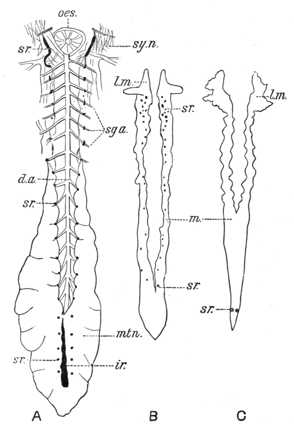
Fig. 203.—Supra-renal and inter-renal bodies of Fishes. A, of Scyllium catulus; B, of Acipenser sturio; C, of Pagellus centrodontus. d.a, Dorsal aorta; tr, inter-renal body; l.m, lymphoid portion of the mesonephros; m, mesonephros; mtn, metanephros; oes, oesophagus; sg.a, segmental arteries; sr, supra-renal bodies; sy.n, sympathetic nerves. (From Swale Vincent.)
The Supra-renal Bodies.—The supra-renal bodies are organs of problematic function, which are present in the Cyclostomata, and probably in all Fishes, and situated in close proximity to the kidneys.
In the Cyclostomata (Petromyzon) these bodies are represented by lobules of cells along the posterior cardinal veins, and also by masses of peculiar cells ("chromaffin cells") along the sides of the aorta and segmental arteries.[406] In Elasmobranchs there are two distinct structures, the paired supra-renals and the inter-renals (Fig. 203, A). The former are a series of pairs of segmentally arranged bodies, situated on the successive pairs of segmental arteries given off from the dorsal aorta. The two bodies which form the first pair are much larger than any of the others, and were formerly spoken of as "axillary hearts." The inter-renal is usually a thin elongated "ochre-yellow" body, from which one or two lobes may be detached in front, and extends for a variable distance in the median line between the two kidneys, or is unsymmetrically placed on the ventral surface of either kidney.[407] Sometimes (e.g. in Raia) the inter-renals are paired, in which case they are applied to the inner and hinder margins of the kidneys. In the Sturgeon (Acipenser sturio) the "supra-renals" appear as numerous "ochre-yellow" bodies, variable in size and distribution (Fig. 203, B). Some of them are visible on the surface of the kidneys, while others are scattered about in their substance, but on the whole are more anteriorly placed than in Teleosts. In the latter group the "supra-renals" are usually two in number (Fig. 203, C), but may be as many as five or reduced to one. They are disposed either on the ventral or the dorsal surface of the kidneys, generally near their hinder extremities, or more or less deeply embedded in their substance. Besides these bodies there are also chromaffin cells in the walls of the anterior cardinal veins.[408]
Histologically, the paired segmentally arranged bodies of Elasmobranchs differ considerably in structure from the inter-renal bodies, the former resembling the "medulla," while the inter-renals, as well as the so-called supra-renals of Acipenser, exhibit a striking resemblance to the alveolar "cortical" substance of the Mammalian supra-renals.[409] In Cyclostomes the cortex is apparently represented by the lobules of cells along the posterior cardinal veins and the medulla by the "chromaffin" cells, while in Teleosts the cortex and the medulla have their respective counterparts in the supra-renals and the "chromaffin" cells in the walls of the anterior cardinal veins. It may be concluded, therefore, that Elasmobranchs, Cyclostomes, and Teleosts possess anatomically distinct representatives of both the "medulla" and "cortex" of Mammalia, although the Sturgeon is at present only known to possess the equivalent of the "cortex." In Amphibia, Reptilia, and Aves both "cortex" and "medulla" are present, and in the varying intimacy of their relations offer a transition to the Mammalian arrangement of a central medulla closely invested by a sheath of cortical substance. A more or less intimate connexion exists between the paired supra-renals of Elasmobranchs and the sympathetic nervous system. The former are usually well supplied with sympathetic nerve fibres, and contain ganglion-cells in their substance.
The primitive origin of these organs is very obscure, and as regards their development there is much diversity of opinion. It seems certain, however, that the cortex and medulla of the higher Vertebrates, including their equivalents in the Elasmobranchs, have independent origins, and the balance of opinion seems to point to the derivation of the cortex from some portion of the germinal coelomic epithelium, while the medulla is derived from the embryonic nerve cells of the sympathetic ganglia.
Lymphoid Tissue.—In addition to certain of the ductless glands, and the local or diffused masses of their characteristic tissue already mentioned in connexion with the alimentary canal, lymphoid tissue is often abundantly present in other parts of the body. There is, for example, a mass of this tissue on the heart of the Sturgeon (Acipenser). The anterior enlarged portion of the mesonephros, commonly termed the "head-kidney" of the Teleostomi (Fig. 203, B, C), is almost entirely composed of lymphoid tissue,[410] which has replaced, wholly or partially, the proper renal structure; and from the presence of free red blood-corpuscles and of crystals of oxy-haemoglobin and other derivatives of haemoglobin, it may be inferred that the "head-kidney," in common with the more orthodox blood-glands, performs a blood-destroying function.[411] On the other hand, the example of the spleen, which is alike the seat of leucocyte-formation and of blood-destruction, renders it unnecessary to reject the view that the "head-kidney" is an organ in which leucocytes or blood-corpuscles are formed. In but few Teleostomi is a purely lymphoid "head-kidney" entirely wanting, as, for example, in the Sun-Fish (Orthagoriscus mola).[412] As previously mentioned the Dipnoi are remarkable for the extraordinary development of lymphoid tissue, inasmuch as it forms a thick investing mass round the kidneys and gonads in addition to its exceptional abundance in the walls of the alimentary canal.
The absence of ordinary lymphatic glands in Fishes is well known, and it is at least probable that, functionally, the want of these lymphoid organs may be compensated for by the superabundance of lymphoid tissue in other parts of the body.[413]