Chapter VIII · (continued 2)
Reading edition
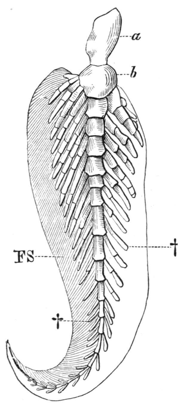
Fig. 147.—The left pectoral fin of Neoceratodus. a, b, First two segments of the axis; FS, preaxial horny fibres; †, †, pre- and post-axial radialia. (After Wiedersheim.)
The Pelvic Fins.—In the simplicity of their endoskeletal supports the pelvic fins of Cladoselache are the most primitive type of paired fins at present known (Fig. 145, B). In general structure they resemble the pectorals, but the radialia are fewer in number, less modified by concentration, and exhibit little, if any, trace of basal fusion. Add to such features as these the apparent absence of any trace of pelvic rudiments, or of basipterygia, and it will be obvious that the pelvic fins differ but little from the median fins of the same Fish except that they are paired. In Pleuracanthus the pelvic fins differ from the corresponding pectorals in being uniserial instead of biserial (Fig. 250). All other Elasmobranchs, including the Holocephali, have uniserial fins, which consist of a large metapterygium, supporting a preaxial fringe of segmented radialia. A propterygium is sometimes present, notably in some of the Skates and Rays, and, like the metapterygium, it is directly connected with the pelvic girdle.
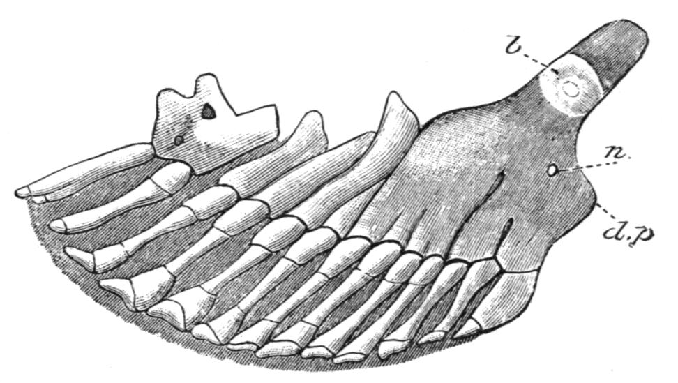
Fig. 148.—Skeleton of a pelvic fin of Polyodon folium, ventral view, with the anterior margin of the fin to the right; to show the partial fusion of the proximal portions of primitively distinct radialia to form a basipterygium. b, Inner or mesial extremity of the basipterygium; d.p, dorsally directed, rudimentary iliac process; n, foramen for nerves. (After Rautenfeld.)
The skeleton of the pelvic fins of the Teleostomi is often extremely degenerate. It is perhaps best developed in the Chondrostei,[220] where each fin is supported by numerous segmented radialia, more or fewer of which fuse towards the base of the fin, and those form a large and slightly ossified basipterygium (Fig. 148). In the living Crossopterygii, Holostei, and Teleostei, the pelvic fins are similar in essential structure, but are very degenerate. The basipterygium is usually well developed and is always bony (Fig. 149), and in many Teleosts it acquires so extensive a sutural connexion with its fellow that, physiologically, it supplies the place of a true pelvic girdle. At its distal end there may be a single row of small cartilaginous or bony nodules, representing vestigial radialia, as in the Crossopterygii, Holostei, and Teleostei, but even these may be absent, and the dermal fin-rays then articulate directly with the basipterygium. Little is known of the skeleton of the pelvic fins in the fossil Crossopterygii, but there is evidence of the existence of a higher grade of structure than in their surviving allies. In Eusthenopteron,[221] for example, the fin is supported by an axis of at least three bony segments, with at least three ossified preaxial radialia; hence, it has obviously undergone less degeneration than in Polypterus, where the fin-skeleton is essentially Teleostean. In the Dipnoi the pelvic fins are similar to the corresponding pectoral fins, but individual variation is more marked and even the central axis may divide.[222] In the males of all existing Elasmobranchs, including the Holocephali, certain of the more distally situated metapterygial radialia become modified to form a supporting skeleton for the copulatory organs, the claspers, or mixipterygia. In the latter group the anterior claspers are also provided with cartilaginous supports articulating with the pelvic girdle directly in front of the pelvic fins.
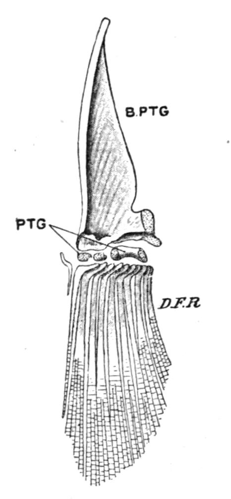
Fig. 149.—Skeleton of the left pelvic fin of a Trout (Salmo fario), seen from the dorsal surface. B.PTG, Basipterygium; D.F.R, dermal fin rays; PTG, distal radialia. (From Parker and Haswell.)