Chapter VII
Reading edition
THE SKIN AND SCALES
The skin of the Cyclostomata and Fishes consists (1) of the epidermis, formed of several layers of epidermic cells, which are constantly being recruited by the division of the cells of the basal layer; and (2) of a stratum of connective tissue with intermingled unstriped muscle-fibres, blood-vessels and nerves, which constitutes the deeper layer or dermis. From the epidermis are formed the various unicellular or multicellular glands with which the skin is provided; and from one or both of the skin layers originate the different calcareous structures which constitute the hard exoskeleton.
In the Cyclostomata the epidermis is particularly rich in goblet-shaped, mucus-secreting, gland-cells. The Myxinoids also possess numerous pockets of so-called "thread-cells." In each of these cells the protoplasm secretes a long spirally-coiled thread, and under the influence of appropriate stimuli the thread is shot out and unwound to a great length. The threads and the mucus are so abundant that one of these animals will convert a bucket of water into a thick mass of jelly. No scales or other hard exoskeletal structures are present in any of the Cyclostomata.
In Fishes mucus-glands are also abundant in the epidermis, and to their activity is due the slimy mucus which lubricates the surface of the body. They are specially numerous in the Dipnoi (e.g. Protopterus), where, in addition, there are many simple multicellular glands which secrete the "cocoon" or capsule in which the Fish is enclosed during the dry season. From the epidermis are derived the poison-glands of some Teleosts, and also the "glandula pterygopodia" in relation with the claspers of the male Elasmobranchs. The glandular structures in connexion with the phosphorescent organs of the deep-sea Fishes will no doubt be traced to the same source.
In the great majority of Fishes the skin becomes the seat of calcareous deposit, and gives rise to such diverse exoskeletal structures as the varied forms of spines and scales with which the surface of a Fish is invested.[161] These structures, probably the most ancient form of Vertebrate skeleton owing its existence to the presence of lime salts in the tissues of the body, present highly characteristic modifications in the different groups.
Exoskeletal structures are of two kinds: (1) those which owe their formation to the secretory activity of cells belonging both to the epidermis and the dermis, and (2) those which are derived solely from the dermis. To the first belong the dermal denticles or so-called placoid scales of most Elasmobranchs, and to the second the scales which form the skin-skeleton of living and extinct Teleostomi and Dipnoi. With the exception of enamel, which is always formed by the cells of the epidermis, the hard exoskeletal tissues owe their existence to the secretion of certain cells of the dermis (scleroblasts),[162] the inclusion of which in a growing calcifying tissue is the cause of whatever cellular structure the tissue may present. It will shortly be apparent that the dermic scleroblasts are by no means uniform in their products, and that in different Fishes they give rise to widely different hard tissues.
The dermal denticles or "shagreen" of the ordinary Sharks and Dog-Fishes (Elasmobranchii) probably represent the most primitive form of exoskeleton. In the development of a dermal denticle a papilla of the dermis grows up into the overlying epidermis, pushing before it the basal layer of epidermic cells, which forms an investment to the papilla and constitutes the so-called "enamel organ" (Fig. 100). The papilla itself subsequently becomes converted into dentine, leaving, however, a central pulp-cavity, while the apex of the papilla is invested by a cap of enamel formed by the enamel organ. Ultimately the base of the papilla widens out into a more or less rhomboidal basal plate formed of bone. In this way there is formed a pointed, enamel-tipped spine of dentine which protrudes through the epidermis, and projects backwards on the surface of the body, but is firmly fixed in the skin by the basal plate with which it is continuous. The centre of the under surface of the basal plate is perforated for the entrance of the blood-vessels which pass to the cellular pulp in the axis of the spine. In the adult Fish the denticles form a fairly close-set covering to the whole body, including the head and even the surfaces of the fins, and are larger on the dorsal than on the ventral surface (Fig. 99). In the Rays (Raia) they are more sparsely scattered, and in different parts of the body may form spines of considerable size for offensive or defensive purposes. The spines vary greatly in shape in different members of the group, sometimes being acutely pointed, and sometimes flattened or depressed, and often they are furnished with smaller accessory spines developed at their bases or from the surface of the basal plate. An arrangement of the denticles in oblique transverse rows is observable in some genera (e.g. Scyllium). In the Saw-Fishes (e.g. Pristis) the denticles which fringe the lateral margins of the long flattened rostrum are not only enormously enlarged, but are implanted in sockets and form the teeth of the saw (Fig. 262). In the Holocephali the smooth skin is almost entirely devoid of exoskeletal structures, but dermal denticles are present on the frontal and anterior claspers, and in the young there may be a double row of small denticles along the back.
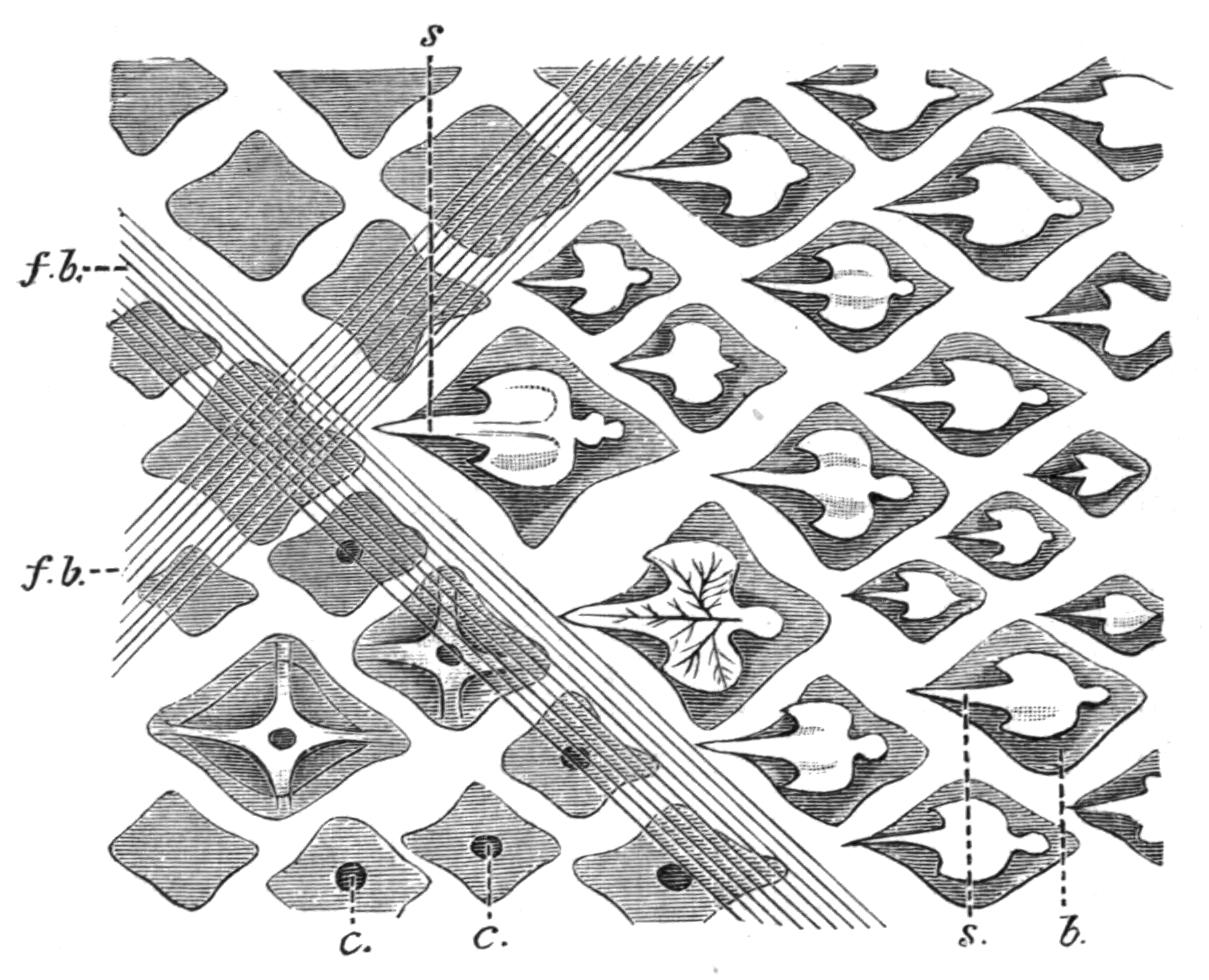
Fig. 99.—Surface view of the dermal denticles of Scyllium sp., showing their arrangement in oblique transverse rows. b, Basal plate; c, canal which perforates the basal plate and becomes the axial pulp-cavity of the spine; f.b, intersecting fibrous bands of the dermis; s, spine; in the spine of one scale the dentinal tubules are shown. The smaller denticles are those most recently formed. (After Klaatsch.)
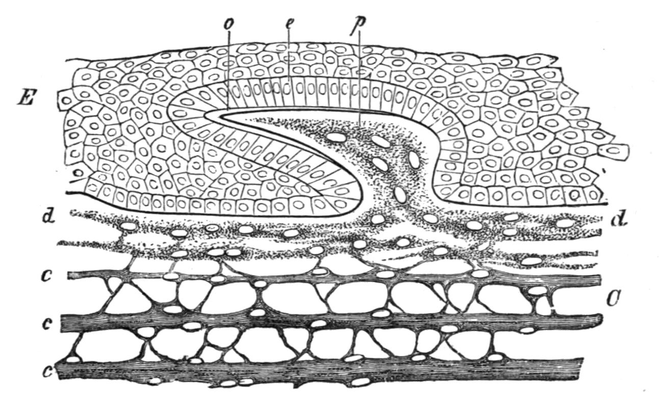
Fig. 100.—Vertical section through the skin of an embryo Shark. C, Dermis; c.c.c.d, layers of the dermis; E, epidermis; e, enamel organ; o, enamel layer; p, papilla of the dermis. (From Wiedersheim, after Gegenbaur.)
In the remaining groups of Fishes, the Teleostomi and the Dipnoi, the spine of the primitive dermal denticle is either evanescent or entirely wanting, while the equivalent of the basal plate remains to form the unit of a scaly armature. Evidence of this may be found in the presence of transitory evanescent spines, provided with an enamel-cap, secreted by the basal epidermis, on the developing rhomboidal scales, as in the young Lepidosteus[163] (Fig. 101); while the entrance of blood-vessels into the scales through perforations on their inner surfaces, as in Polypterus and Lepidosteus, obviously recalls the perforated base of a dermal denticle (Fig. 99). The epidermis now ceases to take any part in the formation of the scales, and hence enamel no longer enters into their structure. A more regular and definite arrangement of the scales is noticeable, and whether distinct, or articulating with one another, or overlapping like the slates on the roof of a house, they are usually disposed in a series of successive oblique transverse rows. In some of these Fishes the embryonic epidermic covering of the scales becomes lost, and their outer surfaces are naked. More frequently, as in the generality of Teleosts, and in the Dipnoi, the reverse is the case, and the scales are more or less completely invested both by the dermis and the epidermis. As regards their shape, size, and minute structure there is much variation. In some Teleostomi the primitive rhomboidal shape of the dermal denticle is retained; in others a rounded or cycloid scale supplants the earlier rhombic type. Within the limits of the same group (e.g. Crossopterygii) there are examples of the independent evolution of a cycloid from a pre-existing rhombic squamation; and with the introduction of the cycloid type an overlapping or imbricated disposition of the scales always takes the place of the marginal articulation of the rhombic type.
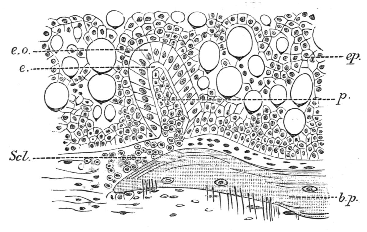
Fig. 101.—Development of a scale in Lepidosteus osseus × 330. b.p, Basal plate, with included bone cells, at first distinct from the spine; e, enamel; e.o, enamel organ; ep, epidermis, with large gland cells; p, dermic papilla which forms the vestigial spine; Scl, scleroblasts. (From Klaatsch.)
As to the causes which may have determined the shape and mutual relations of scales interesting suggestions have been made.[164] Scales bear a segmental relation to the subjacent muscle-segments or myotomes, sometimes being disposed in oblique transverse rows coinciding with the latter, or the rows may be so far increased as to be multiples of the myotomes. From mechanical considerations depending on the sigmoid shape and interdigitating relations of the myotomes and their separating fibrous septa or myocommata, and the attachment of the myocommata to the dermis, the contraction of the myotomes during the lateral flexions of the trunk in swimming has a tendency to wrinkle the skin into definitely circumscribed rhombic areas, thus determining the shape, limits, and disposition of the scales which are developed in those areas. The rhombic was probably the primitive shape of scales, and is certainly characteristic of the palaeontologically older types of scaly Fishes. Generally the rhombic condition is associated with a peg-and-socket articulation between the upper and lower margins of adjacent scales. But a rhombic squamation is not without disadvantages, and would certainly impose some restriction on the lateral flexures of the body in swimming, and hence in the different groups of Fishes it may happen that, in the more specialised forms, an imbricated cycloid squamation supersedes a rhombic condition, and with the change the Fish acquires greater lateral mobility. Even in the same Fish the gradual substitution of the cycloid for the rhombic type may be observed. In the Australian Aetheolepis,[165] a fossil genus related to the European Liassic Dapedius, there is a gradual transition along the sides of the body between the articulated rhombic scales of the relatively immobile trunk and the cycloid overlapping scales of the flexible tail; and it may be mentioned that, even where a typical rhombic squamation exists, the peg-and-socket articulation may be wanting in the caudal region, so as to ensure greater freedom of movement. Mechanical considerations may also explain the overlapping of cycloid scales. From the mode of attachment of the myocommata to the dermis, the contractions of the myotomes, through the pull which they exert on the former, tend to deflect or depress the scale-areas, particularly at their anterior margins.
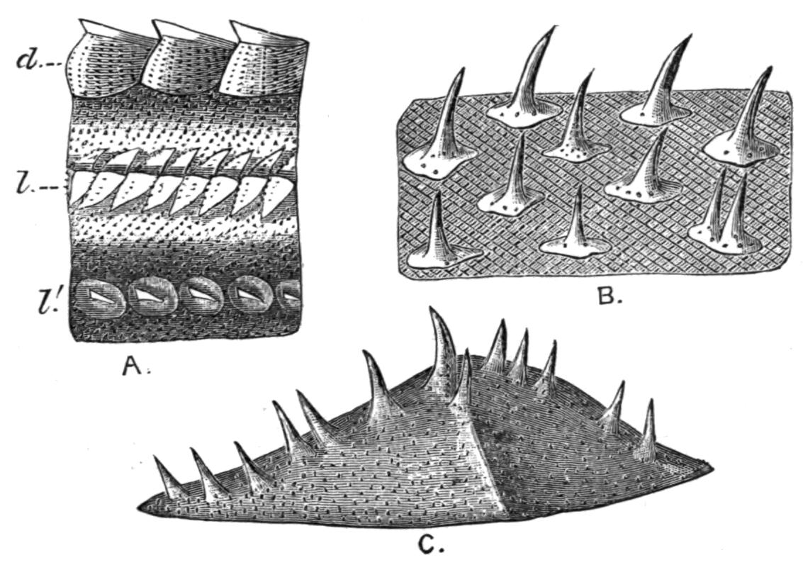
Fig. 102.—Acipenser ruthenus. A, Side view of the trunk of a specimen 30 cm. in length (nat. size); d, dorsal row of plates; l, l′, lateral rows; between the rows of large scutes may be seen the numerous small denticles which are represented (× 10) in B; C, one of the large scutes (× 10). (From Hertwig.)
In the surviving Crossopterygii, as in Polypterus, the scales are rhomboidal and thick, and they only slightly overlap. They articulate with one another by means of marginal peg-and-socket articulations (Fig. 106, B). A thick layer of hard, glistening, enamel-like substance or "ganoin" forms the outer layer of the scale; the inner layer consisting of bone in which dentinal tubules as well as bone-cells are present. In the numerous fossil members of the group the scales are either rhomboidal or cycloid.
The oldest representatives of the Chondrostei, the Palaeoniscidae (Fig. 283) possessed a complete armature of rhombic scales, but in all the surviving members of the group the scales have undergone considerable modification in some respects, and in others are degenerate. In the Sturgeon (Acipenser)[166] the primitive rhombic squamation is retained only on the sides of the terminal part of the tail, and there they are in close apposition in oblique rows. The rest of the body is traversed by five widely-separated longitudinal rows of large bony scutes, which, like the rhombic scales, are furnished with ridges and projecting spines (Fig. 102). Between the rows of large scales there are numerous denticle-like structures arranged in oblique rows. Each of these consists of a basal plate imbedded in the dermis, and of one or more projecting spines which perforate the epidermis. All the scales have the same minute structure, consisting mainly of bone; but the surface layer and the spines seem to be composed of a hard laminated substance from which bone-cells are absent (ganoin). In Polyodon the scutes are wanting, but vestigial denticles are retained.
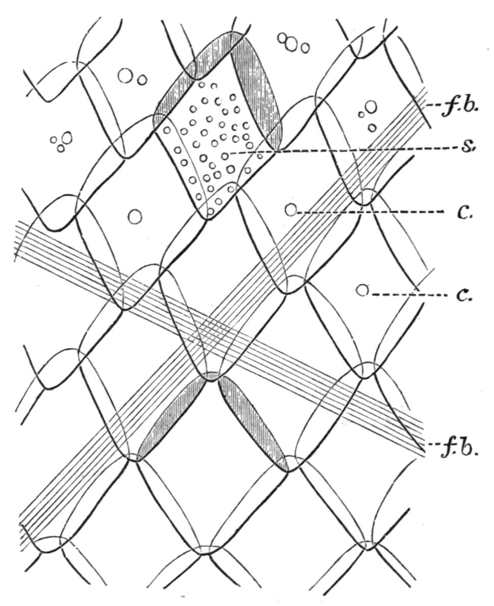
Fig. 103.—Surface view of the rhombic scales of a young Lepidosteus. In two scales the parts which are overlapped by adjacent scales are shaded. c, Position of the central canal which perforates the inner surface of each scale; f.b, intersecting fibrous bands of the dermis; s, vestigial spines. (After Klaatsch.)
Among the Holostei the scales are very different in the two surviving members of the group. In Lepidosteus (Fig. 103) the scales are rhombic, and both in arrangement and structure, as well as in their method of articulating with one another, they closely resemble those of Polypterus. In Amia,[167] on the contrary, the relatively thin scales are cycloid in shape, and in their imbricated arrangement, in their enclosure in pouches of the dermis, and in the absence of any superficial covering of ganoin, they are very similar to the scales of the more typical Teleosts (Fig. 295). The resemblance extends even to histological structure, for each scale consists of an outer layer of bone, which gradually passes into an inner fibrous stratum.
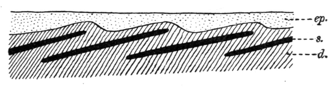
Fig. 104.—Diagrammatic longitudinal section through the skin of a Teleost to show the position of the scales. d, Dermis; ep, epidermis; s, scale. (After Boas.)
In Teleosts the usually thin and flexible scales are primarily developed from dermic papillae, but subsequently they come to lie in pockets or pouches in the dermis. As a rule no spines are developed, and so far no trace of an enamel organ has been detected during their development. The scales in their dermal pouches are disposed obliquely to the surface of the body, so that the hinder part of one scale overlaps the anterior portion of the scale next behind it (Fig. 104). Only the free hinder part of each scale has an epidermic investment (Fig. 105). In minute structure each scale consists of an outer layer of bone, which, like the bone of the endoskeleton, may either be homogeneous except for a feeble lamination, or it may contain bone-cells arranged in successive layers, parallel to the surface of the scale. In addition, there is an inner fibrous stratum in which the fibrous bundles in any one plane cross those in planes above or below them. The scales are either "cycloid," that is, they have smooth, unbroken margins (Fig. 105), or the free margin of each scale is produced into a series of tooth-like spines, and the scale is said to be "ctenoid" (Fig. 106, A).
Some Teleosts, however, have scales which are neither cycloid nor ctenoid, and in certain features seem to be intermediate between ordinary Teleostean scales and dermal denticles. Thus, on certain parts of the body of Centriscus,[168] each scale consists of a rhombic basal plate, produced into a curved, backwardly-inclined spine, the axis of which contains a pulp-cavity opening on the inner surface of the basal plate (Fig. 107). Some Malthidae (e.g. Malthe[169]) have similar scales, but with round basal plates and solid spines (Fig. 108, B). Similar scales (Fig. 109), sometimes rhombic in shape, with one or more spines, which may be simple or branched, are also found in the Sclerodermi (e.g. Balistes, Monacanthus, Triacanthus).[170]
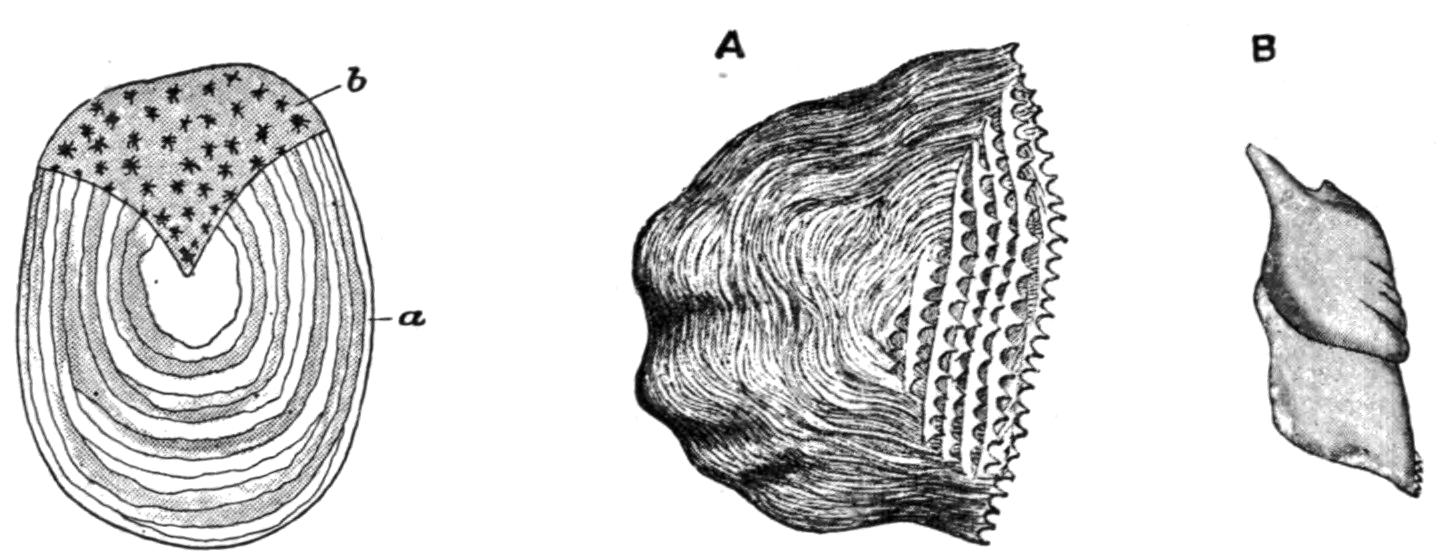
Fig. 105.—Cycloid scale of Salmo fario. a, Anterior portion covered by overlap of preceding scales; b, free portion covered only by pigmented epidermis. (From Parker and Haswell.)
Fig. 106.—A, Ctenoid scale; B, "Ganoid" scale. (After Günther; from Parker and Haswell.)
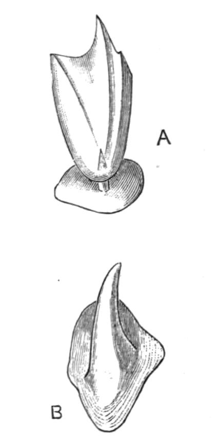
Fig. 107.—Centriscus scolopax. A, Scale from the orbital region, × 50; B, scale from the base of the pectoral fin, × 100. (From Hertwig.)
Amongst some of the usually scaleless Siluroid Fishes the scales assume a very peculiar structure. In Hypostoma[171] (Plecostomus) the sides and back of the Fish are covered by large bony plates, but on the under surface and on the fins these are replaced by much smaller ones. Both kinds, however, carry numerous small movable spines implanted in sockets (Fig. 110), a fact which suggests comparison with a stage in the development of the scales of Lepidosteus, when the independently formed and evanescent spines have not yet fused with the basal plates.
In other Teleosts, as in the Agonidae and some Triglidae, the body is completely cuirassed with large keeled bony plates. The singular appearance of many of the Plectognathi is largely the result of the curious modifications which their scales undergo. In some of the Coffer-Fishes (Ostracion) these structures assume the form of polygonal bony plates which suturally articulate with one another and enclose the trunk in a rigid cuirass, from which the scaleless tail protrudes behind (Fig. 438); while in some Globe-Fishes and Porcupine-Fishes (e.g. Tetrodon, Diodon) the prolongation of the scales into strong erectile spines equally well serves the purpose of protection (Fig. 439).
Most Teleostomi have the scales along the "lateral line" perforated by single or multiple apertures, through which the sensory canal communicates with the exterior.
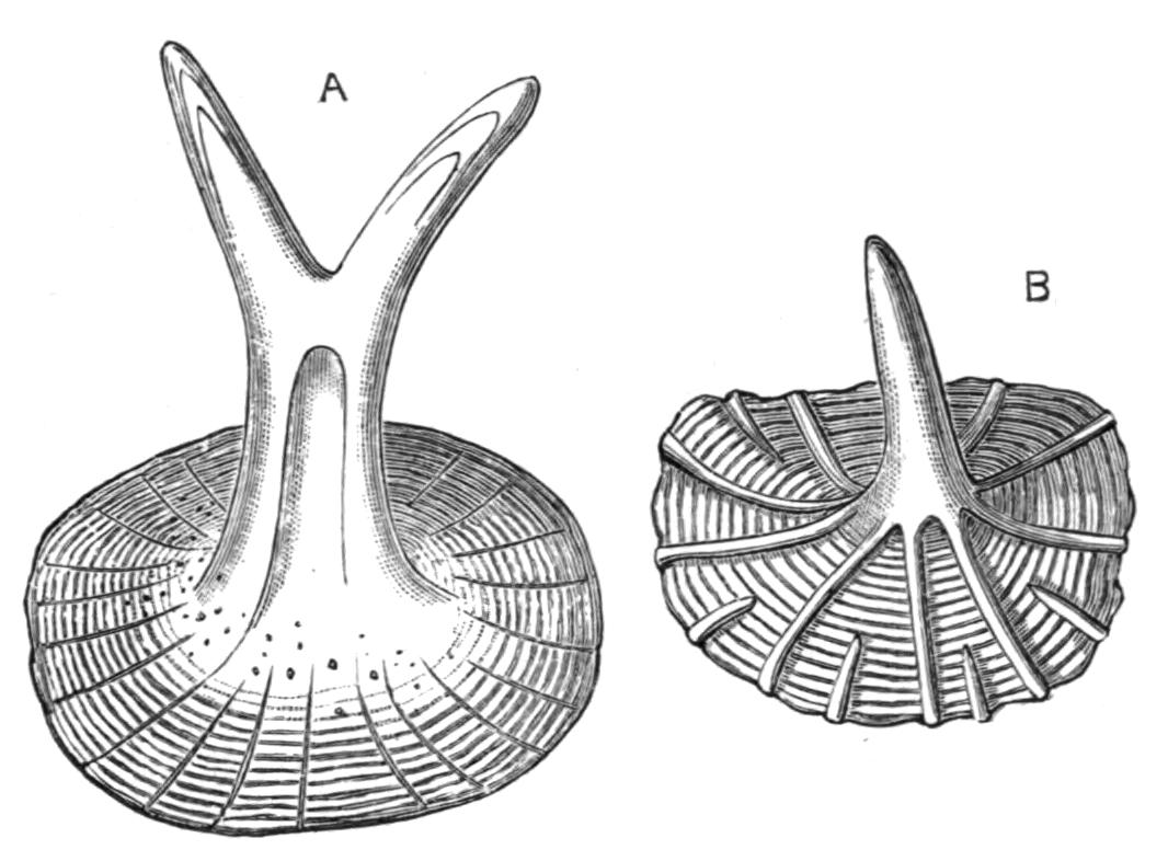
Fig. 108.—A, Scale of Antennarius hispidus, × 100; B, scale of a young Malthe vespertilio, × 100. (After Hertwig.)
In a few Teleosts scales are entirely absent, as in most Siluridae; or they exist only as microscopic vestiges hidden in the skin, as in Eels; or, as in such naked forms as Antennarius marmoratus and Lepadogaster, and in some Siluridae, they become reduced to mere papillae of the dermis.
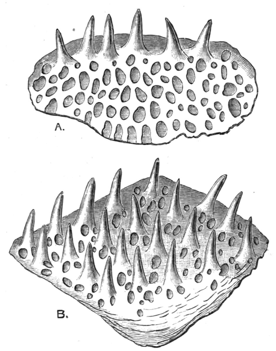
Fig. 109.—A, Scale of Balistes capriscus, × 20; B, scale of Monacanthus scopas, × 20. (After Hertwig.)
The concentric rings observable (Fig. 105) on the surface of many Teleostean scales are an index to the age of the Fish.[172] The formation of these rings depends on the fact that the lines of growth on the surface of the scale are more widely separated from one another on that portion of the scale formed during summer, and relatively closer together on that part which is formed during the winter; the more rapid growth in the warmer season probably being due to favourable conditions as to food and temperature, and the retarded growth of the colder season to the reverse. Hence, by counting the alternating zones of close-set winter lines and less closely approximated summer lines of growth, a reliable clue may be gained as to the age of the Fish.
In the Dipnoi,[173] as in Teleosts, the scales are enclosed in dermal pockets, and exhibit a regular, imbricated disposition in oblique rows (Fig. 304, A). In shape they are nearly cycloid, or slightly oval, with the long axis coinciding with that of the body. Structurally, also, they bear some resemblance to Teleostean scales, although differing in details. On the outer surface of the scales there are numerous small conical spines. No significance, other than as an example of evolutionary convergence, can be attached to the resemblance between the scales of Fishes so widely separated as the Dipnoi and the Teleosts.
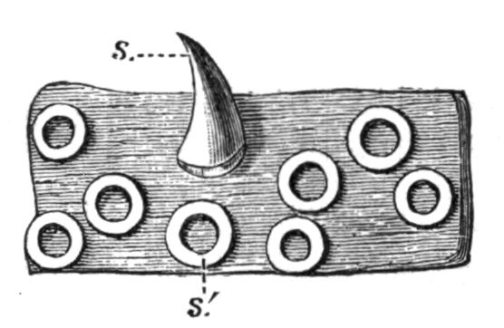
Fig. 110.—Hypostoma commersonii. A scale from the periphery of the caudal fin, × 50; one of the spines (s) is implanted in its socket (s′). (From Hertwig.)
All known fossil Dipnoi had scales of a similar character, although differing greatly in size in different genera. In some (e.g. Dipterus) a layer of enamel-like substance invests the exposed portions of the scales.