Chapter XV
Reading edition
THE KIDNEYS AND THE REPRODUCTIVE ORGANS—BREEDING
The kidneys and the reproductive organs are so intimately connected that it is necessary to deal with them together. Both organs are specialised portions of the coelom and its epithelial lining. The kidneys are essentially a series of tubular and at first segmentally-disposed outgrowths from the coelom (urocoeles) which acquire a connexion with the exterior, while the gonads have their origin from local modifications of the coelomic epithelium. At a very early embryonic stage each lateral half of the coelom presents three well-marked divisions: (1) a series of dorsal portions ("myocoeles"), the cavities of the myotomes or muscle-segments; (2) a longitudinally continuous unsegmented portion extending round the alimentary canal, the "ventral coelom"; and (3) a series of intermediate tubular portions or "nephrotomes," each of which leads from a myocoele to the ventral coelom (Fig. 229, A). The essential components of the kidneys, the urocoeles or renal tubules, are derived from the nephrotomes. In its typical condition each kidney consists of three portions, which, in accordance with their embryological and evolutionary sequence, are termed the "pronephros," the "mesonephros," and the "metanephros." The pronephros, the larval or provisional kidney, is formed from a limited number of the nephrotomes immediately behind the head. From each nephrotome a hollow tubular outgrowth is formed, which grows towards the lateral surface of the body, and then unites with its fellows of the same side to form a main longitudinal duct—the "archinephric" or "pronephric duct" (Fig. 229, A, Fig. 230, A). This duct grows backwards until it opens into the cloaca.[474]
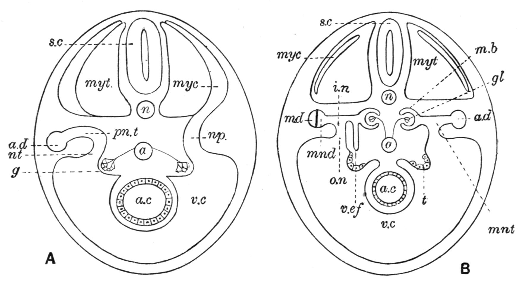
Fig. 229.—Diagrammatic transverse sections through an embryo Craniate to show the mode of development of the pronephros (A) and of the mesonephros (B). The right side of each figure shows an earlier stage than the left. In B (left side) the connexion of a vas efferens with a mesonephric tubule, and the division of the archinephric duct into Müllerian and mesonephric ducts are shown, a, Aorta; a.c, alimentary canal; a.d, archinephric duct; g, glomus; gl, glomerulus; i.n, inner nephrostome; mb, Malpighian body; md, Müllerian duct; mnd, mesonephric duct; mnt, mesonephric tubule; myc, myocoele; myt, myotome; n, notochord; np, nephrotome; nt, nephrostome; o.n, outer nephrostome; pn.t, pronephric tubule; s.c, spinal cord; t, testes; v.c, ventral coelom; v.ef, vas efferens. (After Kingsley and Semon.)
At the same time the nephrotomes lose their connexion with the myocoeles, although they still retain their "nephrostomes" or apertures through which they communicate with the ventral coelom. When fully developed the pronephros consists of a few tubules, more or less convoluted, opening at their inner extremities into the coelom by means of their ciliated nephrostomes, and at their outer ends communicating with the exterior through the archinephric duct. In relation with the pronephros a branch from the dorsal aorta forms a tuft of capillary blood-vessels or "glomus," opposite the nephrostomes, which projects into the ventral coelom on each side. Later, a second series of much more numerous tubules is formed behind the pronephros, which constitute the mesonephros. In forming mesonephric tubules the nephrotomes become disconnected from the myotomes and their myocoeles, and curving outwards they come to open into the archinephric duct, although they do not in any way contribute to its formation (Fig. 229, B). Segmentally-arranged twigs from the dorsal aorta end in tufts of capillaries or glomeruli, each of which projects into a small sac-like enlargement of a mesonephric tubule, pushing before it the wall of the sac. In this way a double-walled "Malpighian body," containing a "glomerulus," is formed in connexion with each tubule. Subsequently, the mesonephric tubules increase in number by budding. New nephrostomes and Malpighian bodies are developed on the secondary branches, and the original segmental arrangement of the tubules becomes obscured. With the growth of new tubules, and the formation of blood-vessels and of connective and lymphoid tissues between them, each mesonephros finally assumes the condition of a compact gland imbedded in the dorsal wall of the coelom, with its ventral surface invested by the peritoneum. A "metanephros," which in the higher Vertebrates replaces the mesonephros as the functional kidney, is perhaps not represented in Fishes.
A more or less well-developed pronephros is present in the embryos or larvae of the Cyclostomes and of all Fishes, but as a rule it completely disappears at an early period and is replaced by the mesonephros. It is retained throughout life, however, in the Myxinoid Cyclostomes (Fig. 230, B), and has its persistent nephrostomes opening into the pericardial cavity.[475] In a few Teleosts the pronephros is also persistent, as in Fierasfer and Dactylopterus, and in others the organ may not completely disappear until the approach of sexual maturity. But with these exceptions the mesonephros is the sole functional kidney in the adults of the Cyclostomes and of all Fishes. As regards the nature of the duct by which the excretion of the mesonephros is conveyed outwards, there are notable differences in different Craniates. The Cyclostomes and the Teleostomi retain that part of the archinephric duct into which the mesonephric tubules open, and which remains after the atrophy of the pronephros (Fig. 230, B, E, F). In Elasmobranchs, and probably also in the Dipnoi, a special mesonephric duct is developed in a way which will be described later (Fig. 230, C, D).
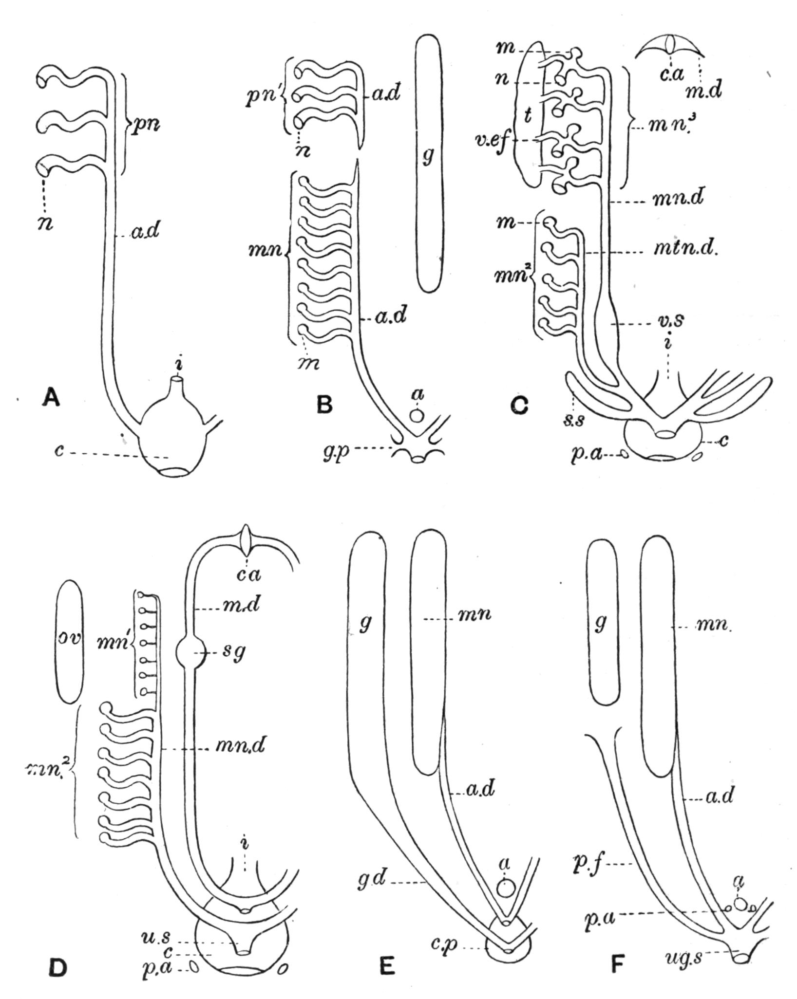
Fig. 230.—Showing the principal modifications of the kidneys and reproductive organs in Cyclostomes and Fishes. A, The pronephros and its duct in the embryo; B, the kidneys and genital pores in Petromyzon, the vestigial pronephros represented as in Myxine; C and D, the urinogenital organs of a male and female Elasmobranch; E, of a male or female Teleost, or a male Lepidosteus; F, of a female Polypterus, Acipenser, Amia, or Osmerus. a, Anus; a.d, archinephric duct; c, cloaca; c.a, the coelomic aperture of the Müllerian duct; c.p, cutaneous pit; g, gonad; gd, gonoduct; g.p, genital pore; i, intestine; m, Malpighian body; m.d, Müllerian duct; mn, mesonephros; mn1, vestigial mesonephros; mn2, excretory portion of the mesonephros ("metanephros"); mn3, genital portion of mesonephros; mn.d, mesonephric duct; mtn.d, metanephric duct; n, nephrostome; ov, ovary; p.a, abdominal pore; p.f, peritoneal funnel; pn, pronephros; pn′, vestigial pronephros; s.g, shell gland; s.s, sperm sac; t, testis; ug.s, u.s, urinogenital sinus; v.ef, vasa efferentia; v.s, vesicula seminalis.
In the males of Elasmobranchs some of the hinder mesonephric tubules unite to form a single main duct opening into the terminal part of the mesonephric duct, and these tubules and their separate duct are sometimes regarded as a metanephros and a metanephric duct. The mesonephric nephrostomes are persistent throughout life in a few Elasmobranchs (e.g. Notidanidae, Heterodontidae, Rhinidae, and some Scylliidae), and also in Amia:[476] in all other Fishes as well as in the Cyclostomes they become closed in early life.
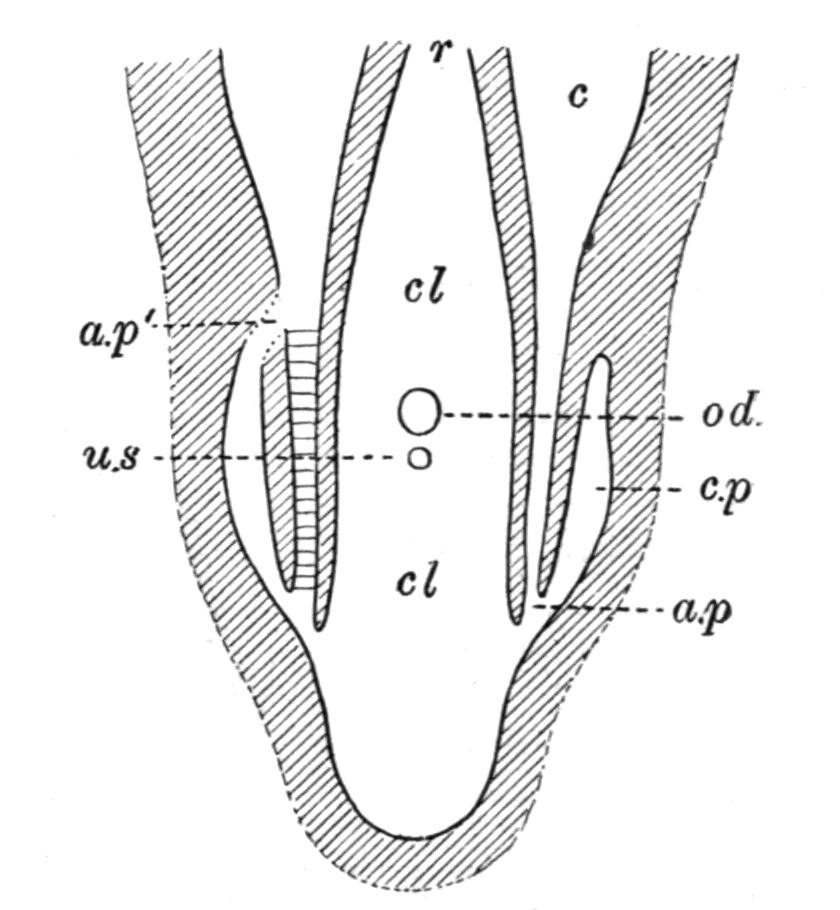
Fig. 231.—Diagrammatic horizontal section through the abdominal pores and cloaca of an Elasmobranch. a.p, Abdominal pore; c, coelom; cl, cloaca; cl.p, cloacal papilla; c.p, cloacal pit; od, oviducal apertures in the female; r, rectum; u.s, cloacal aperture of the urinary sinus (female), or the urogenital sinus (male). In some Elasmobranchs the abdominal pore opens at the base of the cloacal papilla, as shown at a.p1. (Modified from Bles.)
In many Fishes the hinder extremity of the coelom communicates directly with the exterior through "abdominal pores," of which there is usually a pair, rarely a single pore, situated close to the cloacal or the anal aperture.[477] Elasmobranchs usually have a pair, often at the extremities of a pair of cloacal papillae (Fig. 231), but they are absent in some families (e.g. Heterodontidae and Rhinidae); and in some Scylliidae (e.g. Scyllium canicula) they are very variable, being either present or absent on both sides, or an open pore is present on one side only. Pores are present and paired in the Crossopterygii, the Chondrostei, and the Holostei. Amongst the Dipnoi Neoceratodus has a pair of pores. Protopterus sometimes has two pores opening into the cloaca, but as a rule the two become confluent and have a single external aperture. In Lepidosiren pores are wanting. Abdominal pores are rarely present in Teleostei. They exist, however, in the Mormyridae (Gymnarchus and several species of Mormyrus), and also in the Salmonidae,[478] where they are as singularly variable in different species and individuals as in the Elasmobranch Scylliidae. The use of abdominal pores is not certainly known, unless the coelom of those Fishes which possess them continues to retain some measure of its primitive excretory function, and the pores act as excretory ducts. That the nephrostomes are excretory organs has been shown by experiment, and it is worthy of note that there exists a reciprocal relation between these structures and abdominal pores, to the extent that while there are a few Fishes (e.g. certain Elasmobranchs and Amia) in which both coexist, there are many others in which the presence of nephrostomes is correlated with the absence of pores and vice versâ.
The male and female gonads, testes and ovaries, are derived from the coelomic epithelium near the inner or median aspect of the nephrotomes (Fig. 229, B). Here the epithelium remains columnar, and soon projects into the ventral coelom as a continuous longitudinal ridge. It is probable that at first the modified epithelium is segmented as a series of "gonotomes," but if so, the latter must soon coalesce into a continuous ridge. Some of the epithelial cells enlarge to form the primitive sex-cells. In the development of an ovary, portions of the epithelium sink inwards, carrying with them the primitive ova. Certain of the cells form the epithelial walls of a number of ovisacs, each of which encloses an ovum. As the ovisacs increase in number and size the germinal ridges project more and more into the coelom until, as ripe ovaries, they become suspended from its dorsal wall by a double peritoneal fold, the "mesovarium" (Fig. 156). The testes develop in a similar fashion except that the primitive sex-cells, which later give rise to spermatozoa, form the lining of a number of simple or ampulla-like tubules, the seminiferous tubules, and the suspensory fold is termed the "mesorchium."
The Cyclostomes have gonads in the shape of unpaired organs extending nearly the whole length of the coelom, but in all Fishes the organs are primarily paired, although by fusion, or by the absorption of one gonad, the ovaries or the testes sometimes appear as if single. The ovaries may either be naked, as in Elasmobranchs, Dipnoi, Crossopterygii, and Chondrostei, and in Amia amongst the Holostei; or, as in Lepidosteus and most Teleosts, they become enclosed in coelomic sacs. The former, or "gymnoarian," condition is primitive; the latter, or "cystoarian," is secondary, and is brought about by the growth of two peritoneal folds round the ovary and the union of their margins. Into these coelomic sacs the egg-bearing or real ovarian tissue projects either in the form of processes or of transversely- or longitudinally-arranged plates or folds (Fig. 232, B). The testes are composed of seminal ampullae, as in Elasmobranchs, or of radially-arranged and sometimes plexiform tubules opening into the gonoduct, as in nearly all other Fishes (Fig. 232, A).
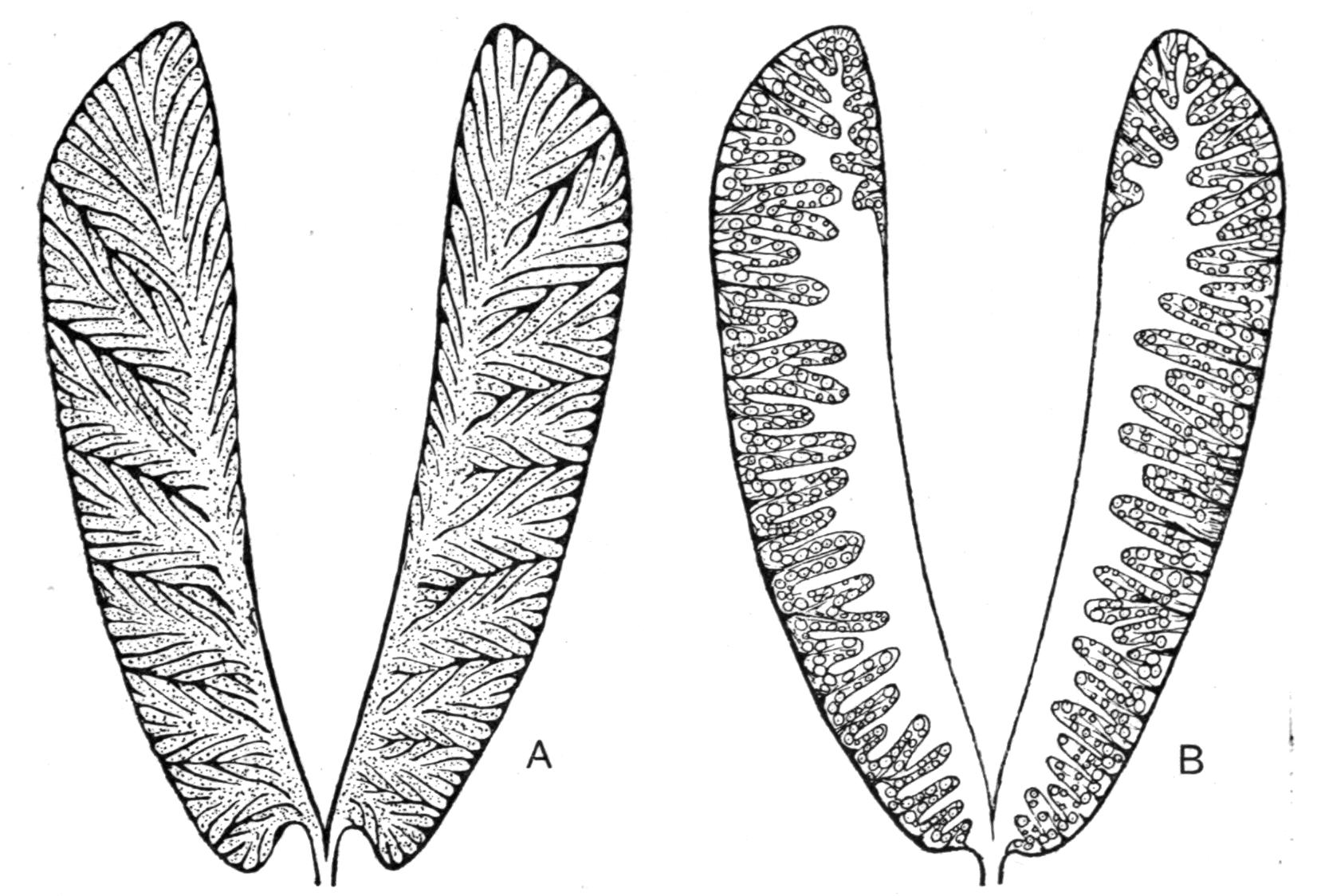
Fig. 232.—Diagrams to show the structure of the testes (A) and of the ovaries (B) in a Herring. (From Cunningham.)
In the Cyclostomes (e.g. Petromyzon) the eggs and spermatozoa are discharged from the gonads into the coelom, whence they reach the exterior through a pair of "genital pores" leading from the hinder end of the coelom into a urinogenital sinus formed by the united extremities of the two archinephric ducts.[479] Myxine has, however, but a single median pore, opening into an integumentary cloaca, which also receives the rectal and urinary orifices. Bdellostoma has two such pores communicating with a similar cloaca.[480]
The nature and homologies of the genital ducts in the different groups of Fishes are amongst the most puzzling of the many problems which vex the soul of the Vertebrate morphologist, and although there is a fairly general agreement on some points, there are others of great importance of which it may be said quot homines, tot sententiae.
Broadly speaking, there are two types of genital ducts in Fishes: (1) those which are obviously derived from some part of the kidney system; and (2) those which are special ducts and appear to have no connexion with kidney-ducts.
The Elasmobranchs offer a typical example of gonoducts of the first kind. At an early embryonic period in both sexes each archinephric duct becomes longitudinally split into two ducts, of which one continues to receive the openings of the mesonephric tubules and remains as a mesonephric duct (Fig. 229, B).[481] The other, which has no connexion with the mesonephros, opens anteriorly into the coelom by means of the united nephrostomes of the pronephros, and is known as the "Müllerian duct" (Fig. 230, C and D). In the adult male the Müllerian ducts are useless vestiges, but in the female they persist and act as oviducts, receiving the eggs set free from the ovarian ovisacs through their coelomic apertures, and thence conveying them to the cloaca. In the male, certain of the anterior mesonephric tubules become connected with the testicular ampullae by means of a network of slender tubules, the "vasa efferentia" or testicular network, and through the latter the spermatozoa pass from the testes to the mesonephric duct (Fig. 230, C). Consequently, the mesonephric duct conveys both spermatozoa and the kidney excretion to the cloaca. It is obvious, therefore, that both the male and female gonoducts are derived from kidney-ducts.
The Teleostei afford an equally typical illustration of the second type. Each female gonoduct (oviduct) is formed by a backward growth of the same two peritoneal folds which enclose the ovary; these are converted into a "peritoneal tube" or canal by the union of their margins. The male gonoducts are also formed in continuity with the testes, that is, as backward prolongations from the latter. Each duct, male or female, seems to be a duct sui generis and to have no connexion whatever with the kidney system (Fig. 230, E). In the Salmonidae, Anguillidae, Galaxiidae, Hyodontidae, Notopteridae, and Osteoglossidae, and also in Misgurnus, the oviducts lose their continuity with ovaries and degenerate to an extent which differs greatly in different families. Thus in some Salmonidae, as in the Smelt (Osmerus eperlanus),[482] the oviducts end anteriorly in wide funnel-like coelomic apertures after the fashion of Müllerian ducts, and do not embrace the ovaries: hence the ovaries are naked and not cystoarian, and their ducts are not peritoneal tubes but "peritoneal funnels" (Fig. 230, F). In other Salmonidae and in the Anguillidae the oviducts appear to have so far degenerated that they are represented either by a pair of very short funnels or by a pair of genital pores, which, as in the Salmon, have a common external aperture behind the anus and in front of the single orifice of the united archinephric ducts (Fig. 233, A). In all such instances the eggs are set free from the ovaries into the coelom, from whence they escape through the peritoneal funnels or genital pores. In the Eels the male gonoducts also degenerate, and, losing all connexion with the testes, they become reduced to genital pores as in the female.
The Holocephali and probably the Dipnoi conform to the Elasmobranch type in the nature of their male and female gonoducts. In the Crossopterygii[483] each testis has its own proper duct, which has no connexion with the kidney system and apparently belongs to the Teleostean type, while the oviduct, which is almost certainly not a Müllerian duct, is probably a peritoneal funnel. On the other hand, the Chondrostei and the Holostei are in the interesting transitional condition of possessing male ducts of the Elasmobranch type and female ducts of the Teleostean type, the latter being either ducts directly continuous with the ovaries, as in Lepidosteus, or of the nature of peritoneal funnels, as in Acipenser, Polyodon, and Amia (Fig. 230, E and F).
How far the distinction between the two types of gonoduct holds good in the case of the male is not quite clear, and it has recently been argued that the Dipnoi offer a connecting link between the two.[484]
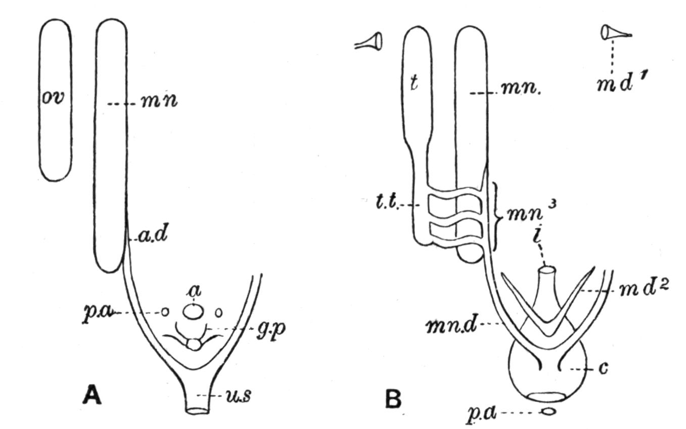
Fig. 233.—Diagram to show the kidneys and gonoducts of a female Salmon (A), and of a male Protopterus (B). md1 and md2, Anterior and posterior vestiges of the Müllerian duct; t.t, tubular posterior portion of the testis (t). Other reference letters as in Fig. 230. (B, after Graham Kerr.)
In Protopterus each testis is divided into an anterior sperm-producing part and a posterior tubular portion which has lost the capacity of producing sex-cells. The testicular network is greatly reduced, and forms but a limited connexion between the tubular portion of the testes and the mesonephric duct (Fig. 233, B). If it be supposed that the testicular network became still further reduced so that the connexion between the testes and the kidney-duct took place directly through a single channel instead of through several, the result would be a gonoduct essentially similar to the male duct of an ordinary Teleost. Should this view prove to be correct, it will follow that the male gonoducts of all Fishes are differently-modified examples of the Elasmobranch type. But there will still remain the female gonoducts of Ganoids and Teleosts, which must be regarded as distinct from Müllerian ducts unless it can be shown that their different methods of development are not necessarily fatal to their homology with Müllerian ducts, or that both types of gonoduct can be derived from some intermediate type. Assuming that some Fishes do possess male or female ducts which have not been derived from the kidney system, but have been independently acquired, there is still the question, which of the two types is the more primitive, or, in other words, has the Elasmobranch type superseded the Teleostean, or vice versa? To this question no decisive answer can at present be given.
The terminal relations of the kidney-ducts and the gonoducts, and the presence of accessory or of vestigial organs in connexion with them, will now be briefly dealt with. In the males of the Elasmobranchs the mesonephric ducts which, as already pointed out, act both as kidney-ducts and gonoducts, dilate posteriorly to form a pair of vesiculae seminales, and then unite to form a urinogenital sinus, opening into the cloaca at the extremity of a median papilla (Fig. 230, C). The sinus also receives ducts from the hinder part of the mesonephros, either separately, as in the female, or by a common duct on each side—the so-called metanephric duct—as in the male. Two tubular caecal outgrowths from the sinus form two sperm sacs. Only the anterior portions of the Müllerian ducts with their coelomic apertures are retained in the adult. In the female the mesonephric ducts are purely excretory, but otherwise they are similar, and the oviducts (Müllerian ducts) open into the cloaca separately or by a common orifice (Fig. 230, D). A glandular dilatation of each oviduct forms the oviducal or shell gland by which the horny egg-cases are secreted. In the males of the Holocephali the gonoducts open into a urinogenital sinus with an external orifice distinct from and behind the anus; but the female has separate apertures for the rectum, the conjoined oviducts, and the united mesonephric ducts. Both sexes have complete Müllerian ducts communicating with the coelom in front, and behind with the exterior. The Dipnoi of both sexes essentially resemble the Elasmobranchs in the general relations of their ducts, but the Müllerian ducts of the male exhibit marked differences in the three genera.[485] In Neoceratodus the ducts are as complete as their functional representatives in the female. Protopterus retains anterior vestiges and the coelomic apertures, and also vestiges of the hinder portions which unite and end blindly in the urinogenital papilla, but the middle sections of the two ducts are suppressed (Fig. 233, B). In the Teleostomi there is a general similarity in the terminal relations of the gonoducts and kidney-ducts. In the Ganoids the archinephric ducts unite and then expand into a urinary sinus or bladder, and the gonoducts of the female, or of both sexes in Lepidosteus, open either into the archinephric ducts or into the common sinus, and therefore both ducts communicate with the exterior by a urinogenital orifice behind the anus. Peritoneal funnels, similar to the functional oviducts of the female, are present in the males of the Chrondrostei and of Amia. In Teleosts the terminal connexions of the ducts tend to become less intimate. The archinephric ducts often dilate into a urinary bladder either before or after their union, and the common duct joins the united gonoducts to form a short urinogenital sinus which opens externally, or the confluent gonoducts have an independent genital orifice between the anus and the urinary aperture. Not rarely the genital or the urinogenital orifice is prolonged into a tubular papilla, which in the male acts as an intromittent organ, or, as in the females of the Cyprinoid Rhodeus amarus, the long oviducal tube serves the purpose of an ovipositor. The males and females of the Siluroid Plotosus have a remarkable vascular and glandular arborescent appendage just behind the urinogenital papilla, the use of which is unknown.[486]
The eggs of different Fishes[487] exhibit considerable diversity in size and shape as well as in the nature of their external coverings and their mode of deposition.[488] The size of the eggs largely depends on the quantity of food-yolk stored up in their substance for the nutrition of the embryo: hence the eggs of Elasmobranchs, which resemble Fowls' eggs in the superabundance of their yolk, are by far the largest. Teleostomi have much smaller eggs. The largest Teleostean ova are those which are heavy and sink (demersal ova); the smallest, those which are buoyant and float (pelagic ova). Of the former, the eggs of Gymnarchus are about 10 mm. in diameter; those of the Salmon about 5 mm.; and those of some species of Arius, 5 to 10 mm. The eggs of the Wolf-Fish (Anarrhichas lupus) are about 6 mm. Smaller demersal ova are those of the Lump-sucker (Cyclopterus) and Heterotis, which are 2.6 and 2.5 mm. respectively. Pelagic eggs are very small, those of the Plaice, which are exceptionally large, varying from 1.65 to 1.95 mm.
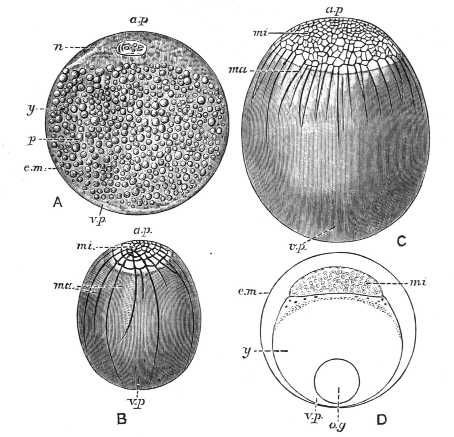
Fig. 234.—Different types of egg-segmentation in Fishes. A, a typical telolecithal egg. Holoblastic and unequal segmentation in Amia (B) and in Lepidosteus (C). D, the meroblastic segmentation of a Teleost. a.p, Animal pole; e.m, egg-membrane; ma, macromeres; mi, micromeres; n, nucleus; o.g, oil globule; p, protoplasm; v.p, vegetative pole; y, yolk. (From Ziegler: A, after Hertwig; B, after Whitman and Eycleshymer; C, after Eycleshymer.)
An egg-cell consists of living protoplasm and a nucleus, a variable quantity of non-living food-yolk, and of certain enveloping and protective egg-membranes. The ova of Fishes differ principally in the amount and disposition of the food-yolk, in the character of the egg-membranes, and in the presence or absence of special perforations in the egg-membranes for the entrance of spermatozoa into the eggs. In the small ova of some of the lower Chordata (e.g. Amphioxus), where the very small quantity of food-yolk is uniformly distributed, and its presence affects all parts of the egg alike, the process of segmentation which follows fertilisation results in the transformation of the entire egg into a mass of approximately equal-sized cells or blastomeres (Fig. 82). The eggs are therefore described as "alecithal," and the segmentation as being "holoblastic" and "equal." On the other hand, all Fishes possess "telolecithal" eggs, that is, ova in which the food-yolk is more or less abundant, and tends to accumulate at one pole of the egg ("vegetative pole"), while the opposite or "animal pole" consists of protoplasm, comparatively free from yolk granules and containing the nucleus (Fig. 234, A). The term telolecithal is, however, a somewhat comprehensive one, and covers important variations in the relations of the inert food-yolk and the living protoplasm in different Fishes, which greatly modify the process of segmentation. Thus there are some Fishes in which the amount of food-yolk at the vegetative pole is sufficient to retard segmentation in that part of the egg without actually preventing it, and consequently segmentation begins in the animal pole, and takes place more rapidly there than it does when it extends into the vegetative pole. Hence it follows that although the entire egg is segmented the blastomeres are of unequal size, the animal pole giving rise to a large number of small cells or micromeres, and the vegetative pole to a smaller number of much larger cells or macromeres. The segmentation of such an egg is said to be holoblastic but unequal (Fig. 234, B and C). This type of egg is characteristic of the Chondrostei, the Holostei, and the Dipnoi. In other Fishes, like the Elasmobranchs and the Teleostei, the food-yolk so greatly preponderates that it entirely prevents segmentation in the vegetative part of the egg, and segmentation is restricted to the small mass of protoplasm (germinal disc) at the animal pole, in which the nucleus is situated (Fig. 234, D). Eggs undergoing partial segmentation in this way are termed "meroblastic." No hard and fast line can be drawn between the two types, and in the Chondrostei and Holostei an interesting transition between the holoblastic and meroblastic ova may be observed. The egg-membranes are formed either by the egg itself or by the epithelium of the ovarian ovisacs, and, as will shortly be seen, the character of the outer egg-membrane greatly influences the mode of deposition of the eggs and their location afterwards. In Elasmobranchs the egg is enclosed in a stout horny egg-shell, secreted by the oviducal shell gland.[489] In many Fishes, as in the Chondrostei, Holostei, and Teleostei, the egg-membranes are perforated at the animal pole of the egg by a small aperture or "micropyle," which is only large enough to admit of the entrance of a single spermatozoon at a time (Fig. 235). Generally, there is only a single micropyle, but, according to Salensky, the Sturgeon (A. sturio) has from 3 to 9, and the Sterlet (A. ruthenus) from 5 to 13.
An important distinction may be made between the ova of different Teleostomi as regards their location after extrusion from the female. From this point of view two types of ova can be distinguished, demersal and pelagic ova. Demersal eggs are characterised by their larger size and greater weight, so that they always sink after extrusion; and by their opacity. They may either have an outer egg-membrane which is viscid and adhesive, so that the eggs readily adhere to one another or to foreign objects, or the membrane is smooth and non-adhesive. The Salmonidae, for example, produce non-adhesive demersal eggs, which remain separate after being deposited on the gravelly bed of a stream. Most freshwater and many marine shore Fishes have adhesive demersal eggs, which are deposited at the bottom of the water, generally adhering to one another in larger or smaller clumps, masses, or sheets, and attached to rocks, stones, or empty shells, like the eggs of many shore Fishes, or to aquatic plants after the fashion of the eggs of the Carp, Perch, and Pike, or even to branching zoophytes, as is the case with the eggs of the Sea-snail (Liparis). In some adhesive eggs the external egg-membrane forms threads for their attachment. The eggs of the Gar-Fish (Belone), and those of the Saury Pike (Scombresox) and of the Flying Fishes (Exocoetus), have viscid threads developed from opposite points on the surface, which are either attached to foreign objects or they become entangled with those of other eggs of the same species. The oval eggs of some of the Gobies have a bunch of fibres at one pole which serves to attach them. In the Smelt (Osmerus eperlanus) a portion of the outer egg-membrane breaks away from the rest and becomes turned back, inside out, but remains attached to the egg at one point. By means of this membrane the egg is attached to rocks or stones. Pelagic eggs are distinguished by their lightness and buoyancy, so that they always float near the surface of the water, and by their smaller size and remarkable transparency (Fig. 235). A conspicuous feature in many of them is the presence of a single large oil globule on the surface of the yolk, and not infrequently the yolk becomes partially or completely broken up into small masses. Pelagic eggs are always non-adhesive and free, and they invariably belong to marine Fishes. Amongst the British food Fishes which produce pelagic ova may be mentioned the Gadidae (e.g. Cod, Whiting, Hake, Ling), the Pleuronectidae (e.g. Turbot, Brill, Sole, Plaice), Scombridae (e.g. Mackerel), Triglidae (e.g. the Gurnards), Percidae (e.g. the Bass), and Clupeidae like the Pilchard and Sprat, but not the Herring, whose adhesive demersal eggs are deposited in clumps on shingly banks in the sea at varying distances from the shore.
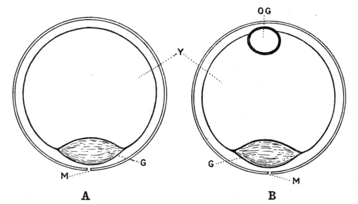
Fig. 235.—Diagrams of the pelagic ova of a Cod or a Plaice (A) and of a Ling (Molva). G, Germinal disc; M, micropyle; O.G, oil globule; Y, yolk. (From Cunningham.)
The eggs of Elasmobranchs are deposited singly or in pairs at considerable intervals, and the period of egg-laying is prolonged over a considerable part of the year. In most other Fishes, as in Teleosts, the period of spawning is limited to a few months, usually in the spring and summer in temperate latitudes; and in the case of a single Fish it may last only a few days or weeks, but the number of eggs produced is often enormous. Thus, in a Ling 61 inches long and weighing 54 pounds the ovaries contained 28,361,000 eggs. A Turbot, 17 pounds in weight, had 9,161,000 eggs; and a Cod of 21½ pounds 6,652,000. The least prolific of the British food Fishes is the Herring, in which the number of ovarian eggs varied from 21,000 to 47,000 in four specimens examined.[490] The extraordinary fecundity of many Fishes seems to bear no relation to the relative abundance of the Fishes themselves, but rather it is to be associated with certain disadvantages attendant on the sexual relations of Fishes, involving a considerable waste of the sex-cells, while in many Fishes it no doubt helps to compensate for any subsequent mortality among the larvae, which may result from an uncertain and precarious food supply and from the attacks of enemies. Whenever internal fertilisation is the rule, or when, as in nest-building and marsupial Fishes, the propinquity of the sexes in the breeding season ensures the fertilisation of a larger proportion of the eggs and the protection of the young, the number of eggs produced is small.
The male sex-cells or spermatozoa are essentially similar to those of other Vertebrates, although in different Fishes they may vary in such details as length, and the shape and size of the head, which may be rod-like and wavy, elliptical or globular.
As a rule, in Fishes females are more numerous than males, and generally they are larger, but to both statements there are notable exceptions. The relations of the sexes in the breeding season are usually very promiscuous, especially in those Teleosts which discharge their sex-cells while swimming together in shoals. A female may, however, consort with several males (polyandry), or a male with several females (polygamy); or, as in some of the nest-building Fishes (e.g. Gastrosteus), there are not wanting examples of the pairing of one male with one female (monogamy).
Fishes often migrate at the commencement of the breeding-season to localities most suitable for the deposition of the eggs. Many marine species seek banks or shallower water near the shore, and some, like the Salmon and the Sturgeon, are anadromous, and ascend rivers for long distances to deposit their spawn.
In all Fishes except the Elasmobranchs and a few Teleosts the fertilisation of the eggs takes place in the water after their extrusion, the male depositing its seminal fluid over the eggs or in their neighbourhood. The waste of the sex-cells is often, no doubt, very considerable, especially when the eggs are adhesive and fixed, and the seminal fluid is liable to drift at the mercy of tides and currents. With pelagic ova the waste is perhaps not so great, inasmuch as the eggs as well as the spermatozoa would probably drift at the same rate and in the same direction. Liability to waste must also be greatly diminished in many Fishes by their habit of living in shoals, or of congregating together in the breeding season, in which they are sometimes aided by their power of emitting characteristic sounds, and in the case of nest-building Fishes by the still more intimate relations of the sexes. Even when the liability to waste is very great, compensation may be afforded by exceptional fecundity. The copulation of the sexes and the internal fertilisation of the eggs occur only in Elasmobranchs and some Teleosts. The copulatory organs of Elasmobranchs are the so-called "claspers" with which the males are provided. Some form of copulation is probably the rule in the viviparous Teleosts, where the eggs are fertilised in the oviducts, or even while they are still in the ovaries, and the young are born alive. As mentioned above, an intromittent organ is often formed by the prolongation of the genital or the urinogenital orifice into a papilla, or a longer or shorter tube.[491] Some Cyprinodontidae[492] (e.g. Anableps) have the anterior part of the anal fin modified in the male to form an intromittent organ along which the urinogenital canal runs (Fig. 374). In the females the genital aperture is covered by a special scale, which is free on one side and not on the other. "The male organ in some individuals is turned to the right, in others to the left, and in some females the opening beneath the special scale is to the right, in others to the left. Copulation thus takes place sideways, a left-sided male pairing with a right-sided female, and vice versa."[493] The anal fin also forms an intromittent organ in the "Half-beak" (Hemirhamphus). In a genus (Girardinus) of the same family the anal fin is modified to form an apparatus for holding the female during sexual congress.[494] The singular method of fertilisation practised by the males and females of Callichthys paleatus is referred to elsewhere.[495]
With the exception of the pelagic Antennarius, which builds its nest in the Sargasso weed in mid-ocean, nest-building and parental solicitude for the young are confined to freshwater Fishes and to marine forms with demersal ova. Pelagic ova must necessarily be beyond the scope of parental care. As a rule it is the male which acts as guardian nurse, the female troubling herself but little about the fate of her eggs or her offspring.
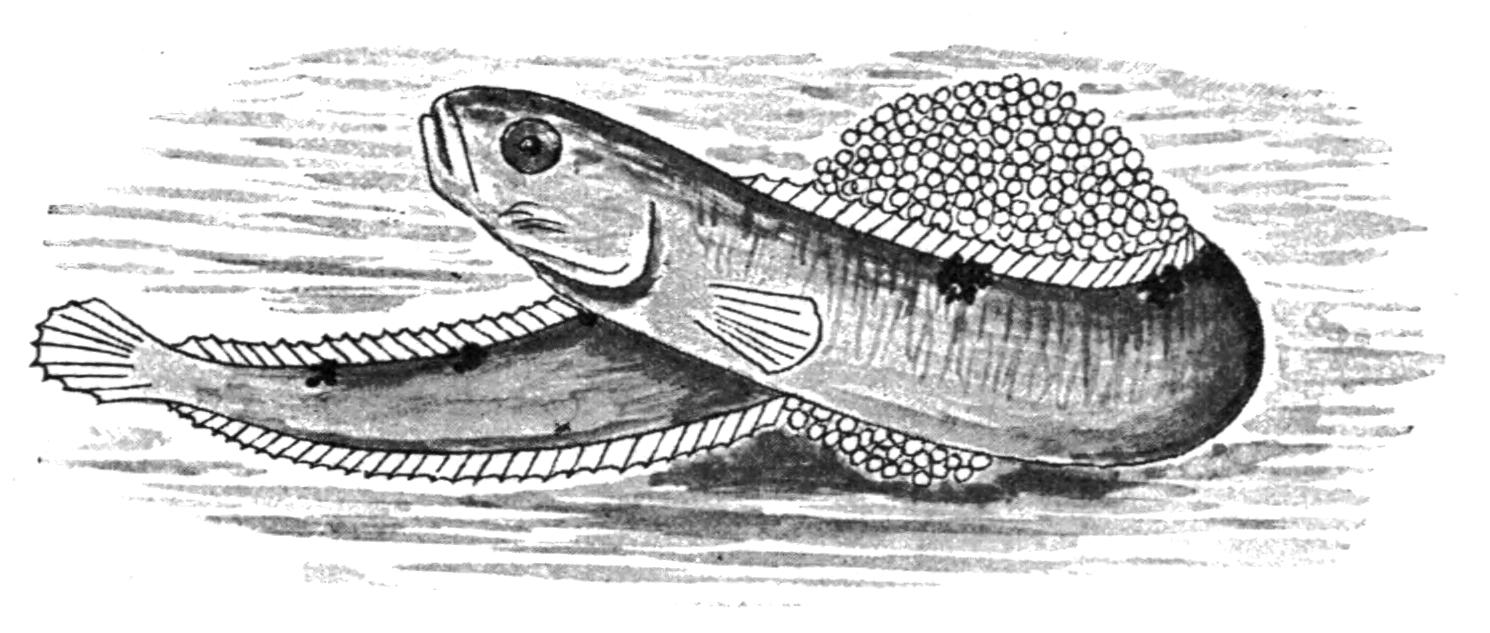
Fig. 236.—The Butter-Fish (Pholis gunnellus) coiling round a mass of eggs. (From Cunningham, after Holt.)
Perhaps the more primitive form of parental foresight is exhibited by those Fishes which, like the females of the Salmonidae, make a furrow in the gravelly bottom of a running stream for the reception of the eggs, and then cover them over with a layer of gravel, or like the Siluroid Arius australis, of the Burnett river in Queensland, which deposits its eggs in circular excavations in the sandy bed of the river and covers them with layers of large stones. But in neither case does it appear that either the male or the female takes any further interest in the eggs or in the young when hatched. Without actual nest-building, or even the preparation of a place for their reception, the eggs may be protected in various ways by the male. The common British Gunnel or Butter-Fish (Pholis gunnellus) rolls its eggs into a rounded mass by coiling its body round them, the male and female taking possession of them alternately. The little clumps of eggs are then deposited in holes made by the boring Mollusc, Pholas. Some British Blennies attach their eggs in a single layer to the sides of cavities in rocks, or between stones, where they are watched over by the male parent. The eggs of the Lump-Sucker (Cyclopterus lumpus) are attached in masses to rocks or to piles and guarded by the male, who aerates them by keeping up a flow of water over the spawn through the action of his pectoral fins. When hatched, the young fry cling to the body of their watchful parent by their suckers. A more decided approach to nest-building is exhibited by the Sand Goby (Gobius minutus). In this species the male scoops out the sand from beneath an empty shell, generally that of a Pecten, and the female deposits her adhesive eggs on the under surface of the shell.
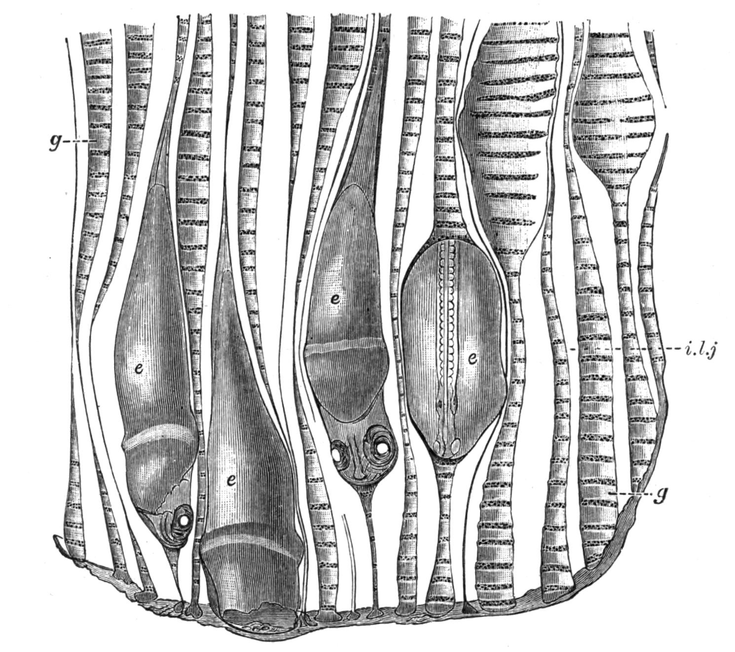
Fig. 237.—Showing the embryos of Rhodeus amarus in the gill-cavities of Unio. e, Embryos; g, inter-lamellar cavities; i.l.j, an inter-lamellar junction. (From Olt.)
The male remains on guard, and by the movements of its pectoral fins promotes the aeration of this rude form of nest. References to some of the more striking examples of true nest-building in Fishes will be found in the systematic part of this volume, especially in those chapters treating of the Dipnoi and Amiidae, and such Teleosts as the Mormyridae, Osteoglossidae, Siluridae, Gastrosteidae, Centrarchidae, Osphromenidae, Labridae, and Antennariidae. Other illustrations of parental care are to be found in the development of marsupial pouches or grooves for the reception of the eggs in the males of the Syngnathidae (Fig. 387) and the females of the Solenostomidae, and the use of the oral cavity for a similar purpose in the males, rarely in the females, of some Siluridae, and the males or females, according to the species, of the Cichlidae. The singular method by which the female Aspredo safeguards both her eggs and her progeny is referred to on p. 596. The Cyprinoid, Rhodeus amarus (the "Bitterling" of Central Europe), is unique in the means which it adopts to secure the same result.[496] By means of its long ovipositor the female Fish deposits its eggs in the mantle cavity of a Unio, or of an Anodon. Here they are fertilised by spermatozoa carried in through the inhalent siphon of the Mollusc with the inspiratory water current, and they complete their development in the gill-cavities (Fig. 237).[497]
The time which elapses between the fertilisation of the egg and the hatching out of the young Fish varies greatly in different Teleosts. The eggs of some Clupeidae hatch in a very short time, two to three days in the Anchovy, and three to four days in the Sprat. In most of the British marine food Fishes the period rarely exceeds twelve to fourteen days. The larger demersal eggs with much food-yolk are longer in hatching; in the Salmon the time ranging from thirty-five to one hundred and forty-eight days. A low temperature lengthens the time. The eggs of the Herring which hatched in eight to nine days at a temperature of 52° to 58° F. took forty-seven days in water at 32° F.
The extent to which the development of the embryo proceeds while it is still enclosed in the egg-membranes, and consequently the condition of the embryo when hatched, depends largely but not exclusively on the quantity of food-yolk which is present in the egg and available for the nutrition of the embryo during its earlier stages. Embryos hatched from pelagic ova are very small and imperfectly developed. The mouth is usually not yet formed. The median fins, which later become isolated, are continuous, and the caudal fin is diphycercal, although it subsequently becomes homocercal after passing through a heterocercal stage. The blood is colourless, and even the gill-clefts may at first be lacking. In this condition the newly-hatched Fish is nourished at the expense of the residual food-yolk, which is enclosed in a yolk-sac projecting from the ventral surface of the body (Fig. 238). As the yolk is gradually used up the mouth is formed, and the young Fish feeds on the minute organisms of various kinds living in the sea, and by degrees the form, proportions, and structure of the more mature Fish are acquired. In the case of the larger demersal eggs the young are not only longer in hatching, but when hatched they are larger and more advanced in development. The young of many Fishes are provided with larval or provisional organs, and they may be so unlike the adult in other respects that their subsequent development assumes the form of a more or less striking metamorphosis. As examples of larval organs, mention may be made of the adhesive or cement organs of the larval Chondrostei and Holostei, and of the Dipnoi (e.g. Protopterus and Lepidosiren), and also of a Teleost, probably the Mormyrid (Hyperopisus bebe, Lacép);[498] the cutaneous gills of the Crossopterygii and some Dipnoi; the so-called external gills of such Teleosts as Cobitis, Gymnarchus (Fig. 239), and Heterotis, which are singularly like those of Elasmobranchs; and the defensive spines which are developed on the scales or scutes of the trunk, and the dermal bones of the skull, in the young of some Plectognathi. The most striking metamorphosis to be found in Fishes occurs in the Flat-Fishes and in the Eels, an account of which will be found in other parts of this volume (pp. 685, 602).
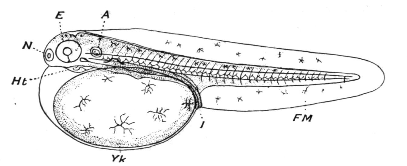
Fig. 238.—Newly-hatched embryo Teleost from a pelagic egg. A, Auditory organ; E, eye; FM, continuous median fin; Ht, heart; I, intestine; N, nostril; Yk, yolk-sac. (From Cunningham.)
The only examples of viviparous Fishes occur in certain families of Elasmobranchs,[499] and in five families of Teleosts, viz. the Blenniidae, the Cyprinodontidae, the Scorpaenidae, the Comephoridae, and the Embiotocidae.[500] In the Teleosts mentioned the eggs are fertilised while they are still either in the ovarian ovisacs or in the cavity of the ovary, and their development may take place in either position. In such Cyprinodonts as Gambusia and Anableps the embryos are developed in the ovisacs, but as a rule both fertilisation and development occur in the ovarian cavity. During a prolonged gestation the young are nourished partly by the food-yolk present in the eggs, and partly by a nutritive secretion derived from the ovarian walls or from the epithelial wall of the ovisacs as the case may be. In Anableps the secretion of the walls of the ovisacs is absorbed by papillae developed on the surface of the yolk-sac of the embryo along the course of its blood-vessels. The eggs of the Embiotocidae have little food-yolk, and the embryos are mainly nourished by the secretion of the ovarian walls, which is swallowed by the embryo and absorbed by villi on the inner surface of the intestine. The number of young produced varies considerably. In the Embiotocidae the ovarian cavity contains 40 to 50 young. The viviparous Scorpaenid, Sebastes norvegicus of Northern Europe, produces, it has been estimated, about 1000 young, while the Blenny (Zoarces viviparus), the only other European viviparous Teleost, produces from 20 to 300 or more, according to the size of the female. In the Blenny the eggs are hatched in about twenty days, but the young are not born until about four months after fertilisation, when they are about an inch and a half long, and in every outward respect similar to the adult Fish.
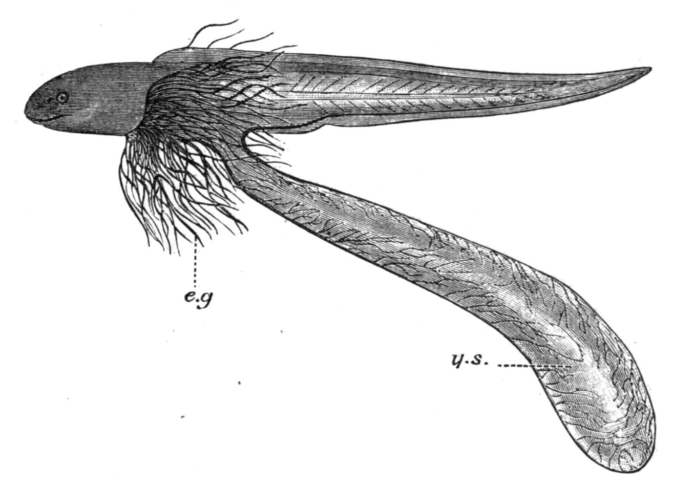
Fig. 239.—Young Gymnarchus niloticus, with its large yolk-sac (y.s) and its long external gills (e.g). (From Budgett.)
Besides the distinction between the sexes resulting from the different nature of their gonads and sex-cells, the males and females are often distinguished by secondary sexual characters ("sexual dimorphism"[501]). As mentioned above, females are usually larger as well as more numerous than the males, although in one or both respects the reverse may be the case. Secondary sexual characters are best marked in Teleosts, where they are generally related to the special rôle which each sex takes in the deposition and fertilisation of the eggs, and the nurture and protection of the young, of which examples have already been given. To a more limited extent they may be associated with the struggle of the males for the females, and in at least a few Teleosts the exuberant coloration of the males in the breeding season suggests that instances of courtship and sexual selection are not altogether wanting.[502]
Although the vast majority of Fishes are dioecious, instances of functional hermaphroditism are not unknown in a few Teleosts.[503] Species of the Percoid genus Serranus (e.g. S. cabrilla, S. hepatus, and S. scriba) are invariably hermaphrodite and self-fertilising. Chrysophrys auratus is an example of successive hermaphroditism, the male and female sex-cells ripening alternately. As an occasional variation hermaphroditism has been recorded in several other Teleosts, including amongst others such well-known Fishes as the Cod, the Mackerel, and the Herring. The relations of the gonads in hermaphrodites is subject to much variation. In the Cod, for example, the testes may be double, each being continuous with the hinder end of the ovary of its side, or there may be only a single testis confluent with the anterior or the posterior portion, or with some other part of the surface, of either the right or left ovary. In other Teleosts individuals occasionally present themselves with a testis and an ovary on opposite sides.