Sub-Division III. FUNGI. (continued 2)
Michael Cresse Potter translation
About 2,000 species of Lichens have been described. If we disregard the Basidiolichenes, which will be considered on page 176, the remaining Lichens (Ascolichenes) may be divided into the two following orders according to the structure of the fruit-bodies:—
Order 1. Pyrenolichenes. The ascocarps (apothecia) are spherical or flask-shaped, as in the Pyrenomycetes, more rarely linear (Graphis).
According to the nature of the thallus, these Lichens may be divided into:—
a. Thallus homoiomerous, but not gelatinous, branching according to the mode of growth of the Algæ: Ephebe (Fig. 133), with Algæ of the genus Stigonema.
b. Thallus homoiomerous, gelatinous: Lichina.
c. Thallus heteromerous, crustaceous: Verrucaria, Pyrenula; Graphis (Fig. 139), which may be considered as Hysteriaceæ with gonidia; several species of Graphis are common on bark.
d. Thallus heteromerous, foliaceous: Endocarpon.
e. Thallus heteromerous, fruticose: Sphærophorus.
Order 2. Discolichenes. These, as in the Discomycetes, have open apothecia, which, as a rule, are cupular, more rarely hemispherical (Cladonia).
According to the nature of the thallus, these Lichens may be divided into:—
a. Thallus homoiomerous, but not gelatinous, branching according to the mode of growth of the Algæ: Cœnogonium.
b. Thallus homoiomerous, gelatinous: Collema (Fig. 132), with Algæ of the genus Nostoc; Leptogium.
c. Thallus heteromerous, crustaceous: Pertusaria (Fig. 140), Lecidea, with apothecia open from the beginning; Lecanora, with apothecia, which in the beginning are closed, later on open, but with a rim formed by the thallus (Fig. 138); Bæomyces, whose apothecia are borne on a stem formed by the thallus.
d. Thallus heteromerous, foliaceous: Parmelia (P. saxatilis; P. parietina, Wall-Lichen, Fig. 141, is yellow, very frequent on tree-stems, stone-walls, tiles); Physcia (P. ciliaris, frequent on tree-stems); Sticta (S. pulmonacea, Lung-Lichen, on tree-stems); Peltigera, especially on the Moss among trees; Umbilicaria, on rocks.
e. Thallus heteromerous, fruticose: Cetraria (C. islandica), “Iceland Moss,” with an olive-brown, flat, furrowed, fringed thallus, on heaths; C. nivalis, white, in the Polar regions; Evernia, Ramalina, Usnea (U. barbata, Beard-Lichen, Fig. 143); Roccella, Stereocaulon, Cladonia, of which the genus C. rangiferina, Reindeer-Moss (Fig. 142) is important; Cladonia has two kinds of thallus, one scaly and leaf-like, the other erect, which bears the apothecia and may be fruticose (Fig. 142), or cupular (Fig. 144); they grow in soil in forests and on heaths.
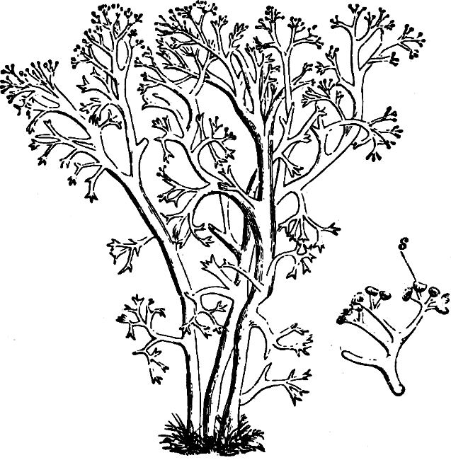
Fig. 142.—Cladonia rangiferina: s ascocarp.
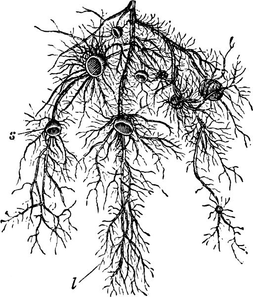
Fig. 143.—Usnea barbata: s ascocarp. (Slightly magnified.)
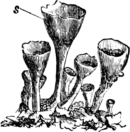
Fig. 144.—Cladonia pyxidata.
Sub-Class 2. Basidiomycetes.
This sub-class embraces the most highly developed Fungi, with large “fruit-bodies,” which in ordinary language we shortly term Funguses, Toadstools, or Mushrooms.
They have no sporangia, but reproduce only by means of basidiospores, conidia, chlamydospores and oidia. The chief characteristic of this sub-class is the basidium (Fig. 145), i.e. the conidiophore, which has a distinctive form, and bears a definite number (generally 4) of characteristically shaped conidia (basidiospores, Fig. 145 c, d, e).
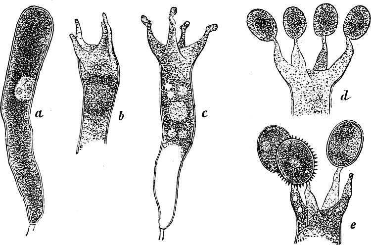
Fig. 145.—Development of spores in Corticium.
The summit of each basidium is produced generally into four conical points (sterigmata, Fig. 145 b), from each of which a basidiospore is abstricted. The basidia may be classified into three principal groups, each of which accompanies a distinctive conidiophore: 1, the long, filamentous, transversely divided basidia, with lateral sterigmata and spores, found in the Uredinaceæ (Figs. 146 D, 153), Auriculariaceæ (Fig. 160 B), and Pilacraceæ; 2, the spherical, longitudinally divided basidia of the Tremellaceæ (Figs. 160 C d; 161 iii. iv.); and 3, the ovoid, or cylindrical, undivided basidia of the Autobasidiomycetes (Figs. 145, 163, etc.); the two last have apical sterigmata and spores.
The first two groups are the septate basidia (protobasidia), of the Protobasidiomycetes; while the unseptate basidia (autobasidia) of the Autobasidiomycetes are the third group. On the formation of the basidiospores, the nucleus of the basidium divides into four nuclei, each of which is transferred to a spore.
In addition to the basidia, simple conidiophores are also found. In the Protobasidiomycetes, the simple conidia are very generally found as accessory methods of reproduction in conjunction with the basidiospores; but less frequently in the Autobasidiomycetes, e.g. among the Dacryomycetes, Tomentellaceæ, Heterobasidion annosum.
The simple conidiophores vary in size, and in the number and shape of the conidia; they, however, resemble the basidia, and are doubtless an early stage in the development of the definitely formed basidia.
Finally, well-defined chlamydospores, formed in various ways, appear in the Basidiomycetes as supplementary reproductive bodies (compare p. 90). Among the Protobasidiomycetes, chlamydospores are at present only found among the Uredinaceæ, but in various forms; in the majority of families of the Autobasidiomycetes oidia frequently occur (Fig. 162), but genuine chlamydospores seldom.
In the same species several of the known forms of reproduction may be distinguished.
The mycelium is generally composed of white, branched strands, consisting of numerous felted hyphæ; in some, sclerotia are found.—The great majority are saprophytes; some (particularly all the Uredinaceæ), are parasites.
Divisions of the Basidiomycetes.
Series 1. Protobasidiomycetes: partly gymnocarpic, partly angiocarpic.
„ 2. Autobasidiomycetes.
Family 1. Dacryomycetes: gymnocarpic.
„ 2. Hymenomycetes: partly gymnocarpic, partly hemiangiocarpic.
„ 3. Phalloideæ: hemiangiocarpic.
„ 4. Gasteromycetes: angiocarpic.
Appended. Basidiolichenes: Lichen-forming basidiomycetes.
Series I. Protobasidiomycetes.
To this series belong the lowest of the Basidiomycetes. The basidia appear in two principal forms (1 and 2 on page 144) and are divided into four cells, either transversely or longitudinally, each division forming a sterigma which abstricts a basidiospore. The first three orders, Uredinaceæ, Auriculariaceæ, and Tremellaceæ have gymnocarpic fruit-bodies, while those of the Pilacraceæ, on the contrary, are angiocarpic.
Order 1. Uredinaceæ (Rusts). All the Rust-Fungi are parasites, their mycelium living in the interior of the stems and leaves of their hosts, causing red, brown, or black spots—hence their name—and malformations, sometimes of considerable size.
The Rust-Fungi are gymnocarpic and destitute of a hymenium; for these reasons they are regarded as the simplest order of the Basidiomycetes. They are entirely parasitic, and their filamentous, branched mycelium ramifies in the intercellular spaces of its host, and often protrudes haustoria into the cells. The mycelium is perennial should it enter a woody tissue; it may also hibernate in the rhizomes of perennial herbs and permeate the shoots springing from them, but in the majority of the Rust-Fungi the mycelium has a very limited growth. The chief means of reproduction of the Rust-Fungi are the chlamydospores, which in the more highly developed species occur in three forms, namely, the teleuto-, æcidio-, and uredo-spores. The spores, in the host, are formed immediately beneath its epidermis, which is ruptured on the ripening of the spores, with the production of “rust,” brown, red, or black spots. Those chlamydospores which produce basidia are termed teleutospores. The spore on germination produces a transversely divided basidium, “promycelium,” on which basidiospores, “sporidia,” generally four in number, are produced on lateral sterigmata. This basidio-fructification is gymnocarpic; the basidia neither form a hymenium nor a fruit-body (only Cronartium and Gymnosporangium have a slight indication of a basidio-fructification).
Many Rust-Fungi, in addition to basidiospores, have small, unicellular conidia, “spermatia,” which are borne in conidiocarps, “spermogonia.”
The TELEUTOSPORES (Winter-spores) may be either unicellular or multicellular; in the majority of cases they are enclosed in a hard outer cell-wall, the exospore, which in some cases is very strongly developed; they have also a long or short stalk, the remains of the spore-bearing hypha. Each cell of the teleutospore has one germ-pore (a thin portion of the wall, for the protrusion of the germ-tube; in Phragmidium and Gymnosporangium there are, however, several germ-pores). The colour of the teleutospores is generally much darker than that of the uredospores, and it is by these that the majority of the Rust-Fungi hibernate.
In Gymnosporangium, two kinds of teleutospores are found (distinguished by their size and thickness of exospore). In many species of Puccinia, the form of the teleutospores varies very much, so that in the same layer spores have been observed with the characteristic form of other, allied genera.—The teleutospores of Endophyllum resemble æcidiospores, since they are united in chains, whose cells are easily separated, and are produced in the interior of a “peridium.” The multicellular teleutospores of Coleosporium function as basidia, and from each cell immediately produce basidiospores.—The teleutospores of Coleosporium and Chrysomyxa, differ from other teleutospores in the absence of exospore and germ-pore.
The ÆCIDOSPORES (Spring-spores) are produced in chains which are generally enclosed in an envelope of hyphæ, the peridium; the peridium enclosing the spores being termed the æcidium. The æcidiospores are unicellular, and generally of an orange colour; they are often separated by intermediate cells which wither and so assist in the distribution of the spores. The exospore is made up of minute, radially arranged rods. Generally germination proceeds immediately, the æcidiospore producing a germ-tube, which developes into a mycelium bearing either uredo- or teleutospores.
The æcidia of many Rust-Fungi were formerly considered as distinct genera. The æcidia of Phragmidium, Triphragmium, and Melampsora, in which the peridium is wanting, were in part considered as Cæoma. The æcidia with fimbriate edge, or those of Gymnosporangium with longitudinal lattice-like splits, were considered as “Rœstelia” (Lattice-Rust); large, sac-shaped æcidia on the Coniferæ were known as Peridermium.
The UREDOSPORES (Summer-spores) are unicellular and arise singly, seldom in chains (Coleosporium). Their colourless, warty exospore bears, in the equatorial plane, 2–8 germ-pores. In the majority, germination proceeds immediately, and a mycelium is produced which at first gives rise to uredospores and afterwards to teleutospores.
The uredospore-formations of Melampsorella and Cronartium are enclosed in an envelope, and hence resemble æcidia.—Between the uredospores sterile, unicellular hyphæ (paraphyses) may be found.
The spermogonia are spherical or pear-shaped conidiocarps, generally embedded in the substratum, and are produced before the æcidia, before or simultaneously with the uredospores, or before the teleutospores. The conidia, as far as observations go, do not generally germinate under ordinary conditions.
Among the Rust-Fungi some species are found which only form basidiospores and teleutospores (Puccinia malvacearum, Chrysomyxa abietis). Other species have in addition uredospores; others spermogonia and uredospores; others spermogonia and æcidia; others spermogonia, uredospores and æcidia. Those species in which all the methods of reproduction are not developed must not be considered as incomplete forms.
As a rule the mycelium, which is produced from the basidiospores, developes æcidia; in the species, however, without æcidia, it developes the uredo-form, and when the uredospores are also absent, the teleutospore-form. It has been established in some species of Puccinia and Uromyces that the formation of æcidia can be suppressed, and it is not a necessary part of the cycle of development of the species.
The majority of Rust-Fungi hibernate in the teleutospore-form. Many species are able to hibernate in the uredospore-form (Coleosporium senecionis). Others pass the winter in the æcidio-form, and develope æcidia on new hosts (Uromyces pisi, on Euphorbia cyparissias; Phragmidium subcorticium, on Rosa; Æcidium elatinum, on Abies alba). In Chrysomyxa abietis, the mycelium, developed from the basidiospores, survives the winter.
Among the Rust-Fungi, with several forms of reproduction, there are about sixty whose development can only be completed by an alternation of hosts, that is, on one host only uredo-and teleutospores are produced, while the further development of the germinating basidiospores, and the formation of the æcidia and spermogonia from its mycelium, can only take place on a second quite distinct and definite host (heterœcious or metoxenous Fungi). Those Fungi which have all their forms of reproduction on the same host are termed autœcious or autoxenous. It is not, however, always necessary that the heterœcious Rust-Fungi should regularly change their hosts; for example, Puccinia graminis can hibernate in the uredo-form on the wild Grasses, and in the spring can distribute itself again in the same form.
As a consequence of the alternation of hosts the various forms of development were considered as independent genera (Uredo, Æcidium, Rœstelia, Cæoma, Peridermium), until De Bary and Oersted established, about the same time (1865), the mutual connection of some forms, and paved the way for the right conception of these Fungi.
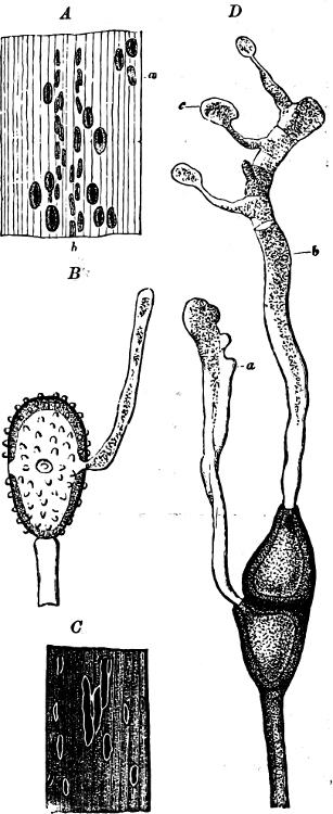
Fig. 146.—Puccinia graminis.
As an example of one of the most highly developed species, Puccinia graminis, the “Rust of Wheat,” holds a prominent position. Its uredospores and teleutospores are produced (Fig. 146) on Grasses (on cereals, especially Wheat, Rye, Oats, and many wild Grasses), while the æcidia and spermogonia are confined to the Berberidaceæ. The teleutospores, developed on the Grasses, hibernate on the dried portions of their host, and in the succeeding year each of the two cells of the teleutospore may develop a basidium with four basidiospores (Fig. 146 D, c). The basidiospores are distributed by the wind, germinate quickly, and only proceed to further development on Berberis or Mahonia. The germ-tube bores through the epidermis of the Barberry-leaf, and forms a mycelium in its interior, its presence being indicated by reddish-yellow spots on the leaf. After 6–10 days the flask-shaped spermogonia appear (Fig. 147 B; C, a; conidia in Fig. 147 D) and a few days later the cup-shaped æcidia (Fig. 147 A; C, c, d, e). The former are generally on the upper, and the latter on the under side of the leaf. The orange-coloured æcidiospores scatter like dust, and germinate only on Grasses; the germination takes place in about two days when placed on any green part of a Grass. The germ-tube enters the Grass-leaf through a stoma; a mycelium is developed in the leaf, giving rise to a small, oval, rust-coloured spot (Fig. 146 A); in about 6–9 days the epidermis is ruptured over the red spot, and numerous reddish-yellow uredospores, formed on the mycelium, are set free. The uredospores (Fig. 146 B) are scattered by the wind, and can germinate should they fall on the green portions of other Grasses: they then emit 2–4 germ-tubes through the equatorially-placed germ-pores. The germ-tubes enter a leaf through a stoma, a new mycelium is then developed, and in about eight days a fresh production of uredospores takes place, which germinate as before. The uredospore-mycelium very soon produces, in addition, the brown teleutospores, which give a brown colour to the rust-coloured spots, the familiar uredospores on the cereals being quite suppressed towards the close of the summer (Fig. 146 C, D). The “Rust of Wheat” hibernates on some wild Grasses in the uredospore-form.
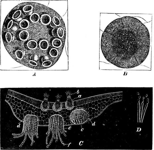
Fig. 147.—Æcidium berberidis. A Portion of lower surface of leaf of Barberry, with cluster-cups (æcidia). B A small portion of leaf, with spermogonia, from above. C Transverse section of leaf on the upper side, in the palisade parenchyma are three spermogonia (a b); on the lower side an unripe æcidium (c d) and two ripe æcidia (d, e, f); f chain of æcidiospores. D Hyphæ, forming conidia.
Genera. Puccinia (Fig. 146, 147) has bicellular teleutospores, each having a germ-pore, and the æcidia when present have an indented peridium; some species, as exceptions, have 1–3-celled teleutospores. Many species are HETERŒCIOUS, for example, P. graminis, described above; P. rubigo, which also infests various Grasses, but whose æcidia appear on Anchusa; the masses of teleutospores are small; they contain paraphyses, and are for a long time covered by the epidermis. P. coronata, on Oats and Rye Grass; its æcidia on Rhamnus; the teleutospores are surmounted by a crown—“coronate processes.” P. phragmitis, on Reeds; æcidia on species of Rumex and Rheum. P. moliniæ, on Molinia cœrulea; the æcidia on Orchids. P. poarum, on Meadow-Grass; æcidia on Tussilago. Various Puccinias growing on species of Carex have their æcidia on Urtica, Lysimachia, Cirsium, Pedicularis, etc.—Of those AUTŒCIOUS species, which have all their generations on the same host, may be noted:—P. galii, P. menthæ, P. violæ, P. epilobii, P. asparagi, which grow on the hosts from which they have taken their specific names.—As representative of a group which have spermogonia, uredo-and teleutospores on the same host, but on different individuals, P. suaveolens, on the Field-Thistle, may be mentioned. The spermogonia have a strong odour.—A peculiar group (Leptopuccinia) has only teleutospores, which germinate immediately, and whilst still attached to their living host. To this group belong P. arenariæ, on a number of Caryophyllaceæ; and P. malvacearum, on various Malvaceæ, introduced in 1873 from South America to Europe, where it soon proved very destructive to Hollyhocks.
Uromyces (Fig. 149) differs only from Puccinia in always having unicellular teleutospores. Among this genus both heterœcious and autœcious species are found. To the first group belong U. pisi, whose æcidia are found on Euphorbia cyparissias, and U. dactylidis, whose æcidia appear on Ranunculus; to the second group belong U. betæ, U. phaseoli, U. trifolii.
Triphragmium has teleutospores with three cells (one below and two above), on Spiræa ulmaria.
Phragmidium (Fig. 150) has teleutospores consisting of a row of cells (3–10) arranged in a straight line; the upper cell has one germ-pore and the others four germ-pores placed equatorially. Both this and the preceding genus have large, irregular æcidia without peridia, but often with bent, club-like paraphyses (150 b and c); they are all autœcious, and are only found on the Rosaceæ.
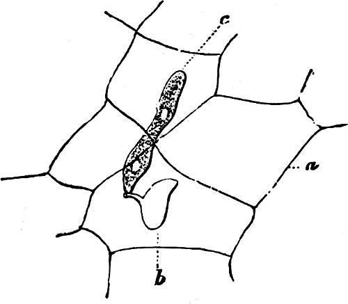
Fig. 148.—Gymnosporangium sabinæ. A small portion of the epidermis of a Pear-leaf (a) pierced at b by the germinating basidiospore (c).
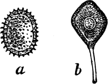
Fig. 149.—Uromyces genisteæ; a uredospore; b teleutospore.
Endophyllum (see above, under teleutospores, p. 147) on species of Sempervivum.
Gymnosporangium (Figs. 152, 154) has bicellular teleutospores collected in large, gelatinous masses formed by the swelling of the long spore-stalks; in each cell 2–4 germ-pores are found. Uredospores are wanting. All the species are heterœcious; the teleutospores appear on Juniperus, the æcidia (Rœstelia) on the Pomaceæ. G. sabinæ, on Juniperus sabina, J. virginiana, etc., has the æcidia (“Rœstelia cancellata”) on Pyrus communis (Figs. 152, 148); G. juniperinum, on Juniperus communis with “Rœstelia cornuta” (Fig. 154 a) on Sorbus aucuparia, Aria nivea (S. aria) and Malus communis; G. clavariæforme on Juniperus communis, the æcidium belonging to it (“Rœstelia lacerata”) on Cratægus oxyacantha.
Melampsora has prismatic teleutospores placed parallel to each other and forming a crustaceous layer; in many species they are divided longitudinally into several cells (Fig. 151). The æcidia, without peridium, belonged to the old genus Cæoma. M. caprearum, on Willows, has the æcidia (Cæoma euonymi) on Euonymus. M. hartigii, on Osiers; the æcidium on Ribes. M. mixta, on Salix repens and Orchids. M. pinitorqua, on leaves of the Aspen, æcidia on Pine branches (Pine shoot fungus); M. populina on Populus monilifera and nigra; M. betulina (Fig. 153), on Birch leaves; M. padi (Fig. 151), on leaves of Prunus padus, developes teleutospores in the epidermal cells; M. lini is the cause of injury to the Flax; M. agrimoniæ.
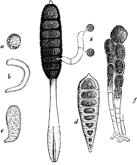
Fig. 150.—Phragmidium gracile: a an uredospore; b and c two paraphyses; d a young teleutospore; e a teleutospore with a basidium and two basidiospores (s); f two series of æcidiospores (Ph. rosæ).
Calyptospora gœppertiana; teleutospores on Vaccinium vitis idæa; spermogonia and æcidia on Abies alba (Firneedle-Rust).
Coleosporium (Fig. 155) forms its uredospores in reddish-yellow chains; for the teleutospores, see page 147. C. senecionis, on the Groundsel; its æcidium (Peridermium wolffii) on Pine-leaves (Fig. 155 a). Other species on Sonchus, Petasites, Campanula, Rhinanthaceæ.
Chrysomyxa (Fig. 156) has bright red, branched teleutospore-chains; each spore developes a 4-celled basidium. C. ledi, on Ledum palustre; its æcidia on the leaves of the Fir. C. abietis (Fig. 156), without uredo-and æcidiospores; teleutospores on the leaves of the Fir. In the first summer, yellow bands are formed on the leaves, and in the following spring the red cushions of spores.
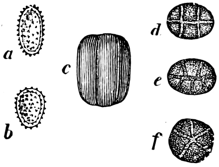
Fig. 151.—Melampsora padi: a and b uredospores; c-f teleutospores, seen from different sides.
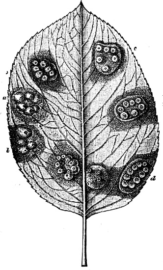
Fig. 152.—Pear-leaf, seen from the under side, with “Rœstelia cancellata”: in different ages (a youngest, d oldest).
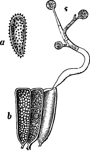
Fig. 153.—Melampsora betulina: a uredospores; b three contiguous teleutospores, one of which has developed a basidium with three basidiospores. (× 400.)
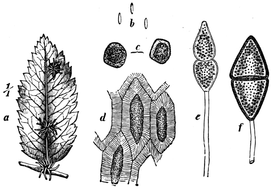
Fig. 154.—Gymnosporanginum juniperinum: a a small leaf with three clusters of æcidia (nat. size); b three conidia; c two æcidiospores on one of which are seen the germ-pores; d a portion of the wall of an æcidium; e, f two teleutospores.
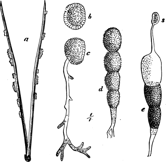
Fig. 155.—Coleosporium senecionis: a Pine-leaves with æcidia (Peridermium wolffii) nat. size; b an æcidiospore; c a germinating æcidiospore; d a chain of uredospores; e a chain of teleutospores of which the terminal one has germinated and produced a basidiospore (s).
Cronartium (Figs. 157, 159) has unicellular teleutospores united in numbers to form erect threads or columns; the uredospores are enclosed in a “peridium”; C. ribicola (Fig. 157), on leaves of Ribes (especially Black Currants); its æcidia (Peridermium strobi, or P. klebahni) on the stems and branches of Pinus strobus (Fig. 159), on which it causes great damage; C. asclepiadeum, on Vincetoxicum officinale; its æcidia (Peridermium cornui) on the stems and branches of Pinus silvestris.
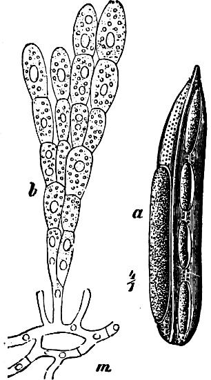
Fig. 156.—Chrysomyxa abietis: a leaf of the Fir, with 5 clusters of basidiospores (× 4); b branched rows of teleutospores springing from the mycelium (m).
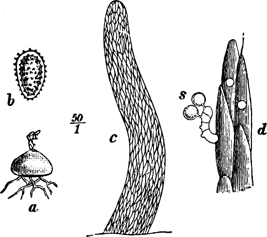
Fig. 157.—Cronartium ribicola: a mass of uredospores (× 50); b an uredospore; c a column of teleutospores (× 60); d a small portion of the same more highly magnified, with a basidium and two basidiospores (s).
To the Fungi of which the æcidium is known, whilst the remaining forms are still undetermined, but which are without doubt heterœcious, belong Æcidium elatinum, which produces the enormous “witches’ brooms” and barrel-shaped swellings on stems and branches of Abies alba; and Æcidium strobilinum (Fig. 158), which attacks Fir-cones, causing all the scales to become covered with clusters of æcidia opening by a lid. Hemileia vastatrix destroyed the coffee plantations in Asia.
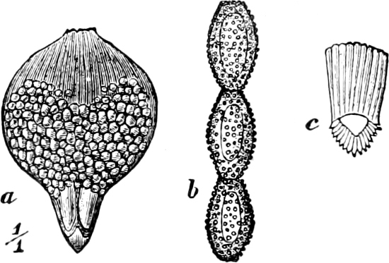
Fig. 158.—Æcidium strobilinum: a scale of cone of Picea excelsa, with numerous æcidia; b æcidiospores arranged in a series; c a cell of the peridium.
Order 2. Auriculariaceæ. The long, transversely divided basidia bear laterally 4 long sterigmata with basidiospores (Fig. 160 B) and are united to form an hymenium on the surface of the fruit-body. Parasites or saprophytes.
Auricularia sambucina (Auricula judæ), Judas’-ear, has large fruit-bodies, which may attain the size of several inches, resembling an ear or a mussel shell. In the moist condition they are flesh-coloured, tough and gelatinous, but when dried, become hard, grey and wrinkled; the exterior is covered with short hairs; while the internal surface bears the hymenium. Habitat: stems and branches of old Elder-trees (Sambucus).
Order 3. Tremellaceæ. The round, pear-shaped, longitudinally divided basidia bear 4 elongated sterigmata, situated apically, and 4 basidiospores (Fig. 160 C, D), and are united into the hymenium on the surface of the fruit-body. The fruit-bodies are frequently gelatinous and quivering; similar fruit-bodies are also found in the Dacryomycetaceæ and Hydnaceæ. Simple conidiophores, which appear not infrequently in the basidiocarps, before the basidia, are known in many species. Saprophytes.
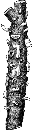
Fig. 159.—Peridermium strobi: æcidia of Cronartium ribicola (nat. size).
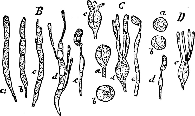
Fig. 160.—B Auricularia sambucina: a-d basidia in various stages of development; e a sterigma bearing a spore.—C Tremella lutescens: a-d basidia seen from various sides (b from above) and in various stages of development; e sterigma with basidiospore (× 400). D Exidia glandulosa: a-c various stages in the development of a basidium; d sterigma with basidiospore (× 350).
Exidia has kidney-shaped, oblong basidiospores, and small, hook-like conidia; E. glandulosa, E. albida, etc., on wood.—Craterocolla has conidiocarps; C. cerasi on Cherry-wood.—Sebacina incrustans; the yellow, fleshy, or cartilaginous fruit-bodies are found in autumn covering the ground in moist woods.—Tremella has round basidiospores; T. mesenterica has irregularly-folded, quivering, orange fruit-bodies, about one inch in breadth; T. lutescens (Fig. 161) has orange-yellow conidial-and yellow basidial-layers; T. frondosa has fruit-bodies upwards of a foot in breadth.
Order 4. Pilacraceæ. The transversely divided basidia have no sterigmata, but sessile basidiospores, and fill up the cavity of a closed (angiocarpic) fruit-body as a gleba without a regular arrangement (hymenium wanting).
Pilacre fagi on the old stems of the Copper-Beech; P. petersii, on dried branches of the Hornbeam, has stalked, capitate fruit-bodies.
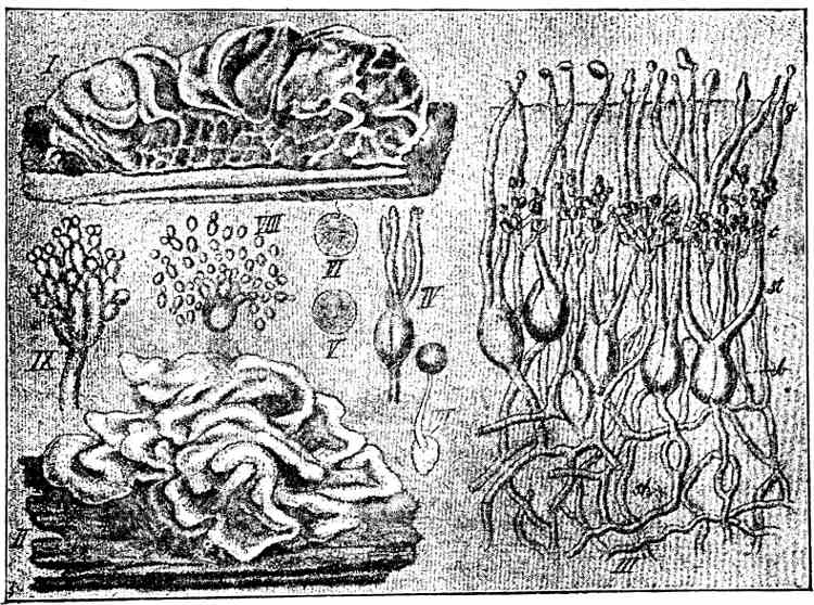
Fig. 161.—Tremella lutescens: I and II fruit-bodies (nat. size); III vertical section through a fruit-body; b basidia; c conidia; IV-VI basidia; VII basidiospore with a second spore; VIII a basidiospore with yeast-like budding (cultivated); IX a conidiophore. (III-IX about 400.)
Series 2. Autobasidiomycetes.
This second and larger part of the Basidiomycetes is characterised by its more highly differentiated, undivided, club-shaped, or cylindrical basidia, which generally bear 4 (seldom 2, 6, 8) apically-placed sterigmata and basidiospores (Fig. 145). The fruit-bodies are partly gymnocarpic (in the first 3 orders and in some Agaricaceæ), partly hemiangiocarpic (in orders 3–6 of the Hymenomycetes and in the Phalloideæ, the fruit-bodies in these orders are in the young conditions more or less angiocarpic, but later on generally open below and bear the hymenium on the under surface of the fruit-body), partly also angiocarpic (in the Gasteromycetes).
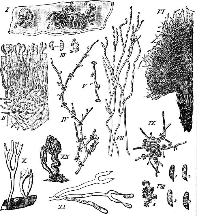
Fig. 162.—Dacryomyces deliquescens: I fruit-body (nat. size); II vertical section through the hymenium; III germinating basidiospore; IV a portion of mycelium with conidia; V a germinating conidium; VI and VII chains of oidia more or less strongly magnified; VIII basidiospore of D. longisporus; IX germinating basidiospore of D. ovisporus; X and XI Calocera viscosa; X fruit-body (nat. size); XI basidia with basidiospores (highly magnified); XII Dacryomitra glossoides (nat. size).
Family 1. Dacryomycetes.
The long, club-shaped basidia bear two tapering sterigmata, which develope remarkably large basidiospores (Fig. 162 II, XI) and form gymnocarpic fruit-bodies with hymenium. 1 order:
Order 1. Dacryomycetaceæ. This order comprises 4 genera of which the first two develope the hymenium on the whole surface of the fruit-body, but the two last only on its apex.
Dacryomyces: the folded, gelatinous, Tremella-like fruit-bodies break out in winter on dried wood (hedges) in the form of red or yellow drops. D. deliquescens is very common (Fig. 121). The following genera have cartilaginous fruit-bodies.—Calocera (Fig. 162), with club-like, simple, or branched, Clavaria-like, fruit-bodies; the orange coloured fruit-bodies of C. viscosa grow aggregated together on the wood of Conifers.—Guepinia resembles a Peziza, and has the hymenium only on the hollow upper surface.—Dacryomitra resembles a Mitrula (Fig. 162).
Family 2. Hymenomycetes.
This family is very rich in species (more than 8000 have been described), and to it belong all the “Mushrooms” and “Toadstools.” The fruit-bodies present very various forms; they are generally fleshy, very perishable, seldom leathery or corky, in the last case often perennial. The basidia are more or less cylindrical and bear generally 4 (seldom 2, 6 or 8) sterigmata and basidiospores. The hymenium in the fully-formed fruit-bodies lies free on the surface: in orders 1 and 2 and a portion of order 6 it is from the commencement exposed, fruit-bodies gymnocarpic; orders 3–6 have hemiangiocarpic fruit-bodies (p. 157). In the first order the basidia (or the hymenium) are developed immediately from the mycelium (Fig. 163); the fruit-bodies of orders 2 and 3 present a higher grade of development, and have between the mycelium and hymenium a special hyphal-tissue, a stroma, which is crustaceous, club-like, or coralloid, etc., and in general bears the hymenium on the largest part of the free, smooth surface. In the forms most highly developed (orders 4–6) a new tissue—the hymenophore—is introduced between the stroma and hymenium, which appears on the under side of the fruit-body in the form of warts, projections, tubes, folds or lamellæ (Figs. 166, 167, 174 bc). Paraphyses are frequently found in the hymenium, among the basidia. In the Hymenomycetes few examples of conidia can be recognised at first. More frequently chlamydospores are found, particularly oidia. The mycelium is richly branched, generally colourless, often perennial; it lives in humus or decaying wood, and is seldom parasitic. The hyphæ generally have clamp-connections and unite, sometimes, to form a rhizomorpha (Fig. 177) or sclerotia with coloured, pseudo-parenchymatous covering.
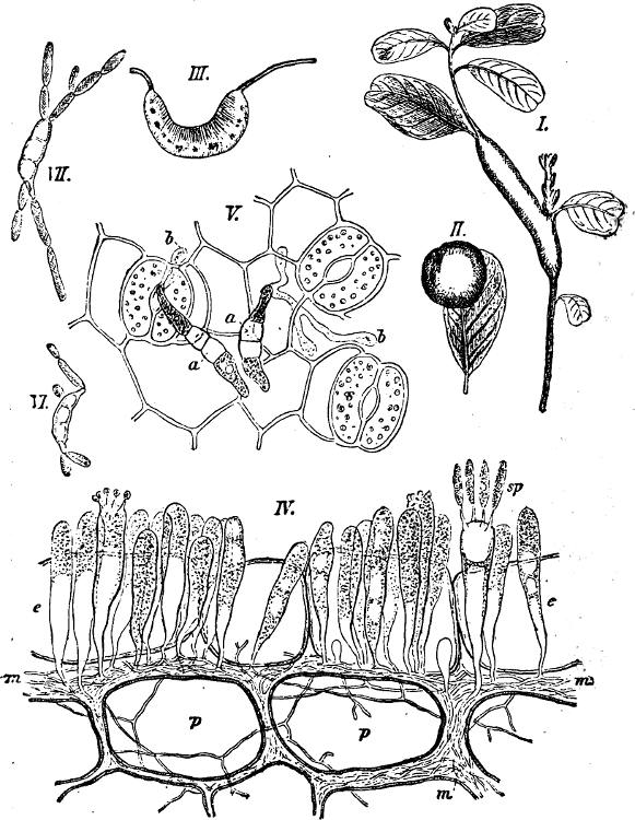
Fig. 163.—Exobasidium vaccinii. I Hypertrophied stem of Vaccinium vitis idæa; II leaf with gall-like swelling; III section of II; IV transverse section: m mycelium between the parenchymatous cells; p hypodermal cells; e epidermis with basidia in various stages of development; V epidermis with germinating spores; VI and VII spores germinating in water (IV-VII × 620).
Order 1. Tomentellaceæ. To this order belong the simplest of the Hymenomycetes. The basidia (Fig. 145) arise free and irregularly from the mycelium; a hymenium is entirely absent or very slightly formed (in Corticium it attains its highest development); fruit-bodies are also wanting.—In general they form flaky, membranous or leathery coverings on bark and wood. Some are parasites.
Hypochnus without conidia.—Tomentella with conidiophores; growing on wood or earth.—Exobasidium vaccinii (Fig. 163), a parasite on Vaccinium, Andromeda, Arctostaphylos, and Rhododendron, forms flaky-powdery, white or red coverings and may cause hypertrophy of the parts attacked. E. warmingii is parasitic on Saxifraga; E. lauri causes outgrowths on the stem of Laurus canariensis as long as a finger, which formerly were regarded as aerial roots.—Corticium forms membranous to leathery layers or crusts; C. quercinum on wood and bark, particularly Oak, is flesh-coloured; C. cæruleum has a blue hymenium; C. giganteum on the bark of fallen Pine-trees.
Order 2. Clavariaceæ. The hymenium is situated on a stroma, and either completely covers the smooth surface of the more or less fleshy gymnocarpic fruit-body, or is confined to a tolerably well defined upper portion of it (Typhula). Paraphyses absent. The vertical, white, yellow, or red fruit-bodies are roundish or club-like, undivided or richly branched (Fig. 125). Generally on the ground in woods, seldom on tree-stems, etc.
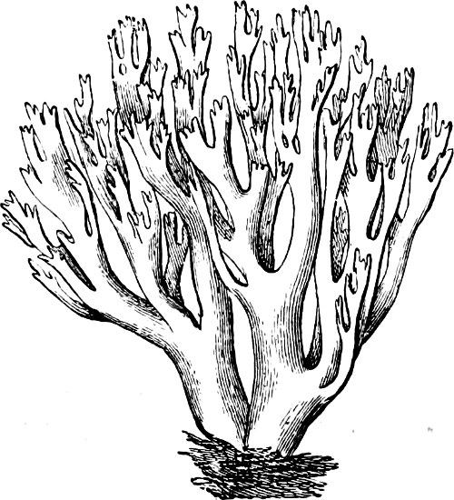
Fig. 164.—Clavaria coralloides (nat. size).
Genera: Clavaria, generally large Fungi with thick, round branches. C. botrytis has a very thick, tubercular stem with numerous short, flesh-coloured branches: it has an agreeable taste. C. coralloides has a brittle, richly-branched fruit-body (Fig. 164); basidia with two large spores. C. pistillaris consists of a single, undivided club of a yellowish-white colour.—Sparassis has compressed, leaf-like, curled branches; S. crispa has fruit-bodies as large as a white cabbage-head, with an agreeable taste.—Typhula and Pistillaria are small Fungi with filamentous stalks, terminating in a small club. The fruit-bodies of the former often arise from a small, spheroid sclerotium; the latter is distinguished by the basidia bearing only two spores.
Order 3. Thelephoraceæ. The hymenium is placed on a stroma and covers the smooth surface of the leathery hemiangiocarpic fruit-body, generally on its under side. The edge of the stroma, which bounds the hymenium, is sometimes especially developed (Stereum). Saprophytes.
Genera: Thelephora. The fruit-bodies in this genus are brown, very irregularly shaped, and often lobed. The spores too are brown, but in the other genera colourless. The species are found growing on barren soil. T. laciniata (Fig. 165) has imbricate, semicircular, dark-brown pileus, which is jagged at the edge and upper surface. The fruit-bodies are very often raised above the ground, and although this species is not a parasite, yet it destroys young seedlings by growing above and smothering them.—Stereum has a stiffer fruit-body, with a distinct, fibrous, intermediate layer. It grows on bark and wood, projecting like a series of imbricate brackets. S. hirsutum is yellow; its free edge is provided with a number of stiff hairs, the upper surface being divided into a number of zones. S. purpureum has a red-violet hymenium which distinguishes it from the previous species.—Cyphella has a membranous cup- or bell-shaped fruit-body, often borne on a stalk, the concave surface being covered with the hymenium. They are small, white Fungi, growing on Moss and dead stems.—Solenia is closely related to Cyphella; its fruit-bodies are smaller and hairy; they are found clustered together forming a crust-like covering on dead wood.—Craterellus has a large, funnel-shaped fruit-body, the hymenium covering the external surface. C. cornucopioides is shaped like a trumpet or a “horn of plenty.” It is dark-grey, several inches in height, and grows gregariously on the ground in forests. It is distinguished by the basidia bearing only two sterigmata.
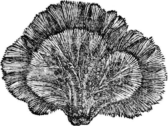
Fig. 165.—Thelephora laciniata (nat. size).
Order 4. Hydnaceæ. The fruit-body is most frequently fleshy, and varies considerably in shape, the simplest forms being resupinate,[14] the higher ones umbrella-like. The hymenophore is found on the free or downward-turned surface, and always takes the form of soft emergences hanging vertically downwards. The emergencies may be thorn-, awl-, or wart-like. The species are found growing on the soil and on dead wood.
Genera: Hydnum has subulate, distinct emergences. H. repandum is yellow, the stalk being placed in the centre of the pileus. It is an edible species, and often forms “fairy rings” in woods. H. auriscalpium (Fig. 166) is dark-brown, with stalk placed at the edge of the pileus. It grows on old Fir-cones. H. erinaceus grows on old tree-trunks. The fruit-body is yellow and very large—as big as a human head—with emergences as much as an inch in length.—Irpex has a leathery fruit-body, partly resupinate, partly with free, projecting edge; the under side bears tooth-like emergences which are arranged in rows, and Irpex thus forms a transition to the Agaricaceæ.—Phlebia is entirely resupinate, with radially-arranged folds on the free side, and pectinate border.
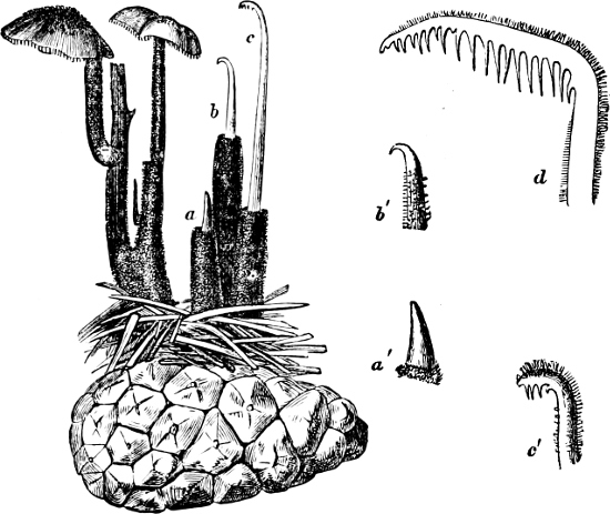
Fig. 166.—Hydnum auriscalpium, upon a Fir-cone, in different stages of development.
Order 5. Polyporaceæ (Pore-Fungi). An order very rich in species (about 2000 species are described). The fruit-body is of very different forms—resupinate, projecting like a bracket, hoof-like, or umbrella-shaped. In some it is fleshy and edible, in others leathery or corky, persisting for several years. The hymenophore is situated on the under side of the fruit-body, and consists of wide or narrow tubes or pores, whose inner surface is clothed with the hymenium (Fig. 167). In some fruit-bodies large cavities are to be found, which have arisen as interstices between the labyrinthine curved and reticulate folds. Chlamydospores are known in some species. Conidia occur very rarely. Many species work considerable damage: some as parasites on trees, others by destroying timber.
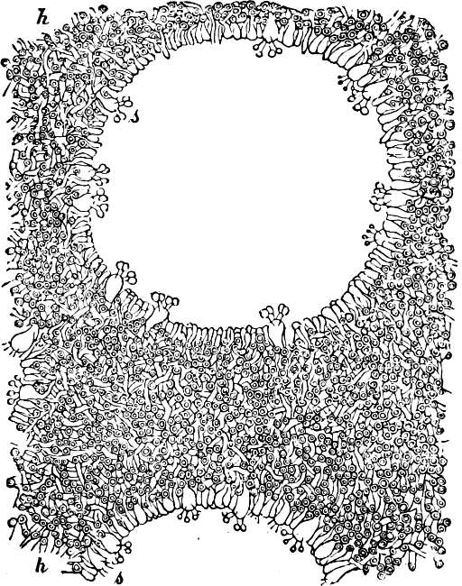
Fig. 167.—Polyporus igniarius. Section through the under side of the Fungus: h-h is hyphal-tissue between the tubes, formed by irregularly felted hyphæ, many of which are seen cut across; s is the hymenium which covers the walls of the tubes, and from which the basidia with the spores protrude.
Genera. Polyporus (Pore-Fungus). The tubes are narrow, accurately fitted together, and forming a thick layer on the under side of the fruit-body, appearing as a number of fine holes. The fruit-body most frequently resembles a bracket, or is hoof-shaped, with one side growing from a tree-trunk; it is very often perennial, and a new layer of tubes arises in each succeeding period of vegetation. Strata, corresponding to the periodically interrupted growth, are thus formed in storeys one above the other, and are visible on the upper surface of the fruit-body, as well as in the interior, as a series of concentric belts, sometimes as many as half a score or more in number. P. fomentarius (Touchwood) attacks trees, especially the Beech. The spores germinate on wounds from broken branches, and the hyphæ, following the course of the medullary rays, find their way into the interior of the tree, from whence the mycelium spreads upwards, downwards, and peripherally, so that the wood becomes rotten (“white-rot”) and thick felts of mycelium are formed in radial and tangential directions. A dark line, caused by the youngest parts of the hyphæ containing a brown juice, marks the boundary between the rotten and the unattacked parts of the stem (Fig. 168); at places where the mycelium extends to the bark, the cambium becomes destroyed and further growth is arrested, so that longitudinal furrows arise on the stem. It is at these places, too, that the hoof-shaped, ash-coloured fruit-bodies are developed, which may attain a circumference of upwards of 7 feet. The interior of the fruit-body consists of a dried-up, loosely felted, red-brown mass of hyphæ, which has been used for tinder and as a styptic (“Fungus chirurgorum”). P. igniarius has a harder, dark-brown, more rounded fruit-body; it grows in a similar manner, but especially attacks Oaks, Poplars, and Plum-trees, the wood of which becomes rotten, and is called touchwood. P. pini (Trametes pini), (Fig. 170), a parasite on the stems of Pinus, causes a kind of “red-rot” in the stem. P. sulphureus has a soft, cheesy, yellow fruit-body; it produces “rot” in Oaks and Apple-trees. P. officinalis, Larch-fungus (“Fungus Laricis” in Pharmocopœia), grows on Larch-trees in the south-east of Europe. P. versicolor has thin, semicircular fruit-bodies, with zones of various colours on the upper side; it is one of the most frequent species on tree-stems. P. frondosus grows on soil in woods, and consists of numerous aggregated fruit-bodies, which become very large and fleshy. This species is edible. P. perennis also grows on the soil in woods; it is very leathery, with central stalk, and has concentric zones on the upper surface of the fruit-body. P. vaporarius destroys the wood of living Pines (Pinus silvestris) and Firs (Picea excelsa), causing it to become red-brown; in timber this Fungus causes “red-strip” followed by a “dry-rot.” P. squamosus destroys many Walnut-trees, and is also very destructive to Limes and Elms. P. fulvus causes a “white-rot” in Abies alba.
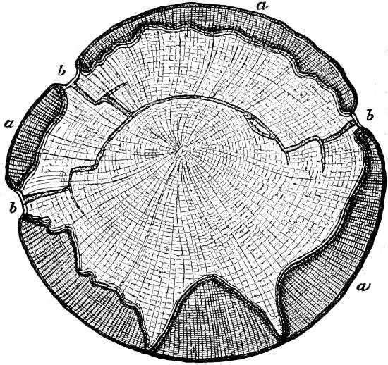
Fig. 168.—Section of stem of a Beech attacked by P. fomentarius: a non-attacked parts of the stem; b the furrows where the mycelium has reached the bark, and where the thick mycelium-strands reach the exterior (⅙th of the nat. size).
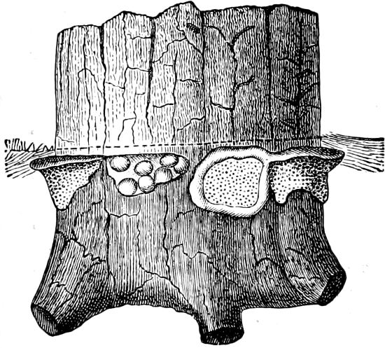
Fig. 169.—Base of a Fir-tree, with a number of fruit-bodies of Heterobasidion annosum just beneath the surface of the soil, indicated by the dotted line (¼th nat. size).
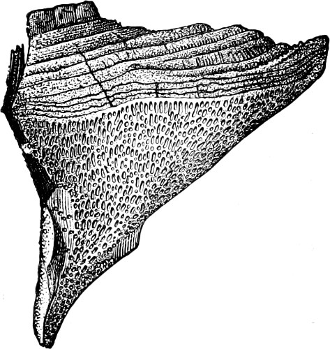
Fig. 170.—A fully developed fruit-body of Polyporus pini (Trametes pini), lateral view (nat. size).
Heterobasidion annosum (Polyporus annosus, Trametes radiciperda, Fig. 169) is characterized by its Aspergillus-like conidiophores. It is a parasite on the Pine, Fir, Birch, Beech, etc., and is the chief cause of a root-disease (red-rot) in Pines and Firs; the fruit-bodies develope a large number of basidiospores; they may be very large and are found just beneath the surface of the soil (on living or dead roots), and exposed to the air (on felled stems and roots, in Scandinavia).
Ptychogaster has cushion-like fruit-bodies, which consist chiefly of chlamydospore-chains, formed of ellipsoidal spores, which alternate with short hyphæ having transverse septa and clamp-connections. The hymenial portion is limited to a small group of tubes. Pt. albus (Oligorus ustilaginoides) grows on stumps of Conifers and forms irregular cushions, at first white and later on brown, which consist almost entirely of chlamydospores.
Boletus (Fig. 171) has a fleshy fruit-body resembling a common Mushroom, with central stalk. The layer of tubes is easily detached from the pileus, and the tubes are easily separable from one another. They grow on the ground in woods. Edible species are: B. edulis, with thick, reticulate stalk; B. scaber, with thin stalk and rough pileus; B. luteus, with a ring on the stalk. B. luridus is poisonous, its tubes have red openings, and the flesh turns quickly blue when broken and exposed to the air.
Fistulina hepatica (Beef-steak Fungus), has a red, fleshy, edible fruit-body, with red juice. The tubes are individually distinct; conidia are also developed. Grows on old Oaks.
Merulius lacrymans (“Dry-rot”) has a resupinate fruit-body with white, cotton-like border, and the remaining portions covered by reticulate, ramified veins of a rust-brown colour. In favourable vegetative conditions it is fleshy and exudes large drops of water—hence its specific name and also the name “Tear Fungus.” The mycelium is at first colourless, and then yellow-brown; when dry it is tough and leathery. It destroys the timber in damp houses, extends far and wide over boards and beams and even over the masonry, giving rise to a disagreeable smell in the rooms in which it lodges. In woods the Fungus lives on Pine-stems. It is brought from the forest on the logs of timber, and is distributed from log to log by the mycelium and the basidiospores. The living mycelium can be recognised by the clamp-connections shooting out branches. The basidiospores are often ejected a distance of a metre; they are elliptical (10–11µ long and 5–6µ broad), and germinate easily on damp wood, or in fruit-juice which has been neutralized with urine or alkaline carbonates.
Dædalea (Labyrinth Fungus), has bracket-like, corky fruit-bodies with irregularly-folded plates or discs on the under side. It forms a transition to the Agaricaceæ. D. quercina is frequent on Oak-stumps.
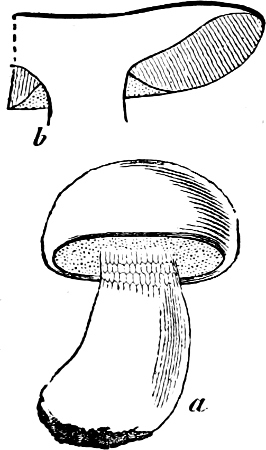
Fig. 171.—Boletus edulis (about ¼th): b longitudinal section of a portion of the pileus.
Order 6. Agaricaceæ (Mushrooms, Toadstools). The hymenophore consists of knife-like plates (lamellæ, gills), which are situated on the under side of the umbrella-like pileus of the fruit-body, and radiate from the central stalk. Those which are first formed extend from the edge of the pileus to the stalk; those formed later reach only a longer or shorter portion of this distance, according to their age. In structure the lamellæ (Fig. 174) consist of a central mass of hyphæ, the trama, continuous with the hyphæ of the pileus; these terminate in a layer of shorter cells, the subhymenial layer, immediately beneath the hymenium which is composed of basidia and paraphyses. In a few species, but not in the majority, the lamellæ are branched, and in some they are decurrent. A few have the stalk placed excentrically, or it may be entirely absent.
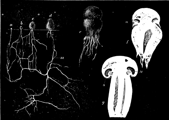
Fig. 172.—Development of Psalliota campestris: a, b, c, d show the various stages of the development of the fruit-bodies and the mycelium (m) (nat. size); e the fruit-body in a somewhat later stage, slightly magnified; f longitudinal section of e; n first formation of the hymenium; g longitudinal section of a more advanced fruit-body (nat. size); n the hymenium; o velum partiale (see Fig. 133.)
In the early stages of its development the fruit-body is more or less enclosed in a hyphal tissue—the “veil” (velum universale, or volva). The veil at first completely encloses the young fruit-body, but is afterwards ruptured as the latter grows, part remaining at the base of the stalk as the “sheath” (annulus inferus), and part on the pileus as scales or warts. In the “Fly Mushroom” (Amanita muscaria) the remains of the veil are especially conspicuous as white patches on the bright red ground of the upper surface of the pileus, and as a sheath at the base of the stalk (Fig. 178 v.). Another veil—the velum partiale—a hyphal tissue (Figs. 178 a; 173) stretches from the edge of the pileus to the stalk, and encloses the lamellæ. This veil is ruptured as the pileus expands, a portion attached to the stalk remaining as the “upper ring” (annulus superus) (Figs. 173, 178 a), or a part attached to the pileus hanging down as a fringe round its edge.—Some genera have no veil, the under side of the pileus being exposed from the first (gymnocarpic Agaricaceæ). Those which have a veil (hemiangiocarpic A.) afford a transition to the angiocarpic Gasteromycetes.
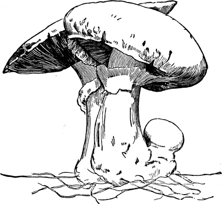
Fig. 173.—The cultivated Mushroom (Psalliota campestris).
The mycelium mostly grows in soils rich in humus or dung, on decaying trees and similar objects. Many species, e.g. Tricholoma personatum and Marasmius oreades, form the so-called “fairy rings.” The fruit-bodies in these species are confined to a larger or smaller surface on which they are very regularly arranged in a ring. The reason for this is found in the radial growth of the mycelium, so that the oldest portion, or the starting point, is found at the centre of the ring, and the younger ones, on which the fruit-bodies are formed, at the circumference. The older hyphæ gradually die, and at the same time, the radial growth continuing, the ring of fruit-bodies becomes larger and larger. The “fairy-rings” are marked not only by the fruit-bodies, but also by the more vigorous growth and darker colour of the grass upon these spots.
Some species are parasites. An example is presented by Armillaria mellea, a remarkable and very destructive Fungus in woods and forests (Figs. 176, 177). In addition to the filamentous, white mycelium, it has also black, or black-brown, horny, root-like mycelium-strands (rhizomorpha) which were formerly considered to belong to a special genus of Fungi described under the name “Rhizomorpha.” The mycelium lives parasitically on the Conifers and other trees, forcing its hyphæ into the bark and between the bark and wood, and thence penetrating into the wood so that the tree is very severely attacked. It may also live saprophytically, and clusters of fruit-bodies are often found on old stumps and stems, on old timber, and in the rich soil of woods. The rhizomorpha, living underground, can extend for considerable distances and infect the roots of neighbouring trees, and spreads in this way the diseases known as “Harzsticken” and “Bark-Canker,” which are very destructive to young trees.
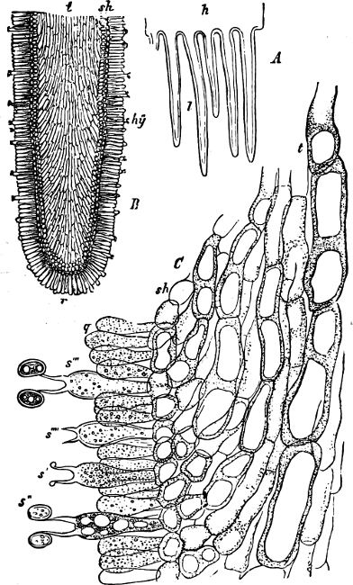
Fig. 174.—Psalliota campestris. A Tangential section of pileus showing lamellæ (l). B Portion of gill more highly magnified; t trama; hy hymenium with basidia and basidiospores; sh, subhymenial layer. C A portion of the same more highly magnified; s′ s′′ s′′′ s′′′′ various stages in the development of basidiospores; q paraphyses.
The chief characteristics by which the numerous genera are separated are the presence or the absence of the two kinds of veils, the nature of the fruit-body, the form, branching of the lamellæ, and their position and relation with respect to the stem, the shape of the pileus, the colour of the spores, etc., etc. A knowledge of the colour may be obtained by placing the pileus with the lamellæ turned downwards on a piece of white or coloured paper, so that the spores, as they fall off, are collected on the paper, and the arrangement of the lamellæ can then be clearly seen.
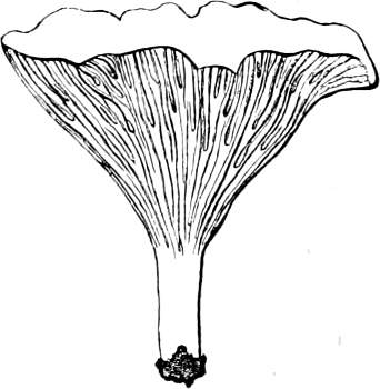
Fig. 175.—Cantharellus cibarius (reduced).
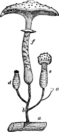
Fig. 176.—Armillaria mellea. (½ nat. size): a root of a Fir; b rhizomorpha-strands; c-f fruit-bodies in four different stages of development.
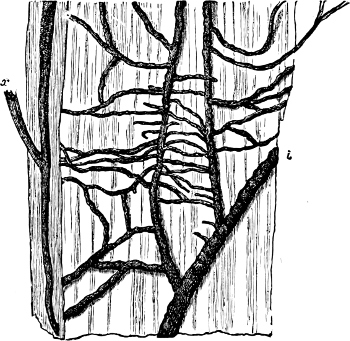
Fig. 177.—The mycelium of Armillaria mellea (“Rhizomorpha”) (nat. size).
About 4,600 species belonging to this order have been described.
On account of the large number of species the order is divided into several sections:
1. Agaricinei; fruit-body fleshy; lamellæ membranous, knife-like, with sharp edge; basidia crowded together. The FOLLOWING HAVE WHITE SPORES:—Amanita (Fly Mushroom), with volva, and generally also the upper ring on the stalk; many are poisonous, such as A. muscaria (Fig. 178) which has bright red pileus with white spots, A. pantherina and A. phalloides; A. cæsarea is edible.—Lepiota procera (Parasol Fungus) is one of the largest Mushrooms; it has a scaly pileus and moveable ring (edible).—Armillaria mellea has been mentioned above (Figs. 176, 177).—Tricholoma, lamellæ indented near the stalk; T. gambosum (Pomona Fungus) belongs to the best of edible Fungi; T. personatum often forms fairy rings (see above).—Clitocybe, lamella decurrent; C. nebularis is edible.—Pleurotus, stalk eccentric; P. ostreatus (Oyster Mushroom) grows in clusters on tree-stems (edible).—Collybia and Mycena, species numerous, small.—Spores rose-red: Volvaria and Hyporhodius.—Spores Brown: Cortinarius, with cobweb-like veil; Pholiota, membranous veil and ring; P. squarrosa in clusters on tree-stems; P. mutabilis, on tree-stumps (edible).—Spores Violet-purple: Hypholoma, Psalliota; to this section the common edible Mushroom (Fig. 172–174) belongs, with annulus and chocolate-coloured lamellæ; it is cultivated for the sake of the fine flavour.—Spores Black: Coprinarius.
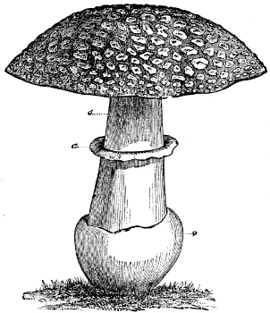
Fig. 178.—Fly Mushroom (Amanita muscaria).
2. Marasmiei. Fruit-body tough, almost leathery, and persistent; spores white. Marasmius oreades forms large, regular fairy-rings on pastures and commons; it is used as seasoning in food.—Panus stipticus with eccentrically-placed stalk, in clusters on tree-stumps.—Schizophyllum has the edge of the lamellæ divided longitudinally, and the split portions revolute.—Lentinus affords a transition to Dædalea among the Polyporaceæ.
3. Russulei. Fruit-body fleshy and fragile, in which two different systems of hyphæ may be distinguished; spores thorny, white, or pale-yellow. Many are poisonous.—Russula has generally fragile and thick lamellæ reaching from stalk to edge of pileus; pileus frequently red.—Lactarius has white or yellow milky juice, which often is very acid. L. deliciosus has red-yellow milky juice, and is of a pleasant flavour. L. torminosus is poisonous.
4. Hygrophorei. Lamellæ thick and waxy, widely separated; spores white. Many species of Hygrophorus have brightly-coloured pileus and grow among the grass on moors and commons.—Nyctalis is parasitic on larger Toadstools. It is remarkable for its abundant formation of chlamydospores, whilst the basidiospores are little developed.
5. Coprinei. Fruit-bodies very soft, quickly perishable; lamellæ membranous and deliquescent. The basidia are separated from each other by paraphyses. Coprinus has coal-black spores, grows on manure, and sometimes developes sclerotia.
6. Paxillei. Fruit-body fleshy; lamellæ easily detached from the pileus and reticulately-joined near the stalk. They form a connecting link between the Agaricaceæ and Boletus.
7. Cantharellei. Lamellæ reduced to dichotomously-divided folds, decurrent on the stalk. Cantharellus cibarius (Fig. 175) is yolk-yellow, and grows on the ground in woods (edible). It is allied to Craterellus.
Family 3. Phalloideæ.
The fruit-bodies before they are ripe are spherical or ovoid, and enclosed by a fleshy covering, the peridium, which is perforated at maturity and remains as a sheath (Fig. 179); the fruit-bodies are hemiangiocarpic.
Order 1. Phallaceæ (Stink-horns). The peridium has a complicated structure and is composed of three layers, the intermediate one being thick and gelatinous. The gleba (the tissue which bears the hymenium) is situated upon a peculiar receptacle which expands into a porous stalk and by its sudden distension, rupturing the peridium, elevates the gleba and hymenium above the peridium, which remains as a sheath. The gleba becomes gelatinous and dissolves away as drops. To this order belong many peculiar and often brightly coloured forms, which are natives of the Southern Hemisphere.
Phallus impudicus (Stink-horn) (Fig. 179), has a fruit-body which at first is white, heavy, and soft, and resembles a hen’s egg in shape and size. The peridium is divided into three layers (Fig. 179 e, g, f) of which the external and internal are membranous, and the middle one very thick and gelatinous; each of these has again a laminated structure. The peridium when ruptured remains as a sheath (k) at the base of the stalk. The receptacle at first is strongly compressed (h) but afterwards expands into a long stalk (l) which bears the conical gleba (m). Prior to the rupture of the peridium the gleba consists of a greenish mass (i) which, when exposed, emits a carrion-like stench serving to attract flies, by whose agency the spores are distributed. It is found commonly in hedgerows and in woods, growing on the ground. The much smaller and less common P. caninus is found on rotten tree-stumps.—In Clathrus cancellatus the receptacle expands into a bright red, reticulate structure. A native of the South of Europe. Colus, Aseroë, Mitromyces.
Order 2. Sphærobolaceæ. An intermediate layer of the peridium swells when ripe, becomes convex, and ejects the remaining spherical portion of the fruit-body which contains the spores. Sphærobolus carpobolus has small, spherical fruit-bodies which open in the form of a star.
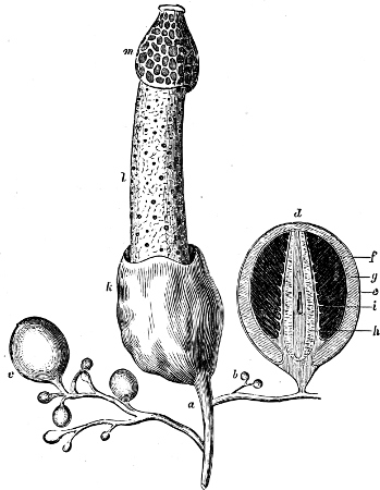
Fig. 179.—Phallus impudicus (Stink-horn), somewhat diminished. Fruit-bodies in all stages of development (b, c, d and k-m) are seen arising from a root-like mycelium (a); d longitudinal sections through a fruit-body before the covering has ruptured.
Family 4. Gasteromycetes.
The fruit-body is angiocarpic, fleshy at first, and later generally more or less hard and continues closed after the spores are ripe. The tissue lying immediately inside the peridium is termed the gleba; it is porous, containing a larger or smaller number of chambers lined with the hymenium, which is either a continuous layer of basidia or else it fills up the entire cavity. The basidia as a rule bear four spores, sometimes eight (Geaster), or two (Hymenogaster). The tissue of the walls (trama) consists often (Lycoperdaceæ) of two kinds of hyphæ, some thin and rich in protoplasm, divided by transverse septa and bearing the basidia; others thicker and thick-walled which do not dissolve like the former on the ripening of the spores, but continue to grow and form a woolly, elastic mass, the capillitium, which may be regarded as highly developed paraphyses. The peridium may be either single or double, and presents many variations in its structure and dehiscence. The mycelium is generally a number of string-like strands, living in soils rich in humus.
Order 1. Tylostomaceæ. Capillitium present. After the rupture of the peridium the remaining part of the fruit-body is elevated on a long stalk. Tylostoma mammosum, on heaths.
Order 2. Lycoperdaceæ. The fruit-body has a double peridium; the external one at length breaks into fragments (Lycoperdon, Bovista), or it has a compound structure of several layers (Geaster) and detaches itself as a continuous envelope from the inner layer, which is membranous and opens at its apex. The interior of the fruit-body consists either solely of the fertile gleba (Bovista, Geaster), or, in addition, of a sterile tissue at the base (Lycoperdon). A capillitium is also present.
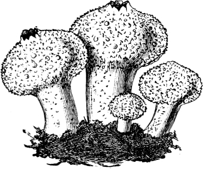
Fig. 180.—Lycoperdon gemmatum (½ nat. size).
Lycoperdon (Puff-ball) has a sterile part at the base of the fruit-body which often forms a thick stalk. The surface of the peridium is generally covered with warts or projections. When young this Fungus is edible, but when ripe it is dry, and used for stopping the flow of blood. L. giganteum, which is often found growing in meadows, attains a considerable size, its diameter reaching as much as eighteen inches. L. gemmatum (Fig. 180) is covered with pyramidal warts; in woods.—Bovista has no sterile basal part; the external peridium is smooth, and falls away in irregular patches. B. plumbea, on links near the sea.—Geaster (Earth-star) has an external peridium composed of several layers, which when the fruit-body opens, split into several stellate segments. These segments are very hygroscopic, and in dry weather bend backwards and so raise the inner peridium into the air. The inner peridium contains the spores and capillitia. G. coliformis has several apertures in the inner peridium. The other species have only one regular aperture at the apex. G. striatus has a pedicellate inner peridium, with conical, striped peristome. G. fornicatus has an external peridium split into four segments. This last and several other species produce “mycorhiza” on the roots of Conifers.
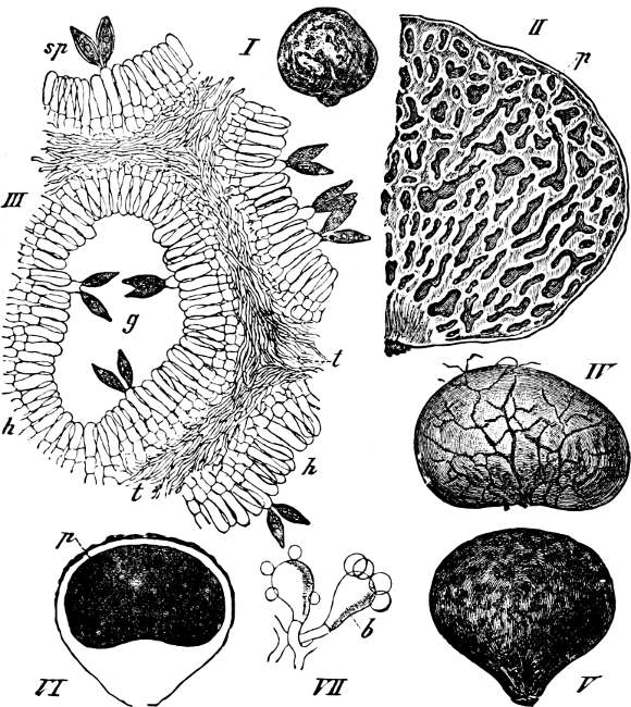
Fig. 181.—I Hymenogaster citrinus (nat. size); II longitudinal section through H. tener (× 5); III portion of a section of H. calosporus; g a chamber; h hymenium; sp. spores; t trama (× 178); IV Rhizopogon luteolus (nat. size); V Scleroderma vulgare, VI section of V; VII basidia with spores belonging to the same Fungus.
Order 3. Sclerodermataceæ. Capillitium wanting. The peridium is simple and thick, gleba with round, closed chambers, which are filled with basidia.
Scleroderma has a corky peridium. The fruit-bodies commence their development under ground. S. vulgare (Fig. 181 V-VII), has a hard, slaty-black gleba.
Order 4. Nidulariaceæ (Nest-Fungi). Small Fungi of which the fruit-body at first is spherical or cylindrical but upon maturity it becomes cupular or vase-like, and contains several lenticular “peridiola” lying like eggs in a nest. The peridiola are the chambers which contain the hymenium, covered by a thin layer of the gleba, all the remaining portion of the gleba becoming dissolved. On decaying wood.
Nidularia has spherical fruit-bodies containing a large number of lenticular peridiola, embedded in a slimy mass.—Crucibulum has fruit-bodies resembling crucibles with discoid peridiola, each with a spirally-twisted stalk.—Cyathus has a fruit-body, which when open is campanulate, with stratified peridium, and long-stalked, lense-shaped peridiola.
Order 5. Hymenogastraceæ. Fruit-bodies tubercular, globose and subterranean, resembling very closely the Truffles, from which they can only be distinguished with certainty by microscopic means. The peridium is simple, capillitium wanting, and the gleba encloses a system of labyrinthine passages covered with a continuous hymenium. The fruit-bodies persist for some time, and form a fleshy mass, the spores being only set free by the decay of the fruit-body, or when it is eaten by animals. The majority are South European. Hymenogaster, Melanogaster, Rhizopogon (Fig. 181 I-IV).
Appendix to the Basidiomycetes:
Basidiolichenes (Lichen-forming Basidiomycetes).
Several Fungi belonging to the Basidiomycetes have a symbiotic relationship with Algæ exactly similar to that enjoyed by certain Ascomycetes, and these are therefore included under the term Lichens (p. 136). They are chiefly tropical.
Order 1. Hymenolichenes. To this order belong some gymnocarpic forms: Cora, Dictyonema, Laudatea.[15]
Order 2. Gasterolichenes. To this belong some angiocarpic forms: Emericella, Trichocoma.
Appendix to the Fungi.
Fungi imperfecti (Incompletely known Fungi).
1. The Saccharomyces-forms are Fungi which are only known in their yeast-conidial form. They are conidia of higher Fungi which can multiply to an unlimited extent by budding in nutritive solutions, and in this way maintain their definite size and shape. The budding takes place only at the ends of the conidia. The wall of the conidium forms at one or at both ends a small wart-like outgrowth, which gradually becomes larger, and is finally separated from its mother-cell as an independent cell, surrounded by a closed cell-wall (Fig. 182 a, b).
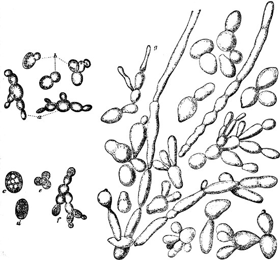
Fig. 182.—Beer-yeast (Saccharomyces cerevisiæ): a-b (× 400); c-f (× 750); c a cell in the process of forming spores; d a cell with four ripe spores; e the spores liberated by the dissolution of the cell-wall; f three germinating spores; g mycelium-like cell-chains. (× 1000: after Em. Chr. Hansen.)
Under very favourable conditions multiplication occurs so rapidly that the daughter-cells themselves commence to form buds, before they have separated from their mother-cell, with the result that pearl-like chains of cells are produced. When the yeast-cells have only limited nutriment, with an abundant supply of air, at a suitable temperature, an endogenous formation of spores takes place. The protoplasm of the cells divides into 1–4 (rarely a greater number) masses (Fig. 182 c, d, e) which surround themselves with a thick cell-wall, and in this state can withstand adverse conditions and periods of dryness lasting for several months.
The sporangia are not asci since they have no definite form, and a definite number, form and size of spores is not found. The spores in the different species and kinds occupy varying periods for their development, although exposed to the same temperature, a fact of importance in determining one from another. On germination the wall of the mother-cell is destroyed, and each spore gives rise to a new cell, multiplication taking place by budding (Fig. 182 f). The majority of Yeast-Fungi are able to produce alcoholic fermentation in saccharine fluids.
The most important of these Fungi is the Beer-yeast (Saccharomyces cerevisiæ) with ovate, ellipsoidal or spherical cells (Fig. 182). It is a plant which has been cultivated from time immemorial, on account of its property of producing alcoholic fermentation in sugar-containing extracts (wort), derived from germinating barley (malt). Carbonic acid is also set free during this process. The “surface-yeast” (Fig. 182 a), which produces ordinary beer when the brewing takes place at higher temperatures, has cell-chains; “sedimentary yeast” (Fig. 182 b), used in the brewing of Bavarian beer, has spherical cells, solitary, or united in pairs. Both these and the following Yeast-Fungi include, according to Hansen, several species and kinds.
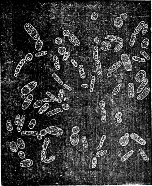
Fig. 183.—Saccharomyces mycoderma.
The “Ferment of Wine” (Saccharomyces ellipsoideus) produces wine in the juice of grapes. Uncultivated yeast-cells are always present on grapes; an addition of this species to the “must” is not necessary to secure fermentation. A large number of other “uncultivated” yeast-cells appear in breweries mixed with the cultivated ones, and cause different tastes to the beer (S. pastorianus, etc.). S. ludwigii, found, for instance, on the slimy discharge from Oaks, produces abundant cell-chains on cultivation. S. apiculatus is very frequently met with on all kinds of sweet fruits, it has orange-like cells. S. mycoderma has cylindrical cells, often united together in chains (Fig. 183): it forms a whitish-gray mass (“fleur de vin”) on wine, beer, fruit-juice, etc., standing in bottles uncorked or not entirely filled. It is thought that this Fungus causes decomposition and oxydises the fluid in which it is found, but it cannot produce alcoholic fermentation in saccharine liquids, and it does not form endospores; hence it is uncertain whether it is true Saccharomyces.
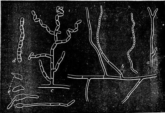
Fig. 184.—Oidium lactis: a branched hypha commonly met with; b a hypha lying in milk and producing aerial hyphæ which give rise to oidia; c a branch giving rise to oidia, the oldest (outermost) oidia are becoming detached from one another; d a chain of divided cells; e germinating oidia in different stages (slightly more magnified than the other figures).
The “Dry-yeast” used in baking white bread is “surface-yeast.” In leaven, a kneaded mixture of meal, barm and water, which is used for the manufacture of black bread, Saccharomyces minor is present, and a species allied to this produces alcoholic fermentation in dough with the evolution of carbonic acid, which causes the dough to “rise.”
2. Oidium-forms. Of many Fungi only the Oidium-forms are known, which multiply in endless series without employing any higher form of reproduction. Oidium lactis (Fig. 184) is an imperfectly developed form which frequently appears on sour milk and cheese. It can produce a feeble alcoholic fermentation in saccharine liquids. Thrush or aphthæ (O. albicans) appears as white spots in the mouths of children. Several similar Oidium-forms are parasites on the skin and hair of human beings, and produce skin diseases, such as scurvy (O. schoenleinii) and ringworm (O. tonsurans).
3. Mycorhiza. These Fungi, which have been found on the roots of many trees and heath-plants, particularly Cupuliferæ and Ericaceæ, consist of septate hyphæ, and belong partly to the Hymenomycetes, partly to the Gasteromycetes. It has been shown that the Mycorhiza enters into a symbiotic relationship with the roots of higher plants.