DIVISION III. PTERIDOPHYTA (VASCULAR CRYPTOGAMS).
Michael Cresse Potter translation
The alternation of generations is as distinct in this Division as in the Mosses, but the sexual generation consists of only a small thallus, the prothallium, which bears directly the sexual organs, antheridia and archegonia; and the asexual generation, which arises from the fertilisation of the oosphere, is no longer a single short-lived sporangium, but a highly developed, generally perennial, plant provided with stem, leaves and true roots (Ferns, Horsetails, etc.), the sporangia being borne on the leaves. In this latter generation the tissues are differentiated into epidermis, ground tissue and vascular tissue; in the last named the bundles are closed, and in the majority of cases concentric.
The sexual generation, gametophyte, or prothallium, is always a thallus, although not always green and leaf-like (Figs. 205, 215, 222, 229, 235, etc.) It is very small, even in cases where it attains the greatest development, and consists only of parenchymatous cells. The prothallium is nourished by hair-like roots (rhizoids) and has only a transitory existence, dying soon after the fertilisation of its oosphere.
The ANTHERIDIA exhibit great variations in structure which, however, must be considered as modifications of the fundamental type which is found in the Mosses. These modifications will be mentioned under the various families. The spermatozoids are always spirally-coiled, self-motile, protoplasmic bodies, with most frequently a large number of fine cilia on the anterior end (Figs. 206, 223, 234). They are formed principally from the nucleus of the mother-cell, and portions of the cytoplasm often remain for a time attached to their posterior end.
The ARCHEGONIA are more uniform throughout the entire Division, and more closely resemble those of the Mosses. They are, as in the previous Division, principally flask-shaped; but the central portion, which encloses the oosphere, is always embedded in the tissue of the prothallium, so that the neck, which is formed of 4 rows of cells, projects above the surface (Figs. 201 3, 222 h). The development of the archegonium in a Fern is seen in the accompanying figure (Fig. 201). The archegonium is developed from a surface cell, which divides into three cells by two walls in a direction parallel to the surface of the prothallium (Fig. 201). The most internal cell becomes the ventral portion of the archegonium. The external one (b) divides perpendicularly to the surface of the prothallium into four cells, which again divide parallel to the surface and form the neck (b, in 2 and 3). The intermediate cell projects upwards into the neck and divides into two, the lower one, after the separation of the ventral canal-cell, becoming the oosphere, and the upper one the neck-canal-cell (c, in 2 and 3).
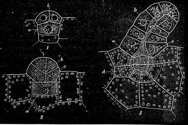
Fig. 201.—Pteris serrulata. Development of archegonia.]
As in the Mosses, the divisional walls of the neck-canal-cells become mucilaginous, causing the rupture of the neck of the archegonium. Fertilisation takes place as in the Mosses, and the passage of the spermatozoids, along the neck, to the oosphere, has been observed. Water (rain or dew) is similarly necessary for the movements of the spermatozoids, and hence for fertilisation. The other classes of the Division chiefly deviate from the Ferns in having the archegonium sunk deeper into the prothallium, and the neck reduced in length (compare Fig. 201 with Figs. 216, 222, 235, 236).
According to the nature of the spores, the three classes of the Vascular Cryptogams are each divided into isosporous and heterosporous groups.
I. The isosporous Vascular Cryptogams have only one kind of spore. The prothallium developed from this is in some cases monœcious, bearing both antheridia and archegonia; but in others there is a distinct tendency for each prothallium to bear only antheridia or archegonia (diœcious)—true Ferns and Lycopodium.
In Equisetum there is only one kind of spore, but two kinds of prothallia are developed, one of which bears only antheridia (male), the other only archegonia (female); but the one that bears antheridia may be transformed into the one that bears archegonia and vice versa.
II. In the higher group, heterosporous Vascular Cryptogams (Selaginella and Isoëtes, etc.), there are two distinct kinds of spores, the small, microspores, and the large, macrospores. The microspores are male, and produce prothallia which bear only antheridia. The macrospores are female, and produce prothallia which bear only archegonia.
Corresponding to this difference in the spores, there is also found a difference in the development of the prothallium. In the Isosporeæ the prothallium is large, and either green, leaf-like, and provided with rhizoids (most of the Ferns, Horsetails, etc.), or subterranean, pale-coloured, and globular (Ophioglossum, Lycopodium). It lives vegetatively for a fairly long time, and generally produces a large and varying number of archegonia and antheridia. The prothallium in the Heterosporeæ is gradually more and more reduced, its independent and vegetative life becomes of less and less importance, it becomes more dependent on the mother-plant, and projects from the spore very slightly, or not at all. The antheridia and archegonia become reduced in number to one, and also degenerate in point of development.
It may here be remarked that the gradual development of the asexual generation, the development of the two kinds of spores, and the progressive reduction of the prothallium and sexual organs which is found in this Division, is continued to the Gymnosperms and Angiosperms. The microspores are in these called pollen-grains, and the male prothallium is very rudimentary. The macrospores are termed embryo-sacs, and the female prothallium, the endosperm.
The asexual generation, sporophyte. When the oosphere, which in this case as in all others is a primordial cell, is fertilised, it surrounds itself with a cell-wall and commences to divide into a number of cells, to form the embryo.
The first dividing wall (basal wall) is nearly horizontal, and in the direction of the longitudinal axis of the archegonium. The next wall is vertical, and the next perpendicular to the other two. The oosphere, therefore, is now divided into eight octants by these three walls. The basal wall divides the embryo into a hypobasal and an epibasal half. From the first one, by continued divisions, the first root is developed; from the latter, the stem and leaves. After the formation of the octants the development proceeds in somewhat different ways in the various classes. In addition to the stem, leaf, and root, a “foot” is developed from the hypobasal half which remains enclosed in the prothallium, and conveys nourishment from the prothallium to the young plant until it is able to sustain itself (Fig. 202). The formation of these members in the embryo depends on the position of the oosphere in the archegonium and prothallium, and is independent of gravity.
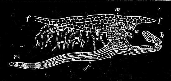
Fig. 202.—Adiantum capillus veneris. Vertical section through a prothallium (f f), with a young plant attached on its under side (mag. about 10 times); r the first root, and b the first leaf of the young Fern-plant; m the foot. In the angle between m and b lies the apex of the stem: h the rhizoids of the prothallium; æ æ unfertilised archegonia.
In the Mosses the asexual generation is the sporogonium, which is limited in its development and in a great measure dependent upon the sexual generation, upon which it is situated; but in the Pteridophyta this generation is an independent and highly developed plant, provided with stem, leaf, and true roots, and has in many instances an unlimited development. The Pteridophyta are the lowest Division with true roots. The root which is first formed is very similar in nature to the primary root of the Monocotyledons; it very soon dies and is replaced by others which are more permanent, and developed upon the stem (adventitious roots); roots are wanting in Salvinia, Psilotum, and some Hymenophyllaceæ. The differentiation is, however, not so complete as in the Flowering-plants, and so many leafy forms are not found. The various members of these plants are anatomically much higher than in the Mosses, having an epidermis, a ground tissue with variously differentiated cells, and a highly developed vascular system. The vascular bundles, like those in the Monocotyledons, are without cambium, and closed; they are therefore incapable of any increase in thickness. In general the bundles are concentric, with the bast round the wood (Fig. 203). The wood is almost entirely made up of scalariform tracheides.
In Isoëtes a secondary thickening takes place by a cambium, which is formed inside the cortex, constructing secondary cortex to the exterior, and secondary wood towards the interior.—Botrychium has also a thickening growth. Collateral vascular bundles occur in Osmundaceæ, Equisetaceæ, and the leaves of many Polypodiaceæ, etc.
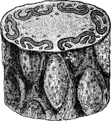
Fig. 203.—Portion of the stem of a Fern. Above is seen the transverse section, with vascular bundles of different form and size. The rhombic figures on the side of the stem are leaf-scars.
It is a point of special interest, that the gigantic forms of Ferns, Equisetums, and Club-Mosses (which flourished in earlier geological periods, when these classes attained their highest development) possessed some means of increasing in thickness.
The sporangia are in all cases capsule-like, and burst open when ripe to eject the spores. They are nearly always situated on the leaves (in Lycopodiaceæ, in the axils of the leaves, or above these, on the stems themselves). In some forms (Leptosporangiatæ), the sporangia are developed from a single epidermal cell; in others (Eusporangiatæ), from a group of epidermal cells, or from cells which lie beneath the epidermis. In the first group a primitive mother-cell (archesporium) is formed, which divides commonly into sixteen special mother-cells. In the latter group, on the other hand, a number of primitive spore-mother-cells are developed. In each sporangium three different tissues are generally developed; an innermost sporogenous one (s in Fig. 204 A), which arises from the archesporangium; an outermost one, which forms the wall (a), and may be one or, more rarely, several layers in thickness; and an intermediate one, the tapetum (Fig. 204 A, B, b t), which is rich in protoplasm, and whose cells are dissolved so that the spores float freely in the fluid thus provided. The spores arise as in the Mosses (in tetrads), by the cross-division of the special mother-cells, and according to the manner in which they are arranged in the mother-cell have either a tetrahedral form, with a large base resembling a segment of a ball, or are oblong (bilateral spores). Their construction is the same as in the Mosses (p. 187).
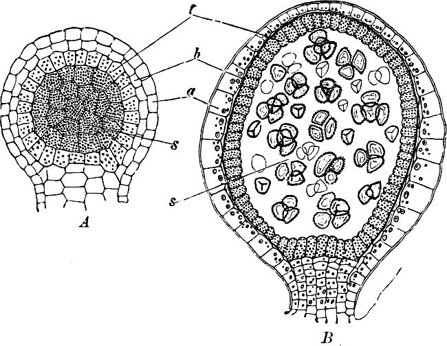
Fig. 204.—Selaginella inæqualifolia. A A young sporangium, which may develope either into a macro-, or a microsporangium. B A microsporangium.
The spore-formation in its earliest commencement takes place in the same way in the Isosporous and the Heterosporous Vascular Cryptogams; but from a certain point, after the tetrahedral division, a difference occurs with regard to the macrosporangia. All the spores formed in the microsporangium may complete their development; but those which are formed in the macrosporangium are generally aborted, with the exception of one or four, and these consequently attain a much larger size (see Fig. 239.—The series to the left are microsporangia; those to the right, macrosporangia).
Apogamy. In some Ferns (Pteris cretica; Aspidium filix mas, var. cristatum; A. falcatum; Todea africana) the young plant is not developed as a consequence of fertilisation, but as a bud from the prothallium. This is known as apogamy, or loss of the power of sexual reproduction. The antheridia are generally more or less developed; archegonia are entirely wanting in Asp. filix mas, var. cristatum. This variety has probably only become apogamous through cultivation. Many specimens of Isoëtes lacustris, in a lake in the Vosges mountains, produce in the place where the sporangia are usually found, a vegetative shoot which grows into a new plant, so that the sexual generation is wanting in this case. Some specimens have sporangia on some leaves, and shoots on others.
Apospory, or the formation of prothallia instead of sporangia and spores on the leaves, is found in Athyrium filix femina, var. clarissimum. In this case the development of the sporangia proceeds only to a certain point, and from these arrested sporangia the prothallia are produced. Normal sporangia are entirely wanting in this variety, and in Aspidium angulare, var. pulcherrimum, sporangia are completely wanting. Compare the Mosses (page 188).
The Vascular Cryptogams are divided into three large classes, in each of which a progressive development can be traced from the isosporous to the heterosporous forms, but some of these are now only known as fossils.
Class 1. Filicinæ (Ferns).—The stem is small in comparison with the leaves, and branches only seldom, and then by lateral shoots. The leaves are scattered, large, often deeply divided, and of various highly developed forms. The undeveloped leaves are rolled up in the bud, having what is termed circinate venation. The sporangia are situated on the edge or on the lower side of the leaves, those on which the sporangia are borne (sporophylls) being often the ordinary foliage-leaves; but in a few cases the fertile differ from the barren ones (a higher stage in development). The fertile leaves are not confined to definite parts of the shoot, and do not limit its growth. The archesporium is most frequently unicellular.
A. Isosporous: Sub-Class 1. Filices (True Ferns).
B. Heterosporous: Sub-Class 2. Hydropterideæ (Water Ferns).
Class 2. Equisetinæ (Horsetails), in its widest meaning.—The leaves in this class are small in comparison with the stem. They are arranged in whorls, and unite to form a sheath. The sporangia are situated on specially modified, shield-like leaves, which are closely packed together and form a “cone.” The cone is borne terminally, and limits the growth of the shoot. The sporangia are developed from a large group of epidermal cells, the archesporium being unicellular. The branches are arranged in whorls, and develope acropetally.
A. Isosporous: Sub-Class 1. Equisetaceæ. Existing forms.
B. Heterosporous: Sub-Class 2. Extinct forms.
Class 3. Lycopodinæ (Club-Mosses).—Roots generally branching dichotomously. The leaves are scattered or opposite, and in proportion to the stem very small, undivided, and simple. They are scale-like and triangular, tapering from a broad base to a point. The sporangia are situated singly (except in Psilotaceæ), and almost in every case on the upper side of the leaf or in the axil of a leaf; but in some cases they are borne on the stem, just above the leaf-axil. The sporangia arise from groups of epidermal cells. The sporophylls are often modified, and differ from the foliage-leaves; they are then arranged in cones placed terminally on branches, thus limiting their growth.
A. Isosporous: Sub-Class 1. Lycopodieæ.
B. Heterosporous: Sub-Class 2. Selaginelleæ.
Class 1. Filicinæ (Ferns).
The characteristics of this class have already been given on page 204.
The class is divided into two sub-classes:—
1. The True Ferns, Filices, have one kind of spore which generally developes monœcious prothallia, relatively large and green. The sporangia are most frequently situated in groups (sori), which are often covered but not enclosed by an indusium.
2. Water Ferns, Hydropteridæ, have microsporangia with many (4 × 16) microspores, and macrosporangia, each with one macrospore. The prothallium is small, and projects but slightly from the germinating spore. The sporangia are situated in groups (sori), which are either enclosed by an indusium, or enveloped in a portion of a leaf, to form “fruits” termed sporocarps.
The old name for the Hydropterideæ, “Rhizocarpeæ,” i.e. the “root-fruited,” originated from the erroneous supposition that the sporocarps were borne on the roots.
Sub-Class 1. Filices (the True Ferns).
Of the eight orders (with about 4,000 species) comprised in this sub-class, the Polypodiaceæ is the largest (having about 2,800 species) and the most familiar; for this reason it will be taken as typical.
The sexual generation. When the spore germinates, the external covering (exospore) is ruptured, as in the Mosses. The internal cell-wall (endospore) grows out as a filament, which soon divides and gives rise to the prothallium, a flat, cellular expansion resembling the thallus of a Liverwort. In its fully developed state the prothallium is generally heart-shaped, dark green, and provided with root-hairs, and it attains a diameter of about one centimetre (Fig. 205). It is formed of one layer of cells, except along the central line near the anterior depression, where it becomes several layers of cells in thickness, forming the “cushion,” on the lower side of which the archegonia are developed. The antheridia are first formed; they are thus found on the oldest parts of the prothallium, on its edge, or among the root-hairs. The archegonia are developed later, and are therefore found near the apex. Several tropical Ferns have prothallia[18] deviating from this typical form; Trichomanes (Order Hymenophyllaceæ) has filamentous, branched prothallia, which resemble the protonema of a Moss. Others, again, have strap-shaped prothallia, which resemble the thallus of certain Liverworts.
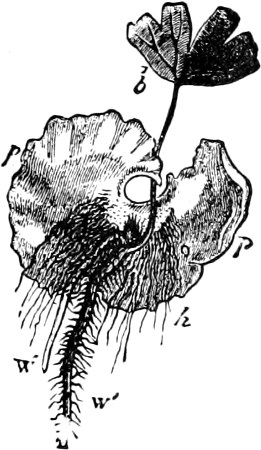
Fig. 205.—Prothallium (p p) of Maiden hair (Adiantum capillus veneris) with a young plant attached: b first leaf; w′ primary root; w″ adventitious roots; h h root-hairs of the prothallium (× abt. 30).
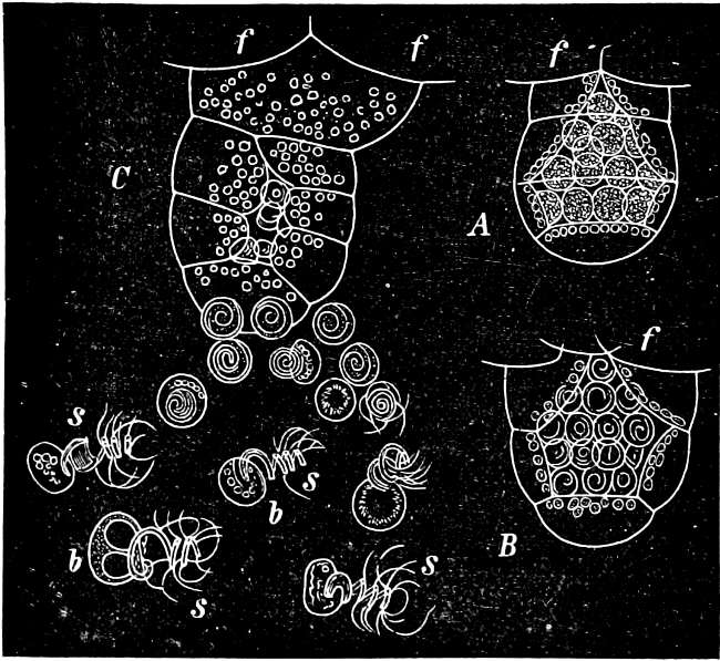
Fig. 206.—Antheridia of Maiden-hair (× 550). A Unripe; B ripe, but unopened; C open and ejecting the spermatozoids (s). Those which have been last ejected are still lying enclosed in their mother-cells, the others are coiled up and drag with them the cytoplasmic remains (b); f cells of the prothallium.
The ARCHEGONIA have been already mentioned (p. 199, Fig. 201). The ANTHERIDIA are hemispherical or slightly conical bodies (Fig. 206). They consist, as in the Mosses, of a wall formed by one layer of cells, which encloses a number of spermatozoid-mother-cells (A and B). The antheridia when ripe absorb water, and are ruptured, and the spirally-coiled spermatozoids liberated (Fig. 206 S). The spermatozoids have been observed to pass down the neck of the archegonium, and to fuse with the oosphere.
The asexual generation. The first leaf, the “cotyledon,” of the embryo developed from the oospore (Figs. 202, 205) is always small, and has a very simple shape. The leaves which occur later become more perfect, stage by stage, until the permanent form of leaf has been attained.—The STEM is most frequently a subterranean or a semi-aerial rhizome; it is only in the tropical, palm-like Tree-Ferns, that the stem raises itself high in the air and resembles that of a tree, with leaf-scars or with the remains of leaves attached (Figs. 207, 203); in certain species the stem is encased in a thick mat of aerial roots (Dicksonia antarctica). When the rhizome is horizontal the internodes are frequently elongated, and the leaves are arranged in two rows, as in Polypodium vulgare and in the Bracken-Fern (Pteridium aquilinum), etc.; it is also generally dorsiventral, having a dorsal side on which the leaves are situated, and a ventral side, different from the former, on which the roots are borne. When the stem ascends in an oblique direction, or is nearly vertical, its internodes are extremely short, and the leaves are arranged in a spiral line with a complicated phyllotaxis, e.g. in Athyrium filix-fœmina, Aspidium filix-mas, etc. The BRANCHING upon the whole is extremely slight, and is generally confined to the petiole (e.g. Aspid. filix-mas), or to the stem near the insertion of the leaves. Several species normally form buds on different parts of the lamina. The buds which are formed on the stem are not confined to the leaf-axil as in the higher plants. The Tree-Ferns, generally, do not branch at all.
The VASCULAR BUNDLES are concentric, with the wood surrounded by the soft bast. In transverse section they are seen as circles or irregularly-shaped figures (Fig. 203), the name of “King Charles and the Oak” (Bracken-Fern) having originated from the appearance which the bundles present in oblique section. In Osmunda they are collateral and resemble those of the Flowering-plants. Round each individual bundle is often a sheath of thick-walled, hard, brown, sclerenchymatous cells, which act as a mechanical tissue; similar strands are also found in other parts of the stem.
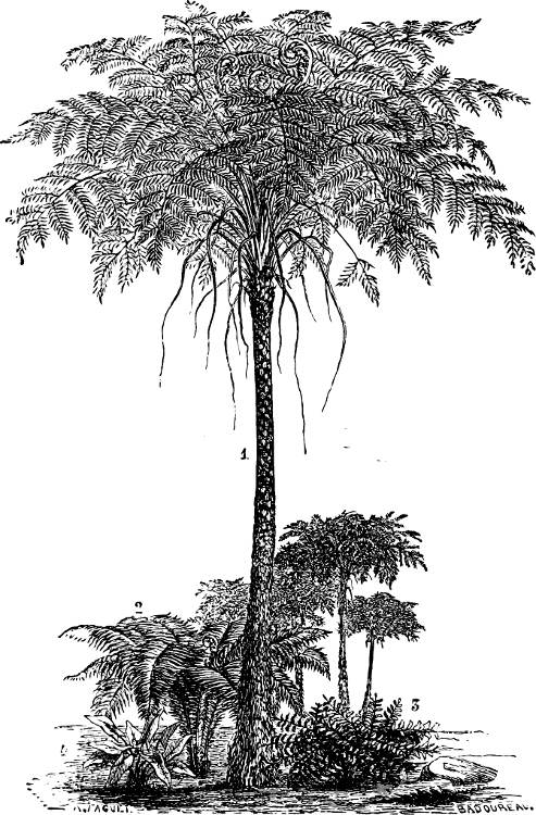
Fig. 207.—Various Ferns (1, 2, 3, 4).
The LEAVES in nearly all species are only foliage-leaves, borne in a spiral. They have an apical growth which continues for a long time, and some require several years for their complete development. In the buds they are rolled up (circinate); not only the midrib, but also all the lateral veins, and even the terminal portions of a leaf are sometimes rolled up together, the tissues of the leaf being already fully developed and only waiting to expand. The leaves are often excessively divided and compound, with pinnate branches, and have an epidermis with stomata and a well-developed system of venation. Stipules are only found in Marattiaceæ and Ophioglossaceæ.
Very often peculiar hairs or scales (paleæ, ramenta), dry, brown, flat and broad, are found on stem and leaf.
The SPORANGIA are small, round capsules, which, in a very large number of Ferns, are formed on the back, but more rarely on the edge of the ordinary foliage-leaves. It is very seldom that there is any difference in form between the barren foliage-leaves and the fertile leaves, as is found for example in Blechnum spicant or Struthiopteris; or that the fertile part of the leaf is differently constructed from the barren portion of the same leaf, as in the Royal-Fern (Osmunda). In such instances the mesophyll of the fertile parts is poorly developed.
The sporangia in the Polypodiaceæ are lens-shaped, with long stalk (Fig. 211 D): their wall consists of one cell-layer on which a single row of cells, passing vertically over the top (that is along the edge of the sporangium), is developed into the “ring” (annulus). The cells of the annulus are very much thickened on the inner and side walls, and are yellowish-brown. The thickened cells, however, do not entirely encircle the sporangium, and on one side, near the stalk, they pass over into large, flat, thin-walled cells. These form a weak point in the wall, and it is here that the sporangium is opened diagonally by the elongation of the annulus. The sporangium of the Polypodiaceæ opens as it dries. The cells of the annulus are very hygroscopic, and in straightening, the annulus bends back with a jerk, thus ejecting the spores to considerable distances. The cells of the annulus absorb water with great readiness. [The sporangium arises as a single epidermal cell, from which a basal stalk-cell is cut off. Three oblique cell-walls, intersecting near the base, are next formed in the upper cell, and a fourth between these and parallel to the free surface; an inner tetrahedral cell enclosed by four others is thus formed, the outer cells become the wall of the sporangium, while the inner cell, by a series of walls, parallel to its sides, cuts off a layer of cells which eventually form the tapetum, the remaining central cell constituting the archesporium.]
The SPORES are either oblong and bilateral, or they are tetrahedric with curved sides, depending upon the way in which the tetrad division has taken place.
The sporangia are almost always situated on the nerves and gathered into groups, sori, which differ in form in the various genera. The sori, in many genera, may be covered by a scale-like structure, the indusium (Figs. 211 B, 212).
In the majority of cases, each sorus is situated on a small papilla (placenta, or receptacle), which is supplied by a small vascular bundle. Between the sporangia, hairs (paraphyses) are often situated, which spring either from the placenta or from the stalks of the sporangia.
Systematic Division. The Ferns may be divided into two groups, characterized by the structure and development of the sporangia. The sporangia in the Eusporangiatæ take their origin from a group of epidermal cells, and their walls are formed by several layers of cells. The archesporium is the (not tetrahedric) hypodermal terminal cell of the axial row of cells which give rise to the sporangium. In the Leptosporangiatæ the sporangia are developed from single epidermal cells, and their walls are uni-layered. The archesporium is a central, often tetrahedric cell, from which sixteen spore-mother-cells are developed.[19] It is difficult to say which form is the oldest (according to Prantl, those which have the sori on the nerve-endings); however, the Eusporangiatæ would seem to have made their appearance long before the others, and also well defined Marattiaceæ and Ophioglossaceæ occur in the Kulm and Coal period, before the true Polypodiaceæ.
About 4,000 species of Ferns are now existing, and they are found especially in tropical and sub-tropical forests.
Family 1. Eusporangiatæ.
Order 1. Ophioglossaceæ. The prothallium differs from that of all other Ferns in being subterranean, free from chlorophyll, pale and tuberous. The stem is extremely short, with short internodes, most frequently unbranched, vertical, and entirely buried in the ground (Fig. 208 st). In several species (among which are the native ones) one leaf is produced every year, which has taken three to four years for its development. In Botrychium a closed, sheath-like basal part of each leaf covers the subsequent leaves during their development. In Ophioglossum and others each leaf has at its base an intrapetiolar, cap-like sheath, which protects the succeeding leaf. The leaves are of two kinds: (a) foliage, which in Ophioglossum vulgatum are lanceolate and entire, but in Botrychium however, are pinnate (b in Fig. 208 A, B); and (b) fertile, which are found facing the upper side of the foliage-leaves. These latter in Ophioglossum are undivided and spike-like (Fig. 209 A), but pinnate in Botrychium (Fig. 208 B). Each foliage and fertile leaf are branches from the same petiole. The large sporangia are placed laterally, and open by two valves. No annulus is formed (Fig. 209).—Ophioglossum reproduces vegetatively by adventitious buds on the roots.
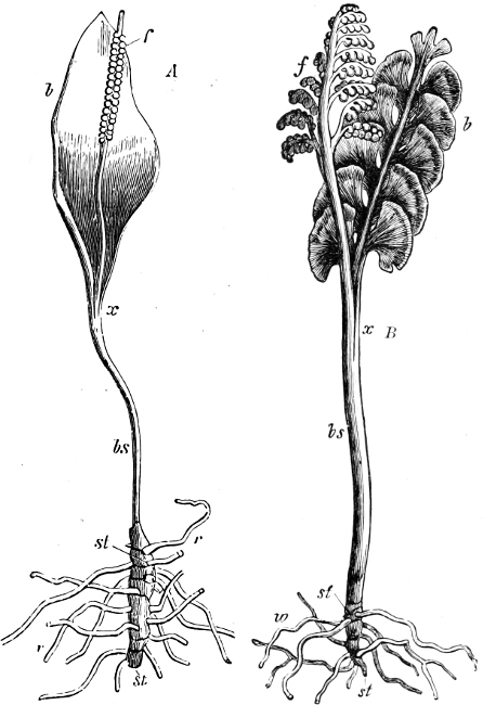
Fig. 208.—A Ophioglossum vulgatum (Adder’s-tongue); B Botrychium lunaria (Moonwort), both natural size; r-r roots; bs leaf-stalk; st stem; b foliage-leaf; f fertile leaf.
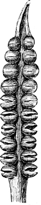
Fig. 209.—Fertile leaf of Ophioglossum.
Three genera with about twelve species.
Order 2. Marattiaceæ are tropical Ferns, whose gigantic leaves resemble those of the Polypodiaceæ, but have stipules in addition. The sporangia are grouped in sori, situated on the lower side of the leaves, the sporangia in each sorus being arranged either in two rows or in a ring. In Angiopteris they are isolated (Fig. 210 A), but in the other species (Kaulfussia, Danæa, Marattia), they are united, and form “synangia” divided into a number of chambers corresponding to the sporangia. These open by clefts or pores. Marattia presents the highest development, as its sporangia are completely united in a capsule-like synangium, which is closed until maturity, and then opens by two valves. In each valve there is a row of three to eleven sporangia, each opening by a slit towards the inside (Fig. 210 B, C). An indusium encloses the sorus, except in Kaulfussia; it is formed of flat and lobed hairs, which resemble the hairs of the other portions of the leaves. In Angiopteris and Marattia the indusium is very rudimentary; in Danæa it forms a kind of cupule.
The numerous fossil Marattiaceæ (15 genera, with 98 species) present similar differences to those now living, but more various forms are found, for example, with solitary free sporangia. Those now living are the last small remnant (4 genera with only 23 species) of a once dominant family, which existed from very early times, and whose culminating point was reached in the Kulm and Coal periods.
The Ophioglossaceæ appear also in the Kulm and Coal periods, and were about as numerous as at the present time (presumably 2 genera, with 19 species). Leptosporangiate Ferns appear however to have occurred first of all in the Trias-formation.
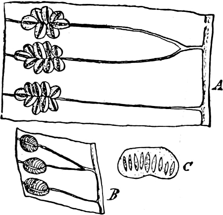
Fig. 210.—Sporangia of the Marattiaceæ: A Angiopteris; B and C Marattia; C is a half sorus with nine sporangia, each of which has opened by a longitudinal cleft.
Family 2. Leptosporangiatæ.
Order 1. Polypodiaceæ. Sporangia on the lower side of the leaves, stalked and provided with a vertical, incomplete annulus; dehiscing by a transverse cleft (Fig. 211 D).—The genera are distinguished by the form of the indusium and the position of the sori, etc.
1. The sporangia cover the entire lower surface of the leaf (Tropical America and Asia). Acrostichum, Platycerium.
2. Sori without indusia, circular or oval. Polypodium (Fig. 211 A). The leaves are most frequently situated in two rows on the dorsal side of the creeping rhizome, and fall off leaving a smooth scar behind.—P. vulgare, common in woods, on stones. (Phegopteris also has no indusium; see page 214).
3. The sporangia are situated in continuous lines just inside the margin of the leaf.—Pteris[20]: the sporangia form a continuous line along the entire margin of the leaf (Fig. 211 C), which bends over and covers the sporangia, forming a “false-indusium.” Pteridium has linear sori situated on a marginal vascular bundle, covered by two linear basal indusia, of which the outer is bent over like the edge of a leaf.—P. aquilinum (Bracken) has a wide-spreading rhizome with large alternate leaves, placed on opposite sides, at some distance apart. Only one leaf is developed from each branch every year.
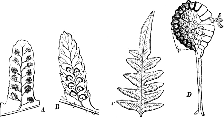
Fig. 211.—Portions of leaves with sori. A Polypodium. B Aspidium. C Pteridium. D A sporangium of one of the Polypodiaceæ: r the annulus; s spores.
Adiantum (Maiden-hair): sori on the underside of small portions of the edge of the leaf, which are bent over (false indusium). Cryptogramme (Allosorus), Cheilanthes.
4. The sori are oval or linear, situated on one side of the vascular bundle.—Asplenium (Fig. 212 A): sori linear; indusium with one of its edges attached at the external side. A. ruta muraria (Wall-Rue); A. septentrionale; A. trichomanes.—Athyrium: sori linear or curved; A. filix-fœmina (Lady-Fern).—Scolopendrium (Fig. 212 B): sori as in Asplenium, but situated in pairs across the lanceolate, entire leaves. Each sorus is covered on the external side by an indusium, whose free edges are parallel and approach each other. S. vulgare (Hart’s-tongue).—Blechnum (B. spicant, Hard Fern; the fertile leaves differ from the barren, the pinnæ being narrower, while the underside is almost entirely covered with sori, and hence they are of a much darker brownish hue than the barren ones).—Ceterach: indusium rudimentary or absent.
5. Sori circular and covered by a shield-like, or reniform indusium.—Aspidium (Fig. 211 B); the leaves wither away and leave no scar upon the root-stock. A. filix-mas (Male-Fern); A. spinulosum.—Phegopteris has no indusium, the withered bases of the leaf-stalks are persistent; P. dryopteris and P. polypodioides.
6. The indusium is situated below the sori, and has the shape of a one-sided scale (Cystopteris, Struthiopteris), or of a cup or cupule, which in Woodsia is sometimes fimbriate (Fig. 212 C, D).
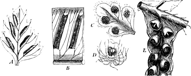
Fig. 212.—A Asplenium. B Scolopendrium. C Woodsia; D single sorus of the same. E Cyathea: the sporangia have fallen off in the upper sori. (All magnified.)
7. The sori are situated on the margin of the leaf, and at the end of a vascular bundle. Indusium, semi-cupular. Davallia. Principally tropical species. 1 in S. Europe.
This order is the greatest, comprising about 2,800 species, the majority being perennial plants. A few are large, and known as Tree-Ferns.
As plants in conservatories and rooms the following are cultivated: species of Gymnogramme (tropical America), Lomaria, Nephrolepis, Pteris (P. serrulata, cretica).
Officinal. Aspidium filix-mas, rhizome and the withered petioles.—Species of Alsophila and Cibotium give Penghawar Djambi. The rhizome of Pteridium aquilinum, var. esculentum, contains so much starch that it is used as food.
The other orders of true Ferns deviate from the Polypodiaceæ, especially in the formation of the annulus, the bursting of the sporangium and its mode of attachment and development, and in the differences in the formation of the prothallium, etc. The principal are:—
Order 2. Hymenophyllaceæ. To this order belong the lowest and most Moss-like Ferns; the leaves, with the exception of the veins, are most frequently formed of only one layer of cells, and consequently stomata are wanting; the formation of the prothallium also somewhat resembles the Mosses. Sori marginal, on the extremities of the vascular bundles, and surrounded by a cupular indusium. The sporangia are sessile, with equatorial annulus. Hymenophyllum (H. tunbridgense, European). Trichomanes (T. speciosum, European). Species about 200, which live especially on rocks and trees in damp and shady tropical forests. Some have no roots.
Order 3. Cyatheaceæ. Annulus complete and oblique. To this order belong, principally, the tree-like Ferns with palm-like habit. The number of species is about 200, they are all tropical and form forests in some regions of Australia. Cibotium and Dicksonia have marginal sori, with cupular, basal indusium. (The stem of D. antarctica is covered with aerial roots.) Alsophila (without indusium); Cyathea with cupular, inferior indusium (Fig. 212 E).
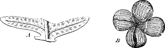
Fig. 213.—Gleichenia: A part of a leaf with sori; B a single sorus.
Order 4. Gleicheniaceæ. Sporangia with equatorial annulus, and longitudinal dehiscence, most frequently groups of 3–4 in sori without indusium (Fig. 213). Gleichenia: the apical growth of the leaves continues for a long time.
Order 5. Schizæaceæ. Annulus apical. To this order belongs Aneimia, which is so commonly cultivated in conservatories. The two lowest pinnæ are metamorphosed, having no leaf parenchyma and being covered with sporangia. Schizæa. Mohria. Lygodium, a climber, whose leaves have unlimited growth and attain a length of several metres. About 70 species. Tropical.
Order 6. Osmundaceæ. The sporangia have at the apex a lateral group of strongly thickened cells, which gradually pass over into the ordinary cells. The sporangia open by a longitudinal cleft. Indusium wanting. Osmunda bears the sporangia upon peculiar, branched pinnæ, without parenchyma (the uppermost in the leaf). O. regalis (Royal-Fern): European.
Sub-Class 2. Hydropterideæ (formerly Rhizocarpeæ), Water Ferns.
The following further characteristics must be added to those given on page 205:—
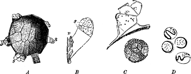
Fig. 214.—Salvinia natans: A microsporangium with germinating microspores and protruding prothallia (s); B a prothallium with the bicellular antheridium (s) growing out of the microsporangium; C the two cells of the antheridium have opened by transverse clefts; beneath is seen the microspores enclosed by the hardened mucilage; D spermatozoids still enclosed in the mother-cells.
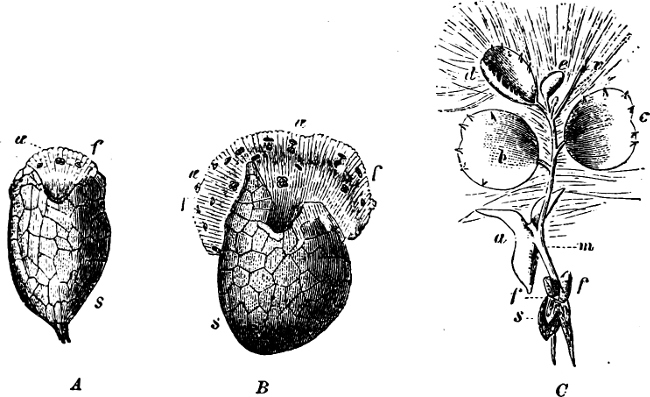
Fig. 215.—Salvinia natans. A, B Female prothallia, f-f, protruding from the macrospore which is still enclosed in the macrosporangium; œ archegonia. C An embryo (× 16) still in connection with the spore (s): a the scutiform leaf; b-e the subsequent foliage-leaves, of which b and c stand singly, d-e-v in a whorl; v the submerged-leaf; f-f wing-like lobes of the prothallium: m the foot.
Sexual generation. The MICROSPORES produce an extremely rudimentary prothallium, formed of only a single cell, and having also a very much reduced bicellular antheridium with a small number of spermatozoid mother-cells in each cell (in Salvinia 4, in Marsilia and Pilularia 16). In Salvinia the microspores remain embedded in a hard mucilaginous mass (at first frothy) which fills up the cavity of the sporangium. The prothallium must therefore grow out through this slime and also through the wall of the sporangium (Fig. 214), and it thus terminates in a relatively long cell.
In Marsilia the microspores are set free from the microsporangium, and the prothallia, with the antheridia, remain in them until the spermatozoids are liberated. The latter are spirally-twisted threads.
The MACROSPORES, on germination, give rise to a very reduced prothallium, which in Salvinia bears 3 archegonia; but, if these are not fertilised, the prothallium may continue to grow and become a fairly large, green body with several archegonia (Fig. 215 A, B). In Marsilia the prothallium is still more reduced, it is enclosed in the macrospore, and only bears one archegonium. The archegonia are similar in structure to those of the Ferns, but are smaller, and sunk more deeply in the tissue of the prothallium.
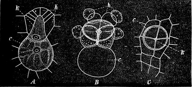
Fig. 216.—Salvinia natans. A An archegonium, unripe, seen in longitudinal section: h the neck-cells; k the neck-canal-cells; c the central cell. B An open archegonium of which the neck-cells have separated off. C An open, old archegonium seen from the top.
The asexual generation is developed from the fertilised egg-cell. It is a dorsiventral, horizontal shoot. In Salvinia it bears at first a shield-like leaf, the scutiform leaf (Fig. 215 C, a), which is succeeded by the ordinary foliage-leaves. The young plants of Marsilia, likewise, have less perfect leaves in the very early stage.
The formation of the sporangium is the same as in the Leptosporangiate Ferns. (The 16 spore-mother-cells originate from one central, tetrahedric archesporium.)
The Hydropterideæ are divided into 2 orders, the chief differences between them being found in the asexual generation.
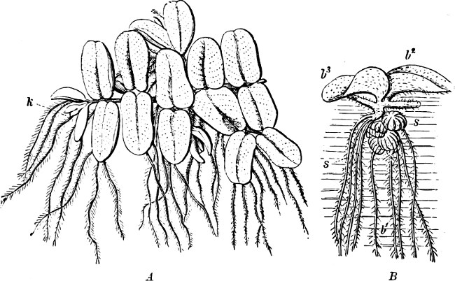
Fig. 217.—Salvinia natans (natural size): A seen from above, floating on the water; B a portion seen from the side in its natural position in the water.
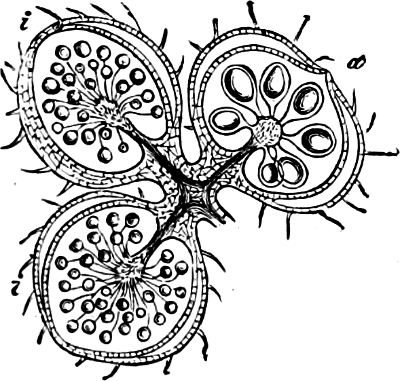
Fig. 218.—Sori of Salvinia in longitudinal section: h microsporangia; m macrosporangia. (× 10.)
Order 1. Salviniaceæ. This order more nearly approaches the true Ferns, especially so on account of the form of the indusium. Only one species is found in Europe, Salvinia natans (Fig. 217). This is a small, floating, annual, aquatic plant, entirely destitute of roots. The dorsiventral, horizontal stem bears two kinds of leaves, which are arranged in whorls of three. Two of these which turn upwards are oval, entire, “aerial foliage-leaves” (Fig. 217 B, b2-b3); the third, the “water-leaf” (b1) is submerged and divided into a number of hair-like segments, similar to the submerged leaves in many aquatic plants, for instance, Water-buttercup (see also Fig. 215 C). The whorls of leaves alternate with each other; there are thus 4 rows of dorsally-placed aerial leaves, and two rows of ventrally-placed submerged leaves. The sporangia are situated in sori, each sorus being borne on a small column (receptacle or placenta) and enveloped by a cupular, but entirely closed indusium (Fig. 218). The sori are situated on the submerged leaves (Fig. 217 B, s-s) and are unisexual, i.e. each sorus contains microsporangia only, or macrosporangia.
Azolla belongs to this order. It is a very small, floating, tropical water-plant (America and East India), with horizontal, root-bearing stem. The stem branches profusely by lateral buds, and bears the two rows of leaves on its dorsal side, the roots on the ventral side. Each leaf is bifid, and divided into an upper dorsal, and a lower ventral portion. The upper segments float on the surface of the water and are arranged like tiles on a roof, each one overlapping its neighbour. In each floating segment a large cavity is found, in which Anabæna is always present. The lower segments are submerged.
Order 2. Marsiliaceæ. The characteristic feature of this order, and one not possessed by other Fern-like plants, is that the sori (2–many) are enveloped in leaf-segments which close round them and form a “sporocarp,” just in the same manner as the carpels, in the Angiospermous Flowering-plants, close round the ovules and form ovaries. The sori contain both micro-and macrosporangia. When the spores are ripe, the sporocarp opens in order to disperse the spores (Fig. 220).
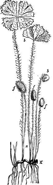
Fig. 219.—Marsilia salvatrix (natural size): K terminal bud; b leaves; f sporocarps; x point of branching of petiole.
The two genera (with 57 species, Temperate, Tropics) are land-and marsh-plants, whose dorsiventral, creeping stem bears roots on the under surface, and the leaves in two rows on the upper side (Figs. 219, 221). The leaves of Marsilia are compound, and divided into four small leaflets springing from the apex of the petiole (Fig. 219), and resemble the leaves of Oxalis. In the bud the leaves are circinate (Fig. 219 b), and at night they exhibit the well-known sleep-movements. The sporocarps are borne on the petioles of the fertile leaves, near their bases (Fig. 219 f); they are oblong and resemble small beans, the outer cells being hard and sclerenchymatous, while the inner ones are divided into a number of loculi arranged in two rows. On germination, water is absorbed, the two sides separate slightly, as valves (Fig. 220 A), and a long vermiform mass of gelatinous, parenchymatous cells (Fig. 220), swollen by the water, emerges, bearing a large number of sori arranged pinnately. Each sorus (sr) is covered by a thin indusium. (The thin covering may be considered an indusium physiologically, though not morphologically).
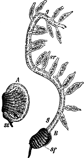
Fig. 220.—Marsilia salvatrix: A the sporocarp commencing to germinate; B a more advanced stage of germination.
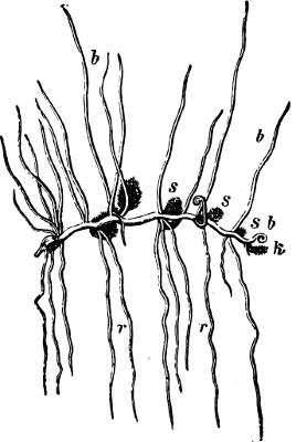
Fig. 221.—Pilularia globulifera (natural size): s sporocarps; b leaves; k the growing point; r roots.
Marsilia quadrifolia, in Europe. Many species are found in Australia. The nutritious sporocarps of M. salvatrix were the means of saving the Burke expedition in the interior of Australia, and hence this species has earned its specific name.
Pilularia has linear leaves, without lamina. The sporocarps are spheroid (Fig. 221), brown and hard, and situated near the base of the leaves. They are 2–4 chambered and open by a corresponding number of valves.
Class 2. Equisetinæ (Horsetails.)
The characteristics of this class have been described on page 204.
It is divided into two sub-classes:—
1. The isosporous Equisetinæ. To this sub-class belong, with certainty, only the Equisetaceæ now existent, which are represented by only one genus, Equisetum.
2. The heterosporous Equisetinæ. Forms which are now extinct.
Sub-Class 1. Isosporous Equisetinæ.
Order. Equisetaceæ (Horsetails).
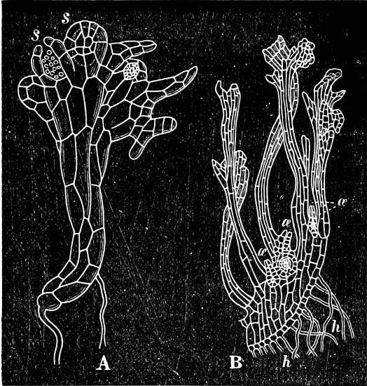
Fig. 222.—Equisetum arvense. The prothallium highly magnified. A Male; s, s antheridia. B Portion of a female, cut through vertically; œ œ archegonia, the central one is fertilised; h h root-hairs.
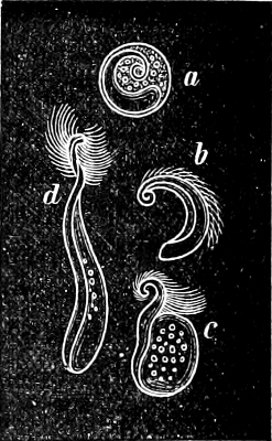
Fig. 223.—Equisetum maximum. Spermatozoids: a shows them still enveloped by the mother-cell.
The sexual generation. The prothallium is green and leaf-like, as in the majority of Ferns, but irregularly branched and curled. It is often unisexual. The male prothallia bear antheridia only, and are smaller and less branched (Fig. 222 A) than the female; the latter may attain a diameter of ½ an inch, and bear archegonia only (Fig. 222 B). The antheridia and the archegonia resemble those of the Ferns, but the spermatozoids (Fig. 223) are larger and less twisted. On the last curve is situated a more or less elongated appendage of cytoplasm (Fig. 223 c).
The asexual generation. The embryo is similar to that of the Ferns. The fully developed Equisetum is a perennial herb, with widely creeping (in some species tuberous) rhizome, from which extend erect, aerial, most frequently annual shoots.
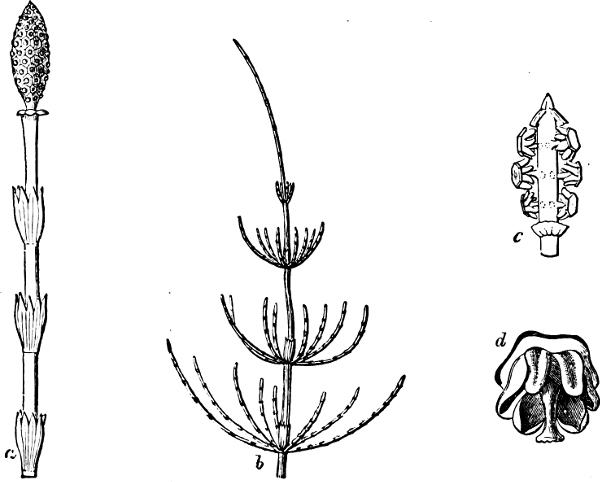
Fig. 224.—Equisetum arvense: a fertile branch with cone; b vegetative shoot; c cone; d sporophylls.
The vegetative aerial STEMS are divided into a number of internodes by the whorls of leaves (Fig. 224). The internodes are hollow, the cavities being separated from each other by the transverse partitions of the solid nodes. The lower portion of the internode, which is encased by the leaves, has much thinner and softer cell-walls, so that the stem is easily separated into segments just above the nodes. Each internode has a large number of ridges and furrows, and bears at its apex a whorl of leaves whose number and position correspond to the ridges of the internode. As in the case of other verticillate plants, the whorls are placed alternately, one above the other; the same arrangement is also found in the ridges on two successive internodes. In addition to the large air-cavity in the centre of each internode (the central cavity), a whorl of tubular air-passages is found in the cortex of the stems, opposite the furrows (vallecular canals). There is also a similar air-passage (carinal canals) in each of the vascular bundles, which are placed in a ring, one opposite each ridge, and therefore alternating with the vallecular canals. The vascular bundles are collateral as in the majority of Flowering-plants, but poorly developed. The xylem of each bundle consists of two groups of annular or spiral vessels, close to the outer border of the carinal canal, and two groups of scalariform tracheides, each placed on a radius passing through a group of spiral vessels. The phloëm is placed between these four groups, each of which has only a few vessels. The stiffness of the stems is mainly due to the large amount of silica in the cell-walls of the epidermis, and to the sclerenchymatous cells of the ridges.
All LEAVES are situated in whorls. The VEGETATIVE are simple, undivided, 1-nerved, and are united into toothed sheaths (Fig. 224 a, b). The branching of the stems in some species (E. arvense) is very abundant. The branches break through the base of the leaf-sheaths (Fig. 224 b), and generally alternate with the teeth (leaves).
The FERTILE LEAVES (sporophylls) are different from the barren ones. They are free, shield-like, each one having a short stalk bearing usually an hexagonal plate (Fig. 224 d), and closely compressed into an ear or cone (Fig. 224 a, c). The Equisetums thus present an advance in development distinctly beyond that of the Ferns, which is further emphasized by the circumstance that a transition from the sheath-leaves to the fertile-leaves is found in the involucre or annulus, a “collar” of specially modified leaves situated at the base of the cone (Fig. 224 a and c). The cone may be considered as a very rudimentary flower, and the annulus may be regarded as a very early stage in the formation of a flower (perianth). See page 235.
The SPORANGIA are situated on the underside of the sporophylls, one at each angle; they are sac-like, and open inwardly by a longitudinal cleft (Fig. 224 d). An annulus is wanting; but in the wall of the sporangium, as in the pollen-sacs of the Flowering-plants, a layer of cells, with annular or spiral thickenings, is developed, which assists in the dehiscence of the sporangium.
The SPORES are green; the walls composed of four distinct layers, of which the outer is gradually separated, except at one point, and becomes split into four long bands (elaters) (Fig. 225). The elaters are extremely hygroscopic, coiling round the spore when moistened, and expanding as soon as dry, presenting a most lively object under the microscope when breathed upon and allowed to dry. The second layer, when germination commences, becomes detached from the inner wall, which is formed of the exospore and endospore.
The order has become much reduced, and at the present time includes only one genus, Equisetum, with about twenty-five species, which are distributed over the entire globe, particularly in damp situations. In SOME SPECIES the barren shoots are green and very much branched, but the fertile ones are unbranched, pale brown, and possess no chlorophyll (E. arvense, Field-Horsetail, Fig. 224, and E. maximum). In others the fertile and barren shoots are alike green, and either both unbranched (E. hiemale), or branched (E. palustre, E. limosum, etc). The fertile shoots of E. silvaticum, up to maturity, resemble those without chylorophyll of E. arvense, but after that period they produce green branches, and thus resemble the barren ones.
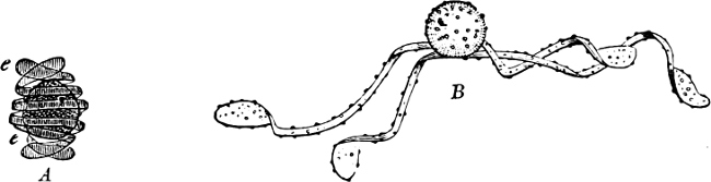
Fig. 225.—Spores of Equisetum: A damp, with elaters (e) coiled round the spore; B dry, with elaters expanded.
Extinct isosporous Equisetinæ. In addition to several true species of fossilized Equisetums, the order of the Calamites, which no doubt is closely allied to the Equisetinæ, is also found in the fossil state. These were gigantic forms, attaining about twenty times the size of those of the present day, and stems of nearly 10–12 metres in height are known. They reached the culminating point of their development in the Carboniferous period, and died out towards the close of the Palæozoic. The stems had hollow internodes and alternating grooves, similar to their relatives of the present day. The leaves must either have been absent or very perishable, since they have not been identified with certainty. If the determinations of certain remains of cones which of late have been discovered are correct, they were heterosporous and had two kinds of sporangia as in the following sub-class. A cambium formation and an increase in thickness has been found in the stems.
Their USES are very limited. A few species, such as E. hiemale are used for polishing on account of the hard siliceous cell-walls of the epidermis, found in all species of Equisetum.
Sub-Class 2. Heterosporous Equisetinæ.
The two orders which come under this head are united by the characteristics, that the verticillate leaves are not united into sheaths (Fig. 226), and that between each whorl of fertile leaves there is also a whorl of barren ones. The fertile whorls in Annulariæ are situated about midway between the barren ones (Fig. 227), but in Asterophylliteæ they occur immediately above a barren whorl (Fig. 228) and contain only half as many members as the latter. The lower whorls bear macrosporangia with one macrospore, the upper, microsporangia with many microspores.
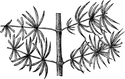
Fig. 226.—A. fragment of Annularia.
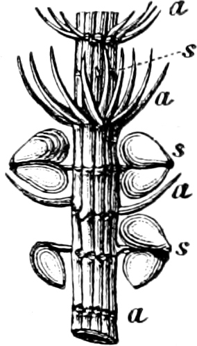
Fig. 227.—Fragment of Annularia longifolia, with sporangia; the leaves have partly fallen off: a barren whorls; s fertile whorls.
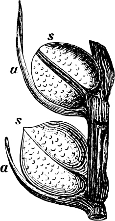
Fig. 228.—Fragment of cone of Asterophyllites (Volkmannia elongata): a and s as in Fig. 227.
The Annulariæ were distichous (Fig. 226), and presumably floating plants. The Asterophylliteæ had verticellate branches. These also died out after the Carboniferous period, at the close of the Palæozoic.
Class 3. Lycopodinæ (Club-Mosses).
The characteristics of this class have been given on page 205. It consists of two sub-classes, one embracing isosporous, the other heterosporous forms.
Sub-Class 1. Lycopodieæ (Isosporous Lycopodinæ).
One kind of spore. Prothallium large, partly green. Leaves without ligule.
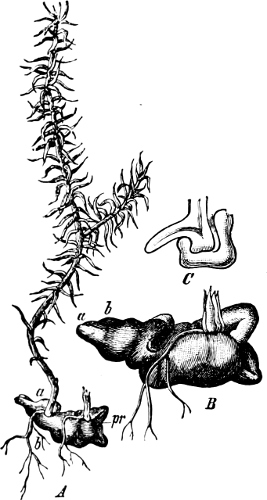
Fig. 229.—Lycopodium annotinum: A embryo (nat. size), with prothallium (pr), one embryo is broken off; B the prothallium (slightly magnified); C section through the prothallium and embryo in the direction a-b of A, and vertically in the plane of the paper.
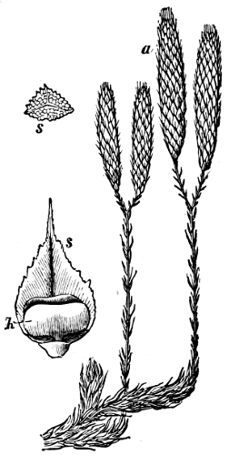
Fig. 230.—Lycopodium clavatum: portion of a stem, bearing cones (a); s a spore; h sporangium in the axil of a leaf, s.
Order 1. Lycopodiaceæ. The PROTHALLIUM is only known in a few species at present, but in these it is more or less tubercular, and bears both antheridia and archegonia.
In L. annotinum the prothallium is a relatively large mass of cells, without chlorophyll, and subterranean, in which the antheridia and archegonia are embedded (Fig. 229). In the widely distributed tropical species, L. cernuum, and in L. inundatum, it is a small tubercular body which has a subterranean portion, with either little or no chlorophyll; and an aerial green portion. The prothallia of L. phlegmaria and others live saprophytically in the crevices of the bark of trees; they are partly filamentous, branched, and possess no chlorophyll.
The asexual generation. Perennial plants. The stem branches monopodially (often apparently dichotomously), and is thickly covered by small, simple, triangular or scale-like leaves. The leaves are spirally arranged in some species (Figs. 229, 230), and in others, whose stem is compressed with unequal sides, opposite (Fig. 231). The roots of Lycopodium are dichotomously branched.
The SPORANGIA in Lycopodium are situated singly at the base of the leaves, almost in their axils; they are reniform, unilocular and open like a mussel-shell by two valves (Fig. 230 h). The sporangia are developed from a group of surface cells. The archesporium is formed from one hypodermal cell (or perhaps a cell-row).
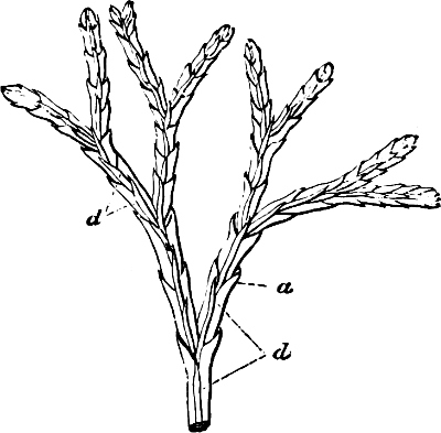
Fig. 231.—Lycopodium complanatum: a leaves on the edges of the stem; d leaves on the sides.
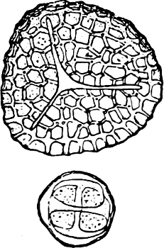
Fig. 232.—Lycopodium clavatum. A tetrahedral spore seen from above, where the three borders join; and a tetrad of bilateral spores, still lying in the mother-cell.
The fertile leaves are collected upon definite regions of the stem. They are either similar to the barren ones, and then the fertile portions of the stem pass gradually, without any break, into the barren portion (L. selago); or they differ from the barren leaves, and are then collected into special apical cones (Fig. 230 a). The SPORES are tetrahedral or bilateral (Fig. 232).
About 100 species, chiefly tropical.
Five species of Lycopodium are found in Great Britain. L. clavatum and L. selago are common in mountainous districts. L. annotinum is common in the Highlands of Scotland. The other genus of the order is Phylloglossum, with one species, P. drummondi (Australia, Tasmania, and New Zealand), a small plant only a few centimetres high, with two tubers, and about eleven linear leaves at the base of the stem which is terminated by a cone of sporophylls.—Fossil Lycopodiaceæ in the Carboniferous period.
Officinal: “Lycopodium,” the spores of L. clavatum.
Family 2. Psilotaceæ. The sporangia are placed on the apex of short, two-leaved stems, as 2–3, seldom four, small capsules. Small herbs, with angular stems; leaves small, simple, and one nerved. Only four species.—Psilotum (Madagascar, Moluccas, Sandwich Islands, etc.) is destitute of roots, their place being supplied by special underground stems which bear a few modified leaves, very much reduced, especially when buried deeply in the soil. Three species.—Tmesipteris (Australia), one species.
Sub-Class 2. Selaginelleæ (Heterosporous Lycopodinæ).
Micro-and macrospores. The prothallia are very much reduced, especially the male; the female does not leave the spore. The leaves are ligulate.
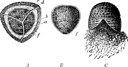
Fig. 233.—Germination of the microspores of Selaginella: A the spore rendered transparent, seen from above. In the interior is seen the prothallium (f), and the first divisions of the antheridium (a, b, c, d); in B the spore-wall is removed and all the spermatozoid-mother-cells formed; in C, the microspore has opened and the spermatozoids and the mother-cells are escaping together.
The sexual generation. In the MICROSPORES are formed: (1) a very small “vegetative” cell, representing the vegetative part of the prothallium (f in Fig. 233 A, B), and (2) a cell many times larger and which divides into a number (4–8) of primordial cells, each of which divides into four spermatozoid-mother-cells, though all of these may not develope spermatozoids. On germination, when the spore-wall is ruptured, the spermatozoids and spermatozoid-mother-cells are ejected into the water.
The SPERMATOZOIDS in Selaginella are elongated and club-shaped, with two cilia (Fig. 234); but in Isoëtes lacustris they are spirally-twisted threads which differ from all other spermatozoids by having a bunch of cilia at each end; the other species of Isoëtes have cilia only at the anterior end.
The MACROSPORES. Shortly after the macrospores have been set free, or in Selaginella, while still enclosed in the sporangium of the mother-plant, they germinate and soon become filled with the cellular tissue of the prothallium, and even in Selaginella the archegonium begins to be formed before the rupture of the spore-cell-wall has commenced (Fig. 235 A).
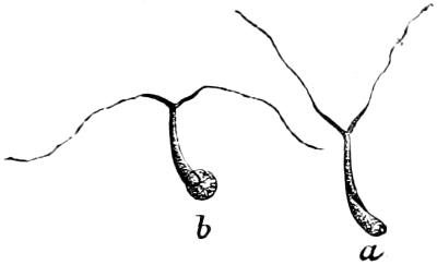
Fig. 234.—Spermatozoids of Selaginella: b with a remnant of cytoplasm.
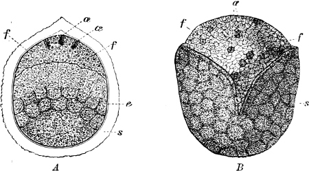
Fig. 235.—Macrospore of Selaginella: A longitudinal section, before the rupture of the wall, six weeks after being sown. The endosperm (e) has not yet filled the entire chamber. Cell-formation is still proceeding in the lower part of the spore. The endosperm and prothallium (f f) are separated by a distinct line (diaphragm). B Germinating macrospore seen from outside: s wall of the spore; æ archegonia.
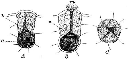
Fig. 236.—Archegonia of Selaginella: A unripe, in longitudinal section; c the central cell; k neck-canal-cell, which is wedged in between the two-storied neck-cells; B ripe; u ventral canal-cell; C seen from above, open. It will be noticed that the neck is formed of two tiers of four cells each.
The ARCHEGONIA are constructed on the same plan as those of the other Archegoniatæ, but are quite embedded in the prothallium (Figs. 235 æ, 236).
The asexual generation varies very much in the different orders.
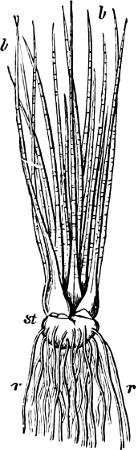
Fig. 237.—Isoëtes lacustris (slightly diminished): st the stem; r roots; b leaves.
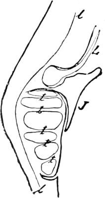
Fig. 238.—Isoëtes lacustris. Longitudinal section through the base of the leaf with a microsporangium. The edge of the groove, in which the microspangium is placed, is continued as a thin covering which envelopes the sporangium. The inferior edge of the ligular groove (L) forms a lip (J); t sterile cell-rows (trabeculæ) which divide the sporangium into compartments; l vascular bundle.
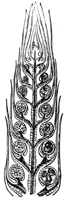
Fig. 239.—Selaginella inæqualifolia. Cone in longitudinal section; microsporangia are seen on the left side, macrosporangia on the right (most frequently each with four macrospores).
Order 1. Isoëtaceæ (Quill-worts). The only known genus, Isoëtes (Quill-wort), has an extremely short, tuberous, unbranched stem with very short internodes (Fig. 237). The STEM is remarkable as being the only one among the Vascular Cryptogams which increases in thickness (see page 202). The meristematic cells are situated round the axial cylinder, and form, especially, parenchymatous tissue in two or three directions, giving rise to 2–3 grooves in which the dichotomously-branched ROOTS are produced. The LEAVES are arranged spirally in a close rosette. They are awl-shaped and have at the base a semi-amplexicaul sheath, with a groove (fovea), in which a sporangium is situated (Fig. 238). The ligule is a foliar outgrowth from the upper edge of the groove.—The MACROSPORANGIA (each with a number of macrospores), are situated on the outer leaves, the MICROSPORANGIA (Fig. 238), on the inner ones. Between each cycle of fertile leaves there are a number of imperfect or barren ones as in the case of the female plant of Cycas. The spores are liberated by the decay of the sporangium. The two kinds of sporangia develope at the commencement in the same way. The archesporium is, at first, a hypodermal layer of cells which grow out in the direction perpendicular to the surface of the leaf, and divide by a number of walls parallel to this direction, forming a sporogenous mass of cells. Some of the cell-rows of this sporogenous mass lose their rich protoplasmic contents, and are arrested in their growth; thus incomplete divisional walls of sterile cells, “trabeculæ” arise in the sporangium, dividing it into a number of compartments one above the other (Fig. 238 t). (The trabeculæ, according to Goebel, play the same part as the nutritive cells of the sporangium of Riella; the tapetal cells, as in the Ferns, are in a great measure dissolved at a later period.) The sporogenous cell-rows, in the microsporangia, give rise to a large number of spore-mother-cells, but in the macrosporangia only one spore-mother-cell, with tapetum, is developed from each fertile archesporial cell.
The two native species, and several others, are aquatic plants, the remaining species are land plants, or are amphibious. About 50 species. In temperate and tropical regions.—Fossil species in the Tertiary period.
Order 2. Selaginellaceæ. This order contains only one genus, Selaginella. The STEM, in the majority of species, is dorsiventral, long and slender, and apparently branches dichotomously, but in reality monopodially, with well developed lateral shoots. The LEAVES are small, round, or ovate, in the majority of species arranged in whorls of two leaves each; these whorls, however, are not decussate, but are considerably inclined towards each other, an arrangement by which four rows of leaves are produced, each whorl having one large and one small leaf. The two leaves in each whorl are of unequal size, the smaller one being placed on the upper surface and the larger on the lower surface of the stem (Fig. 240). Some species have spirally-arranged leaves, more resembling the arrangement in the Lycopodiums.
The FERTILE LEAVES most frequently differ from the barren ones, and are collected into spike-like cones (a kind of flower; Fig. 239). Micro-and macrosporangia are found in the same cone (Fig. 239). Each sporangium arises from a group of superficial cells of the stem, directly over the leaf on which it will be situated later on. Each sporangium has a hypodermal, unicellular archesporium, and contains a layer of tapetal cells; these are dissolved later, when the spores are ripe, and not before as in the Ferns. In the very early stages of their development, the micro-and macrosporangia are precisely similar, and the differences between them arise later on. In the microsporangium all the spore-mother-cells divide, and each forms four tetrahedrically-arranged microspores (Fig. 204); but in the macrosporangium only four macrospores are formed, by the division of a single mother-cell, while the remaining spore-mother-cells are aborted. It is rarely that the macrosporangia contain 2 or 8 macrospores.
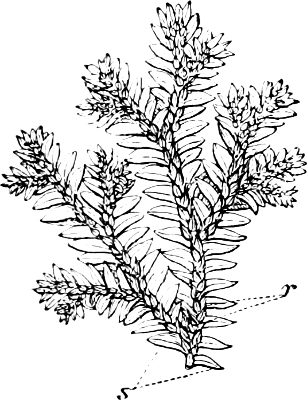
Fig. 240.—Selaginella martensii: s lower leaves; r upper leaves.
For the GERMINATION OF THE SPORES, see pages 228, 229. The prothallium arises in the macrospore (f-f, in Fig. 235 A), probably by division of the meniscus-shaped protoplasmic mass, which is marked off at the apex of the spore; primordial cells are thus formed which later on are surrounded by a cell-wall. In six to seven weeks after sowing, the spore-wall is ruptured by the growing prothallium, which already has developed archegonia (Fig. 235 œ-œ). The prothallium so formed does not occupy the entire cavity of the spore, but four to five weeks after sowing, the large-celled parenchyma is developed in the lower portion of the spore by free cell-formation; this has been termed by Pfeffer, “endosperm,” since it is similar to the endosperm of Flowering-plants. Goebel, however, has termed it “secondary prothallium,” as the homology with the endosperm of the Angiosperms is very doubtful.
The FERTILISED OOSPHERE divides into an upper (hypobasal) and a lower (epibasal) cell; from the latter alone the embryo is developed with its root, stem, foot, and two cotyledons, and the former gives rise to an organ which appears in this instance for the first time, but which occurs in all Flowering-plants, viz. the suspensor. This forces the embryo down into the “endosperm,” which is entirely or partially absorbed by the embryo. In the case of the Flowering-plants the embryo is developed with its longitudinal axis in the elongation of the suspensor, but in Selaginella the embryo is situated transversely to it.
Selaginella (300–400 species), is essentially tropical, only one species living in the North (S. spinulosa), but others grow in Central and South Europe.
Order 3. Lepidodendraceæ are extinct, tree-like Lycopods, which are found especially in the Lower and Middle Carboniferous. Vegetatively they are most nearly related to Lycopodium, but the stem attained much larger proportions (about eleven metres in height and one metre in thickness), and had a cambium by which it increased in thickness. It was regularly dichotomous, and closely studded with spirally-placed leaves, which left behind them peculiar rhombic scars. The large cones resemble Pine-cones, and bore sporangia much larger than any which are now produced (the male ones as much as 2 cm.’s in length). The macrosporangia were situated at the base, and the microsporangia at the apex.
Order 4. Sigillariaceæ. These are, presumably, another group of extinct tree-like Lycopods (especially in the Middle Carboniferous). The name has been derived from the seal-like scars, which the fallen leaves have left behind in longitudinal rows on the grooved stem. The rhizomes of these plants were formerly termed Stigmaria, and placed in a separate genus.
Order 5. Sphenophyllaceæ form an entirely extinct group. They do not definitely belong to any of the three large classes of Vascular Cryptogams, but it is perhaps best to place them in juxtaposition to these. They were herbaceous plants with verticillate, wedge-shaped leaves, with nerves branching dichotomously into equally strong branches. Micro-and macrosporangia were formed in the same cone; and were situated in the axils of the leaves, as in the Lycopods.
The Transition from the Cryptogams to the Phanerogams.
All the plants considered in the preceding chapters are included in the term Cryptogams; all in the following chapters under the head of Phanerogams (see page 3). Hofmeister’s pioneer works (1851, Vergleichende Untersuchungen der höheren Kryptogamen, etc.) and the numerous researches published later by other investigators, have closed the gap which was formerly thought to exist between these plants; so that we now, in the series: Bryophyta—Pteridophyta—Gymnospermæ—Angiospermæ see the expression of a single line of development in accordance with a definite plan. The forms through which this gradual development has taken place have in course of time, however, to a great extent died out, and only single links of the chain connecting the lowest to the highest still remain.
The alternation of generations, which we found indicated in certain Thallophytes, can be proved with the greatest clearness in all the higher Cryptogams, from the Mosses upwards; it is also found in the Phanerogams, but not in such a pronounced degree, because one of the generations is so far reduced that it has almost given up its independence. For the sake of greater clearness, we will begin with the comparison of the sporophyte, asexual (second) generation.
The asexual (2nd) generation of the Cormophytes.
The asexual generation which follows from the further development of the fertilised oosphere, is, in the Mosses, only the sporogonium (according to one theory it is perhaps homologous with a spore-bearing leaf, situated upon a short stem, see p. 187); in Filicinæ, Equisetinæ, and Lycopodinæ, on the other hand, it is a highly developed plant differentiated into stem, leaf, and true root, and bearing the sporangia on its leaves. The MODIFICATION OF THE SHOOT is very slight in Filicinæ. The first leaves of the embryo are very simple in form (Fig. 205), but after a certain age all the leaves which arise are essentially alike. The fertile leaves do not differ from the barren ones, and are found associated with them, and their formation does not limit the growth in length of the stem. It is only in a few of the true Ferns, and in the Hydropterideæ, that the fertile leaves differ considerably from the barren ones. A division of labour in which certain leaves are set apart for nutrition, and others for reproduction, is found more pronouncedly in the Equisetinæ and Lycopodinæ, for in these groups, with a few exceptions, the fertile and barren leaves are very dissimilar; the former are collected in special ear-like cones, which terminate the further growth of the short stems on which they are borne. In connection with the cone, leaves are sometimes developed which form a transition from the barren to the fertile ones (the “annulus” in Equisetaceæ), and in these cases the first indication of a flower with perianth or floral-leaves is to be traced. Among the Cryptogams the highest division of labour is found in Selaginella and Isoëtes, which have the two kinds of sporangia borne on different leaves. The division of labour (modification) is, however, still more pronounced in the Phanerogams: the leaves which bear the microsporangia (“pollen-sacs”) have quite different forms from those which bear macrosporangia (the “nucellus” in the ovule), the former are termed stamens, the latter carpels; in certain instances, too, there is even a contrast between the “male plants” and the “female plants.” Moreover, a division of labour, in a much greater degree, takes place in the leaves which do not directly take part in reproduction, and it is thus possible in many plants to draw a sharp line not only between stamens and carpels, but also between four or five distinct kinds of leaves, which differ in form, structure, and corresponding functions, and which appear in regular sequence on the shoot: namely, between “scale-leaves” and “foliage-leaves,”[21] both of which occur in the Cryptogams, and the “floral-leaves,” including the bracts and leaves of the “perianth,” which latter often differ from each other in form and colour, and are then separated into sepals and petals. The leaves—stamens and carpels—which bear the sporangia are termed sporophylls, and the shoot, or extremity of a shoot, whose leaves are modified into sporophylls, is terminated in its further growth by their production, and is known as a flower. The flower which is most completely furnished has calyx, corolla, stamens, and carpels arranged in this order. If the only sporophylls present are stamens, then it is said to be a male (staminate) flower, and if only carpels, then a female (pistillate) flower, and in both these cases the flowers are unisexual, or diclinous. If stamens and carpels are both present in the same flower, it is termed hermaphrodite. Diclinous plants in which the female flowers are situated on one plant, and the male flowers on another, are termed diœcious; and those in which the same plant bears the two kinds of flowers are termed monœcious. When the male, female, and hermaphrodite flowers are found in the same species, the plant is said to be polygamous.
The sporangia-bearing leaves—Sporophylls. In the Mosses the asexual generation is only represented by the sporogonium, and if the theory is correct which considers the sporogonium to be an embryo consisting of a rudimentary stem and terminal leaf, then the spores are produced on the leaves in these plants. The sporangia in the Filicinæ are situated in groups (sori) on the back or on the edge of the leaves. The number of sporangia in the sorus diminishes very greatly in the Marattiaceæ and Gleicheniaceæ (three to four in the latter, Fig. 213). In the Equisetinæ the sporangia are situated in a small number on the underside of shield-like leaves, and in Lycopodinæ, singly, in the axils of the fertile leaves, which are alike and bear either micro- or macrosporangia. In the Phanerogams there is a great difference between the stamens and carpels.
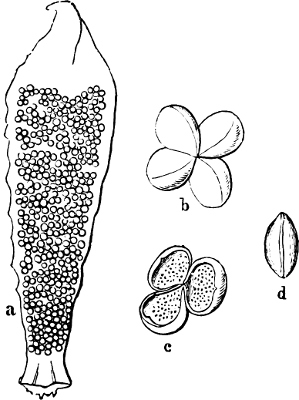
Fig. 241.—Cycas: a stamen (nat. size) seen from the under side; b four pollen-sacs, not yet open, forming a “sorus”; c three open pollen-sacs; d a pollen-grain.
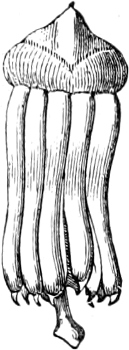
Fig. 242.—Stamens of Araucaria (pollen-sacs long and pendulous).
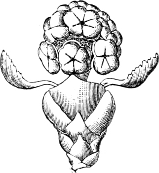
Fig. 243.—Male flower of Taxus.
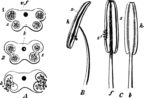
Fig. 244.—A Cross section through a quadrilocular anther in different stages of development: s the seam where it bursts open; vf vascular bundle; k connective. B A stamen. C Another stamen seen from the front (f) and from the back (b).
Stamens. In the lowest Phanerogams (Cycadeæ) there are many indications of relationship to the Ferns. The stamens are flat and broad, and have on the back many pollen-sacs (microsporangia) arranged in small groups (true sori), which even have a small “placenta,” similar to the one possessed by the Ferns, and open towards the inside by a longitudinal cleft (Fig. 241, compare Fig. 213). A section of the Coniferæ agree more closely with the Equisetaceæ, in having a few (three to eight) pollen-sacs arranged on the underside of more or less shield-like leaves (Figs. 242, 243, compare with Fig. 224 a, c, d). In the Abietaceæ the number of sporangia is diminished to two, which are placed also on the lower side (Fig. 267) of a stamen. The number of pollen-sacs (microsporangia) in the Angiosperms is nearly always four to each stamen; they are longitudinal projections which are placed in pairs on each side of the central line of the stamen, two on the edge, and the other two generally on the side which is turned inwards; the pollen-sacs generally dehisce longitudinally (quadrilocular anthers, Fig. 244). A few, for instance Orchidaceæ and Asclepiadaceæ, have only two pollen-sacs (bilocular anthers); and in others, such as Solanum and the Ericaceæ, they open by pores; in Lauraceæ and Berberidaceæ, by valves. The part of the stamen which bears the pollen-sacs is termed the anther. Most frequently this is supported by a stalk known as the filament.
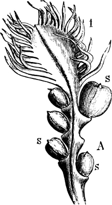
Fig. 245.—A carpel of Cycas revoluta with 5 ovules (s), at half to one-third nat. size.
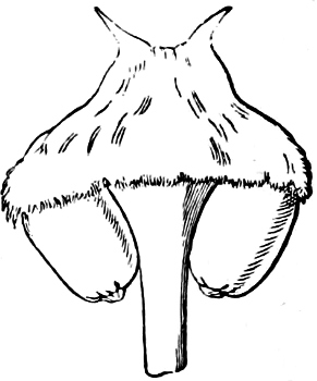
Fig. 246.—Carpel with 2 ovules of Ceratozamia robusta (1/1).
Carpels. The simplest forms of carpels are found in Cycas. In this genus both the foliage and fertile leaves are pinnate, and hence present great similarity; the ovules (macrosporangia) are situated on the margin of the central portion, just as the sporangia are placed on the edge of the fertile leaf of Ophioglossum (Fig. 245, compare with Fig. 209). The carpels of the other Cycadeæ present greater divergence from the foliage-leaves, being peltate, for instance, in Zamia and Ceratozamia (Fig. 246). The ovules in the Coniferæ are situated on the upper side and near the base of the ovuliferous scales, almost in the same position as the sporangia in the Lycopodinæ (Figs. 269, 272, 273 H, compare Figs. 230, 239). In Taxus the uninclosed ovule is placed on the apex of a shoot (Fig. 264). In all these plants the ovules are not enclosed by the carpels, that is, they are not enclosed in chambers formed by the turning in of the walls of the carpel, and hence the name Gymnospermæ is given to them. In the higher Flowering-plants, the Angiospermæ, the ovules are distinctly situated on the edge, the upper surface, or base of the carpel; but the carpel closes round the ovules which are therefore enclosed in a chamber—the ovary. In a few cases, for example in the Polygonaceæ, an ovule is situated apparently on the apex of the stem itself, as in the Yew; in other cases, as in the Primulaceæ, many ovules are apparently developed on the apex of the stem, which seems to have been specially adapted as a placenta, but it is also possible and correct in these cases to suppose that the ovules are in reality developed on the carpels.[22] A single fully-developed carpel or a collection of carpels joined together is termed the pistil. The extremity of the carpel, which is specially developed to catch the pollen-grains and form a suitable nidus on which they may germinate, is called the stigma. The united edges of a carpel which bear the ovules are termed the ventral suture. The back of the carpel forms the dorsal suture. The Marsiliaceæ take a position among the Hydropterideæ analogous to that occupied by the Angiosperms; the sporangia are in a corresponding manner enveloped in a closed leaf.
The collection of stamens in a flower is termed the andrœcium, and all the carpels, whether individually free or united into one pistil, the gynœceum.
The Sporangia. The asexual generation of the Mosses is the sporogonium, in which the spores arise in tetrads from the mother-cells. The sporangia in the Filicinæ take their origin either from a single cell (Leptosporangiatæ) or, what probably may be regarded as an older stand-point, from a group of cells (Eusporangiatæ). In both cases there may be distinguished in a mature sporangium three tissues, which have different significance (Fig. 204): (1) an external layer, the sporangium-wall, most frequently composed of one layer of cells made up of cells of dissimilar structure, so that on desiccation the wall is ruptured and the sporangium opens in a definite manner; (2) an internal group of cells, consisting of the spore-mother-cells, developed from an archesporium, and which by division into four gives rise to the spores; (3) a layer of cells lying between the two already mentioned, which is dissolved before maturity. The intermediate cellular layer, which directly surrounds the spore-forming cells, is in form and contents more worthy of note than the others, and is termed the tapetum. The construction of the sporangium in the Equisetinæ and Lycopodinæ is in the main the same.
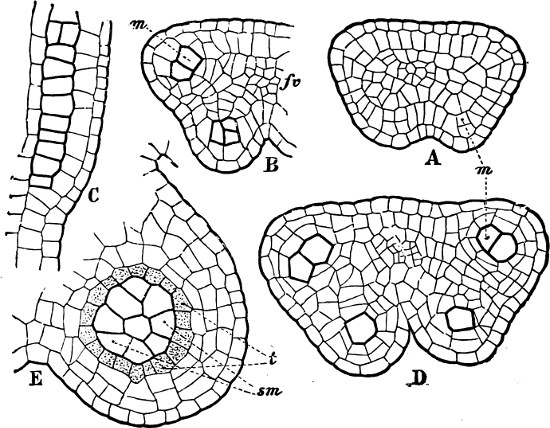
Fig. 247.—Development of an anther. A Transverse section of a young anther of Doronicum macrophyllum. The formation of the 4 pollen-sacs commences by divisions of the hypodermal cells (at m, for instance). These cells divide by periclinal walls into external cells which only take part in forming the anther-wall; and internal cells, which correspond to the Archesporium, and from which the spores are derived. These spore-forming cells are drawn with thicker walls in B-E. The commencement of the vascular bundle is seen in the centre. B An older stage; the pollen-sacs already project considerably. It is the cells in the hypodermal layer which are active and in which tangential divisions particularly occur; fv vascular bundle. C A corresponding longitudinal section. D Transverse section through an older anther, the thickness of the wall outside the mother-cells of the pollen-grains is already increased, and it becomes still thicker by the division of the hypodermal cells: its most external layer of cells but one, becomes transformed into the “fibrous cells.” E Transverse section of a still older pollen-sac of Menyanthes; sm are the mother-cells of the pollen-grains surrounded by the tapetum (t), external to the tapetum is the anther-wall, which is still far from being fully developed. The sub-epidermal layer becomes “fibrous,” and the cells lying inside it become dissolved, together with the tapetum.
In the Phanerogams the Microsporangia are termed Pollen-sacs. They take their origin from a large group of cells, which, in the Angiosperms, lie immediately beneath the epidermal cells of the anther. In the developed, but not yet mature, sporangium (pollen-sac) there are to be found: (as in the Vascular Cryptogams) (1) an internal group of mother-cells which give rise to the pollen-grains (microspores), in this case also formed in tetrads; (2) a group of cells surrounding these, of which the internal ones form a tapetal layer, similar to that in the Vascular Cryptogams; the tapetum and some of the cells surrounding it in this group, become dissolved before maturity; the more external ones, on the other hand, are provided with peculiar thickenings, and form the “fibrous” layer by the aid of which the dehiscence of the anther takes place; (3) an external layer, the epidermis, enclosing all the other layers (Fig. 247).
In some Coniferæ (Cupressus, Thuja, and several species of Juniperus) the microsporangia (pollen-sacs), which are situated on the under side of the stamen, are covered by a thin structure which seems to be a continuation of the lamina and which is supposed to be homologous with the indusium of the Ferns.
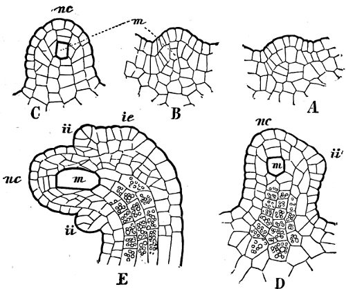
Fig. 248.—Development of the ovule in the Red Currant, Ribes rubrum, arranged alphabetically in the order of development. A Is the youngest stage, E the oldest. ii Inner integument; ie outer integument; nc nucellus; m archespore (mother-cell of the embryo-sac).
The Ovule in the Phanerogams arises most frequently on a projecting portion of the carpel, termed the placenta. The ovules (compare the sporangium of the Eusporangiatæ and especially the pollen-sac) take their origin from a group of cells which lies beneath the epidermis (Fig. 248 A, B). First of all a small papilla is formed, which is later on provided with a vascular bundle and becomes the funicle; this probably has the same value as the projections (“placenta”) on which the sori in the Ferns are attached. Only one macrosporangium (nucellus; Fig. 248 nc) is developed at the apex of the funicle. This arises by a process of cell-division exactly corresponding to that by which the pollen-sacs are formed (Fig. 248 C-E), with this difference only, that while a great many cells may be distinguished in each pollen-sac, which forms pollen-grains by tetrad-division, only a few are found in the ovule, and all these moreover are suppressed, with one single exception which developes into the macrospore (embryo-sac) without undergoing a division into tetrads. The wall of the embryo-sac, in the Gymnosperms, may be thick and divided into two layers and partly cuticularized, as in the spores of the Cryptogams which are to be set free. In the Angiosperms, on the other hand, the wall is extremely thin.
The pollen-sac thus stands in the same relation to the nucellus as the microsporangium does to the macrosporangium: in the pollen-sacs and microsporangia a number of spores arise by the tetrad-division of several mother-cells; in the nucellus and macrosporangium, a reduction of the cells already formed takes place to such an extent that the number of macrospores becomes one (Salvinia, Marsilia, Phanerogams) or four (Selaginella), or rarely a large number as in Isoëtes.
In the Ferns, as stated on page 210, etc., indusia covering the sori very often occur. Horsetails and Club-Mosses have no indusium; but in all Phanerogams cupular or sac-like structures (integuments) are found which envelop the nucellus. These develope from the upper end of the funicle (ii and ie, in Fig. 248; y and i, in Fig. 249) and enclose the nucellus on all sides as a sac, leaving only a small channel at the apex of the nucellus—the micropyle—(Fig. 249) through which the pollen-tube proceeds to the embryo-sac. The ovules of the Gymnosperms have only one integument (Figs. 251, 264, 269, 274) and the same is the case with the majority of the Sympetalæ and a few Choripetalæ; but the Monocotyledons and most of the Choripetalæ have two integuments (Fig. 249).
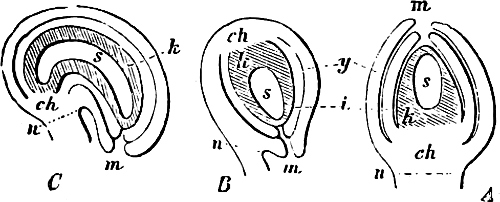
Fig. 249.—Various forms of ovules: A an erect ovule (orthotropous); B reversed (anatropous); C curved (campylotropous): k the nucellus (shaded in all the figures); s the embryo-sac; ch the base of the ovule (chalaza); y and i the external and internal integuments, the dotted line denotes the place where the scar (hilum) will form when the seed is detached from the funicle.
In shape the integuments resemble very closely the cupular indusium of the Hymenophyllaceæ, certain Cyatheaceæ (Fig. 212 E), and Salvinia (Fig. 218); that they are really homologous with these is probable, but is not proven. Some authorities regard them as structures found only in the Phanerogams.
The ovule is thus a “monangic” (i.e. reduced to 1 sporangium, the nucellus) sorus, situated on a funicle, and enclosed by one or two cupular indusia, the integuments. Some of the ovules are erect (orthotropous), others curved (campylotropous), the majority reversed (anatropous) (Fig. 249).
[Goebel (1884 and earlier) with Strasburger considered the entire ovule of the Phanerogams as homologous with the macrosporangium, the integuments however as new structures in contradistinction to the Ferns: the funicle then corresponds to the stalk of the sporangium. The integuments of the ovule (according to Goebel, 1882) differ from the indusium of the Fern-like plants in being developed from the basal portion of the nucellus and are not, as in the Ferns and Isoëtes, a portion (outgrowth) of the leaf which bears the sporangia (K).]
The nucellus is the only macrosporangium which never opens; the macrospore remains enclosed in it, and the macrosporangium remains attached to the mother-plant. It is therefore essential that the method of fertilisation which is employed should be very different from that of the Cryptogams. The pollen-grains must be transferred to the ovule, and retained either by a drop of mucilage at the micropyle (Gymnosperms) or by the stigma on the carpels (Angiosperms). Fertilisation by spermatozoids, which are freely motile in water, is abandoned in the Phanerogams.
Many other modifications, unknown in plants of more simple structure, take place, for instance, in the shoots which bear the fertile leaves; especially in the form of the stem or thalamus (hypogynous, perigynous, epigynous); in the development of the perianth which stands in intimate connection with the special means employed to effect fertilisation; with respect to the different grades of union found in the leaves; in the union of the flowers into aggregations of a higher order (inflorescences), and at the same time the production of “floral-leaves” (page 235).
The sexual generation. The Fertilisation.
The sexual generation in the Mosses is relatively well developed, because not only the protonema, but all the other vegetative parts of the Moss-plant, in addition to the archegonia and antheridia, belong to it. In the groups which follow, a gradual but increasing reduction of the sexual generation takes place, and at the same time an indication of sex is found in the prothallia, which finds expression in the forms of the spores themselves. In the majority of cases among the isosporous Vascular Cryptogams, the sexual generation—prothallium—is a green, leafy expansion which can sustain itself by the assimilation of carbonic acid, and by the absorption of nutriment from the soil by means of root-hairs. In some plants (Ophioglossaceæ, Lycopodium annotinum) the prothallium is a subterranean, pale, tubercular body, but in these instances it is relatively large. In the heterosporous Vascular Cryptogams and in the Phanerogams, the prothallium is much more reduced, both as regards its size, and also with respect to the number and structure of the antheridia and archegonia.
1. The Microspores. The PROTHALLIUM in all Vascular Cryptogams which have unequal spores, consists of a single, vegetative (barren) cell, which plays a very unimportant part in the life of the prothallium (Fig. 233 A). In Salvinia it is somewhat elongated and tubular, because it must break through the sporangium (Fig. 214); but in other cases it is very small and lenticular. In all these plants only one antheridium is formed. In Salvinia it consists of 2 cells whose walls are ruptured in order that the spermatozoids may be liberated (Fig. 214 B, C). In Marsilia, Isoëtes, and Selaginella the prothallium does not leave the spore, and consists for the most part of primordial spermatozoid-mother-cells without cell-wall, which on germination are ejected so that the spermatozoids are set free.
In the Phanerogams, the microspores have from olden times been termed pollen-grains.
In the GYMNOSPERMS the prothallium is reduced to 1, 2 or 3 small cells, placed on one side of the mature pollen-grain (at the top in Fig. 250 I, II, and in Fig. 267 N) and which do not play any part in the germination of the pollen-grain. The antheridium is represented by the remaining portions of the interior of the pollen-grain, that is, it consists of a large cell with a nucleus which does not even go so far as the antheridium of Selaginella and become divided into spermatozoid-mother-cells without cell-wall, for even these cells are not formed. The unicellular antheridium grows, on the germination of the pollen-grain, into a tubular body known as the pollen-tube, formed from the inner wall of the pollen-grain (Fig. 250), which works its way down the micropyle to the oosphere. The fertilisation takes place by diosmosis through the cell-wall, and consists here also of the coalescence of the nucleus of the pollen-tube (the sperm-nucleus, male pronucleus) with that of the oosphere.
In the ANGIOSPERMS the reductions proceed still further. The barren cell, which represents the prothallium, was in the last group separated from the antheridium by a true cell-wall, but in the Angiosperms a membrane at most, but no firm cell-wall, is formed. The pollen-grain contains two cells, a vegetative and a free generative cell. Both these pass into the pollen-tube, but the vegetative cell disappears about the time the pollen-tube reaches the ovule; while the generative cell divides into two: one, the sperm-nucleus coalescing with the nucleus of the oosphere, the other being absorbed (Lilium, after Guinard).
The Gymnosperms prove in yet another point that they are more closely related to the Cryptogams than are the Angiosperms. When the pollen-grain begins to germinate the external wall ruptures as in the Cryptogams (Fig. 250), but in the Angiosperms special germ-pores are formed in the cell-wall for the emergence of the pollen-tube.
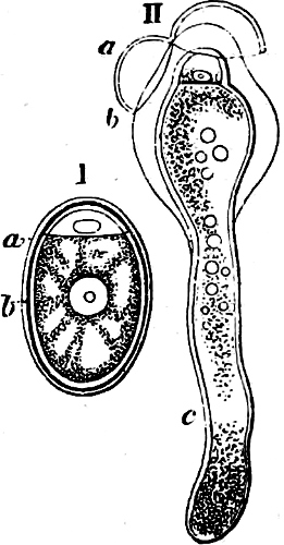
Fig. 250.—I Pollen-grains of Cupressus; at the top is seen one prothallium-cell. II Germinating; c pollen-tube; a the extine; b the intine.
2. The Macrospores. The prothallium in Salvinia and Marsilia is still rather large, green, and capable of the independent assimilation of carbon. It projects more or less from the macrospore and bears (in Marsilia only one, in Salvinia several) archegonia, which however are embedded to a greater degree in the prothallium, and are more reduced than the archegonia of the true Ferns and Horsetails (Figs. 215, 216). The prothallium is still more reduced in Isoëtes and Selaginella; partly because it is smaller and is in a higher degree enclosed in the spore, it also contains less chlorophyll, or is entirely without chlorophyll, and in consequence incapable of independent existence, whilst the number of archegonia is less; and partly because the archegonia are themselves reduced, the cells of the neck are fewer and embedded to the level of the surface of the prothallium without any, or with only a very slight projection (Figs. 235, 236).—Finally, the prothallium with its archegonia begins to develope in Selaginella while the macrospore is still within its sporangium, and before it is set free from the mother-plant. After the spores are set free and germination has commenced, the spore-wall ruptures and the prothallium is exposed.
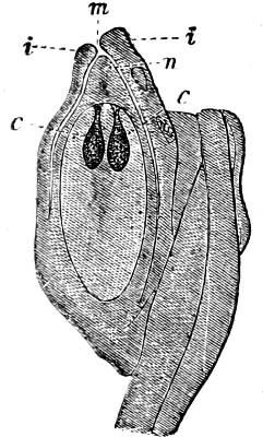
Fig. 251.—Longitudinal section of ovule of Abies canadensis. Inside the integument (i) is seen the nucellus, n; m the micropyle. In the interior of the nucellus is seen an oval mass of cells, the endosperm, and at its top two archegonia, c. The ovule is turned in such a way that the micropyle points upwards, but usually it turns downward in the Abietineæ.
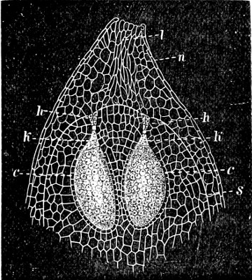
Fig. 252.—The apex of the nucellus (n) of an ovule of Abies: l long-shaped cells which guide the pollen-tube; s the wall of the macrospore (embryo-sac); h the neck-cells of the archegonium; k the ventral canal-cell; and c the central cell (oosphere). The archegonia of the Cryptogams should be compared with this (see pages 181, 208, 216).
The GYMNOSPERMS go still further. The macrospore (embryo-sac) germinates and forms internally a cellular tissue, designated in former times by the name of albumen (endosperm), which is homologous with the prothallium. It always remains enclosed in the embryo-sac, and is a parenchymatous mass containing a large supply of nourishment. In the upper part of the endosperm a number of archegonia are developed which are in the main constructed in the same manner as those in the Cryptogams, but are still more reduced, the neck consisting only of 4, 2, or 1 cell (Figs. 251, 252). The ventral canal-cell is also formed, in the majority, as a small portion cut off from the large central cell just beneath the neck; the larger remaining portion becomes the oosphere. When the pollen-tube has passed down to the oosphere (Fig. 253) and fertilisation has been effected, the oospore commences a cell-formation, the final result of which is the formation of an embryo (the asexual generation) which is provided with a thinner, lower end, termed the suspensor. The embryo is forced more or less into the endosperm in which it may rest for a longer or shorter time, and generally is developed to such an extent that it has a distinct primary-root (radicle) and stem (plumule) with one or more embryo-leaves (cotyledons).
When the oosphere has been fertilised its nucleus sinks down to its lower end, and by repeated division into two, forms four cells lying in one plane (Fig. 253, see base of the left archegonium). Three tiers of cells are now formed by transverse division of these four. It is the intermediate one of these which elongates and forms the suspensor, or four suspensors, if they separate from each other, which push the lowermost four cells deep down into the endosperm. It is from these four lower cells that the embryo (or four embryos when the suspensors separate) is developed, but never more than one embryo attains full development. As several archegonia are contained in one and the same ovule, all of which are capable of forming embryos, there is the possibility that several embryos may be developed in a seed (polyembryony), but usually only one embryo attains perfect development.
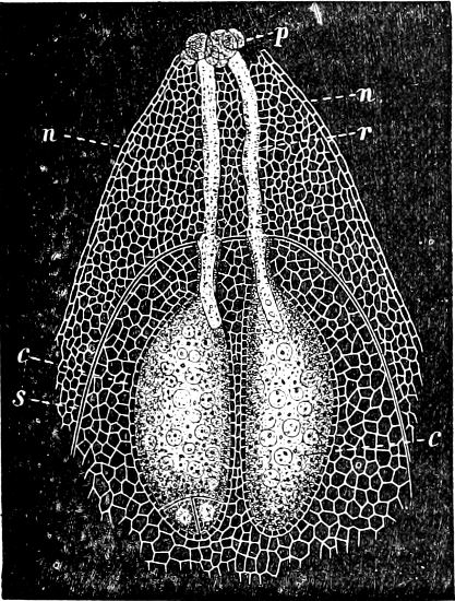
Fig. 253.—The apex of the nucellus (n) of Abies in longitudinal section: c, c the oospheres of the two archegonia; the embryo-formation has commenced at the bottom of the left archegonium; s wall of the macrospore; p pollen-grains; r pollen-tubes.
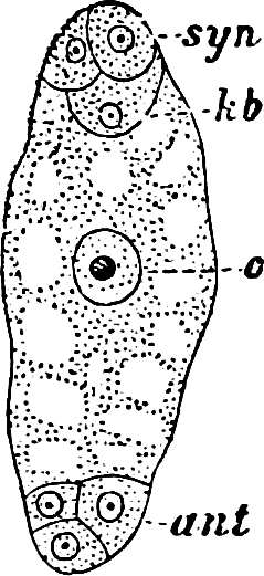
Fig. 254.—Embryo-sac of Carex præcox: syn synergidæ; kb the oosphere; c the central nucleus; ant the antipodal cells.
At the same time that the embryo is being developed, other changes are taking place in the ovule, especially in the integument which becomes the shell of the seed (testa). The endosperm grows, and the embryo-sac supplants the cells of the nucellus. The seed is now formed, and it consists in its most complete development, as in this instance, of three parts:
(1) The testa of the seed, formed by the enveloping integuments, with the remainder of the tissue of the nucellus lying outside the embryo-sac (the macrosporangium).
(2) The endosperm or prothallium.
(3) The embryo.
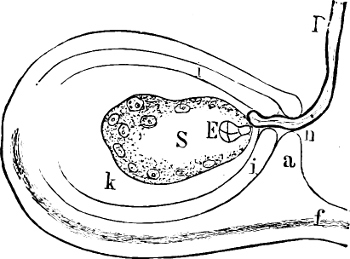
Fig. 255.—Diagrammatic longitudinal section through an anatropous ovule shortly after fertilisation; a and i are the two integuments; f the funicle; k the nucellus; S the embryo-sac, with the incipient formation of nutritive-tissue; E the embryo; P the pollen-tube passing through the micropyle (n) to the oosphere.
The reduction in the Angiosperms is carried to the extreme limit. In the embryo-sac (the macrospore) the nucleus by continued division produces a prothallium consisting of primordial cells (Fig. 254). In the upper end of the embryo-sac (which is nearest the micropyle) are three cells, two of which are termed the “co-operating cells” (synergidæ) and the third is the oosphere. Three others are placed at the opposite end of the embryo-sac and are therefore termed the “antipodal cells.” Finally, a large cell is also formed, which occupies the space between the two groups and whose cell-nucleus, the central definitive nucleus, lies in the centre of the embryo-sac. These primordial cells are the slight remnant of the prothallium. The entire structure of the archegonium, with its neck and canal-cells, has disappeared, and nothing is left but the indispensable oosphere. When the oosphere has been fertilised, and has commenced the cellular divisions which lead to the formation of the embryo (Fig. 255), the synergidæ and antipodal cells are absorbed, and a cell-formation begins by a new process which emanates from the definitive nucleus and by which a parenchymatous cell-tissue, the nutritive-tissue, arises which may perhaps be considered as homologous with the endosperm of the Gymnosperms. The difference is that the nutritive-tissue of the Angiosperms is formed in two parts with an intervening interruption; the primary nutritive-tissue is first formed, and after fertilisation is absorbed, with the exception of one cell, which continues the development and gives rise to the nutritive-tissue proper, which is formed in the first instance of primordial cells, and later on of a cellular tissue; this nutritive-tissue formed in the embryo-sac is termed “endosperm”; in a few instances[23] a tissue which is derived from the nucellus functions as nutritive-tissue, and is termed “perisperm.” In many plants the seeds, when ripe, contain a very rich nutritive-tissue, in addition to the embryo, for the purpose of its nourishment during germination. These are termed albuminous (endospermous) seeds, in distinction to the ex-albuminous, or those in which the nutritive-tissue is stored in the embryo itself, before it is completely developed, and used for its sustenance.
In addition to the changes which fertilisation produces in the ovule itself, it also gives the impetus to a series of changes in the entire shoot which bears the ovule. The perianth, stamens, and style, generally wither, because the part they play is at an end; the wall of the ovary grows and becomes the wall of the fruit (pericarp). The entire gynœcium of a flower, transformed as a consequence of fertilisation, is termed a fruit. It consists of two parts, the pericarp and the seeds, and according to the nature of the pericarp, the fruit is termed a capsule, nut, berry, or drupe.
The chief characteristic of the Phanerogams does not lie in the formation of the flower (although they may quite properly be termed “Flowering-plants”), because Equisetums and Lycopods have reproductive shoots as highly differentiated as those of certain Gymnosperms and other Phanerogams. As regards the SEXUAL GENERATION the characteristics are found:—(1) in its great reduction; (2) in the transmission of the microspore (pollen-grain) to the macrosporangium, and its germination, with the formation of a pollen-tube (antheridium), the protoplasm of which is not differentiated into spermatozoids; (3) in the fact that the macrospore (embryo-sac) never leaves its sporangium (nucellus); and further in the Angiosperms, (4) in the peculiar development of the nutritive-tissue in two parts; and (5) in the great reduction of the archegonium.
As regards the ASEXUAL GENERATION the characteristic feature is that this generation is formed whilst the sporangium is still attached to the mother-plant, and for a long time is nourished by it; and that after the sporangium has become detached from the mother-plant, it spends a longer or shorter resting period as the embryo in the seed (enveloped by the testa), and does not make its appearance until the “germination” of the seed. In addition the shoot which bears sporangia undergoes greater modification than in the case of the Flowerless-plants.
The Phanerogams are separated into two Divisions as follows:—
Division 4. Gymnospermæ. The ovules, as well as the seeds, are borne naked on the surface of open carpels, or on the apex of a stem (ovary wanting). The pollen-grains are conveyed by the wind to the ovules, and caught by drops of mucilage, secreted by the micropyle. A “stigma” is wanting. The entire female prothallium (the endosperm), which serves for the nourishment of the embryo, is formed before fertilisation. The archegonia are embedded in the upper part of the prothallium. The pollen-grains are “multicellular,” i.e. there is always in their interior a distinct prothallium, formed by 1–3 cells, and a larger cell which gives rise to the pollen-tube.
Division 5. Angiospermæ. The carpels surround the ovules and form an entirely closed chamber (ovary), in which the ovules mature and ripen into seeds. The surface of a portion of the apex of the carpel is transformed into the “stigma,” which, by a sticky fluid and also by hair-structures, is capable of retaining the pollen-grains conveyed to it by the wind, or more frequently by insects. The pollen-tube grows from the stigma, through the “conducting cellular tissue” (style), to the ovules. The pollen-grains contain two cells, a vegetative and a free generative cell. The latter passes into the pollen-tube and there divides into two, one of which is the sperm-nucleus. The female prothallium, which is intended to serve as nutritive-tissue, is formed after fertilisation. Archegonia are wanting.