Sub-Division II.—ALGÆ.
Michael Cresse Potter translation
Mode of Life. The Algæ (except most of the Bacteria) are themselves able to form their organic material by the splitting up of the carbonic acid contained in the water, or air in some cases, and for this purpose need light. The majority live in water, fresh or salt, but many are present on damp soil, stones, bark of trees, etc.
With the exception of the Bacteria, no saprophytes have actually been determined to belong to this group, and only very few true parasites (for instance, Phyllosiphon arisari, Mycoidea, etc.), but a good many are found epiphytic or endophytic on other Algæ, or water plants, and on animals (for instance, certain Schizophyceæ and Protococcoideæ; Trichophilus welckeri in the hairs of Bradypus, the Sloth), and several species in symbiotic relation to various Fungi (species of Lichen), to Sponges (e.g. Trentepohlia spongiophila, Struvea delicatula), and to sundry Infusoria and other lower animals as Radiolarias, Hydra, etc. (the so-called Zoochlorella and Zooxantella, which are perhaps partly stages in development of various Green and Brown Algæ).
Vegetative Organs. The cells in all the Algæ (excepting certain reproductive cells) are surrounded by a membrane which (with the exception of the Bacteria) consists of pure or altered cellulose, sometimes forming a gelatinous covering, at other times a harder one, with deposits of chalk or silica formed in it. The cell-nucleus, which in the Schizophyta is less differentiated, may be one or more (e.g. Hydrodictyon, Siphoneæ) in each cell. Excepting in the majority of the Bacteria, colour materials (of which chlorophyll, or modifications of it, always seems to be found) occur, which either permeate the whole cytoplasm surrounding the cell-nucleus, as in most of the coloured Schizophyta, or are contained in certain specially formed small portions of protoplasm (chromatophores).
The individual at a certain stage of development consists nearly always of only one cell; by its division multicellular individuals may arise, or, if the daughter-cells separate immediately after the division, as in many of the simplest forms, the individual will, during the whole course of its existence, consist of only a single cell (unicellular Algæ). In multicellular individuals the cells may be more or less firmly connected, and all the cells of the individual may be exactly alike, or a division of labour may take place, so that certain cells undertake certain functions, and are constructed accordingly; this may also occur in parts of the cell in the large unicellular and multinuclear Algæ (Siphoneæ, p. 62).
The cells in most of the Algæ belong to the parenchymatous form; these, however, in the course of their growth, may very often become somewhat oblong; in many Algæ (particularly Fucoideæ and Florideæ) occur, moreover, hyphæ-like threads, which are very long, often branched, and are either formed of a single cell, or, more frequently, of a row of cells, having a well-pronounced apical growth. The parenchymatous as well as the hyphæ-like cells may, in the higher Algæ (especially in certain Fucoideæ and Florideæ), be further differentiated, so that they form well-defined anatomico-physiological systems of tissue, i.e. assimilating, conducting, storing, and mechanical.
With regard to the external form, the thallus may present no differentiation, as in many unicellular Algæ, or in multicellular Algæ of the lower order, which are then either equally developed in all directions (e.g. Pleurococcus, Fig. 47), or form flat cell-plates (Merismopedium) or threads (Oscillaria, Fig. 21). The first step in the way of differentiation appears as a difference between apex and base (Rivularia, Porphyra); but the division of labour may proceed so that differences may arise between vegetative and reproductive cells (Œdogonium, Fig. 54); hairs and organs of attachment (rhizoids and haptera), which biologically serve as roots, are developed, and even leaves in certain forms of high order, belonging to different classes (e.g. Caulerpa, Fig. 59; Characeæ, Fig. 61; Sargassum, Fig. 72; and many Florideæ).
The non-sexual reproduction takes place vegetatively, in many instances, simply by division into two, and more or less complete separation of the divisional products (Diatomaceæ, Desmidiaceæ (Fig. 36), many Fission-plants, etc.), or by detached portions of the thallus (e.g. Caulerpa, Ulva lactuca, etc.; among many Schizophyceæ, small filaments known as hormogonia are set free), or asexually by special reproductive cells (spores) set free from the thallus; these may be either stationary or motile. The stationary reproductive cells (spores) may either be devoid of cell-wall (tetraspores of the Florideæ), or may possess a cell-wall; in the latter case they may be formed directly from the vegetative cells, generally by the thickening of the walls (akinetes), or only after a process of re-juvenescence (aplanospores). Aplanospores, as well as akinetes, may either germinate immediately or may become resting-cells, which germinate only after a period of rest.
The motile asexual reproductive cells are spherical, egg- or pear-shaped, naked, swarmspores (zoospores), which have arisen in other cells (zoosporangia), and propel themselves through the water by means of cilia; or they are Phyto-Amœbæ, which have no cilia and creep on a substratum by means of pseudopodia. The cilia, which are formed from the protoplasm (in the Bacteria, however, from the membrane), are mostly situated at the pointed and colourless end, which is directed forwards when in motion, and are 1, 2 (Fig. 5 B), 4 or more. Both the cilia in the Brown Algæ are attached to one side (Fig. 65); they are occasionally situated in a circle round the front end (Œdogonium, Fig. 6 a, and Derbesia), or are very numerous and situated in pairs distributed over a large part or nearly the whole of the zoospore (Vaucheria). Besides being provided with one or more nuclei (Vaucheria), they may also have a red “eye spot” and vacuoles, which are sometimes pulsating, i.e. they appear and reappear at certain intervals. The swarmspores move about in the water in irregular paths, and apparently quite voluntarily, revolving round their longer axes; but they come to the surface of the water in great numbers either because of their dependence on light, or driven by warm currents in the water, or attracted by some passing mass of food material. The swarmspores germinate, each forming a new plant, as their movement ceases they surround themselves with a cell-wall, grow, and then divide; in Fig. 6 b, two may be seen in the condition of germination, and about to attach themselves by means of the front end, which has been developed into haptera (see also Fig. 5 B, lowest figure).
The sexual reproduction here, probably in all cases, consists in the coalescence of two masses of protoplasm, that is, in the fusion of their nuclei.
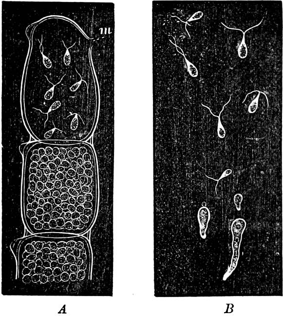
Fig. 5.—Cladophora glomerata. A The lower cells are full of swarmspores, whilst from the upper one the greater part have escaped through the aperture m. B Free and germinating swarmspores.
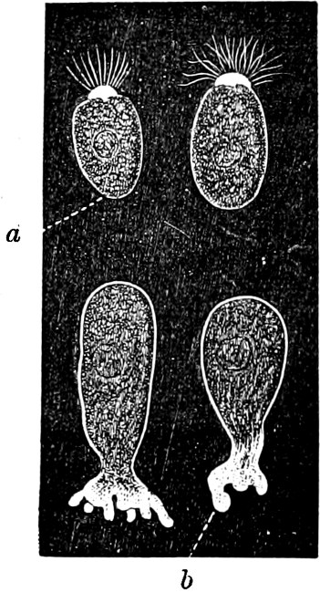
Fig. 6.—Œdogonium: a (free), b germinating swarmspores.
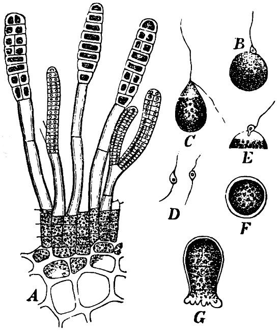
Fig. 7.—Zanardinia collaris. A Male gametangia (the small-celled) and female gametangia (large-celled). C Female gamete. D Male gamete. B E Fertilisation. F Zygote. G Germinating zygote.
The simplest and lowest form is termed conjugation, or isogamous fertilisation, and is characterized by the fact that the two coalescing cells (termed gametes) are equal, or almost equal, in shape and size (the female gamete in the Cutleriaceæ, e.g. Zanardinia collaris, Fig. 7, is considerably larger than the male gamete). The cell in which the gametes are developed is called a gametaugium, and the reproductive cell formed by their union—which generally has a thick wall and only germinates after a short period of rest—is termed a zygote or zygospore. The conjugation takes place in two ways:—
(a) In the one way the gametes are motile cells (planogametes, zoogametes, Fig. 8), which unite in pairs during their swarming hither and thither in the water; during this process they lie side by side (Fig. 8 d), generally at first touching at the clear anterior end, and after a time they coalesce and become a motionless zygote, which surrounds itself with a cell-wall (Fig. 8 e). This form of conjugation is found in Ulothrix (Fig. 8 d), Acetabularia, and other Algæ (Figs. 45, 56, 66).
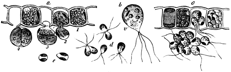
Fig. 8.—Ulothrix zonata: a portion of a thread with zoospores, of which two are formed in each cell (zoosporangium), the dark spots upon them are the “red eye-spots”; 1, 2, 3, 4 depict successive stages in the development of the zoospores; b a single zoospore, at v the pulsating vacuole; c portion of a thread with gametes, of which sixteen are formed in each gametangium; d gametes free and in conjugation; e conjugation has been effected, and the formed zygotes are in the resting condition.
(b) Among other Algæ (e.g. Diatomaceæ and Conjugatæ), the conjugating cells continue to be surrounded by the cell-wall of the mother-cell (aplanogametes in an aplanogametangium); the aplanogametangia generally grow out into short branches, which lie close together and touch one another, the wall at the point of contact is then dissolved (Fig. 39). Through the aperture thus formed, the aplanogametes unite, as in the first instance, and form a rounded zygote, which immediately surrounds itself with a cell-wall. Various modifications occur; compare Figs. 37, 39, 41, 43.
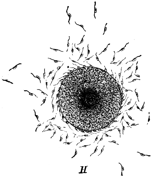
Fig. 9.—Fertilisation in the Bladder-wrack (Fucus vesiculosus).
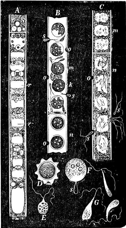
Fig. 10.—Sphæroplea annulina.
The highest form of the sexual reproduction is the Egg- or Oogamous fertilisation. The two coalescing cells are in the main unlike each other in form as well as size. The one which is considered as the male, and is known as the spermatozoid (antherozoid), developes as a rule in large numbers in each mother-cell (antheridium); they are often self-motile (except in the Florideæ, where they are named spermatia), and are many times smaller than the other kind, the female, which is known as the egg-cell, (oosphere). The egg-cell is always a motionless, spherical, primordial cell which can either float about freely in the water, as in the Fucaceæ (Fig. 9), or is surrounded by a cell-wall (oogonium); generally only one oosphere is to be found in each oogonium, but several occur in Sphæroplea (Fig. 10). The result of the spermatozoid coalescing with the egg-cell is, as in this case, the formation of a oospore, which generally undergoes a period of rest before germination (the Florideæ are an exception, a fruit-body, cystocarp, being produced as the result of coalescence).
An example of fertilisation is afforded by the Alga, Sphæroplea annulina (Fig. 10). The filamentous thallus is formed of cylindrical cells with many vacuoles (r in A); some cells develope egg-cells (B), others spermatozoids (C), the latter in a particularly large number. The egg-cells are spherical, the spermatozoids of a club- or elongated pear-shape with two cilia at the front end (G; E is however a swarmspore). The spermatozoids escape from their cells through apertures in the wall (o in C) and enter through similar apertures (o in B) to the egg-cells. The colourless front end of the spermatozoid is united at first with the “receptive spot” of the egg-cell (see F), and afterwards completely coalesces with it. The result is the formation of a oospore with wart-like excrescences (D).
The female (parthenogenesis) or male (androgenesis) sexual cell may, sometimes without any preceding fertilisation, form a new individual (e.g. Ulothrix zonata, Cylindrocapsa, etc.).
Systematic division of the Algæ. The Algæ are divided into the following ten classes:
1. Syngeneticæ; 2. Dinoflagellata, or Peridinea; 3. Diatomaceæ; 4. Schizophyta, Fission-algæ; 5. Conjugatæ; 6. Chlorophyceæ, Green-algæ; 7. Characeæ, Stone-worts; 8. Phæophyceæ; 9. Dictyotales; 10. Rhodophyceæ.
Among the lowest forms of the Algæ, the Syngeneticæ, the Dinoflagellata, and the unicellular Volvocaceæ (Chlamydomoneæ), distinct transitional forms are found approaching the animal kingdom, which can be grouped as animals or plants according to their method of taking food or other characteristics. Only an artificial boundary can therefore be drawn between the animal and vegetable kingdoms. In the following pages only those forms which possess chromatophores, and have no mouth, will be considered as Algæ.
Class 1. Syngeneticæ.
The individuals are uni- or multicellular, free-swimming or motionless. The cells (which in the multicellular forms are loosely connected together, often only by mucilaginous envelopes) are naked or surrounded by a mucilaginous cell-wall, in which silica is never embedded. They contain one cell-nucleus, one or more pulsating vacuoles, and one to two band- or plate-like chromatophores with a brown or yellow colour, and sometimes a pyrenoid.
Reproduction takes place by vegetative division, or asexually by zoospores, akinetes (or aplanospores?). Sexual reproduction is unknown. They are all fresh water forms.
To this class may perhaps be assigned the recently arranged and very little known orders of Calcocytaceæ, Murracytaceæ, Xanthellaceæ, and Dictyochaceæ, which partly occur in the free condition in the sea, in the so-called “Plankton,” and partly symbiotic in various lower marine animals.
The Syngeneticæ are closely related to certain forms in the animal kingdom, as the Flagellatæ.
Order 1. Chrysomonadinaceæ. Individuals, uni- or multicellular, swimming in free condition, naked or surrounded by a mucilaginous covering. The cells are generally oval or elongated, with 2 (rarely only 1) cilia, almost of the same length, and generally with a red “eye-spot” at their base, and with 2 (rarely 1 only) band-shaped chromatophores. Reproduction by the longitudinal division of the individual cells either during the swarming, or during a resting stage; in the multicellular forms also by the liberation of one or more cells, which in the latter case are connected together.
A. Unicellular: Chromulina, Cryptoglena, Microglena, Nephroselmis.
B. Multicellular: Uroglena, Syncrypta (Fig. 11), Synura.
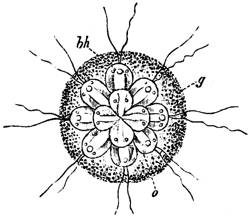
Fig. 11.—Syncrypta volvox: the multicellular individual is surrounded by a mucilaginous granular envelope.
Among the unicellular Chrysomonadinaceæ are probably classed some forms which are only stages in the development of the multicellular, or of other Syngeneticæ.
Order 2. Chrysopyxaceæ are unicellular, and differ mainly from the preceding in being attached either on a slime-thread (Stylochrysalis), or enclosed in an envelope (Chrysopyxis, Fig. 12). They have two cilia, and multiply by longitudinal (Chrysopyxis) or transverse division, and the swarming of one of the daughter-individuals (zoospore). Division may also take place in a motionless stage (palmella-stage).
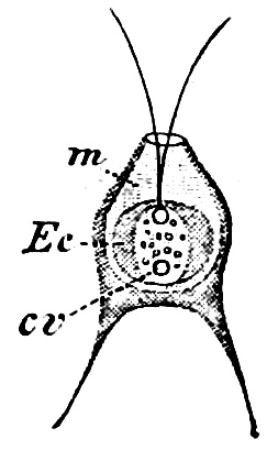
Fig. 12.—Chrsopyxis bipes: m envelope, Ec chromatophore, cv contractile vacuole.
Order 3. Dinobryinaceæ. The individuals are originally attached, uni- or multicellular; each individual cell is distinctly contractile, and fixed at the bottom of a cup-shaped, open envelope. Cilia 2, but of unequal length. Asexual reproduction by zoospores, which are formed by straight or oblique longitudinal division of the mother-cell, during a palmella-stage which is produced in the winter aplanospores. Epipyxis, Dinobryon.
Order 4. Hydruraceæ. The individuals are attached, without cilia, multicellular, branched, and with apical growth. The cells are spherical, but in the final stage almost spindle-shaped, and embedded in large masses of mucilage. Asexual reproduction by zoospores which are tetrahedric, with 1 cilia, and by resting akinetes. Hydrurus is most common in mountain brooks.
Class 2. Dinoflagellata.
The individuals are of a very variable form, but always unicellular, and floating about in free condition. The cell is dorsiventral, bilateral, asymmetric and generally surrounded by a colourless membrane, which has no silica embedded in it, but is formed of a substance allied to cellulose. The membrane, which externally is provided with pores and raised borders, easily breaks up into irregularly-shaped pieces. In the forms which have longitudinal and cross furrows, two cilia are fixed where these cross each other, and project through a cleft in the membrane; one of these cilia projects freely and is directed longitudinally to the front or to the rear, the other one stretches crosswise and lies close to the cell, often in a furrow (cross furrow). The chromatophores are coloured brown or green and may either be two parallel (Exuviella), or several radially placed, discs, which sometimes may coalesce and become a star-shaped chromatophore. The coloring material (pyrrophyl) consists, in addition to a modification of chlorophyl, also of phycopyrrin and peridinin; this colour is sometimes more or less masked by the products of assimilation which consist of yellow, red or colourless oil (?) and starch. Cell-nucleus one: in Polydinida several nuclei are found; contractile vacuoles many, which partly open in the cilia-cleft (Fig. 13 gs). In some an eye-spot, coloured red by hæmatochrome, is found. Pyrenoids occur perhaps in Exuviella and Amphidinium.
The reproduction takes place as far as is known at present, only by division. This, in many salt water forms, may take place in the swarming condition, and, in that case, is always parallel to the longitudinal axis. The daughter-individuals, each of which retains half of the original shell, sometimes do not separate at once from each other, and thus chains (e.g. in Ceratium) of several connected individuals may be formed. In others, the division occurs after the cilia have been thrown off and the cell-contents rounded. The daughter-cells then adopt entirely new cell-walls. A palmella-stage (motionless division-stage) sometimes appears to take place, and also aplanospores (?) with one or two horn-like elongations (e.g. in Peridinium cinctum and P. tabulatum); at germination one, or after division, two or more, new individuals may be formed.
Sexual reproduction has not been observed with certainty.
The Dinoflagellata move forward or backward, turning round their longitudinal axes; in their motion they are influenced by the action of light. The motion possibly may be produced only by the transverse cilium, which vibrates rapidly; whilst the longitudinal cilium moves slowly, and is supposed to serve mainly as a steering apparatus. They live principally in salt water, but also in fresh.
Besides the coloured forms, which are able to make their own organic compounds by the splitting up of the carbonic acid contained in the water, there are a few colourless forms (e.g. Gymnodinium spirale), or such as do not possess chromatophores (Polykrikos); these appear to live saprophytically, and may be able to absorb solid bodies with which they come in contact.
Dinoflagellata occur in the “Plankton” of the open sea, where they form together with Diatomaceæ the basis for the animal life. It is known with certainty that some salt water forms (like the Noctiluca, which belongs to the animal kingdom and to which they are perhaps related) produce light, known as phosphorescence.
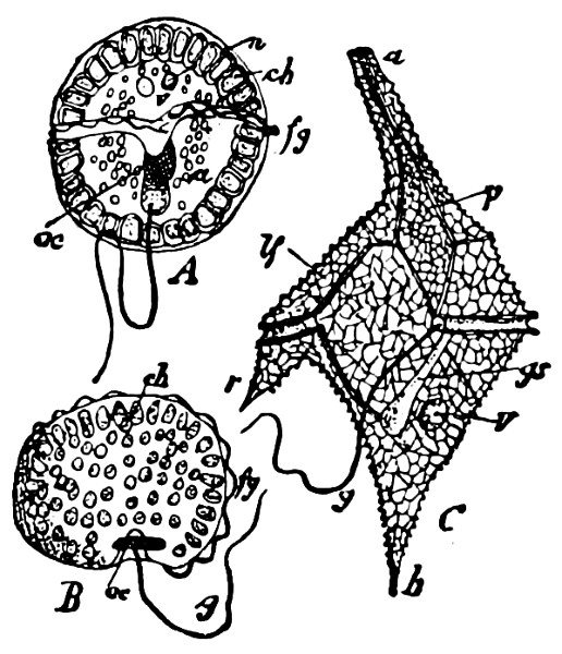
Fig. 13.—A and B Glenodinium cinctum. A seen from the ventral side, B from behind; fg transverse cilium; g longitudinal cilium; ch chromatophores; a starch; n cell-nucleus; v vacuole; oc eye-spot; C Ceratium tetraceros from the ventral side; r the right, b the posterior horn; lf longitudinal furrow; gs cilium-cleft; v vacuole; g longitudinal cilium. (A and B mag. 450 times, C 337 times.)
Dinoflagellata (Peridinea, Cilioflagellata) are allied through their lowest form (Exuviella) to the Syngeneticæ and especially to the order Chrysomonadinaceæ. They may be divided into three orders.
Order 1. Adinida. Without transverse or longitudinal furrows, but enclosed in two shells, and with two parallel chromatophores in each cell. Exuviella, Prorocentrum.
Order 2. Dinifera. With tranverse and generally longitudinal furrow. Many radially-placed, disc-formed chromatophores. The most common genera are—Ceratium (Fig. 13), Peridinium, Glenodinium (Fig. 13), Gymnodinium, Dinophysis.
Order 3. Polydinida. With several transverse furrows, no chromatophores, and several cell-nuclei. Only one genus—Polykrikos.
The order Polydinida deviates in a high degree from the other Dinoflagellata, not only by its many tranverse furrows, each with its own transverse cilium, and by the absence of chromatophores, but also in having several cell-nuclei and a kind of stinging capsule, which otherwise does not occur within the whole class. It may therefore be questionable whether this order should really be placed in the vegetable kingdom.
Class 3. Diatomeæ.
The individuals—each known as a frustule—assume very various forms and may be unicellular or multicellular, but present no differentiation; many similar cells may be connected in chains, embedded in mucilaginous masses, or attached to mucilaginous stalks. The cells are bilateral or centric, often asymmetrical, slightly dorsiventral and have no cilia; those living in the free condition have the power of sliding upon a firm substratum. The cell contains 1 cell-nucleus and 1–2 plate-shaped or several disc-shaped chromatophores. The colouring material “Melinophyl” contains, in addition to a modification of chlorophyl, a brown colouring matter, diatomin. 1 or 2 pyrenoids sometimes occur. Starch is wanting and the first product of assimilation appears to be a kind of oil (?).
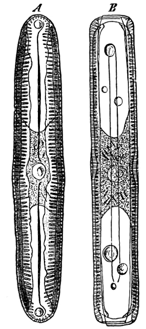
Fig. 14.—Pinnularia: B, from the edge, shows the valves fitting together; A, a valve.
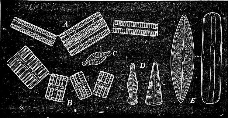
Fig. 15.—Various Diatomaceæ. A Diatoma vulgare. B Tabellaria flocculosa. C Navicula tumida (lateral views). D Gomphonema constrictum (lateral views). E Navicula west[=i][=i] (lateral views).
The cell-walls are impregnated with silica to such a degree that they are imperishable and are therefore able to contribute in a great measure to the formation of the earth’s crust. The structure of their cell-wall is most peculiar and differs from all other plants (except certain Desmidiaceæ); it does not consist of a single piece but is made up of two—the “shells”—(compare Exuviella and Prorocentrum among the Dinoflagellata) which are fitted into each other, one being a little larger than the other and embracing its edge, like a box with its lid (Fig. 14 B). The two parts which correspond to the bottom and lid of the box are known as valves. Along the central line of the valves a longitudinal rib may often be found, interrupted at its centre by a small cleft (perhaps homologous with the cilia-cleft of the Dinoflagellata), through which the protoplasm is enabled to communicate with the exterior (Fig. 14 A). It is principally by reason of the valves, which bear numerous fine, transverse ribs, striæ or warts, etc. (Figs. 14, 15, 17), that the Diatomeæ have become so well known and employed as test objects in microscopical science. When the division takes place, the two shells are separated a little from each other, and after the cell-contents have divided into two masses, two new shells are formed, one fitting into the larger valve, the other one into the smaller valve of the original frustule. The latter cell (frustule) is thus, upon the whole, smaller than the mother-cell, and as the cells do not increase in size, some frustules are smaller than the ones from which they are derived, and thus, by repeated divisions, it follows that smaller and smaller frustules are produced. This continued diminution in size is, however, compensated for by the formation, when the cells have been reduced to a certain minimum, of auxospores, 2–3 times larger. These may either be formed asexually by the protoplasm of a cell increasing, rounding off and surrounding itself with a new wall (e.g. Melosira) or after conjugation, which may take place with various modifications: 1. Two individuals unite after the secretion of a quantity of mucilage, and the valves then commence to separate from each other, on the side which the two individuals turn towards each other. The protoplasmic bodies now release themselves from their cell-wall, and each rounds off to form an ellipsoidal mass; these two protoplasmic masses (gametes) coalesce to form a zygote, the cell-nuclei and chromatophores also fusing together. The zygote increases in size, and surrounds itself with a firm, smooth, siliceous wall—the perizonium. The auxospores, whichever way they arise, are not resting stages. The germination of the zygote commences by the protoplasm withdrawing itself slightly from the cell-wall and constructing first the larger valve, and later on the smaller one; finally the membrane of the zygote bursts (e.g. Himantidium). 2. The conjugation occurs in a similar manner, but the protoplasm of the cells divides transversely before conjugation into two daughter-cells. Those lying opposite one another conjugate (Fig. 16) and form two zygotes. The formation of the perizonium, and germination take place as in the preceding instance (e.g. Epithemia). 3. Two cells place themselves parallel to each other, and each of the two cell-contents, without coalescing, becomes an auxospore. The formation of the wall takes place as in the preceding case. This is found in the Naviculeæ, Cymbelleæ, the Gomphonemeæ (e.g. Frustulia, Cocconema).
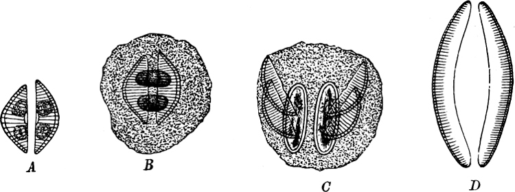
Fig. 16.—Conjugation of Cymbella variabilis. A, The protoplasm in the two cells has divided into two masses; B these masses coalesce in pairs; the cells (B C) enclosed in a mucilaginous matrix. C D Auxospores and their formation.
The Diatomaceæ may be found in salt as well as in fresh water (often in such masses that the colour of the water or mud becomes yellow or brown; in the same manner the genera Chætoceros, Rhizosolenia, Coscinodiscus, and several others, form large slime-masses, “Plankton” on the surface of the sea), on damp soil and in dust blown by the wind. They occur as fossils in the recent formations, often in large deposits (siliceous earth, mountain meal), as in the cement lime in Jutland, the alluvial deposits beneath Berlin, in clay strata beneath peat bogs, in guano, etc. These accumulations of fossilized diatoms are used in the manufacture of dynamite and in various manufactures.
The Diatomaceæ appear nearest to, and must be placed as a group co-ordinate with the Dinoflagellata, as they doubtless may be supposed to derive their origin from forms resembling Exuviella, and to have lost the cilia. The resemblances to the Desmidiaceæ which are striking in many respects, can only be conceived as analogies, and cannot be founded upon homologies, and it is therefore impossible to regard them as proof of genetic relationship. The family contains only one order.
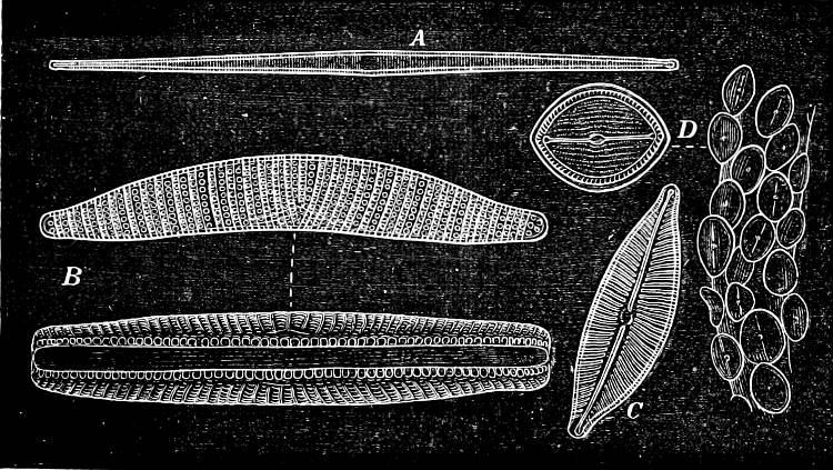
Fig. 17.—Various Diatomeæ. A Synedra radians. B Epithemia turgida (from the two different sides). C Cymbella cuspidata. D Cocconeis pediculus (on the right several situated on a portion of a plant, on the left a single one more highly magnified).
Order 1. Diatomaceæ. This order may be divided into two sub-orders, viz.—
Sub-Order 1. Placochromaticæ. The chromatophores are discoid, large, 1 or 2 in each cell; the structure of the valves is bilateral and always without reticulate markings. The following groups belong to this sub-order: Gomphonemeæ, Cymbelleæ, Amphoreæ, Achnantheæ, Cocconeideæ, Naviculeæ, Amphipleureæ, Plagiotropideæ, Amphitropideæ, Nitzchieæ, Surirayeæ, and Eunotieæ.
Sub-Order 2. Coccochromaticæ. The chromatophores are granular, small and many in each cell. The structure of the cells is zygomorphic or centric, often with reticulate markings. The following groups belong to this sub-order: Fragilarieæ, Meridieæ, Tabellarieæ, Licmophoreæ, Biddulphieæ, Anguliferæ, Eupodisceæ, Coscinodisceæ, and Melosireæ.
Class 4. Schizophyta, Fission-Algæ.
The individuals are 1—many celled; the thallus consists in many of a single cell, in others of chains of cells, the cells dividing in only one definite direction (Figs. 18, 21). In certain Fission-Algæ the cell-chain branches (Fig. 30) and a difference between the anterior and the posterior ends of the chain is marked; in some, the cells may be united into the form of flat plates by the cell-division taking place in two directions; and in others into somewhat cubical masses, or rounded lumps of a less decided form, by the divisions taking place in three directions; or less defined masses may be formed by the divisions taking place in all possible directions.
The cell-walls rarely contain cellulose, they often swell considerably (Figs. 20, 22), and show distinct stratifications, or they are almost completely changed into a mucilaginous mass in which the protoplasts are embedded, e.g. in Nostoc (Fig. 22), and in the “Zooglœa” stage of the Bacteria (Fig. 27). Sexual reproduction is wanting. Vegetative reproduction by division and the separation of the divisional products by the splitting of the cell-wall or its becoming mucilaginous; among the Nostocaceæ, Lyngbyaceæ, Scytonemaceæ, etc., “Hormogonia” are found; in Chamæsiphon and others single reproductive akinetes are formed. Many Fission-Algæ conclude the growing period by the formation of resting akinetes or aplanospores.
The Schizophyta may be divided into 2 families:
1. Schizophyceæ.
2. Bacteria.
Family 1. Schizophyceæ,[5] Blue-Green Algæ.
All the Blue-green Algæ are able to assimilate carbon by means of a colouring material containing chlorophyll (cyanophyll); but the chlorophyll in this substance is masked by a blue (phycocyan), or red (phycoerythrin, e.g. in Trichodesmium erythræum in the Red Sea) colouring matter which may be extracted from them in cold water after death. The colouring matter, in most of them, permeates the whole of the protoplasm (excepting the cell-nucleus), but in a few (e.g. Glaucocystis, Phragmonema), slightly developed chromatophores are to be found. Where the cells are united into filaments (cell-chains) a differentiation into apex and base (Rivulariaceæ) may take place, and also between ordinary vegetative cells and heterocysts; these latter cannot divide, and are distinguished from the ordinary vegetative cells (Fig. 22 h) by their larger size, yellow colour, and poverty of contents. Branching sometimes occurs and is either true or spurious.

Fig. 18.—Microcoleus lyngbyanus: a portion of a filament, the thick sheath encloses only one cell-chain; in one place a cell is drawn out by the movement of the cell-chain; b the cell-chain has divided into two parts (“hormongonia”) which commence to separate from each other.
The cell-chain in the spurious branching divides into two parts, of which either one or both grow beyond the place of division (Fig. 18) and often out to both sides (e.g. Scytonema), the divisions however, always take place transversely to the longitudinal direction of the cell-chain. In the true branching a cell elongates in the direction transverse to the cell-chain, and the division then takes place nearly at right angles to the former direction (Sirosiphoniaceæ).
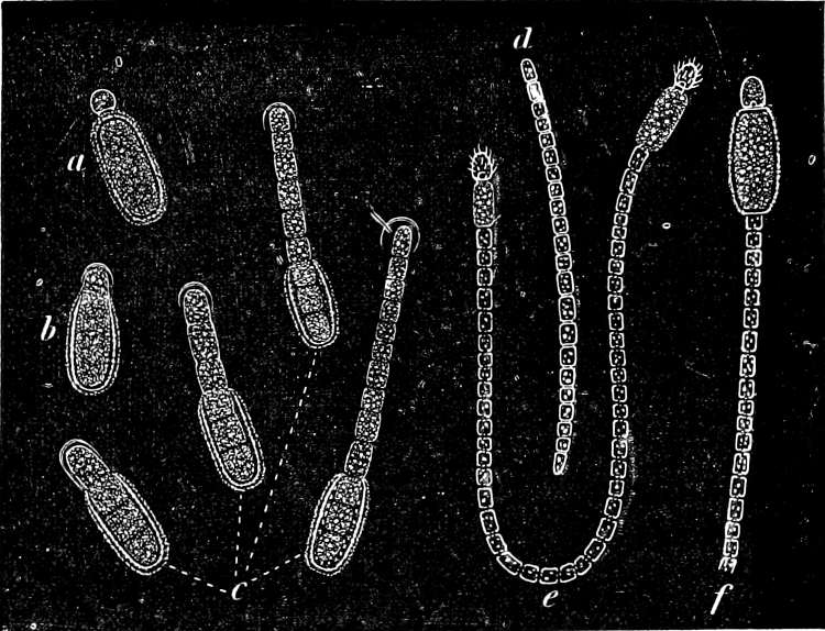
Fig. 19.—Cylindrospermum majus: a resting akinete with heterocyst; b-d germinating stages of a resting akinete; e filament with two heterocysts and the formation of new akinetes; f part of a filament with a heterocyst, and mature resting akinete.
Cilia are wanting, but the filaments are sometimes self-motile (e.g. hormogonia in Nostoc) and many partly turn round their axes, partly slide forward or backward (Oscillaria).
Reproduction takes place by spores and hormogonia in addition to simple cell-division. Hormogonia are peculiar fragments of a cell-chain capable of motion, and often exhibit a vigorous motion in the sheath, until at last they escape and grow into a new individual (Fig. 18). The spores are reproductive akinetes (Chamæsiphon, etc.) or resting akinetes; these latter arise by the vegetative cells enlarging and constructing a thick cell-wall (Fig. 19 e f). On germination, this cell-wall bursts and the new cell-chain elongates in the same longitudinal direction as before (Fig. 19 b c). Many (e.g. Oscillaria) may however winter in their ordinary vegetative stage. Aplanospores are wanting.
The Fission-Algæ are very prevalent in fresh water and on damp soil, less so in salt water; they also often occur in water which abounds in decaying matter. Some are found in warm springs with a temperature as high as 50° C.
The Family may be divided into 2 sub-families:
1. Homocysteæ (heterocysts are wanting): Chroococcaceæ, Lyngbyaceæ and Chamœsiphonaceæ.
2. Heterocysteæ (heterocysts present): Nostocaceæ, Rivulariaceæ, Scytonemaceæ and Sirosiphoniaceæ.
Order 1. Chroococcaceæ. The individuals are 1—many-celled, but all the cells are uniform, united to form plates or irregular masses, often surrounded by a mucilaginous cell-wall, but never forming cell-chains. Multiplication by division and sometimes by resting akinetes, but reproductive akinetes are wanting. Chroococcus, Aphanocapsa, Glœocapsa (Fig. 20), Cœlosphærium, Merismopedium, Glaucocystis, Oncobyrsa, Polycystis, Gomphosphæria.
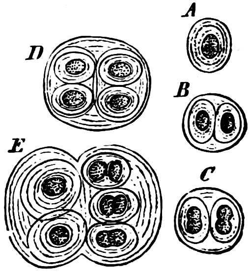
Fig. 20.—Glœocapsa atrata: A, B, C, D, E various stages of development.
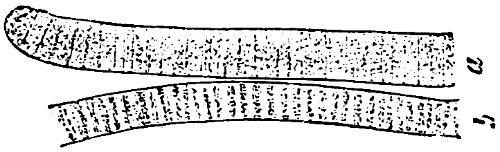
Fig. 21.—Oscillaria; a terminal, b central portion of a filament.
Order 2. Lyngbyaceæ (Oscillariaceæ). The cells are discoid (Fig. 21), united to straight or spirally twisted, free filaments, which are unbranched, or with spurious branching. The ends of the cell-chains are similar. Heterocysts absent. Reproduction by synakinetes, resting akinetes are wanting. Oscillaria (Fig. 21), Spirulina, Lyngbya, Microcoleus, Symploca, Plectonema.
Order 3. Chamæsiphonaceæ. The individuals are 1—many-celled, attached, unbranched filaments with differentiation into apex and base, without heterocysts. Multiplication by reproductive akinetes; resting akinetes are wanting. Dermocarpa, Clastidium, Chamæsiphon, Godlewskia, Phragmonema.
Order 4. Nostocaceæ. The individuals are formed of multicellular, unbranched filaments, without differentiation into apex and base; heterocysts present. Reproduction by synakinetes and resting akinetes.
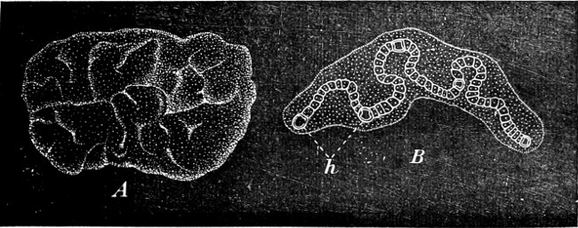
Fig. 22.—Nostoc verrucosum. A The plant in its natural size; an irregularly folded jelly-like mass. B One of the cell-chains enlarged, with its heterocysts (h), embedded in its mucilaginous sheath.
Some genera are not mucilaginous, e.g. Cylindrospermum (Fig. 19). The cell-chains in others, e.g. Nostoc, wind in between one another and are embedded in large structureless jelly-like masses, which may attain the size of a plum or even larger (Fig. 22); sometimes they are found floating in the water, sometimes attached to other bodies. Other genera as follows: Aphanizomenon and Anabæna (in lakes and smaller pieces of water); Nodularia is partly pelagic. Some occur in the intercellular spaces of higher plants, thus Nostoc-forms are found in Anthoceros, Blasia, Sphagnum, Lemna, and in the roots of Cycas and Gunnera; Anabæna in Azolla.
Order 5. Rivulariaceæ. The individuals are multicellular filaments, with differentiation into apex and base; spurious branching, and a heterocyst at the base of each filament, reproduction by synakinetes and resting akinetes, rarely by simple reproductive akinetes. Rivularia, Glœotrichia, Isactis, Calothrix.
Order 6. Scytonemaceæ. The individuals are formed of multicellular filaments with no longitudinal division; differentiation into apex and base very slight or altogether absent; branching spurious; heterocysts present. Reproduction by synakinetes, rarely by resting akinetes and ordinary reproductive akinetes. Tolypothrix, Scytonema, Hassalia, Microchæte.
Order 7. Sirosiphoniaceæ. The individuals are formed of multicellular threads with longitudinal divisions; true branching and heterocysts, and often distinct differentiation into apex and base. Reproduction by synakinetes, rarely by resting akinetes and ordinary reproductive akinetes. Hapalosiphon, Stigonema, Capsosira, Nostocopsis, Mastigocoleus.
Family 2. Bacteria.[6]
The Bacteria (also known as Schizomycetes, and Fission-Fungi) are the smallest known organisms, and form a parallel group to the Blue-green Algæ, but separated from these Algæ by the absence of their colouring material; chlorophyll is only found in a few Bacteria.
The various forms under which the vegetative condition of the Bacteria appear, are termed as follows:
1. Globular forms, cocci (Figs. 27, 30 c): spherical or ellipsoidal, single cells, which, however, are usually loosely massed together and generally termed “Micrococci.”
2. Rod-like forms: more or less elongated bodies; the shorter forms have been styled “Bacterium” (in the narrower sense of the word), and the term “Bacillus” has been applied to longer forms which are straight and cylindrical (Figs. 28, 29, 30 E).
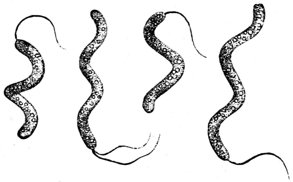
Fig. 23.—Spirillum sanguineum. Four specimens. One has two cilia at the same end, the sulphur grains are seen internally.
3. Thread-like forms: unbranched, long, round filaments, resembling those of Oscillaria, are possessed by Leptothrix (very thin, non-granular filaments; Fig. 30 A, the small filaments) and Beggiatoa (thicker filaments, with strong, refractile grains or drops of sulphur (Fig. 31); often self-motile). Branched filaments, with false branching like many Scytonemaceæ, are found in Cladothrix (Fig. 30 B, G).
4. Spiral forms: Rod-like or filamentous bodies, which more or less strongly resemble a corkscrew with a spiral rising to the left. In general these are termed Spirilla (Fig. 23); very attenuated spirals, Vibriones (standing next to Fig. 30 M); if the filaments are slender and flexible with a closely wound spiral, Spirochætæ (Fig. 24).
5. The Merismopedium-form, consisting of rounded cells arranged in one plane, generally in groups of four, and produced by divisions perpendicular to each other.
6. The Sarcina-form, consisting of roundish cells which are produced by cellular division in all the three directions of space, united into globular or ovoid masses (“parcels”) e.g. Sarcina ventriculi (Figs. 25, 26).
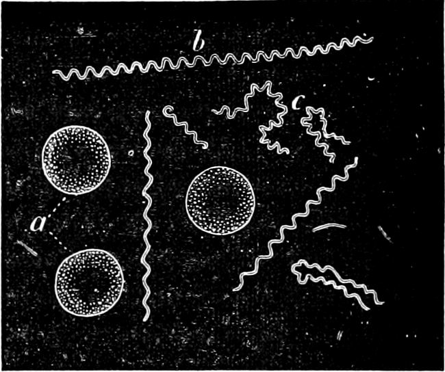
Fig. 24.—Spirochæte obermeieri, in active motion (b) and shortly before the termination of the fever (c); a blood corpuscles.
All Bacteria are unicellular. In the case of the micrococci this is self-evident, but in the “rod,” “thread,” and “spiral” Bacteria, very often numerous cells remain united together and their individual elements can only be recognised by the use of special reagents.
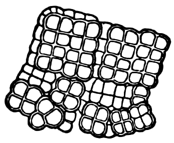
Fig. 25.—Sarcina ventriculi. One surface only is generally seen. Those cells which are drawn with double contour are seen with the correct focus, and more distinctly than those cells lying deeper drawn with single contour.
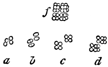
Fig. 26.—Sarcina minuta: a-d successive stages of one individual (from 4–10 p.m.); f an individual of 32 cells.
The condition termed “Zooglœa,” which reminds us of Nostoc, is produced by the cells becoming strongly mucilaginous. A number of individuals in active division are found embedded in a mass of mucilage, which either contains only one, or sometimes more, of the above-named forms. The individuals may eventually swarm out and continue their development in an isolated condition. Such mucilaginous masses occur especially upon moist vegetables (potatoes, etc.), on the surface of fluids with decaying raw or cooked materials, etc. The mucilaginous envelope is thrown into folds when the Bacteria, with their mucilaginous cell-walls, multiply so rapidly that there is no more room on the surface of the fluid.
The cells of the Bacteria are constructed like other plant-cells in so far as their diminutive size has allowed us to observe them. The cell-wall only exceptionally shows the reactions of cellulose (in Sarcina, Leuconostoc; also in a Vinegar-bacterium, Bacterium xylinum); a mucilaginous external layer is always present. The body of the cell mostly appears to be an uniform or finely granulated protoplasm. Very few species (e.g. Bacillus virens) contain chlorophyll; others are coloured red (purple sulphur Bacteria); the majority are colourless. Bacillus amylobacter shows a reaction of a starch-like material when treated with iodine before the spore-formation. Some Bacteria contain sulphur (see p. 37). The body, which has been described as a cell-nucleus, is still of a doubtful nature.
Artificial colourings with aniline dyes (especially methyl-violet, gentian-violet, methylene-blue, fuchsin, Bismarck-brown and Vesuvin) play an important part in the investigations of Bacteria.
Movement. Many Bacteria are self-motile; the long filaments of Beggiatoa exhibit movements resembling those of Oscillaria. In many motile forms the presence of cilia or flagella has been proved by the use of stains; many forms have one, others several cilia attached at one or both ends (Fig. 23) or distributed irregularly over the whole body; the cilia are apparently elongations of the mucilaginous covering and not, as in the other Algæ of the protoplasm. In Spirochæte the movement is produced by the flexibility of the cell itself. Generally speaking, the motion resembles that of swarm-cells (i.e. rotation round the long axis and movement in irregular paths); but either end has an equal power of proceeding forwards.
The swarming motion must not be confounded with the hopping motion of the very minute particles under the microscope (Brownian movement).
Vegetative reproduction takes place by continued transverse division; hence the name “Fission-Fungi” or “Fission-Algæ,” has been applied to the Bacteria.
Spores. The spores are probably developed in two ways. In the ENDOSPOROUS species (Figs. 28, 29), the spore arises as a new cell inside the mother-cell. The spores are strongly refractile, smaller than the mother-cell, and may be compared to the aplanospores of other Algæ. In addition to these there are the ARTHROSPOROUS species in which the cells, just as in Nostoc and other Blue-green Algæ, assume the properties of spores without previously undergoing an endogenous new construction, and are able to germinate and form new vegetative generations (Fig. 27). The formation of spores very often commences when the vegetative development begins to be restricted.
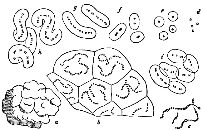
Fig. 27.—Leuconostoc mesenterioides: a a zooglœa, natural size; b cross section of zooglœa; c filaments with spores; d mature spores; e-i successive stages of germination; in e portions of the ruptured spore-wall are seen on the external side of the mucilaginous covering. (b-i magnified 520.)
The spores germinate as in Nostoc by the bursting of the external layer of the cell-wall, either by a transverse or longitudinal cleft, but always in the same way, in the same species (Fig. 28, example of transverse cleft).
Distribution. Bacteria and their germs capable of development, are found everywhere, in the air (dust), in surface water, and in the superficial layers of the soil. The number varies very much in accordance with the nature of the place, season, etc. They enter, together with air and food, into healthy animals and occur always in their alimentary tract.
Growth and reproduction depend upon the conditions of temperature. There is a certain minimum, optimum and maximum for each species; for instance (in degrees Centigrade)—
Minim. Opt. Maxim.
Bacillus subtilis + 6 c. 30 + 50
B. anthracis 15 20–25 43
Spirillum choleræ asiaticæ 8 37 40 (but grows only feebly if under 16°).
Bacterium tuberculosis 28 37–38 42
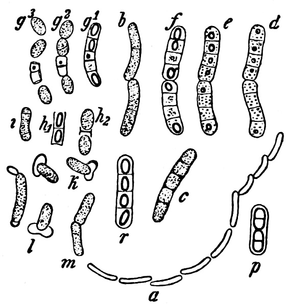
Fig. 28.—Bacillus megaterium: a outline of a living, vegetative cell-rod; b a living, motile, pair of rods; p a similar 4-celled rod after the effects of iodine alcohol; c a 5-celled rod in the first stages of spore-formation; d-f successive stages of spore-formation in one and the same pair of rods (in the course of an afternoon); r a rod with mature spores; g1–g3 three stages of a 5-celled rod, with spores sown in nutritive solution; h1–h2, i, k, l stages of germination; m a rod in the act of transverse division, grown out from a spore which had been sown eight hours previously. (After de Bary; a mag. 250, the other figures 600 times).
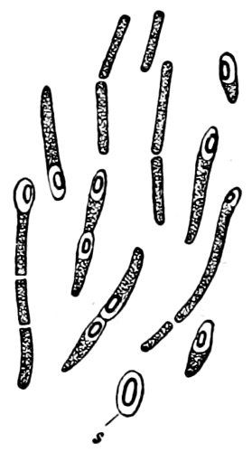
Fig. 29.—Bacillus amylobacter. Motile rods, partly cylindrical and without spores, partly swollen into various special shapes and with spore-formation in the swelling. s Mature spore, with thick mucilaginous envelope. (After de Bary; mag. 600 times, with the exception of s, which is more highly magnified.)
The functions of life cease on a slight excess of the maximum or minimum temperature, numbness setting in when either of these limits is passed. Crenothrix-threads provided with mucilaginous envelopes may, according to Zopf, sustain a temperature of-10°. Some Bacteria are said to be able to resist the exposure to as low a temperature as-110° for a short time. It is not known at what degree of cold the death of the Bacteria occurs: the greatest degree of heat which the vegetative cells can withstand is about the same as that for other vegetative plant-cells, namely, about 50–60° C. Certain Bacteria, e.g. B. thermophilus, grow and thrive vigorously at 70° C. Many spores, on the contrary, are able to bear far higher temperatures (in several species a temperature for some duration of above 100°, those of Bacillus subtilis, for instance, can withstand for hours a temperature of 100° in nutrient solutions; the spores remain capable of development after exposure to a dry heat of 123° C.).
The Desiccation of the air, if prolonged, kills many forms when in the vegetative condition. The spores however can bear a much longer period of dryness, some even several years.
Oxygen. Some species cannot live without a supply of free oxygen (Aerobic), e.g. the Vinegar-bacteria, the Hay-bacilli, the Anthrax-bacilli, the Cholera-Microspira. Other species again thrive vigorously without supply of free oxygen, and are even checked in their development by the admission of air (Anaerobic), e.g. the butyric acid Bacterium (Clostridium butyricium = Bacillus amylobacter). A distinction may be drawn between obligate and facultative aerobics and obligate and facultative anaerobics. Several Bacteria, producing fermentation, may grow without the aid of oxygen when they are living in a solution in which they can produce fermentation; but, if this is not the case, they can only grow when a supply of oxygen is available. A great number of the pathogenic Bacteria belong to the facultative anaerobics.
A luminous Bacterium (Bacillus phosphorescens) which in the presence of a supply of oxygen gives a bluish-white light, has been found in sea-water. Phosphorescent Bacteria have frequently been observed upon decaying sea-fish, as well as on the flesh of other animals; by transferring the Bacteria from cod fish to beef, etc., the latter may be made luminous.
Organic carbon compounds are indispensable for all Bacteria, (except, as it appears, for the nitrifying organisms), as they can only obtain the necessary supplies of carbon from this source. The supplies of nitrogen, which also they cannot do without, can be obtained equally as well from organic compounds as from inorganic salts, such as saltpetre or ammonia-compounds. The various “ash-constituents” are also essential for their nourishment.
While Moulds and Yeast-Fungi grow best in an acid substratum, the Bacteria, on the other hand, generally thrive best in a neutral or slightly alkaline one.
In sterilization, disinfection, and antisepsis, means are employed by which the Bacteria are killed, or checked in their development, for instance, by heat (ignition, cooking, hot vapours, hot air, etc.), or poisons (acids, corrosive sublimate). The process of preserving articles of food, in which they are boiled and then hermetically sealed, aims at destroying the Bacteria, or the spores of those which already may be present in them, and excluding all others.
As the Bacteria are unable to assimilate carbon from the carbonic acid of the air, but must obtain it from the carbon-compounds already in existence in the organic world, they are either saprophytes or parasites. Some are exclusively either the one or the other, obligate saprophytes or parasites. But there are transitional forms among them, some of which are at ordinary times saprophytes, but may, when occasion offers, complete their development wholly or partly as parasites—facultative parasites; others are generally parasitic, but may also pass certain stages of development as saprophytes—facultative saprophytes.
All chlorophyll-free organisms act in a transforming and disturbing manner on the organic compounds from which they obtain their nourishment, and while they themselves grow and multiply, they produce, each after its kind, compounds of a less degree of complexity, i.e. they produce fermentation, putrefaction, sometimes the formation of poisons, and in living beings often disease.
Those organisms which produce fermentation are called ferments; this word, however, is also employed for similar transformations in purely chemical materials (inorganic ferments or enzymes). Many organic (“living”) ferments, among which are Yeast-cells and Bacteria, give off during their development certain inorganic and soluble ferments (enzymes) which may produce other transformations without themselves being changed. Different organisms may produce in the same substratum different kinds of transformation; alcoholic fermentation may for instance be produced by different species of Fungi, but in different proportions, and the same species produces in different substrata, different transformations (e.g. the Vinegar-bacteria oxydize diluted alcohol to vinegar, and eventually to carbonic acid and water).
In the study of Bacteria it is absolutely necessary to sterilize the vessels employed in cultivation, the apparatus, and nutrient solutions, i.e. to free them from Bacteria germs and also to preserve the cultures from the intrusion of any foreign germs (“pure-cultures”). A firm, transparent, nutritive medium is frequently employed. This may be prepared by adding to the nutrient solutions (broth) either gelatine, or—when the Bacteria are to be cultivated at blood-heat—serum of sheep’s or calf’s blood, agar-agar or carragen; serum alone may in itself serve as a nutrient medium. The so-called “plate-cultures” are frequently employed, i.e. the germs are isolated by shaking them with the melted liquid nutrient gelatine, which is then spread on a glass plate and allowed to coagulate; when later on the individual germs grow into colonies, these remain separate in the solid substratum and it is easy to pursue their further development. Similar plate-cultures may also be cultivated in test-tubes and on microscopic slides. The slides and glass plates must be placed in “moist chambers” free from Bacteria. By sowing a few cells (if possible one) using a fine platinum wire, pure cultures for further investigation may be obtained.
In order to prove the relationship between pathogenic Bacteria and certain diseases, the experimental production of pathogenic Bacteria by the inoculation of Bacteria from pure cultures into healthy animals, is very important.
It has not so far been possible to establish a classification of the Bacteria, as the life-history of many species, has not yet been sufficiently investigated.[7] The opinions of botanists are at variance, in many cases, about the forms of growth of a particular kind. Some species are pleomorphic (many-formed) while others possess only one form.
The following Bacteria are Saprophytes:—
Cladothrix dichotoma is common in stagnant and running water which is impregnated with organic matter; the cell-chains have false branching. According to Zopf, Leptothrix ochracea is one of the forms of this species which, in water containing ferrous iron (e.g. as FeCO3), regularly embeds ferric-oxide in its sheath by means of the activity of the protoplasm. Leptothrix ochracea and other Iron-bacteria, according to Winogradsky (1888), do not continue their growth in water free from protoxide of iron; while they multiply enormously in water which contains this salt of iron. The large masses of ochre-coloured slime, found in meadows, bogs, and lakes, are probably due to the activity of the Iron-bacteria.
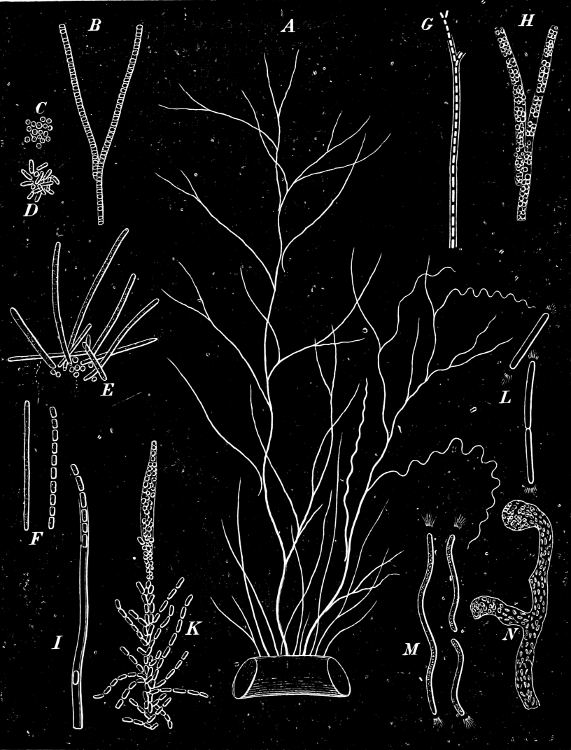
Fig. 30.—Cladothrix dichotoma.
Those forms which, according to Zopf’s views, represent the forms of development of Cladothrix dichotoma are placed together in Fig. 30. A represents a group of plants, seventy times magnified, attached to a Vaucheria. The largest one is branched like a tree, with branches of ordinary form; a specimen with spirally twisted branches is seen to the right of the figure, at the lower part some small Leptothrix-like forms. B shows the manner of branching and an incipient Coccus-formation. C a Coccus-mass whose exit from the sheath has been observed. D the same mass as C after the course of a day, the Cocci having turned into rods. E a group of Cocci in which some have developed into shorter or longer rods. F one of these rods before and after treatment with picric acid, which causes the chain-like structure to become apparent. G a portion of a plant with conspicuous sheath, two lateral branches are being formed. H part of a plant, whose cells have divided and form Cocci. The original form of the cells in which the Cocci are embedded may still be recognised. I. Leptothrix-filaments with conspicuous mucilaginous sheath, from which a series of rods is about to emerge; the rod near the bottom is dead, and has remained lying in the sheath. K part of a plant which is forming Cocci, those at the top are in the zooglœa-stage, at the base they are elongating to form rods and Leptothrix-filaments. L a portion of a branched Cladothrix, which divides into motile Bacillus-forms; the rays at the free ends indicate the currents which the cilia produce in the water. M a spirally-twisted, swarming filament, before and after division into halves. N part of a tree-like zooglœa with Cocci and short rods.—All of these spirilla, zooglœa, etc., which Zopf has connected with Clad. dichotoma, are according to Winogradsky, independent organisms.
Micrococcus ureæ produces urinal fermentation (transformation of urinal matter into ammonium carbonate); aerobic; round cells generally united to form bent chains or a zooglœa.—Several other kinds of Bacteria have the same action as this one: in damp soil containing ammonia-compounds, saltpetre-formations are produced by M. nitrificans and several different kinds of Bacteria.
Micrococcus prodigiosus is found on articles of food containing starch; “bleeding bread” is caused by this Bacterium, which has the power of forming a red pigment; it also occurs in milk, and produces lactic acid.
Leuconostoc mesenterioides is the frog-spawn Bacterium (Fig. 27) which is found in sugar manufactories, and has the power of producing a viscous fermentation in saccharine solutions which have been derived from plants, e.g. in beetroot-sugar manufactories, where large accumulations of mucilage are formed at the expense of the sugar, with an evolution of carbonic acid. The cell-rows, resembling somewhat a pearl necklace, have thick mucilaginous cell-walls, and form white “Nostoc”-lumps. The mucilage eventually deliquesces and the cells separate from each other; arthrospores?—Similar viscous deteriorations occur in beer and wine, which may then be drawn out into long, string like filaments—“ropiness.”
Bacterium aceti, the Vinegar-bacterium, oxidizes alcohol into acetic acid (acetous-fermentation) and forms a greyish covering of Bacteria (“Vinegar-mother”) on the surface of the liquid; the acetic acid formed, becomes by continued oxidization by B. aceti, again transformed into carbonic acid and water. Aerobic; short cylindrical cells, often united into chains, or to form a zooglœa; sometimes also rod-and spindle-shaped. The Vinegar-bacteria and other kinds with ball- or rod-forms sometimes become swollen, spindle-shaped, or oval links; they are supposed to be diseased forms[8] (“Involution-forms”).
Bacillus lacticus (Bacterium acidi lactici, Zopf) is always found in milk which has stood for some time, and in sour foods (cabbage, cucumbers, etc.); it turns the milk sour by producing lactic acid fermentation in the sugar contained in the milk; the lactic acid formed, eventually causes the coagulation of the casein. It resembles the Vinegar-bacteria, occurring as small cylindrical cells, rarely in short rows; not self-motile.—Several other Bacteria appear to act in the same way, some occurring in the mouth of human beings; some of these Bacteria give to butter its taste and flavour.
The kefir-grains which are added to milk for the preparation of kefir, contain in large numbers a Bacterium (Dispora caucasica) in the zooglœa-form, a Yeast-fungus, and Bacillus lacticus. Kefir is a somewhat alcoholic sour milk, rich in carbonic acid; it is a beverage manufactured by the inhabitants of the Caucasus, from the milk of cows, goats, or sheep, and is sometimes used as a medicine. In the production of kefir, lactic acid fermentation takes place in one part of the sugar contained in the milk, and alcoholic fermentation in another part, and the casein which had become curdled is partially liquefied (peptonised) by an enzyme of a Zooglœa-bacterium.
Bacillus amylobacter (Bacillus butyricus), the Butyric-acid-bacterium (Fig. 29), is a very common anaerobic which produces fermentation in sugar and lactic-acid salts, and whose principal product is butyric acid. It destroys articles of food and (together with other species) plays a part in the butyric acid fermentation which is necessary in the making of cheese; it is very active wherever portions of plants are decaying, in destroying the cellulose in the cell-walls of herbaceous plants, and is thus useful in the preparation of flax and hemp. The cells are self-motile, generally cylindrical, sometimes united into short rows; endosporous; the spore-forming cells swell, assume very different forms, and show granulose reaction. The germ-tube grows out in the direction of the long axis of the spore.
Bacillus subtilis, the Hay-bacillus, is developed in all decoctions of hay; a slender, aerobic, self-motile Bacillus; endosporous (aplanospores); the spore-wall ruptures transversely on germination.
Crenothrix kuehniana occurs in the springs of many baths, in wells, in water or drain-pipes.
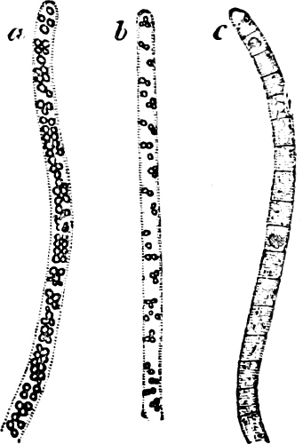
Fig. 31.—Beggiatoa alba: a from a fluid containing abundance of sulphuretted hydrogen; b after lying 24 hours in a solution devoid of sulphuretted hydrogen; c after lying an additional 48 hours in a solution devoid of sulphuretted hydrogen, by this means the transverse walls and vacuoles have become visible.
Beggiatoa (parallel with the Blue-green Alga Oscillaria). Long filaments formed of cylindrical cells which are attached by one of the ends, but which are nearly always free when observed. The filaments, like those of Oscillaria, describe conical figures in their revolutions, the free filaments slide upwards and parallel with one another; sheaths are wanting; strongly refractive sulphur drops are found in the interior. The Beggiatoas are the most prevalent Sulphur-bacteria. They occur, very commonly in large numbers, wherever plant or animal remains are decaying in water in which sulphuretted hydrogen is being formed; thus, for example, B. alba (Fig. 31) occurs frequently as a white covering or slimy film on mud containing organic remains. B. mirabilis is remarkable for its size and its strong peristaltic movements. The Sulphur-bacteria oxidize the sulphuretted hydrogen, and accumulate sulphur in the shape of small granules of soft amorphic sulphur, which in the living cell never passes over into the crystalline state. They next oxidize this sulphur into sulphuric acid, which is immediately rendered neutral by absorbed salts of calcium, and is given off in the form of a sulphate, thus CaCO3 is principally changed into CaSO4. In the absence of sulphur the nutritive processes are suspended, and consequently death occurs either sooner or later. The Sulphur-bacteria may exist and multiply in a fluid which only contains traces of organic matter, in which organisms devoid of chlorophyll are not able to exist. The Beggiatoas very frequently form white, bulky masses in sulphur wells and in salt water, the traces of organic material which the sulphur water contains proving sufficient for them. The cellulose-fermentation, to which the sulphur wells in all probability owe their origin, mainly procures them suitable conditions for existence. The CaCO3 and H2S, formed during the cellulose fermentation by the reduction of CaSO4 is again changed into CaSO4 and CO2 by the Sulphur-bacteria (Winogradsky, 1887).—Other Sulphur-bacteria, the so-called purple Sulphur-bacteria, e.g. B. roseo-persicina, Spirillum sanguineum (Fig. 23), Bacterium sulfuratum, etc., have their protoplasm mixed with a red colouring matter (bacterio-purpurin) which, like chlorophyll, has the power, in the presence of light, of giving off oxygen (as proved by T. W. Englemann, 1888, in oxygen-sensitive Bacteria). The three purple Sulphur-bacteria mentioned, are, according to Winogradsky, not pleomorphic kinds but embrace numerous species.
Many Spirilli (Spirillum tenue, S. undula, S. plicatile, and others) are found prevalent in decaying liquids.
Bacteria (especially Bacilli) are the cause of many substances emitting a foul odour, and of various changes in milk.
Parasitic Bacteria live in other living organisms; but the relation between “host” and parasite may vary in considerable degree. Some parasites do no injury to their host, others produce dangerous contagious diseases; some choose only a special kind as host, others again live equally well in many different ones. There are further specific and individual differences with regard to the predisposition of the host, and every individual has not the same receptivity at all times.
The harmless parasites of human beings. Several of the above mentioned saprophytes may also occur in the alimentary canal of human beings; e.g., the Hay-bacillus, the Butyric-acid-bacillus, etc.; but the gastric juice prevents the development of others, at all events in their vegetative condition. Sarcina ventriculi, “packet-bacterium,” is only known to occur in the stomach and intestines of human beings, and makes its appearance in certain diseases of the stomach (dilation of the stomach, etc.) in great numbers, without, however, being the cause of the disease. It occurs in somewhat cubical masses of roundish cells (Fig. 25).
Less dangerous parasites. In the mouth, especially between and on the teeth, a great many Bacteria are to be found (more than fifty species are known), e.g. Leptothrix buccalis (long, brittle, very thin filaments which are united into bundles), Micrococci in large lumps, Spirochæte cohnii, etc. Some of them are known to be injurious, as they contribute in various ways to the decay of the teeth (caries dentium); a Micrococcus, for instance, forms lactic acid in materials containing sugar and starch, and the acid dissolves the lime salts in the external layers of the teeth: those parts of the teeth thus deprived of lime are attacked by other Bacteria, and become dissolved. Inflammation in the tissues at the root of a tooth, is probably produced by septic materials which have been formed by Bacteria in the root-canal.
Dangerous Parasites. In a large number of the infectious diseases of human beings and animals, it has been possible to prove that parasitic Bacteria have been the cause of the disease. Various pathogenic Bacteria of this nature, belonging to the coccus, rod, and spiral Bacteria groups, are mentioned in the following:—
Pathogenic Micrococci. Staphylococcus pyogenes aureus produces abscesses of various natures (boils, suppurative processes in internal organs). The same effects are produced by—
Streptococcus pyogenes, which is the most frequent cause of malignant puerperal fever; it is perhaps identical with—
Streptococcus erysipelatis, which is the cause of erysipelas in human beings.
Diplococcus pneumoniæ (A. Fränkel) is the cause of pneumonia, and of the epidemic cerebro-spinal meningitis.
Gonococcus (Neisser) is the cause of gonorrhea and inflammation of the eyes.
Pathogenic Rod-Bacteria. Bacterium choleræ gallinarum, an aerobic, facultative parasite which produces fowl-cholera among poultry; it is easily cultivated on various substrata as a saprophyte. The disease may be conveyed both through wounds and by food, and may also be communicated to mammals.
Bacillus anthracis, the Anthrax bacillus (Fig. 32), chiefly attacks mammals, especially herbivorous animals (house mice, guinea-pigs, rabbits, sheep, cattle), in a less degree omnivorous animals (including human beings), and in a still less degree the Carnivores. Aerobic. Cylindrical cells, 3–4 times as long as broad, united into long rod-like bodies, which may elongate into long, bent, and twisted filaments. Not self-motile. Endosporous. Germination takes place without the throwing off of any spore-membrane (compare Hay-bacillus p. 37 which resembles it). Contagion may take place both by introduction into wounds, and from the mucous membrane of the intestines or lungs, both by vegetative cells and by spores; in intestinal anthrax, however, only by spores. The Bacillus multiplies as soon as it has entered the blood, and the anthrax disease commences. The Bacilli not only give off poison, but also deprive the blood of its oxygen. Vegetative cells only occur in living animals. This species is a facultative parasite which in the first stage is a saprophyte, and only in this condition forms spores.
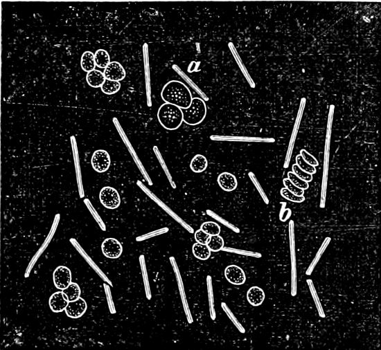
Fig. 32.—Anthrax bacillus (Bacillus anthracis) with red (b) and white (a) blood-corpuscles.
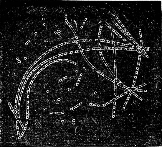
Fig. 33.—Anthrax bacillus. The formation of the spores; magnified 450 times.
Bacillus tuberculosis produces tuberculosis in human beings, also in domestic animals (perlsucht). It is a distinct parasite, but may also live saprophytically. It is rod-formed, often slightly bent, and is recognised principally by its action with stains (when stained with an alkaline solution of methyl-blue or carbolic fuchsin, it retains the colour for a long time even in solutions of mineral acids, in contrast with the majority of well-known Bacteria): it probably forms spores which are able to resist heat, dryness, etc.
Bacillus lepræ produces leprosy; Bacillus mallei produces glanders; Bacillus tetani, tetanus (the tetanus bacillus is very common in soil; anaerobic); Bacillus diphtheriæ, diphtheria; Bacillus typhosus, typhoid fever, etc.
Pathogenic Spiral Bacteria. Spirochæte obermeieri (Fig. 24) produces intermittent fever (febris recurrens); it makes its appearance in the blood during the attacks of fever, but it is not to be found during intervals when there is no fever. Obligate parasite.
Spirillum choleræ asiaticæ (Microspira comma) without doubt produces Asiatic cholera; an exceedingly motile spirillum, which is also found in short, bent rods (known as the “Comma-bacillus”), it lives in the intestines of those attacked by the disease, and gives off a strong poison which enters the body. It is easily cultivated as a saprophyte.
A great many circumstances seem to show that a number of other infectious diseases (syphilis, small-pox, scarlet-fever, measles, yellow-fever, etc.) owe their origin to parasitic Bacteria, but this has not been proved with certainty in all cases.
It has been possible by means of special cultivations (ample supply of oxygen, high temperature, antiseptic materials) to produce from the parasitic Bacteria described above (e.g. the fowl-cholera and the anthrax Bacteria) physiological varieties which are distinct from those appearing in nature and possess a less degree of “virulence,” i.e. produce fever and less dangerous symptoms in those animals which are inoculated with them. The production of such physiological varieties has come to be of great practical importance from the fact that they are used as vaccines, i.e. these harmless species produce in the animals inoculated with them immunity from the malignant infectious Bacteria from which they were derived. This immunity is effected by the change of the products of one or more of the Bacteria, but we do not yet know anything about the way in which they act on the animal organism. The white blood corpuscles, according to the Metschnikoff, play the part of “Phagocytes” by absorbing and destroying the less virulent Bacteria which have entered the blood, and by so doing they are gradually enabled to overcome those of a more virulent nature.
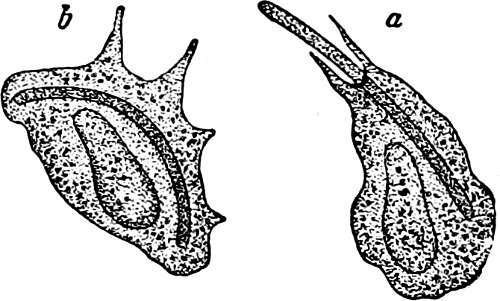
Fig. 34.—a and b The same blood-cell of a Frog: a in the act of engulfing an anthrax-bacillus; b after an interval of a few minutes when the bacillus has been absorbed.
Class 5. Conjugatæ.
The Algæ belonging to this class have chlorophyll, and pyrenoids round which starch is formed. The cells divide only in one direction, they live solitarily, or united to form filaments which generally float freely (seldom attached). Swarm-cells are wanting. The fertilisation is isogamous (conjugation) and takes place by means of aplanogametes. The zygote, after a period of rest, produces, immediately on germination, one or more new vegetative individuals; sometimes akinetes or aplanospores are formed in addition. They only occur in fresh or slightly brackish water.
Order 1. Desmidiaceæ. The cells generally present markings on the outer wall, and are mostly divided into two symmetrical halves by a constriction in the middle, or there is at least a symmetrical division of the protoplasmic cell-contents. The cell-wall consists nearly always of two layers, the one overlapping the other (Fig. 35 C). The cells either live solitarily or are united into unbranched filaments. The mass of protoplasm formed by the fusion of the two conjugating cells becomes the zygote, which on germination produces one (or after division 2, 4 or 8) new vegetative individual. The chromatophores are either star-, plate-, or band-shaped, and regularly arranged round the long axis of the cell.
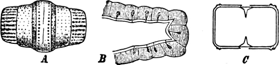
Fig. 35.—A Cell of Gymnozyga brebissonii, external view showing the distribution of the pores. B A portion of the membrane of Staurastrum bicorne with pores containing protoplasmic projections. C Cell-wall of Hyalotheca mucosa during cell-division: the central part, being already formed, shows the connection with the divisional wall.
The Desmidiaceæ are not able to swim independently, many species, however, show movements of different kinds by rising and sliding forward on the substratum. These movements, which are partly dependent upon, and partly independent of light and the force of gravitation, are connected with the protrusion of a mucilaginous stalk. The mucilage, which sometimes surrounds the whole individual, may acquire a prismatic structure, it is secreted by the protoplasmic threads which project through certain pores definitely situated in the walls (Fig. 35 A, B).
Vegetative multiplication takes places by division. A good example of this is found in Cosmarium botrytis (Fig. 36 A-D). The nucleus and chromatophores divide, and simultaneously the central indentation becomes deeper, the outer wall is then ruptured making a circular aperture through which the inner wall protrudes forming a short, cylindrical canal between the two halves to which it is attached (Fig. 36 C). After elongation the canal is divided by a central transverse wall, which commences as a ring round its inner surface and gradually forms a complete septum. The dividing wall gradually splits, and the two individuals separate from each other, each one having an old and a new half. The two daughter-cells bulge out, receive a supply of contents from the parent-cells, and gradually attain their mature size and development (Fig. 36 B-D). Exceptions to this occur in some forms.
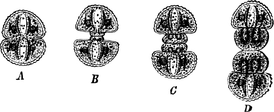
Fig. 36.—Cosmarium botrytis. A-D Different stages of cell-division.
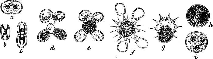
Fig. 37.—Cosmarium meneghinii: a-c same individual seen from the side, from the end, and from the edge; d-f stages of conjugation; g-i germination of the zygote.
Conjugation takes place in the simplest way in Mesotænium, where the two conjugating cells unite by a short tube (conjugation-canal), which is not developed at any particular point. The aplanogametes merge together after the dissolution of the dividing wall, like two drops of water, almost without any trace of preceding contraction, so that the cell-wall of the zygote generally lies in close contact with the conjugating cells. The conjugating cells in the others lie either transversely (e.g. Cosmarium, Fig. 37 d; Staurastrum, etc.), or parallel to one another (e.g. Penium, Closterium, etc.), and emit a short conjugation-canal (Fig. 37 d) from the centre of that side of each cell which is turned towards the other one. These canals touch, become spherical, and on the absorption of the dividing wall the aplanogametes coalesce in the swollen conjugation-canal (Fig. 37 e), which is often surrounded by a mucilaginous envelope. The zygote, which is often spherical, is surrounded by a thick cell-wall, consisting of three layers; the outermost of these sometimes bears thorn-like projections, which in some species are simple (Fig. 37 f), in others branched or variously marked; in some, however, it remains always smooth (e.g. Tetmemorus, Desmidium). Deviation from this mode of conjugation may occur within certain genera (e.g. Closterium, Penium). Upon germination the contents of the zygote emerge, surrounded by the innermost layers of the wall (Fig. 37 g, h) and generally divide into two parts which develop into two new individuals, placed transversely to each other (Fig. 37 i); these may have a somewhat more simple marking than is generally possessed by the species.
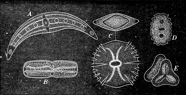
Fig. 38.—Desmidiaceæ. A Closterium moniliferum; B Penium crassiusculum; C Micrasterias truncata (front and end view); D Euastrum elegans; E Staurastrum muticum (end view).
The most frequent genera are:—
A. Solitary cells: Mesotænium, Penium (Fig. 38 B), Cylindrocystis, Euastrum (Fig. 38 D), Micrasterias (Fig. 38 C), Cosmarium (Fig. 36, 37), Xanthidium, Staurastrum (Fig. 38 E), Pleurotænium, Docidium, Tetmemorus, Closterium (Fig. 38 A), Spirotænia.
B. Cells united into filaments: Sphærozosma, Desmidium, Hyalotheca, Gymnozyga, Ancylonema, Gonatozygon.
Order 2. Zygnemaceæ. Cell-wall without markings. The cells are cylindrical, not constricted in the centre, and (generally) united into simple, unbranched filaments. The whole contents of the conjugating cells take part in the formation of the zygote, which on germination grows out directly into a new filament.
Spirogyra is easily recognised by its spiral chlorophyll band; Zygnema has two star-like chromatophores in each cell (Fig. 40); both these genera are very common Algæ in ponds and ditches.
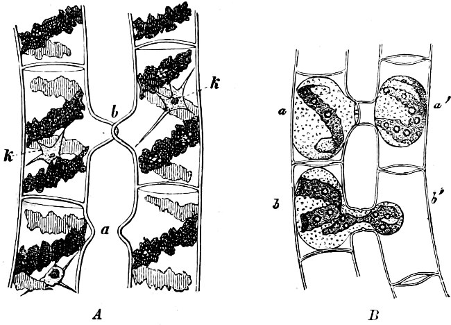
Fig. 39.—Spirogyra longata. A At the commencement of conjugation, the conjugation-canals begin to protrude at a and touch one another at b; the spiral chlorophyll band and cell-nuclei (k) are shown. B A more advanced stage of conjugation; a, a’ the rounded female and male aplanogametes: in b’ the male aplanogamete is going over to and uniting with the female aplanogamete (b).
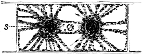
Fig. 40.—A cell of Zygnema. S Pyrenoid.
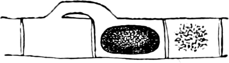
Fig. 41.—Zygnema insigne, with zygote.
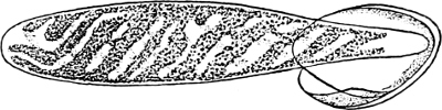
Fig. 42.—Germinating zygote of Spirogyra jugalis: the young plant is still unicellular; the end which is still in the wall of the zygote is elongated and root-like; the chromatophore divides and forms the spiral band.
The conjugation among the Zygnemaceæ takes place in the following manner: the cells of two filaments, lying side by side, or two cells, the one being situated above the other in the same filament (Fig. 41), push out small protuberances opposite each other (Fig. 39 A, a, b); these finally meet, and the dividing wall is absorbed so that a tube is formed connecting one cell with the other; the protoplasmic contents round off, and the whole of these contents of one of the cells glides through the conjugation-tube and coalesces with that of the other (Fig. 39 B), the aggregate mass then rounds off, surrounds itself with a cell-wall, and becomes a zygote. A distinct difference may be found between the cells in the two filaments, those in the one whose protoplasmic contents pass over being cylindrical, while those of the recipient one are more barrel-shaped, and of a larger diameter. The former may be regarded as a male, the latter as a female plant. The zygote germinates after a period of rest, and grows out into a new filament (Fig. 42).
Order 3. Mesocarpaceæ. The cell-walls are glabrous, unconstricted in the centre, and united into simple unbranched filaments. The chromatophore consists of an axial chlorophyll-plate, with several pyrenoids. The zygote is formed by the coalescence of two cells (Fig. 43) (sometimes three or four), but the whole protoplasmic contents of the cells do not take part in this process, a portion always remaining behind; the aplanogametes coalesce in the conjugation-canal. The zygote thus formed appears incapable of germination until after 3–5 divisions. Of the cells so formed, only one is fertile, the sterile cells, according to Pringsheim, constituting a rudimentary sporocarp. The germinating cells grow out into a new filament. In this order, conjugation has been observed between two cells of the same filament. The Mesocarpaceæ thrive best in water which contains lime.
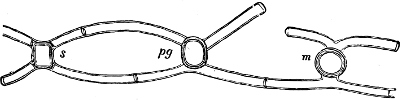
Fig. 43.—Mougeotia calcarea. Cells showing various modes of conjugation: at m tripartition; pg quadripartition; s quinquipartilion of the zygote.
Class 6. Chlorophyceæ (Green Algæ).
These Algæ are coloured green by chlorophyll, seldom in combination with other colouring matter, and then especially with red. The product of assimilation is frequently starch, which generally accumulates round certain specially formed portions of protoplasm termed pyrenoids. The thallus is uni- or multicellular; in the higher forms (certain Siphoneæ) the organs of vegetation attain differentiation into stem and leaf. The asexual reproduction takes place in various ways; the sexual reproduction is effected by conjugation of motile gametes, or by oogamous fertilisation. The swarm-cells (zoospores, gametes, and spermatozoids) are constructed symetrically, and have true protoplasmic cilia, these generally being attached to the front end of the swarm-cells. Most of these Algæ live in water (fresh or salt); some are found upon damp soil, stones, or tree-stems, and some live enclosed in other plants.
The Class is divided into three families:—
1. Protococcoideæ: Volvocaceæ, Tetrasporaceæ, Chlorosphæraceæ, Pleurococcaceæ, Protococcaceæ, Hydrodictyaceæ.
2. Confervoideæ: Ulvaceæ, Ulothricaceæ, Chætophoraceæ, Mycoideaceæ, Cylindrocapsaceæ, Œdogoniaceæ, Coleochætaceæ, Cladophoraceæ, Gomontiaceæ, Sphæropleaceæ.
3. Siphoneæ: Botrydiaceæ, Bryopsidaceæ, Derbesiaceæ, Vaucheriaceæ, Phyllosiphonaceæ, Caulerpaceæ, Codiaceæ, Valoniaceæ, Dasycladaceæ.
Family 1. Protococcoideæ.
The Algæ which belong to this group are uni- or multicellular with the cells more or less firmly connected, sometimes in a definite, sometimes in an indefinite form (Fig. 47). Colonies are formed either by division or by small unicellular individuals becoming united in a definite manner; the colonies formed in this latter way are termed Cœnobia. Apical cells and branching are absent. Multiplication by division; asexual reproduction by zoospores, rarely by akinetes. Sexual reproduction may be wanting, or it takes place by isogamous, rarely by oogamous fertilisation.
Some are attached by means of a stalk to other objects (Characium, Fig. 49), others occur as “Endophytes” in the tissues of certain Mosses or Phanerogams, e.g. Chlorochytrium lemnæ, in Lemna trisulca; Endosphæra, in the leaves of Potamogeton, Mentha aquatica, and Peplis portula; Phyllobium, in the leaves of Lysimachia nummularia, Ajuga, Chlora, and species of Grasses; Scotinosphæra in the leaves of Hypnum and Lemna trisulca; the majority, however, live free in water and in damp places. Many species which were formerly considered to belong to this family have been proved to be higher Algæ in stages of development.
Order 1. Volvocaceæ. The individuals in this order are either uni- or multicellular, and during the essential part of their life are free-swimming organisms. They are generally encased in a mucilaginous envelope, through which 2–6 cilia project from every cell. The vegetative reproduction takes place by the division of all, or a few, of the cells of the individual; in some a palmella-stage is found in addition. The sexual reproduction takes place by isogamous or oogamous fertilisation.
The Volvocaceæ may be considered to include the original forms of the Chlorophyceæ, because, among other reasons, the motile stage is here the most prominent; they also form the connecting link between the animal Flagellata, and forms intermediate to the Syngeneticæ may perhaps be found amongst them. Three series of green Algæ may be supposed to have taken their origin from the Volvocaceæ: Conjugateæ (Desmidiaceæ) which have lost the swarming stage, but whose conjugation is the nearest to the fertilisation in Chlamydomonas pulvisculus: the Protococcaceæ in which the vegetative divisions have disappeared, while the swarming stage continues to be present, though of shorter duration; and Tetrasporaceæ, in which the vegetative divisions are more prominent, whilst the swarming stage is less so.
A. Unicellular Individuals. The principle genera are: Chlamydomonas, Sphærella, Phacotus.—Sphærella nivalis is the Alga which produces the phenomenon of “Red Snow,” well known on high mountains and on ice and snow fields in the polar regions. The red colouring matter which appears in this and other green Algæ, especially in the resting cells, is produced by the alteration of the chlorophyll.
Phacotus lenticularis has an outer covering incrusted with lime, which, at death, or after division, opens out into two halves. Species may be found among Chlamydomonas, in which conjugation takes place between gametes of similar size without cell-wall, but in C. pulvisculus conjugation takes place between male and female aplanogametes which are surrounded by a mucilaginous envelope.
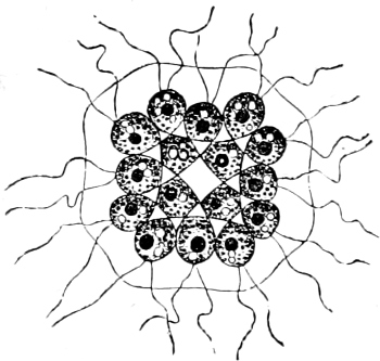
Fig. 44.—Gonium pectorale.
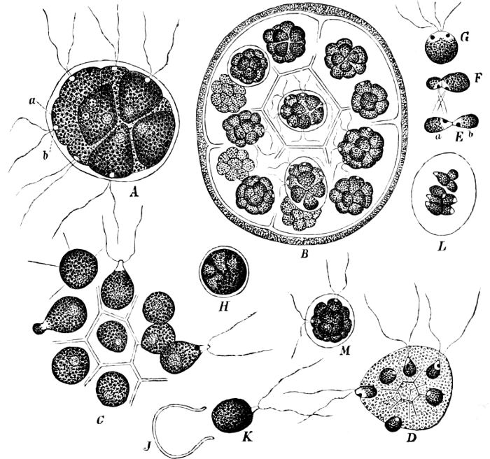
Fig. 45.—Pandorina morum.
B. Multicellular Individuals. The most important genera are Gonium, Stephanosphæra, Pandorina, Eudorina, Volvox.—Gonium has 4 or 16 cells arranged in a definite pattern in a flat plate (Fig. 44). Pandorina (Fig. 45), has 16 cells arranged in a sphere (Fig. 45 A). The vegetative reproduction takes place in this way: each cell, after having rounded off, and after the withdrawal of the cilia, divides itself into 16 new ones (Fig. 45 B), each forming a new individual, which soon grows to the size of the mother-individual. It was in this Alga that the conjugation of self-motile gametes was first discovered by Pringsheim, 1869. When conjugation is about to take place, each cell divides into sixteen, as in vegetative reproduction, but the 16 × 16 cells all separate from one another (Fig. 45 C, female gametes, and D, male gametes), and swarm solitarily in the water. The male are, most frequently, smaller than the female, but otherwise they are exactly alike; they are more or less pear-shaped, with a colourless anterior end, 2 cilia, a red “eye-spot,” etc. After swarming for some time they approach each other, two and two, generally a large and a smaller one, and come into contact at their colourless end; in a few moments they coalesce and become one cell (Fig. 45 E, F), this has at first a large colourless anterior end, 4 cilia, and 2 “eye-spots” (Fig. 45 G), but these soon disappear and the cell becomes uniformly dark-green and spherical, and surrounds itself with a thick cell-wall, losing at the same time its power of motion: the zygote (Fig. 45 H) is formed, and becomes later on a deep red colour. On the germination of the zygote, the protoplasmic cell-contents burst open the wall (Fig. 45 J), and emerge as a large swarmspore (Fig. 45 K) which divides into 16 cells, and the first small individual is formed (Fig. 45 L, M).
Eudorina is like Pandorina in structure, but stands somewhat higher, since the contrast between the conjugating sexual cells is greater, the female one being a motionless oosphere.
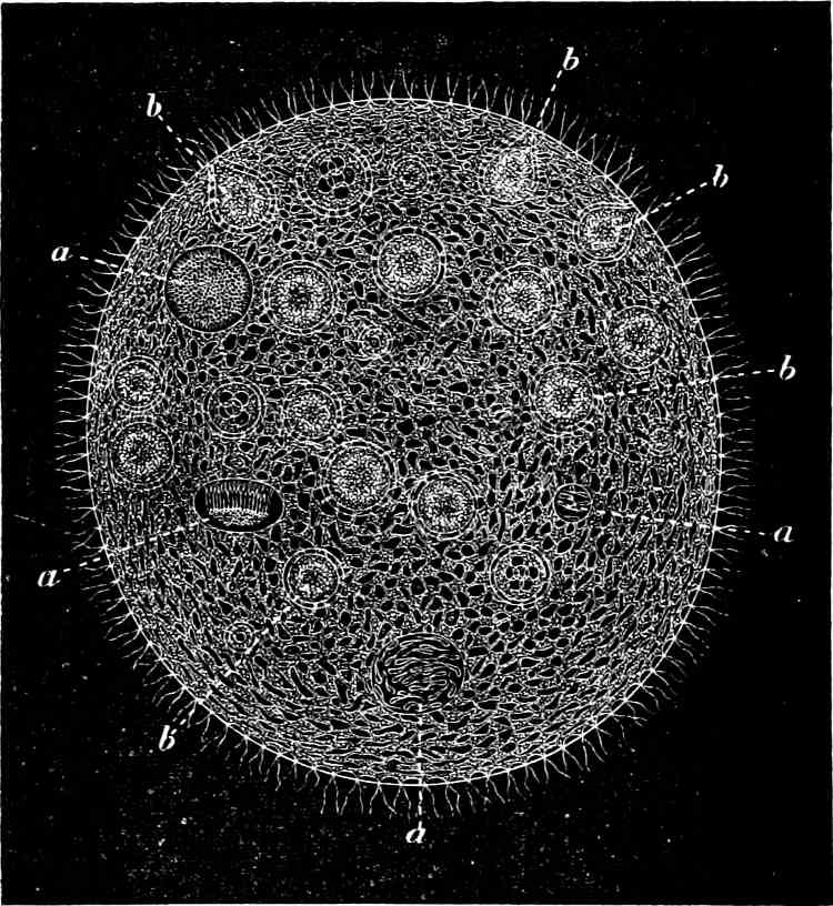
Fig. 46.—Volvox globator, sexual individual: a antheridia which have formed spermatozoids; b oogonia.
The highest stage of development is found in Volvox (Fig. 46). The cells are here arranged on the circumference of a sphere, and enclose a cavity filled with mucilage. The number of these cells may vary from 200–22,000, of which the majority are vegetative and not reproductive, but some become large, motionless oospheres (Fig. 46 b); others, which may appear as solitary individuals, divide and form disc-shaped masses of from 8–256 small spermatozoids (Fig. 46 a). After the oosphere has been fertilised by these, the oospore surrounds itself by a thick, sometimes thorny cell-wall, and on germination becomes a new individual of few cells. A few cells conspicuous by their larger size may be found (1–9, but generally 8) in certain individuals, and these provide the vegetative reproduction, each forming by division a new individual.
Order 2. Tetrasporaceæ reproduce both by vegetative divisions and swarmspores, some have also gamete-conjugation. The principal genera are: Tetraspora, Apiocystis, Dactylococcus, Dictyosphærium, Chlorangium.
Order 3. Chlorosphæraceæ. Chlorosphæra.
Order 4. Pleurococcaceæ. In this order the swarm-stages and sexual reproduction are entirely absent. Vegetative reproduction by division. The principal genera are: Pleurococcus (Fig. 47), Scenedesmus (Fig. 48), Raphidium, Oocystis, Schizochlamys, Crucigenia, Selenastrum.—Pleurococcus vulgaris (Fig. 47) is one of the most common Algæ throughout the world, occurring as green coverings on tree-stems, and damp walls, and it is one of the most common lichen-gonidia.
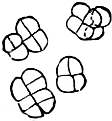
Fig. 47.—Pleurococcus vulgaris.
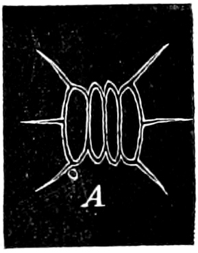
Fig. 48.—Scenedesmus quadricauda.
Order 5. Protococcaceæ. The cells are motionless, free or affixed on a stalk (e.g. Characium, Fig. 49), either separate or loosely bound to one another; they never form multicellular individuals. Multiplication by division is nearly always wanting. Reproduction takes place by swarmspores, which have 1 or 2 cilia, and sexual reproduction in some by gamete-conjugation. The principal genera are: Chlorococcum, Chlorochytrium, Chlorocystis, Scotinosphæra, Endosphæra, Phyllobium, Characium, Ophiocytium, Sciadium.
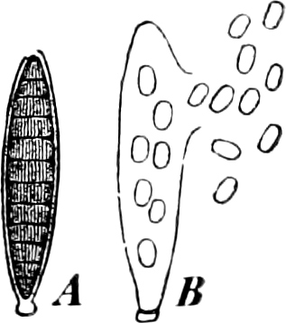
Fig. 49.—Characium strictum. A The cell-contents have divided into many swarmspores. B Swarmspores escaping.
Order 6. Hydrodictyaceæ. The individuals are unicellular but several unite after the zoospore-stage into definitely formed families (cœnobia). Ordinary vegetative division is wanting, but asexual reproduction takes place by zoospores (or by motionless cells without cilia), which unite and form a family similar to the mother-family, inside the mother-cell, or in a mucilaginous envelope. Where sexual reproduction is found it takes place by gamete-conjugation. The principal genera are: Pediastrum (Fig. 50), Cœlastrum, Hydrodictyon (Fig. 51).
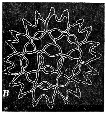
Fig. 5O.—Pediastrum asperum.
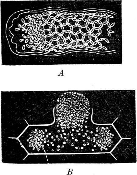
Fig. 51.—Hydrodictyou reticulatum. A A cell where the zoospores are on the point of arranging themselves to form a net. B A cell with gametes swarming out.
The cœnobium of Hydrodictyon reticulatum (Water-net) is formed of a large number of cells which are cylindrical, and attached to one another by the ends (Fig. 51). The asexual reproduction takes place by zoospores, which are formed in large numbers (7,000–20,000) in each mother-cell, within which they move about for a time, and then come to rest and arrange themselves into a new net (Fig. 51 A) which is set free by the dissolution of the wall of the mother-cell, grows, and becomes a new cœnobium. The sexual reproduction takes place by gamete-conjugation. The gametes are formed in the same manner as the zoospores, but in larger numbers (30,000–100,000), and swarm out of the mother-cell (Fig. 51 B). The zygote forms, on germination, 2–5 large zoospores, each with one or two cilia, these generally swarm about for a time, and after a period of rest become irregular thorny bodies (polyhedra); their contents again divide into zoospores, the thorny external coating of the polyhedra is cast off, and the zoospores, surrounded by the dilated internal coating, unite to form a small family, which produces several others in the manner described.
Family 2. Confervoideæ.
The individuals are always multicellular, the cells firmly bound together and united into unbranched or branched filaments, expansions, or masses of cells which grow by intercallary divisions or have apical growth. In the first seven orders the cells are uninuclear, but the cells of the remaining three orders contain several nuclei. Asexual reproduction by zoospores, akinetes or aplanospores. Sexual reproduction by isogamous or oogamous fertilisation.
The Confervoideæ, through the Ulvaceæ, are connected with the Tetrasporaceæ, and from the Coleochætaceæ, which is the most highly developed order, there are the best reasons for supposing that the Mosses have taken their origin. The Cladophoraceæ show the nearest approach to the Siphoneæ.
Order 1. Ulvaceæ. The thallus consists of one or two layers of parenchymatous cells, connected together to form either a flat membrane (Monostroma, Ulva) or a hollow tube (Enteromorpha), and may be either simple, lobed, or branched. Reproduction takes place by detached portions of the thallus; or asexually by zoospores or akinetes. Gamete-conjugation is known to take place in some members of this order, the zygote germinating without any resting-stage. The majority are found in salt or brackish water.
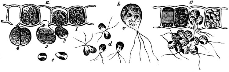
Fig. 52.—Ulothrix zonata: a portion of a filament with zoospores, which are formed two in each cell (zoosporangium); the dark spots are the red “eye-spots”; 1, 2, 3, 4, denote successive stages in the development of the zoospores; b a single zoospore, v the pulsating vacuole; c portion of a filament with gametes, sixteen are produced in each gametangium; d free gametes, solitary or in the act of conjugation; e the conjugation is completed, and the formed zygote has assumed the resting-stage.
Order 2. Ulothricaceæ. The thallus consists normally of a simple unbranched filament (sometimes a small expansion consisting of one layer of cells is formed, as in Schizomeris and Prasiola which were formerly described as separate genera). Asexual reproduction takes place by means of zoospores (with 1, 2, or 4 cilia), akinetes or aplanospores; the last named may germinate immediately, or only after a period of rest. Sexual reproduction takes place by the conjugation of gametes of about the same size, each having two cilia (Fig. 52 d). The zygote of Ulothrix, on germination, produces a brood of zoospores which swarm for a time and then elongate to become Ulothrix-filaments (alternation of generations). The gametes may also germinate without conjugation in the same manner as the zoospores. The principal genera are: Ulothrix, Hormidium, Conferva, Microspora.—Ulothrix zonata is very common in running fresh water. Nearly all the species of Hormidium occur on damp soil, tree-stems and stones.
Order 3. Chætophoraceæ. The thallus consists of a single, branched, erect or creeping filament of cells, often surrounded by mucilage. The cells have only one nucleus. Asexual reproduction by zoospores with 2 or 4 cilia, by akinetes, or aplanospores. In many, conjugation between gametes with 2 cilia may be found. They approach on one side, Ulothricaceæ, and on the other, Mycoideaceæ. The principal genera are: Stigeoclonium, Draparnaldia, Chætophora, Entoderma, Aphanochæte, Herposteiron, Phæothamnion, Chlorotylium, Trichophilus, Gongrosira, Trentepohlia. Most of the species of Trentepohlia are coloured red by the presence of a red colouring material, which occurs in addition to the chlorophyll. They are aerial Algæ which live on stones (T. jolithus, “violet stone,” so named on account of its violet-like odour in rainy weather), on bark and old wood (T. umbrina), or on damp rocks (T. aurea). Trichophilus welckeri lives in the hair of Bradypus.
Order 4. Mycoideaceæ. The thallus is discoid, consisting of one or more cell-layers, and is always attached. Asexual reproduction by zoospores with 2 or 4 cilia. Sexual reproduction in some species by the conjugation of gametes with 2 cilia. This order forms the connecting link between Chætophoraceæ and Coleochætaceæ. The species occur in fresh water (Chætopeltis) as well as in salt (Pringsheimia), on the carapace of tortoises (Dermatophyton = Epiclemmydia), or endophytic between the cuticle and the epidermal cells of the leaves of tropical plants, destroying the leaf-tissue (Mycoidea).
Order 5. Cylindrocapsaceæ. The thallus consists of a simple (rarely, in parts, formed of many rows) unbranched filament, attached in the young condition, which has short cells with a single nucleus, and is enveloped in a thick envelope with a laminated structure. Asexual reproduction by zoospores with 2 cilia, which are formed 1, 2, or 4 in each vegetative cell. The antheridia are produced by a single cell, or a group of cells, in a filament, dividing several times without increasing in size. Two egg-shaped spermatozoids, each with 2 cilia (Fig. 53 D), are formed in each antheridium, and escape through an aperture in the side; in the first stages they are enclosed in a bladder-like membrane (Fig. 53 B, C). Other cells of the filament swell out and form oogonia (Fig. 53 A), which resemble those of Œdogonium. After fertilisation, the oospore surrounds itself with a thick wall, and assumes a reddish colour. The germination is unknown. The unfertilised oospheres remain green, divide often into 2–4 daughter-cells, and grow into new filaments.
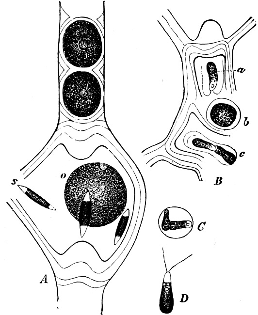
Fig. 53.—Cylindrocopsa involuta. A Oogonium with oosphere (o) surrounded by spermatozoids (s). B Two antheridia, each with two spermatozoids. C Spermatozoids surrounded by their bladder-like membrane. D Free spermatozoid.
This order, which only includes one genus, Cylindrocapsa, forms the connecting link between Ulothricaceæ and Œdogoniaceæ. The few species (4) occur only in fresh water.
Order 6. Œdogoniaceæ. The thallus consists of branched (Bulbochæte) or unbranched (Œdogonium) filaments, attached in the early stages. The cells may be longer or shorter, and have one nucleus. Asexual reproduction by zoospores, which have a chaplet of cilia round the base of the colourless end (Fig. 6 a). Sexual reproduction takes place by oogamous fertilisation. On the germination of the oospore, 4 zoospores are formed (Fig. 54 F). They occur only in fresh or slightly brackish water. The division of the cells takes place in quite a peculiar and unusual manner. At the upper end of the cell which is about to divide, a ring-shaped thickening of soft cellulose is formed transversely round the wall; the cell-nucleus of the mother-cell and the protoplasm then divide by a transverse wall into two portions of similar size, and the cell-wall bursts transversely along the central line of the thickened ring. The cell-wall thus divides into two parts—the upper one short, the “cap,” and the lower one much longer, the “sheath.” The portions of the original cell-wall now separate from each other, the cellulose ring extending, and supplying an additional length of cell-wall between them. The cap and sheath will project a little in front of the piece thus inserted. The dividing wall between the two new cells is formed near to the uppermost edge of the sheath, and gradually becomes thicker and firmer. The inserted piece of wall forms the larger part of the wall of the upper cell: the remainder is formed by the cap. This mode of division is repeated exactly in the same way, and new caps are formed close below the first one, one for every division.
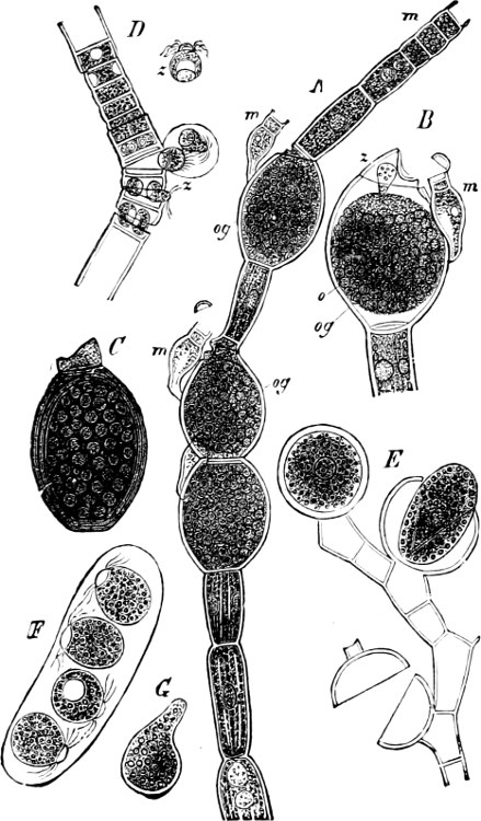
Fig. 54.—A Œdogonium ciliatum. A Female plant with three oogonia (og) and dwarf-males (m). B An oogonium with spermatozoid (z) seen entering the oosphere (o) having passed through an aperture near the summit of the oogonium; m dwarf-male. C Ripe oospore. D Œdogonium gemelliparum. F Portion of a male filament from which spermatozoids (z) are emerging. E Portion of filament of Bulbochæte; the upper oogonium still encloses the oospore, in the central one the oospore is escaping while the lower one is empty. F Four zoospores developed from an oospore. G Zoospore germinating.
Fertilisation takes place in the following way. The oogonium is a large ellipsoidal, swollen cell (og, in Fig. 54 A), whose contents are rounded off into an oosphere with a colourless receptive-spot (see B); an aperture is formed in the wall of the oogonium, through which the spermatozoids are enabled to enter (B). The spermatozoids are produced either directly, as in D (in pairs), in basal cells of the filament, or indirectly. In the latter case a swarmspore (androspore) is formed which comes to rest, attaches itself to an oogonium, germinates, and gives rise to a filament of a very few cells—dwarf-male (A, B, m). The spermatozoids are formed in the upper cell of the dwarf-male (m), and are set free by the summit of the antheridium lifting off like a lid. On the germination of the oospore (C), which takes place in the following spring, 4 zoospores are produced (F) (i.e. the sexual generation); these swarm about for a time, and ultimately grow into new filaments.
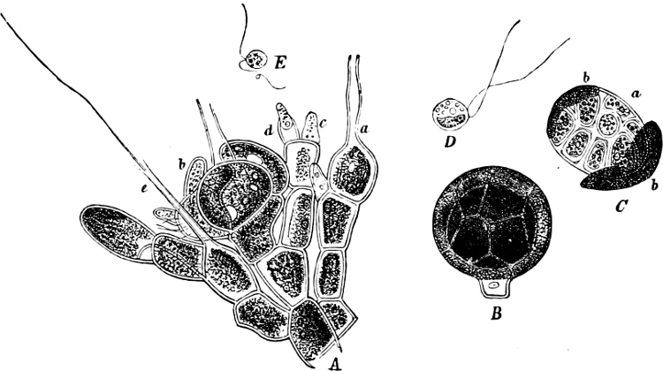
Fig. 55.—Coleochæte pulvinata. A A portion of a thallus with organs of reproduction; a oogonium before, b after fertilisation; c an antheridium, closed; d open, with emerging spermatozoid. B Ripe oogonium, with envelope. C Germination of the oospore. D Zoospore. E Spermatozoid.
Order 7. Coleochætaceæ. The thallus is always attached, and of a disc- or cushion-shape, formed by the dichotomous branching of filaments of cells united in a pseudo-parenchymatous manner. Each cell has only one nucleus. Asexual reproduction by zoospores with 2 cilia (Fig. 55 D), which may arise in all the cells. Sexual reproduction by oogamous fertilisation. The spermatozoids resemble the swarmspores, but are smaller (E), and originate singly (in the species figured) in small conical cells (c, d in A). The oogonia are developed at the extremities of certain branches: they are bottle-shaped cells with very long and thin necks (trichogyne), open at the end (a in A); at the base of each oogonium is a spherical oosphere. The spermatozoids reach the oosphere through the trichogyne, or through an aperture in the wall when the trichogyne is absent, and fertilisation having taken place, the oogonium becomes surrounded by a cell-layer (envelope), which grows out from the cells near its base (b in A), and in this way a kind of fruit is formed (B) (spermocarp, cystocarp).
The oospore, next spring, divides and forms a parenchymatous tissue (homologous with the Moss-sporophyte); this bursts open the envelope (C), and a zoospore (homologous with the spores of the Moss-capsule) arises in each of the cells, and produces a new Coleochæte. We have then, in this case, a still more distinct alternation of generations than in Œdogonium. Only one genus, Coleochæte, is known, but it contains several species, all living in fresh water.
Order 8. Cladophoraceæ. This order is probably derived from the Ulothricaceæ. The thallus consists of a single, unbranched or branched filament, generally with an apical cell. The cells have each 2 or more nuclei. Asexual reproduction by zoospores with 2 or 4 cilia, and by akinetes. Conjugation of gametes with 2 cilia is found in some genera. They occur in salt as well as in fresh water. The principal genera are: Urospora, Chætomorpha, Rhizoclonium, Cladophora; of the last named genus the species C. lanosa and C. rupestris are common in salt water; C. fracta and C. glomerata in fresh water.
Order 9. Gomontiaceæ. Gomontia polyrrhiza, the only species hitherto known, is found on old calcareous shells of certain salt water Molluscs.
Order 10. Sphæropleaceæ. The thallus consists of free, unbranched filaments, with very elongated multinuclear cells. The vegetative cells form no zoospores. Sexual reproduction by oogamous fertilisation (see page 13, Fig. 10 B). The oospore has a thick wall (Fig. 10 D) studded with warts, and assumes a colour resembling red lead. It germinates only in the following spring, and produces 1–8 zoospores, each with 2 cilia (Fig. 10 E), which grow into new filaments. Only one species, Sphæroplea annulina, is known.
Family 3. Siphoneæ.
The thallus has apical growth, and in the vegetative condition consists generally of one single (in the Valoniaceæ most frequently of more) multinuclear cell, which may be much branched, and whose separate parts in the higher forms (e.g. Bryopsis, Fig. 57; Caulerpa, Fig. 59, etc.) may be differentiated to perform the various physiological functions (as root, stem and leaf). Vegetative multiplication by detached portions of the thallus (gemmæ); asexual reproduction by zoospores, akinetes, or aplanospores. Sexual reproduction by gamete-conjugation, rarely by oogamous fertilisation. The zygote or oospore germinates as a rule without any resting-stage.
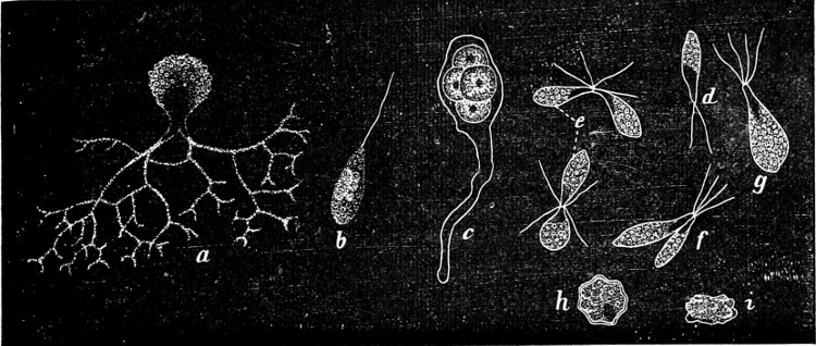
Fig. 56.—Botrydium granulatum: a an entire plant forming swarmspores; b swamspores; c an individual with gametangia; d, gamete; e, f, g conjugation; h zygote seen from above; i the same in a lateral view.
Most of the Siphoneæ occur in salt water or on damp soil. Many (e.g. Dasycladaceæ) are very much incrusted with lime, and occur, in the fossilized condition, in the deposits from the Cretaceous period to the present time. The Siphoneæ are connected by their lowest forms (Botrydiaceæ or Valonia) with the Protococcaceæ, but show also, through the Valoniaceæ, points of relationship to the Cladophoraceæ.
Order 1. Botrydiaceæ. The thallus in the vegetative condition is unicellular, club-shaped, with a small single (Codiolum) or repeatedly dichotomously branched system of colourless rhizoids (Botrydium, Fig. 56 a), by which it is attached to objects immersed in salt water (Codiolum) or to damp clay soil (Botrydium). Asexual reproduction by zoospores with one (Botrydium) or two cilia, and by aplanospores. The sexual reproduction is only known in Botrydium, and takes place in the following manner: in the part of the thallus which is above ground and in an active vegetative condition, several round cells (Fig. 56 c) are formed, which may be green or red according as they grow under water, or exposed to the strong light of the sun. These cells must be considered as “gametangia” as they produce many gametes (d) provided with two cilia. The zygote (h, i) formed by the conjugation (e, f, g) may either germinate immediately, or become a thick-walled resting-cell of an irregular, angular form.
Order 2. Bryopsidaceæ. The thallus in the vegetative condition is unicellular, and consists at the lower extremity of branched rhizoids, while the upper portion is prolonged into a stem-like structure of unlimited growth, producing, acropetally, branches and leaf-like structures. The latter have limited growth, and are separated by a cross wall from the stem, and become gametangia, or drop off. The gametes have two cilia, and are of two kinds: the female, which are green and large and the male, which are of brownish colour and smaller. Zoospores or any other method of asexual reproduction are unknown. Only one genus, Bryopsis, living in salt water.
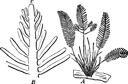
Fig. 57.—Bryopsis plumosa. A the plant, natural size. B A portion (enlarged) which shows the growing point (v), and the leaves derived from it in acropetal succession.
Order 3. Derbesiaceæ. Only one genus, Derbesia, living in saltwater. The zoospores, which are formed in a few lateral, swollen zoosporangia, possess one nucleus which has arisen through the coalescence of several, and they resemble the zoospores of Œdogonium by having a circle of cilia attached at the base of the colourless spot.