Sub-Division I.—MYXOMYCETES, SLIME-FUNGI.
Michael Cresse Potter translation
The Slime-Fungi occupy quite an isolated position in the Vegetable Kingdom, and are perhaps the most nearly related to the group of Rhizopods in the Animal Kingdom. They live in and on organic remains, especially rotten wood or leaves, etc., on the surface of which their sporangia may be found.
They are organisms without chlorophyll, and in their vegetative condition are masses of protoplasm without cell-wall (plasmodia). They multiply by means of spores, which in the true Slime-Fungi[3] are produced in sporangia, but in some others[4] free. The spores are round cells (Fig. 1 a) which in all the true Slime-Fungi are surrounded by a cell-wall. The wall bursts on germination, and the contents float out in the water which is necessary for germination. They move about with swimming and hopping motions like swarmspores (e, f), having a cilia at the front end and provided with a cell-nucleus and a pulsating vacuole. Later on they become a little less active, and creep about more slowly, while they continue to alter their form, shooting out arms in various places and drawing them in again (g, h, i, k, l, m); in this stage they are called Myxamœbæ.
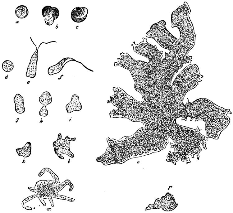
Fig. 1.—a-l Development of “Fuligo” from spore to Myxamœba; a-m are magnified 300 times; m is a Myxamœba of Lycogala epidendron; l´ three Myxamœbæ of Physarum album about to unite; o, a small portion of plasmodium, magnified 90 times.
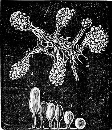
Fig. 2.—The plasmodium (a) of Stemonitis fusca, commencing to form into sporangia (b); drawn on July 9. The dark-brown sporangia were completely formed by the next morning; c-e shows the development of their external form.
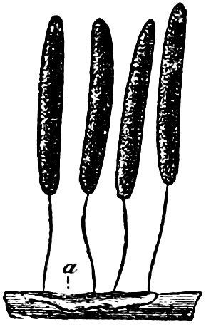
Fig. 3.—Four sporangia of Stemonitis fusca, fixed on a branch. a The plasmodium.
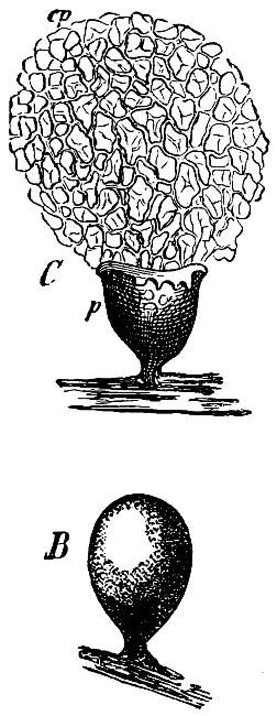
Fig. 4.—Sporangium of Arcyria incarnata. B closed; C open; p wall of sporangium; cp capilitium.
The Myxamœba grows whilst taking up nourishment from the material in which it lives, and multiplies by division. At a later stage a larger or smaller number of Myxamœbæ may be seen to coalesce and form large masses of protoplasm, plasmodia, which in the “Flowers of Tan” may attain the size of the palm of a hand, or even larger, but in most others are smaller. The plasmodia are independent, cream-like masses of protoplasm, often containing grains of carbonate of lime and colouring matter (the latter yellow in the Flowers of Tan). They creep about in the decaying matter in which they live, by means of amœboid movements, internal streamings of the protoplasm continually taking place; finally they creep out to the surface, and very often attach themselves to other objects, such as Mosses, and form sporangia (Fig. 2). These are stalked or sessile and are generally cylindrical (Fig. 3), spherical or pear-shaped (Fig. 4); they rarely attain a larger size than that of a pin’s head, and are red, brown, white, blue, yellow, etc., with a very delicate wall. In some genera may be found a “Capillitium” (Fig. 4 cp), or network of branched fine strands between the spores. Flowers of Tan (Fuligo septica) has a fruit-body composed of many sporangia (an Æthalium), which has the appearance of flat, irregular, brown cakes, inside the fragile external layer of which a loose powder, the spores, is found. It generally occurs on heaps of tanners’ bark, and appears sometimes in hot-beds in which that material is used, and is destructive by spreading itself over the young plants and choking them.
All the motile stages may pass into resting stages, the small forms only surrounding themselves with a wall, but the large ones at the same time divide in addition into polyhedral cells. When favourable conditions arise, the walls dissolve and the whole appears again as a naked (free-moving) mass of protoplasm.
To the genuine Slime-Fungi belong: Arcyria, Trichia, Didymium, Physarum, Stemonitis, Lycogala, Fuligo, Spumaria, Reticularia.
Some genera wanting a sporangium-wall belong to the Slime-Fungi: Ceratiomyxa, whose fruit-body consists of polygonal plates, each bearing stalked spores; Dictyostelium, in which the swarm-stage is wanting and which has stalked spores. Plasmodiophora brassicæ preys upon the roots of cabbages and other cruciferous plants, causing large swellings. Pl. alni causes coral-shaped outgrowths on the roots of the Alder (Alnus). Phytomyxa leguminosarum may be found in small knobs (tubercles) on the roots of leguminous plants. It is still uncertain whether it is this Fungus or Bacteria which is the cause of the formation of these tubercles.