Part 5 — CHAPTER XXVI. CYSTS AND TUMORS. (continued 2)
Reading edition
Fig. 84
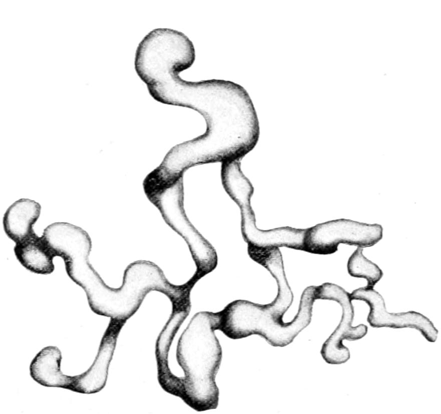
Plexiform neuroma, dissected free from all adherent tissues. (Lexer.)
Fig. 85
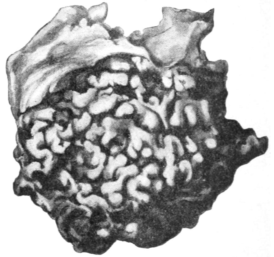
Plexiform neuroma of chest wall in a young child. Illustrating its gross external resemblance to lymphangioma. (Lexer.)
Plexiform Neuroma.
—Plexiform neuroma is relatively rare. This is a type of nerve tumor in which all the branches of a given nerve which are distributed to a particular area become enlarged and elongated, the overlying skin being stretched and thin. Such a tumor seems like a loose bag containing a number of vermiform bodies, resembling the sensation given when palpating a varicocele. On section each of the affected nerves reveals a quantity of myxomatous tissue replacing the nerve sheath. They are in large measure congenital. The skin overlying a plexiform neuroma will frequently be found to be pigmented, variously altered in thickness, and covered with fine hair. These growths have been frequently mistaken for lymphangioma (Figs. 84 and 85).
Malignant Neuroma.
—Malignant neuroma (so called) will generally be found to be a true sarcoma of nerve structures, usually of the spindle-cell variety. Traumatic neuroma is often seen in amputation stumps, where the terminations of the divided nerves become bulbous, attaining the size of cherry stones, the tumors being composed of a mixture of connective tissues and nerve fiber, from which in time the true nerve structure usually recedes or vanishes. They form when suppuration has been profuse or healing long delayed, and when sufficient care has not been exercised to prevent entangling of the nerve ends in the scar of the wound. They give rise to much pain, and often necessitate re-amputation. The bulbous enlargement is the result of prolonged irritation in a nerve, and has been noted around various foreign bodies.
True neuroma is innocent in tendency, though often painful. It is the sarcoma of nerve tissue which produces signs of malignancy. A true neuroma which causes unendurable pain should, when accessible, be removed. It is sometimes possible to separate the tumor mass from the balance of the nerve trunk, and thus to remove it without excision of the nerve. At other times it is impossible to avoid division and ensuing paralysis. Divided nerve ends should be brought together by catgut suture, by which means it may be possible to avoid permanent loss of function. Nerve grafting is also resorted to for repairing such defects. Removal of painful neuromas due to injuries to the head has more than once been the means of curing traumatic epilepsy.
6. Tumors Derived from Epithelium.
These tumors consist of specific epithelial elements supported and more or less bound together by a vascular connective-tissue stroma. The only apparent exception to this statement is tumor of dental tissue. The teeth are positively modified and petrified or calcified epithelial products.
Odontoma.
—The odontomas are tumors composed of one or more of the dental tissues, arising either from tooth changes or teeth in process of development. They may be divided, according to Sutton, as follows:
1. Epithelial Odontomas.
—These are provided with a capsule, and present usually as a series of cysts separated by thin septa, containing mucoid fluid, while the growing portions have a reddish tint not unlike sarcoma. They are most frequent about the twentieth year of life, but may occur at any age. They probably arise from persistent remains of the epithelium of the original enamel organs.
2. Follicular Odontomas.
—These are often called “dentigerous cysts.” They arise in connection with permanent teeth, and especially with the molars, sometimes attaining great size and producing conspicuous deformity. The tumor consists of a wall representing the expanded tooth follicle, and a cavity containing viscid fluid, with some part of an imperfectly developed tooth, occasionally loose and more or less displaced in location. The cyst wall always contains calcareous material. These tumors rarely suppurate. They occur also in animals.
3. Fibrous Odontomas.
—These consist of condensed connective tissue in a developing tooth, presenting as a tumor with a firm outer wall and a loose inner texture, blending at the root of the tooth with the dental papilla and indistinguishable from it. The developing tooth thus becomes enclosed within the capsule before it protrudes from the gum. These tumors are most common in ruminants, being often multiple.
4. Cementoma.
—A tumor of fibrous character whose capsule has ossified or calcified, the developing tooth thus becoming embedded in a mass of dental cementum. These tumors occur most frequently in horses.
5. Compound Follicular Odontomas.
—These are tumors containing a number of masses of cementum resembling small teeth, or even amounting to well-formed but ill-shaped teeth composed of all three dental elements. In such a tumor teeth may be found in great numbers. They occur in the human subject as well as in animals.
6. Radicular Odontomas.
—These are tumors which arise after the crown of the tooth has been completed and while its roots are yet in process of formation. The crown, being unalterable enamel, does not enter into the composition of these growths, which then consists of dentine and cementum in varying proportions. They are rare in man, but frequent in other animals, and often multiple.
7. Composite Odontomas.
—These are hard tumors, bearing little or no resemblance in shape to normal teeth, occurring in the jaws, consisting of a conglomeration of enamel, dentine, and cementum, presenting abnormal growth of all the elements of the tooth germ. So far this tumor has only been found in man.
Little is said about the odontomas in general surgical literature. These tumors, as they grow, are often regarded as due to necrosed bone or to unerupted teeth, while fibrous odontomas have been often regarded as myeloid sarcomas. No tumor of the jaw, especially in young people, should lead to excision of the jaw until it has been demonstrated that the tumor is not one of the above forms. When diagnosticated as true odontoma its complete removal is all that is necessary.
Papilloma, or Fibro-epithelioma.
—The type of papilloma is this common wart, consisting of a central stem of fibrous tissue and bloodvessels covered by epithelial projections and proliferations. Papillomas are usually sessile and villous.
1. Warts.
[14]—These are sessile papillomas, most common on the skin, often seen on mucous surfaces, and occurring sometimes singly, often in crops. They are exceedingly common about the perineum, where skin and mucous membrane meet, and are regarded as due to the irritation of specific discharges. The papillomas occurring about the genitalia are known as condylomas. The growths in these instances are frequently so luxuriant and proliferative that they assume fungoid shape, and are called mulberry growths. Warts grow slowly or rapidly according to circumstances. Warty growths may attain enormous size and become vascular. Late in life they are frequently the starting points of epithelial ingrowths, and then become true epitheliomas—i. e., cancer. Warty growths sometimes line the buccal cavity and complicate cases of macroglossa. They occur also in the larynx, and when situated near the glottis may cause dyspnea and fatal obstruction to respiration. It is claimed by some that cutaneous warts will disappear with continued small internal dosage of Fowler’s solution. (See Plate XXI.)
Fig. 86
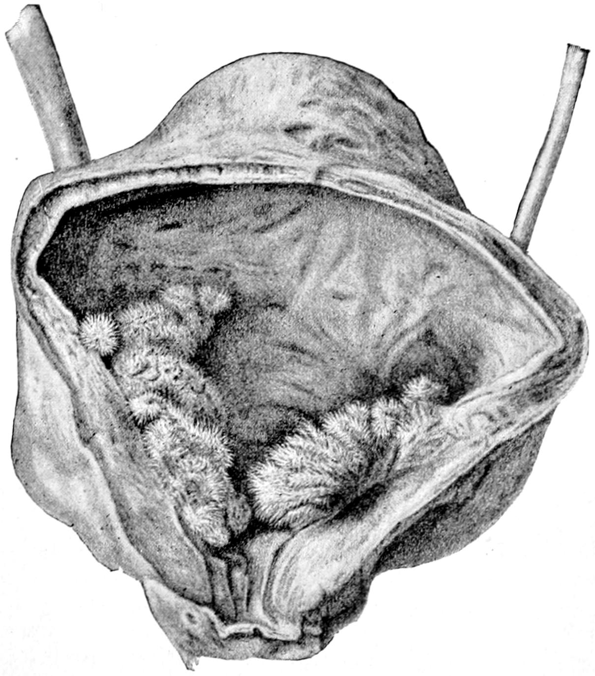
Papilloma of the bladder.
2. Villous Papillomas.
—These are met with most commonly in the bladder, occasionally in the pelvis of the kidney. They are identical with chorionic villi, and occur most often singly. It frequently happens that long, fine tufts are detached and carried away with the escaping urine. Another form of villous growth arises from the choroid plexuses of the lateral ventricles in the brain. These may grow and attain a size sufficient to produce disturbance (Fig. 86).
3. Intracystic Villous Growths.
—These are seen, for example, in mammary cysts. These, of course, are lined with epithelium, which acts here as it does in other localities, and proliferates more or less rapidly under unknown circumstances. In dealing with paroöphoritic cysts the presence of these growths has also been alluded to.
4. Ovarian Papilloma.
—There is a form of ovarian papilloma which partakes of the nature of a malignant tumor, in that separated particles seem to attach themselves to peritoneal surfaces, where they grow luxuriantly. Either this is an expression of parasitism or infectivity, or else of the implantation of tumors, which, to the writer’s mind, constitutes a strong argument for the parasitism of cancer. After abdominal section, with removal of the original focus, these growths often disappear. This affords a parallel to the instances of cure of tuberculous peritonitis after the same procedure.
PLATE XXI
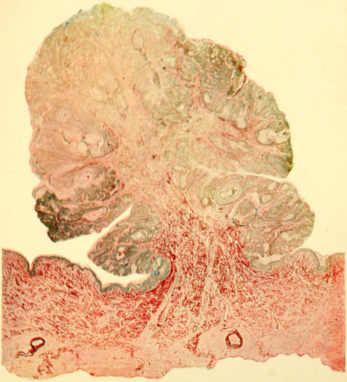
Photographic Reproduction of Papilloma. Low power. (Gaylord.)
5. Cutaneous Horns.
—These are also epithelial outgrowths, and are met with in four varieties (Sutton):
(a) Sebaceous horns, quite common, arising by protrusion of contents of a sebaceous cyst through a rupture in its wall or through its duct, with consequent desiccation by exposure to the air, while fresh material is consequently added at the basis so long as sebaceous secretion continues. These growths soften when soaked in weak liquor potassæ.
(b) Warty horns, structurally identical with the above, but growing from warts instead of from sebaceous cysts. Both these forms are often found about the head. Cutaneous horns are also met with in ovarian dermoids. They are common in the lower animals and may attain large size.
(c) Horns growing from cicatrices, especially of bones, are rare, but a cornified condition of the cicatrix itself, with formation of scales resembling those from horns, is not uncommon.
(d) Nail horns are simply overgrown nails, occurring on the digits and toes of bedridden patients who never walk (Fig. 87).
Fig. 87
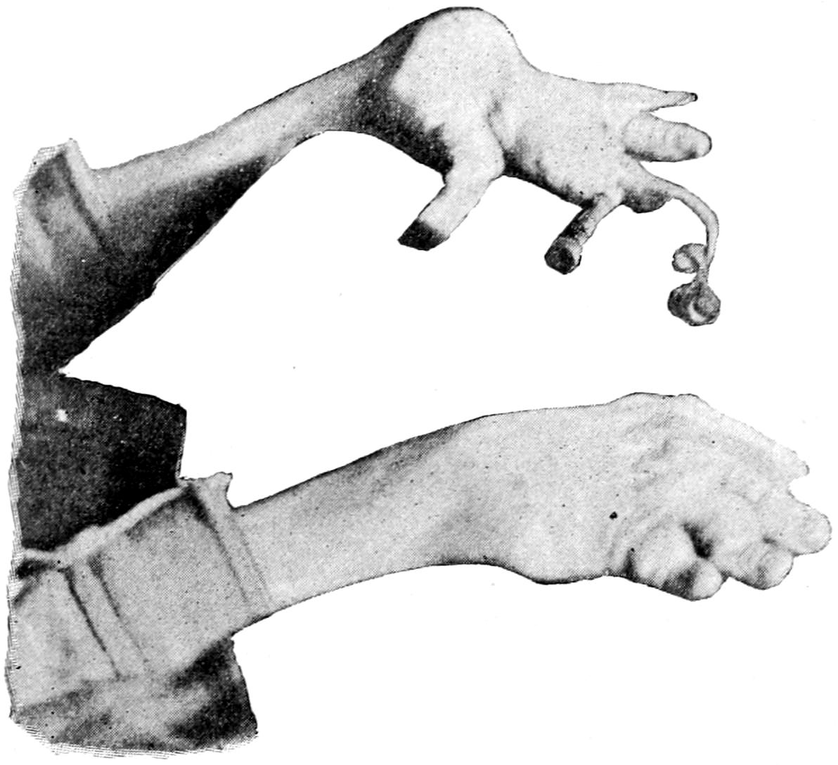
Nail horns. (Original.)
Treatment.
—All these forms of epithelial outgrowth call for radical removal, which implies complete extirpation of the membrane or tissue from which the growth occurs, after which, if effected, there is no recurrence. If some be left there is tendency to recedive.
Mucous Polyp.
—Similar papillary and often pedunculated epithelial tumors frequently hang or project from the mucous membrane—e. g., the rectum. The pedicle really projects from the submucosa. Between the layers of the overgrown mucosa are found altered glands. So long as the growth of these polyps is toward the exposed surface they are innocent and wellnigh harmless, unless they attain fair size; but so soon as they grow inward and the boundary of the submucosa is transgressed they assume malignant aspects at once. Such transformation is by no means rare, and constitutes a strong argument for their prompt removal.
Goitre; Struma.
—Pathologically the various enlargements of the thyroid known as goitre or struma constitute essential neoplasms. (See chapter on Regional Surgery of the Neck.) In this condition either the epithelial or the connective tissue may be primarily at fault.
1. Struma Parenchymatosa Nodosa.
—This includes also the colloid and the cystic varieties, and refers to an enormous overproduction of the epithelial elements (parenchyma) in distended alveoli, where they often undergo colloid softening. So marked are these changes in numerous instances that multiple cysts (minute or large) result. The collective volume of such altered tissue may be very large.
2. Struma Fibrosa.
—This presents itself in the way of dense enlargement of the thyroid, the stroma being the tissue now involved, even to the extent of causing much of the alveolar structure to disappear or become obliterated. In this condition calcification is common, and calcareous concretions or patches are often found.
Even benign tumors of the thyroid show occasionally a tendency to metastases. Cases are on record of benign goitre causing general metastases, and even of metastasis without noticeable thyroid enlargement. These occur most often in the bones, less frequently in the lungs and other organs. They are more common when the goitre has undergone colloid changes. The reasons for these changes are unknown.
In either form hemorrhages are common, with their resulting blood cysts or their solid residue, in which case pigment is usually found. Both forms are often accompanied by enlargement of the vessels, and sometimes these become enormously dilated and constitute an almost insuperable obstacle to successful removal. (See Thyroidectomy.)
Ovarian Cystoma.
—The cystomas of the ovarian region assume two types: (1) Glandular cystoma, and (2) papillary cystoma.
1. Glandular Cystoma.
—The glandular type produces the multilocular forms, with numerous small and large cavities, filled with fluid which varies in color and appearance within wide limits, having usually the consistency of mucus or thin pus, and containing a small number of cylindrical epithelial cells. The cyst wall may contain tubular glandlike structures reaching into the surrounding connective tissue.
2. Papillary Cystoma.
—The papillary type presents projections into cavities of papillomatous outgrowths from their walls, which are covered by cylindrical epithelium, which latter also lines the cavities. It is most common in the parovarium.
It is rare to find a pure type of either variety; both forms are usually blended. Malignant transformation, of the latter type especially, occurs easily and insidiously, and explains many disappointments in result.
Adenoma and Fibro-adenoma.
—Adenoma is a tumor whose type is the normal secreting gland, from which it differs in being an abnormal outgrowth or product, but particularly in that it has no power of producing the secretion peculiar to the gland tissue or type from which it grows. The adenomas occur for the most part as circumscribed tumors in the mammæ, parotid, thyroid, liver, and in the mucous membranes of the bowels and the uterus. They may be single or multiple; in the intestine they are usually multiple. In certain locations (e. g., the mammæ) they attain enormous dimensions, and in the ovary tumors of this character may be met with weighing forty or fifty pounds. The true adenoma shows no tendency to infection of neighboring lymphatics, and gives rise to no secondary deposit, and when it causes death it is usually because of size or pressure upon important organs. It displays a marked tendency to cystic alteration, while the relative proportion of epithelium and connective tissue or stroma varies within wide limits. In some cases, in which the former is small in amount, the preponderance of the latter has caused the use of the term adenosarcoma, which is really a misleading name.
The distinction between adenoma and true carcinoma is in some respects but slight, and this fact will account for the conversion which many innocent gland tumors seem to undergo from one into the other. As soon as the epithelial cells lose their regularity of disposition and collect in groups, or make their way outside of the acini into the tissues, then the change from the benign to the malignant tumor has begun, and the entire clinical aspect of the case has altered. This change may be the result of external irritation, of such tissue changes as pregnancy and lactation, or of the undefined changes which advancing years seem to produce. (See Plate XXII, Fig. 2.)
Adenoma occurs in the breast as cystic adenoma or fibro-adenoma. The former often attains large size, is encapsulated, the acini are much dilated, while from the walls of the epithelium-lined cavities frequently project papillomatous processes, forming what are called intracystic growths. Cystic adenomas grow slowly, produce atrophy of mammary tissue by pressure, occur after puberty until the menopause, and rarely give rise to pain until they become large. As they grow they distort the breast until it may become pendulous. When the growth of connective tissue, peculiar to the tumor in that it is rich in nuclei, forms well-marked partitions between alveoli, the growth is called pericanalicular adenofibroma, which may assume a tubular or an acinose type. When the alveoli and ducts are themselves invaded by ingrowth of this tissue, then we have the intracanalicular adenofibroma, which constitutes a growth sometimes bordering on the malignant. When the arrangement of epithelial cells in the acini and ducts becomes irregular and atypical, then malignant transformation has begun.
PLATE XXII
FIG. 1
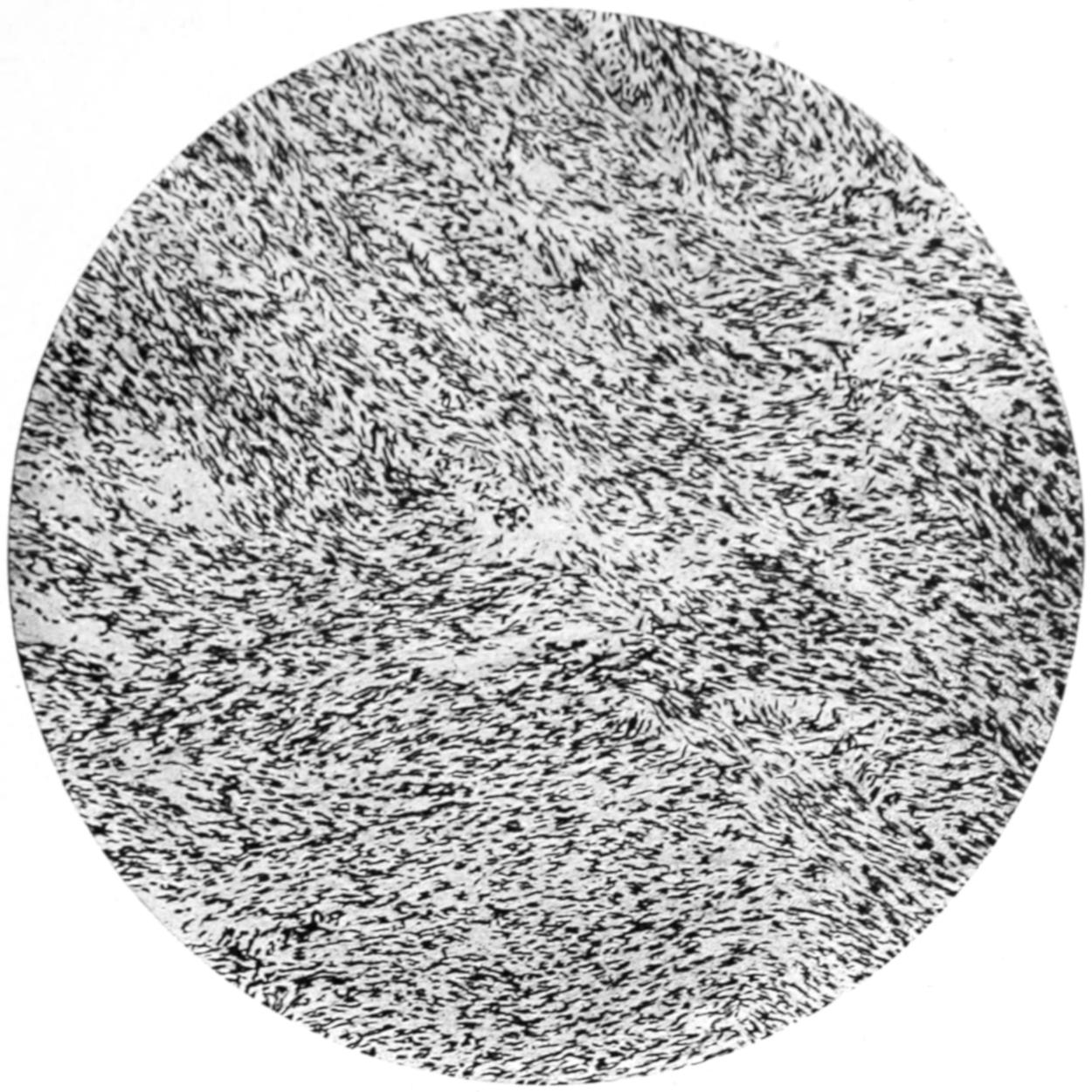
Fibromyoma of Uterus. (Low power.)
FIG. 2
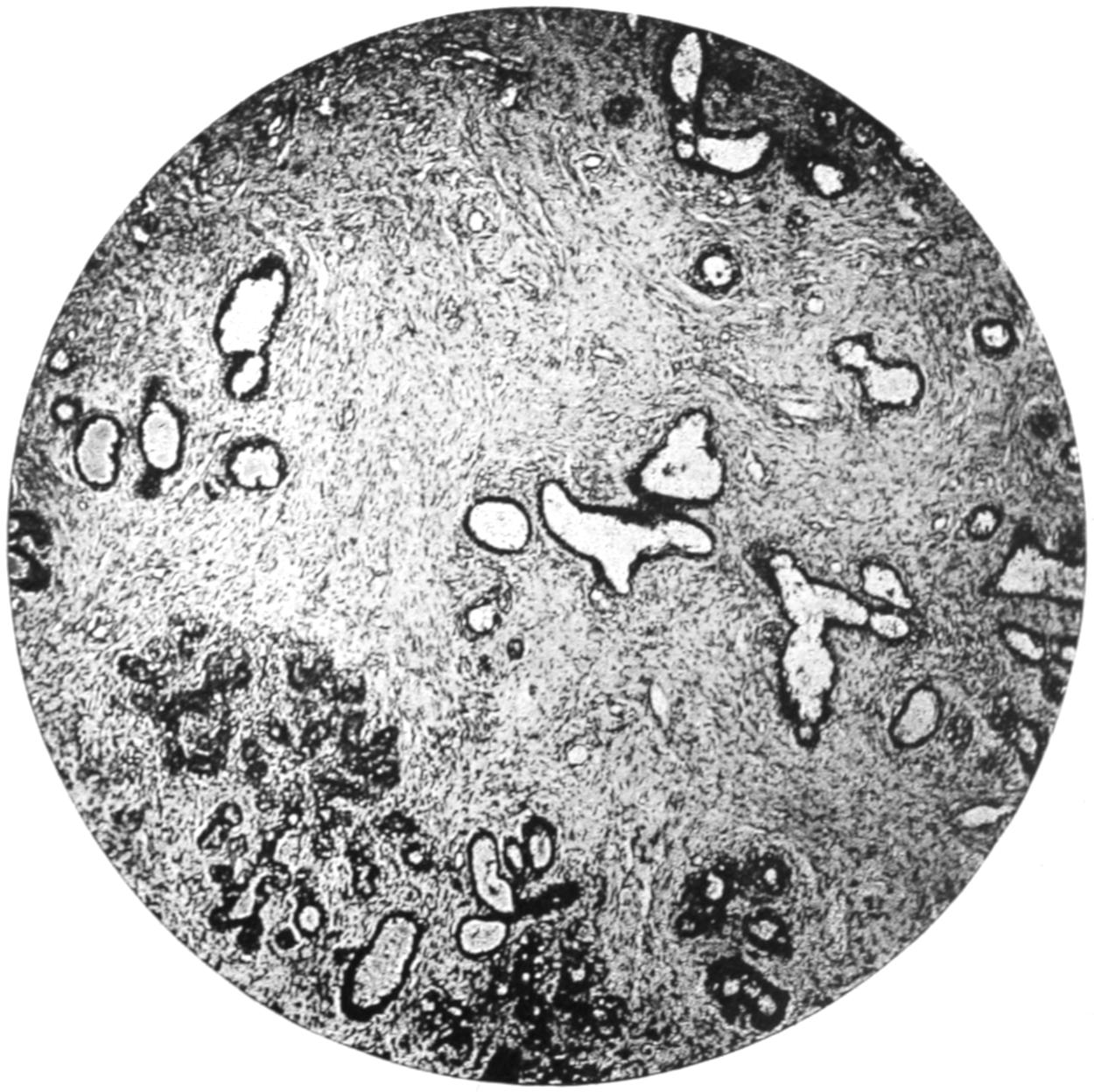
Fibro-adenoma of Breast. (Low power.)
PLATE XXIII
FIG. 1
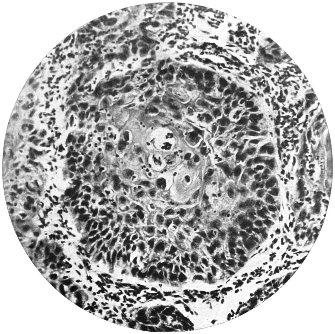
Epithelial Pearl Formation in Squamous Epithelioma. (Middle power.)
FIG. 2
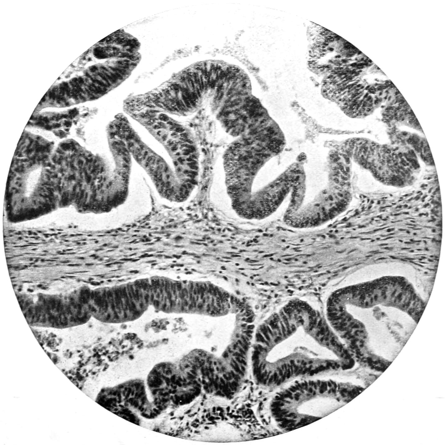
Malignant Adenoma of Rectum. (Middle power.)
Fibro-adenoma occurs also in the breast as a small tumor, encapsulated, usually superficially placed, movable in its site, often multiple; most common between the twentieth and thirtieth years of life; often painful, especially during menstruation; tender upon pressure. Both forms may occur in young men. A form of fibro-adenoma in which fibrous tissue is greatly in excess, which never attains great size, is common in the breasts of unmarried women. It gives rise to much pain and distress, but is clinically not malignant. (See Plate XXII, Fig. 2.)
Adenoma occurs frequently in sebaceous glands as:
1. Sebaceous Cysts.
—Sebaceous cysts are generally known as wens. These tumors commonly begin as retention cysts, the ducts of the sebaceous glands becoming occluded. But in many cases there is no occlusion of the ducts, and their secretion may be easily expressed. They occur wherever sebaceous glands abound, but especially upon the scalp. They are usually multiple, vary greatly in size, are easily movable over the bone, and are intimately related to the skin, while the duct orifice is frequently recognized by a black spot, after removing which sebum can be expressed. These cyst-adenomas are encapsulated, and can be easily shelled out of their matrices, save when inflamed, in which case they are often astonishingly adherent. Their contents consist of pultaceous debris resembling old epithelial scales, fat, cholesterin, etc. The contents of these cysts are very prone to decompose, and they become as offensive as anything with which the surgeon has to deal. Putrefaction may be independent of inflammation or coincident with it. When irritated these gland cysts become inflamed and may suppurate, suppuration being tantamount to cure by spontaneous processes. They may also ulcerate, without suppurating, and form foul-smelling ulcers, or give rise to cutaneous horns.
Fig. 88
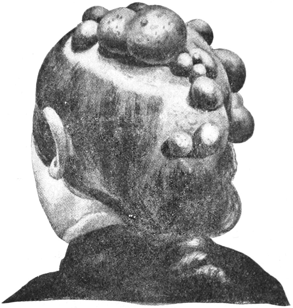
Multiple atheromatous cysts (wens). (Lexer.)
2. Sebaceous Adenomas.
—These arise from the sebaceous glands, which are lobulated, like those about the nose and ear. Adenomas from this source are extremely liable to ulceration, may undergo calcification, and are often mistaken for epithelioma because of the fungous ulcerations to which they give rise.
3. Adenocarcinoma.
—Sutton has also described an adenocarcinoma of the peculiar sebaceous glands named after Tyson. These are found particularly at the base of the prepuce, this form of tumor being rare. Adenomas arising from the mucous glands, which are usually transformed into cysts, are also known, as well as other gland tumors springing from the glands of Bartholin, Cowper, etc. (See Plate XXIII, Fig. 2, and Plate XXIV.)
Pituitary adenomas are either analogous to struma or belong to the mixed tumors of dermoid or teratomatous type.
Prostatic adenoma is in large degree fibromyoma of that body, with more or less hypertrophy of its glandular structures. Minute cystic alterations may occur also, as well as growth resembling intracanalicular fibro-adenoma.
Adenoma is occasionally observed in the salivary glands, where it is usually encapsulated, and may undergo cystic changes. It has been observed in the liver and pancreas. In the former its pseudo-ducts often contain inspissated material of bile-green tint.
The lesions of the kidney referred to as cystadenoma are now grouped among the teratomas, and are described under that heading. They present interesting examples of mixed tumors.
In the testis, as in the ovary, epithelial tumors frequently present themselves, but they partake less often of the type of pure adenoma, and incline rather to that already described under Ovarian Cystoma. Even in the paradidymis tumors of this same character are found, with cystic or even papillary alterations.
In the mucous membrane of the stomach and bowels adenoma usually presents as an ovoid tumor, attaining such size as to give rise to mechanical obstruction either by pressure or by traction. Adenoma of the pyloric region is a repetition in structure of the pyloric glands. In the rectum it presents usually as a polypoid outgrowth, often seen in young children. Such tumors are generally small, and when solitary they often hang by a distinct stalk.
Similar polypoid tumors present in the cervical canal of the uterus, where are also found sessile and racemose tumors, all of which are structural repetitions of the glands met with in the cervix uteri. Adenoma of the uterine cavity is seldom seen; it is also rare in the Fallopian tube, but occasionally presents as a dendritic outgrowth from the mucous membrane distending the tube.
Epithelioma.
—Epithelioma is common, especially where there is transition from one kind of epithelium to another, and, of all other localities, particularly where skin and mucous membrane meet—e. g., the lips, the vulva, and the anus. Epithelioma differs from papilloma in that the former is no longer limited by basement membrane, but passes beyond it into the underlying connective tissue and presents down—rather than up—growth. Characteristic of epithelioma are the so-called cell nests or pearly bodies, where there seems to be a tendency to globular arrangement of cells with such condensation or alteration that they lose their ability to take stains, and appear as a more or less lustrous mass, showing off by contrast among the standard surrounding tissue. On this account they are often called pearly bodies. Recognition of these is tantamount to diagnosis of epithelium. (See Plate XXIII.)
This form of neoplasm is essentially the same, no matter what its clinical varieties. These comprise a wart-like growth or nodule, which quickly becomes an ulcer with elevated edges, ulceration being due to necrosis of cells farthest from the periphery; or, again, the disease may start as an ulcerated fissure, ulceration and infiltration keeping pace, in which case there is a sharply defined ulcer with undermined edges. A third variety, often seen upon the lips, comprises a projecting mass, with more or less horny surface. In nearly all of these, however, the characteristic cell nests with their onion-like arrangements of cells will be found.
Epithelioma, especially when exposed to the air or to surface irritation, quickly ulcerates and tends to involve all the surrounding tissues, while occasionally the distinctive cells proliferate so rapidly as to give the ulcer more or less of a bursal or a cauliflower-like arrangement. From such a surface there is a constant discharge of foul-smelling detritus or of sloughs. Even bone cannot resist its progressive invasion and slowly disintegrates before the advancing mass. Cartilage is resistant, and usually preserves its integrity. In other words, the tendency of epithelioma is toward constant encroachment and infiltration, and toward a fatal termination from hemorrhage by ulceration, from septic infection, exhaustion, or other accidents. The wart-like forms run the slowest course of all, but even here the malignant tendency is most evident.
Lymph-node Infection.
—A striking characteristic of epitheliomas is the invasion of the adjoining lymph nodes, which attain a size disproportionate and bearing no necessary relation to that of the primary growth. This constitutes one of the most serious complications of the condition. This lymphatic invasion partakes of the malignant character of the disease, and from every focus of this character infiltration and destruction proceed. Infected nodes also show an early tendency to central degeneration and to spurious cyst formation. When the overlying skin becomes involved we have extensive sloughing and the conversion into large malignant ulcers. Dissemination to a distance (i. e., metastasis) is rare in epithelioma—much more so than in carcinoma. (See Plate XXV, Fig. 2.)
About the mouth epithelioma is not common before the thirty-fifth year, though I have seen it on the lip of a twenty-year-old woman. It is vastly more common in men than in women, and more frequent on the lower than the upper lip. In the tongue it seldom occurs before the fortieth year. It seems to be more common both on the lip and tongue in men with bad teeth and in confirmed smokers, thus giving rise to the view often held that it is purely a matter of irritation. It may, however, be due to contact infection should it be regarded as of parasitic origin. In one-fifth of the cases of epithelioma of the tongue there are preceding lesions, usually described as leukoplakia or ichthyosis of the tongue—conditions characterized by epithelial reduplication and the formation of dense plaques or scales. These lesions are usually regarded as precancerous conditions. (See Plate XXVI.)
PLATE XXIV
FIG. 1
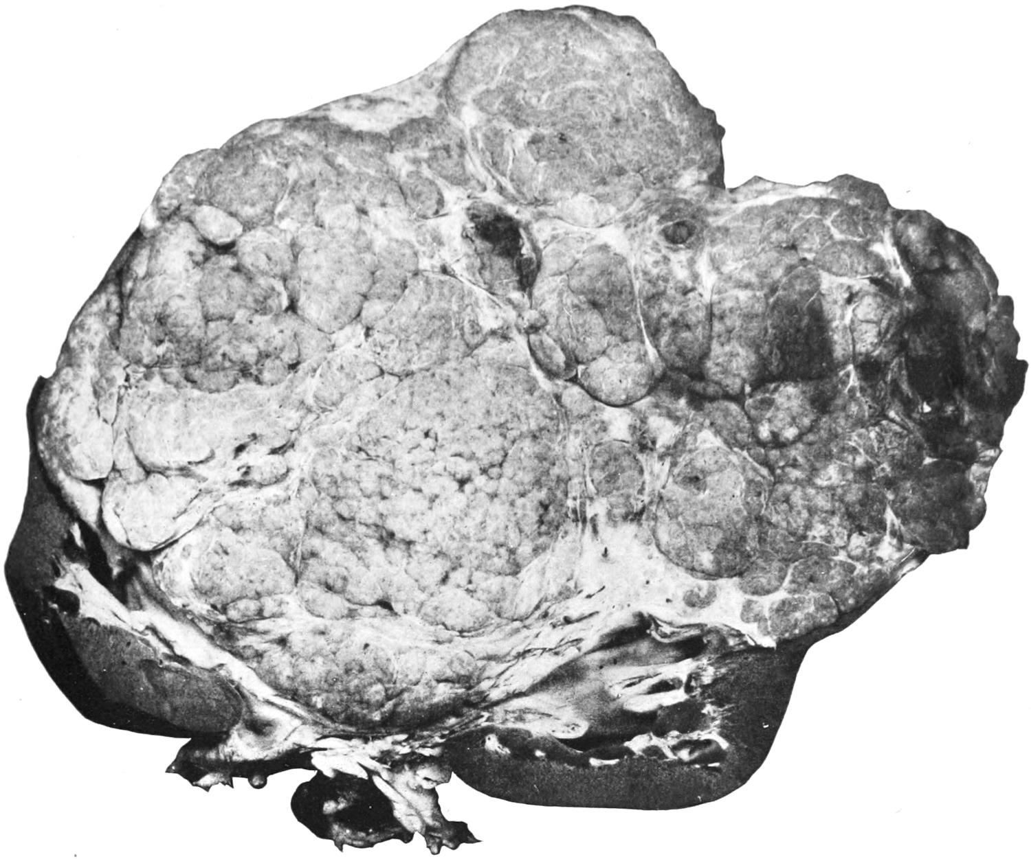
Primary Papillary Adenocarcinoma of the Kidney. (One-half original size.) (Gaylord.)
FIG. 2
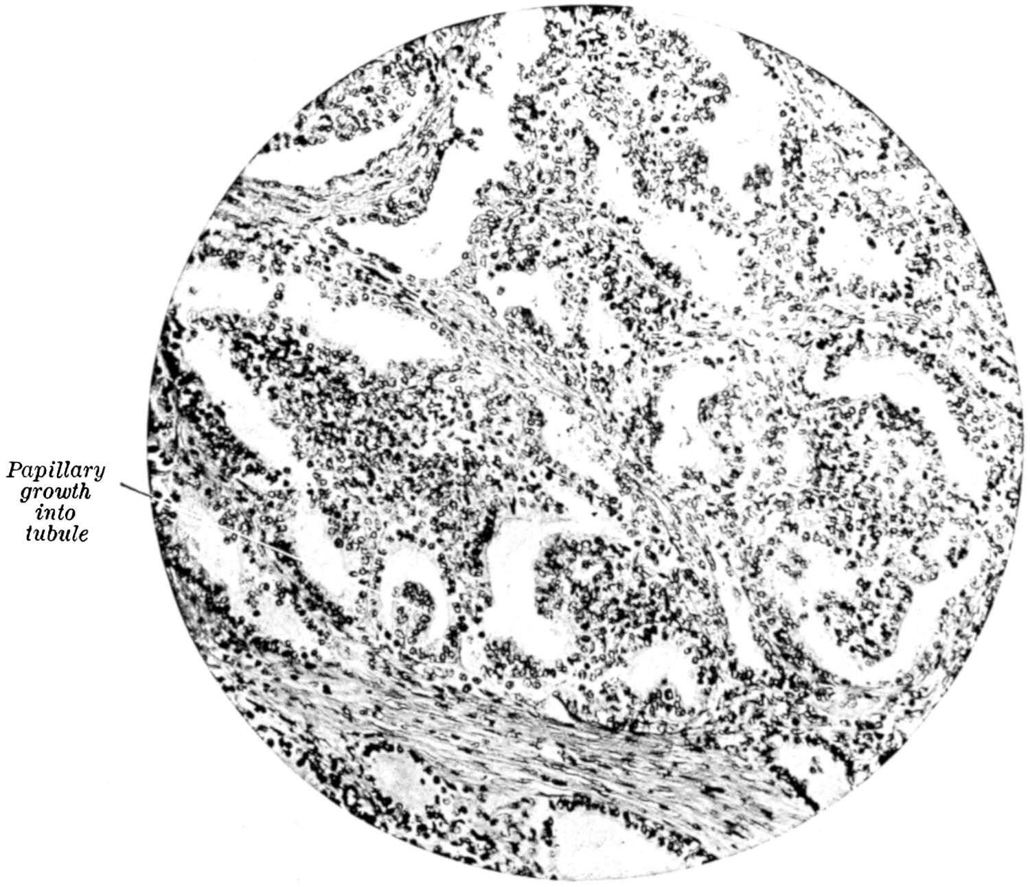
Section of the Primary Growth. (Gaylord.)
PLATE XXV
FIG. 1
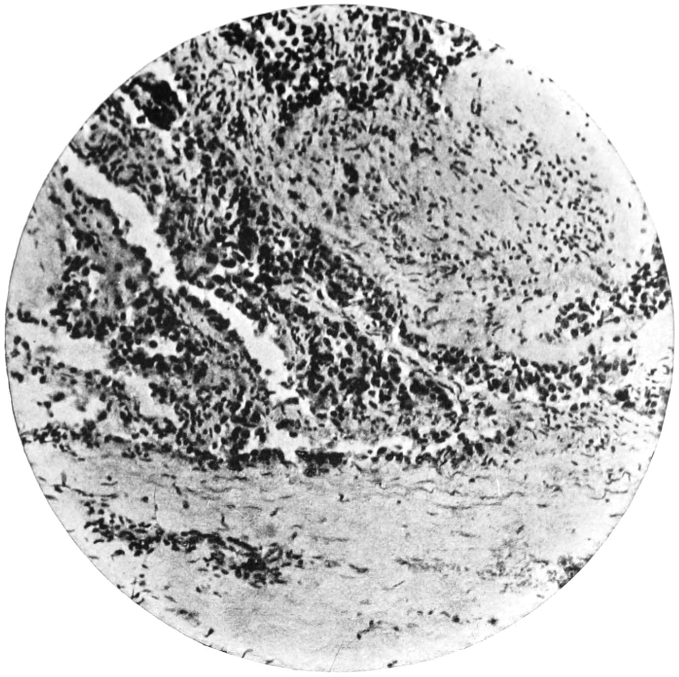
Carcinoma developing in a Thrombus in the Portal Vein. (Middle power.) (Gaylord.)
FIG. 2
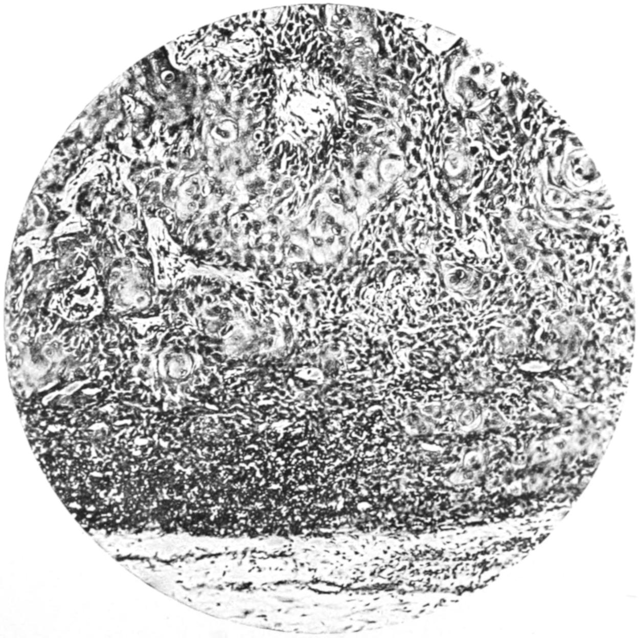
Metastasis of Squamous Epithelioma in a Lymph Node. Pearl Formation. (Middle Power.)
PLATE XXVI
FIG. 1
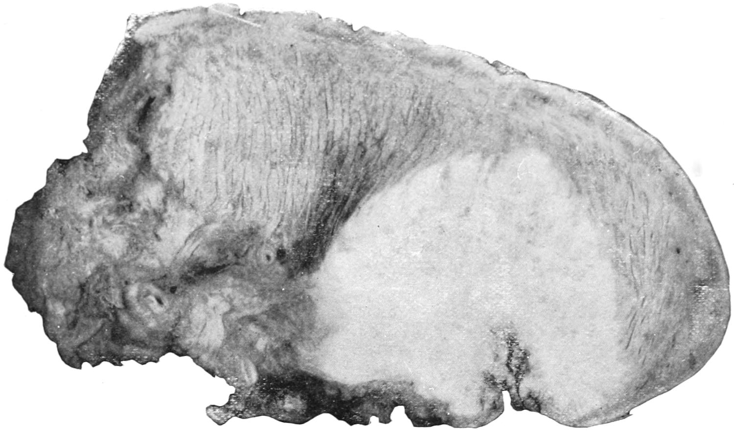
Epithelioma of Tongue. Enlarged three diameters.
FIG. 2
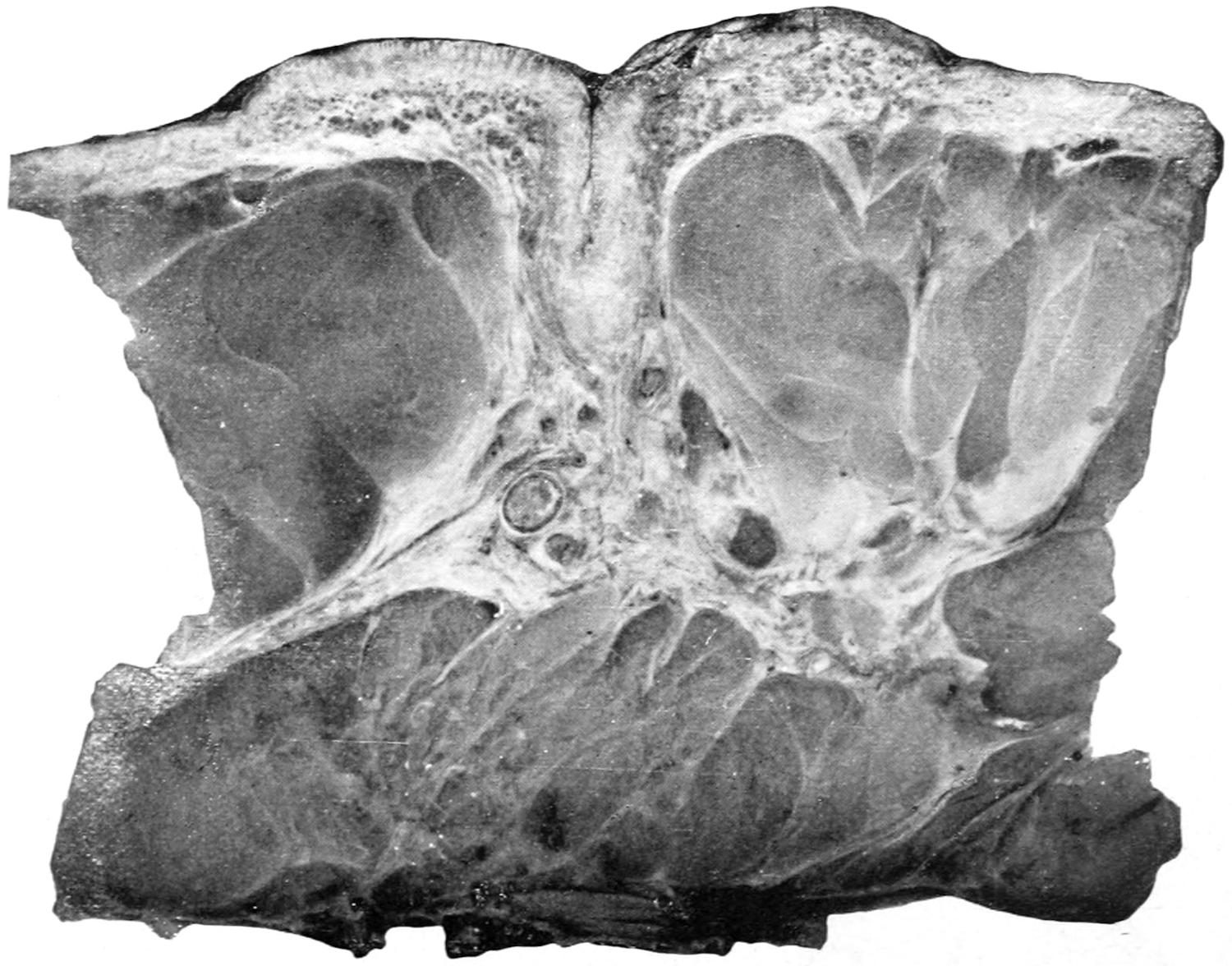
Paget’s Disease of the Nipple. Enlarged two diameters.
Photographs from hardened unstained specimens.
The disease often starts near the stump of a carious tooth, in which case infiltration and erosion begin promptly and progress rapidly. Epithelioma of the tongue has been known to follow along the obliterated track of the thyrolingual duct, and in this way to bring about a perforating ulcer.
Epithelioma of the esophagus is a common cause of stricture of this passage-way. It leads to ulceration, and usually to perforation into the trachea or some other cavity or passage (i. e., a bloodvessel). In the larynx the disease is well known, and gives rise to intense and finally fatal symptoms, but has been dealt with successfully by radical operations for extirpation of the entire organ. (See Chapter XLI.)
Fig. 89
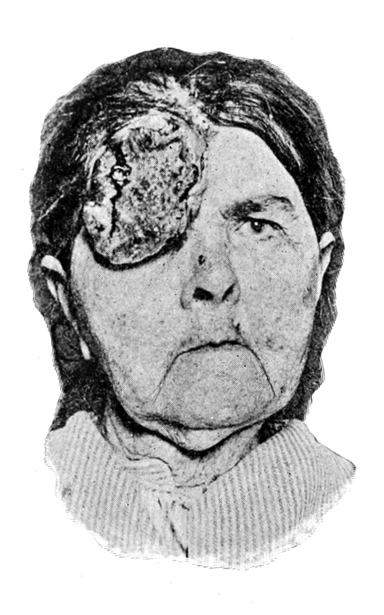
Epithelioma of forehead and eyelid. (Neisser.)
Fig. 90
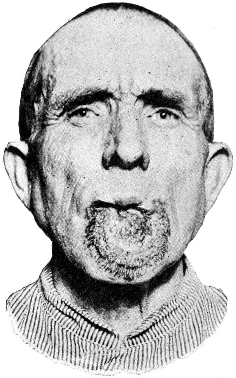
Epithelioma of lip. (Neisser.)
Occurring upon the scrotum, epithelioma has been called chimney-sweeper’s cancer, or soot-warts, and has been ascribed to the irritation of foreign material. Ulceration and infection of the inguinal nodes usually proceed rapidly and disastrously. It is believed also that tar and paraffin may produce similar irritation, and paraffin cancer has been described by various writers. It usually occurs upon the scrotum.
The skin lesions which precede the formation of paraffin cancer resemble those seen in chimney-sweeper’s cancer. The skin becomes dry, thickened, parchment-like, while the openings of the sebaceous glands become obstructed by the tar or other material, producing acne-like lesions. Warty outgrowths then occur, and these become the seat of malignant ulceration. In chimney-sweeper’s cancer the scrotum is usually first affected in a chronic dermatitis, to which warty outgrowths succeed, these enlarging and growing downward as ulceration takes place.
About the external genitalia epithelioma is not uncommon, particularly in and about the prepuce. Such a degree of phimosis as leads to retention of smegma is certainly a predisposing cause, not only in man but in the lower animals. Epithelioma of the vulva has been described under the name esthiomène, and requires to be recognized and dealt with promptly if the surgeon should attempt a radical cure. In the vagina and about the cervix uteri it is common, a large proportion of cases of cancer of the uterus being essentially epitheliomas of the cervix.
In and about scars and upon granulating ulcers epithelioma is quite common. One danger to which a chronic ulcer is always exposed is that of epitheliomatous transformation. These growths also attack lupus scars, or even any tissues actively involved in the lupoid process. This is particularly true between the fortieth and sixtieth years of life.
Among the viscera the gall-bladder is probably more often involved in distinct epitheliomatous changes than any other. It presents as a uniform thickening, and causes augmentation in size, so that a distinct tumor projects from beneath the liver. In this location dessemination is rare.
Epithelioma is to be regarded as having an essential malignant tendency. Its treatment demands early removal of diseased parts and complete extirpation of involved lymph nodes. It is only the small and incipient growths which should be attacked by such destructive agencies as cancer pastes or the electrolytic current.
Rodent Ulcers.
—Under the name of rodent ulcers, lupus exedens, noli-me-tangere, etc., writers, mostly English, have described a variety of epithelioma, met especially upon the face, to which a separate classification has usually been assigned. Until recently it has been generally regarded as a local ulceration, distinct from cancer. In some text-books it is described as lupus exedens. It is preceded usually by a nodular condition of the skin, vascular, breaking down into a regular ulceration, but little elevated, the base of the ulcer deeply excavated, with a striking disproportion between ulceration and new-growth. In this particular variety infiltration seems to be continuously in advance of the rodent process, the former being excessive, the latter but slight. This variety of epithelioma rarely produces lymphatic involvement; the discharge is slight, the pain complained of inconsiderable. Occasionally it entirely alters its aspect, and may present features of the conventional epitheliomatous type.
Fig. 91
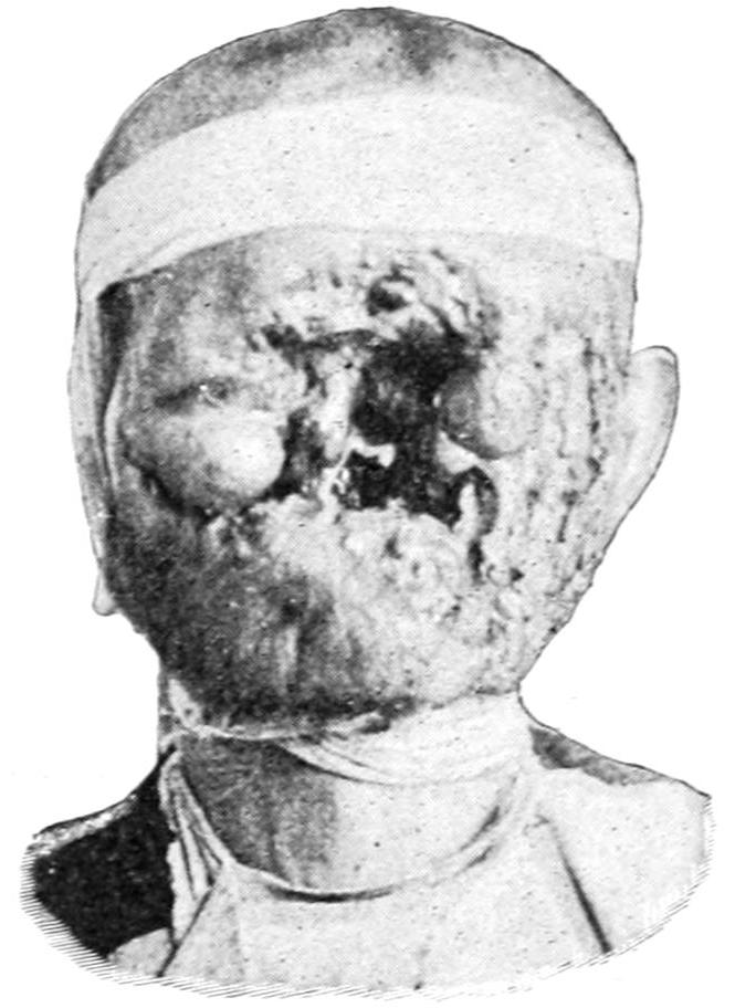
Fig. 92
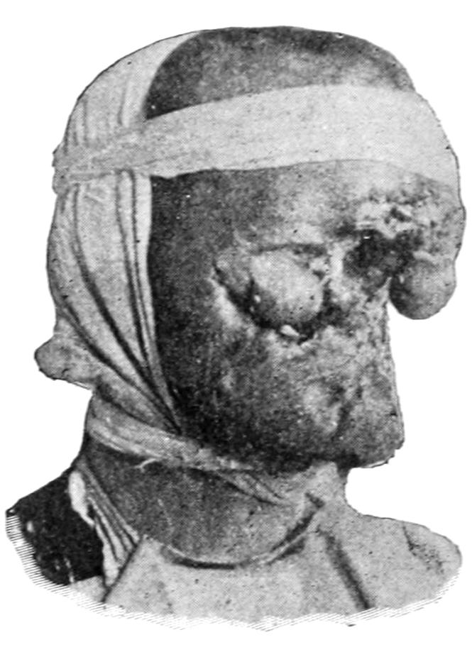
Rodent ulcer. (Original.)
The development of cancer in lupus areas is now of sufficiently frequent occurrence to demand attention. Whether the epithelium which gives rise to it is to be accounted for by Cohnheim’s hypothesis, as having been cut off in the course of healing and become a cell rest to subsequently undergo malignant degeneration, is not yet settled. It has been suggested that curettage might cause fragments of epidermis to be loosened and then entangled in the cicatrix, and thus be responsible for subsequent malignant changes. When lupus thus degenerates it assumes usually the papillomatous form, which rarely involves lymph nodes, while the change which follows x-ray treatment often succeeds a hyperkeratosis and rapidly involves gland structure.
Rodent ulcer allies itself with the type of tubular epithelioma springing from the outer sheath of the hair follicle, sending out cylindrical processes which freely blend with one another. It is to be regarded as an equally malignant type of ulceration with other cancerous ulcers, and demands the same thorough and radical measures for its relief as do other forms of epithelioma. It is perhaps the most favorable one with which to deal, because of the usual freedom from involvement of deep lymphatics. No distinctive measures are necessary for its relief—only those which are thorough.
Carcinoma.
—Carcinoma is a tumor springing from preëxisting gland tissue, which it more or less closely resembles in type, save that the structural similarity is incomplete, the epithelial cells now collecting in irregular clusters, or filling the acini and obstructing the ducts, or bursting beyond the basement membrane and invading the surrounding tissues. They frequently so fill the ducts as to appear in columnar arrangement when seen under the microscope, and this has given rise to the use of a term so vague as to have no place in pathology—i. e., cylindroma. Carcinomas may arise from any of the secreting glands, but more commonly from some than from others. They have no capsules. They infiltrate the surrounding tissues, usually involve the lymphatics early, are liable to spread to the superficial tissues and to ulcerate, and to undergo various degenerative changes. Nearly all cancerous tumors abound in lymphatics, which will explain the rapidity with which the lymph nodes become infected, as well as the tendency to dissemination, which is characteristic of these growths. Dissemination leads to so-called secondary or metastatic growths, which may make their appearance in any organ or tissue, even in the bones, where they give rise to changes of texture that make spontaneous fracture easy. It is characteristic of carcinoma that the metastatic tumors which it may produce will reproduce almost perfectly the type of the primary tumor whence the embolic fragments which have produced them spring. The amount of dissemination varies exceedingly: it may even become so marked and widespread as to produce a condition analogous to that met with in miliary tuberculosis—miliary carcinosis. A similar condition, much more rare, is seen in dissemination of sarcoma, and is known as miliary sarcomatosis. A constantly spreading cancerous infiltration of the superficial tissues, which is noted most often after mammary cancer, is described under the form of cancer en cuirasse, or jacket or corset cancer. Instances will be seen in which this infiltration of the surrounding structures has extended nearly or even completely around the thorax. It gives rise to a brawny induration which is unyielding, and is studded here and there by nodules that tend to ulcerate, to fungate, and to bleed easily. It is perhaps the most hopeless form of cancerous disease.
The older writers have constituted two or three clinically distinct forms of carcinoma, based mainly upon the relative hardness or softness of the tumor and the invaded tissues. The term scirrhus is thus applied to a tumor in which connective tissue preponderates and epithelial cells are relatively deficient. On the other hand, the term encephaloid has been applied to a tumor in which the connective tissue seems barely sufficient to hold the mass together, while the epithelial cells are in vast preponderance. These are all tumors of the round epithelial-cell type, and these distinctions are of clinical interest, yet have no great pathological import, save that in a general way the greater the proportion of epithelial elements the sooner will life be terminated by destructive processes. In other words, the more the tumor may partake of the encephaloid type the worse the prognosis or the shorter the probable duration of life. Again, these tumors pursue a varying clinical course. In those tumors, particularly of the scirrhus type, where the connective tissue largely preponderates, there is often an eventual reduction in the size of the part involved, and such reduction of vascularity and of nutritive activity that the rate of growth is thereby perceptibly checked. The so-called atrophying cancers of the breast are the best examples of this type of cancerous disease. Here the volume of the gland is diminished rather than augmented, and the disease may last for a number of years. It is questionable whether it is well to operate.
The so-called colloid forms of cancer are simply the expression of pathological changes occurring in growths of more distinct type. Thus colloid softening may occur in any tumor in which cancer cells predominate, and the so-called colloid cancers of the peritoneum, the ovary, etc., are either examples of such alterations or are possibly endotheliomas arising in these locations. The term villous cancer, with other terms like it, should be expunged from all scientific literature, unless these terms are used in purely adjective and clinical sense, for they imply nothing accurate as to histological structure, and are often misleading and inaccurate.
Carcinoma is most common in the following regions:
In the breast it appears particularly in two forms:
1. Acinous Cancer; and
2. Duct Cancer.
1. Acinous Carcinoma.
—Acinous carcinoma is usually of the scirrhus type. It may arise at any portion of the breast, and if anywhere near the nipple it will cause retraction of that prominence, which is always pathognomonic; elsewhere it leads to puckering and adhesion of the overlying skin. These tumors infiltrate widely, especially along the connective-tissue stroma and the fibrous tissue which intersperses the fat of the breast. They are usually firm and sometimes exceedingly dense. A form of scirrhus known as atrophying scirrhus consists largely of strands of fibrous tissue, injected here and there with epithelial cells. It is the slowest in growing of all the forms of cancer, and by its contraction tends to reduce rather than augment the size of the mamma.
Acinous cancer is rare before the age of thirty, most common between forty and fifty. It occurs in women in all conditions of life, married and single, but is rarely noted in the male breast. The most dangerous form is that which appears during lactation. Ordinarily its progress is slow. As it augments in volume it infiltrates the surrounding tissues, becomes adherent to the pectoral fascia, infiltrates the muscle fibers, and finally attaches itself to the periosteum of the ribs. The infiltrated tissues tend to shrink rather than to increase in volume. Lymphatic injection occurs early in this form, and is a pathognomonic sign. It occurs mostly in the axillary lymphatic nodes, but may often be detected in the neck above the clavicle. When the skin is involved there is a tendency toward ulceration and fungoid condition. This is preceded by the purplish appearance of the tense skin. (See Plate XXVII.)
Fig. 93
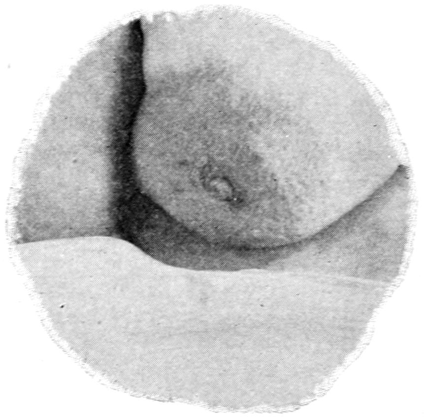
“Pig-skin” appearance of cancerous breast.
Pain is an uncertain and variable feature. It is important to emphasize this fact, as many of these conditions have been lightly regarded because of freedom from pain. Pain is not a constant phenomenon in cancer. On the other hand, it is sometimes intense, either localized or radiating and referred to distant points. Pain is particularly noticed in cases which assume the form of cancer en cuirasse. Secondary deposits in viscera frequently occur, particularly in the abdominal organs and the lungs; but any organ may be the seat of secondary infection, and this is found occasionally in the bone-marrow, not alone of the sternum or ribs, but of distant bones, and is called marrow injection. As the result of cancerous affection of serous membranes effusions of fluid frequently take place, as in the pleura, peritoneum, and pericardium, and this fluid is often blood-stained.
In consequence of pressure upon the venous trunks in the axilla there is often a swelling of the arm upon the affected side, dropsical in character, known as lymphatic edema. The arm grows heavy, the patient loses control of it, and the skin may become so distended by effusion as to cause the limb to resemble a cast. This is due not alone to pressure upon the veins but to involvement of the lymphatics, and upon careful examination positive dilatation of the lymphatic vessels may be noted. Pain is a usual accompaniment of this form of edema.
2. Duct Carcinoma.
—This appears especially about the time of the menopause, when glandular structure has disappeared and only ducts remain. It is common, without reference to cancer in these instances, to find cystic dilatation of numerous ducts, which vary in size from a mustard seed to that of a cherry. These are referred to by Sutton and others as involution cysts. They are filled with mucoid material and have a bluish tint. They occur usually upon the under surface of the gland. Such cystic breasts are common, and when appearing in diffused form may be easily mistaken for cancer. Pain is not frequent. This condition is certainly a precancerous stage, since the dilated ducts are often the starting points of cancer, and occasionally of papillomatous or villous outgrowths from their walls.
Duct cancer implies the form which arises in these dilated ducts, most commonly in the terminal branches, appearing ordinarily as a single tumor, but sometimes as a mass of separate nodules. Intracystic and intracanalicular growths of this character are often found. When assuming the truly cancerous phases they may be spoken of as duct cancers, otherwise as duct papillomas. They have generally been referred to as intracanalicular fibromas. Duct cancers are less tense than the preceding variety, and when situated near the surface often discolor the skin. It is from these cases that there is seen a more or less abundant discharge of fluid resembling bloody milk. These tumors grow slowly, lymphatic involvement is late, and in general they present the least malignant forms of breast cancer.
PLATE XXVII
FIG. 1
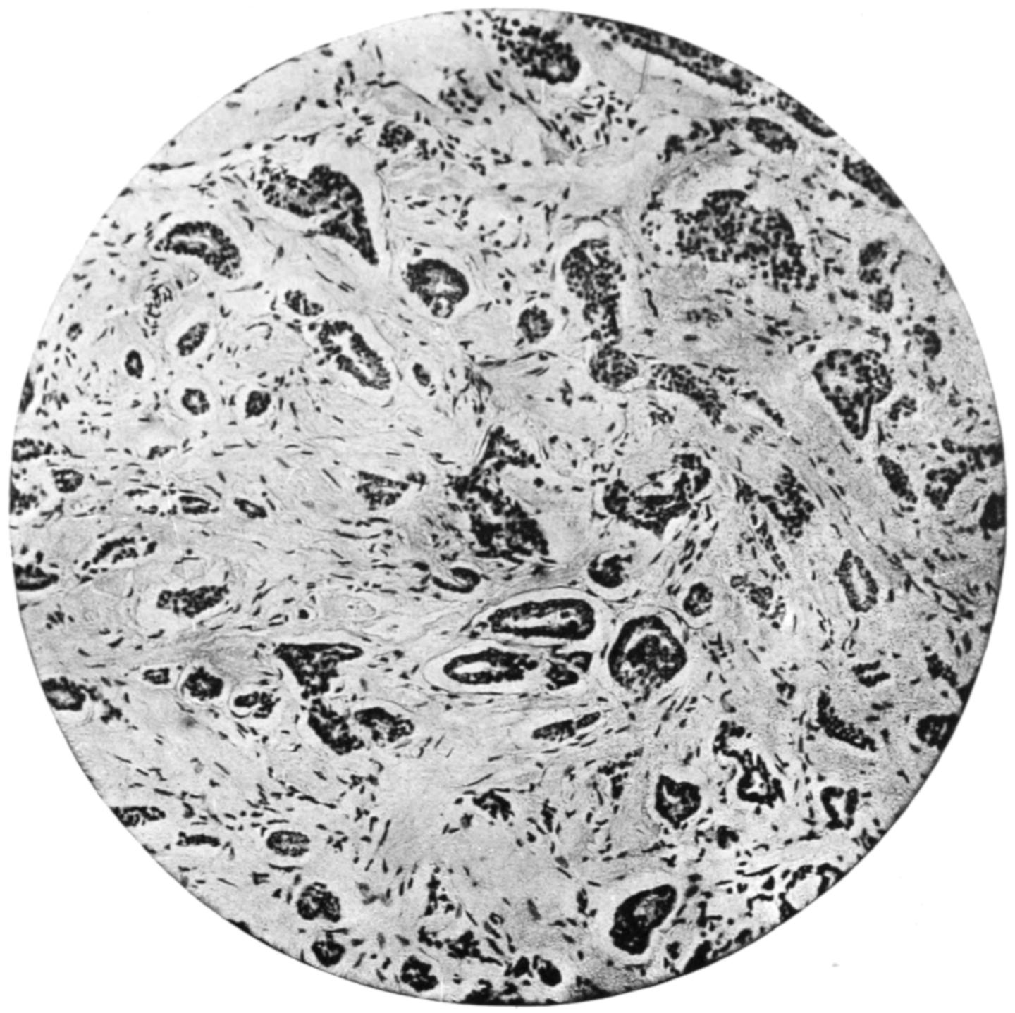
Scirrhus Carcinoma of Breast. (Middle power.)
FIG. 2
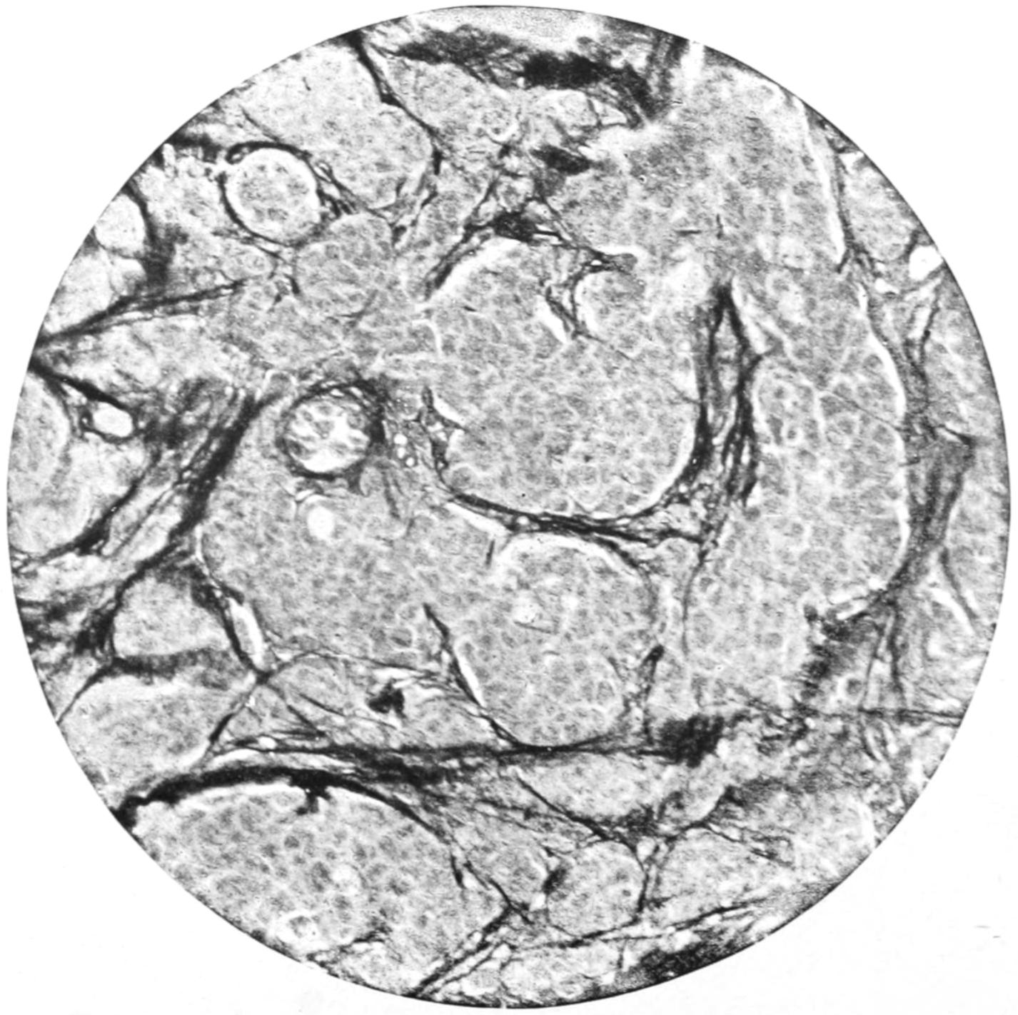
Soft Infiltration Carcinoma of Breast, showing Stroma. (Mallory’s connective-tissue stain.)
Carcinoma of sebaceous glands is by all means most common in those specialized glands named after Tyson, occurring about the prepuce. They give rise to the usual forms of cancer in this locality.
Carcinoma in the prostate is not common, and is usually confined to old men. Infiltration proceeds around the base of the bladder at the same time and binds the pelvic viscera together. The pelvic lymphatics become early infected and dissemination is frequent. (See Prostatic Hypertrophy.)
Fig. 94
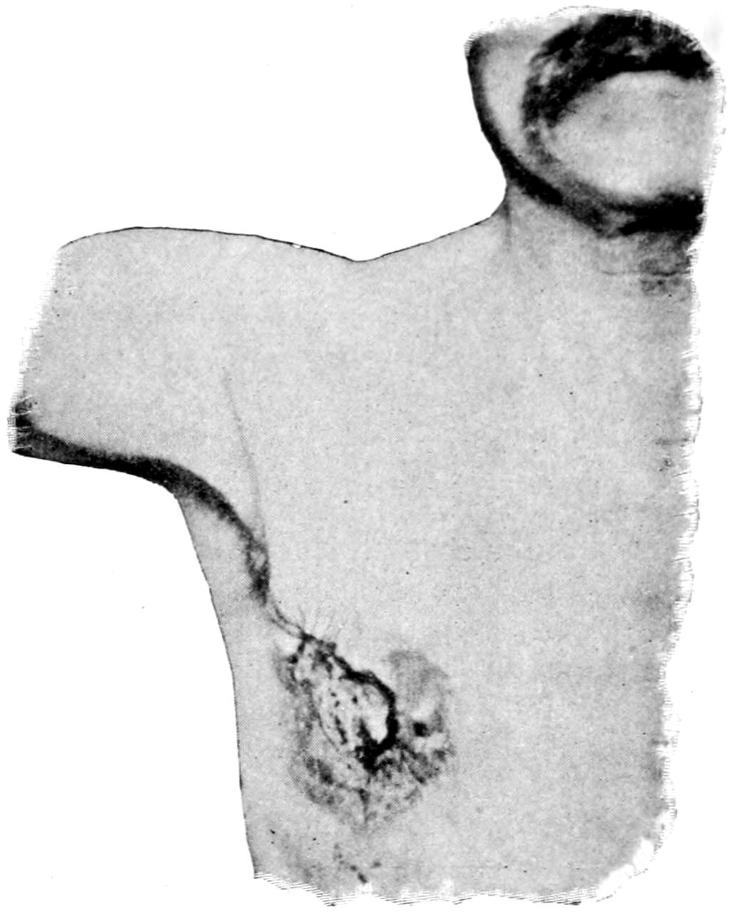
Recurring carcinoma of male breast. (Original.)
Carcinoma in the salivary glands is not common; it is more frequent in the parotid region, occurring at middle life, growing rapidly, infiltrating surrounding parts, and tending to ulceration.
Carcinoma of the liver varies in its arrangement and appearance. Sometimes it appears in the form of nodules; at other times, as a more diffuse malignant infiltration by cells relatively abundant in number, so that the clinical aspects of the case conform rather to the encephaloid or medullary type.
Carcinoma of the kidney was formerly described as encephaloid, meaning thereby simply a malignant tumor of soft structure. It is probable that a large proportion of these tumors were sarcomas. Nevertheless, true carcinoma of the kidney is possible.
Carcinoma of the ovary may originate as such, or be the result of a transformation from an ovarian cystoma (see above). No better illustration can be offered of the infectivity of cancer cells (be the secret of this infectivity what it may) than the rapid dissemination of cancer throughout the peritoneal cavity, which sometimes follows the removal of an apparently non-malignant tumor which is undergoing this change.
On the other hand, in the testicle such tumors are common—more so than sarcomas. It is likely that many of them arise from the paradidymis.
Carcinoma of the stomach is a frequent disease. It involves the tubular glands, especially in the pyloric region, and conforms to them in type. After involving the mucosa it spreads to the entire coats of the stomach and infiltrates adjacent structures, while the mesenteric lymphatics are usually early and notably involved. Were it possible to recognize this involvement early in the course of the disease diagnosis of pyloric cancer and operative interference would be much more common and hopeful. Secondary involvement is generally in the adjoining viscera, but may be seen at a distance. Miliary carcinosis has been noted after pyloric cancer. This form usually occurs between the fortieth and sixtieth years of life, the duration of the disease not being long.
In the intestine, and particularly in the rectum, carcinoma proceeds also from the mucous glands, and tends constantly to extend at its periphery and involve the entire lumen of the bowel. It seems to be inseparable from a tendency to contraction of the gut and consequent annular stricture. Ulceration, favored by surface irritation and infection, occurs almost always early. Above the rectum it usually occurs in the neighborhood of the sigmoid flexure. Cripps has observed that when cancer of the rectum spreads downward and involves the anus, it loses its typical glandular character and assumes the type of epithelioma, or squamous-cell cancer. In these cases the pelvic and mesenteric lymphatics are infiltrated and metastatic affections are common.
Carcinoma may appear in any portion of the uterus, but is more common in the lower than in the upper half. It assumes the type of the cervical glands, spreads rapidly, infiltrates widely, ulcerates early, and disseminates frequently. By extension of ulceration the formation of urinary and of fecal fistulæ is common. Pyosalpinx and hydrosalpinx are also favored, while the spread of the disease is, in fact, more common when it involves the cervix than when it involves the uterine fundus.
Malignant Chorion Epithelioma.
—This has also been called deciduoma malignum, a malignant growth of chorionic epithelium. Inasmuch as this tumor also includes a syncytial layer it has been known as syncytioma. Such tumors usually contain elements derived from both layers of the chorion. They follow pregnancy, generally within a few months, and are often preceded or accompanied by a hydatidiform mole. This growth constitutes a malignant neoplasm. It pertains to ulcerating uterine growths characterized by early extensive metastasis, which prove fatal. It has been shown that similar growths occur not only in the uterus but also in the testicle, and thus the scope of the term has been much enlarged. In its biology it resembles the sarcoma; in its histology, the carcinoma. It is more malignant than any other known growth. (See Plate XXVIII.)
Occurring within the uterus its most important clinical feature is a tendency to frequent and alarming hemorrhage. When occurring about the testicle this trouble rapidly becomes fungoid, bleeding easily and excessively, the lungs being among the first organs to show metastasis, which takes place through the blood as well as the lymphatic vessels, for the cells of these growths seem to penetrate the capillaries. By the time a diagnosis is made a case is likely to be too far advanced to admit of radical treatment. If scrapings could be examined early, shreds of syncytioma would be found, and it might be possible that a complete hysterectomy would be of use.
Metastatic nodules consist mostly of round, dark masses presenting a more or less pronounced fibrous structure. These are generally found in the lungs and cerebrum, where the vessels are large and the tissues soft. There is usually a sharp contrast between such a tumor and the surrounding tissues. The time which elapses between delivery and the appearance of the growth is from three to ten weeks. The tumor rapidly spreads to the upper portion of the vagina. The trouble probably begins some time before delivery.
The latest tendency among pathologists is to refer a growth of this kind to the teratomas. In women this tumor is particularly a teratoid growth, some cells of the fecundated ovum giving rise to neoplasms, while the ovum itself thus derived may misdevelop into a hydatidiform mole. The tumor may be properly regarded as consisting in effect of fetal cells; it is built up of these cells, without bloodvessels and connective tissue, and so belongs to a class by itself. Occurring in women it is almost always a consequence of pregnancy; occurring in the testicle or in the ovary it should be regarded as proceeding from ectodermal cells. For their treatment the earliest and most radical measures only will suffice.
Suprarenal Epithelioma; Hypernephroma.
—Grawitz has distinctly established the right of these tumors to separate consideration, for he first determined their origin and identity. Hypernephroma is a tumor, found mainly in the kidney, composed of adrenal rests, or bits of accessory suprarenal tissue imprisoned within the renal capsule. Their minute structure is often that of the adrenals, with a tendency toward the type of perithelioma. They have hitherto been considered examples of sarcoma of the kidney, but are to be abruptly distinguished from it in most instances. Tumors of this character have also been found within the capsule of the liver and along the spermatic artery. In the kidney the tumor portion is usually distinct from the renal tissue; it is often enclosed within a sort of capsule, and rarely connects with the pelvis. Hence, though exceedingly liable to hemorrhages, blood rarely escapes by the ureter. Hypernephroma is delicate in structure, and its vessels give way readily. After this has happened a true hematoma may result. (See Plate XXVIII.)
Similar neoplasms form in the adrenals themselves. These tumors vary in degree of malignancy, some of them scarcely deserving the designation malignant. They may be met at any age, but are more common in adult life. Before removal they are not to be differentiated from other tumors of the kidney. Their cells manifest this peculiarity in that they contain a notable percentage of glycogen. It should also be added that even in true sarcoma of the kidney proliferating adrenal elements may be found.
PLATE XXVIII
FIG. 1
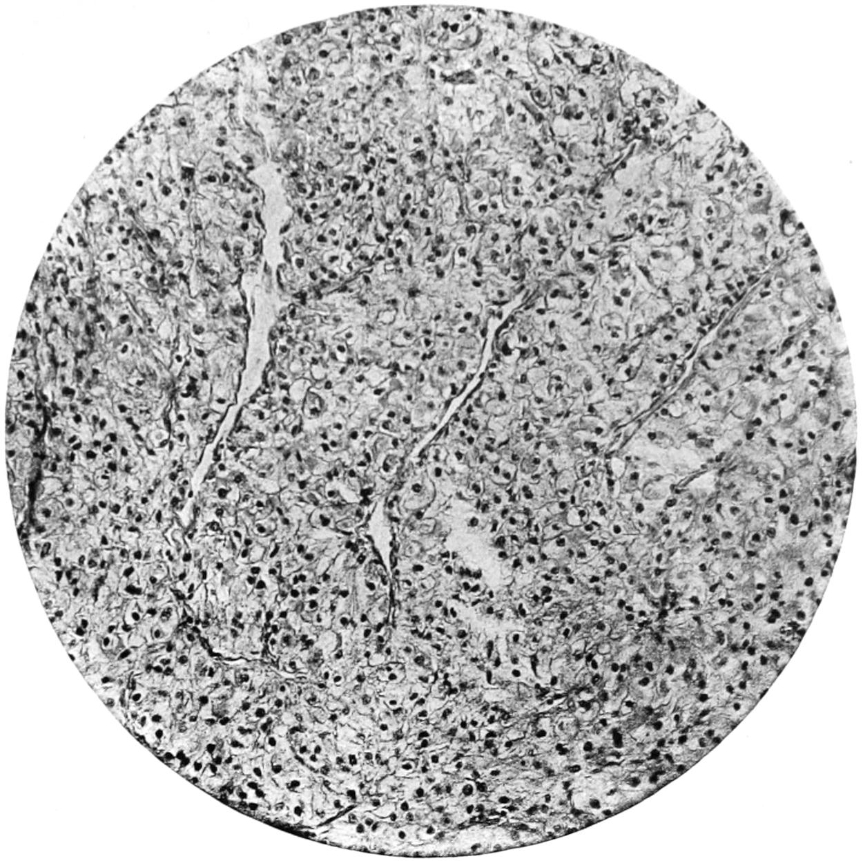
Hypernephroma Renalis. (Medium magnification.)
FIG. 2
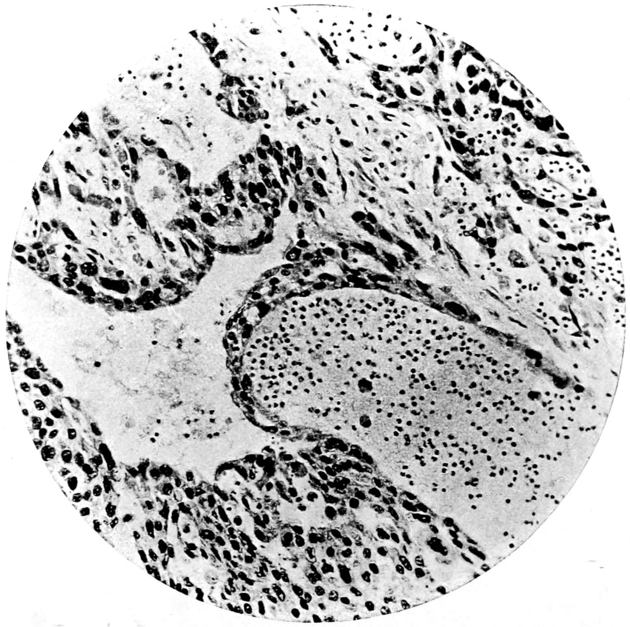
Chorion Epithelioma.
GENERAL DIAGNOSTIC FEATURES OF MALIGNANT GROWTHS.
The following tables are here inserted, trusting that they may aid the young practitioner in distinguishing in a general way between benign and malignant tumors, and even in making a diagnosis between sarcoma and carcinoma. I have also inserted a table differentiating the clinical appearances of epithelioma and of lupus. In these tables comprehensiveness has not been aimed at, rather simplicity, while it is not denied that cases are met with in which diagnosis may be exceedingly difficult, and in which the common signs herein mentioned may be found either absent or misleading:
Table I.—Differentiation between Benign and Malignant Growths.
Benign Growths. Malignant Growths.
Common at all ages. Rare in early life.
Usually slow in growth. Usually rapid in growth.
No evidences of infiltration or dissemination. Infiltration in all cases, dissemination in many.
Are often encapsulated, nearly always circumscribed. Never encapsulated, seldom circumscribed.
Rarely adherent unless inflamed. Always adherent.
Rarely ulcerate. Often ulcerate—nearly always when surface is involved.
Overlying tissue not retracted. Overlying tissue nearly always retracted.
No lymphatic involvement when not inflamed. Lymphatic involvement an almost constant feature.
No leukocytosis. Leukocytosis often marked.
Elimination of urea unaffected. Deficient elimination of urea (?).
Table II.—Diagnosis between Sarcoma and Carcinoma.
Sarcoma. Carcinoma.
Occurs at any age. Rare before thirtieth year of life.
Disseminates by the bloodvessels (veins). Disseminations by the lymphatics.
Arises from mesoblastic structures. Arises from glandular (epithelial) tissues.
Distant metastases are more common. Less so.
Contains blood channels rather than complete bloodvessels. Contains vessels of normal type.
Less prone to ulceration. More so.
Involvement of adjacent lymphatics not common. Almost invariably adjacent lymphatics are involved.
Secondary changes and degenerations are more common. Degenerations not common; other secondary changes rare.
(Sugar present in the blood?) (Peptone present in the blood?)
Differential diagnosis between epithelioma and ulcerating gumma will be found in Chapter X.
Table III.—Diagnosis between Epithelioma and Tuberculosis (Lupus).
Epithelioma. Tuberculosis (Lupus).
Preceded usually by continued irritation or warty growths. Irritation plays no figure. Preceded usually by nodules.
Diathesis plays no known part. Diathesis evident. Coincident evidences of tuberculous disease elsewhere.
Rarely multiple. Often multiple.
Area of thickening ahead of ulceration. Extension of ulceration not preceded by thickening.
Ulceration advancing from a central focus. Various foci, which may coalesce.
Border usually raised and everted, regular in outline. Border abrupt, eaten, irregular, thickened, firm, often inverted, irregular in outline.
Often assumes fungoid type. Never fungoid.
Base may be deeply excavated. Base nearly level with surface.
Usually painful. Seldom painful.
Bleeds easily. Seldom bleeds.
Never tends to cicatrize. As marginal ulceration proceeds there is often cicatrization at centre.
Most rare in the young. Common in the young.
Discharge is very offensive. Discharge rarely offensive.
Lymphatic involvement nearly always. Rarely.
GENERAL CONSIDERATIONS CONCERNING CANCER.
Cancer is one of the most fatal of diseases, yet has no symptomatology of its own. It produces no symptoms which may not be produced by other affections, and this lack of pathognomonic features constitutes one of the great difficulties in diagnosis. It may disturb every function of the part involved. Experimenters have sought in vain for a distinctive feature by which the disease can be recognized; neither in the blood nor in the various organic tissues have such changes been found that can be explained only on the hypothesis of cancer. The pain which it is supposed to cause is often lacking, and is extremely variable and uncertain. The cachexia of its terminal stages is not characteristic, no matter how pronounced, and may be explained by a variety of conditions, all of which may accompany the disease. The search for the suspected parasites cannot be made with such certainty as to lead to any definite conclusions. It is known by a complex of clinical conditions or by microscopic sections of tissues already removed.
When the disease is superficial it is easily recognized, but when deep-seated, recognition comes later.[15]
The microscopic picture may explain considerable in regard to the future as well as the past. For instance, in a case of sarcoma the presence of small, round cells, and especially of pigment, bespeaks a degree of malignancy which probably nothing yet known can baffle. A chemical examination of the tumor after removal may make the surgeon alert regarding the future of the case, according to the amount of glycogen contained within the mass, since the glycogen content is in direct proportion to its malignancy. For a while some reliance was placed upon the percentage of urea elimination, but this is influenced by so many factors as to have proved unreliable.
The relations which cancer bears to other diseases are of considerable interest. Those between cancer and trauma have been discussed; tuberculosis perhaps is the condition which, next to pure local irritation, predisposes to cancerous invasion. The transformation of tuberculous into cancerous lesions can be best appreciated where it can be most readily inspected, i. e., on the skin, and it is well known that lupus lesions frequently undergo this change. This is also true of large ulcers, which may undergo a direct transformation into epithelioma, or pass through the intermediate stage of tuberculous infection. Cancer in tuberculous lymph nodes is also a matter of interest. Again, cancers and tuberculous lesions may exist side by side in the same organ, as in the lung or the brain. Distinct sarcomatous nodules have been found in infiltrated lungs and alongside of tuberculous cavities, while cancer of the face will not infrequently be found associated with tuberculosis of the cervical lymphatics. Lubarsch has claimed that 4 to 5 per cent. of tuberculous patients suffer also from cancer, and that about 20 per cent. of cancer patients suffer from tuberculosis.
The method of death in cancerous patients is as free from distinctive characteristics as the course of the disease. It is usually associated with two prominent features, malnutrition and some terminal infection. At the last there is usually some toxemia, which renders the closing hours free from actual pain, while if the toxemia be profound patients may linger unconscious for several days.
GENERAL REMARKS ON THE TREATMENT OF CANCER.
Accepting the views expressed when discussing the nature of the cancerous process, the following may be assumed to be true: Cancer begins as a local disease. There is therefore a period in its history when if it be recognized in time, if it be or can be made accessible, and if it be thoroughly removed, it can be frequently cured. The “ifs” in the foregoing statement afford such insuperable obstacles in so many cases that the difficulties in the way of treatment are very great. It has been said that, “The resources of surgery are rarely successful when practised upon the dying.” It happens too often that these cases are not submitted to the surgeon until long after the favorable period above indicated is past. This is explained by the difficulties of diagnosis, by the inaccessibility of many primary cancers, and by the unwillingness of patients to submit to the knife. Nevertheless the best time to treat a cancer is when its existence is first suspected, and the best way is the most radical, i. e., by thorough extirpation.
While such extirpation should include a wide area of apparently healthy tissue and of the entire organ which seems to be involved, for instance, in the case of the liver, this last may be impossible; and yet by removal of a considerable area of healthy liver around a cancerous gall-bladder the writer has seen complete and apparent final recovery follow. The principal direction is to be thorough.
That cancer so often returns after operative attack is largely due to the fact that the general practitioner, under whose observation most of these cases first come, is slow to recognize the malady, and timid to advise radical methods.
It has been recognized that in cancer the internal administration of arsenic has been beneficial. In order to obtain the best results from its use, it must be pushed to the physiological limit and in preparations of the most active and reliable kind.[16]
Treatment by Toxins of Erysipelas.
—A number of years ago Fehleisen, calling attention to the fact that cancers had seemed to improve or possibly even disappear after an attack of erysipelas, suggested deliberate infection of the surface of such a growth from a case of erysipelas. In this procedure he met with some success, but there were numerous objections to it, one being the impossibility of controlling the spread of the infection thus produced. Coley, of New York, then undertook a much more systematic study of the relation between the two diseases, and devised a method of injecting the toxins produced by the streptococci of erysipelas and of reinforcing them, if necessary, by those of the bacillus prodigiosus. The intent of this treatment is to produce reasonable reaction in the hope of mitigating the rapidity of the growth, checking its progress, or even causing its disappearance. It has been on trial now for several years, and while in a few cases of sarcoma, especially in the hands of its originator, the treatment has apparently been of service, it has proved disappointing in the majority of instances.
Liquid Air.
—The application of liquid air to superficial malignant growths has proved successful in a number of instances, but inasmuch as this is practicable in only one or two of the largest cities of the country, it is not a measure which need be discussed here at length. The liquid seems to act as an almost painless escharotic, and its use produces sloughing, or a drying up under a scab, which after a day or two will loosen and be easily detached.
Radium.
—This remarkable element has aroused within the past few years an amount of scientific interest and experimentation with which there is little else to compare. The enormous expense of a preparation of any great activity, and the rather bewildering contradictory statements which have been made by those who use the weaker preparations, have caused it to occupy a doubtful position in any list of reliable therapeutic agencies. It is undeniable that certain rodent ulcers, tuberculous lesions of the skin, and a few carcinomatous lesions have been much improved or apparently cured by its use. It is ordinarily used in glass or aluminum tubes or capsules, which are applied upon the surface of the growth to be treated. It has also been used sprinkled upon a plaster whose surface has been prepared with Canada balsam, and thus directly applied. Again, it has been enclosed in a capsule to which a strong silk thread has been fastened so that the former may be swallowed, retained in the stomach for a few hours, and then withdrawn. These last means of using it are of questionable value. Of still less value are the suggestions to dissolve it in water or to administer water in which a receptacle containing radium has been allowed to stand. There is much of interest and perhaps something of value in radiotherapy, but nothing as yet of positive value in the hands of the profession generally.
Ultraviolet Light
, or, as it is often named after its promoter, Finsen light, has proved of value in many cases of lupus, and in some cases of superficial epithelioma. Its effects, however, can scarcely be made to penetrate into the deeper tissues, and in its use it is even necessary to make pressure upon the part treated with quartz compressors, because ordinary glass shuts out a great proportion of these rays from whatever source may produce them, and because it is necessary to create a temporary anemia of the lesions, as the fluids of the body have the same effect as does glass. For these reasons the method, which is of but limited value, can be made serviceable in but a small proportion of cases.
X-ray Therapy.
—The Röntgen or cathode rays have played a large part during the last few years in the therapy of cancer. Such varying statements have been made concerning their value as to keep them still on trial and nothing very positive can be said regarding their efficacy. It may be said, however, that the nearer the malignant growth is to the surface of the body the more promptly can their effects be produced. The superficial growths, especially of the epitheliomatous variety, often yield readily to their use; the deeper the lesion the more vague the effect, both in character and permanence. It has been the writer’s experience that they furnish the best method of relieving pain, in a large number of these growths, short of the anodyne effects produced by powerful drugs, which are in every other respect undesirable. He holds that no one can predicate with certainty what may be their effect in any given instance, but that they are worthy a trial in every inoperable, painful, or otherwise hopeless case. Occasionally improvement follows their use, while in the next, apparently a similar case, one may be doomed to great disappointment. There are as yet no indications by which the cases which are most amenable can be easily recognized. Even in cases of extensive and disseminated abdominal cancer marvellous improvement may follow, but never a cure. It is indeed questionable whether deep cancer can ever be really cured by these means. As against their undoubted and unchallenged value in some instances, certain disadvantages are met in the difficulty of selecting a proper vacuum tube, the frequency and duration of exposure, the distance, etc. Dermatitis, sometimes mild, sometimes severe, has too often followed the injudicious use especially of a “high” tube, and more painful, irritable, or intractable ulcers are seldom seen than some following so-called “x-ray burns” of the skin. Moreover this is not the worst of these cases, for efforts intended for the best have been in repeated instances turned into a travesty by the development on surfaces thus burned of epithelioma, necessitating later mutilating operation. A well-known American surgeon suffered amputation of one hand and nearly all of the other as a penalty for inattention to the destructive effects of too prolonged exposure of his hands. It has, therefore, impressed itself upon the writer that the x-rays should not be indiscriminately employed. Nevertheless in skilled hands and used with great discretion they can be made a powerful instrument for good in many cases, especially for the relief of pain. They should never be regarded as a substitute for operation if operation be feasible, but they may often be employed to advantage after operating, in serious cases, where there is reason to fear recurrence.
The efficiency of the x-rays is apparently enhanced by the simultaneous administration of thyroid extract; although the explanation for this improvement is not known, it is, however, of enough importance to be borne in mind. The extract should be given in 5-grain doses three or four times a day. All the remarks above made may pertain as well to the employment of cathode rays in non-malignant affections, i. e., tuberculous lesions, neuralgia, etc.
Miscellaneous Measures.
—A large number of suggestions concerning the treatment of cancer have emanated from various sources and from men of widely different views. Beaston, of Glasgow, being impressed by the physiological relationships and sympathies between the ovaries and the mammary glands, has suggested the benefit of the removal of the ovaries in hopeless cases of mammary cancer, holding that the nutrition of the mamma being thus influenced there would be more or less subsidence of pathological activity. He has reported instances in which, apparently, this measure had the desired effect; nevertheless it has not found general favor.
Based upon views concerning the hyperacidity of the blood and tissues in the cancerous condition, it is believed that there is a pronounced indication for the internal use of alkalies; and the hypodermic injection of 5 minims of a 1 per cent. solution of a chemically pure soap has been recommended by Webb, on the theory that it promotes the separation of cholesterin from the living cell. He would increase the dose until 60 minims are given at one time, every other day. A 20 per cent. solution of Chian turpentine, dissolved in sterile oil, has also been recommended to be used in the same way. These are recent suggestions of unknown value.
In the general management of cancer patients, two things should be kept in mind: (1) That they are entitled to relief from suffering in the least harmful way in which it may be offered, and (2) there comes a time in the history of many of these cases when all other considerations may be set aside in favor of comfort and tranquillity. Opium and other “drugs that enslave” have their disadvantages, but these cannot outweigh the benefit which they may confer in the last stages of cancer. The terminal pains of malignant disease should he assuaged at any necessary cost of other considerations.
But while all this is going on elimination must not be neglected. Opiates are peculiarly liable to diminish secretions and peristaltic activity. The skin, the kidneys, and the bowels should be kept active by measures which serve this purpose, and if it be desirable to prolong life, nutrition should be regulated and frequently administered, but it is absolutely necessary to maintain elimination.
The latest suggestion, viz., to treat cancer by injections of pancreatic ferments (trypsin and amylopsin), seems to the writer to be based upon erroneous notions concerning the nature and causation of the disease, and to hold out only specious hope of self-justification.