Part 6 — CHAPTER LVII. AMPUTATIONS.
Reading edition
Amputations are performed for (a) the results of injury, (b) the results of disease, and (c) removal of deformity or mutilation, or the possible results of congenital defects. While generally they are measures of necessity, made such by traumatism or by actively advancing disease, there are occasional instances where an individual decides that an artificial limb will be more useful or that he will be freed from an intolerable annoyance by the sacrifice. The principal diseases which may require such mutilation are the exceedingly acute, e. g., osteomyelitis, the slower destructive forms of ulcer, tuberculosis or of sepsis, the gangrene caused by vascular or diabetic conditions, or the slow involvement of tumors, usually malignant, but sometimes benign.
When a serious and mutilating injury has been received, if there have been complete crushing of a limb or avulsion, of course no doubt exists as to the necessity. Amputation is not now made for compound fractures nearly as often as in former times, for if only the vascular supply be good much may be done by resection of bone ends, wiring, or other expedients; and the attempt should always be made to save a limb unless it appear that even should the effort succeed the limb itself would be too useless to justify the attempt. With the possibilities of modern asepsis, and with immediate attention to the injury, the question of what should be done with an injured limb is largely a question of its blood supply. Extensive contusions with lacerations do not of themselves necessitate amputation, neither does injury to the skin unless it be most extensive.
It is unfortunate when vacillation or any misguided effort at conservatism call for great delay. While this may sometimes be advantageous, at other times the favorable moment has passed before permission to amputate may be obtained. Should delay seem advisable the surgeon should use his best endeavor to effect suitable antisepsis, to provide physiological rest, and to preserve the circulation, all of which require a thorough antiseptic technique, which will include the removal of blood clot, of fragments, and of all tissue which evidently cannot live, and suitable splinting or its equivalent, and of everything that can be done by local warmth and general stimulation to maintain the vigor of the circulation. When once infection has occurred, and especially been allowed to spread, the possibility of recovery inheres only in immediate amputation.
Such mutilations as necessitate immediate amputation are usually accompanied by profound degrees of shock, as well as perhaps by other complicating injuries, whose existence may change the whole complexion of the case. For example, with a patient suffering from probably fatal fracture of the skull one would hardly seriously discuss the matter of immediate amputation of a foot; nevertheless he should take such care of the local lower injury as to permit operation to be done under still favorable circumstances should the head condition justify it. Wide discretion is therefore called for in all these cases. Furthermore the condition of lowered blood pressure or shock may be so extreme that the operator is compelled to delay, for at least a certain time, in order that by the employment of those measures already considered in the chapter on this subject the circulation may be sufficiently restored to make it adequate for the purpose, remembering that scarcely anything predisposes to infection more than such lowered vitality. While resorting to general stimulation, hypodermoclysis, or infusion, with or without adrenalin, the use of such antiseptics should not be omitted, as the local condition may require in order to combat what otherwise may be actively occurring.
Amputations are sometimes referred to as typical, when done according to long-established methods, or atypical, when the entire procedure is planned to fit the necessities of the case. Amputation at a joint is usually spoken of as disarticulation. Amputations, again, are classified as (a) immediate, i. e., before complete reaction from shock or within the first few hours; (b) primary, when done after reaction has occurred, but before visible occurrence of inflammatory changes, (c) intermediate, as done when suppuration is threatening, but before its actual occurrence; (d) secondary, i. e., after the occurrence of suppuration.
The control of hemorrhage is one of the most conspicuous and necessary features of any amputation method. Below the shoulder and at the hip this may be effected by the old-fashioned tourniquet, or the modern elastic bandage, which may or may not be combined with the more complete bloodless method with which Esmarch’s name will always be connected. The pure rubber gum bandage, I believe, was introduced by Martin, of Boston, but the method of its use for bloodless operations upon the extremities is to be credited to Esmarch. The surgeon may avail himself of this method in all suitable cases, but should never resort to it in septic or malignant disease. It includes the application of a Martin elastic bandage from the tip of the extremity to the necessary height, by turns which shall make gentle and equable pressure, gradually forcing the blood from the compressed tissues and out of the limb, and up to a height where another elastic, or, at all events, suitable constricting band is placed with a sufficient degree of tightness to completely shut off access of blood. To so apply a bandage in septic and malignant cases would be to coax septic and malignant material into the veins, and would evince the worst possible judgment. A sufficiently strong rubber tubing forms an effective tourniquet, which, however, should be applied over a folded towel, or in some manner so that it does not too deeply constrict and compress the soft tissues of the limb. Instead of tying a knot it may be secured with an ordinary clamp forceps.
The tourniquet should never be applied over the leg or forearm, for it can here make no impression upon the interosseous vessels. Its application should be begun by pressure upon the vascular, i. e., the adductor side of the limb, so that venous choking may be avoided. After it is once in place the limb should not be completely flexed nor extended, lest the tissues firmly enclosed by the constriction be more or less spontaneously torn; nor should the tourniquet be too long left in place, as injury to the vessels is the possible result.
The bloodless method of Esmarch is furthermore subject to the following disadvantages. It is sometimes followed by serious and permanent paralysis of the limb, the result of prolonged or excessive constriction and compression of the motor nerve trunks. Similar results (in the arms) follow the use of crutches as well as of pressure of the side of the operating table when the limbs are allowed to hang over it. Again after removal of the bandage there is sometimes most pronounced capillary oozing due to vasomotor paresis. This may be controlled by the stimulation of hot irrigation or applications, and by more or less massage of the limb. The dangers of forcing undesirable material into the circulation have been mentioned, in addition to which should be recorded the increased absorption of toxic substances.
When there is good reason for not using the elastic bandage, save as a tourniquet, much of the desired effect may be obtained by holding the limb for a few minutes in a vertical position, so that its contained blood is drained out of it by gravity, after which the tourniquet may be applied as before.
The cocainization of nerve trunks, as they are exposed and divided, is one of the new measures for the prevention of shock for which we are largely indebted to Crile. It has proved to be a most valuable expedient which should not be neglected. (See Chapter XVIII.)
Under modern methods more is expected of an amputation stump than in days gone by, and the first demand is that it shall be useful, to which end it is necessary that it be both movable and that its end be not too irritable, nor the scar too sensitive to stand at least a certain amount of pressure. It is expected that suitable prosthetic apparatus, i. e. artificial limbs, shall take the place of severed lower extremities and of most arms or hands removed. The skill and the mechanical ingenuity of the maker of artificial limbs have now reached a point where most acceptable substitutes are thus provided, but for them suitable stumps should be afforded by the surgeon, and there should be coöperation from each direction. Thus it used to be held that the bone end in every stump should be covered with periosteum, yet it has been recently shown, especially by Hirsch, that such bone ends are as acceptable, and perhaps more so, when stripped of rather than covered with this membrane, the latter being sensitive, and there being no advantage in the presence of such new bone as may be formed by its preservation.
Many a good stump may be molded in various ways, but always provided that the end of the bone be smoothly divided, and have no corners or osteophytic outgrowths to make pressure upon the sensitive scar. For this reason it should be manipulated as early as possible, and should not be allowed to undergo the atrophy noticeable in stumps left after old operations. If primary union be gained, so long as the cicatrix and the nerve ends be kept out of the way, one may expect a stump which is serviceable in every respect. The ideal method is that the skin and the periosteum should retain their normal relationship, an ideal best attained in the supracondyloid operation after Gritti’s method. Various osteoplastic methods have been devised, first by Walther, in 1813, and since him especially by Ollier, Pirogoff, Gritti, and Bier. The latter would cover every bone end not merely with periosteum, but with a bone flap so arranged that its lower surface is one normally covered by periosteum. The introduction of the x-rays has permitted a more thorough study of bone ends in stumps which are, on one hand, extremely tender, or, on the other, extremely serviceable, and the osteoplastic methods seem to conduce to the latter condition. Another matter of great importance is to so place the scar that it shall be neither subject to pressure nor to traction. If, therefore, the sawed surfaces be covered with a periosteum which shall retain its normal relation to its coverings, a minimum of disturbance in the scar is the result.
The value of early use of the stump and of accustoming it to pressure is considerable, as atrophic stumps are tender, like other disused parts, and there is, therefore, every reason for resorting to prosthetic apparatus as early as possible. As Kocher puts it, the following is the best procedure for the normal operation: “An oblique incision, combined if necessary with a longitudinal one, in the form of a racket or lanceolate incision through skin and fascia. After retracting the elastic skin the muscles are divided obliquely down to the bone. The periosteum is also to be divided obliquely. Periosteum is then separated along with the superficial layer of the cortex of the bone, by means of a sharp raspatory or chisel, or, when possible, a flap of bone having a movable periosteal hinge is made by means of the saw. Lastly, if only a thin shell of the cortex have been raised up along with the periosteum, the end of the bone is simply rounded off, while if a distinct flap of bone, by any osteoplastic method, have been divided, the end of the bone must be sawed in a curved direction so as to fit it. The periosteal or bony flap is sutured over the sawed surface of the bone to its periosteum, and the stumps of the muscles or tendons are sutured to each other, or to the surface of the bone at a distance from the sawed surface. Finally the skin and fascia are sutured; but in case where a periosteal flap or flap of bone and periosteum cannot be obtained in normal relation to other soft parts it is better to remove the periosteum entirely from the end of the stump, to scrape out the medullary cavity, and to round off edges of the bone as dentists do.”
While these methods give better results than those formerly in vogue, they also consume more time; but the days of brilliancy and rapidity in amputation are past, as time should be devoted to careful work, except only in those cases where emergency demands the most rapid and dexterous removal of a limb in the shortest possible time, and where every other consideration is sacrificed to the principal interest of preserving life.[75]
AMPUTATION METHODS.
With a view to simplifying this subject as much as possible the following methods alone will be considered: (1) The circular with its modifications, the oblique, the elliptical, the ovoid, etc.; (2) the flap method; (3) the mixed or skin flap and deep circular.
Choice of method sometimes leaves much, sometimes nothing, to the tastes or wishes of the operator. It should be based solely on the primary consideration of saving life and the secondary consideration of furnishing the most useful possible stump. To obtain the latter it is necessary that the bone be amply covered, except that its coverings be not adherent, that there be a minimum of disturbance of blood supply, that nerves be drawn down and divided as far from the stump end as possible, in order that they may not be entangled in the scar, and that the scar be so planned for and arranged that it shall be at one side, at all events in such position that no pressure shall be made upon it, and, if possible, also no tension by muscle action.
Elasticity of skin and contractility of the muscles vary much in different individuals, and it is not always easy to estimate either of them previous to their division. Consequently it is much better to make cuffs or flaps too long at first rather than too short. The existence of previous disease will always modify these local conditions, but, in general, the rule is laid down that the external flaps should be longer than the bone by from one-third to one-half the diameter of the limb.
1. Circular Method.
—The simple circular method is the simplest and easiest of all. It may be so performed as to furnish a solid musculotegumentary division, or skin cuffs may be made, which being turned up, permit a further circular division of the muscles and other tissues directly down to the bone. The former is preferable when possible. With an ordinarily long amputating knife the skin, down to and including the superficial fascia, is divided by one clean circular incision, made in one stroke; then by further circular cuts the muscles are divided in sections, the outer group being allowed to retract and expose the deeper layers, which are then divided at a higher plane. In this way the periosteum is reached. If sufficient time be afforded it may be circularly divided at the level of the last incision through the muscles, and then separated with a strong elevator or, as done by Kocher, with a chisel, in order that some portion of the exterior of the bone be raised with it. In this way a cuff of periosteum, or enough of it to cover the bone end, is detached upward, to the level where the bone itself is finally divided. The bone division is done with the ordinary amputating saw, or with the wire or chain saw.
The skin-cuff (Manchette) method differs in that the exterior flap is made wholly of skin, which is dissected as a cuff nearly up to the level of bone section, at which point the muscles are divided directly down to the bone. In this method the skin, fat, and superficial fascia should be raised together, and at no points separated from each other.
Modification of the cuff method, by which it is more easy to evert the circular flap, is made by one or two vertical incisions, by which the cuff is split some distance on one or both sides, thus transforming the cuff proper into two nearly square skin flaps. At other times the first method may be similarly modified, in which case we have to deal with two square flaps, including not only the skin, but all the tissues down to the bone.
Neudörfer still further modified the circular method for certain purposes by first making an incision along the outer or least vascular part of the limb, carrying the knife directly down to the bone, retracting the wound edges, and thus exposing the bone, which is then divided with a chain or Gigli saw. After the bone is divided the soft coverings are lifted to a sufficient distance below the saw line to ensure ample covering, then divided as above. The method is a slow one and is especially serviceable for amputation of the thigh, at its middle, for diabetic or senile gangrene, where it is so desirable to protect vascular supply from injury (Fig. 677).
The so-called elliptical method is practically a circular incision carried obliquely around the limb, the upper and lower ends of the ellipse being indicated by previous small incisions at the proper height. The skin and superficial fascia are retracted from the lower portion of the ellipse by turning them up to the level of the highest point, at which level the muscles are divided transversely by a plain circular incision. A modification of this method is the so-called ovoid or racket, which is simply an oval division with a pointed end, the margins of the flap being united in the long axis of the bone. This method is frequently applied in amputation of the fingers. (See Fig. 683.)
2. Flap Methods.
—Flaps are either cutaneous or musculocutaneous. In every case the skin surface must be larger than the muscular. They are objectionable in that the skin flap is apt to slough, although least so about joints. The flap method is advantageous in that one flap may usually be made much longer than the other, and the longer one so doubled over at its end as to place the scar out of harm’s way. In certain injuries where the skin is much more injured on one side of the limb than on the other the operator is compelled to resort to flaps, unless he divide the limb much higher than might otherwise be demanded. Double flaps may be anteroposterior or lateral. A double flap practically results from a circular incision, carried through to the bone, with lateral division on either side, while a double flap with one long member may be similarly furnished by an oblique circular incision with the lateral prolongations.
Fig. 677
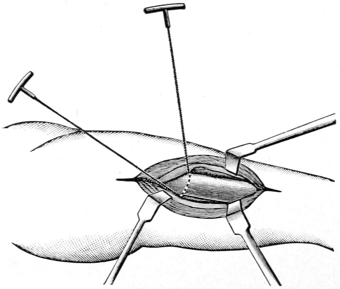
Neudörfer’s method of amputation by primary division of the bone, before shaping the flaps. Neudörfer used the chisel, but one may use the Gigli saw with special advantage in performing this operation. The method is applicable to any portion of the upper or lower extremity, especially in the continuity of the long bones. (Matas.)
Flaps may be formed by transfixion, for which purpose a long, sharp, amputating knife is required. Inasmuch as it makes an oblique and irregular division of the principal vessels, which are in consequence more difficult to secure, and by which nutrition of flaps is endangered, it is not to be commended, save perhaps in certain amputations about the wrist. A better method of making the flap is to divide the skin and fascia with an ordinary stout scalpel, and then, permitting them to retract, to divide the muscles obliquely toward the bone in such a way as to leave a flap wedge-shaped at its base. The anteroposterior amputations of the foot, thigh, and arm are better performed in this way, each flap being in length preferably three-fourths the diameter of the limb. (Matas.) An extension of this method furnishes the possibility for various subperiosteal amputations to be described below.
The osteoplastic methods of today furnish desirable operative procedures. One of the earliest of the good ones was Teale’s method, as applied to the leg, of double quadrangular flaps, the anterior being much the longer. A minor degree of this work includes simply the preservation of a cuff of periosteum, which is supposed to afford protection to the marrow cavity and a smoothly rounded bone end, without adhesions to the overlying soft parts; but much more complete operations are afforded by Pirogoff’s amputation at the heel, and by Wladimiroff and Mikulicz’s amputation of the foot (practically an exsection of the heel), or by Gritti’s and the other methods of supracondyloid knee amputation, with preservation of the patella. Bier and other foreign and domestic surgeons have also devised methods of reflecting or raising bone flaps from the continuity of bone shafts, which, being still connected by periosteal bridges, are so turned and fastened in place as to furnish a complete bone end over the stump (Figs. 678 and 679).
The choice of method must depend, to a large extent, on the character of the case. Some injuries will leave parts so exposed that a portion of a limb can still be utilized if only flaps be cut in an atypical way. One need never hesitate to resort to these, especially about the hand and upper extremity, where it is so desirable to save every inch of tissue. It is not necessary to preserve every possible inch of tissue in the foot and leg, as the makers of artificial limbs can adapt an artificial leg to any kind of a stump. The intent in making these statements is that while it is best to follow conventional methods under ordinary circumstances, there need be no hesitation in departing from them when occasion demands it.
Fig. 678
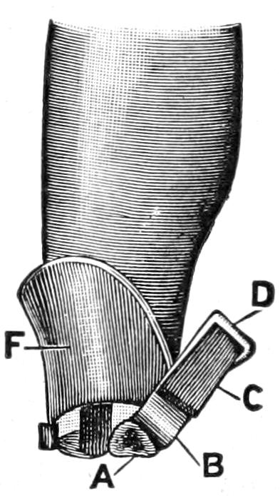
Bier’s osteoplastic amputation of the leg (procedure advocated by Bier in 1897 and 1899): F, long anterior flap reflected on the tibia; A, cross-section of tibia; B, periosteal flap after excision of intervening section of bone; C, osteoperiosteal flap; D, projecting border of periosteum to be sutured to tibial periosteum.
Fig. 679
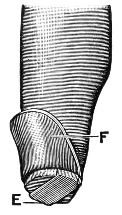
Bier’s osteoplastic amputation of the leg, with osteoperiosteal flap in position.
It is essential in caring for every stump, after the actual amputation has been performed, (1) that bleeding be absolutely controlled; (2) that nerve ends be placed out of the way of cicatricial entanglement; (3) that proper drainage be provided; (4) that the soft parts be so brought together as to unite in the promptest and most perfect fashion. The possibility of the latter will depend very much on the occasion for the operation and the condition of the tissues. Operating in the presence of previous disease, as when the parts are inflamed or edematous, or as when one amputates at a point where more or less sloughing and separation of tissues have already occurred, the surgeon cannot look for such primary repair as furnishes an ideal termination, nor should he endeavor to make such close suturing or approximation as he would otherwise attempt. In fact, under these circumstances, it is often desirable to leave the wound widely open, perhaps packing it with yeast, in order to hasten sloughing and secure healthy granulating surfaces, which may be then brought together by secondary suture or by suitable strapping and bandaging. Nothing worse can happen than imprisonment of the debris resulting from the sloughing process.
But an amputation wound made with faultless technique, and in tissues previously healthy, may be closed with a minimum of drainage, or often without any, providing it be so closed as to leave no dead spaces in which blood clot may accumulate. This requires careful suturing, by numerous buried sutures, of muscle to muscle, tendon to tendon or to periosteum, and the like, the wound being gradually closed from its depth, and finally so bandaged that equable pressure shall be made, with comfortable support, but without undue pressure at any point. In aseptic cases animal ligatures and sutures (chromic gut) will prove reliable and efficient. In septic cases it would probably be better to trust to (secondary) silk, especially if parts are to be long exposed, so that it can be later removed. For the superficial wound silkworm sutures answer admirably.
For drainage a gauze packing for the worst cases, one or two tubes for ordinary cases, and for those which scarcely need it strands of catgut or of silkworm-gut, or two or three little rolls of oiled silk, will be sufficient.
In this country Link and in Germany Credé have practised the method of bringing parts together merely by equable pressure and bandaging. This has been of late modified by the use of strips of sterile adhesive plaster; and in certain instances, everything else favoring, it has given good results. It might be advantageously adopted in cases where it is feared that it may be necessary to reopen the wound, as it would permit an easy method of so doing.
Dressings should be copious and snugly applied, and the limb involved should be immobilized. Thus after a leg amputation it is well to bind the leg and thigh upon a suitably arranged splint, physiological rest, which is so essential to success, being in this way attained. The same is also true of the arm.
AMPUTATIONS OF THE UPPER EXTREMITY.
Amputations of the Finger and Thumb.
—It is desirable in the upper extremity to save every portion which can be preserved and still made useful. This is particularly true of the fingers, where every half-inch adds to their usefulness. When it is possible the palmar surface should be saved and made to cover the stump end, as it is not only more sensitive but denser and stands wear better. This is equally true of disarticulations or of divisions between the joint ends of the phalanges, which are best exposed by bending the finger, cutting the dorsal flap in this position, then stretching it and cutting the palmar flap (Fig. 680).
The vessels and nerves lying on the lateral aspect should be secured against hemorrhage, and cocaine solution introduced if local anesthesia is being practised. It is important also to remember the arrangement of the common palmar synovial bursa, with the digital prolongations to the thumb and the little finger, and that the three middle fingers are ordinarily shut off from it. Nevertheless if tendons be divided near the hand, and short finger stumps be made, it is easy to infect this common palmar bursa through retraction of the tendon and the consequent opening up of a tunnel directly into that cavity.
Fig. 680
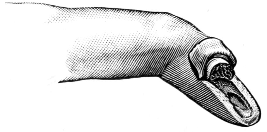
Typical amputation of finger in continuity (through a phalanx); long palmar and short dorsal flaps. (Farabeuf.)
Fig. 681
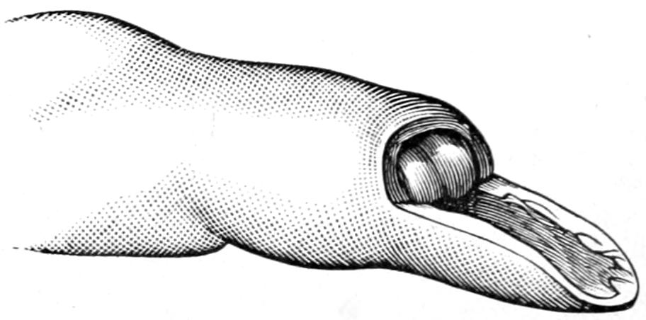
Typical or preferred method of disarticulating a finger by long single palmar flap. (Farabeuf.)
Figs. 680 and 681 illustrate the best methods of amputating fingers through a phalanx or at the joints, while Fig. 682 shows the best method of closing the wound. In this way a serviceable finger-tip is preserved which will stand every irritation to which it will probably be subjected.
When the finger is to be disarticulated from the hand a modified oval flap is preferable, with its long flap on the radial side and the scar on the dorsum rather than in the palm. The thumb is perhaps best separated at an articulation by a single palmar flap, without the preservation of the sesamoids which belong to its short flexor. Fig. 683 illustrates the various flaps and methods preferable at the bases of the different fingers.
When two or more fingers have to be removed the incision should be planned to meet the indications. When the first three fingers have to be removed, with or without that portion of the hand to which they are attached, leaving only the thumb and little finger, I have repeatedly followed to advantage the suggestion of Lauenstein, and through a small incision properly placed have, with cutting forceps, divided the first and fifth metacarpal bones at about their middle, and have then given to each of the remaining digits a quarter of a revolution toward each other, in such a way that when their tips are flexed there was better prehensile power, the hand acting similarly to a more perfect claw. If they are to be maintained in this position during healing they must be suitably held upon the splint to which the entire hand and forearm should be attached.
Fig. 682
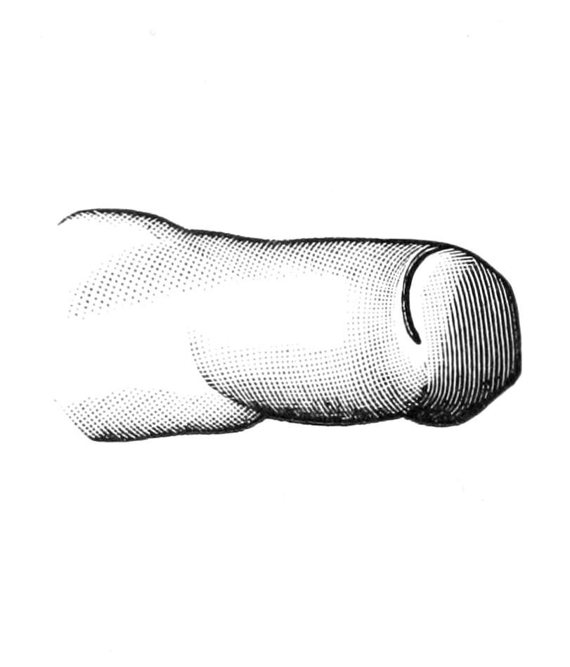
Stump resulting from the procedure shown in Fig. 681.
Fig. 683
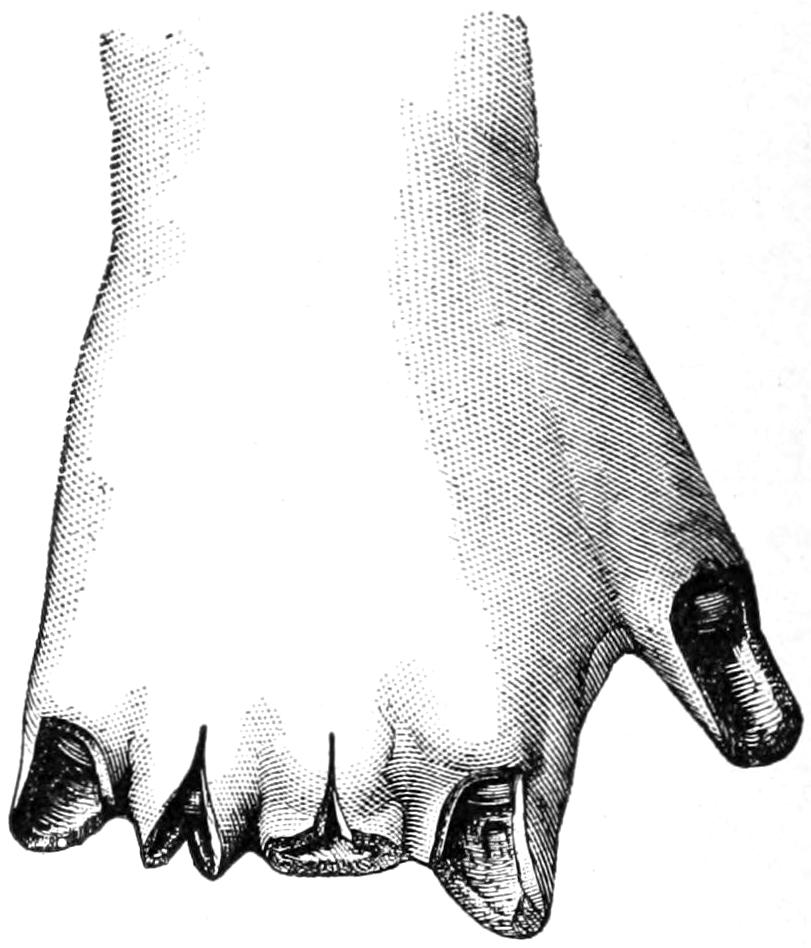
Illustrating various finger amputation. (Farabeuf.)
Fig. 684
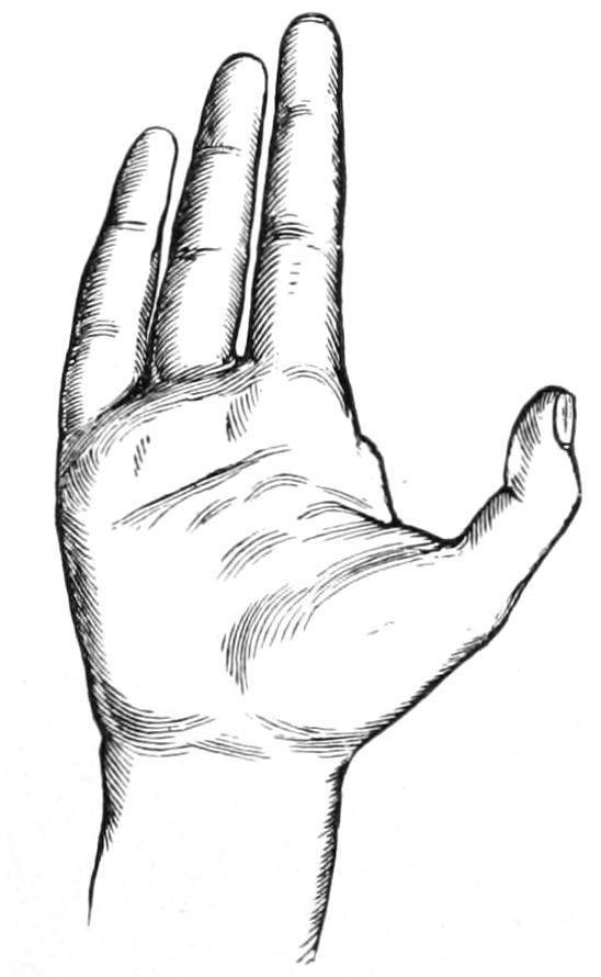
Removal of index finger. (Erichsen.)
Fig. 685
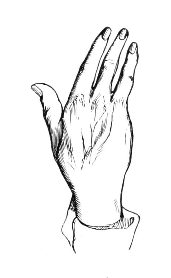
Removal of little finger. (Erichsen.)
Fig. 686
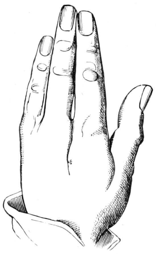
Results of amputation above metacarpo-phalangeal articulation in middle, index, and ring fingers. (Erichsen.)
Fig. 687
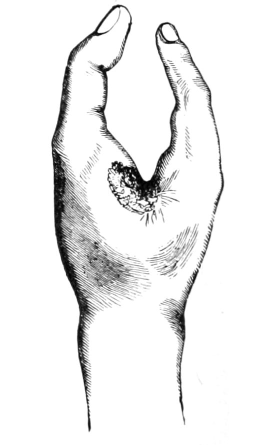
Hand after removal of metacarpal bones and three fingers, leaving thumb and little finger. (Erichsen.)
When an entire finger is to be removed it is a question whether the metacarpal belonging thereto should also be sacrificed for cosmetic purposes. In general this is undesirable except in the case of the fifth metacarpal with the little finger. This is easily exposed by lateral incision along the ulnar border of the hand, sufficient to disclose the bone and permit its disarticulation from the carpus. The same is also true, in at least some instances, of the thumb, but it is unwise to expose the carpal joints to the possibility of infection when this can be avoided; moreover, the deep palmar arch crosses just in front of the bases of the second to the fourth metacarpals, where it must be carefully avoided. If, then, the metacarpal is to be sacrificed this should be done rather from the dorsal side, while for cosmetic purposes alone it is usually sufficient to disarticulate the finger at its base and then simply remove the head end of the corresponding metacarpal. Figs. 688 to 692 furnish illustrations of how the incisions may be best planned to effect either of these purposes.
Hand Amputations and Wrist Disarticulations.
—While it makes but little difference whether the metacarpals be disarticulated from the carpus or the latter from the radial end, it is advisable to adopt whichever line of separation will best meet the indications. For a removal of the hand at or near the wrist two flaps usually afford the most serviceable method, the palmar tissues being preserved, if possible, in order that they may cover the stump. This operation is usually done for injury, and it is more than likely that one will have to plan his flap according to the tissues which still are serviceable.
Fig. 688
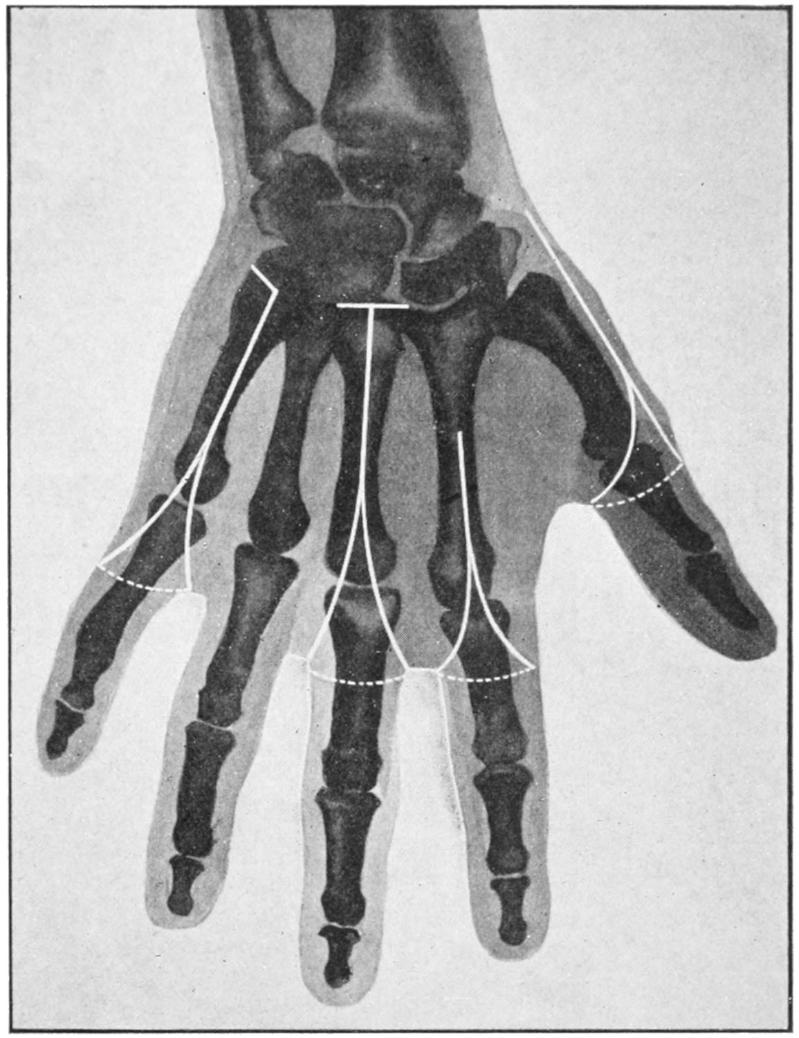
Outline of amputation of fingers, with their metacarpals. (Modified by Matas from Mignon.)
In the lower part of the forearm the flap method furnishes a serviceable stump. As the elbow is approached the circular or elliptical methods are preferable, as illustrated in Fig. 690.
The Elbow.
—With elbow disarticulations caution should be observed to have flaps of sufficient length. The joint is opened more readily from its radial side. The integument of the back of the elbow region lies closely upon the bone, is thin, and retracts but slightly. Anteriorly there is more muscular covering and consequently a tendency to retraction. Therefore the anterior flap should be made longer than might otherwise seem to be required. Here the ideal scar will be behind the end of the humerus, but it is difficult to obtain because of the tendency to drag it around beneath the end of the bone. An elliptical incision, directed obliquely downward and forward, is the easiest method and furnishes the best stump. The lower end of the posterior part of the flap should be at a distance below the articulation, at least equal to the transverse diameter of the joint itself, i. e., in an adult nearly one hand-breadth from the line of the joint to the point of dissection. (Matas.) (Fig. 690.)
The Arm.
—The arm furnishes that nearly cylindrical outline best adapted for circular amputations. Here, as at the elbow, the greatest retraction is on the flexor side. With the arm should be saved all that is possible even up to its upper extremity.
Remembering the greater tendency of the flexors to contraction the truly circular method should be modified to a somewhat elliptical incision, in order to compensate for this difficulty, while an external liberating incision is often of assistance. Abrupt transverse division of the muscle down to the bone should be made after the oblique incision of the skin.
Fig. 689
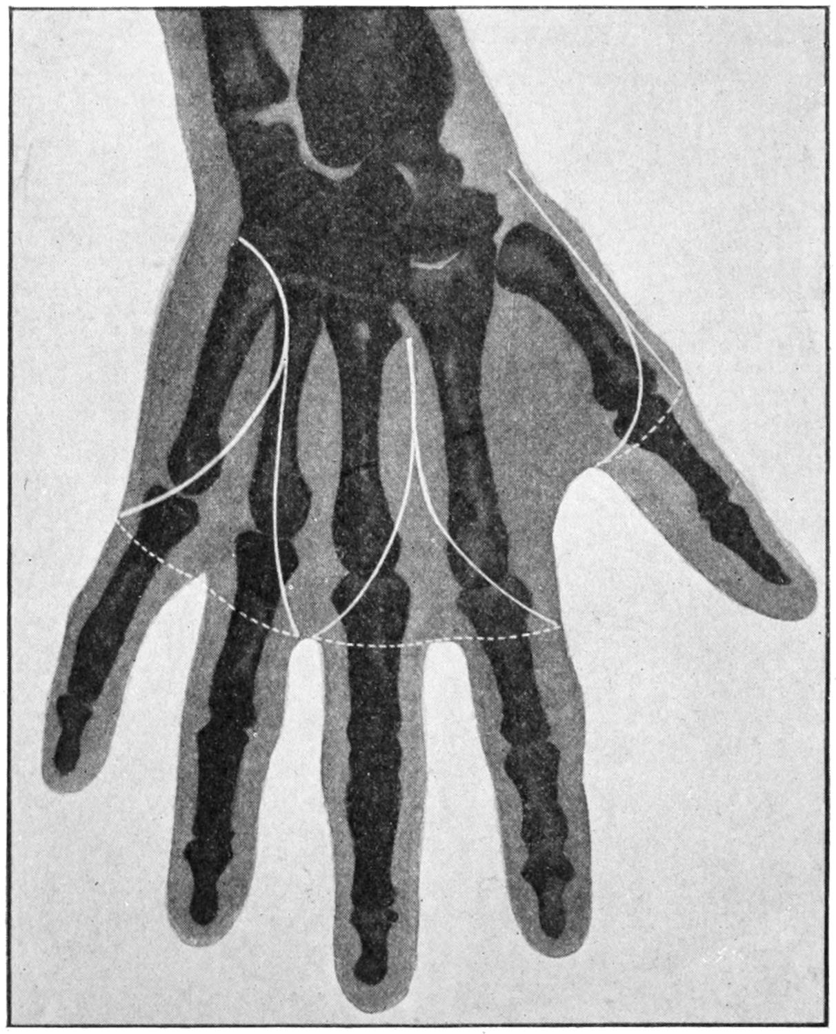
Outline of amputation of two fingers simultaneously with their metacarpals; also thumb with its metacarpal. (Modified from Mignon.)
Disarticulation at the Shoulder.
—Until accurate methods of blood control were introduced this was an amputation viewed usually with disfavor, in spite of the fact that compression of the axillary artery in theory is easy. The older methods comprised this compression, either above the clavicle, or by exposure of the vessel and its proximal ligation, or by opening and separating the joint and then seizing the vessels within the inner flap, and controlling them by digital pressure until their division. Now with the use of Wyeth’s pins and the elastic bandage, effectual control may be secured without resorting to any of the former expedients. If the removal is to be a high amputation, just below the neck of the humerus, the method shown in Fig. 691, of application of the tourniquet and its control by a constricting strap, may be adopted.
If the surgeon expect to disarticulate he should resort to the pins of Wyeth (i. e., to the use of long mattress needles), which are passed through from above downward, or from the axilla upward, one of them being passed anteriorly and the other posteriorly, and brought out at corresponding points on the upper aspect of the shoulder, where, their points being protected by sterilized corks, they serve to prevent sliding of the elastic bandage or tourniquet, which is now placed proximally to them, and is thus held more securely than is possible in any other way.
PLATE LVII
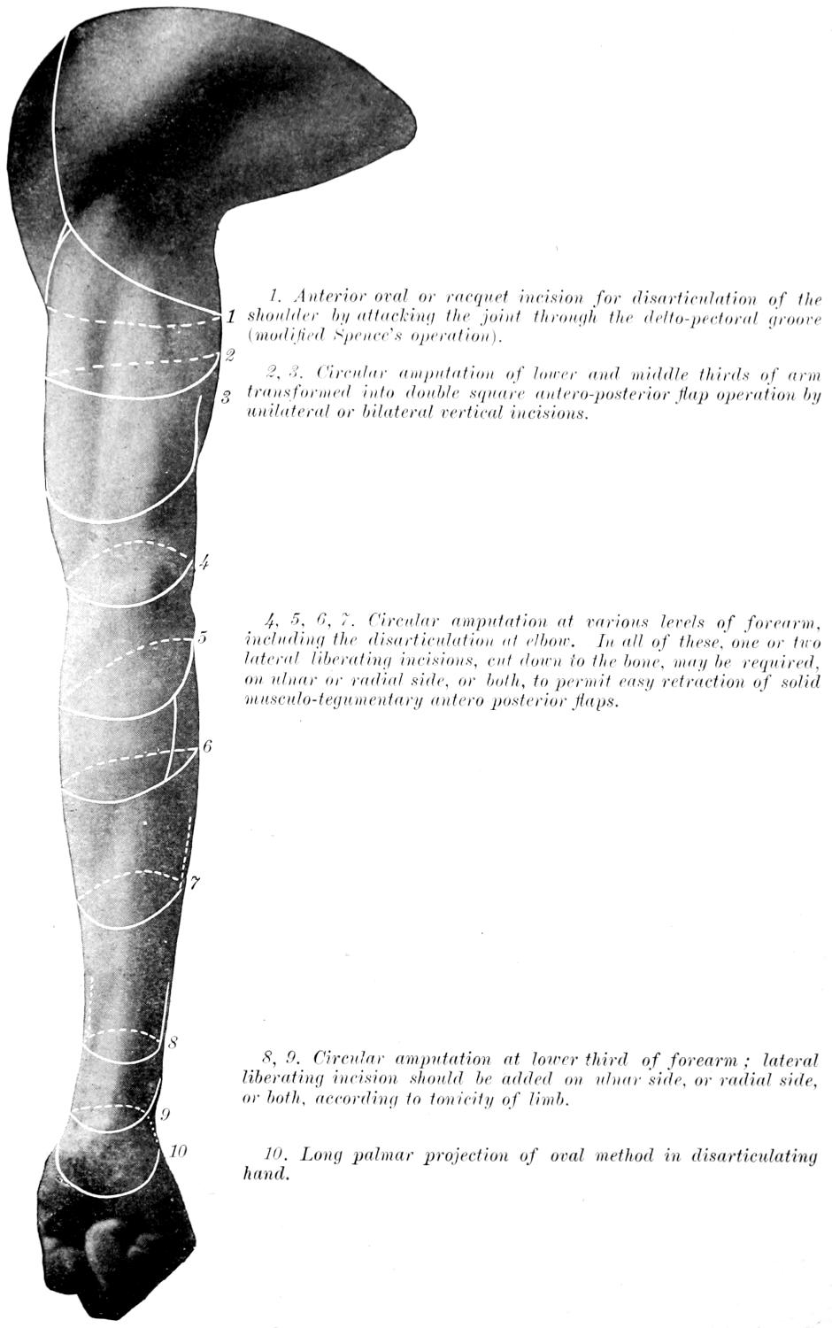
Cutaneous Incisions in Amputations of the Upper Extremity (Ventral or Flexor Side).
1. Anterior oval or racquet incision for disarticulation of the shoulder by attacking the joint through the delto-pectoral groove (modified Spence’s operation).
2, 3. Circular amputation of lower and middle thirds of arm transformed into double square, antero-posterior flap operation by unilateral or bilateral vertical incisions.
4, 5, 6, 7. Circular amputation at various levels of forearm, including the disarticulation at elbow. In all of these, one or two lateral liberating incisions, cut down to the bone, may be required, on ulnar or radial side, or both, to permit easy retraction of solid musculo-tegumentary antero posterior flaps.
8, 9. Circular amputation at lower third of forearm; lateral liberating incision should be added on ulnar side, or radial side, or both, according to tonicity of limb.
10. Long palmar projection of oval method in disarticulating hand.
PLATE LVIII
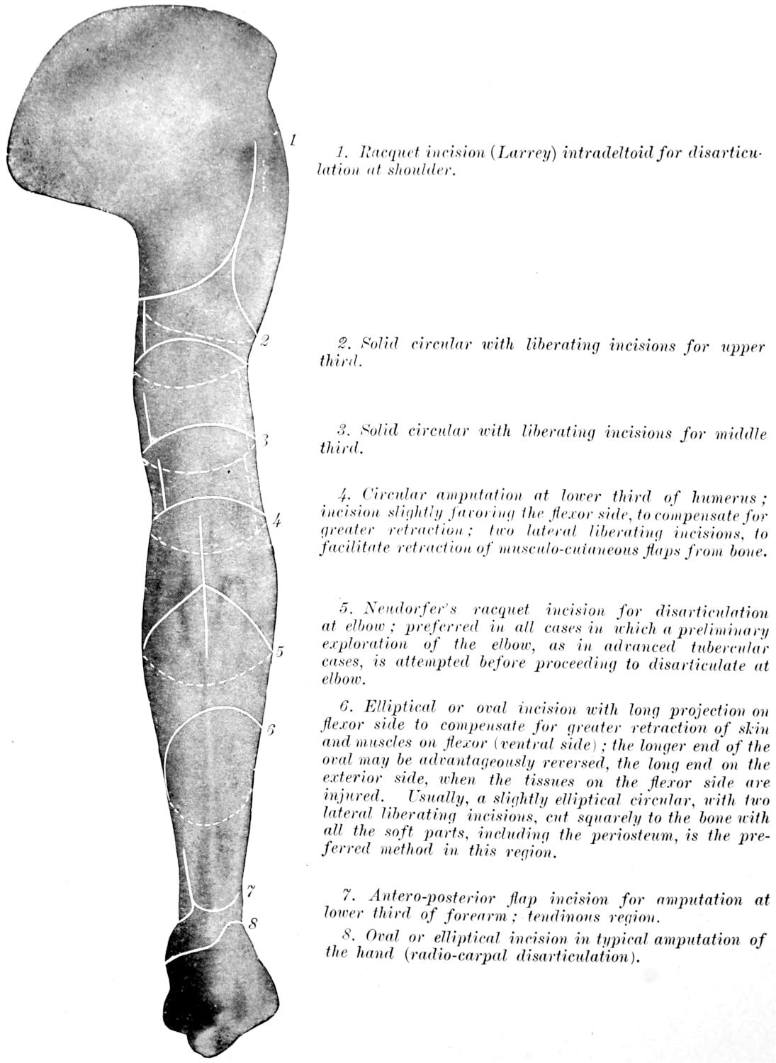
Surface Tracings showing Some of the More Useful Lines of Skin Incision in Amputations of the Upper Extremity (Dorsal or Extensor Surface).
1. Racquet incision (Larrey) intradeltoid for disarticulation at shoulder.
2. Solid circular with liberating incisions for upper third.
3. Solid circular with liberating incisions for middle third.
4. Circular amputation at lower third of humerus; incision slightly favoring the flexor side, to compensate for greater retraction; two lateral liberating incisions, to facilitate retraction of musculo-cutaneous flaps from bone.
5. Neudörfer’s racquet incision for disarticulation at elbow; preferred in all cases in which a preliminary exploration of the elbow, as in advanced tubercular cases, is attempted before proceeding to disarticulate at elbow.
6. Elliptical or oval incision with long projection on flexor side to compensate for greater retraction of skin and muscles on flexor (ventral side); the longer end of the oval may be advantageously reversed, the long end on the exterior side, when the tissues on the flexor side are injured. Usually, a slightly elliptical circular, with two lateral liberating incisions, cut squarely to the bone with all the soft parts, including the periosteum, is the preferred method in this region.
7. Antero-posterior flap incision for amputation at lower third of forearm; tendinous region.
8. Oval or elliptical incision in typical amputation of the hand (radio-carpal disarticulation).
Fig. 690
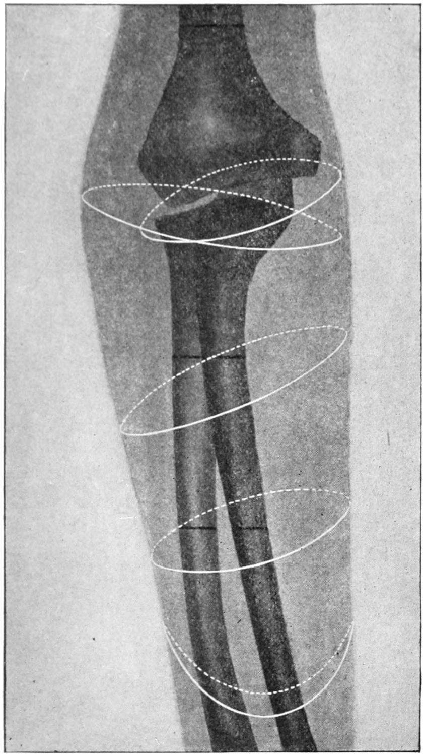
Lines of amputation in lower third of forearm, of elbow, and lower third of arm. (Modified from Mignon.)
Circulation being thus controlled, a modified circular operation may be made or a long external and superior flap cut, matching it with another one dissected from the axillary aspect. In the former case the circular incision is made on a level a little below the anatomical axillary border. Then a cuff of skin being raised while the arm is held in adduction, all the soft parts are divided to the bone and separated from it. Now a liberating incision may be made from the anterior border of the acromion to the coracoid process, then over to the deltoid groove, and along it to the first circular incision. Through this all the soft tissues surrounding the glenoid margin are separated, and then the bone is enucleated by opening the capsular ligament, reserving perhaps the detachment of the group of scapular tendons until the last. If one have any fear as to the efficiency of his hemostatic precautions he may secure the axillary vessels so soon as they are divided and then proceed with the disarticulation as above. In some cases it may be preferable to cut a wide flap from the deltoid region, preserving that muscle or not as may be desired, and, after having thus exposed the joint, make the disarticulation, separating the head of the bone sufficiently to allow the passage of an amputating knife behind it and down along the shaft to a distance sufficient to justify turning it abruptly and toward the surface, and then cutting out the axillary flap. The attempt should be to cut all the vessels at right angles rather than obliquely.
Plates LVII and LVIII, prepared by Professor Matas, afford a synoptic view of the more useful lines of skin incision in the principal amputations of the arm and shoulder.
Interscapularthoracic Amputations.
Removal of the Entire Upper Extremity.
—This includes removal not only of the arm, but of the scapula and clavicle as well, or at least its outer portion. It is not often required, and inasmuch as the circumstances which justify it are seldom duplicated, a suitable method for each individual case should be planned, rather than try to make one set of directions cover them all. Much will depend upon whether sufficient skin can be saved in order to cover the large defect thus made. In general, however, an incision should first be made along the clavicle, exposing it and dividing it near its middle. It is convenient to take out the middle portion at this time, and in this way to afford ample room through which a proximal ligation of the subclavian vessels may be made, they being here carefully dissected out, secured by double ligation, and divided. From the outer part of the above incision another is carried downward and outward toward the deltoid groove and then beneath the axilla to its posterior margin. The posterior flap is then furnished by an incision continuous with the last one, which terminates below about opposite the lower angle of the scapula, and is then continued upward along the inner scapular border and over the shoulder until it reaches the outer end of the incision first made. In this manner will be furnished a sufficient covering. The balance of the operation consists in the gradual separation of the entire mass from the outer wall of the thorax. With a preliminary ligation of the subclavian vessels there will be no hemorrhage which cannot be easily checked by pressure and forceps.
The above, however, is only a general description, which may need to be modified in most cases. If the amputation be done for injury all the skin which is still viable should be utilized, no matter how shaped, while if done for disease the incisions may have to be modified materially, taking more skin from one side and less from the other, in order to avoid that involved in the disease process.
In the majority of cases the result is satisfactory, in spite of the mutilation thus afforded.
Fig. 691
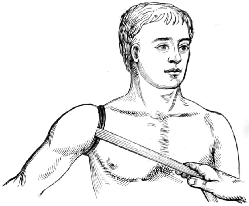
Esmarch’s elastic constrictor applied and held in place by a bandage or a strap (Wyeth’s pins may also be used to hold the constrictor in place) in high amputation of the arm. (S. Smith.)
AMPUTATIONS OF THE LOWER EXTREMITY.
The most important physiological purposes of the foot are those of support and locomotion, not mere tactile sensibility nor prehension. Its purposes being different from those of the upper extremity, the tenets previously held regarding the advantage of conservatism may be changed to some degree, for a tender foot or leg-stump is sometimes extremely annoying, even disabling, and it is in the end far better to so plan an amputation of this extremity as to make the stump most serviceable, without primary reference to its exact length. As in the hand, foot-stumps should be covered with dense plantar (instead of palmar) tissues, and the long flap should, therefore, be made from the sole. When this is impossible it would be wiser to shorten the stump. Moreover, as there will be constant friction upon the resulting cicatrix, this should be placed in the most protected location, on the dorsum of the foot.
The most important indication, then, in all foot amputations is to furnish a complete plantar flap and to place the scar on top of the foot.
The Toes.
—Amputations of the toes are, by virtue of their shortness, nearly always disarticulations. The basal row of phalanges should be preserved when possible, and even here the covering of the stump should be as far as possible fashioned from the sole.
The big toe may be removed by either internal or oval plantar flaps, which should be long enough to cover the metatarsal head, otherwise the latter must be decapitated. These same principles also apply to the little toe. When all or most of the toes have to be severed it may as well be done by a single dorsal incision, as seen in Fig. 692, which will permit either their disarticulation or their removal along with that of the ends of the metatarsals. These methods are shown in Figs. 692 and 693.
Amputation of a toe with its metatarsal is best effected by a racket incision. This may extend up to the posterior tarsal joint. Some have recommended to enucleate the metatarsals subperiosteally, through dorsal incisions, all the soft parts being scrupulously left behind. With the first and fifth toes the scar should be so placed as to be removed from the edge of the foot (Fig. 693).
Partial amputations of the foot have been suggested and devised in great numbers, and the subject has been greatly complicated by the number of methods that were taught. Modern ideas of conservative surgery have caused a complete departure from the anatomist’s standard, and it has been shown that with aseptic technique there is no advantage in disarticulating when it leaves irregular lines. As Matas says: “As Agnew taught long ago in this country, and others elsewhere, the skeleton of the foot must be considered a surgical unity, to be treated by the knife and saw just as the femur and humerus would be, at the exact point which will yield the longest and most useful stump to the patient. What is essential in every case is the application of the principle of plantar flaps—preservation and scar protection.”
Concerning the utility of many of these methods and the usefulness of the resulting stumps we may learn more from the makers of artificial limbs than from almost any other source. Thus, Truax, for instance, who has had large experience as a mechanic, has given this advice, as quoted by Matas: “Avoid amputation within three inches of the ankle-joint; do not amputate between the metatarsal bones and the junction of the lower and middle thirds of the tibia. At other points save all you can, and you will in every case have done the best for your patient.” Should one be rationally governed by this advice a large number of amputation methods which cumber most text-books would be discarded.
Fig. 692
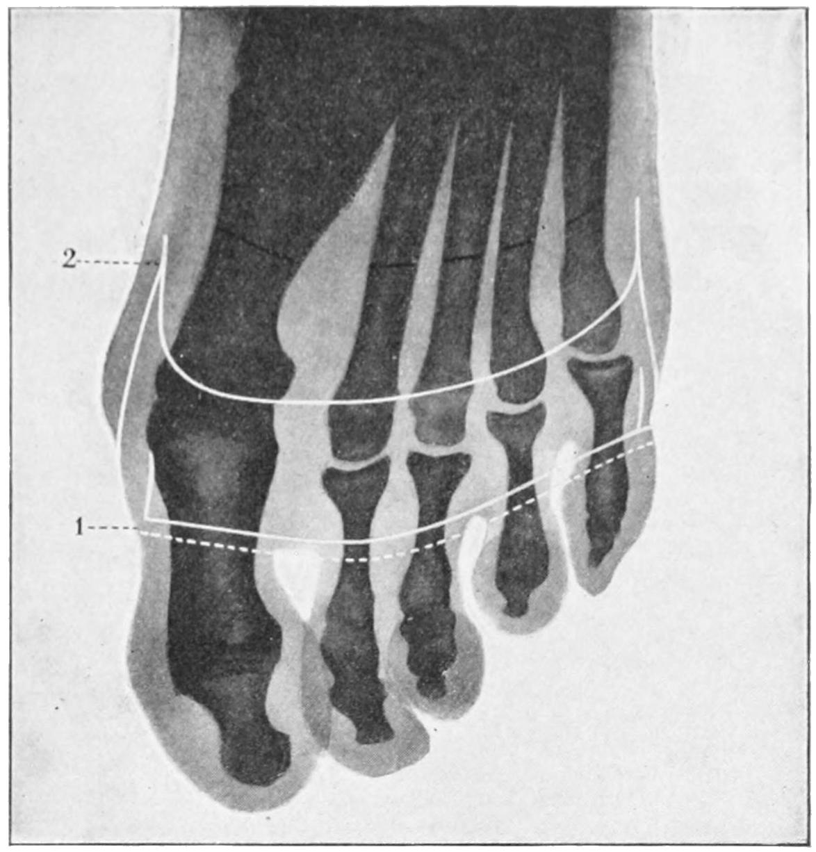
1, simultaneous disarticulation of all the toes; 2, amputation of the toes in their continuity. (Mignon.)
Fig. 693
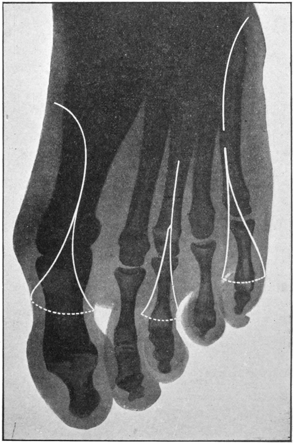
1, partial amputations of the third and fifth metatarsals; 2, disarticulation of the first and fifth metatarsals. (Mignon and Matas.)
Fig. 694
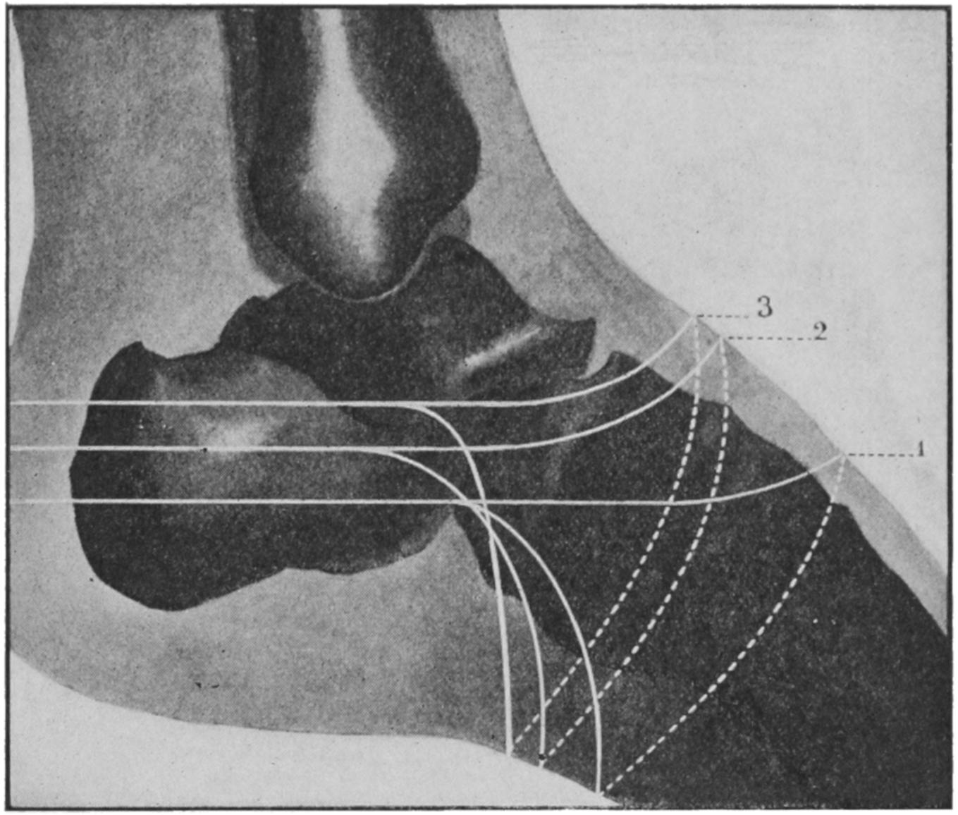
Tracings of intratarsal amputations at various levels (outer side): 1, subastragaloid; 2, tibiotarsal; 3, tibiocalcaneal; the different lengths of flaps shown in relation to skeleton.
Fig. 695
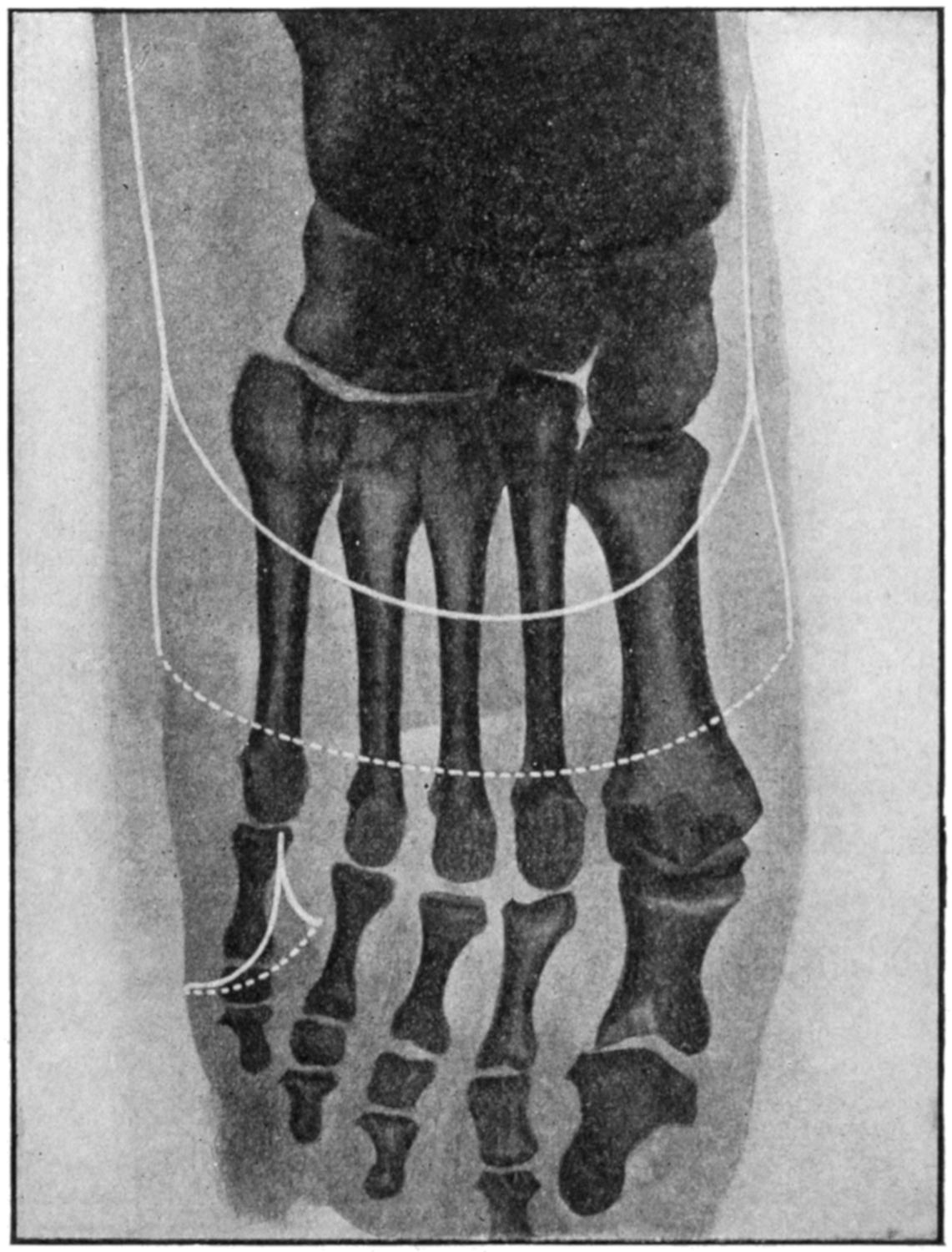
Tracings of incisions in mediotarsal amputations and total amputation of fifth toe. (Mignon and Matas.)
For my own part I would advise to save all of a foot that can be saved, providing a sufficiently long plantar or heel flap can be retained; but if these are not available, then I would advise amputation, at least three inches above the ankle.
I would advise, moreover, to discard the complicated rules and technique of stilted methods and to use the saw whenever it can be made useful, rather than to go farther back to a row of joints simply because they are joints (Fig. 694).
Figs. 695 and 696 illustrate conservative modern methods, which are perfectly available for most purposes, and from which departure need be made only when peculiar circumstances obtain, which so complicate the case that none of the ordinary rules would apply. A surgeon of judgment and experience is competent to devise a flap for a given case, whether it complies with standard methods or not. It seems to me, therefore, worth while to describe only the so-called mediotarsal disarticulation of Chopart, in which but the astragalus and calcis remain of the proper bones of the foot. The joint line extends from just behind the tuberosity of the scaphoid to the outer side of the body of the calcis, where a tubercle can be usually felt. Across this line an incision is carried obliquely over the dorsum of the foot. The plantar flap is the long one, and the line of division is just behind the balls of the toes. Two lateral incisions can be made to facilitate disarticulation if desirable.
Fig. 696
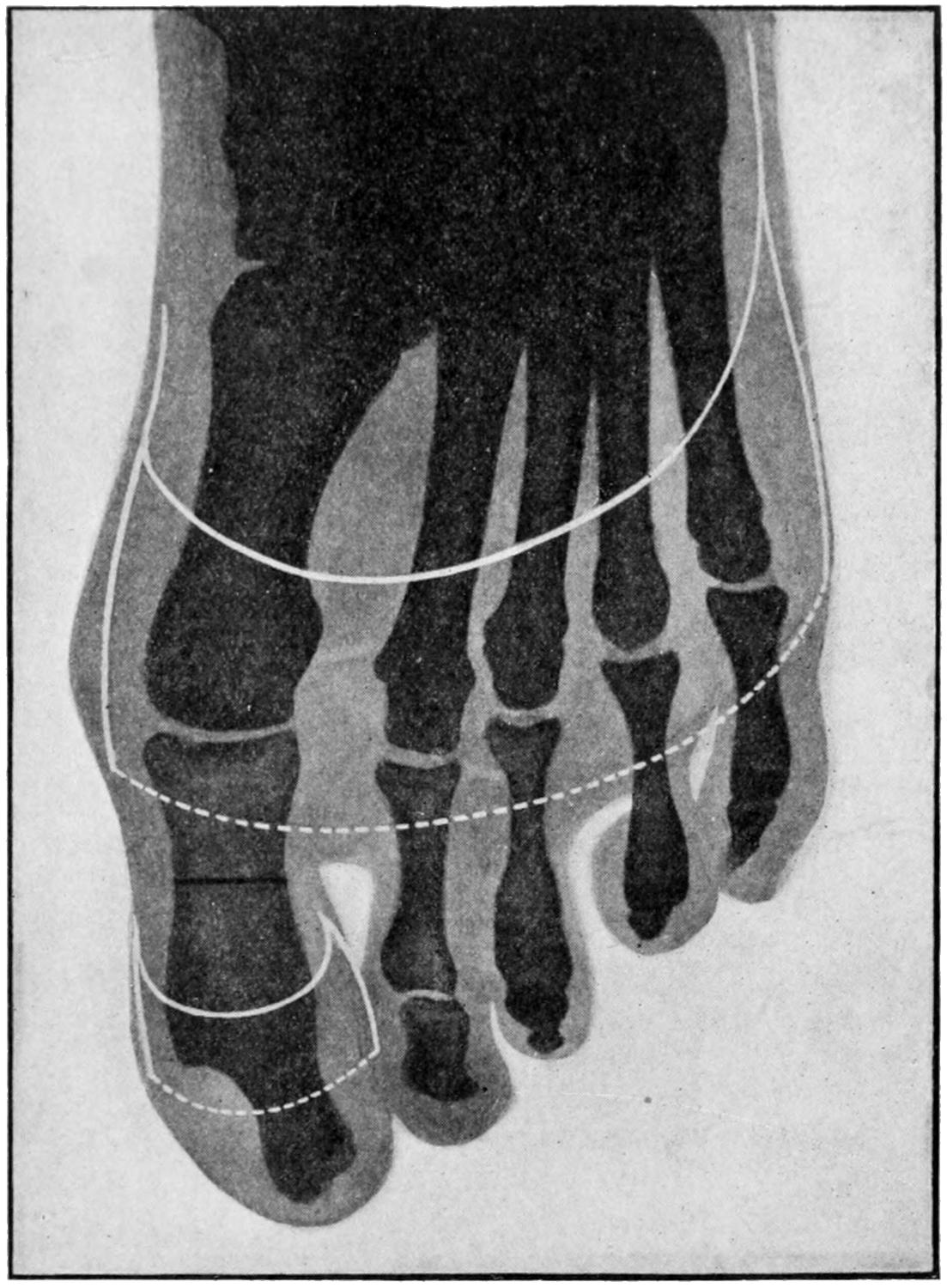
1, disarticulation at the tarsometatarsal joint, showing length of flaps; 2, disarticulation of the big toe in continuity. (Mignon.)
This operation sometimes leaves the foot in a bad equinus position; accordingly it is wise to make tenotomy of the tendo Achillis, as well as to attach the dorsal or extensor tendons to the stump end. (See Figs. 699 and 700.)
Amputations of the Foot (Tibiotarsal).
—The former favorite method of amputating the foot proper was that of Syme, illustrated in Fig. 701. This preserved the plantar surface and tip of the heel, thus forming a cutaneous hood, which was made to cover the lower ends of the tibia and fibula, whose malleoli were usually removed close to their terminations. The incision is made directly down to the bone, from the apex of one malleolus across the sole and up to the other. Then the foot is forced into extreme flexion and another incision carried directly across the dorsum, by which the upper ends of the first are joined. This permits opening the ankle-joint in front, after which the lateral ligaments are divided and the astragalus dislocated forward. The heel hood is next carefully separated from the calcis with the knife, and it and all the parts retracted from the ends of the leg bones, which are divided just above the articular surfaces, or the latter may be left and the malleoli alone removed. Now the heel hood is raised, made to cover the end of the leg, and united to the anterior incision, drainage being made by a small opening on the outer side of the tendo Achillis, as the plantar surface of the flap should not be opened (Fig. 703).
Fig. 697
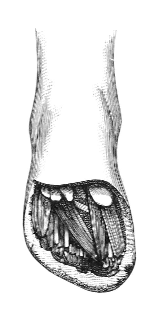
Lisfranc’s tarsometatarsal disarticulation. (Farabeuf.)
Fig. 698
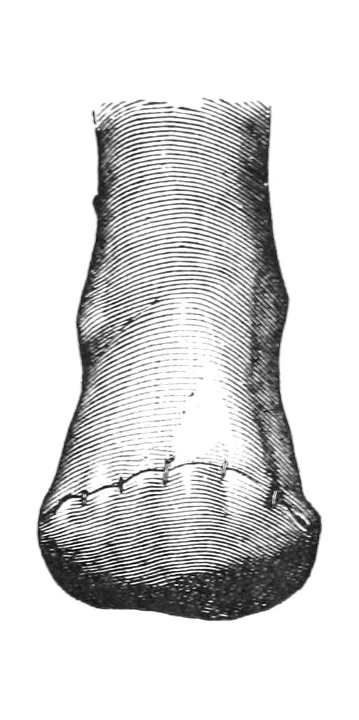
Stump after same.
Fig. 699
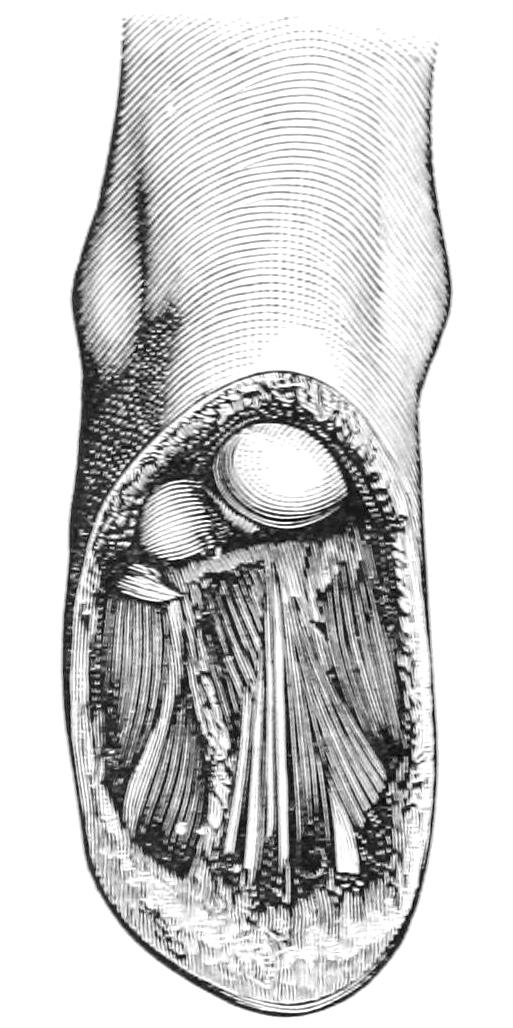
Classical mediotarsal amputation (Chopart), showing length of plantar flap. (Farabeuf.)
Fig. 700
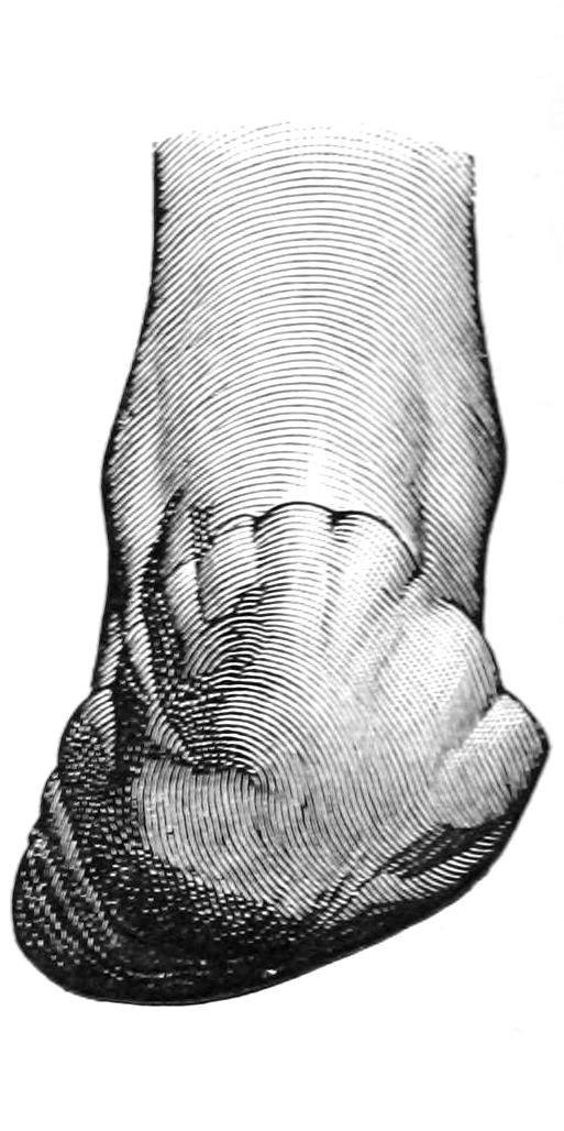
Stump after Chopart’s amputation. (Farabeuf.)
Fig. 701
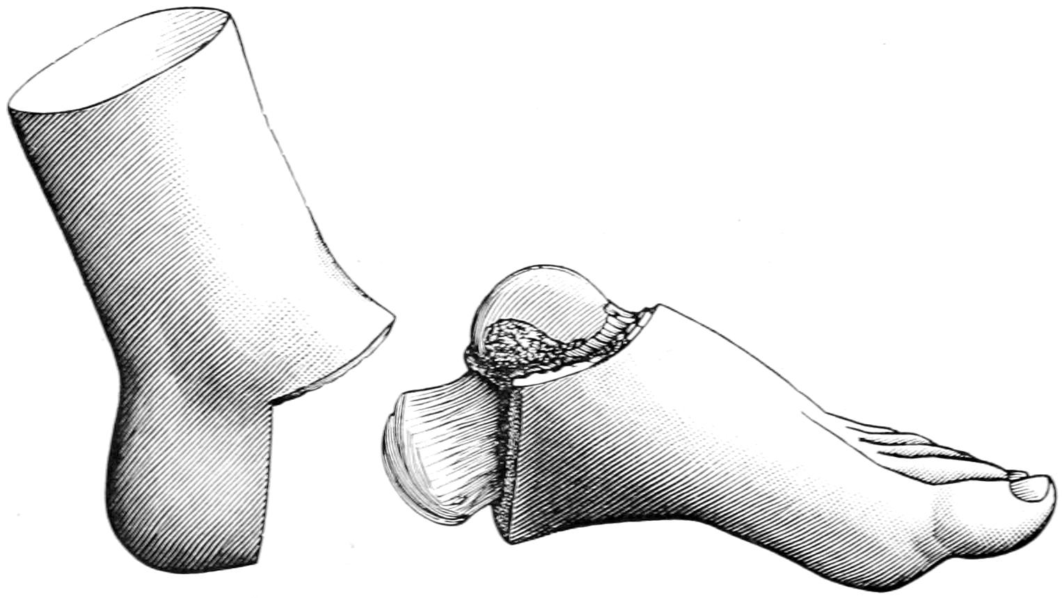
Syme’s tibiotarsal operation, showing part removed and lines of section, before division of malleoli. (Farabeuf.)
Fig. 702, the complement of Fig. 701, illustrates the appearance of the stump after the completion of the disarticulation and before the removal of the malleoli.
Pirogoff introduced a serviceable modification by obliquely dividing the os calcis in front of the heel, turning up its sawed surfaces without any dissection of the hood from the heel, and uniting its fresh bone aspect with that made by removal of the articular ends of the leg bones. This would seem to be preferable to Syme’s disarticulation, affording a better walking stump. (See Figs. 704 and 705.)
A reverse of this operation was suggested by Mikulicz and Wladimiroff, independently, and at about the same time, the heel being excised by an incision across the sole and then behind the ankle, the calcis being divided and its posterior end removed, while the articular surfaces of the leg bones are also removed. The foot is then brought down so that these surfaces can be brought in contact, it being expected that after their reunion the individual will walk in the exaggerated equinus position and upon the ends of the metatarsal bones. The operation is, in effect, an exsection rather than an amputation, and is applicable to but a very small number of cases, in which, however, it sometimes gives excellent results. (See Fig. 707).
Fig. 702
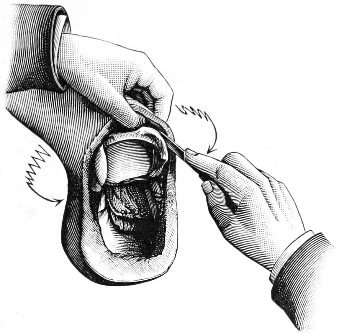
Syme’s amputation after disarticulation of the foot at the ankle-joint. The soft parts are being cleared from the malleoli preparatory to sawing the malleoli and lower articular surface of tibia. (Farabeuf.)
Fig. 703
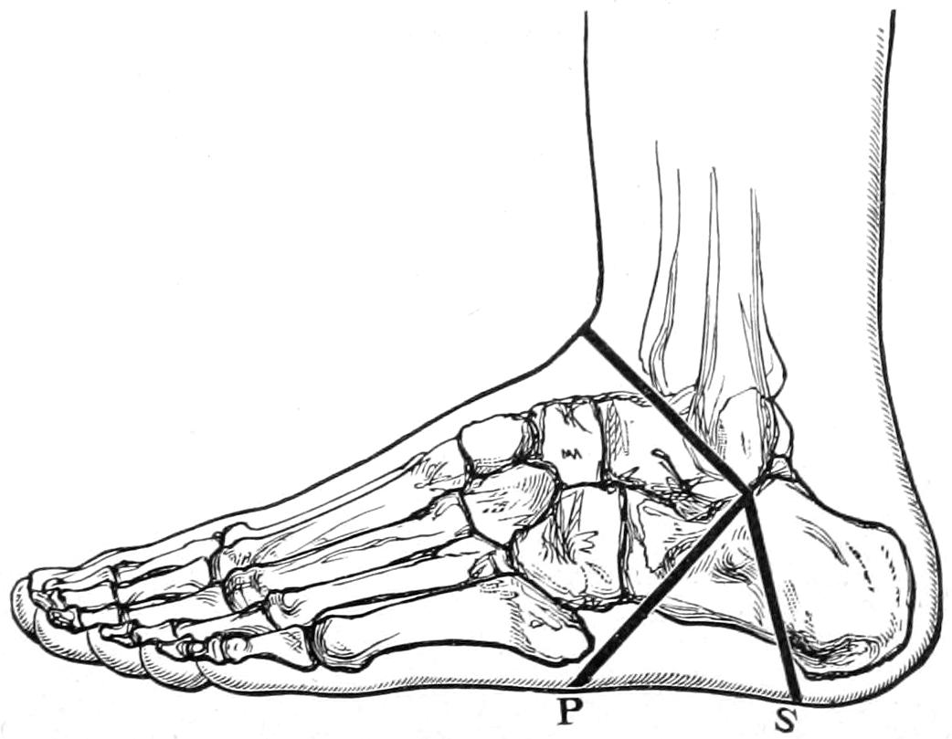
S, line of incision for Syme’s operation; P, line of incision for Pirogoff’s operation. (Erichsen.)
Amputations Of the Leg.
—Modern prosthetic methods have materially changed the indications in amputating the leg. The pressure in artificial limbs is not borne upon the end of the stump, but is rather circumferential and borne by a conical socket. It is now, therefore, an object to preserve as much of the limb as practicable, in order to have better leverage or control of the artificial member. Consequently the point of election is now the middle of the leg, unless the amputation may be made even below this point. The objection to a short leg stump is the inevitable flexion which the hamstring flexors will produce; in such cases the pressure will be borne upon the knee, while the appearance of the stump is by no means ideal. If, therefore, one is forced to make a high amputation of the leg it would be far preferable to make a good knee disarticulation, or, better still, a supracondyloid amputation, with preservation of the patella, thus furnishing a stump which affords perhaps the only exception to the general rule, i. e., that weight cannot be borne upon the stump end.
Fig. 704
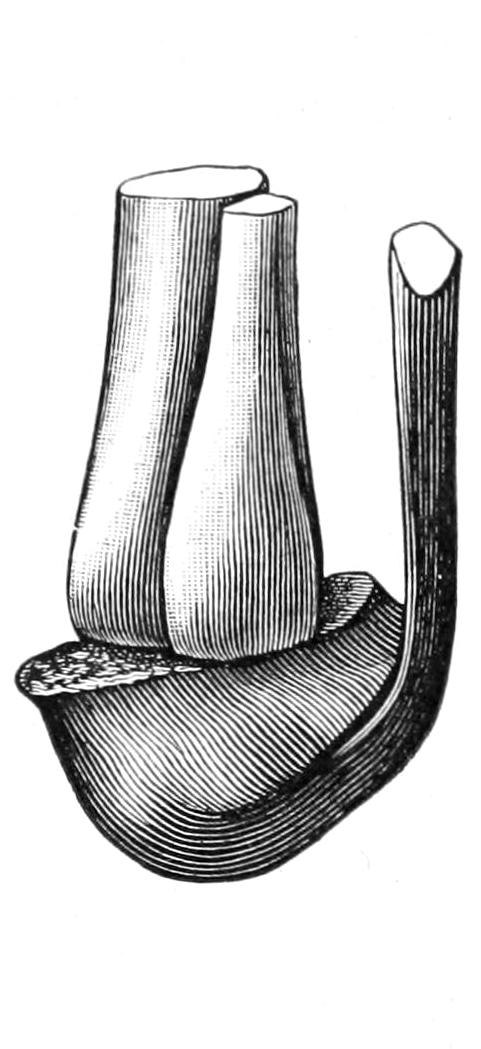
Skeleton of stump after Pirogoff’s osteoplastic amputation. The sawed surface of the calcaneum in apposition with divided surface of tibia. (Farabeuf.)
Fig. 705
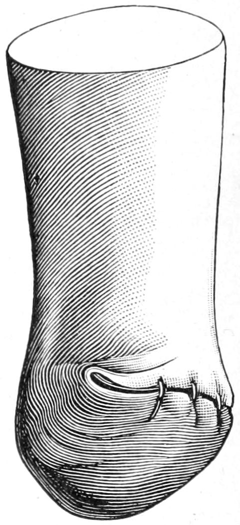
Stump after Pirogoff’s operation. The weight of the body must rest upon the thick plantar skin of the heel and never on the thin skin of the retrocalcaneal surface. (Farabeuf.)
Fig. 706
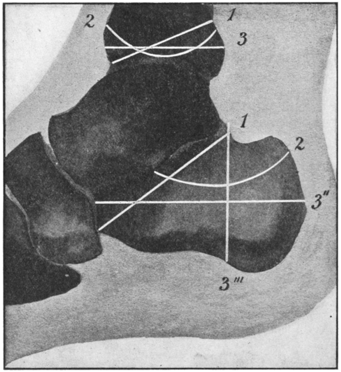
Sections of bone in Pirogoff’s amputation and its modifications: 1, oblique section of calcaneum to correspond with (1) oblique section of tibia. (Gunther.) 2, curvilinear or concave section of calcaneum to correspond with (2) convex section of tibia. (v. Bruns.) 3″, horizontal section of calcaneum to correspond with (3) similar section of tibia. (Pasquier Le Fort.) 3‴, vertical section of calcaneum to correspond with (3) horizontal section of tibia. (Typical Pirogoff.) (Modified by Matas from Mignon.)
Fig. 707
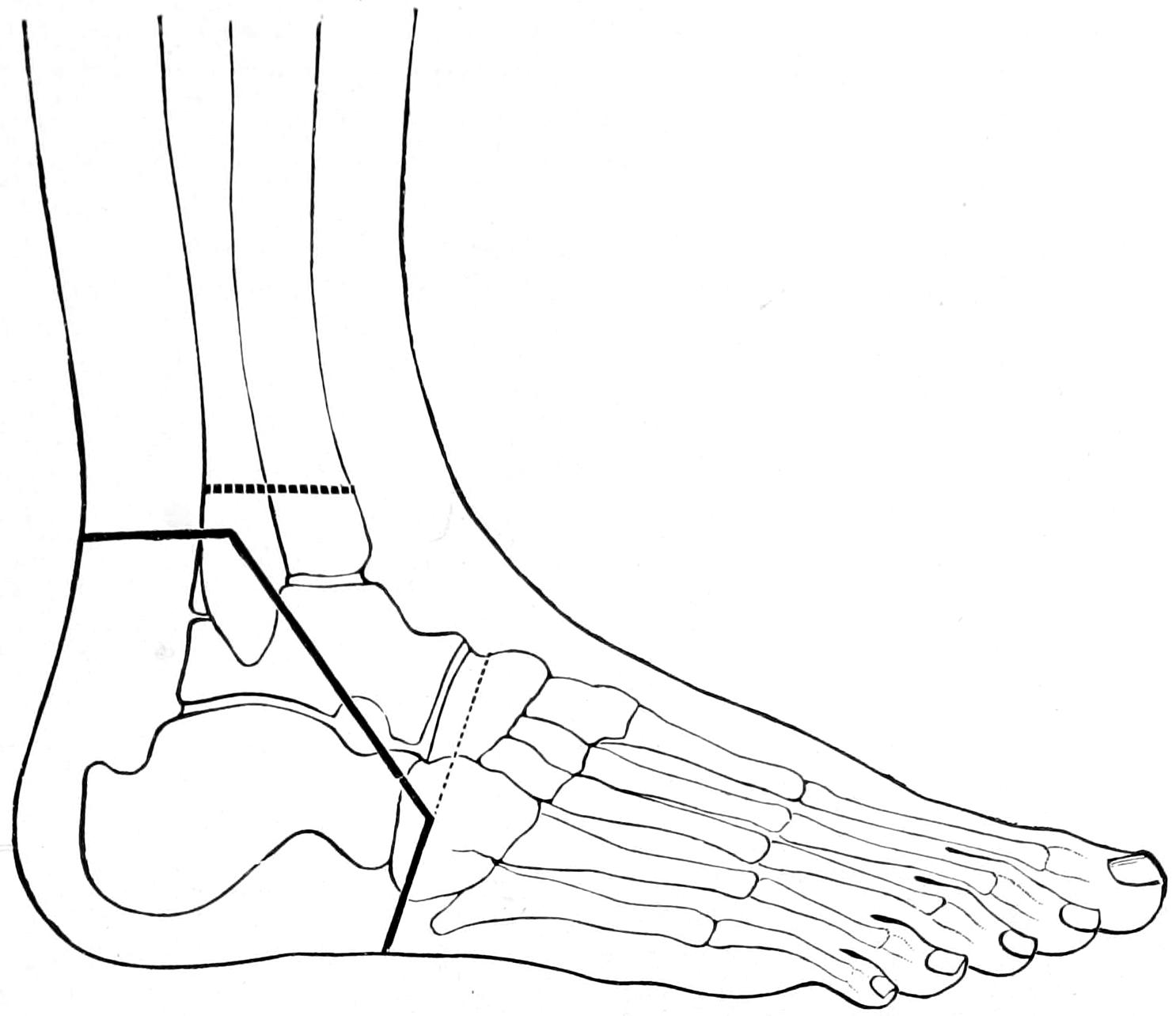
Osteoplastic excision of foot. (Mikulicz.)
Let us, then, consider but one or two amputations of the leg—that low down or near the middle and that at the knee. Whatever the method it is most desirable that the scar be kept off to the side, and especially away from the front of the shin. This can be best accomplished by a modified circular (Fig. 708) or a bilateral flap method (Fig. 711), or by the oblique method with lateral incisions, which practically convert it into an anteroposterior operation, while for certain instances the method of Teale may be preferred, i. e., that with a long anterior and short posterior flap, or its modification by which the flaps are made more lateral, or the even long flap method of Bell.
Fig. 708
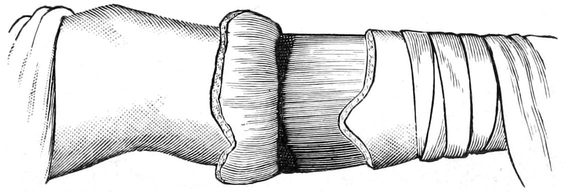
Modified circular amputation of upper third of leg. (Erichsen.)
Whichever of these be selected, after division of the muscles and exposure of the bone, it is usually helpful to retract the flaps, whatever their shape, by a cloth retractor made of a piece of sterile bandage torn into three strips, the middle of which should be inserted between the bones of the leg, the interosseous membrane being divided for this purpose; by this they are held more perfectly out of the way during the act of dividing the bones. The anterior border of the tibia, which is practically a sharp ridge, should be divided obliquely (bevelled), either by a small oblique section before the transverse division is made, or by effecting this later, in order that there shall not remain a sharp point to project through the skin or be subject to constant irritation. The tibia is usually divided transversely, with the above exception. The fibula may be divided slightly obliquely. It is customary, however, to make the division simultaneously, and to so conduct the sawing process as to divide the fibula completely before the last strokes of the saw cut through the tibia.
There is greater difficulty in the recognition and securing of vessels in leg amputations than in any other, especially if they have been divided obliquely. The principal vessels may be found from their known anatomical location. They nevertheless sometimes tend to retract and they must be followed up in order to properly secure them. The accompanying nerve trunks should also be seized firmly, drawn down, and divided two or three inches above the line of division of the other tissues, in order that they may retract out of harm’s way. Every nerve which can be recognized, even in the skin, should be thus treated. Before closing the wound it is well, unless one is absolutely sure of his work, to release the tourniquet and ascertain if any vessel which would otherwise bleed be not yet secured. Oozing may be checked with hot water, while muscle surfaces which leak too much blood may be lightly enclosed within catgut sutures inserted with a curved needle.
Providing that these stumps have well-nourished flaps, and that no sharp or angular bone ends interfere with subsequent comfort, and that the scars be kept away from their lower surfaces, they serve their purpose admirably.
Supposing, then, that amputation is to be about the middle, the first incision, made with a stout scalpel, is begun at the anterior border of the tibia and carried downward along it until it is turned abruptly backward to the posterior aspect, and then upward until a point is reached opposite that of commencement. The skin is dissected up for perhaps an inch. Then the flap on the other side is cut after the same fashion, after which, with a short or long knife, the muscles are divided transversely or circularly down to the bone. Much will depend now upon whether the desire is to resort to the more modern osteoplastic methods or adhere to the old. In the latter case it is well to separate the flaps for the necessary distance from the bone, with or without the periosteum, dividing the bones after suitable retraction, as above suggested. If preference be for an osteoplastic flap it is planned and made at this time, the bone being divided at the same level as the muscles, and the amputation being thus practically completed, after which the osteoplastic flap is arranged, it being now necessary to carefully preserve the periosteal bridge and to again divide the bone at the base of this periosteal flap, this being the true end of the bone stump. In this case the fibula is divided at a higher level.
It is perhaps less desirable to preserve periosteum in young children than in adults, for if bone be permitted to grow too rapidly conical stumps result, sometimes even with protrusion of bone ends. Ollier and his pupil Mondan have shown that this so-called atrophic elongation of bone is a consequence of abnormally rapid growth from the upper epiphyseal direction, permitted by lack of pressure from below, and that conical stumps will often happen in children in spite of every precaution. Nevertheless it should not be encouraged, and for this reason periosteum should not be preserved.
The method of Teale was to cut a long anterior flap, raising the coverings from the bone with the least possible disturbance, to divide the bone at the high level, then to double the flap upon itself in such a way as to bring the scar at a level one inch or more above the stump end. Heine modified this slightly by raising the periosteum with the rest of the anterior flap. In cases which permit such a long flap to be formed from one aspect of the leg the method gives excellent results (Fig. 709).
Fig. 709
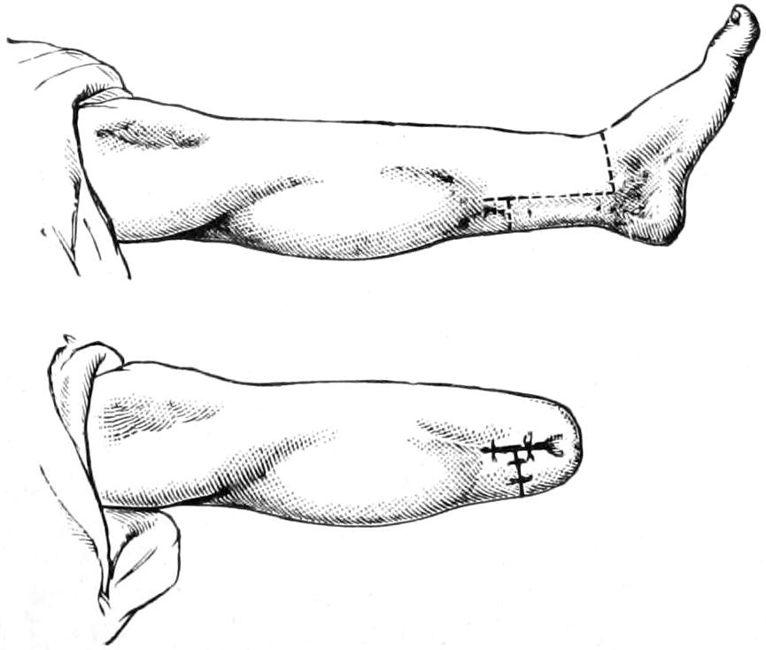
Teale’s method of amputation.
Bruns devised a method which is begun almost as an exsection, by an oblique circular incision, with liberating lateral incisions, and division of all the tissues over the inner border of the tibia and the outer side of the fibula; after which, without disturbing skin attachments in front, the periosteum is separated from the bones as high as the liberating lateral incisions permit, and then the fibula first and later the tibia divided. It is practically a subperiosteal excision of the leg bones and affords a well-protected stump. In effect it is an anteroposterior flap method.
Fig. 710
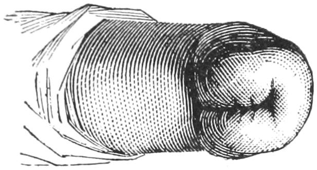
Stump after Stephen Smith’s amputation at knee.
The Knee.
—It was Brinton who, in 1872, suggested the preservation of the semilunar cartilages in all knee disarticulations, as in this way all the normal relations are preserved and retraction is prevented. But the makers of prosthetic apparatus have urged to abandon all true disarticulations, and to substitute for them the supracondyloid method, which affords ideal stumps. Disarticulations are supposed to produce less shock, less loss of blood, and less danger of sepsis from opening up the bone-marrow, while muscle insertions are less disturbed and the stump covering usually is mobile and not very sensitive. No disarticulation should be thought of unless the joint involved be free from disease and unless about it there be met sufficient healthy integument to furnish a satisfactory flap.
For a true disarticulation Stephen Smith’s bilateral method is now almost universally adopted. Here the incision is begun one inch below the tubercle of the tibia and is carried directly down to the bone, downward and forward around the side of the leg, and then inward and upward toward the middle of the popliteal space, the lateral flaps thus made being nearly duplicates. The flaps thus cut out are completely separated from the bone up to the joint level, where the ligaments are divided, the joint being manipulated as may be necessary to best expose them and facilitate division. In this operation the patella is usually removed, the joint being opened by separating its ligament at its insertion into this bone. One should remember that the internal condyle is lower and longer than the external, and that the internal flap should be perhaps made on this account a little the longer of the two. Fig. 710 illustrates the stump resulting from this operation and shows the cicatrix drawn up out of harm’s way and resting in the fossa between the condyles. Fig. 711 illustrates the simple method by lateral flaps.
Fig. 711
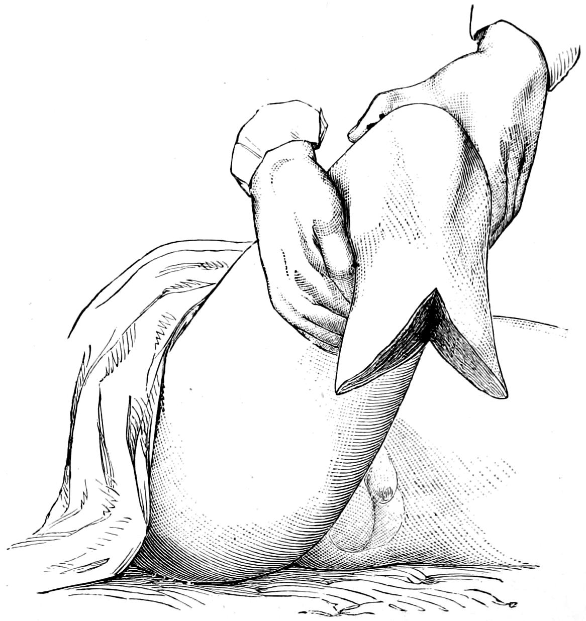
Amputation at knee by lateral flap. (Erichsen.)
Amputation of the Thigh.
—Under this head, rather than that of amputation at the knee, should be described the supracondyloid amputations which give decidedly the best results of all, and which are preferable to any others for the middle of the lower extremity. Of these the best is that suggested by Gritti, which consists in not only removing the condyles but sawing off the articular surface of the patella, which is then drawn upward and applied to the end of the femur, the division of the latter being made at a point above the condyles, where the diameter of both bones will nearly correspond, this latter perhaps being a suggestion of Stokes rather than of Gritti, who did not divide the bone quite so high. (See Fig. 712.)
Fig. 712
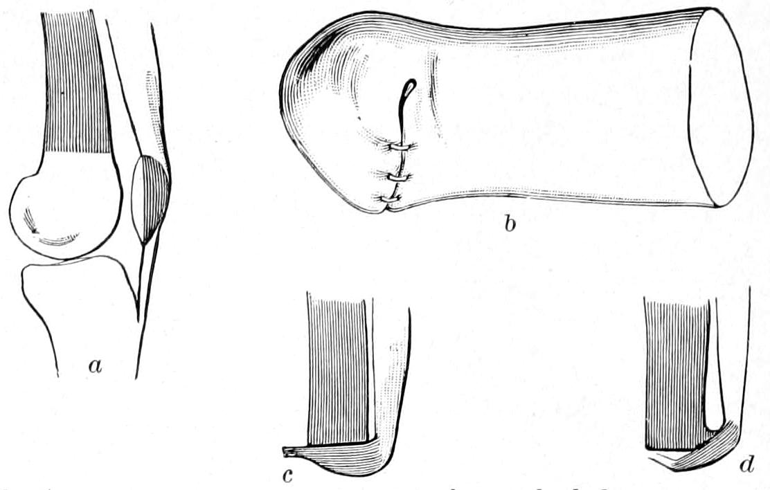
Gritti’s osteoplastic supracondyloid knee amputation, patella utilized: a, shaded parts are those brought in apposition; b, appearance of Gritti stump after suture; c, correct apposition of patella to femur; d, defective apposition. (Farabeuf.)
Fig. 712 will best illustrate the intent of the method as well as its performance. The incisions are planned much as in the Stephen Smith disarticulation method, only they are placed higher, and the patellar tendon is divided as low down as possible, or even separated from the tibia, in order that it may be made of use in attaching the divided patellar surface to the femoral end. The rest of the operation is performed as by other methods, the attachment of the patella being effected by tendon sutures, or, if necessary, by an ivory peg, or even a metal tack or nail which may be left in place.
The beauty of this method is that the anterior surface of the patella is preserved with its natural weight-bearing facilities and the bursa between it and the skin, while the latter is undisturbed. On the end of this stump as much weight can be steadily borne as when one ordinarily kneels, and to it a most serviceable kind of artificial limb can be attached, with which one may walk as though nothing had ever happened.
Another osteoplastic method, namely, that of Sabanejeff, is illustrated in Figs. 713 and 714. In this instance the bone covering over the end of the femur is taken from the upper end of the tibia, the patella not being disturbed. It permits a lower division of the femur and the formation of a stump which is of practically the same length as the original thigh.
Fig. 713
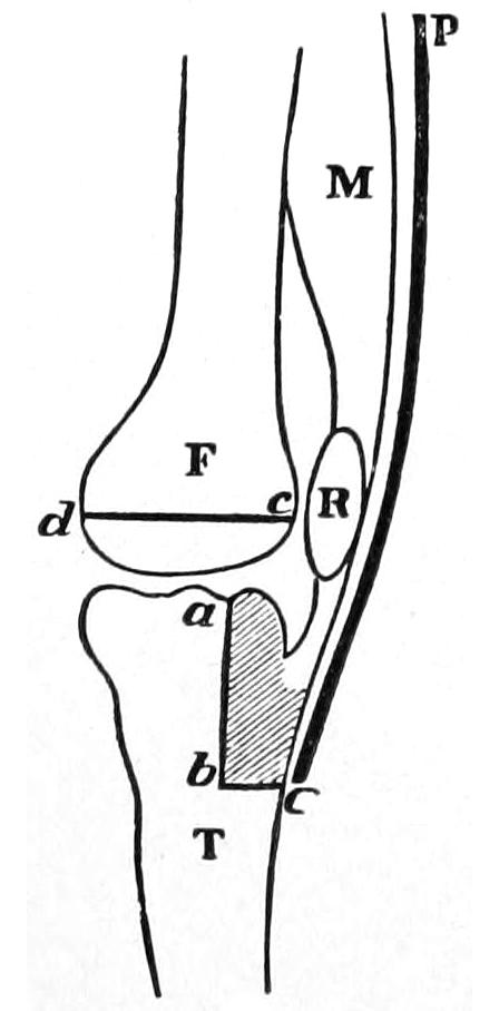
Sabanejeff’s knee amputation. (Chalot.)
Fig. 714
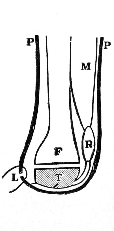
Stump made as in Fig. 713. (Chalot.)
Amputation of the Thigh above the Knee.
—For removal of the thigh it is well to preserve as much of its length as possible, and yet not at the expense of all other considerations. A thigh stump too short is likely to be pulled awkwardly upward by the psoas muscle, and upon such a stump it is difficult to secure an artificial limb tolerable of control against such action of this muscle. On this account, then, thigh stumps should be long. So far as the method is concerned the circular, or some modification thereof, gives the best results in the majority of instances. It may easily be modified into one of the oblique methods, or liberating incisions may be used whenever they will be of service. If it be absolutely necessary to make the amputation high hemostasis can be secured by the same methods that are used in hip-joint amputations. The dense and strong fascia lata, which lies beneath the superficial fascia, should be divided at the same level with the skin, since it serves admirably, when secured by a separate set of sutures, to make a good covering for the ends of the muscles, after these have been themselves carefully united by buried sutures. The sciatic nerve should be especially sought, thoroughly stretched, and divided high up. The vessels often evince a tendency to retract within Hunter’s canal; it is not, however, difficult to separate the vastus internus from the adductor longus, between which they lie, and in this way gain access to them. Even for high work on the thigh one may, if necessary, do as some have done at the hip, make a preliminary ligation of the femoral artery. This may be especially serviceable as an emergency measure, or in special cases of tumors which have attained large size, are placed high up and call for somewhat atypical methods.
The Hip-joint.
—Amputation here is essentially a disarticulation and constitutes one of the usually formidable and serious operations of major surgery. Although the joint itself is generally easily reached there are many things to be considered in the performance of this operation, of which the mere arthrotomy is by no means the most important.
Preparations being all made, the first consideration is the control of hemorrhage, for which several methods have been suggested, but of which but two or three are in general use. Such procedures as compression of the abdominal aorta, either with the hand or by tourniquet, or of the common aorta through the rectum, with a lever, as suggested by Davey, or with the hand, as suggested by Woodbury, or the exposure of the common iliac, either within the peritoneum as practised by McBurney, or externally, or exposing the common femoral above Poupart’s ligament, are now adopted by very few surgeons. Langenbeck used to be fond of preliminary ligation of the femoral where it is most accessible in the groin, and this is probably the best of all of these methods. But they have been all practically discarded since Wyeth introduced the simple method of transfixing the limb with his pins (i. e., long mattress needles or skewers made for the purpose), these serving to hold in place an elastic cord or tourniquet (Fig. 715). This has been found to be a great improvement on the suggestion of Senn, who excised the femoral head and then compressed each half of the limb with a separate elastic band.
Fig. 715
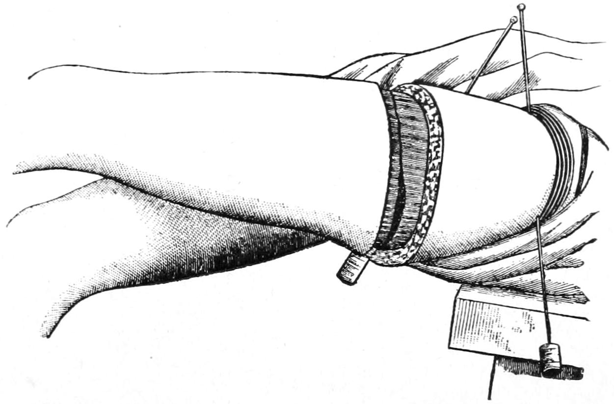
Wyeth’s bloodless method: pins inserted and tube applied.
The directions for the use of Wyeth’s pins are simple. Here, as in other cases, it may not be practicable to use the elastic bandage from the lower end of the limb, but one may at least elevate the limb and thus coax the blood out of it by gravity or by gentle manipulation. While it is still in this position one of the long pins is introduced just below the anterosuperior spine and a trifle to its inner side, and made to emerge on a level with and about three inches from the point of its entrance. The other needle is inserted just to the inner side of the saphenous opening, and below the level of the crotch, and brought out about one inch below the tuberosity of the ischium. Corks should then be placed upon them so as to protect the needle points. Next a piece of elastic tubing or band is placed around the limb above these pins and tightened, each turn being made a little tighter, so as to absolutely control the circulation. The effect of this is felt upon practically every vessel in that part of the body, and if the method be properly practised it affords absolute security.
The surgeon now has his choice of various methods of disarticulation, either that by anteroposterior flaps or lateral flaps, or by the circular, with the free liberating lateral incision; or he may devise any method of his own which will best meet the indication in a given case. Fig. 715 illustrates the employment of Wyeth’s pins and the first circular incision made as for the circular method. Of these all the latter seems preferable when circumstances permit. It should be combined with a sufficient lateral incision, which should be made to pass well over the great trochanter. The cuff raised through this incision should extend down to the deep fascia and up to the level of the lesser trochanter, at which level the deeper tissues are divided transversely or by a circular cut.
It is well next to lay down the knife and secure the large vessels, after which the deep muscles are separated from the upper end of the shaft and the proximity of the joint, while the entire limb may be still used as a lever in so stretching the joint capsule as to better expose and divide it. So soon as the capsule has been opened, and the entrance of air thus permitted, it will be easy to expose and divide the teres ligament, after which the balance of the disarticulation is easily effected. The large nerve trunks are now sought, retracted, and divided high up, all visible vessels are secured firmly, after which the elastic constriction may be gradually released and any vessels that spurt may still be secured. There will nearly always be troublesome oozing from the cut ends of the large muscles, and here, if hot water prove insufficient to check it, with large curved needles and catgut sutures the muscle ends may be secured by ligature en masse, before they are brought together for the purpose of closing the stump.
Whatever the method selected as perfect a closure of the wound as possible should be made, with ample provision for drainage. By careful deep suturing, with tiers of buried sutures, it is possible to avoid leaving dead spaces at any point except perhaps the acetabulum. Through retaining sutures may also be used to advantage. It is most desirable to so plan the incisions and the closure of the wound as to keep them, so far as possible, away from the region of the perineum. Therefore the longer the inner flap or inside of the stump the better. As conditions which necessitate removal of the limb at the hip-joint are always serious, and have each their own peculiarities, any method which will best serve the purpose should be used.
Plates LIX and LX, designed by Prof. Matas, afford the best and briefest epitome of the choicest amputation methods which can be furnished.
THE STUMP.
An amputation having been effected, and the stump closed, there is still occasion to consider how it may best be treated to fit it for its future purposes. When entire chapters, or even small monographs, can be written on the subject of “diseases of stumps” it would appear that the consideration is not one of merely trifling import.
A good stump has a regular outline, with a protected scar, and should be firm, yet mobile, and without tender or sensitive surfaces. It should constitute the lower end of a truncated cone, and needs to be of sufficient length to permit leverage within the socket of the artificial limb which will be fitted about it.
A stump failing in these characteristics is a bad stump, the features which especially tend to make it bad being undue conicity (Fig. 716) or sensitiveness of surfaces, ulceration from friction, or, worse yet, occurring without it, and neuralgia from inclusion of nerve ends, or from bone ends which present osteophytic outgrowths and thus distort and displace tissues (Fig. 717). Acute osteomyelitis occurs in stumps, as do slower carious processes which may call for re-amputation, perhaps even at a distance. The stump is for a long time more or less tender and troublesome, and its owner may be a sufferer from hyperesthesia or perverted sensations.
The possibility of the production of a conical stump in children as the result of atrophic elongation was mentioned early in this chapter. While this cannot always be prevented it may sometimes be foreseen, and one should be prepared at any time in such cases to circumcise the bone, forcibly retract the tissues, and then divide the bone ends on a higher level.
PLATE LIX
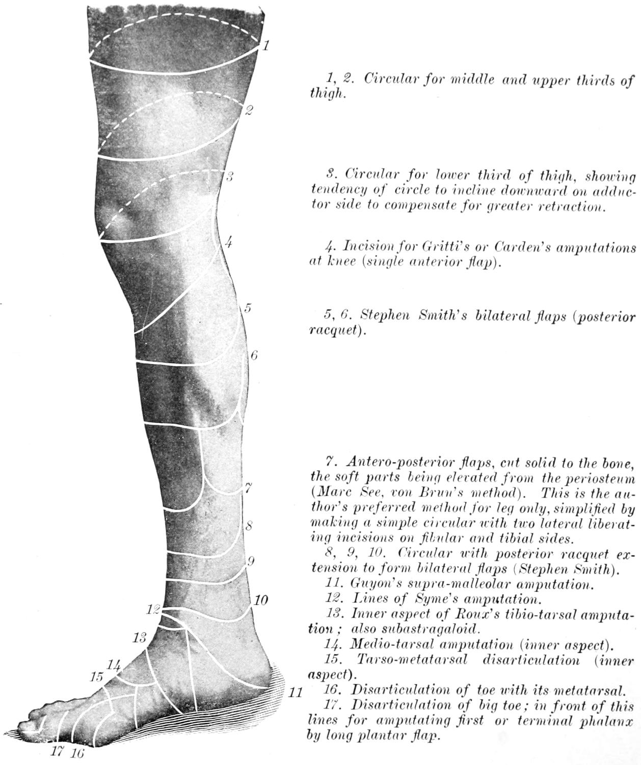
The Right Lower Limb, Internal Lateral View (Surface Incisions).
1, 2. Circular for middle and upper thirds of thigh.
3. Circular for lower third of thigh, showing tendency of circle to incline downward on adductor side to compensate for greater retraction.
4. Incision for Gritti’s or Carden’s amputations at knee (single anterior flap).
5, 6. Stephen Smith’s bilateral flaps (posterior racquet).
7. Antero-posterior flaps, cut solid to the bone, the soft parts being elevated from the periosteum (Marc See, von Brun’s method). This is the author’s preferred method for leg only, simplified by making a simple circular with two lateral liberating incisions on fibular and tibial sides.
8, 9, 10. Circular with posterior racquet extension to form bilateral flaps (Stephen Smith).
11. Guyon’s supra-malleolar amputation.
12. Lines of Syme’s amputation.
13. Inner aspect of Roux’s tibio-tarsal amputation; also subastragaloid.
14. Medio-tarsal amputation (inner aspect).
15. Tarso-metatarsal disarticulation (inner aspect).
16. Disarticulation of toe with its metatarsal.
17. Disarticulation of big toe; in front of this lines for amputating first or terminal phalanx by long plantar flap.
PLATE LX
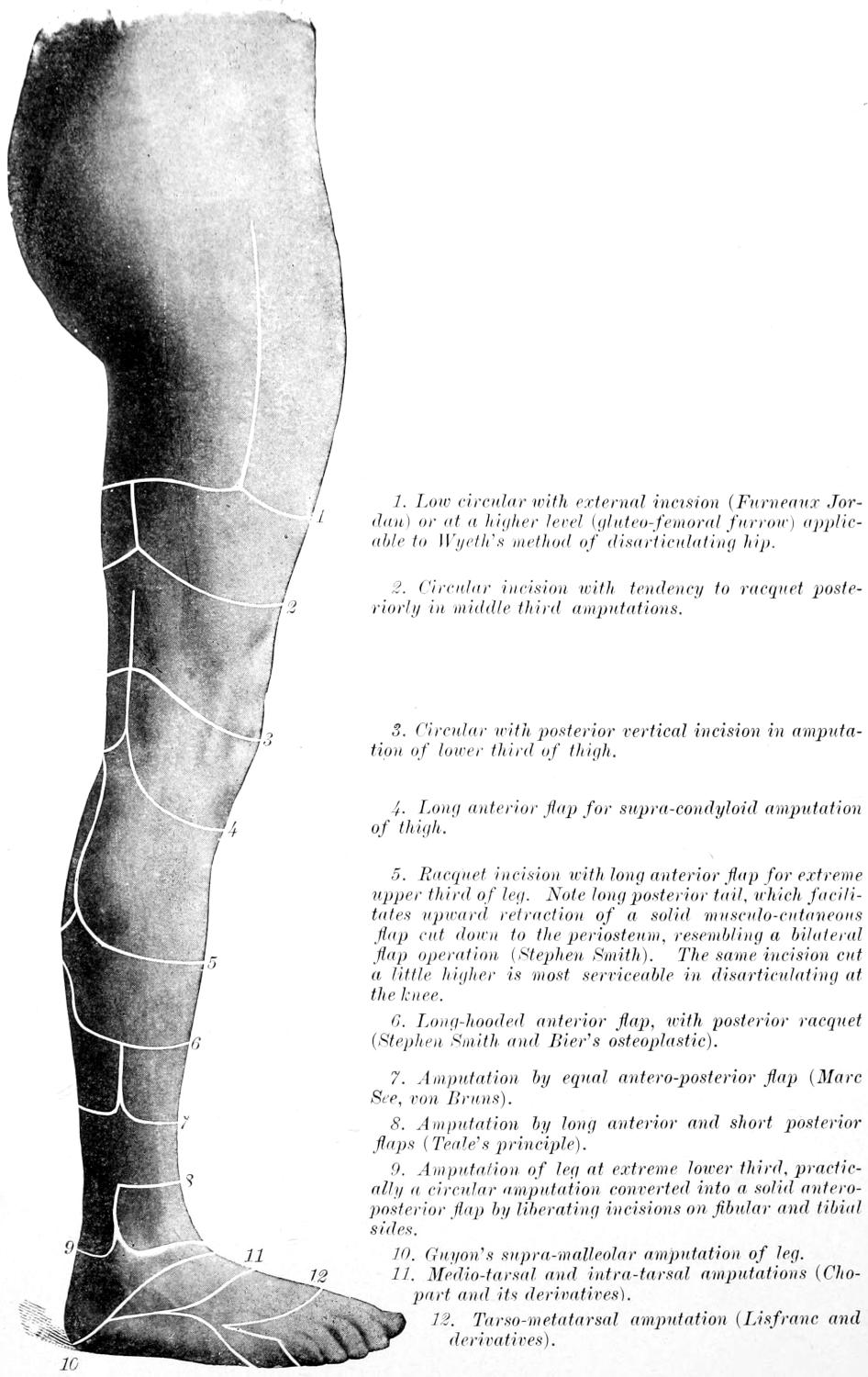
Surface Outlines of Amputations Practised in the Lower Extremity.
1. Low circular with external incision (Furneaux Jordan) or at a higher level (gluteo-femoral furrow) applicable to Wyeth’s method of disarticulating hip.
2. Circular incision with tendency to racquet posteriorly in middle third amputations.
3. Circular with posterior vertical incision in amputation of lower third of thigh.
4. Long anterior flap for supra-condyloid amputation of thigh.
5. Racquet incision with long anterior flap for extreme upper third of leg. Note long posterior tail, which facilitates upward retraction of a solid musculo-cutaneous flap cut down to the periosteum, resembling a bilateral flap operation (Stephen Smith). The same incision cut a little higher is most serviceable in disarticulating at the knee.
6. Long-hooded anterior flap, with posterior racquet (Stephen Smith and Bier’s osteoplastic).
7. Amputation by equal antero-posterior flap (Marc See, von Bruns).
8. Amputation by long anterior and short posterior flaps (Teale’s principle).
9. Amputation of leg at extreme lower third, practically a circular amputation converted into a solid antero-posterior flap by liberating incisions on fibular and tibial sides.
10. Guyon’s supra-malleolar amputation of leg.
11. Medio-tarsal and intra-tarsal amputations (Chopart and its derivatives).
12. Tarso-metatarsal amputation (Lisfranc and derivatives).
An exquisitely neuralgic stump is usually made so by the entanglement of nerve ends and their subsequent enlargement into so-called amputation stump neuromas (which are histologically fibromas), from pressure upon nerve terminals. Under these circumstances their excision through incisions planned for the purpose, or the exsection of a portion of the nerve trunk at a higher level, may be necessitated (Fig. 717).
Fig. 716
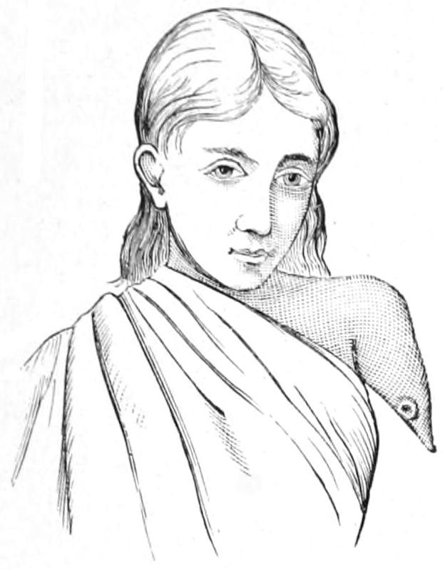
Extreme case of conical stump.
Fig. 717
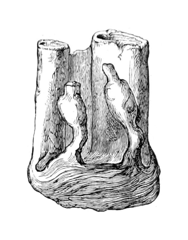
Neuromatous endings of nerves in a stump.
Fig. 718
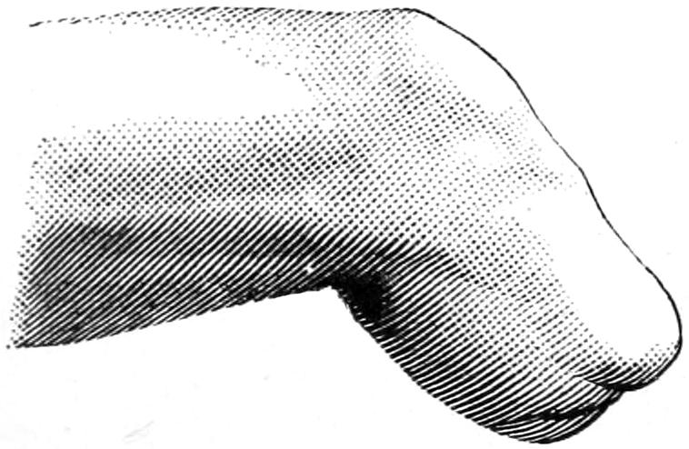
Ideal stump.
Fig. 719
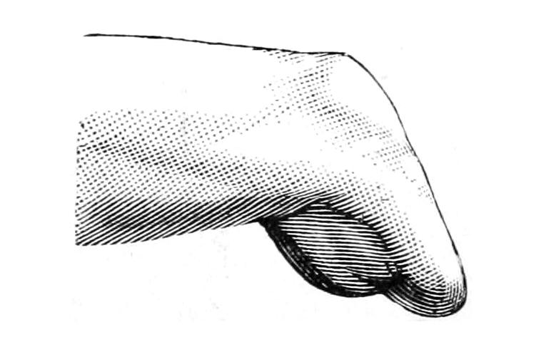
Bad stump, because posterior flap was cut too short, and there has been great retraction of all soft tissues. (Farabeuf.)
While patients may prefer disuse of a stump for as long a time as possible the judicious surgeon will prepare it as rapidly as he may for early application of the expected artificial limb. Inasmuch as leg stumps allowed to hang downward become cyanotic and edematous it is well to keep them bandaged, and the makers of artificial limbs prefer to have the bandages kept wet. When the stump is healed, passive motion of the remainder of the limb should be begun, in order that there may be a minimum of stiffening of joints. If, then, such a stump be bathed, massaged, moved, and then bandaged with comfortable snugness with cold, wet bandages, over which oiled silk may be fastened, and if this be done at least once each day, the stump will be prepared for the artificial limb, on the average, in two to three months. One should not wait for this expiration of time if it be thoroughly healed; or, on the other hand, he may have to wait much longer under unfortunate circumstances; but the above general principles of treatment and general statements will be found to prevail. Figs. 718 and 719 illustrate the difference between good and bad stumps, while Plates LIX and LX (reproduced from Matas) furnish the surface outlines for selection of the various amputations of the lower limb.
CINEPLASTIC OR CINEMATIC AMPUTATIONS OF THE UPPER EXTREMITY.
The most pronounced and illustrative of recent methods is perhaps the “cinematic” or “cineplastic” procedure of Vanghetti. This Italian surgeon proposed a prosthetic method, in 1898, which is illustrated in Figs. 720, 721 and 722. He has shown that tendon terminations may be left exposed in stumps, under favorable conditions, and so utilized as to serve remarkably useful purposes—though under exceptional conditions. For a description of these methods the reader is referred to his monograph. (G. Vanghetti, Plastica e Protesi Cinematiche, Empoli, 1906.)
Fig. 720
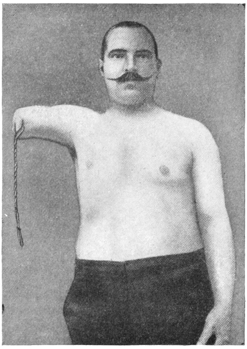
Fig. 721
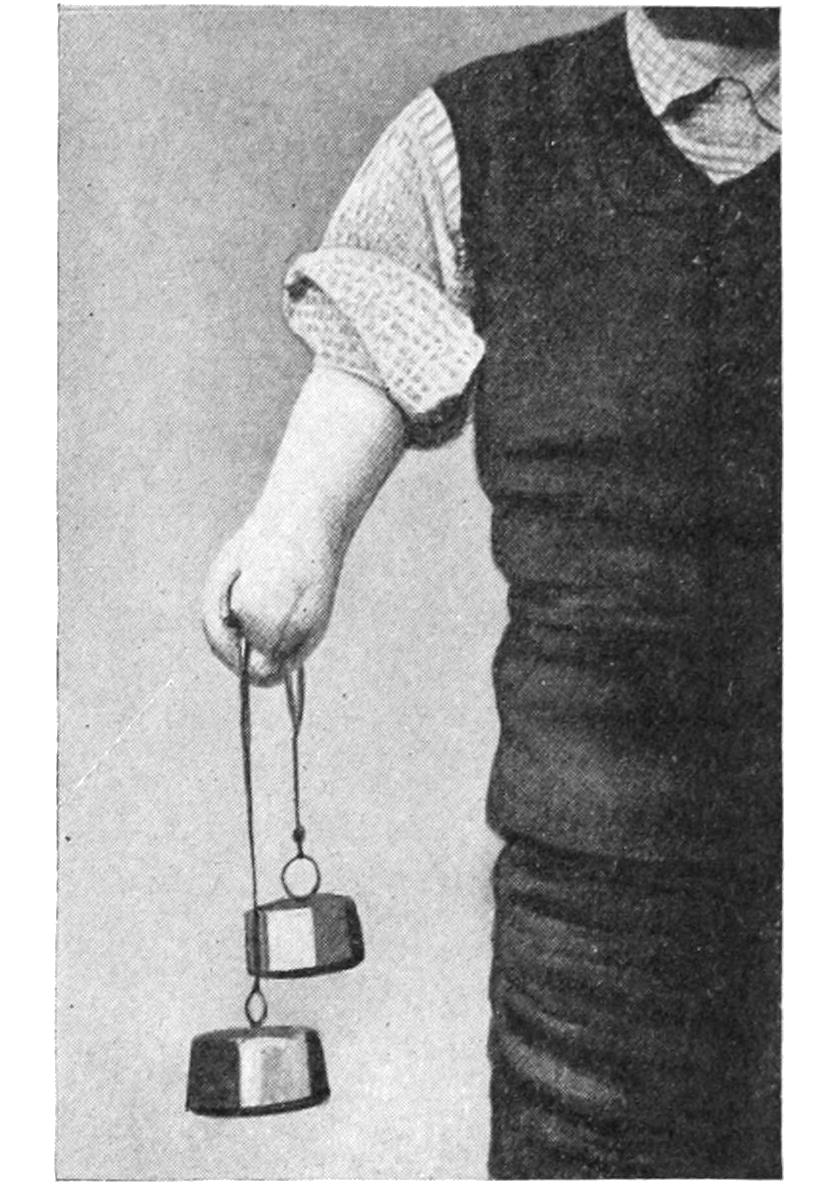
Fig. 722
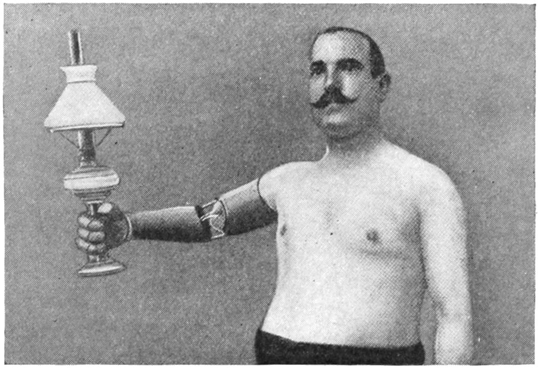
Results of Vanghetti’s “cinematic” method, with preservation and utilization of tendons.