Part 5 — CHAPTER XXXIV. FRACTURES.
Reading edition
The term fracture is, in surgery, applied to such injury of bone and cartilage as effects break in continuity. This injury is effected instantly, and it is rarely that fracture is produced by any slowly acting cause, although this latter may so affect or disintegrate bone as to permit fracture upon the application of a mild degree of force. Fractures are variously classified and grouped for convenience of description; thus we speak of traumatic and pathological fractures, implying by the former those which occur by violence in normal conditions of health, and by the latter those which are produced only because of some previous disease in the bone. The difference is that in the former case there is no preëxisting disease, whereas in the latter it is an essential feature of the case. Fractures are also classified as complete or incomplete, the former term implying injury to the whole thickness of the bone, while the latter are separately classified: (a) Fissure, in which there is a line of fracture by which there is no complete separation of fragment, it being essentially a crack; (b) the green-stick fracture, such as occurs in the young, where the bone is not thoroughly calcified, but is capable of bending to some extent, while a portion of it breaks; (c) depressed fracture, which is generally produced by direct violence, and occurs in a flat bone, i. e., the skull, the scapula, etc.; (d) detachment of a fragment or separation of an epiphysis; (e) partial fractures, corresponding much to the green-stick, but without deformity or change in shape or position.
Fig. 276
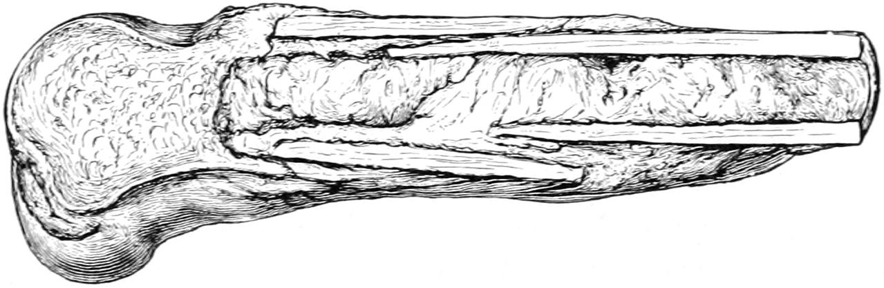
Impacted fracture of the shaft of the femur produced by a fall upon the knee in a man aged eighty-three years. Illustrating impaction. (Bryant.)
Fractures are also described by means of the following adjectives, which practically explain themselves, for instance:
A. Complete, transverse, oblique, longitudinal, dentated, etc. Spiral fracture is also described and occasionally seen. It involves only the long bones, and not only implies a considerable degree of violence, but is itself regarded as exceedingly serious.
B. In number they are single, multiple, or comminuted, as when there are a number of fragments.
C. They are often impacted, which means that one fragment is driven into and more or less embedded in the other. This impaction or interlocking of fragments occurs usually in the neck of the femur and the lower end of the radius. In the former locality it is advisable not to interfere with it; in the latter it should always be dislodged in order to restore the fragment to its proper position (Fig. 276).
D. As to their nature and location, fractures are referred to as pathological, gunshot, intra-articular, or extra-articular, etc., the latter terms referring to involvement of a joint. If blood can escape from the site of the fracture into a joint cavity, or if synovial fluid can escape from the latter into the former, then the fracture is called intra-articular.
Pathological fractures imply preëxisting disease. This may be constitutional, as in the case of the fragilitas ossium, already described in the chapter on the Bones, or it may be due to some secondary deposit of cancer or a primary sarcoma. In adults, especially those with a cancerous history, any spontaneous fracture, or even one occurring with trifling violence, should lead to suspicion of a metastatic focus in the bone at the site of its yielding. The atrophic changes which notably occur in various bones as old age comes on lead also to a condition which is pathological, i. e., it permits of fracture from what would appear to be a trifling injury.
Gunshot fractures are practically always comminuted, save perhaps some of those inflicted with the modern military weapons. A Mauser bullet will frequently make an almost clean perforation, but the gunshot fractures met with in civil practice are almost invariably comminuted, especially those of the skull (Fig. 277).
E. The term compound is applied to any fracture in which there is wound of the soft tissues and so located as to permit access of air to the injured bone. There is a distinction between a compound and a complicated fracture. A fracture of the femur accompanied by a gash or extensive wound, so long as air cannot come in contact with the broken bone, would be described as a fracture of the femur complicated by a lacerated wound. On the other hand, if through the slightest puncture of the skin, even at a distance from the fracture, air can even theoretically enter and come in contact with bone surfaces at the site of the fracture, such an injury constitutes a compound fracture. This distinction is not a trifling one, for upon the exclusion of air, which to a certain extent means the exclusion of germs, depends very much the rapidity and perfection of recovery. Compound fractures are all dangerous in proportion as they permit of infection, and while air infection is not necessarily the most serious of any, it nevertheless is often sufficiently so to set up sepsis and interfere with consolidation, even if it does not prevent it. Fractures are made compound by direct violence from the outside or by indirect violence, as where a bone end perforates soft parts and the skin. Even if a sharp point of bone thus protruded from within is quickly drawn back again it is enough, since both the skin and the air in contact with it are sources of germ activity. Thus it may happen that a slight and apparently trivial injury of this kind is more serious than one which is extensive.
Fig. 277
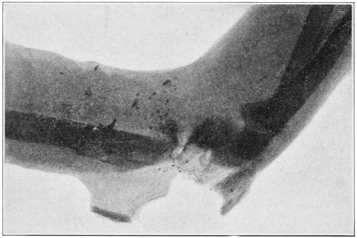
Skiagram of compound comminuted (gunshot) fracture of elbow, inflicted with a Dumdum bullet. Illustrating the extreme of comminution. (Lexer.)
F. Epiphyseal separations constitute a somewhat distinct form of injury, having at the same time the importance and dignity of fractures in the truer sense of the term. In the chapter on Diseases of the Joints will be found a table of the ages at which epiphyses unite. In childhood and youth a fracture near the joint is most likely to partake of this character, and it is of importance that it should be recognized as such when it occurs. Injuries occurring beyond the ages mentioned in the table are not likely to be of this character unless ossification is delayed by some morbid process.
By virtue of their occupations and habits men suffer fractures more frequently than women. Fractures are, moreover, ten times as frequent as are dislocations. The aged, by virtue of their atrophic changes, are more subject to fractures than others. Fracture in the vicinity of certain joints predisposes as well to dislocation of these joints, and it often happens that the treatment for the dislocation is reduction and treatment of the fracture. So far as the external causes of fracture are concerned they are frequently referred to as (a) fracture by external violence, and (b) fracture by muscle activity. The former are easily explained; the latter occur from excessive muscle action, as in violently throwing a ball, or, as in one case with which the writer was conversant, where a colored preacher in the vehemence of his gesticulations fractured his own humerus. Obviously the long or large bones are more liable to fracture than those which are short and irregular. Certain bones, especially the clavicle, are peculiarly exposed.
Intra-uterine fractures have not as yet been mentioned. These occur during the intra-uterine life of the fetus; this term does not include such fractures as may be inflicted during delivery with or without instruments. In a fetus already affected with congenital rickets it may not require any severe contusion upon the abdomen of the mother to inflict a fracture. Starvation (i. e., scurvy, syphilis, and struma) in the mother may so disturb nutrition as to weaken the osseous system of her offspring.
Such previous conditions as ensue from osteomyelitis (i. e., caries and necrosis) may often weaken the bone. Nevertheless with distinct necrosis there is usually so much new bone formation as to strengthen rather than weaken the part. Bones may also become fragile as the result of syphilis, especially when gummas develop within them.
Fractures frequently produce certain deformities which are more or less conspicuous and easily recognized. They are designated as angular, lateral, or axial (i. e., when the axes of bone are considerably displaced, even though they may be more or less parallel), longitudinal (when ends overlap), rotary, etc.; while by the interposition of muscles and other soft tissues more or less wide separation may be produced, the same result occurring when the olecranon or the upper half of the patella is widely separated from the main bone or portion by muscle pull.
DIAGNOSIS OF FRACTURES.
Fractures give rise to subjective symptoms and objective signs. In diagnosis the history is also of value, especially in those cases where it is a question of some constitutional affection and a minimum or absolute absence of violence. The apparent immunity which the intoxicated enjoy is in large measure due to the fact that by virtue of their condition one of the predisposing causes of fracture is avoided. There can be no doubt but what muscle tension, due to voluntary or instinctive efforts to avoid harm, is a contributing factor in the separation of many bones or their processes. A patient stupidly drunk will not make these efforts, and will fall in a relaxed condition, in which violence will probably be much less extensive, and the consequences less disastrous than if he made an effort to save himself from falling.
Pain and tenderness are evidences of injury, and will often serve for its location; even the reference of pain is somewhat suggestive. It is stated as a universal rule that when pressure is applied laterally or in the long axis of a bone and evokes pain, referred to a distance from the point where pressure is made, it will indicate fracture at the point to which it is referred. There is always impairment, usually loss of function, while effort to move a thus injured limb will give rise again to localized pain and tenderness. The pain of contusion is usually diffuse, and that of fracture is referred to a limited area. The tenderness produced by handling or examination will vary with the stolidity, the age, and the character of the patient, as well as the nature of the injury.
Objective signs are crepitus, mobility, deformity, ecchymosis, redisplacement. Crepitus means the sensation of grating or rubbing produced when fractured bone surfaces are moved upon each other. It is recognized by the sense of touch, sometimes by that of hearing. Its presence is pathognomonic, but its absence is a negative sign, and an effort should be made to obtain it. To repeat the demonstration, especially to demonstrate it to others, means superfluous manipulation, which is not to the best interest of the patient. Crepitus, then, should be carefully sought for; once detected it should be sufficient.
Abnormal mobility is explained only by fracture. It is easy to detect it in the shaft of a long bone, but when near the joint it is confusing. Its determination by manipulation is not seen in green-stick or impacted fracture unless these are further broken up by manipulation. When evident it should serve as a caution against unnecessary or rough handling, for if it be easily recognizable crepitus need not be sought.
Deformity is a striking and pathognomonic feature of fracture. It may be imitated by hematoma or sudden swelling of the soft parts or of joints. It may consist of shortening or of angular, lateral, or rotary displacement, or perhaps of depression or indentation. Careful inspection, then, and palpation should precede other methods of examination, as they are often sufficient to indicate the location, the nature, and sometimes even the character of the active causes.
Inspection of the injured part alone is not always sufficient. Careful comparison between the two sides of the body should be made in order that actual measurement or comparative examination may reveal what mere inspection would not. In connection with inspection it should be ascertained whether the individual has ever received previous injuries. The writer recalls a case where a physician claimed a recovery after fracture of the femur, treated by incompetent method, yet with ideal result, inasmuch as he said there was absolutely no shortening. A personal question, however, to the patient revealed the fact that he had had the other thigh broken some years previously, and that an apparently similar amount of shortening followed in each case.
The ordinary indications of fracture are frequently followed by ecchymosis. This will appear at a date corresponding with the depth of the injury beneath the skin (it may occur within an hour or three or four days). The blood will follow the fascial planes and work its way to the surface along them. The sign is of the greatest value in the diagnosis of basal fractures of the skull and certain fractures of the hip and pelvis. When it occurs after an interval it is a confirmatory rather than a promptly available sign.
Redisplacement implies that the parts when properly put into apposition quickly fall out of it unless mechanically supported—that is, they do not stay reduced. This sign is not universally applicable. It applies especially to the fractures of the long bones of the extremities, and particularly to the humerus, the femur, or double fractures of the radius and ulna in the forearm or both bones of the leg.
Diagnostic Aid Afforded by the Fluoroscope and the Skiagram.
—Since Röntgen’s memorable discovery the cathode or x-rays have been of greater and greater use in the diagnosis and portrayal of injuries and morbid conditions in the osseous system. To such an extent is this now true that well-equipped hospitals have ample conveniences for fluoroscopic and photographic work, while many medical men are doing it in their private practice. There can be no question but that diagnosis and methods of treatment have been made more perfect since this new method of investigation has been made available. On one hand, however, it has led perhaps to something of neglect of the methods previously in vogue, which necessitated anatomical knowledge and logical reasoning. On the other hand, the knowledge thus obtained has been sometimes a two-edged sword, since the display of skiagrams, or x-ray pictures, in court has too often worked harm or discredit to the surgeon or the institution with which he was connected. Moreover, even this method of diagnosis, with its apparent certainties, is not always reliable, and disappointments have sometimes followed.
Intra-articular Fractures
are subject to peculiar complications which enhance the difficulty of treatment and jeopardized the result. Among the more common of these are the following:
1. Too wide separation of fragments by hemorrhage or distention, with failure in resorption of fluid before fixation in bad position has resulted.
2. Complete or partial rotary displacement, preventing proper apposition of bone surfaces.
3. Interposition of soft or fibrous tissues between fragments by which bony union is prevented. This is conspicuously common in fractures of the olecranon and patella, and is of itself sufficient reason to justify operation in otherwise suitable cases.
4. Separation of a fragment within a joint capsule, by which its blood supply is cut off, making it essentially a foreign body. This occurs especially at the anatomical necks of both the humerus and femur.
5. Exuberance of callus with consequent limitation of motion.
6. Insufficient amount or absence of callus, which, when bone ends are bathed in joint fluids, is not often thrown out.
All of these are immediate consequences. The following are among the more undesirable remote consequences of the same injuries:
1. Exuberant callus, which may be the result of too early attempt to move the parts, or may result from other causes; it offers more or less mechanical obstruction to joint movements.
2. Separation of fragments to an extent precluding the possibility of repair, and interfering with function.
3. Pseudo-ankylosis, as a result of condensation and organization of blood clot between joint surfaces.
4. Adhesion of tendons to surrounding callus or within their own sheaths.
5. Displacement and distortion of bone ends with vicious union, for which the medical attendant is sometimes responsible. Unfortunate consequences of this kind are generally seen at the elbow after fractures of the condyles; at the wrist, after incomplete reduction of Colles’ fracture; at the hip, when insufficient traction has been made; at the ankle, after the complete form of Pott’s fracture.
6. Exostoses and osteophytic outgrowths, which often complicate fractures.
7. Absorption of bone, which is usually seen after fractures of the neck of the femur.
8. Involvement of nerves by pressure of callus, most often seen about the elbow.
9. Thrombosis leading to obliteration of the deeper and enlargement of the more superficial veins.
10. Edema, also the result of venous obstruction by pressure of callus.
11. Chronic hydrarthrosis.
12. Arthritis deformans traumatica. This is usually a remote result of fractures, and manifests itself by slow changes in shape and position, with deformity and disability. It occurs most often in the aged.
13. Necrosis, which may be the result of failure in the process of repair and will probably necessitate operation.
14. Malignant changes. These have to do with the occurrence of sarcoma in bone callus, a complication which is known to occasionally arise. (See Sarcoma.) It also refers to primary sarcoma, by which bone is weakened, or secondary carcinoma, which produces the same result.
15. Syphilis. Chronic syphilitic disease is well known to weaken bones by atrophic processes as well as by the deposition of gumma. It is known also to delay, or sometimes almost prevent, the process of callus formation, ossification, and later absorption. Syphilitic patients with fractures need to be kept under antispecific medicines.
REPAIR OF FRACTURES.
The immediate consequence of a fracture is outpour of blood both from the broken-bone surfaces and from whatever other tissues may have been lacerated. This produces, first, a hematoma, which is followed by a certain degree of local edema, perhaps even of general edema of the distal parts. The latter will subside with a rapidity proportionate to the promptness of suitable treatment and the nature of the injury. The blood begins to coagulate within a short time, while with the disappearance of the more fluid portion granulations begin to form from the periosteum, as well as bone surfaces, externally and internally, and even from the marrow. The clot loses its original characteristics and is permeated more or less rapidly by granulations. With the site of the injury wrapped in a mass of granulation tissue we speak of the so-called provisional callus, whose amount will depend upon the severity of the injury and the accuracy of the replacement of the parts. If laceration has been but trifling and the bones are accurately apposed the amount of callus will be small, otherwise it may be large; so large, in fact, as to be easily palpated and even to cause edema and pain by pressure. Repair of the fracture is effected by the gradual conversion of this callus into cartilaginous tissue and then into bone. So much of it, at least, as lies on the outer side of the bone and is known as external callus goes through this change. The internal callus, i. e., that within the marrow cavity, undergoes a more direct transformation, which amounts to immediate ossification. The internal callus usually ossifies completely, and then forms a medullary plug that serves as an internal splint and affords support and strength. In time it completely disappears, this time varying in different cases.
The external callus is converted into bone by passing through the intermediary condition of cartilage. Between the broken-bone ends granulation occurs more slowly, and repair at this point is delayed, partly because of poor circulation and nutrition; but the internal callus acting as a bobbin within, and the external callus acting as a solder on the outside, give sufficient support and strength to effect a final and absolute ossification of all the interfragmentary granulation tissue. When the time comes when callus is no longer necessary it begins to disappear by absorption. When everything proceeds normally callus is absorbed in a proportion commensurate with its loss of utility. When bone ends have badly united considerable callus remains permanently. When apposition has been ideal it almost completely disappears, even the medullary cavity being restored.
Fragments which are completely detached may be reunited by practically the same primary process, but fragments of considerable size usually become surrounded by granulation tissue, by which they are nourished and may be finally reunited, with more or less departure from their original shape and location. It is in this way that a comminuted fracture may heal. Fragments that are separated sometimes necrose and have to be removed.
Fig. 278
Fig. 279
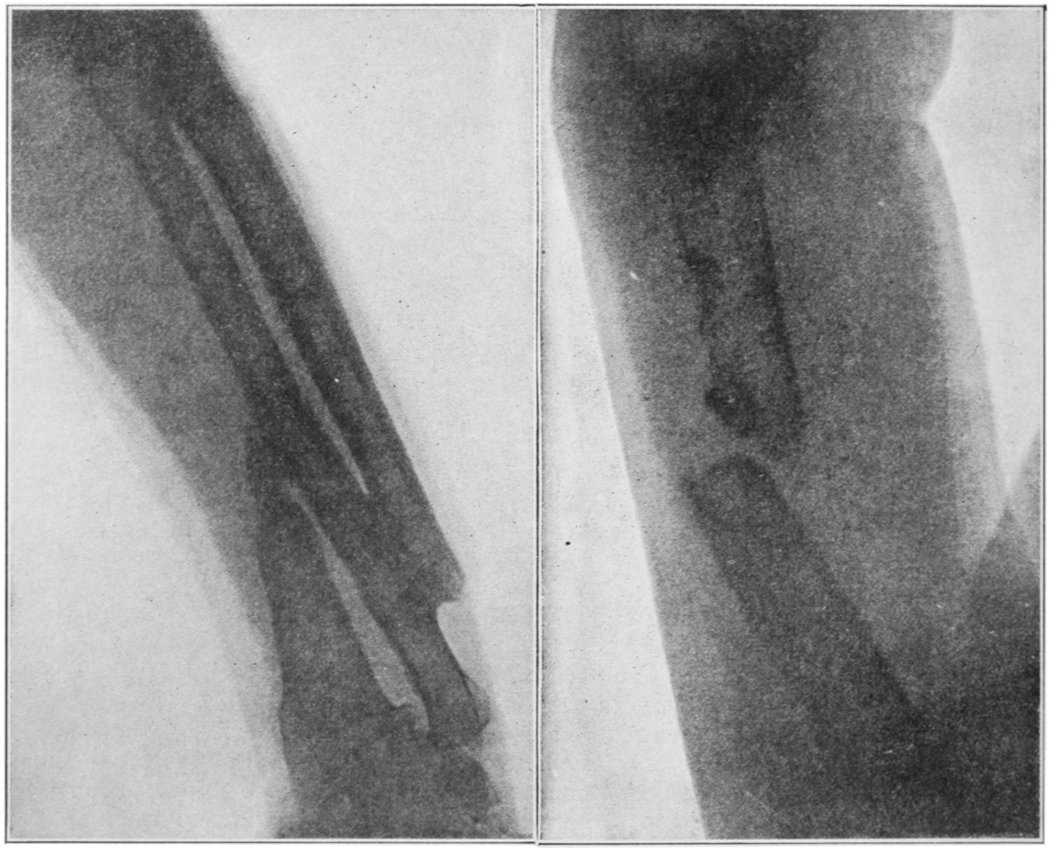
Compound fractures resulting from arm being caught in belting and wound around shafting. End of radius united to ulna and lower end of ulna to the radial fragment. Pseudarthrosis of humerus, thrice operated, the third time in the Buffalo Clinic. (Skiagram by Dr. Plummer.) (Arch. Phys. Therap., May, 1905.)
The repair of the flat bones is effected by a similar process, which is referred to as callus formation. In the skull it is brought about chiefly through the agency of the diploë, whose powers in this direction are somewhat limited. Cancellous bone tissue usually throws out but little callus. Its repair occurs from within. Cartilage heals by a very similar process, though it is not now ossific tissue but fibrous which reunites the fractured surfaces. Instances of both kinds can be seen when a fracture has crossed a joint surface.
In a compound fracture much will depend upon the existence or absence of septic complications. In a clean wound, whence blood and fluid may have escaped, there will be little but granulation tissue. Should this wound suppurate the exposed bone surfaces will undergo at least a superficial necrosis, necrotic particles being removed by the same granulation tissue which will later bind the bone ends together. Here, too, the internal callus plays the largest role in the process of repair. The bone tissue first formed is always coarse and soft. Complete calcification and restoration of original density and vascularity occur slowly. Neither cartilage nor bloodvessels alone appear capable of forming bone; the latter is produced only under the influence of the osteoblasts, which penetrate from the periosteum and the bone itself along the course of the bloodvessels.
The process is one of conversion of blood clot into provisional callus, which then changes into granulation tissue or into cartilage, both of these materials undergoing subsequent conversion into bone through the medium of the osteoblasts and osteoclasts (or giant bone cells), the neighboring bone itself undergoing a rarefying ostitis, to change back into its original condition with the final changes of the callus.
Repair of intra-articular fractures has already been described as influenced by the presence of synovial fluid and cartilage. The latter does not proliferate, and the line of fracture usually appears as a groove on its surface. At epiphyseal junctions union is usually rapid and satisfactory, for the changes taking place at this point are in the direct line of what is needed for repair.
DELAYED UNION; NON-UNION.
The above description refers to the process which is supposed to take place in normal bone repair. When, however, this is disturbed, as it may be from a variety of causes, there may be delayed union; when it completely fails we have non-union. General conditions have bearing on these local failures. Whatever makes a strain upon the system may interrupt the process, e. g., pregnancy, lactation, exhausting hemorrhages, acute diseases, starvation. Again, failure may result from purely local conditions, such as marked displacement, and particularly the intervention of some of the soft tissues, or any foreign body. Suppuration will also frequently cause great disappointment. The humerus is the bone most often troublesome in this direction; next the bones of the leg, the femur, and the bones of the forearm. It is necessary to distinguish between delayed union and absolute non-union. In the former normal processes may be simply retarded. When thus delayed they may be stimulated by rough handling, rubbing the bones together, or by perforating the callus with the point of a drill, from several directions. This method of drilling was introduced by Brainard, of Chicago. The existence of syphilis has much to do with delay, and should be combated by free use of antispecifics. Many patients will be found to have phosphaturia, i. e., to be eliminating phosphates which should go to repairing the bone. Such patients should be given phosphoric acid, with some of the phosphates, preferably of calcium, in order to make up for loss in this direction. Much can be done also by massage, and by everything which stimulates nutrition and general health (fig. 280).
Fig. 280
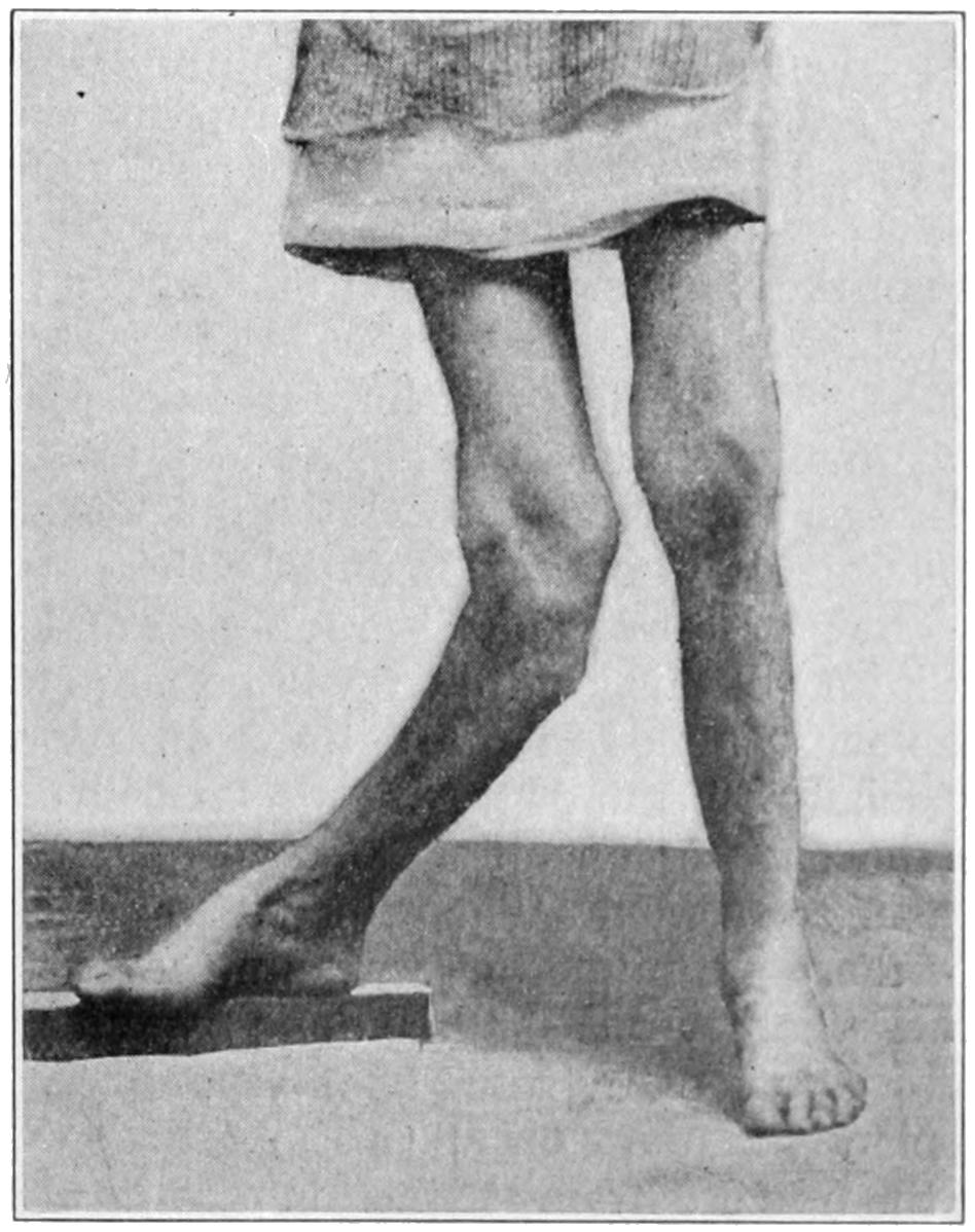
Vicious union with great deformity after fracture, requiring extensive operation. (Buffalo Clinic.)
In non-union efforts at repair are at a standstill; the bone ends become rounded off, the marrow cavity is plugged on either side, while in time the surrounding granulation or connective tissue undergoes condensation, as well as organization, and a capsule is formed in which a certain amount of fluid resembling true synovia collects, and thus is formed sometimes an almost perfect pseudarthrosis or false joint, whose perfection as a joint must be admired, although its presence is so deplored. The causes of non-union are now better understood than formerly, and consist largely in the interposition of fibrous and muscular tissues, that act as a barrier and keep the granulation tissue or the callus on one side from coalescing with that on the other.
Treatment of these cases will vary with their causes. In delayed union patients should be encouraged to use the parts, thereby causing greater activity, but in the presence of an actual false joint no method is of avail except that of actual exposure, by incision, with removal of all intervening fibrous tissue, and freshening of the bone surfaces by saw or chisel, the endeavor being to so shape them that they may lie in contact, and then be so maintained, by some mechanical expedient, such as a wire nail or suture, an ivory peg, a chromicized tendon, a bone ring, a small metal brace fastened with screws, or by any other expedient which may suggest itself to the ingenuity and the means of the operator. There are, however, occasions when one deliberately endeavors to secure a pseudarthrosis, as after ankylosis of the shoulder-joint, if in making powerful effort to break up adhesions the neck of the humerus should snap it would be better to prevent union rather than favor it, as in this way something resembling the original joint, so far as function is concerned, would be obtained. At the hip, also, after such an accident, the same principles may be adhered to or more deliberately secured by a subcutaneous osteotomy, as is sometimes done for relief of deformity.
Fibrous union implies such organization of granulation tissue as converts it into simple fibrous or ligamentous tissue, the change stopping here and not going on to formation of cartilage or bone. There are three localities especially where fibrous union is sometimes the best that can be obtained and often proves sufficient of itself; these are the olecranon, the patella, and the neck of the femur. Even though the halves of the patella be separated by two inches of ligamentous tissue the patient may still have reasonable use of the limb. A separation of half an inch to one inch at the olecranon does not materially disable the arm, while at the hip-joint two or three inches of ligamentous tissue between the main end of the bone and the fragment will not totally interfere with locomotion, except so far as it permits an equivalent amount of shortening of the leg. There are, then, occasions especially when the hip is involved in elderly and decrepit people, when ligamentous union is the best that can be hoped for or attained.
TREATMENT OF FRACTURES.
In principle the treatment of fractures is very simple. It consists in putting the parts in apposition and maintaining them there for sufficient time to permit of complete repair. That which is so simple in theory is often very difficult and sometimes even impossible in practice, made so by the nature of the injury or the disposition of the patient. In the aged, who cannot lie long in one position for fear of pulmonary stasis; also in the insane, in the epileptic, and in those suffering from delirium tremens, will be met difficulties which are insuperable. In such instances the first indication is to preserve the life of the patient, the second is to get a good result, the third is to do the best we can. Good management is not the least important feature of such treatment. This will include suitable nutrition, provision for elimination, prevention of bed-sores or pressure-sores, and many other less important features.
Diagnosis having been made, the surgeon should study how he may best carry out the fundamental principle of putting the parts in apposition and so maintaining them.
The greatest obstacle to reduction and maintenance in position is muscle pull. After an injury of this kind there will be more or less muscle spasm, the more powerful groups displacing bones in the natural direction of their pull. In the humerus and femur especially all arm or thigh muscles will coöperate to produce shortening. As indicated in the chapter on Joint Affections, nothing so thoroughly overcomes chronic muscle spasm as traction. The principle underlying treatment by traction is exceedingly simple, but there are numerous ways and mechanical expedients for effecting it. In the lower limb, whether this shall be done by anterior suspension, by weight and pulley, by elastic contraction, or by some of the more complicated splints, matters little so long as it be efficiently made. Of all these methods it may be said in general the simplest is the best. In the upper extremity traction may be made by similar methods with the patient in bed, or the patient may be allowed to rise and be about with a weight hanging from the elbow or some simple expedient of this kind.
The method of traction is one to be combined usually with further protection, by which not only longitudinal but lateral displacement maybe overcome. This suggests the use of splints in addition to mere traction methods.
It is not always possible to put in operation at first that method which we may prefer a little later, as swelling is usually so pronounced as to make it advisable only to put the parts at rest and hasten absorption. The same is true of hemorrhage. In rarer instances it may be a question as to whether the distal parts may undergo gangrene from the disturbance of circulation. These are matters to be duly regarded before the later and more complete dressing. Mechanical aids, usually in the shape of splints, are therefore necessary. The physiological rest which it is so necessary to ensure will lead to a certain wasting of muscles and stiffening of joints, which are only temporary, but which by no means lessen disability when splints are removed. That splint is best for a given case which best fits it and permits the surgeon to carry out its peculiar indications. The writer is opposed to manufactured splints, as they seldom fit the part. This can be obviated by packing cotton or other compressible material into the splint. For temporary purposes they will frequently suffice. For fixed dressing, however, it is preferable to make a splint which shall fit the limb to which it is affixed. Immobilization is difficult of accomplishment and at many points impossible. Thus in fracture of the ribs or clavicle it is impossible to avoid a certain amount of motion with each respiratory effort, even though an uncomfortably tight dressing be applied.
Splints are made of various materials, metal, wood, various compositions hardened in molds, plaster of Paris, or some of its substitutes, i. e., glue, soluble glass, or a composition like one made of equal parts of powdered starch and fine isinglass, added to a solution of potassium silicate, this being allowed to stand for several days, after which a little fine boric acid powder is added; when this is painted over gauze dressings it solidifies and forms a light and rigid splint. There is one objection to all methods which comprise a solution that hardens slowly—that is, that during the time required for the purpose redisplacement may occur. It is not advisable to dress a recent fracture in a wet pasteboard splint or in such a composition as that mentioned above. Later, when a certain amount of consolidation has already occurred they may serve a useful purpose.[38]
There are two methods of using plaster of Paris: one is gauze bandages sprinkled with it, rolled, and kept ready for use, to be placed in water at the time of their employment. A limb may be enveloped in these, after being covered with a layer of wadding or some other protective material, by which the plaster shall not come in actual contact with the skin. It is also a good plan to place a strip of tin or pasteboard along the exposed surface of the limb, over which the surgeon cuts to remove the splint. Thus one may avoid any danger of injuring the skin with the point of the knife. It is also a good plan to make at least a part of this cut before the plaster has sufficiently hardened, i. e., to do most of the work, leaving perhaps a layer or two of gauze to be cut through some time later. It is necessary to impress the fact that when a quickly hardening fixed dressing is used approximation should be ensured by the greatest attention, maintaining it until the splint is so hardened that redislocation is impossible. Another method of using plaster of Paris is by sopping strips of surgeons’ lint, ordinary canton flannel, or almost any other similar material, in plaster-of-Paris cream, then molding these to the injured limb, maintaining the same rigid precaution as to the proper position of the same while the splint hardens. In this way a splint can be adapted to the part, and, at the same time, made removable, permitting as frequent access to it as may be desired.
COMPOUND FRACTURES AND THEIR TREATMENT.
As already stated, it is the communication of fractured bone surfaces with the external air which makes a fracture compound in the strictly surgical sense. This may occur through a minute and tortuous opening or through a large and extensive wound. Although the communication is with the atmosphere the danger comes not so much from germs floating in the air as from those on the surface of the body and within the pores of the skin, or else from foreign material admitted through the external wound. Obviously the great danger is of septic infection. Whether the tissues may prove more or less susceptible, and thus resist or break down, cannot at the outset be foretold. This leaves but one imperative ride to follow, to act in every instance as though serious injection had occurred and to take precautions accordingly. Even a small puncture made by a spicule of bone may permit germs to be withdrawn into the tissues as the bone is replaced. If, then, the surgeon seals such a puncture he necessarily takes the chances and must abide the result. Whether he shall do this or not will depend upon the patient and the injury. At all events, the site of puncture should be carefully cleansed and disinfected and the case so dressed that it may be carefully watched. Complete sterilization of every particle of exposed tissue is absolutely necessary, and for this purpose hydrogen dioxide or some of its later substitutes will prove effective. A protruding splinter of bone should be removed with cutting forceps, unless the wound must be enlarged as a part of the treatment of the fracture. In most instances it will be safer to pursue this course, i. e., to extend the wound which makes the fracture compound, to a degree permitting thorough exploration and cleansing. Not infrequently fragments of bone will be found, which when nearly or completely detached should be removed. Such a free opening permits also of wiring, or other means of fastening together bone ends, by which apposition may be more perfectly secured. A compound fracture which has been long unattended may be safely assumed to be septic. Here free incision, with cleansing and ample drainage, will be a far safer course than non-compliance with the general rule.
Compound fractures of the skull are nearly always depressed fractures, and practically always call for operation. Their proper treatment will be dealt with when considering Injuries to the Head. A fracture of the ribs may be made compound by penetration of a sharp bone end, and such injury to the lung as may permit air to escape into the pleural cavity. Such a pneumothorax may be followed by a hemothorax and hydrothorax, and these perhaps by empyema. Compound fractures of the pelvis are not infrequently complicated by perforation of the bladder or bowel, or rupture of the urethra, or some other serious visceral injury which may determine their fate. Compound fractures are difficult of treatment because they entail frequent changes of dressing and prevent the use of desirable splints. These fractures are also sometimes so serious as to necessitate amputation, which may be necessitated either by such comminution of bone as to make repair impossible, or such injury to vessels as may determine gangrene. If the circulation can be shown to be sufficient, either at the time or perhaps by delay of a few hours or a day, a limb may be saved by the resection of one or both bones, which in pre-antiseptic days would have required amputation.
The surgeon does not always see these cases in their recent or fresh state. He may be called to a case complicated by suppuration, cellulitis, and sepsis. Here though amputation may be required he may still delay it, hoping to improve local conditions, and thus to make it more promising, or he may have to resort to various expedients, such as suspension with constant irrigation, or temporary packing with yeast, in order to justify any further attack upon the parts already involved.
In the treatment of compound as of simple fractures we should never lose sight of the dangers of too tight bandaging and of pressure sores. I have seen both these lead to gangrene, with its necessary mutilation, in cases where the attendant has forgotten the proneness of injured parts to swell, and has either not allowed for this within the dressings or has not atoned for it in time when it has already occurred.
In the treatment of all these cases the operator should never forget the medicolegal aspects of such a case nor the necessity for constant attention and caution on his part. He should remember that his minutest precautions will often be disobeyed. He may, however, be cheered by the fact that only in cases of carelessness will he incur legal responsibility.
SPECIAL FRACTURES.
Fractures of the skull and of the vertebræ will be considered under the respective headings of Injuries to the Head and to the Spine.
FRACTURES OF THE NOSE.
The nose is the most frequently broken of all the bony parts about the face. One nasal bone or both may be broken, and each may be separated from its bony supports as well as from the other. The fracture may be compound in either direction, most frequently so into the nasal cavity, as a result of which infection may as easily take place from within as from without. The cartilages may also participate in the injury.
The injury would be easy of recognition were it not for the amount of swelling that often accompanies it. The signs are mobility and crepitus, with more or less deformity. So long as the nose can be grasped between the fingers recognition of fracture is easy. If swelling prevents this an instrument or the finger can be passed into one nostril and combined manipulation practised. There is generally more or less bleeding from the nose, and sometimes considerable emphysema. Swelling and ecchymosis are also often pronounced. This will all subside under cool and soothing applications. The most important indication is to replace the nose and hold it where it should remain. The difficulty is increased by the efforts which the patient instinctively makes to dislodge clot or secretion. The importance of accurate reposition is in some cases sufficient to justify an anesthetic and instrumental help. This will permit of the application of such force as may be necessary to elevate or to shift fragments, while a gutta-percha splint may be molded upon the outside, or a sterilized pin or needle made to transfix the nose from one side to the other (Mason), passing behind the fragments and through the septum in such a way as to keep it from dropping backward. A good plan is to introduce a tube into each nostril, perhaps a piece of silk catheter, around which a certain amount of gauze can be packed, and which can thus be used as an internal splint, while on either side and externally a little roll of gauze is held in place by adhesive plaster crossing the cheeks. The operator should take as much pains to see that the septum is in its original position as in attending to outside and cosmetic effects. The septum can be controlled by a pair of forceps.
A nose properly held in place will heal within a few days, to a point requiring little if any support. A transfixion pin should not be needed, if used, for more than four or five days. An internal splint should be removed each day, so that the nose may be sprayed with cleansing solution (Dobell’s) and retained secretions removed.
The disfigurement resulting after this injury is dropping in at the root of the nose, constituting the so-called saddle-nose defect. Such disfigurement as results can be later atoned for by subcutaneous injection of paraffin. (See chapter on Surgery of the Face.)
FRACTURES OF THE SUPERIOR MAXILLA, WITH OR WITHOUT OTHER BONES OF THE FACE.
The more protected portions of the upper jaw are rarely fractured, save by extreme violence. The alveolar process, with one or several teeth, may be partially or completely detached. Such fractures are compound, and after replacement need antiseptic mouth-washes as well as other attention. Usually the teeth in the fragment can be utilized for the purpose of fastening it back into place by means of the uninjured teeth, retention being secured by wire or waxed silk. Extensive detachment may necessitate sutures through drill holes. The lower jaw can usually be utilized as a splint for the upper by binding the jaws firmly together and feeding the patient on fluid food. When one or two teeth are loosened or displaced it will often be possible, if they can be promptly secured, to successfully reimplant them in their sockets. Both the sockets and the teeth should be thoroughly cleansed. After replacement it will be necessary only to ensure absolute rest and retention in position.
In regard to other facial bones there is no injury which may not occur, as the result of direct violence. The zygoma and the malar bone may be broken away, or the entire collection of facial bones may be loosened from their connection with the bones of the skull proper. The margins of the orbit, or its walls, may also be injured, and the sinuses opened, with perhaps more or less entrance of foreign material. These fractures are generally compound and are accompanied sometimes by injuries to the soft tissues. It becomes then a question not merely of cosmetic result, but of avoiding infection and saving life. The latter is the more important, and measures should first be directed to that object. Satisfactory results can be attained by drilling and holding bone fragments together with tendon or other sutures, and by neatly trimming and cleaning wounded surfaces and bringing them together. Subcutaneous sutures should be used for this purpose.
FRACTURES OF THE INFERIOR MAXILLA.
This bone is broken nearly as often as the nose, and almost invariably by direct violence. Here, as in the upper jaw, there may be trifling or serious fractures of the alveolar process, which should be treated on the same principle as above set forth. Fractures of the rami occur more often in those parts which are occupied by teeth, or from which teeth have dropped out by senile changes, the jaw being weakened at these locations. The most frequent seat of fracture is near the middle line. Fractures of the ascending ramus and of the upper processes are rare. Double fractures are not infrequent, the lines of separation being rarely symmetrical. The gum and the skin are often torn and the majority of these fractures are compound. The bone is considered to be weakened at the dental foramen; at all events it often yields in this vicinity. By fracture with much displacement posterior to this opening the inferior dental nerve may be injured or torn. The condyle, after extreme violence, has been known to have been driven up into the cranial cavity through the base of the skull. Gunshot fractures are nearly always comminuted (Figs. 281, 282 and 283).
Fig. 281
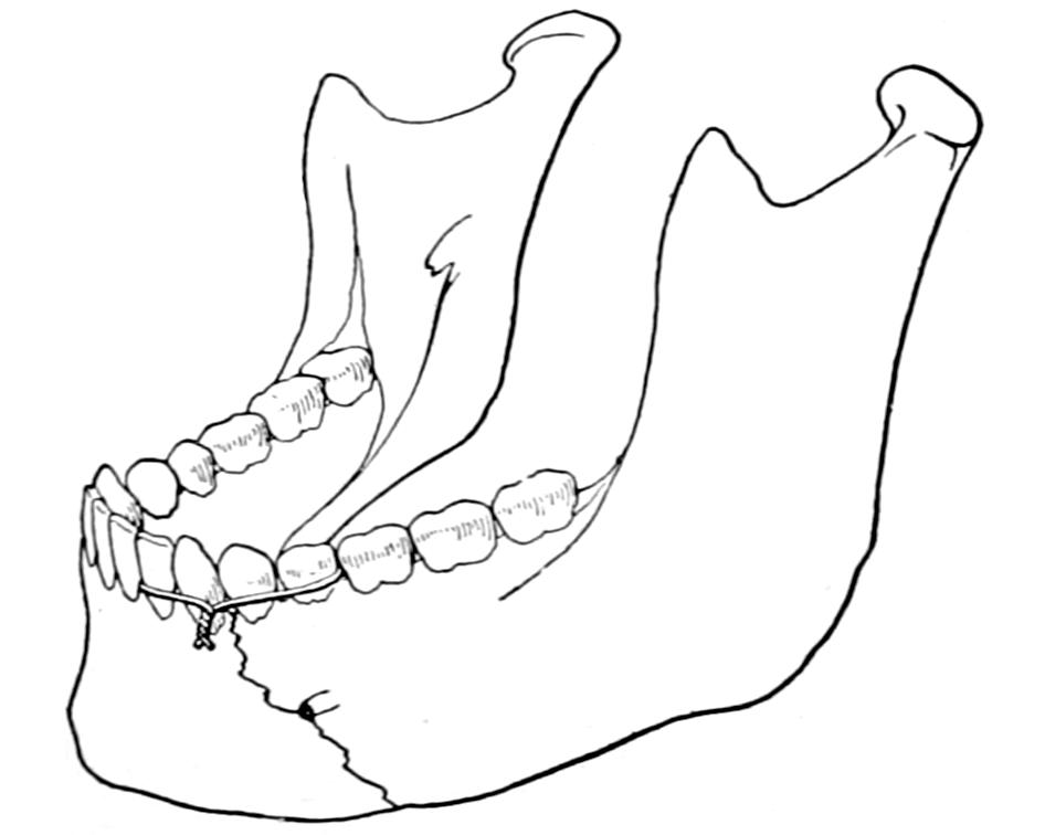
Fig. 282
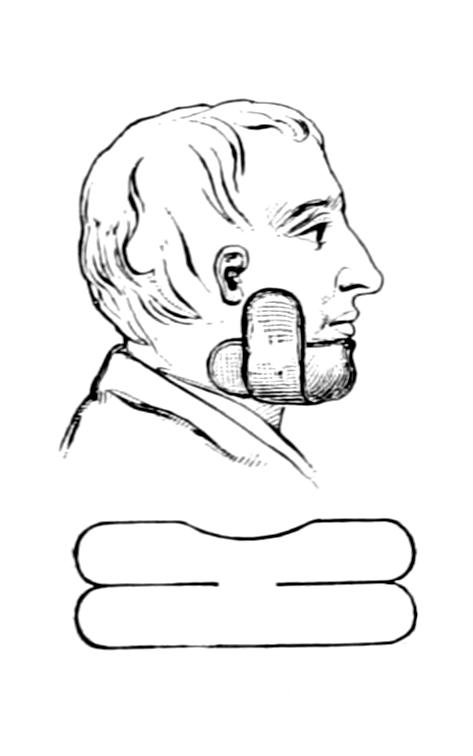
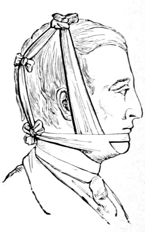
Fig. 283
Use of silver wire in fixation of fragments by utilizing the teeth.
Bandage and splint for fracture of lower jaw. (Bryant.)
The signs of fracture of the lower jaw are unnatural mobility, crepitus, displacement, pain, and loss of function. No bone in the body is more easily investigated by sight and touch, and recognition of these fractures is usually easy. Pain is provoked by attempting to move the jaw, even in talking, and depends on the extent to which the inferior dental nerve is injured. Irregularity in the line of the teeth will sometimes permit recognition. These fractures furnish excellent illustrations of the effect of muscles in producing displacement. Those of the tongue and the floor of the mouth, as well as the anterior muscles of the neck, will pull the fragments in various directions, according to the direction of the line of fracture and its location. This displacement may be trifling or serious. These fractures are often compound, internally or externally, such injuries constituting an unpleasant complication, but affording occasionally an opportunity for fastening fragments by drill or wire suture, which would otherwise require an opening to be made. In every instance antiseptic mouth-washes should be frequently used.
Treatment.
—The treatment is simplified when the dentition is good and regular so that the fragments may be fastened together with wire or waxed silk ligature around the adjoining teeth, and then fixation accomplished with a simple molded gutta-percha or plaster-of-Paris splint, by which the lower jaw is held firmly against the upper. Such a dressing is held in position by a four-tailed bandage (Fig. 283). A silk or wire loop, used for the purpose just mentioned, should include two teeth on either side of the fracture, for by constant tension the nearest tooth will soon loosen, and if this were next to the break the effect of such displacement would be injurious (Fig. 281). When the line of fracture is oblique there is often greater difficulty in adjustment.
While the simplest means by which the fragments may be kept in position are the best, there should be no hesitation in serious cases to resort to operative measures having for their purpose the insertion of wire sutures or their equivalent. These are inserted after drilling the bone at suitable points, and are introduced with a view to their subsequent removal, the ends being left projecting in order to facilitate this. In clean cases, where the incision is made in unbroken skin, the ends may be twisted short and turned in, previously to closing the wound. Such operative treatment is required when there has been a double fracture, the central fragment being badly displaced by groups of muscles which tend to pull it downward and backward.
A dentist should be consulted, as he may be able to make a mold and then construct a plate or interdental splint, by which a more perfect reposition may be effected.
Swelling, emphysema, ecchymosis, etc., may be treated in the usual way. Irritation is likely to provoke free secretion of saliva; this may be combated by small doses of belladonna. Patients should be fed by fluid or thin semifluid food, and mouth-washes should be frequently used.
FRACTURES OF THE HYOID BONE.
The hyoid may be broken by direct violence, either locally applied or by forcing the head backward. Fracture of the bone itself is not so serious as the lesions which accompany or follow it, either hemorrhage or inflammation, with edema of the larynx, which may impede respiration or cause strangulation. Fracture produces difficulty in breathing, swelling, and pain on talking. It is doubtful if bony union is attained, but fibrous union answers equally well. The treatment consists essentially of physiological rest. Edema may necessitate tracheotomy, and dysphagia feeding by an esophageal tube or by the rectum. Should the fracture be compound, or should a fragment be displaced so as to be detected, it may be removed through suitable incision.
FRACTURES OF THE LARYNX.
This may be fractured by injuries of the same character as those which fracture the hyoid, except that it is more exposed to the direct violence of a blow, as from a baseball. In elderly people in whom calcification of the laryngeal cartilages has occurred fracture is more dangerous than in the young. Injuries which produce these lesions are of a serious nature, as prompt swelling, either from hemorrhage or edema, occurs and threatens respiration. For illustration a death occurred on the baseball field within a few minutes after reception of a blow upon the front of the neck with laryngeal fracture; the cause of death was suffocation due to swelling, which might have been averted if tracheotomy could have been performed. In the milder injuries of this kind much can be done with sprays of cocaine and adrenalin, to quiet laryngeal irritation and reduce vascularity.
FRACTURES OF THE STERNUM AND RIBS.
Fracture of the sternum in childhood is exceedingly rare. In adults it may occur in connection with other injuries or as a solitary lesion. Such a fracture, of itself, would indicate in most cases excessive violence. It is usually more or less transverse, the periosteum being rarely so torn as to permit of much escape of blood. Cases are recorded in which it has been broken in straining during the act of parturition. It is most commonly injured by compressing and crushing injuries.
Sternal fractures are followed by much pain, aggravated by deep respiration and made worse by pressure. Sometimes displacement can be made out, while crepitus may be detected with the stethoscope. Occasionally there is sufficient deformity to make the injury apparent at a glance.
Displacement should be reduced and apposition then maintained by a plaster-of-Paris jacket or other suitable apparatus. It is advisable in some cases to anesthetize the patient and to make a sufficient opening that instruments may be used by which fragments may be lifted or pried into place. This should be done under aseptic precautions.
Diastases or separations of ribs or cartilages from the sternum or from each other have essentially the dignity of fractures, are recognized by the same general signs, and are treated in the same general way. A cartilage may snap in the young, and in the old, when calcified, may break as would a bone or even a pipe-stem.
The ribs are usually broken in their lateral aspects, but rarely between the head and angle. They may be fractured by muscle action or by external violence, examples of the former being violent efforts at lifting or sneezing. Violence may be applied in so many ways that it is not necessary to specify them. Fractures may pertain to one or to several ribs in proportion to the extent and violence of the exciting injury. In some crushing injuries an entire section of the chest wall may be broken loose and depressed, this corresponding to a depressed fracture of the skull. Rib fractures are usually of themselves innocent, but may be made serious by complications, as when the pleura is torn, or an intercostal artery bleeds profusely, or when a jagged fragment of bone first scratches and then perforates a lung. This will lead first to the outpour of blood and then of pleuritic fluid, by which in a short time the lung will be separated from the chest wall. Should infection occur through the injured lung, i. e., entrance of germ-ladened air, then empyema may seriously complicate matters and later necessitate operation. Even the heart has been injured, in several reported cases, by projecting fragments of bone. Gunshot fractures of the thoracic wall imply those features pertaining to every compound fracture, plus the injury possibly done to the lungs, heart, or mediastinal contents, such as hemothorax or pneumothorax.
The first and second ribs are so protected and the eleventh and twelfth so movable that by far the greater proportion of rib fractures pertain to the eight intervening ribs.
Symptoms.
—These are often vague, when but a single rib has been cracked through and not displaced, and comprise pain on pressure, as well as that provoked by deep breathing, coughing, and certain other movements. Should this pain be limited, or constant and made worse by pressure, fracture of the rib may be suspected. If auscultation crepitus can be heard, diagnosis is at once made. When abnormal mobility is unmistakable, or when by any means crepitus is elicited, the signs are positive. Sometimes the patient himself will recognize crepitus. This may be learned either by auscultation or by pressure with the flat hand over the affected area. Emphysema is an unmistakable evidence of fracture with perforation, while the signs of the presence of fluid in the chest cavity will also indicate fracture.
Treatment.
—Fracture of one or two ribs with displacement is ordinarily a matter of trivial import, the adjoining ribs acting as splints. It necessitates practically nothing but physiological rest, which may be best afforded by keeping the patient in bed, with firm compression around the chest, made either with a binder of strong cloth or a broad piece of adhesive plaster carried nearly around the body, or in more aggravated cases by a plaster-of-Paris jacket. In thin individuals the formation of callus can be recognized by the sense of touch. So soon as this is fairly formed displacement is less likely to occur and uncomfortable compression may be relaxed. Should there be external angular displacement this may be corrected by pressure. A projecting fragment which threatens to perforate should be cut away with bone forceps through a small incision, taking pains to permit as little air as possible to enter. If there be a traumatic pneumothorax the air should be removed with an aspirating needle. When it is evident that there is serious injury to the chest wall and that air has already separated the lung from it (traumatic atelectasis) the parts should be freely exposed, to permit the rounding off of bone ends, the seizure of intercostal vessels, the cleansing out of the pleural cavity, with perhaps later wiring of fragments or else their complete removal and closure of the external wound with or without drainage, as may be required. If blood or air has already escaped into the pleural cavity the blood should be speedily removed. The same plan is advisable in fractures of the cartilages. Sedatives to check cough, e. g., heroine, are also indicated.
FRACTURES OF THE CLAVICLE.
The clavicle and the radius are the two bones most frequently broken, the former more often in the young, the latter in the elderly; the clavicle yields both to direct violence, as by blows on the shoulder, and that which is transmitted through the arm from the elbow or hand. For convenience of description the bone is divided into thirds, the most common location for fracture being near the junction of the middle and outer third. Save for epiphyseal separations the extremities of the bone are seldom broken. In spite of its subcutaneous position and its proximity to large vessels, compound injuries or other complications are quite uncommon.
The clavicle is the brace which keeps the shoulder proper from falling upon and around the thorax. Consequently when it is broken the shoulder tends to drop downward, forward, and inward, except in a green-stick fracture, while even then there may be some displacement in these directions. Deformity is usually easily recognized, one or other fragment projecting beneath the skin in such a way as to be easily palpated. There is enough spasm of cervical muscles to draw the head over toward the affected side, while there is loss of function in the affected arm. Pain is made worse by pressing the shoulder inward as well as by moving it in any direction.
In young children the bone is often broken with a minimum of displacement. Fracture of both clavicles is not so very rare. Trouble may occur later in the course of the case from pressure of exuberant callus upon nerves and even vessels. This is to be prevented by foresight and by careful attention to maintenance of parts in proper position.
Treatment.
—The multiplicity of dressings which have been suggested for fractures of the clavicle attest the fact that so long as primary indications are observed the treatment can be made very simple. These indications are to keep the shoulder upward, outward, and backward, as it tends to drop in the opposite way. The action of three muscles is of great importance in considering the proper treatment of these cases, i. e., the sternomastoid and the trapezius, because they tend to pull fragments upward, and the pectoralis major because advantage can be taken of its arrangement to overcome upward displacement. It was Moore, of Rochester, who taught many years in Buffalo, who showed how this could be done. The fibers of the great pectoral which arise highest, i. e., from the clavicle, are those which are inserted lowest along the bicipital groove of the humerus, because of the semi-revolution made by the tendon of this muscle as it passes to its insertion. By putting the arm in such a position that these fibers are pulled upon the operator may counteract the upward pull of the other muscles just mentioned. This is the underlying feature of Moore’s suggestion; to force the elbow far backward, into a position which is for the time being uncomfortable, in order thus to pull down fragments which jut up beneath the skin. Any dressing which permits this position to be maintained will be equally serviceable. Moore suggests for this purpose what he calls a double figure-of-eight, which is shown in Figs. 284 and 285. It is put on as follows: A strip of cloth, sheeting, or anything of the kind, about two yards in length and folded sufficiently to make a strong strip eight inches wide, is held near its middle over the surgeon’s hand. This hand is placed beneath the elbow of the injured side, so that the strip crosses the under surface of the flexed forearm at the elbow. One end, which should be the longer, lying to the inner side, is passed upward and in front of the arm, carried over the shoulder across the back and under the opposite axilla, then over in front of the sound shoulder, meeting on the back the other end, which is carried up first over the outside of the forearm, then behind the shoulder and across the spine. This bandage should be pulled tightly, while an assistant holds the elbow as far backward and upward as the patient can tolerate it, as the more the position is exaggerated the more are the clavicular fibers of the muscle pulled upon and the better are the fragments held in place. This dressing not only meets the three primary indications laid down, but gives the added advantage just described. By it the shoulders are drawn backward and fixed to each other. The elbow should be lifted as the dressing is applied, so as to lift the shoulder. Most of the cloth materials used for such a dressing are more or less elastic, and it may need to be tightened once or twice a day during the time that it is worn. After a few days, when consolidation should have occurred, it may be changed for some other less irksome form of dressing. The hand should be supported in a sling. This dressing is useful in dislocations of the clavicle, especially of its outer end, and in every kind of injury in which the indication is to hold the shoulder upward and backward. In simple cases without much displacement the primary indications may be more simply met by a dressing of adhesive plaster, known in the East as Sayre’s and in the West as Freer’s. It consists of two strips of plaster of about the width of the arm itself. One of them is wound around the upper end of the arm, close to the shoulder, in such a way that, as it is passed around the back and brought over the chest, the arm and shoulder are pulled backward. The other strip passes from beneath the elbow of the injured side obliquely up and over the opposite shoulder. When it is applied the elbow should be firmly lifted. After the completion of either of these dressings the injured shoulder should appear at least one inch higher than the well one. Should the patient’s arm and chest be hairy they should be shaved before the application of the plaster strips. Like other material, plaster will stretch and slip, and these, like other dressings, should be readjusted every day or two, for the shoulder should be kept elevated for at least a week.
Fig. 284
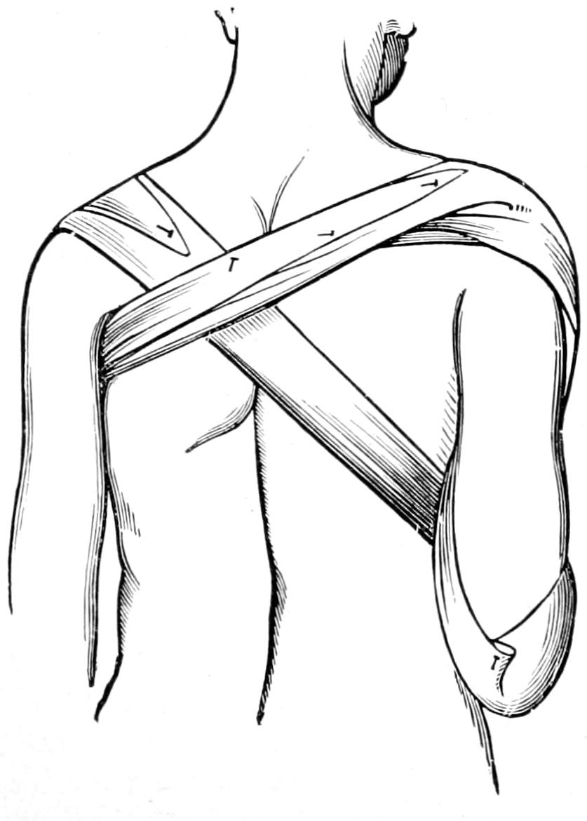
Moore’s apparatus (back view).
Fig. 285
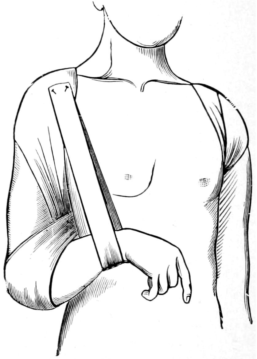
Moore’s apparatus (front view).
When the case is complicated by other injuries necessitating confinement in bed it is sufficient to keep the patient flat upon the back and without a pillow. In this position the shoulder falls naturally in the direction desired, and perhaps no other attention will be required. Many other methods are combined with a figure-of-eight bandage, crossing the back and forming a loop over each shoulder, so as to keep it from dropping forward.
While the results of treatment are nearly always good, if one is insistent upon a minimum of deformity, confinement upon the back on a hard bed is the surest way to obtain satisfactory results. Cases in which there is little or no tendency to deformity need only the simplest support by which rest may be ensured.
Epiphyseal separations are to be treated as fractures.
FRACTURES OF THE SCAPULA.
The most frequent fracture of the scapula is that of the acromion; this is usually the result of direct violence, such as a fall upon the tip of the shoulder. Detachment of this fragment permits a peculiar flattening of the shoulder, but without dislocation. The fragment can be easily felt, while the deltoid is displaced and its rounded contour lost. Treatment consists solely in forcing the arm upward, by dressings applied beneath the elbow, thus lifting the fragment into its place; fibrous union occurring here much more often than osseous, the latter is possible only in case a good apposition be maintained. Any form of dressing, then, by which the elbow is crowded upward and rest maintained will be appropriate.
The surgical neck is occasionally detached, sometimes with and sometimes without the coracoid process. As the humerus is attached to it by the capsular ligament the arm drops with the fragment when the patient is in the upright position, and the elbow will be found lower than that of the injured side. The arm is unduly mobile, and the fragment can usually be seized and crepitus obtained within the axilla. Here it is necessary to hold the arm up, as it controls the position of the fragment. It is usually sufficient to lift the elbow up and bind the arm firmly to the side, the scapula being immobilized by broad straps of adhesive plaster.
The coracoid process is occasionally detached, usually by muscular violence, i. e., it is pulled off by the coracobrachialis and the coracoid head of the biceps which arise from it. The injury is recognized by failure to detect the process in its proper place, and usually by discovery of the fragment at a point below its normal position, to which it has been drawn out by the muscles arising from it. Ligamentous union can be secured by relaxing these muscles, which is done by placing the hand over the opposite shoulder and dressing the arm firmly against the chest. I have seen paralysis of the arm result from excessive callus after fracture of the coracoid.
The spine, body, and the angles of the scapula are occasionally broken by severe violence. In the aged comminution may occur. Crepitus can be nearly always obtained. It may be necessary to distinguish the scapular fracture from one of the ribs beneath it. The treatment consists in simply fixing the shoulder-blade upon the chest, to which it is naturally adapted, by firm bandages, which shall immobilize not only it but the arm as well.
FRACTURES OF THE HUMERUS.
At the upper end of the humerus we deal with fracture of the processes, i. e., the tuberosities, which may be torn off by violent action of the muscles therein inserted; of the anatomical neck, which is rare and occurs most often in the aged; of the surgical neck, which is the most common; or, in the young, epiphyseal separation, which is the equivalent of the last named. Separation of the tuberosities is diagnosticated mainly by exclusion, possibly by x-rays. The anatomical neck lies within the capsule, and should the head be thus detached it might remain as a foreign body in the joint, having no means of securing nutrition. Fractures of the head of the bone are not classical and are usually the result of gunshot injuries or extreme violence. In all of these injuries there will be swelling, loss of function, while crepitus is sometimes obtained, but is very difficult to locate, even under an anesthetic. The diagnosis is to be made mostly by exclusion.
The surgical neck is the most frequently broken; the line of fracture passing below the tuberosities and above the muscles inserted along the bicipital groove. Therefore the pectoralis and the latissimus muscles will both conspire to pull the upper end of the shaft toward the thorax to such an extent that it can be felt in the axilla. This gives its axis a different direction, while all the muscles extending from the shoulder to the forearm will tend to produce shortening. Deformity is usually distinct, crepitus is easily obtained, and undue mobility is well marked. The head of the bone can be detected in its proper place beneath the deltoid, but does not rotate with the shaft. In rare instances a certain amount of impaction may make this evidence of fracture obscure. Epiphyseal separation will give the same signs and symptoms.
Treatment.
—The primary indication here is to overcome muscle pull by traction in a direction toward the crest of the pelvis of the same side. At the same time, with a certain degree of coaxing of the upper end of the shaft outward and a little forward, it may be possible to so re-apply broken surfaces to each other, and so affix the arm to the thorax, as to be effective. When serious difficulty, however, is encountered the writer advises traction, applied to the arm alone, if the patient be able to be upright, or to the arm and forearm, if he be confined in bed. It will take considerable stretching to overcome the combined action of all the muscles which tend to produce displacement. Along with such treatment a coaptation splint should be applied, the best being that which can be carefully molded to the parts and adapted to their needs. For this purpose a molded plaster-of-Paris splint is preferable to one of metal made to some standard size. In the dressing it is necessary to include not only the shoulder and arm but also the forearm, otherwise the principle of physiological rest would not be enforced. Fig. 286 illustrates the common tendency to displacement in these injuries.
Fracture of the surgical neck is occasionally combined with dislocation of the head of the humerus, by which such an injury is seriously complicated. Reduction may be attempted by manipulation. Until recently it was generally advised to wait for a week or ten days, and until consolidation had occurred, and then to make the attempt at reduction; but Porter and McBurney have shown that it is advisable to cut down upon the dislocated upper fragment, and, fixing it with forceps or with an instrument shaped like a corkscrew or hook, to force it back into place again. If this be done under the strictest precautions it lends no serious features to the case, while, in most respects, such a procedure would greatly simplify it, the wound being closed with or without drainage, and the usual fracture dressing being applied.
In cases of old fracture and dislocation the head of the bone should be exsected, the functional result thus obtained being excellent.
Epiphyseal separation has been too often mistaken for dislocation. Fig. 287, from Moore, shows how the periosteum is not necessarily entirely detached, but is stripped up to form a hinge, the fragment displaced forward, and its outer aspect often turned upward. This makes traction in an outward direction an essential feature of the replacement of the fractured surfaces, the manipulation being combined with fixation of the fragment so far as it can be seized through the axilla. If the epiphysis is properly slipped over upon the end of the humerus the case assumes ordinary features, and is to be dressed as usual.
Fig. 286
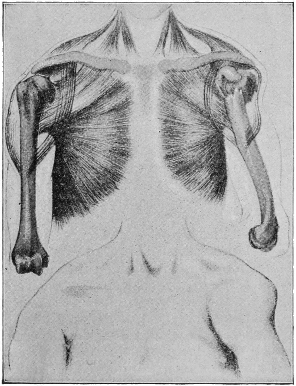
Fracture of the surgical neck of humerus. (Hoffa.)
Fig. 287
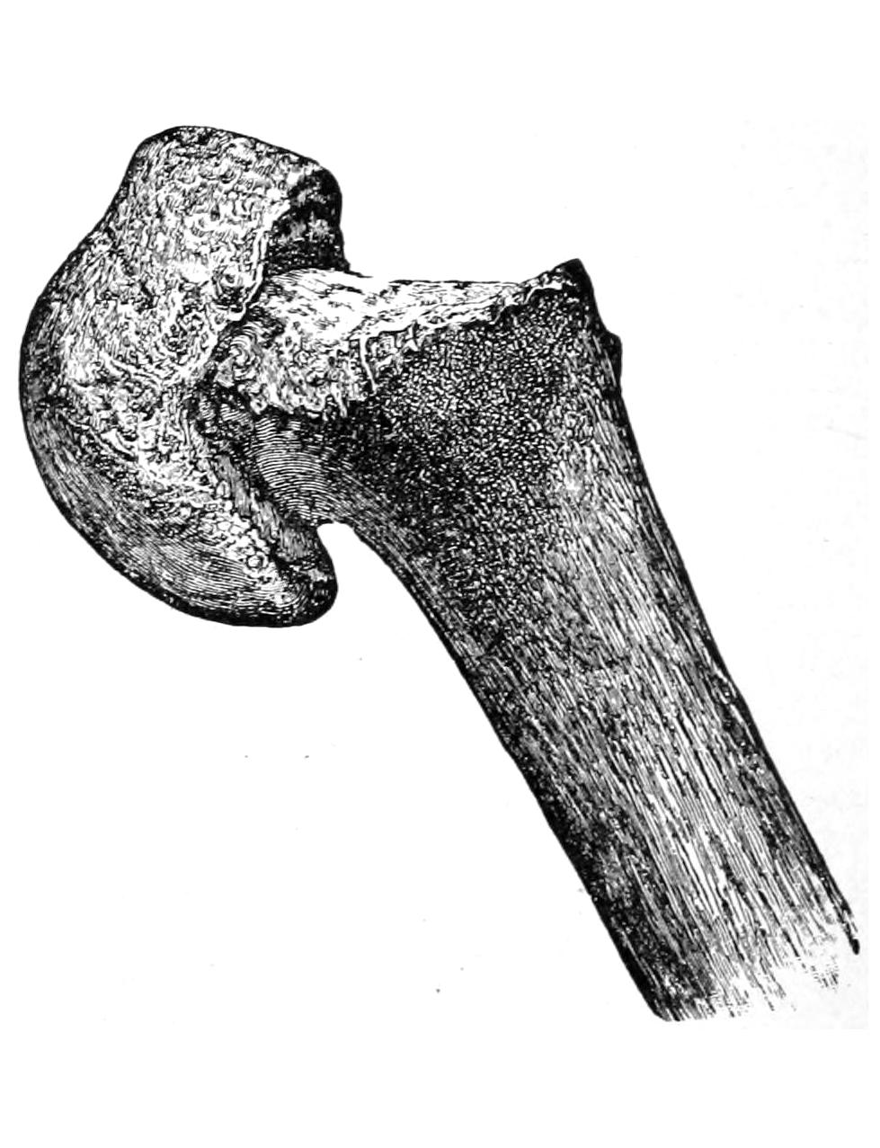
Separation of the upper epiphysis of the humerus; displacement forward of the lower fragment. (Moore.)
The shaft of the bone is frequently broken, lines of fracture running in all directions and occurring at all levels. A variety of displacement may take place. The evidences of fracture are usually recognizable and diagnosis is not difficult. The brachial artery and the musculospiral nerve are occasionally involved, either in callus or by primary injury from a spicule of bone. These fractures are more liable to delay in union or even to non-union than almost any others. These occur often without evident cause, while more or less absorption of bone has been known, by which complications are produced.
In the treatment of fractures of the shaft posture is necessary to observe, the fragments not only being held in position, but the axis of the bone being maintained. An external splint, extending up to and rounded over the shoulder, and an internal splint molded to the inner side of the arm, taking in the elbow and forearm, and placed at a right angle, and then the immobilization of the entire arm by its fixation to the body will give the best result. The writer prefers to make these of plaster of Paris, by molding strips of surgeons’ lint sopped in plaster cream, and maintaining the limb in the desired position while they harden. Should comminution be extreme, or shortening difficult to overcome, a few days’ confinement in bed, with traction upon the forearm, either extended or included in the above dressing, by the usual method, with weight and pulley, will give the best result. So soon as callus has bound the ends of the bone together the patient may be released from bed and the arm left in the right-angle position, in plaster, as above. Or over such a splint as has been described, made of molded plaster, may be hung by a bandage at the elbow sufficient weight (a bag containing shot) to maintain constant traction upon the lower fragment, while the patient is in the upright position, and to influence for good any overlapping or displacement of any kind during the critical period when the bone ends are being united by callus.
The epicondyles are occasionally chipped off from the condyles, the internal being the more frequently injured. These detachments are extra-articular and are relatively unimportant, the fragments being kept from displacement by their fibrous investments. If such an injury should be compound any fragment completely loosened should be removed. It is sufficient to dress such an injured elbow with cold wet compresses in the flexed position. Supracondyloid fracture, or its equivalent in the young (an epiphyseal separation) are somewhat similar, the latter occurring nearer to the articulation than the former. In each of these injuries the arm is flexed and shortened, the fragment lying usually in front of the shaft and the olecranon protruding posteriorly. The more the arm is extended the more prominent the deformity, while by flexion it is much diminished. Hence the advantage of dressing it in the position of overflexion sometimes called Jones’ position.[39] Injury to the vessels at the bend of the elbow may occur in these fractures. If not dressed in this position the elbow should be put at a right angle, while a weight is slung over the elbow, as already mentioned above. Joint function will be greatly hampered if complete extension and reduction be not effected (Fig. 288).
Fig. 288
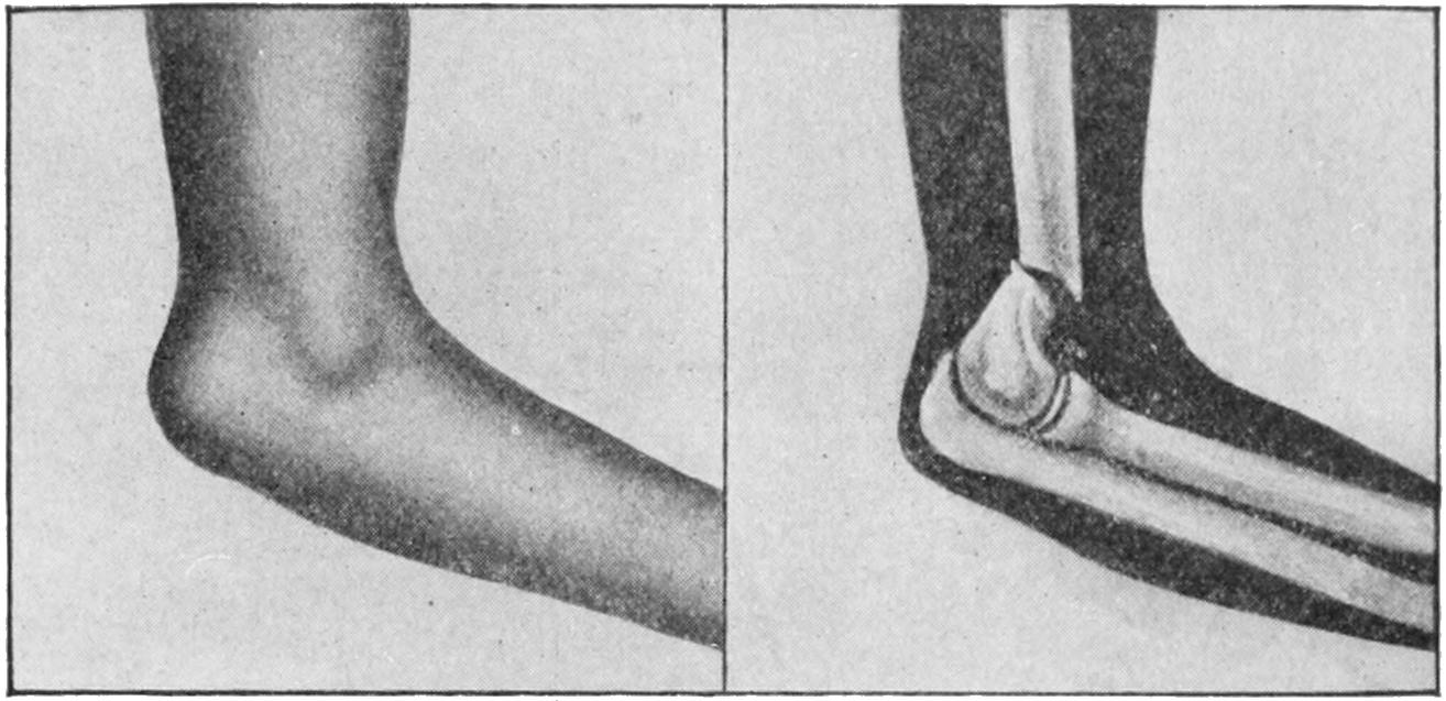
Supracondyloid fracture or epiphyseal separation. (Lejars.)
In considering fractures about the elbow no greater aid can be obtained than by a study of the relations of the three prominent or salient anatomical points to each other. These are the internal and external condyles and the tip of the olecranon. They afford a key to nearly all the displacements which may be produced after fracture or even dislocation, and the only conditions under which they cannot be made available are those where there has been tremendous swelling before the case is seen by the surgeon. A fourth prominent feature, the head of the radius, is also of much assistance, but is less often available, especially in muscular or swollen forearms. When a normal arm is flexed to a right angle and viewed from behind the three points above mentioned constitute the angles of a nearly equilateral triangle. When seen from the side the point of the olecranon is just below the external condyle and in the same plane; when the arm is completely extended and viewed from behind these three points are practically in the same line. By a careful study of the variations from the above relations which are produced by injury diagnosis can be greatly facilitated.
Fig. 289
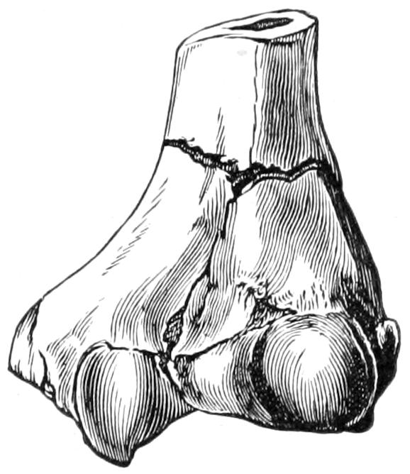
T-fracture of humerus. (Helferich.)
Fig. 290
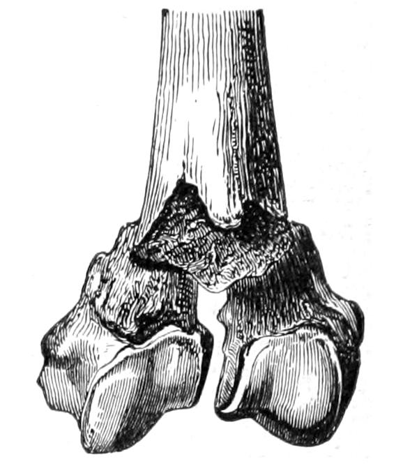
T-shaped fracture of lower epiphysis of humerus.
Fig. 291
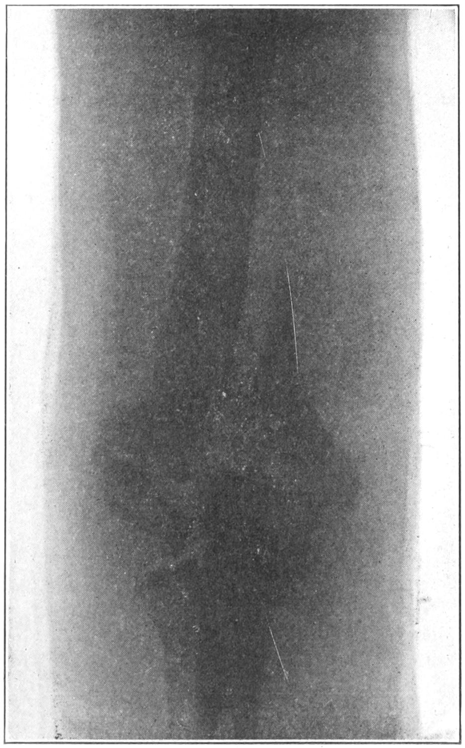
Intracondyloid fracture of humerus. Almost perfect functional result. (Parmenter.)
Fig. 292
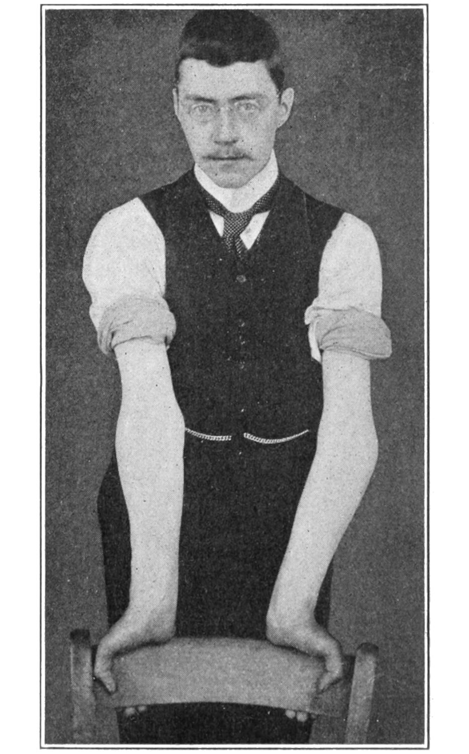
Gunstock deformity after fracture of internal condyle, illustrating neglect of precautions mentioned in text. (Beatson.)
The condyles may each be broken loose by itself, or they may be both broken at the same time. Fig. 291 illustrates what is known sometimes as a T-fracture, where the lower extremity is not only separated from the shaft but is broken into halves; such fractures imply great violence, and are particularly difficult to treat. Should the condyles be detached in such a way as to leave the lower end of the humerus in pointed wedge shape it may perforate or do much harm to the soft parts (Fig. 290). In these intercondyloid fractures the writer would advise dressing in the extended position, with a molded plaster-of-Paris anterior splint and a gentle degree of traction, the patient being confined to bed for a few days. In applying such a splint the surgeon should give extreme care to holding the fragments in proper position while the splint hardens, and in preserving the “carrying function” (Fig. 291). (See below.)
PLATE XXXIX
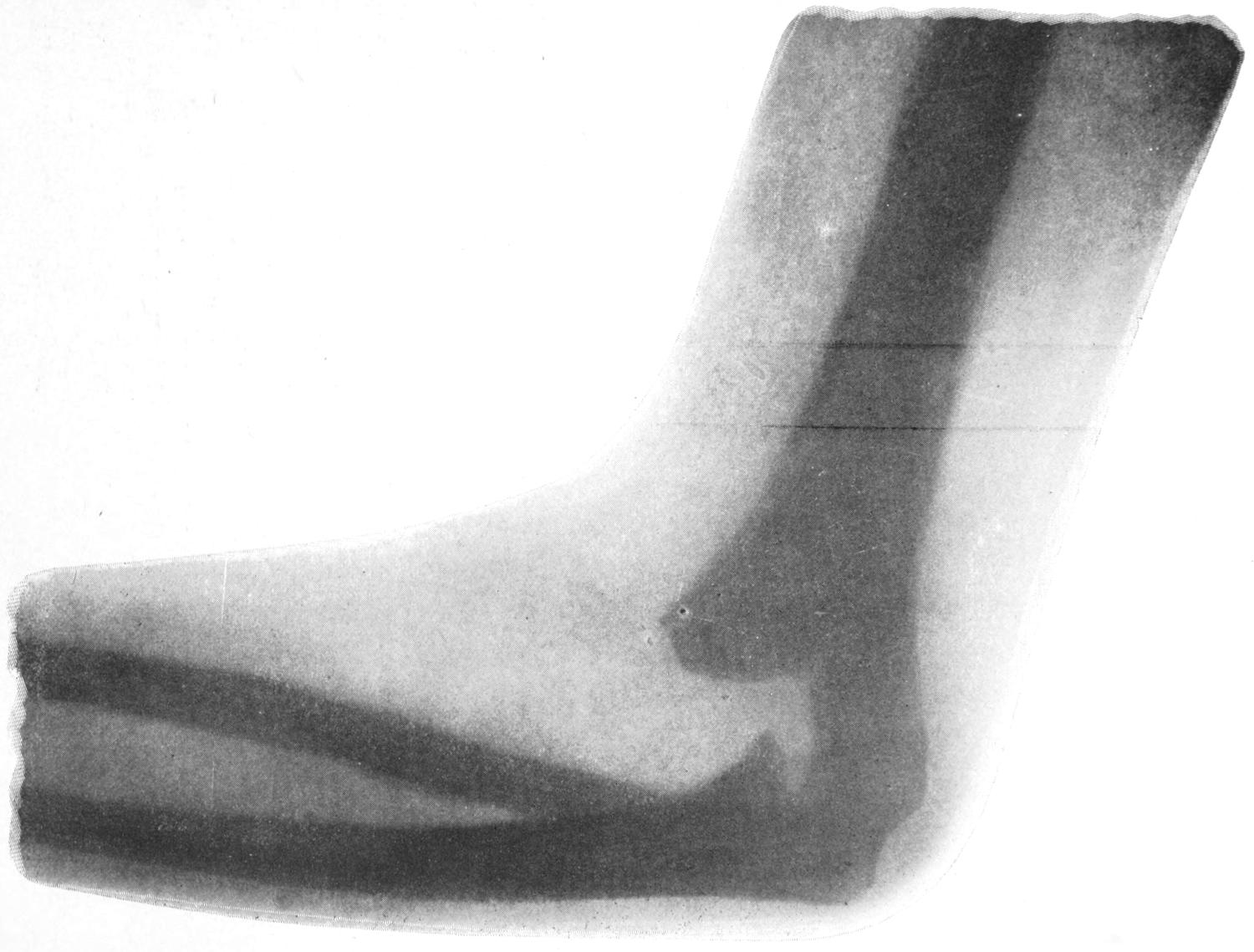
Supracondyloid Fracture. (Child, nine years old.) Union with deformity, fragment so joined to lower end of shaft of humerus at an angle that when forearm is completely flexed upon this fragment it yet is only at right angle with the arm. Operation indicated. (X-ray picture.)
The external condyle when fractured is displaced by muscle pull; when the internal condyle is broken the tendency is to backward displacement of the fragment and widening of the joint.
Fracture of the internal condyle is often an exceedingly serious matter, because it is so often associated with more or less dislocation and with permanent deformity, as a result of inattention to the anatomical relations of the bones. The ulna sustains peculiar relations to the inner condyle; at its upper end it is wrapped around the process, holding it much as a monkey-wrench can be made to seize an ordinary object, and being held to it by the internal lateral ligament. Herein lies the secret of success or failure in treatment, for the fragment, being so fixed to the ulna, should be controlled by it, i. e., the position of the ulna is the most essential feature of the treatment of the fracture. The forearm makes an angle with the arm proper, by which a considerable degree of divergence is maintained. This has been alluded to by Allis and others as the “carrying function.” It can only be estimated in the extended position, and be accurately judged by comparison with the other arm. If the arm be flexed all possibility of estimating it is lost; therefore to dress such a fracture in the right-angle position is bad practice (Fig. 292). The only position in which the carrying function can be preserved is the extended, or one a little short of it for the purpose of comfort. If the ulna is put in the proper position the fragment will be held equally so or as nearly as possible (Fig. 294).
Fig. 293
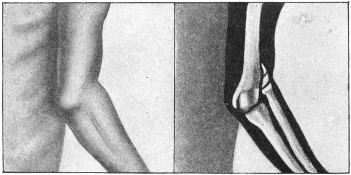
Fracture of external condyle. (Lejars.)
In the treatment of fractures of the inner condyle the patient, if a child, should be anesthetized, the upper part of the body exposed, both arms extended, and the injured arm made to correspond exactly with the other so far as concerns the angle of divergence. Upon the arm so placed an anterior plaster-of-Paris molded splint should be carefully applied, extending from axilla to wrist, and then lightly secured with bandages, the surgeon holding the arm in the proper position until the plaster is sufficiently hardened to permit no displacement. The arm should be kept in this position for at least ten days, after which the splint may be removed and gentle motion practised. It may then be reapplied for two or three days, after which we may begin to flex the arm, applying either a new plaster splint or any other that seems suitable, and in such a way that at the expiration of another week the forearm is brought to a comfortable position of right angle, where it may be maintained with a light splint or simply with a sling, according to the age and tractability of the patient. Fig. 294 illustrates the splint and the position, which is the only one in which the surgeon maintains his own security and can properly estimate the carrying function. The mistake has been in dressing this fracture, like most others at the elbow, in the right-angle position.
In fractures of the outer condyle these anatomical conditions do not prevail, and these may be dressed in whatever position best meets the indications of comfort and accurate reduction. Intercondyloid fractures are subject to the same conditions as those of the internal condyle, plus others which are added, and should therefore be dressed in the same position.
Fig. 294
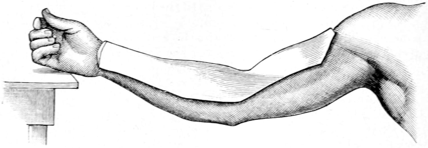
Molded plaster splint for entire arm, and especially for fractures of the internal condyle, showing proper position for dressing same.
Epiphyseal separations, as well as supracondyloid fractures, should be dressed either with traction in a somewhat extended position, or in that of extreme flexion, called also Jones’ position, according as the fragments may best fall into place in one or the other.
Fig. 295
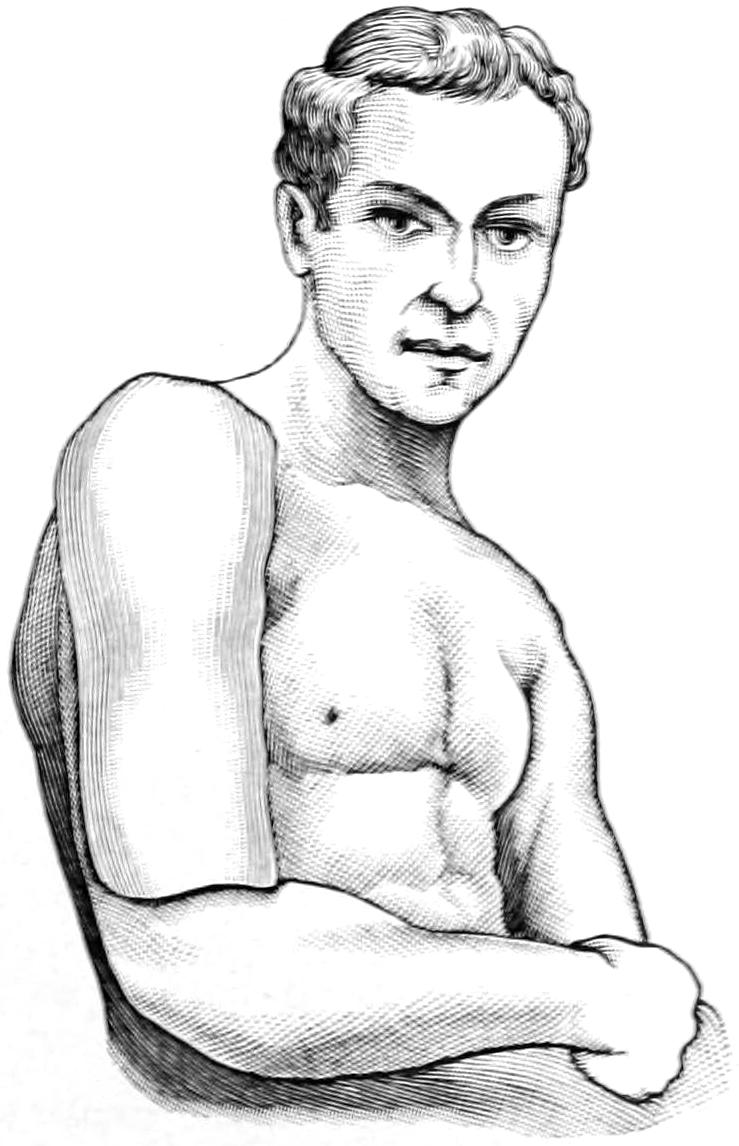
Molded plaster splint for arm.
Fig. 296
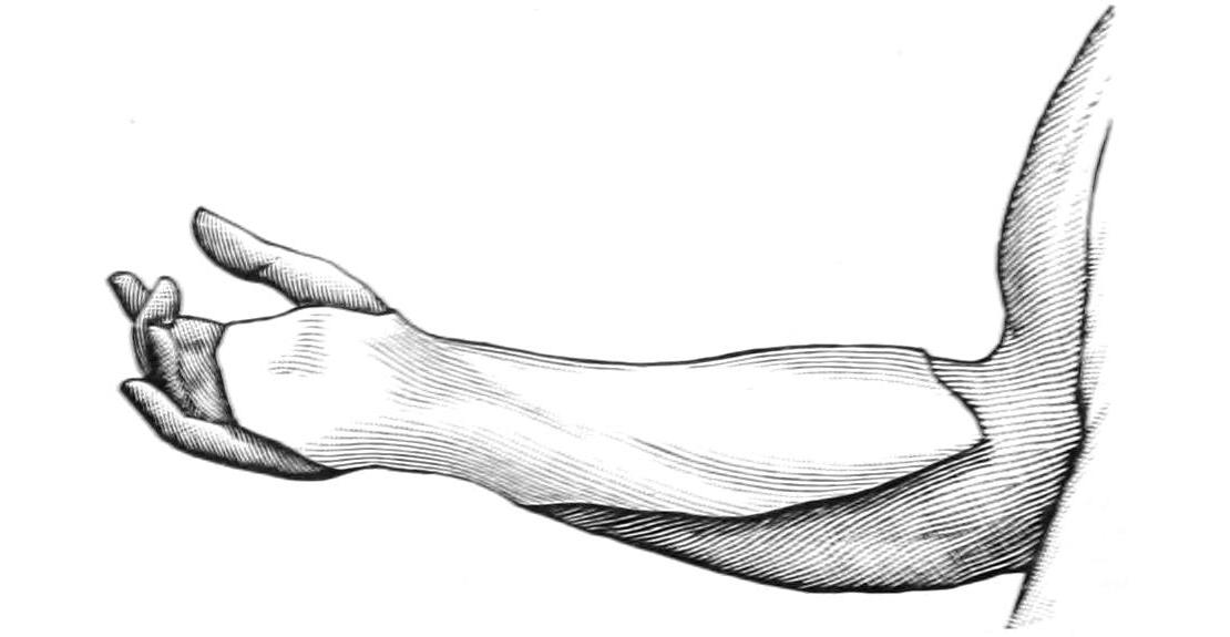
Molded plaster splint for forearm.
The writer has for his own purposes discarded almost all other splint material for the upper extremity in favor of the plaster-of-Paris splints already mentioned. Figs. 294, 295 and 296, may illustrate the method and purpose of their use; many other modifications can be devised as may be demanded. It is customary, after such a splint is hard and firm, to remove it for a few moments, trim it, smooth the edges, line it with a fresh piece of soft lint or its equivalent, and then reapply it to the arm with a roller or starch bandage, the arm meantime not having been disturbed, but maintained in its proper position, and being restored to the splint and made to take its previous position. Such a splint fits accurately the individual for whom it is made. It is worthless for anyone else; nor should it ever be used again, the intent being to mold a splint for each case which shall serve its individual purpose and none other.
Too early passive motion with the intent to regain mobility is inadvisable and often dangerous. A fractured joint should be kept at rest until the bone is consolidated. If callus be thus reduced to the minimum, and consolidation be undisturbed, the patient will, in due time, recover motion, often to the extreme limit. In fractures of the humerus five or six weeks are required for the attainment of perfect union. In spite of precaution callus formation will sometimes be excessive and interfere with motion. Absorption of exuberant material then is most desirable. This can be encouraged by constant but gentle pressure. Thus when callus in front of the lower articular surface of the humerus obstructs the coronoid process of the ulna and prevents complete flexion the patient should wear for several hours at a time an elastic sling, made with a piece of Martin rubber bandage sufficiently long to make a loop around the neck, into which the hand is passed. It should be made so tight as to exert gentle but constant pressure; the result of this will be to cause rapid disappearance of the callus upon which it is made. Conditions may be reversed when necessary, and the patient may have some weight affixed to the hand by which, when the arm hangs down, reversed pressure shall be made, or when desirable these measures may be alternated. One should not, however, be tempted into resorting to them too early, since much is done, even in unfavorable cases, by purely natural processes, this being especially true of children who are growing rapidly.
FRACTURES OF THE FOREARM; THE ULNA.
At the upper end of the ulna the most frequent fracture is that of the olecranon, whose separation by direct or indirect violence corresponds to fracture of the patella. The fragment is pulled upward along the back of the arm by the triceps muscle, and the power of extension is almost lost. There is rarely any difficulty in diagnosis, except in conditions of extreme swelling, which of itself would be suspicious, as under hardly any other circumstances could a joint be so distended (Figs. 297 and 298).
Fig. 297
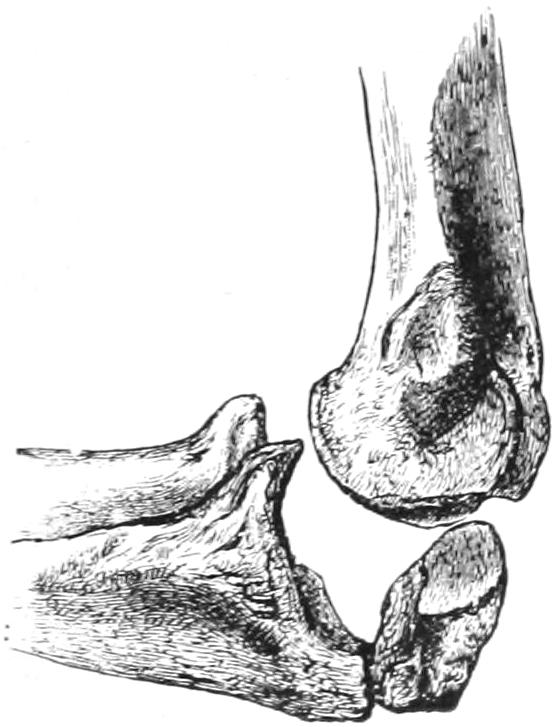
Fracture of olecranon. (Erichsen.)
Fig. 298
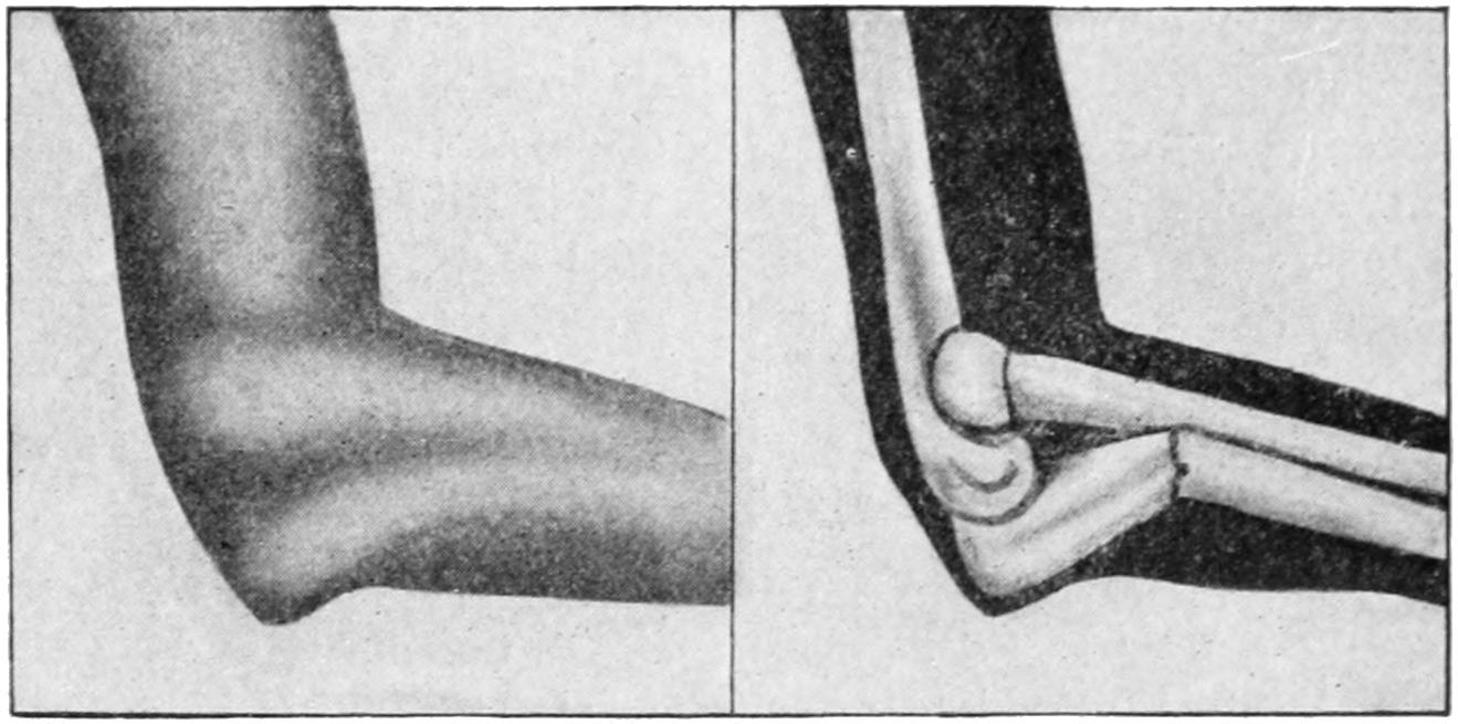
Fracture of ulna, upper end. (Lejars.)
Fig. 299
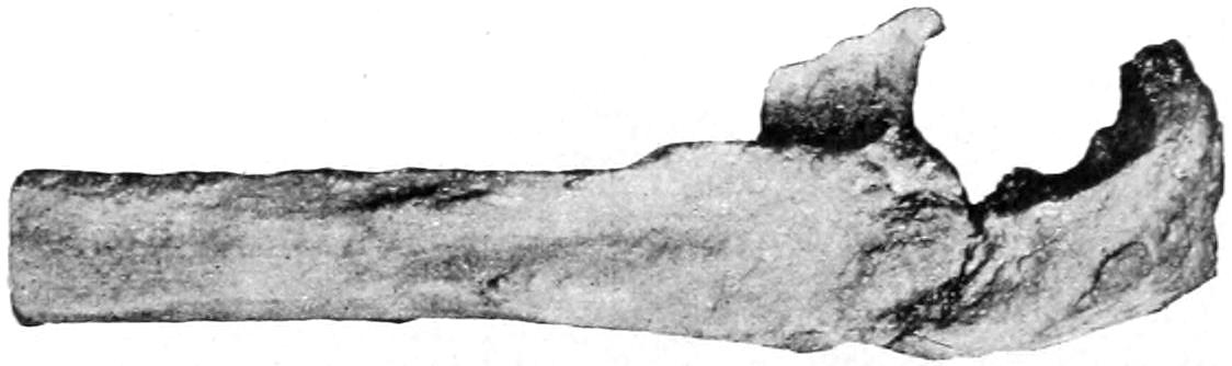
Fracture of olecranon with fibrous union. (Park.)
Treatment.
—The difficulty here, in treatment, consists in the necessity for counteracting the pull of the triceps. The arm first of all should be dressed in the extended position. Sometimes it is possible, by partly encircling the posterior surface of the arm just above the fragment with a strong piece of adhesive plaster, to which is attached some rubber tubing, to make a constant elastic pull upon the fragment, the tubes being brought down and attached to the sides of the anterior splint below the elbow. In the absence of swelling this can often be made quite effective. So long as much fluid is present no means will be efficient. It may, therefore, be well to wait two or three days until the fluid has disappeared, aspirating the joint if necessary. In young and otherwise healthy subjects there is strong reason for advising operation, as only by absolutely approximating the fragment to the main bone and maintaining it in position can bony union be secured. In properly selected cases, and when performed with every precaution, this measure frequently gives ideal results. A short ligamentous union is represented in Fig. 299. At other times the fibrous band will stretch out to an inch or more, not completely disabling the arm but weakening it. The extended position may be relaxed within a week after operation, but not for at least two weeks after other treatment. Passive motion should not be begun too early in the latter cases.
Fracture of the coracoid process is often combined with backward dislocation of the forearm, which is no doubt an incident of the injury or may occur later by mere muscle pull. The brachialis anticus, which is inserted into it, will pull the fragment up against the anterior surface of the humerus. This fracture should be dressed in the right-angle position, in order to relax the muscle, taking care to prevent backward displacement, while ligamentous union is ordinarily all that can be hoped for.
The ulnar shaft may be broken at almost any point, usually as a result of direct violence. As it is weaker in its lower half the greater number of fractures occur here. Fracture of the shaft is easily recognized, crepitus being always obtained, unless muscle tissue has intervened, this being a condition which will occasionally prevent bony union. If it can be established by x-rays that bony surfaces are not in contact and cannot be so placed, it is advisable to cut down upon the site of the fracture, remove the obstacle, and fasten the fragments together. So long as one bone is broken in the forearm the other may be relied on to act as a more or less efficient splint. There is but one position in which any of these fractures can be dressed with safety, that is midway between pronation and supination, i. e., with the thumb pointing toward the patient’s face. Splints used for this purpose should always be wider than the forearm itself, lest by pressure the ends be forced toward the other bone. Some hold that by gentle pressure along the line between the bones, as by a narrow pad or splint, the muscles may be made to press the injured bone away from the other; nevertheless only moderate pressure can be tolerated for this uncertain purpose. It has been generally customary to use two light wooden splints, one along the palmar, the other along the dorsal surface of the forearm, padding them properly and securing them in position by strips of adhesive plaster and suitable bandages. The same plaster-molded splints mentioned above can, however, be made just as effective for this purpose, if properly applied.
When either bone is broken near the wrist, and especially when both are broken, we have to combat the tendency of the pronator quadratus, which tends to pull the lower fragments together.
The styloid process is occasionally detached, as in violent sprains, or broken off in connection with other injuries. Inasmuch as it carries the upper end of the internal lateral ligament its detachment can be quickly recognized by the abnormal freedom of motion which such an injury would permit.
Fig. 300
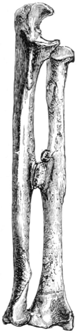
Obliteration of the interosseous space in a fracture of the forearm.
FRACTURES OF THE RADIUS.
The radius vies in frequency of fracture with the clavicle. The head is seldom broken, its fracture being most likely when the shaft is driven against the humerus by falls upon the open hand. The neck is more frequently broken in children than in adults. These fractures have sometimes to be determined by a process of exclusion or by the use of the x-rays. In muscular forearms they lie so deeply that it is not always possible to recognize them. Ordinarily, however, if the head of the bone can be found to remain stationary while the rest of the bone is being rotated, and if, at the same time, crepitus be felt, the matter may be regarded as settled. So far as the shaft of the radius is concerned the remarks made above regarding the ulna mostly hold true for its fellow-bone. When the neck of the bone is broken the shaft will be pulled upward by the biceps tendon, while when the shaft is broken below its insertion the upper fragment is displaced by it. In either of these cases, then, the forearm should be dressed at a right angle with the arm in order to relax the muscle. The supinator brevis and the pronator radii teres should also not be neglected, for the former will tend to rotate and the latter to more or less displace the upper portion of a shaft broken high up. With a fracture near the upper end, in a powerfully muscular arm, diagnosis is not always easy. Fractures in the lower portion of the shaft are to be treated like those of the ulna. But those high up should be dressed with the elbow at a right angle and the forearm supinated. A plaster-of-Paris molded splint here can be adapted to the needs of every individual case if the surgeon will give minute care at the time of its hardening to placing the parts just as he desires them to remain.
FRACTURES OF BOTH BONES OF THE FOREARM.
These are not uncommon, though much less frequent than injuries to either bone alone. They may occur at the same level or be quite widely separated. The loss of function is complete in these instances, while deformity will depend largely upon whether the fractures lie near together or not. It is of the greatest importance to remember, in these cases, that the mass of muscles around the upper fragments tends to crowd them together, while the lower fragments are brought together especially by the pronator quadratus. Everything then conspires to convergence of the four fragments, a tendency which it is sometimes difficult to combat. Every large museum contains specimens showing a common callus, in which all four bone ends are involved, and illustrating the permanent loss of rotation that ensues. This is to be combated, not alone by rest, which tends to limit callus formation, but by position with the arm midway between pronation and supination, in which the bones are naturally farthest separated, and by splints applied with such gentle pressure as may hold the bones apart. This pressure should be applied between the dorsal and palmar surfaces, while the lateral aspects of the forearm should be kept absolutely free from it (Figs. 300 and 301).
Fig. 301
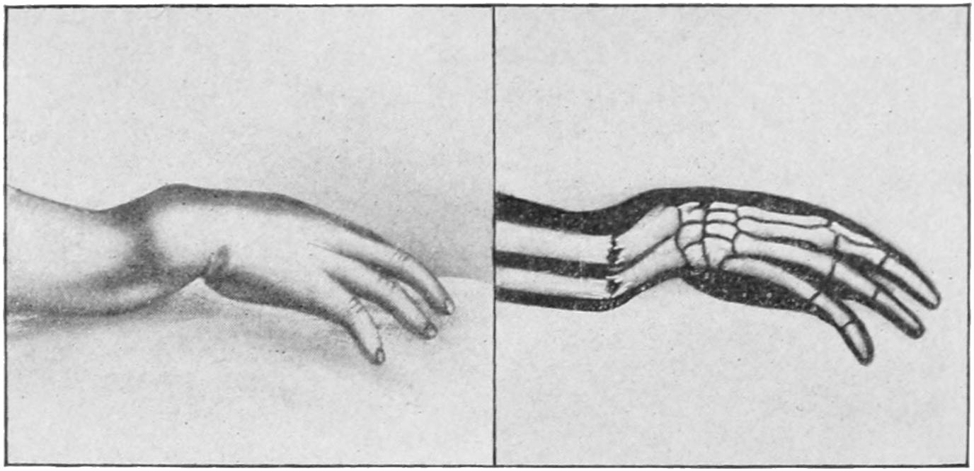
Fracture of both bones near lower extremity. (Lejars.)
It is a serious matter to dress any of these injuries with moistened pasteboard, or other material which does not take its desired shape and strength promptly, for no matter how carefully the desired position may be enforced at first the very nature of a material which remains too long plastic will permit the loss of all that should be maintained. The writer has seen malpractice suits instituted and men forced out of the State by inattention to this precaution.
FRACTURE OF THE LOWER END OF THE RADIUS; COLLES’ FRACTURE.
This is perhaps one of the commonest fractures in the body, occurring at all ages; when seen in growing children it is to be regarded as an epiphyseal separation rather than as a distinct fracture. It derives its name from the fact that until Colles, a Dublin surgeon, over one hundred years ago, described this injury as a fracture it had been always regarded as a peculiar dislocation at the wrist. It is produced by falls upon the hand in the hyperextended position, the force being usually transmitted through the carpus to the radial end. The name is usually limited to those fractures which occur within one and a quarter inches of the articular surface (Fig. 303). Here the structure of the bone is cancellous and impaction may easily occur, this being a decided feature in many of these accidents, and making replacement more difficult. The deformity which results from the fracture is characteristic and more or less uniform. This is called the “silver-fork appearance,” the lower fragment being so displaced, and usually more or less tilted, as to raise the tendons and the structures on the back of the wrist; at the same time it is usually drawn toward the radial side. The more the fragment is impacted or driven into the shaft of the bone the less easily is crepitus elicited.
The fracture is more common than is supposed, and there is no doubt but that many alleged sprains of the wrist illustrate cracks in the bone without displacement, which, nevertheless, are slow to heal and are sometimes followed by thickening and impairment of function. (See Plate XL.)
Along with the radial fracture separation of the styloid process of the ulna may also occur, or, as Moore has shown, the process itself may perforate the internal lateral ligament so as to protrude through the skin; and the surgeon has occasionally to withdraw the styloid from the ligament which has been impaled upon it. The radio-ulnar ligament is also frequently injured, and this permits the ulna to become more prominent than normal. If the styloid has perforated the skin it lends a compound feature to the case. The interarticular fibrocartilage may also be displaced.
Treatment.
—The secret of obtaining a good result and the explanation for failures lie in the completeness or incompleteness of the reduction of the fragment. If the latter be absolutely and accurately replaced it makes but little difference what dressing is applied. On the other hand any fragment not completely restored will lead to subsequent deformity and impairment of function. Successful reduction, then, is the keynote to success, and should be accomplished at any reasonable cost. Sometimes it is not difficult, and then no anesthetic is required; sometimes it is extremely difficult, and the operator has to exert all the strength he has in his arms, aided by profound anesthesia. Moderate cases can usually be dealt with successfully under nitrous oxide gas. The surgeon grasps the hand as if to shake hands, i. e., with his corresponding hand, the elbow being firmly held by an assistant. Traction is then made upon the hand to which the fragment is affixed, while with his other hand the operator makes such pressure, rotation, or coaxing manipulation with his thumb and fingers as may assist in restoring the fragment to its place. With whatever other effort may be made traction should be combined. Forcible swaying movements, combined with hyperextension, may be necessary to dislodge an impacted fragment. Any degree of force is preferable to failure in this respect. Perfect reduction is the key of success; without it, no dressing is efficient; with it, almost anything will suffice.
Fig. 302
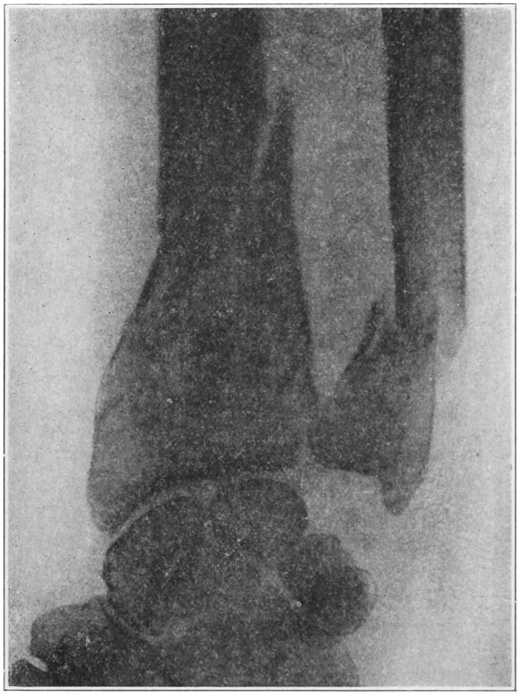
Comminuted but not compound fracture of wrist. (Beatson.)
Reduction once accomplished it is usually an easy matter to hold the arm in position. The writer prefers above all other means a molded plaster-of-Paris splint, which should extend from the line of the knuckles upon the palmar surface well up toward the elbow. It should be fitted neatly to the hand and forearm, bandaged comfortably upon it, while as it solidifies the surgeon should hold the hand slightly flexed to the ulnar side as well as anteriorly. When the splint is hardened and bandaged a simple sling will suffice. The hand should be dressed with the thumb pointing toward the face, while upon the back of the wrist an ice-bag can be applied. Ecchymosis is sometimes extreme; I have seen it extend even to the shoulder after an apparently simple break (Figs. 304 and 305).