Part 6 — CHAPTER XXXVI. INJURIES AND SURGICAL DISEASES OF THE HEAD. (continued 2)
Reading edition
Diagnosis.
—The diagnosis in adults is difficult, but in children, when convulsions occur after exhausting illness, with the signs just noted, marasmic thrombosis may ordinarily be diagnosticated.
Infective thrombosis, the other variety, is due exclusively to the invasion of pyogenic organisms. It is observed mostly in the basal sinuses; its origin is local, and it is always secondary to some external infection. Its most frequent cause is middle-ear disease; consequently the sigmoid sinus is the one most often involved. It may follow carbuncle, erysipelas, or cellulitis of the external parts, or nasal ulceration, as well as dental caries, suppuration of the tonsils, etc. Infection may be propagated by tissue continuity, or through the circulation.
Symptoms.
—Infective thrombosis presents few distinctive symptoms. Local ischemia, perversion of function, extracranial edema are too vague. Headache is nearly always constant and vomiting is frequent; temperature runs high, with marked remissions; the pulse is small and rapid, and remains so even under an anesthetic. Chills are frequent, of the pyemic type, and are followed by copious sweats. Should pulmonary infarct occur there will be typical thoracic signs, although at first physical examination may give negative results. Later, however, we get prune-juice expectoration, putrid sputum, etc. Cerebral function is disturbed late rather than early. The duration of the disease ordinarily is from two to four weeks. Should meningitis complicate the case there is more violent headache, persistent high temperature, great excitement, muscle spasm, strabismus, delirium, and coma; if the sigmoid sinus be involved there is usually retraction of the head. Should leptomeningitis extend down the spine, complaint of girdle pains will be made.
Differential Diagnosis.
—The two conditions which are most likely to be confused with sinus thrombosis are meningitis and brain abscess. In thrombosis there are pain and tenderness over the mastoid, extending down the neck. Fever is high, pulse rapid, respiration not affected, rigidity not usually present. Chills are frequently followed by profuse perspiration. The general picture is one of sepsis and the typhoid state. There are no special eye symptoms. Death is finally due to pyemic processes. In meningitis pain is an early, constant, and severe symptom. Headache is frontal or general, fever is not characteristic, pulse is rapid until the accumulation of pus causes slowness by pressure, breathing is short and rapid, and finally of the Cheyne-Stokes variety. Rigidity of the neck and back, with retraction of the head, is nearly always present, with spasmodic contractions or convulsions about the neck. Chills are not so pronounced, vomiting is almost invariably of the projectile type, optic neuritis is frequent, and the intellect is early impaired. In brain abscess pain is at first localized and severe, extending and becoming excruciating. This increases on pressure, and does not disappear until relief is obtained or the patient becomes unconscious. Temperature is normal or subnormal until the abscess ruptures. The pulse is slow, as in compression from other causes; breathing is slow and stertorous. Rigidity and vomiting are like those of meningitis. Eye symptoms are almost always present, photophobia at first, later inequality of pupils, with dilatation on the affected side, optic neuritis and irregular movements of the eye and lids. Drowsiness, dizziness, and impaired intellect are features when the abscess is in the cerebellum. Death occurs in coma unless the case be complicated by meningitis.
We may also have exophthalmos on one side or both, with conjunctival injection, edema of the lids, and disturbances of vision, due to thrombosis of the cavernous sinus and stasis in the ophthalmic vein. In thrombosis of one transverse sinus only the internal jugular on that side will carry less blood. So long as that on the other side is free it will take that which cannot pass through the obstructed one. Consequently the jugular on the other side will carry more. But if the contained clot extend so far that direct communication with the internal jugular is interfered with then the internal jugular of the affected side will be almost empty, while the external of the same side will be the more distended. When the eye is protruded and the frontal vein distended it is evident that the cavernous sinus on that side is involved. If the superficial veins of the scalp be distended it is the superior longitudinal sinus which is at fault. When the veins of the mastoid region are involved, we may locate the thrombus in the transverse sinus; when there are no localizing symptoms, it can only be said in a general way that thrombosis has occurred.
Prognosis.
—Prognosis is always unfavorable, though recovery is not impossible. The therapeutics are in the main prophylactic. By actual physiological rest the possibility of pulmonary complications can be diminished. The treatment, aside from this, is purely operative, and will be discussed elsewhere.
C. Sinus Phlebitis.
—This may be the result—
(a) Of thrombosis; or,
(b) The continuation of suppurative processes from neighboring tissues.
Symptoms.
—The symptoms are seldom diagnostic. Sinus phlebitis is often accompanied by meningitis, even encephalitis. The first symptom is usually severe headache, often localized, made worse by pressure. Anorexia with early mental disturbance and often delirium follows, with vomiting, restlessness, and mania, changing to stupor and coma. Rigidity or spasm of cervical muscles, or of those of the extremities, followed by paralyses, is often seen. Evidences of irritation of special nerves, particularly the oculomotor or the vagus, are not rare. When pyemic symptoms occur they are vague and are most conspicuous in the lungs and liver. Taken in conjunction with aggravating brain symptoms they make prognosis unfavorable.
Symptoms will in large measure depend upon the sinus most involved. They are characteristic if this be the cavernous sinus. There are disturbances in the eye on the same side, congestion of orbital veins, pain and photophobia, and, later, cloudiness of the cornea and edema with exophthalmos. Finally the pupil becomes paralyzed and dilated, the cornea loses its polish, the upper lid cannot be raised, and, if the case persists, the cornea ulcerates. Along with these local evidences there will be complaint of frontal pain, usually with paralysis of the hypoglossal nerve and consequent thickness of speech. When the transverse sinus is involved there are, first, vagus irritation, then paralysis with paralytic sequences in the muscles of the jaw, the tongue, palate, pharynx, etc. Diaphragmatic motions are interfered with and the character of the respiration altered. As the trouble extends to the internal jugular we have further paralysis of accompanying nerves, especially of the hypoglossal. As the irritation extends down the vein there will be tenderness, rigidity, and often swelling. The local signs and symptoms vary obviously as the lesion extends from one sinus to the other, for when one cavernous sinus is involved the trouble nearly always extends to the other, and local symptoms are repeated upon the opposite side.
D. Meningitis.
—The dura has a duplicate anatomical character. Its outer surface, having the structure of periosteum, functionates as such; its inner surface, being lined with endothelium, partakes of the nature of a true serous membrane. When the former texture is mainly at fault we have pachymeningitis externa, or endocranitis, which is rarely a primary, but usually a propagated lesion met with after injury or external infection. It may lead to infiltration with purulent products, and, if speedy exit for pus be not provided, to involvement of the pia within. Extradural suppuration without external injury is very rare, but should there have been a subdural hemorrhage with external lesion the blood clot may become infected and break down. Pachymeningitis externa is most common after chronic lesions of the cranial bones—i. e., caries and necrosis. Symptoms are not characteristic and often not distinguishable. When chronic there will be local tenderness, evidence of the presence of pus, with focal symptoms.
Treatment.
—The treatment is always surgical, save possibly in certain cases due to syphilis, where delay may be justifiable for the purpose of testing the action of antispecific drugs.
Pachymeningitis Interna.
—Pachymeningitis interna is often confounded with chronic hydrocephalus. It is frequently the occasion of a firm, membranous exudate upon the internal surface of the dura, which forms in time a new membrane rich in small and extremely friable vessels, from which hemorrhages easily occur, thus giving rise to the condition of pachymeningitis hæmorrhagica. Trifling hemorrhages will produce little or no disturbance; when of greater extent they may give rise to localizing brain symptoms. These extravasations may absorb or undergo fluidification—i. e., produce localized or cystic collections of fluid. The condition sometimes occurs after other acute infections, especially pneumonia, pleurisy, typhoid, whooping-cough, etc. Recovery is possible, but usually at the expense of adhesions, which lead to subsequent complications.
The symptoms of pachymeningitis hæmorrhagica are headache, which will increase in intensity with every new escape of blood, usually localized in the vertex, with more or less paralysis following each new extravasation. The final result may be atrophy. Absence of disturbance in the cranial nerves points to lesions in the convexity rather than basal or ventricular. In chronic cases there is optic neuritis, and toward the end coma, usually coming on slowly. Dennis has recommended trephining under these circumstances, and has practised it with great benefit.
Treatment.
—The treatment should be in a large degree surgical, for little short of eradication will bring about the desired result.
Leptomeningitis.
—This term refers to inflammation (i. e., infection) of the pia mater, in whose texture we encounter tissue quite different from that composing the dura, and in which, when inflamed, distinction as between the arachnoid and pia has disappeared. Leptomeningitis suppurativa is an exceedingly common expression of intracranial infection, and may result not merely by extension, but as a primary infection. When begun it spreads rapidly, the fluid contained within the meningeal cavities, mixed with pyogenic agents, helping to disseminate the active agents to the ultimate limits of the membranous involvement. Consequently basilar meningitis usually extends down the spinal canal. Next to injury the most frequent cause is middle-ear disease, with its infectious complications and extensions. Next to this come sinus phlebitis and endocranitis. Infection from the teeth and the nasal cavity may occur. It is also known to result from panophthalmitis: in traumatic cases, when primary, it sets in early, even from four to thirty-six hours after injury. So rich is the pia in loose connective tissue that even from the outset the inflammation may assume the phlegmonous type. The cerebrospinal fluid, as well as that of the ventricles, becomes cloudy, contains numerous flocculi, and is often blood-stained.
Symptoms.
—When the disease is limited to the vertex and follows several days after injury it usually begins with chills and malaise, with increasing temperature; after which the symptoms assume the pyemic type, distinguished from true pyemia by their comparatively early onset. The pulse becomes frequent, first full and then small; patients are disturbed, restless, or uncontrollable, and complain of headache, moan, grate the teeth, become delirious, with glistening eyes and congested face. After a while delirium subsides into stupor and restlessness into insensibility. The pupils contract and remain inactive to light. Paralyses and cramps are not infrequent. Traumatic basilar leptomeningitis occurs often with fracture of the base. Signs and symptoms are less distinctive here; paralyses occur more easily and are less distinctive, save those which involve the special cranial nerves. When ptosis occurs with dilatation of the pupils and glossopharyngeal paralysis we should be quick to suspect extension of the process along the brain. Cramp or stiffness of cervical muscles mean the same thing, and are signs of grave import which may be considered pathognomonic. Albuminuria is frequent, with marked increase of phosphates in the urine.
In the non-traumatic cases the symptoms of leptomeningitis are those of increasing brain pressure and temperature. The disease usually commences with headache followed by vertigo, hyperesthesia, restlessness, delirium, insomnia followed by somnolence, muscle spasm, paralyses, coma, and death. If the disease extends from the middle ear there is frequently facial paralysis before the meningeal symptoms appear.
The type of fever is one of gradual increase, though before death temperature often falls even below the normal. Pathognomonic fever should not be mistaken for the elevation of temperature which often accompanies absorption of intracranial hemorrhages. In these latter cases temperature may mount to 39° C., but if rising higher than this meningeal complications should be suspected.
Diagnosis.
—The diagnosis as between sinus phlebitis and leptomeningitis depends principally upon the existence of pyemic symptoms. When the latter are entirely wanting we may at least say that the predominating symptoms of sinus phlebitis are absent.
Prognosis.
—The prognosis is unsatisfactory. Many cases end in forty-eight hours; others may live for two weeks or more.
Treatment.
—Treatment seems almost futile, though one should endeavor by energetic purgation, venesection, etc., to do what he can. The only prospect or hope comes from the possibility of relieving the compression from effusion of purulent fluid, and of irrigating and draining what is now an enlarged abscess cavity. Since we do not hesitate to open and wash out other serous cavities when thus affected—e. g., peritoneum, pericardium, joints, pleura—we should no longer hesitate to open the dura and wash out the subdural space, even though this necessitate more than one trephine opening. The measure was suggested by S. W. Gross, in 1873, when he reported cases thus treated with success, and has since been practised by other surgeons, among them by Souchon, who has advised multiple puncture with the small drill and irrigation and disinfection through numerous small openings. Of 11 cases collected by Gross more than twenty-five years ago, 45 per cent. recovered.
E. Encephalitis.
—The etiology of this condition is practically that of leptomeningitis. It may proceed from sinus phlebitis or from the veins emptying into the sinus, infection travelling backward rather than forward. In many cases the primary infection occurs from without, as in gunshot fractures. It is also transmitted along the lymphatic channels, since I have operated on abscess in the frontal lobe following intranasal operation. It assumes practically always the suppurative type, and may run either an acute or a chronic course. When acute the lesion is usually limited in area, and the result is an acute abscess with irregular boundaries. It may be distinguished from uremic coma by examination of the blood (leukocytosis) as well as that of the urine.
OPERATIVE TREATMENT OF INTRACRANIAL SUPPURATIONS.
In dealing with pus the surgeon can never follow a safer rule than to go according to this dictum: i. e., that pus left alone is a greater source of danger than the surgeon’s knife judiciously used. Consequently ubi pus, ibi evacua, applies to intracranial collections as well as others. For its detection and evacuation operations are now regarded as not merely justifiable, but indicated whenever there is presumption of its presence. Discussion now hinges entirely upon the wisdom of exploration when absolutely no diagnosis can be made. Save where an opening already exists, trephining is a necessary preliminary. Among other indications is spontaneous escape of pus through a previous opening or any of the natural outlets of the cranium, with or without localizing phenomena. Further indications are those pertaining to the bone—i. e., loosening of pericranium; or to the scalp—i. e., edema, puffy tumor, etc.; and certain other indications are those of a more general character, chills and pyrexia. When the dura is exposed much can be determined by the existing brain tension, it being now well established that brain pulsation is often intensified by the presence of pus beneath the dura. The most feasible method for detection of subdural or deep collections is the use of the aspirating needle—a method now generally in vogue and everywhere accepted.
MASTOID DISEASE AND THE MASTOID OPERATION.
In all cases of infection and suppuration of the middle ear the adjoining portions of the cellular structure of the mastoid undoubtedly participate. Fortunately morbid activity is usually so limited that the clinical evidences of what is called mastoiditis occur in a relatively small proportion of cases, but otitis media purulenta is so common that mastoiditis is consequently a complication of sufficient frequency, and occasionally of such severity, that it is as likely to come under the supervision of the general surgeon as that of the specialist. Moreover, the region affected is such common ground, as it were, between the broad field of the former and the restricted field of the latter that it seems to me that every general surgeon or student of general surgery should be familiar with the condition and its surgical treatment.
Several of the specific germs, of diseases like pneumonia, la grippe, etc., are known to set up acute mischief within the tympanum as well as the commonly known pyogenic organisms. They have easy access to the middle ear through the Eustachian tube, as well as by the deeper blood and lymph channels. The nasopharynx is never free from the presence of organisms, while the specific fevers, like scarlatina, and notably such infections as diphtheria, predispose to germ activity in the region into which the inner end of the Eustachian tube opens. The Schneiderian membrane, which is practically continuous from the ethmoid cells to the membrana tympani, affords easy travelling, and in all directions, for infecting organisms. The violence of reaction will depend upon two uncertain and indeterminable factors, the virulence of the organism and the susceptibility of the patient. To what extent the mastoid cells and antrum, around an infected tympanum, shall participate may be, to a considerable degree, a matter of their anatomical arrangement. When, however, they do participate to any great extent the fact is made known by symptoms of unmistakable character. These constitute the added features of what is known as mastoiditis.
The cavity in the mastoid known as the mastoid antrum, no matter what may be the arrangement of the other cells, is always present, and in the presence of deep disease the antrum should be first opened. In close proximity to the antrum are cavities like the sigmoid sinus, the horizontal semicircular canal, the facial canal, and the interior of the cranium. While opening the antrum care should be taken to avoid encroachment upon the other cavities or structures, except in those instances where there is evidence of intracranial mischief, in which case it may be desirous to expose the sinus wall, or even a considerable area of brain surface. The mastoid prominence varies in different individuals, extending outward to accommodate the sigmoid groove for the lateral sinus.
According to the intensity of the process the pathological condition of the mastoid may vary between an empyema of its cavities, an osteomyelitis of its osseous structure, or osteoperiostitis of its external surface. Nevertheless all three of these may be combined in the same case.
Symptoms.
—The symptoms of mastoiditis are pain, referred to the mastoid, as well as to the region around it, although when pressure is not made by retained pus pain may not be intense; local tenderness is present in nearly all cases, and will depend upon the proximity of the trouble to the surface. This tenderness is evoked by gentle pressure, which will sometimes produce pitting, or by tapping lightly with the finger. When the trouble is superficial there will often be edema, with all the local evidences of suppuration. In addition to this there will be coincident symptoms of disease of the middle ear, with discharge, earache, etc., and frequently edema or actual phlegmon of the auditory canal.
The different directions in which destructive processes may extend, and their consequences, are as follows: (a) Externally, with well-marked local evidences of the proximity of pus; (b) anteriorly into the meatus, with phlegmonous appearances in that canal; (c) upward, through the roof of the tympanum or the antrum, with disastrous cerebral symptoms or extradural abscess; (d) inward, toward the sinus, with consequent thrombophlebitis, extradural abscess, and perhaps cerebellar abscess; (e) downward, and away from the mastoid, with phlegmon deep in the neck.
The first appearance of symptoms of any of these complications should awaken apprehension and demand scrupulous attention. Any collection of pus along the auditory canal should be promptly incised, and the first indication of mastoid tenderness or inflammation should cause a prompt application of leeches, followed by antiseptic irrigations. In this way it may be possible to avert serious symptoms, provided these measures be instituted early.
But with either the access of local symptoms indicating the presence of pus, or of more general symptoms, elevation of temperature, acceleration of pulse, headache, or anything else suggestive of dural irritation or cerebral complication, no time should be lost in making free and radical operation. The mastoid operation, so called, is then demanded in these cases. When thus indicated the first objective point should be the antrum. In order to reach this the customary incision of many writers, back of and parallel to the posterior convex border of the ear, is insufficient and uncertain. The antrum lies within what Macewen has described as the suprameatal triangle, and is to be regarded as the key to the situation. It is necessary to recognize the posterior zygomatic root, which projects behind and above the ear, as well as the tip of the mastoid process, and then to make a perpendicular linear incision, about a quarter of an inch behind the posterior border of the external osseous meatus, extending from this posterior root down to or nearly to the mastoid tip. The surgeon should cut down directly upon the bone, without dissecting or scratching his way through the different tissue layers. The posterior auricular attachments are thus fully exposed, and should be reflected forward, so that the posterior aspect of the external meatus is fully exposed. After thus exposing the bone the surgeon notes the position of the superior meatal triangle, which is formed by the posterior zygomatic root, the upper posterior segment of the external osseous meatus, and an imaginary line uniting these two, extending from the most posterior portion of the osseous meatus to the zygomatic root. Within this triangle the mastoid antrum may be entered, its depth being proportionate to the depth of the middle ear from the surface. So long as care is exercised the sigmoid groove will not be injured. The depth at which it lies from the surface varies. It is more superficial in children, while in adults with chronic ostitis of the region it may have a thick covering. When opened it should be thoroughly cleansed, for it may contain not only pus but granulation tissue or masses of cholesterin. After cleansing the antrum the passage between it and the middle ear should be noted, as well as the position of the facial canal, which traverses its inner side obliquely from without inward as it passes into the inner wall and roof of the tympanum. It is recognizable by a ridge of harder osseous tissue. If changes have occurred in the surrounding bone it may not be recognized. If the operator keeps to the upper and outer part of the antrum he will avoid the nerve. Any injury to it will produce facial twitching. The bony canal may be eroded by granulations, so that the nerve itself may be exposed when the antrum is being cleansed.
The mastoid cells lie posteriorly and below this antrum, and should be exposed, when cleaning out their morbid contents, by removing the external mastoid wall. In this part of the operation the sigmoid groove should not be forgotten, as it may have been disintegrated by granulations which have extended into the fossa and separated the dura from the bone. When granulations have thus formed there is usually more or less thrombosis of the sigmoid sinus in addition to the localized pachymeningitis.
The instruments which may be employed during this work are a matter of choice. It can be done with the ordinary bone instruments of the general surgeon, which should, however, include gouges and curettes of small size as well as delicate chisels and mallet. A dental or surgical engine is advisable, which will serve admirably and for the desired purpose. Just what instrument should be used and how manipulated will depend upon the more or less pneumatic (i. e., cellular) character of the bone. Some mastoids are richly cellular. Pus or granulation tissue should be followed wherever it may lead.
When both mastoid cells and tympanum participate in the morbid process, and are practically filled with pus, debris, or granulations, there may then be added to the operation those features which entitle it to be called tympanomastoid exenteration, as devised by Schwartze, Zaufal, Stacke, and others, and frequently described under their names. It is an extension of the measures already described, and results in converting the mastoid cells and antrum, the tympanic cavity, and the auditory canal into one common cavity. Not only is the bony barrier between the antrum and the tympanum removed, but the ossicles as well. This leaves a large cavity, which should be partially closed and lined by granulation and cicatricial tissue, epithelial lining being furnished so far as it may extend from the exterior.
The operation may be begun practically as already described, the incisions being more extensive and the auricle more freely detached, so as to be reflected forward. There need be no particular effort to save the periosteum over the area of the attack, although there is no objection to reflecting it with the softer tissues. Some operators prefer to detach the cartilaginous meatus and the ear from its osseous insertion and to shift them all farther forward. The antrum and the mastoid cells having been exposed, opened, and cleaned out, the surgeon next passes forward and upward to the external wall of the epitympanum, and the dividing barrier of bone between the tympanum and the mastoid. This cavity being uncovered, the incus, if present, may be lifted out of its position, or all of the ossicles removed in as gentle a manner as circumstances will permit. All the bony prominences and partitions between the tip of the mastoid and the anterior wall of the tympanum are then smoothed off with a curette, or surgical engine, while granulation tissue is followed in to any recesses which may be occupied by it, or along any of the cranial outlets which it may be seen to traverse. One gives the greatest care to avoidance of injury to the horizontal semicircular canal, to the aqueduct of Fallopius, or to inadvertent puncture of the sigmoid groove. The Fallopian aqueduct, or canal, lies in the ridge between the mastoid and the meatus, along the floor of the aditus, and it should be spared in the process of cutting away the bone.
If the membranous portion of the meatus has been split, as advised by some operators, its margins may be brought together with chromic gut. At all events the auricle should be brought back into place after the cleansing is finished, where it should be fastened and retained by sutures as well as by the dressings. Should there be insufficient skin to cover the opening thus made, slide a flap, or even cover the exposed raw area with a skin graft. The former will usually be the better plan. The cavity left after such closure should be packed with gauze, on which balsam of Peru should be used. This may be left for two or three days, after which a daily dressing, with irrigation or suitable cleansing, will suffice.
Most of the mechanism of the middle ear is apparently destroyed, but loss of hearing is not complete.
CEPHALOCELE.
The term cephalocele is applied to tumor of the endocranium, presenting through defects in the cranial bones, of essentially congenital origin, and containing more or less of intracranial contents. It comprises—
A. Meningocele, which means a tumor consisting of a membranous protrusion and containing cerebrospinal fluid; and,
B. Encephalocele, referring to tumors which contain also more or less of actual brain substance.
Fig. 380
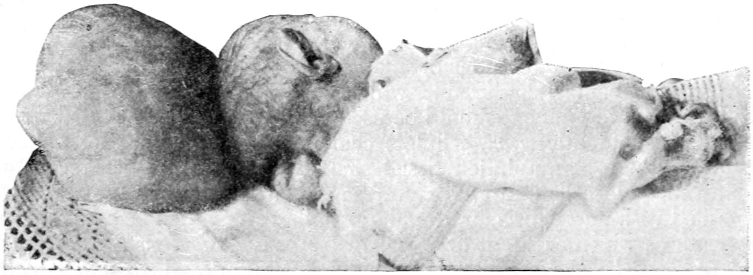
Occipital cephalocele.
Such tumors of non-traumatic origin can only be explained by the existence of congenital defects which permit the escape of that which the normal bone retains within normal limits. In most instances the defect is in the middle line, at either one or the other extremity of the skull. In some instances the arches of the atlas, or even of other cervical vertebræ, are lacking. The most common cephaloceles are the occipital, which are known as inferior when below the occipital spine, or superior when above it. Those appearing anteriorly are known as sincipital, and are met with most often at the root of the nose, where they may communicate with the orbit or the nasal cavity. Other and irregular forms are laterally or unsymmetrically located (Figs. 380, 381 and 382).
Cephaloceles have an elastic feeling, many of them an exquisite fluctuation. Sometimes by touch alone we recognize both their fluid and solid contents. A meningocele with thin walls is translucent. By pressure they can be reduced in size, such pressure usually producing brain symptoms, often paralysis or convulsions. Many children thus affected cannot lie upon the tumor without becoming restless. When the patients cry or make violent straining efforts it becomes larger and its covering more vascular, while during quiet sleep it is usually reduced in size or tension.
Fig. 381
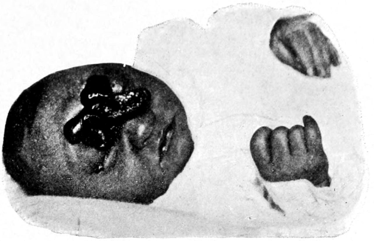
Sincipital meningocele.
A large proportion of patients with these congenital defects die shortly after birth. The tumor, when large, may be ruptured during delivery. Occasionally the sac ruptures spontaneously, which accident is usually followed by purulent meningitis from infection, though it may possibly lead to spontaneous recovery. The principal danger is the liability to such accident.
Fig. 382
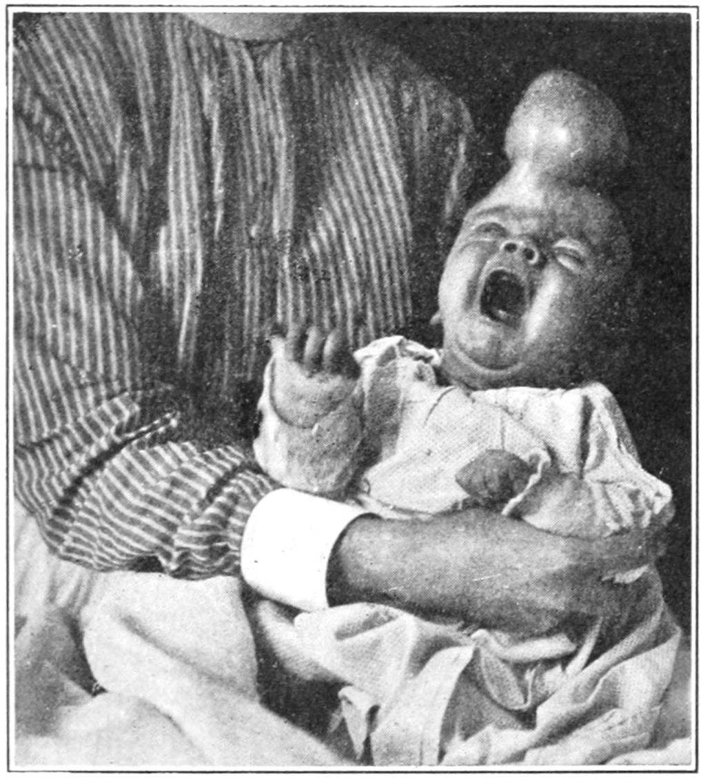
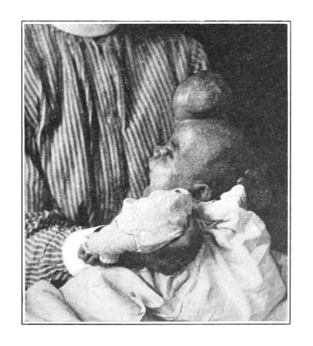
Sincipital hydrencephalocele; two views. (All of these from the Buffalo Clinic.)
The encephaloceles are divided into the cenencephaloceles, containing solid brain substance, and hydrencephaloceles, consisting of the protrusion of a dilated brain cavity—i. e., a thin area of brain enclosing fluid communicating with one of the ventricles. Most of the large tumors pertain to the latter class. The more brain material such a tumor contains the more it pulsates, especially if the patient cry or strain; the smaller, too, is the skull—i. e., the greater the tendency toward microcephalus.
Congenital cysts of brain and membranes, in a measure traumatic, are classified by Rawling, as follows:
1. According to situation: (a) within the calvarium, subosteal, subdural, subarachnoid or intracerebral; (b) projecting through an opening in the skull, with or without ventricular communication.
2. According to origin, i. e., whether they arise from blood clot or other causes.
3. According to contents, whether they contain clear fluid, altered clot, or brain substance.
Cysts of this character are to be differentiated from the cephaloceles already considered because there is about most of them the element of traumatism, although this may have been intra-uterine or produced during parturition. Those which are associated with premature synostosis and microcephaly, with hydrocephalus, with marked deformity, or situated below the external occipital protuberance, are generally considered inoperable, while those considered operable consist of limited protrusions without any of the above defects. This practically excludes the greater proportion of these cases from operation, which is always dangerous. Nevertheless if success is to be achieved the risks should be taken. Osteoplastic methods of closing cranial openings may be perhaps of value in rare cases, although in the young the skull is too thin to furnish an external table which can of itself be detached. In inoperable cysts of this kind, with a tendency to increase, while the rest of the brain lags behind in the rate of growth, the edges of the opening become everted, and operation is thus made more difficult and less desirable.
Treatment.
—Treatment should, first of all, be protective, by a shield of some device held in place by a suitable bandage or dressing. Compression, with or without puncture, has given at times satisfactory results, but not much should be expected from any method or combination. Most of the cases are such that extirpation would seem applicable, but the impossibility of absolute asepsis in young infants and the liability to fatal shock preclude many of these attempts. In some instances ligature of a meningocele has been successfully applied. Operation may be attempted in young children in selected cases. Plastic operations may be resorted to, or plastic maneuvers combined with extirpation. It may be possible by the insertion of a celluloid plate to atone for a small defect in the skull after extirpation of a tumor of this kind. I have successfully practised this method in spina bifida.
HYDROCEPHALUS.
This term is applied to abnormal collections of cerebrospinal fluid within the cranial cavity. We speak of—
A. Hydrocephalus ventriculorum or internus, when the fluid is confined to the dilated ventricles of the brain; or of—
B. Hydrocephalus meningeus or externus, when the fluid collects between the brain and the dura.
The former condition is much the more common. The cause of hydrocephalus in the young is essentially congenital, and inseparable from imperfect development within the cranium. The forms are occasionally combined. At the time of commencing trouble the skull may be of natural size, but yields to the accumulation of fluid within until it attains relatively enormous dimensions. Most children thus affected die early, some shortly after birth. It is most common in rachitic children. Hydrocephalus developing in the adult is the result almost solely of atrophy of the brain. Pachymeningitis interna (see p. 572) may also produce subdural exudate leading to hydrocephalus externus. Encapsulated collections of cerebrospinal fluid due to pachymeningitis interna are known as hygromas of the dura. A ventricular form of hydrocephalus may also result from meningitis and tuberculous disease. The condition is essentially chronic, the fluid collecting in the dilated lateral ventricles, though the third or forth are sometimes also distended: 4000 Cc. of cerebrospinal fluid have been found in more than one instance. As the result of the presence of the fluid there is atrophy of brain, with arrest of development, to such an extent even that the hemispheres are changed into great sacs, being merely spread out upon the outer wall of cystic cavities; all the surface markings are lost, and gray and white substances are scarcely to be differentiated.
In the cranium itself the bones of the vertex separate, and instead of sutures there is a tightly stretched membrane. There is also congenital or acquired aplasia—i. e., absolute defect of bone between the dura and pericranium. All these changes give to hydrocephalic heads a distinctive appearance. Other developmental defects—hare-lip, club-foot, etc.—are common in these patients. Many infants thus affected die during delivery unless skilful help is at hand. The resulting disproportion between the enlarged head and the small face is most distinctive. Children in this condition suffer from disturbed digestion, are emaciated, with rachitic curvatures of the long bones; special senses are seldom developed perfectly; strabismus and nystagmus are frequent, while cramps and stupor are by no means infrequent.
Prognosis.
—While spontaneous recovery is possible, as already stated, the tendency is always toward fatality.
Treatment.
—Treatment by compression of the enlarging skull, with elastic bandages or their equivalent, is an abandoned method since compression which can be effective is too great to be tolerated. Treatment by mere aspiration is also useless. Tapping is an old operation long discontinued, recently revived, but again proved disappointing. The establishment of permanent drainage is a more recent suggestion. It depends upon the demonstration of the fact that the tension of the cerebrospinal fluid and of the blood in the cerebellar veins is the same, and that intracranial pressure forces fluid into the veins and away from the skull. Thus subdural or autodrainage was suggested. Sutherland and Cheyne, in 1898, were the first to operate in this manner. They opened the dura near the lower angle of the anterior fontanelle, through the opening carried a strand of catgut into the ventricle, and passed the outer end beneath the dura; but the method again proved disappointing. Mikulicz passed a gold tube into the right ventricle, leaving its outer end in the subcutaneous tissues about 5 Cm. from the middle line. After being three weeks in this position it ceased to drain, and was then inserted into the other ventricle. The child died, unbenefited, in six weeks. In another case he used a glass-wool drain, making it subdural rather than subcutaneous. This case seemed to be benefited. Senn has modified the method by making a large pocket in the subcutaneous tissues of the cervical region, inserting one end of a rubber tube into it and carrying the other into the ventricle between the temporal and frontal bones. Even this proved disappointing. I have twice tried conducting fluid by a small rubber tube from the ventricle into the cellular tissue in the neck, passing the tube beneath the skin by suitably curved forceps. This method, however, showed no advantage over the others mentioned above. Taylor has endeavored to make a permanent fistula between the ventricles and the subdural space by passing chromicized catgut into the ventricle and letting it drain into the latter. His results, however, were not encouraging, in spite of the plausibility of the theory upon which they were based. Drainage through the spinal canal into the abdominal cavity has also been practised by a very few surgeons. The ingenuity and theory of the method are most attractive, though but very few little patients are in condition to bear the abdominal section which is necessitated for the purpose.[43]
Permanent drainage, then, has been a most disappointing procedure, although there need be no hesitation in tapping the lateral ventricles when there is indication for it. This can easily be done at any time by an opening about 3 Cm. behind the external auditory meatus and the same distance above the base-line of the skull. By directing the puncturing instrument to a point on the opposite side, 6 Cm. above the meatus, the lateral ventricle will be entered. (This same general direction will serve for opening an abscess in the temporosphenoidal lobe.) The best results in hydrocephalus seem to have been obtained by lumbar puncture, as first suggested by Quincke, the method being the same as that now in general use for intraspinal cocainization. As directions for entering the spinal canal with the aspirating instrument would be identical with those mentioned in the chapter on Anesthesia, when describing intraspinal cocainization, the reader is referred to that section for further direction (p. 208). The only case of well-marked hydrocephalus which I have ever apparently cured was one repeatedly tapped in this fashion, a considerable amount of fluid being withdrawn at each little operation.
SURGICAL TREATMENT OF DEFECTS OF INTRACRANIAL DEVELOPMENT.
There are numerous causes which produce imbecility and kindred conditions in the young. Some are in effect congenital, some are postnatal. Within the past few years a number of these cases have been subjected to surgical operation, in many instances with more or less success. Mental defect may occur from injuries at the period of birth—mainly hemorrhages, more commonly cortical, though sometimes deep. In either case the clots thus formed frequently undergo cystic alterations. The term porencephalon is modern, and applied to changes comprising disappearance of real nerve tissue with partial substitution by connective tissue, often with other degenerations, the result being atrophic alterations which apparently permit of no remedy. In a case of true porencephalon the outlook for operation is not at all encouraging, nor is it in any cases which are accompanied or caused by a genuine arrest of cerebral development. On the other hand, when the mental condition can be ascribed to the result of injuries, to hemorrhages, to meningeal irritation, to premature ossification, or too early closure of the fontanelles, or when it is accompanied by evidence of meningeal irritation or symptoms which point to a definite area of the brain as being the site of the principal disturbance, operation as a legitimate experiment may be conscientiously suggested and performed.
Fig. 383
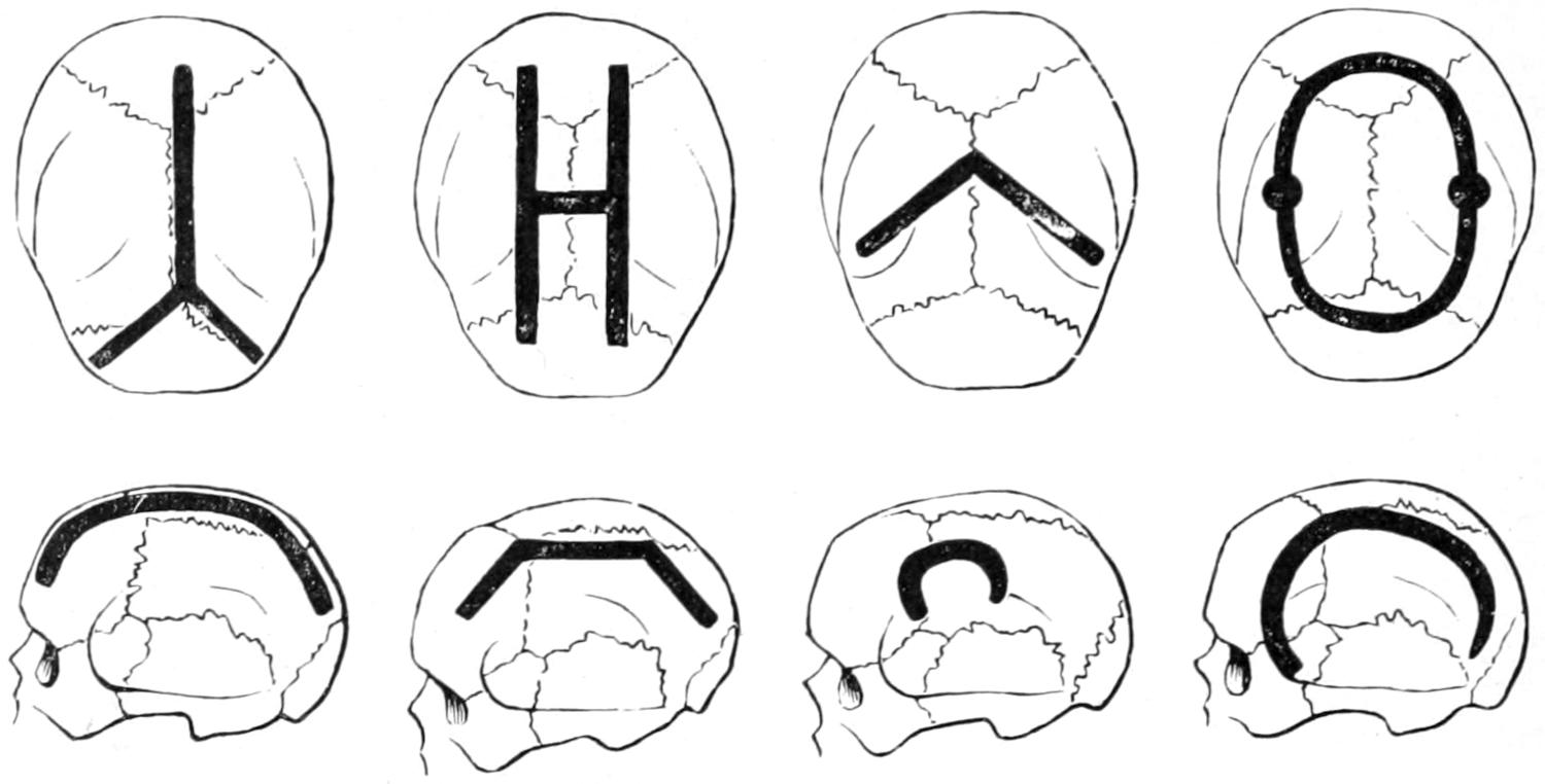
Lines of removal of bone as practised by the author, by Lannelongue, and by others.
Fig. 384
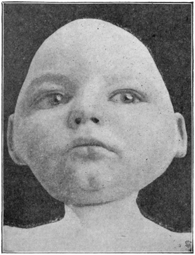
Defective cerebral development. (Buffalo Clinic.)
The operation is usually described as craniotomy or craniectomy, and is apt to be successful in many cases of microcephaly combined with idiocy. An acquired form will give a better prognosis than will the congenital condition. The danger of the operation is often great, and especially so since it is called for in puny, ill-nourished, and badly cared-for children. To be successful it ought to be extensive. It should vary in character and degree—from simple division of the skull along the middle line, from near the root of the nose to the occiput on one or both sides, to the formation of large bone flaps by cutting away a wide groove of bone so as to relieve pressure upon the hemispheres. Fig. 383 presents the various ways of performing the operation.
It can usually be made bloodless, or nearly so, by an elastic tourniquet around the skull. The incision in the skin should not correspond to the groove in the bone, but should overlap it some little distance. For my own part I prefer to do most of these operations in two sittings. I would advise, as a rule, to prepare the scalp carefully for operation, to divide the skin along the proposed line, separate it from the pericranium and check all oozing; then, after opening the skull with the trephine, to cut away with proper forceps (rongeur) along the desired line, or, if provided with it, to remove the bone by some surgical engine or revolving saw operated by electricity. The strip of bone thus removed should be at least half an inch wide, and the overlying periosteum should be removed with it, as only in this way can the undesirably rapid regeneration of bone be prevented. By this means the dura is exposed, but not opened. In some cases this will be sufficient.
In many others, however, it will be insufficient; and, could this be foreseen, it would be well to combine the above measures in one as a first operation, and then, a few days later, to open the dura as the second procedure—this, however, only on the discovery by careful inspection that the wound is absolutely free from possibility of infection. Could infection be prevented, this is certainly the safer procedure, since in weak, puny young children to make a long scalp incision, to remove a long strip of bone, and then to widely open the dura is more than can safely be done in the majority of instances.
It should have been carefully explained to those interested in the case that improvement will in all probability be extremely slow, and that little or nothing is to be expected at first, even if prompt recovery from the operation ensue. Neither would I advise any one to perform the operation unless parents are willing to assume all risks and abide by the results.
SURGICAL TREATMENT OF EPILEPSY AND THE PSYCHOSES.
Operations for relief of epilepsy seem to date back even to the prehistoric era, and were for centuries done as a purely empirical measure; later, to have been practised with more or less plausible reason; then to have fallen into discredit for long periods of time, with occasional revivals of the practice, until within the past twenty-five years the operation has been again revived upon its merits and upon the recognition of more or less accurate indications.
Operations of this character are based upon two fundamental facts: the first, the widespread experience that after various operations epileptic patients have been benefited; and, second, that a certain proportion of these cases, especially those of traumatic origin, are characterized by a localized and definite aura, and by a systematic and practically invariable order of muscle involvement, according, it would seem, to some fixed law, and pointing definitely to a certain area of the brain from which apparently the irritation arises and spreads. This form of epileptic seizure is that generally known as the Jacksonian, and is that in which operation is most often of real service. The statements of patients regarding these phenomena should never be accepted; only those made by a trained observer (nurse or physician) are reliable.
In spasms of the Jacksonian type there is a certain order of progression which is scarcely ever violated. Thus, irritation beginning in the leg centre can hardly reach the face centre without traversing that of the arm. It is possible also to have sensory equivalents for Jacksonian attacks, as when they commence with peculiar sounds indicating irritation in the centre of hearing, or with optical phenomena, or with disturbances of smell or taste, the former indicating occipital irritation, the latter irritation in the temporosphenoidal region.
The surgeon will often be consulted as to the wisdom of operation in the presence of this condition. In brief, and in a general way, the following statements may be made: It is necessary, first of all, to establish a traumatic origin, and epilepsy which has preceded a severe head injury can in no sense be ascribed to it. If it can be clearly established that it has followed injury, and if a distinct scar—especially a scar which is adherent—or depression can be discovered, or any area which is always irritable and which seems epileptogenic when irritated; or if, again, by close study of the case it can be determined that the aura and the initial muscle symptoms arise always in the same part—as, for instance, a finger, thumb, foot, etc.—and proceed according to a constant program—then it may be said that operation is not merely justifiable, but advisable. On the other hand, when neither distinct scar nor history of localizing phenomena can be obtained operation should rarely be attempted.
Again, in epilepsy of the non-traumatic type, operation may be advised when it assumes the distinctly Jacksonian form—i. e., when everything points to irritation proceeding from a localized portion of the brain. In the absence of Jacksonian symptoms operation is even more of an experiment than in the traumatic form. Such cases should be studied a long time on their merits before a decision is made to trephine.
The operation itself is directed to excision of irritable scars, to exposure of the dura at the point of opening, to the detection and suitable treatment of depressed fragments, dural adhesions, tumors, foreign bodies, etc. It is essential in every case that it be represented to those interested that the operation itself removes the cause, but cannot be, per se, expected to complete the cure, especially in cases of long standing, and that the final cure must depend in large measure upon the avoidance of subsequent irritation, upon the establishment of perfect habits of diet and excretion, which are often perverted, and perhaps upon the long-continued administration of drugs, of which the bromides are those most constantly given. The reader need not be reminded that old cases are the least favorable, and that recent cases are the most so for operation, and that the longer the diseased condition has existed the harder it will be to cure by any method.
Besides these direct operative attacks it has been suggested by Alexander to tie the vertebral arteries (now practically abandoned) and by Jonnesco to excise the superior and middle cervical sympathetic ganglia. This seems to me particularly indicated in those cases where a convulsion can be aborted by prompt administration (by inhalation) of amyl nitrite as soon as the preliminary aura is recognized. The operation is described in the chapter on Surgery of the Cranial and Cervical Nerves. Many encouraging results of this treatment have been reported.
I believe thoroughly in operating in selected cases. I am equally confident that indiscriminate operation must lead only to disappointment and to occasional disaster. In the presence of long-standing lesions, like bone depressions, cystic degeneration of old clots, etc., the brain may have been so long pressed upon as to have become atrophied.
The whole subject of the modern surgical treatment of epilepsy is inseparable from the topic of prompt and efficient treatment of all head injuries. Were the indications in these always met at the time of the accident we should have a much smaller proportion of cases of traumatic epilepsy.
Inasmuch as one object of many of these operations is to break up adhesions between the dura and the pia, there is generally anxiety to know the result after such operations as to whether they do not speedily form anew. There is always this theoretical danger, and it is my custom in such cases to insert beneath the dura, at the point where such adhesions have been divided or torn, a piece of delicate gold-foil, duly sterilized, in order that it may separate these surfaces and prevent the recurrence of the old condition. Foil used for this purpose is harmless, and I have numerous patients in whom it has been used, apparently without producing the slightest disturbance. (Foils of silver or aluminum answer as well or better.)
Mental and psychic disturbances after head injuries have been long known and the suggestion to operate upon the skull in cases of so-called traumatic insanity is not new. In a general way it may be said that whenever distinct mania follows a recognized lesion of the vertex of the skull, and fails to subside within a reasonable time and under proper treatment, there are the best of reasons for raising the scalp, trephining, and exploring as to the deeper conditions. Patients might be released from asylums who have long been inmates had this measure been practised at the beginning of their mental alienation.
The same measure will give relief in certain cases of cephalalgia, or headache, where the pain is always ascribed to a particular region, and especially when there is tenderness over this region. These operations are, of course, empirical, yet, as the result of altered nutrition and allayed irritation, relief follows in a fair proportion of instances.
INTRACRANIAL TUMORS.
Until within recent years these were regarded as having interest mainly for the pathologist and clinician, but as essentially hopeless so far as surgical help is concerned. Recent discoveries in the field of cerebral localization and recent experience with extensive openings into the cranium have shown, however, that a small proportion of intracranial tumors are of such a character and so located as to make them amenable to surgical relief. These tumors occur with about equal frequency in childhood and adult life. In the order of frequency they stand about as follows: Tuberculous gumma, glioma, sarcoma, cysts, carcinoma, and syphilitic gumma, with a small proportion of fibroma, etc.
Of 100 cases of brain tumor selected at random not more than 5 to 7 per cent. are so placed as to justify surgical attack. In as many more, at least, the tumors are so located as to justify opening the cranium for mere relief of pressure without any notion or endeavor to attack the tumor itself. Before opening the cranium diagnosis should be made as carefully as possible—first, as to location; second, as to whether cortical or subcortical; third, as to the number of tumors present; fourth, as to their general character. Location is determined in the main by study of pain complained of, by watching patients during convulsive seizures, by determining the extent of local or general paralysis, by careful history which shall reveal the method and rate of extension of these symptoms, and by the study of the optic disks, of vision, and by noting the presence or absence of stupor, nausea, coma, slow pulse, or other compression symptoms.
Symptoms.
—A brief epitome of the principal features attending cases of brain tumor will include:
1. Pain and headache, rarely localized with much accuracy; the former sometimes increased by percussion or pressure, occasionally periodical and usually intense. The location of the pain sometimes corresponds with that of the tumor.
2. Vomiting, usually without pain or nausea, and often projectile. I have repeatedly seen obstinate constipation in brain-tumor cases which has gone almost to a degree of acute obstruction, and which has caused serious error in diagnosis.
3. Vertigo, independent of indigestion or the condition of the stomach or bowels. It is most frequent in cerebellar tumors, but occurs in about 50 per cent. of all cases. It is sometimes quite severe.
4. Eye symptoms such as optic neuritis, choked disk, usually double, indicating pressure, but telling little or nothing as to the location of the tumor causing blindness. Ophthalmoplegias are of little value by themselves as symptoms. Hemianopsia, when homonymous, usually indicates a lesion of the cuneus of the same side, the blind half, according to the patient, indicating the side.
5. Localizing symptoms which may be due to the destruction of brain tissue or to indirect pressure. Those of importance comprise paralysis or spasms, indicating involvement of the motor area; sensory aphasia, indicating trouble in Broca’s area, ataxia or staggering, due to cerebellar lesions; loss of sense of position, sometimes seen in lesions of the parietal regions; anesthesia, which is rare unless the internal capsule is involved. Other symptoms are: word-deafness, which indicates a lesion of the posterior part of the first temporal convolution; agraphia, indicating deep lesions under Broca’s speech centre, and alexia, usually produced by lesions of the lower left parietal lobe. Tumors in the sensory zone affect vision and speech, and reveal themselves by irritative symptoms. For instance, a patient with verbal deafness and marked hemiplegia probably has tumor involving the left superior or dorsotemporal gyrus, which, as it grows, would involve loss of muscle sense and anesthesia on the opposite side of the body. A patient with headache, vomiting, choked disk, stupor, increasing hemianesthesia, lateral hemianopsia, without spasm or hemiplegia, probably has a tumor in the white substance of the occipital lobe. If hemianopsia alone be present there is almost always a tumor upon the inner aspect of the occipital lobe, on the side opposite to the dark half-fields, which by downward growth may cause cerebellar symptoms. Psychic and mental disturbances are present in many cases, but not in all; most frequently in frontal lesions. They are met with in about one-third of the cerebellar tumors and two-thirds of the temporal tumors; they assume the epileptic type, with hallucination, mania, or sometimes convulsions of Jacksonian type, the latter, of course, indicating lesions of the motor area.
6. Finally there are frequent constitutional disturbances, including anomalies of thirst and appetite, and disturbances of heart and respiration. In two or three instances the writer has seen such serious obstruction of the bowel as to lead to mistake in diagnosis, the obstruction in each case being finally fatal, but apparently not justifying operation.
The above symptoms pertain to the brain tumors in general. When it comes to tumors of the cerebellum these constitute, in a measure, a class by themselves. Those which are operable comprise tumors located in one lateral lobe, or invading the vermis or middle lobe, or those found at the junction point of the cerebellum, medulla, and pons, those first mentioned being by far the more favorable for attack. It is not relatively difficult to decide upon the presence of a tumor in the cerebellum, but to minutely locate it is extremely difficult. In addition to the symptoms already rehearsed above the following features may be mentioned: Headache is often intense, sometimes agonizing. While usually referred to the back of the head it is occasionally frontal. Nausea and vomiting are generally present, at least for a time. Sometimes they subside to recur later. Optic neuritis and choking of the disk occur earlier and oftener than in other tumors. Blindness sometimes comes on promptly. Vertigo, as in other brain tumors, is commonly due to irritation of those branches of the fifth nerve which supply the inner surface of the dura, this irritation being reflected to the bulbar nuclei of the fifth, and thence to the nuclei of the pneumogastric. This is partly true of those growths which are in relation with the dura, though sometimes it is true of tumors which make pressure at the base of the brain. It is important to distinguish, if possible, between mere vertigo and cerebellar ataxia. The more directly focal symptoms are: nystagmus, which may be present when the eyes are quiet or only when they are in use; paralysis, when the pyramidal tracts are involved; muscle weakness, seen more often in the legs, which is nearly always a cerebellar symptom; and sometimes a peculiar posture of the head, where the spinal column becomes concaved toward the affected side, the face looking almost backward. Incoördination is a common indication; in about four-fifths of the cases patients stagger in their gait.
To determine whether a given tumor is an irritative or destructive lesion special study should be made of the spastic or non-spastic condition of the limbs, and note to which side the eyes are turned. Tonic spasms and contractures are rare in cerebellar tumors. A tremor of the head and upper part of the body is not infrequent, and muscle sense is rarely lost.
Between cerebellar tumors and those of the parietal region the chief diagnostic points are muscular and cutaneous sensibility in the former, with nystagmus and peculiar and extreme vertigo. From frontal growths they may sometimes be differentiated by the clearness of the mental processes and the absence of those symptoms which point especially to involvement of the temporocortical region, e. g., aphasia. In cerebellar tumors convulsions, one-sided or general, are not infrequent, and incontinence of urine and feces is often noted. The convulsions are accompanied by subjective sensations and noises, vertigo, and by sudden blindness, with loss of consciousness, while such tonic spasms as occur are generally of the extensor type, and last from one to ten minutes.
Basal tumors of the cerebrum produce a collection of symptoms which sometimes are significant. Owing to their location they involve the functions of several of the special nerves. In tumors in the anterior fossa there is involvement of the optic, the oculomotor, and the first branch of the fifth. In tumors of the pituitary body there is involvement of the optic, the chiasm, the oculomotor, and the first branch of the fifth, as well as the abducens. In tumors resting on the middle fossa and situated above the dura the oculomotor, the patheticus, and the chiasm are involved. If situated beneath the dura there is paralysis of the three ocular nerves and also the fifth nerve. In tumors of the posterior fossa there is involvement of the facial, the trigeminus, the auditory, the glossopharyngeal, the vagus, the accessorius, and the abducens.
Neurofibroma of the Acoustic Nerve.
—Fränkel and Hunt have recently shown that basal tumors spring from the acoustic nerve, which are essentially neurofibromas. They have their site upon the nerve at the point where it merges from the junction of the pons and the medulla; in other words, where the function of the nerve is more or less disturbed, and the patient thereby made to complain of deafness, tinnitus, and vertigo. They slowly displace surrounding tissues. They vary in size from a cherry to that of a robin’s egg, are loosely attached, and when exposed easily enucleated. Their general symptoms are those common to all brain tumors, but focal symptoms may include ataxia, paralyses (especially of the fifth, sixth, and seventh nerves), inequality of the pupils, and loss of coördinate movements of the eyes; these symptoms are in addition to those of the auditory already mentioned.
Access to these tumors is a serious matter. It should be undertaken in two stages: the first including a large lateral exposure, with or without an osteoplastic flap, comprising the lower portion of the squamous, a part of the occipital, and perhaps even the posterior aspect of the mastoid. Drainage will be required for a few hours as in other similar operations.
As to depth and number the former may only be learned by studying the nature and location of the signal symptoms, the presence and order of appearance of the same, presence or absence of headache, and local changes in temperature. Tumors occurring in tuberculous individuals are probably multiple. When different centres or systems are involved multiple lesions are usually present.
It has been held that the three cardinal symptoms of brain tumor are optic neuritis, headache, and vomiting; and while each of these is significant, and all of them are corroborative, they are not necessarily present nor does their absence exclude possibility of tumor. Other signs indicating the presence of tumor, it is a mistake to wait for the development of these three. The most distinctive feature of intracranial neoplasms is the progressive character of such symptoms as are present.
There is but one form of brain tumor which is amenable to internal treatment—namely, syphilitic gumma; and in case of doubt it may be justifiable to keep the patient actively under the influence of iodides for a reasonable length of time. This, however, need never be prolonged beyond six weeks, after which time, should no improvement occur, operation should not be delayed.
Operation.
—Brain tumors are operated for two purposes: First, for relief of pain and other distressing symptoms in incurable cases; second, for radical cure. Operation is justifiable in any case when pressure symptoms become severe, particularly so when pain is localized to a reasonable extent. Choking of the optic disks is not infrequently relieved and threatened disability postponed. The complete operation consists in the exposure of the tumor and in its removal.
The osteoplastic method should be used in exposing the tumor, by which a bone flap is raised, along with the overlying scalp, from which it is not detached. The centre of this flap is supposed to be calculated to overlie the centre of the deep lesion which it is proposed to attack. In many instances the operation should be divided into two distinct procedures, the first consisting in removal of the bone and exposure of the dura; this exposure should be ample, including the whole lateral region if necessary, as Horsley has shown; the second, a week or two later, comprising the balance of that which is to be done. But comparatively little shock attends removal of the tumor in the second stage of such a divided operation. After removal of the growth its cavity is best packed with a gauze tampon, after prompt ligation of all bleeding vessels within the field of operation, although it is usually required merely on account of venous oozing, as it is often possible to cut to the depth of an inch in the brain without a single artery spurting except those in the pin. The tampon is of value if allowed to remain for forty-eight hours, as preventing filling of the cavity with clot or excessive bleeding during the vomiting which may follow the administration of the anesthetic. The vasoconstricting properties of adrenalin may prove of great service here; it should be used in the standard 1 to 1000 solution, diluted 1 to 3. I have no hesitation in spraying this upon the brain or in saturating tampons with it, which may be left in situ so long as necessary. A number of the old-fashioned small serrefines, properly sterilized, can also be resorted to, if needed, for securing vessels, which may not be easily tied. They can be left in place along with the tampon and all may be removed together.
Next to the danger from hemorrhage is that of rapid edema of the brain, which may result from increased tension in the arteries or through venous stasis, which later produces lymph stasis, by which fluid collection in the tissues is still further facilitated. Another reason for using tampons is to prevent such relaxation of veins as may predispose to this edema. In most respects the operations for removal of brain tumors differ slightly from those whose general principles are elsewhere mentioned in this work. I am greatly in favor of using secondary sutures (i. e., those tied with bow-knots), which may be loosened on the second or third day, permitting the raising of the flap, removal of tampon, etc., and I employ them largely after all sorts of operations upon the cranium. If we desire to prevent any attempt at union of wound margins we may employ the green silk protective introduced by Lister, which should have been previously carefully sterilized by boiling.
The operative treatment of cerebellar tumors is made doubly difficult by their protected position and the large sinuses with which this part of the brain is surrounded. The cavity is restricted in size, intradural tension is greater than above the tentorium, and there is no room for easy displacement or retraction of parts. The occipital bone varies much in thickness and at points is somewhat thin. Operation which is begun either as an exploration or with a fixed purpose may prove palliative, even should the original purpose fail of accomplishment, as relief may be afforded by reducing tension, such relief consisting perhaps in freedom from headache, vomiting, and vertigo. Incision should extend from the tip of the mastoid process, a little above the superior curved line, to beyond the median line, with a vertical median incision by which a flap sufficiently large may be reflected downward. It is best to reflect the periosteum with the other soft tissues in order to expose the bone. The bone should be bitten away with forceps or removed with a reliable engine as rapidly as possible, hemorrhage being controlled with Horsley’s wax.
The operation may be divided into two stages, confining the first stage to the exposure of the cerebellar surface, or the operator may attempt all at one time.
The second stage consists in raising a dural flap, by which the cerebellar surface is exposed for inspection. It will protrude promptly through the opening, so that, with the finger, it may be possible to detect a tumor by the sense of touch. If no tumor appear on or near the surface deeper exploration should be made, with the aid of a retractor and by removal of a portion of the cerebellar hemisphere. This may require further exposure of the lateral region of the skull. Tumors situated deeply or at the junction of the cerebellum and pons require all the room that can be afforded from the outside, and are better approached from the lateral region than from above or below. It is comforting to realize what considerable portions of the cerebellum can be removed without serious or extensive disturbance, but as the medulla and pons are approached there is need of great care. The opening may be extended across the middle line, and either the lateral or the longitudinal sinus, or both, may be doubly ligated and divided. The tentorium may also be divided nearly to the petrous portion, after the lateral sinus has been thus divided, and so better access given to the deep location.
These remarks apply especially to operations for tumors of the cerebellum. The other features of such operative attack are those common to brain tumors in any location.
In all operations for brain tumor, but particularly for cerebellar tumor, it will prove of the greatest advantage to have the operating table so inclined that the patient’s head will be three or four feet above his heels. In this position the veins are drained by gravity, and the operation is complicated by but little venous oozing. Crile’s pneumatic suit, or at least the lower part of it, should be worn, and an assistant should watch and report on the blood pressure. These two precautions permit such an operation to be conducted with an ease and safety hitherto unknown.[44]
Cushing, dealing especially with a group of brain tumors in which radical procedures are impossible, where nevertheless relief from symptoms would prove a therapeutic desideratum, has proposed to afford this by removal of a portion of their bony covering, in order to allow a part of the brain to protrude, and thus provide a means of relief for the constantly increasing pressure. The incomplete union of the bones in infancy permits something of this kind to occur through natural causes, but after fusion of the elements of the cranial vault it is no longer possible, save in those rare cases where an opening results from the process of slow pressure absorption, which comes only when the tumor is in actual contact with the bone.
It would be mechanically ideal if, during adult life, a dislocation of cranial sutures could be produced similar to that observed in very small children. The dangers of such operation are many, among them being the possible injury to the functions of that portion of the cortex which protrudes through the opening thus made, by which, for example, preëxisting paralyses might be aggravated. For this reason it is preferable to establish the hernia over some “silent” or unimportant part of the cortex and to avoid making it unnecessarily large. Cushing, after various trials, recommends to make the bone defect under the temporal muscle, which not only affords a certain degree of protection, but exposes an area where few important motor centres are involved. He has reported several cases, with gratifying results, with a minimum of undesirable sequels.
Obviously in tumors below the tentorium the opening would best be made in the suboccipital region. Nevertheless, Cushing believes that even here the final result would be no more effectual than were the defect placed elsewhere.
Beck has called attention to the value of the temporal fascia as a substitute for the other firm coverings, by which the brain should be left enclosed after exposure, and when these latter are not available. For the purpose he would fold over a flap made from the temporal muscle and the adjoining periosteum in such a manner that fascia originally external should now be placed deeply and in contact with the cortex.
PLATE XLIV
FIG. 1
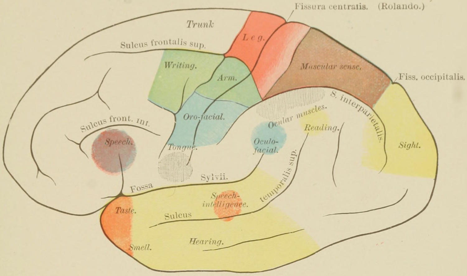
Topographical Anatomy of Cortex. Localization of Functions. (Ziehen.)
FIG. 2
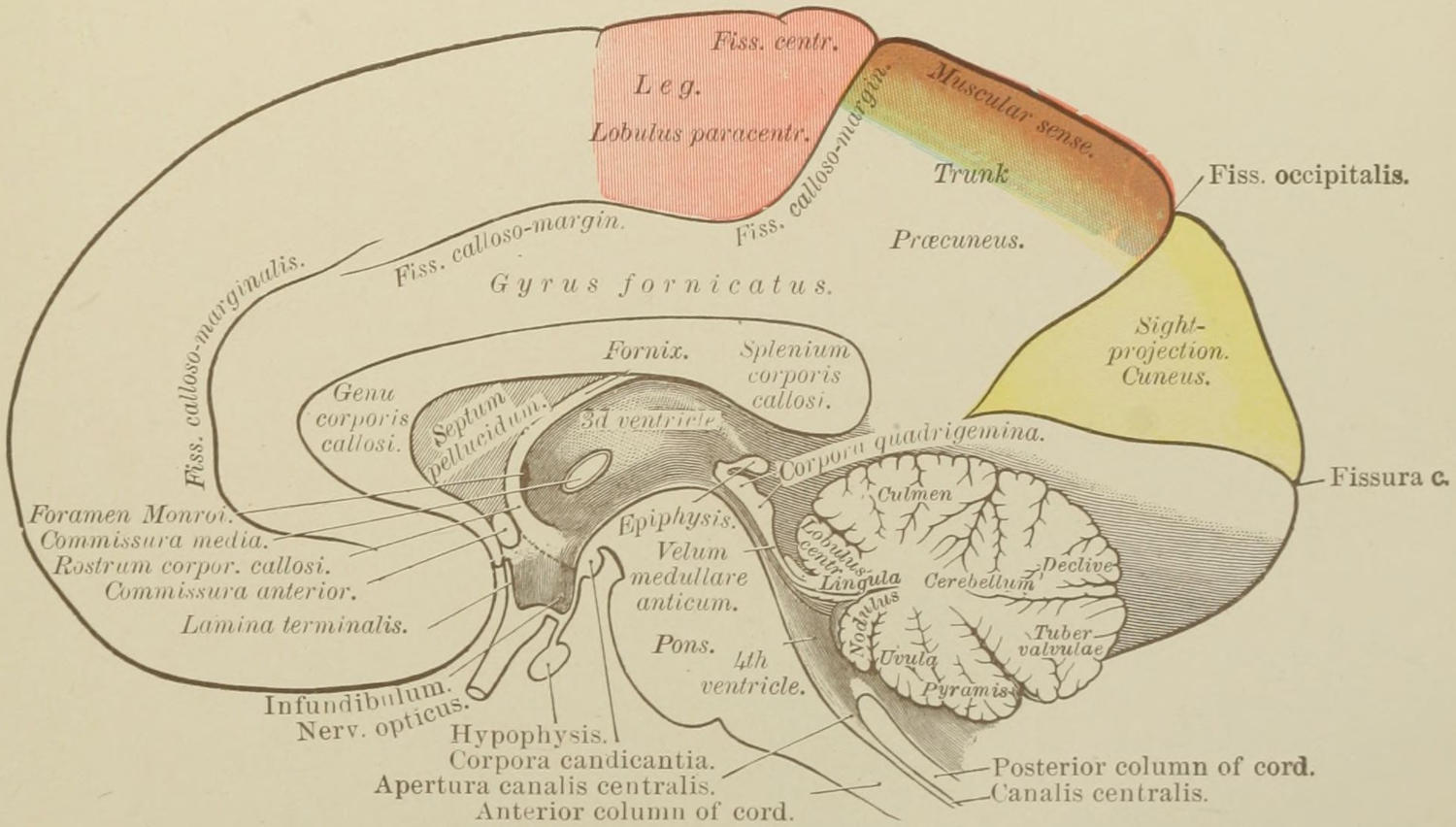
Topographical Anatomy of Inner Surface of Right Hemisphere. Localization of Functions. (Ziehen.)
OPERATIONS UPON THE CRANIUM.
The fissure of Rolando is the anatomical landmark whose position it is important to determine with reference to a number of modern surgical procedures, for around it cluster most of the motor areas or centres. It commences at the middle line about 56 per cent. of the distance backward from the glabella (root of the nose) to the inion (occipital protuberance), and, passing downward and forward, makes with the middle line an angle of 67 to 69 degrees. For most purposes it begins half an inch back of a point midway between the glabella and inion. It may be easily found by Chiene’s method, which consists in folding a square piece of paper diagonally and folding this again; after which it is three-quarters unfolded, the acute angle then representing 67¹⁄₂ degrees. If this be properly applied to the skull, one edge of its surface can be made to fall directly over the Rolandic fissure. The fissure may also be located by a simple instrument known as the cyrtometer—a gauged metal strip having a sliding arm upon it, which, when the long strip is placed over the longitudinal sinus (i. e., the middle line of the skull), can be made to fall directly over the fissure. While neither of these methods is invariably and minutely exact, either of them is sufficiently accurate for all practical purposes.
The fissure of Sylvius may be indicated by a line drawn from a point 3 Cm. behind the external angular process to a point 2 Cm. below the most prominent part of the parietal eminence. The short and ascending limb of this fissure is of relatively small importance in this connection.
Reid’s base-line, so called, is a line drawn from the inferior margin of the orbit backward through the centre of the external auditory meatus. It is a line often alluded to in cranial topography. The colored plate (see Plate XLIV) will indicate with reliable accuracy the relations of the motor centres to each other and to the principal fissures and convolutions. It pertains merely to the left hemisphere of the brain, in whose third frontal convolution is placed Broca’s centre for speech, the corresponding area upon the right side having no exactly corresponding function. The centre for vision, it will be seen, is located in the cuneus, the most basal portions of the hemispheres being the seat of the special senses of taste, smell, and hearing.
Operation.
—The word trephine is at present used both as a noun and as a verb, the older term trepan being now wellnigh discarded. The instrument consists of a section of a tube, one of whose extremities is arranged with sharply cut saw teeth, the whole provided with a grip or handle, which revolves in a plane parallel to that in which the saw teeth cut. The best instrument is that arranged in a slightly conical manner, so that it may less easily burst through the skull and do harm to parts within. The trephine proper is manipulated by the hand. A variety of substitutes have resulted from applications of human ingenuity to the problem of opening the cranial bones. Some of these are operated by foot or hand power, with reduplicated mechanisms, and others by electricity. The more complicated the mechanism the more likely it is to get out of order, and there are but few of these substitutes which give anything like lasting satisfaction.
The operation of trephining is made to include any method by which an opening is made in the uninjured cranium or by which an opening already existing is enlarged and made to subserve the surgeon’s purpose. Aside from the saws already alluded to, there are in use a variety of cutting bone forceps, rongeurs of various device, and a variety of chisels, which are to be used in connection with the mallet or hammer. In order to use any of the latter instruments to advantage the first attack should be made with a trephine of reasonable size, say 2 to 3 Cm. in diameter, after which forceps, chisel, or saw may be used. Straight saws also are of occasional usefulness. I do not favor the use of the chisel and mallet, feeling that the concussions resulting from blows of the hammer add to the shock of the operation. The common trephine is provided with a centre pin, which can be withdrawn after a shallow groove has been cut. To prevent slipping of the centre pin the point to which it is to be applied should be marked by cutting a nick with the point of a chisel.
The Gigli saw should be in every surgeon’s outfit. It consists of a piece of steel wire having a thread cut around and along it by a die, by which it is made as effective as a series of saw teeth. Two small trephine openings are made, and it is then passed into one and out of the other, the dura protected by depressing it, and the wire then handled as though it were a chain saw. It can thus be made to cut its way quickly through the bones of the skull.
Other aids in mechanical procedures are revolving small saws and the surgical engine.
Fig. 385
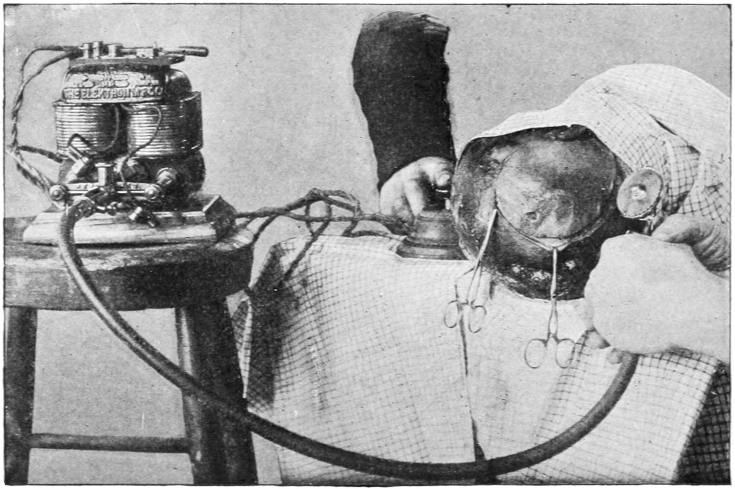
The Powell electric saw cutting a “trap-door” in the skull. (Illustrating the operation upon a cadaver.)
In the absence of a wound a flap of scalp is raised before applying the instrument. This flap is ordinarily of horseshoe shape, and should be made with its convexity pointing toward the occiput, as drainage is best afforded later by this arrangement. The old crucial incisions are now wellnigh abandoned. The pericranium is detached, after incision, with the periosteum elevator, and it should be turned up with its overlying scalp without completely separating it. The scalp flap can be held out of the way by temporarily sewing it to some other part of the scalp, every portion of which should be previously shaved closely and thoroughly scrubbed. The operator has his choice—to seize vessels as they bleed or to make the operation in large degree bloodless by applying an elastic tourniquet tightly around the scalp above the eyebrows and beneath the occiput, the ears preventing it from sliding. If the tourniquet be used the vessels will often bleed in an annoying way after the wound is closed. If the operation be performed for fracture of the skull, should there be an opening already made by the depression of fragments, it may not be necessary to use the trephine, but with suitable bone forceps fragments may be removed or detached. In this case, however, there are often sharp points of bone which will require removal by cutting bone forceps, for the surgeon should leave the margin of the bone opening comfortably round and smooth. Should there be no opening into which the point of an elevator or of bone forceps can be inserted, then one should be made; it is for this purpose that the trephine is mainly used in cases of fracture of the skull. It should now be applied upon a firm and undetached surface of bone, one which will bear the pressure necessary in the process of perforation. As used for this purpose it should be so applied that at least two-thirds of the circle cut by its teeth will be upon unbroken skull; the remaining segment of the circle may be over the fractured area. After it has begun to cut a distinct groove the centre pin should be withdrawn and the instrument maintained in its position during its work by a firm and steady hand, which will force it evenly through the bone and not exercise undue pressure. As the diploë is perforated the bone-dust becomes soft and bloody and the resistance is diminished. As the instrument sinks deeper the operator should frequently intermit its use, and determine his position by means of the irrigator and of the probe or other instrument. The nearer the inner surface is approached the more caution must be exercised, remembering that the bone is likely to be of unequal thickness. When the skull has been completely perforated at one or two points around the little circle the operator should introduce the point of an elevator and pry up the disk of bone, or by rocking the handle of the trephine he may be able to remove the button with that instrument. When the operation is performed in the ideal manner the dura is scarcely touched, certainly not raggedly injured by the teeth of the instrument (Figs. 386, 387 and 388).
Fig. 386
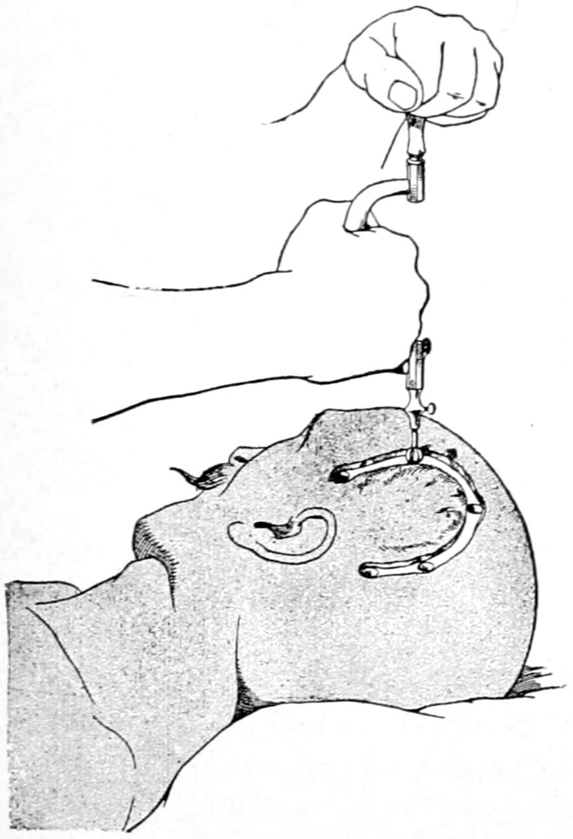
Construction of an osteoplastic flap; bone is exposed; first openings are made with a hand trephine or burr. (Marion.)
Fig. 387
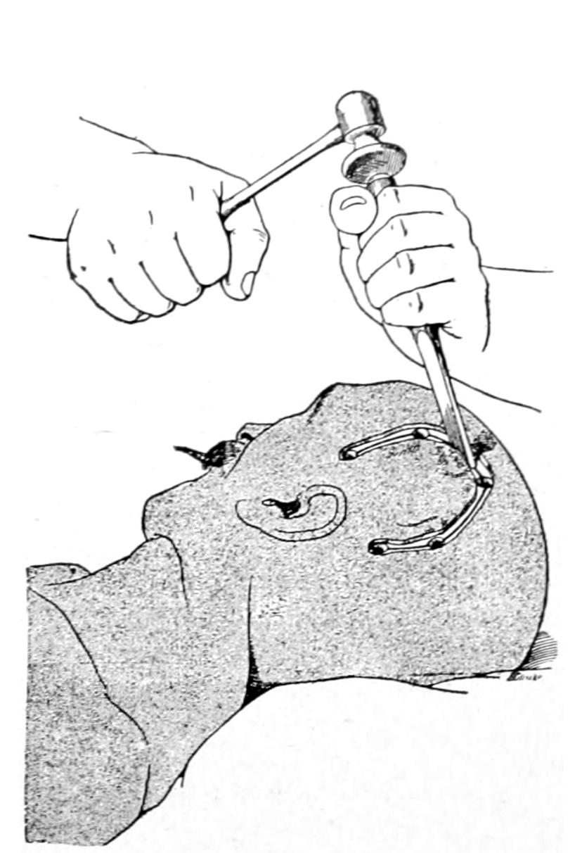
Division of bone by use of hammer and chisel. (Marion.)
Before opening the dura every loose particle of bone and every splinter should be removed, depressed fragments should be picked out, and those which are semidetached should be raised to their proper level. Through the opening thus made the dura is carefully examined; extradural collections of blood are recognized instantly, while some idea as to the amount of intracranial tension may be secured, even through a small opening. Absence of pulsation means probably the presence of cyst, tumor, or abscess deeper. Edema of the membranes usually subsides after nicking or opening them. A yellowish discoloration of the dura often indicates the existence of a tumor beneath. Nothing abnormal being discovered outside of the dura, should brain tension be great or should the dura be discolored, as by blood beneath, the membrane should be opened, by a triangular or horseshoe flap, and the subdural condition accurately estimated. In some cases of meningeal hemorrhage clots will be ejected with some force the instant the dura is opened. In other cases of intracranial pressure, either from tumor or from intraventricular hemorrhage, the brain will instantly protrude to such an extent as to make its reposition difficult or even impossible. Horsley’s dural separator is exceedingly useful, both outside and inside the dura, for detecting and separating adhesions, and as a retractor.
Incisions in the dura should be made, so far as possible, parallel with its vessels rather than across them. When accessible, dural vessels can always be secured and tied. Vessels of the pia can also be picked up and secured with fine catgut ligatures. When the brain tissue itself is diseased it should be carefully excised. The cortex itself is not so vascular as to afford much trouble. Upon any portion of the membranes or cerebral surface a sterilized solution of adrenalin can be sprayed or applied without hesitation. In all deliberate operations sinuses are avoided. When exposed or when necessary to attack them they may be ligated and divided, or may be packed with tampons of sterilized gauze, or may be seized with serrefines or light hemostatic forceps, which may be left for a day or two included within the dressings.
Any of the exposed motor areas or centres can be stimulated, when desired, if the patient be not too deeply anesthetized, by the faradic current of mild degree, applied to surfaces which have not been bathed with antiseptics, nor long exposed to the vapor of the anesthetic, through a double brain electrode made for the purpose, or by sterilized probes connected with the battery.
Buttons of bone or chips of the skull may be replaced after suture of the dura, when desired, though this is seldom advisable. When fragments are thus to be replaced they should be placed in warm sterile salt solution at once after removal, and kept warm. When a button is thus put back the periosteum may be sewed over it with buried catgut sutures.
The dura should be stitched with fine catgut as closely as possible. I have often placed beneath the dural opening a piece of gold, silver, or aluminum-foil, carefully sterilized, with a view to preventing dense adhesions between the dura and the membrane or cortex beneath. I have never known it to do harm.
Fig. 388
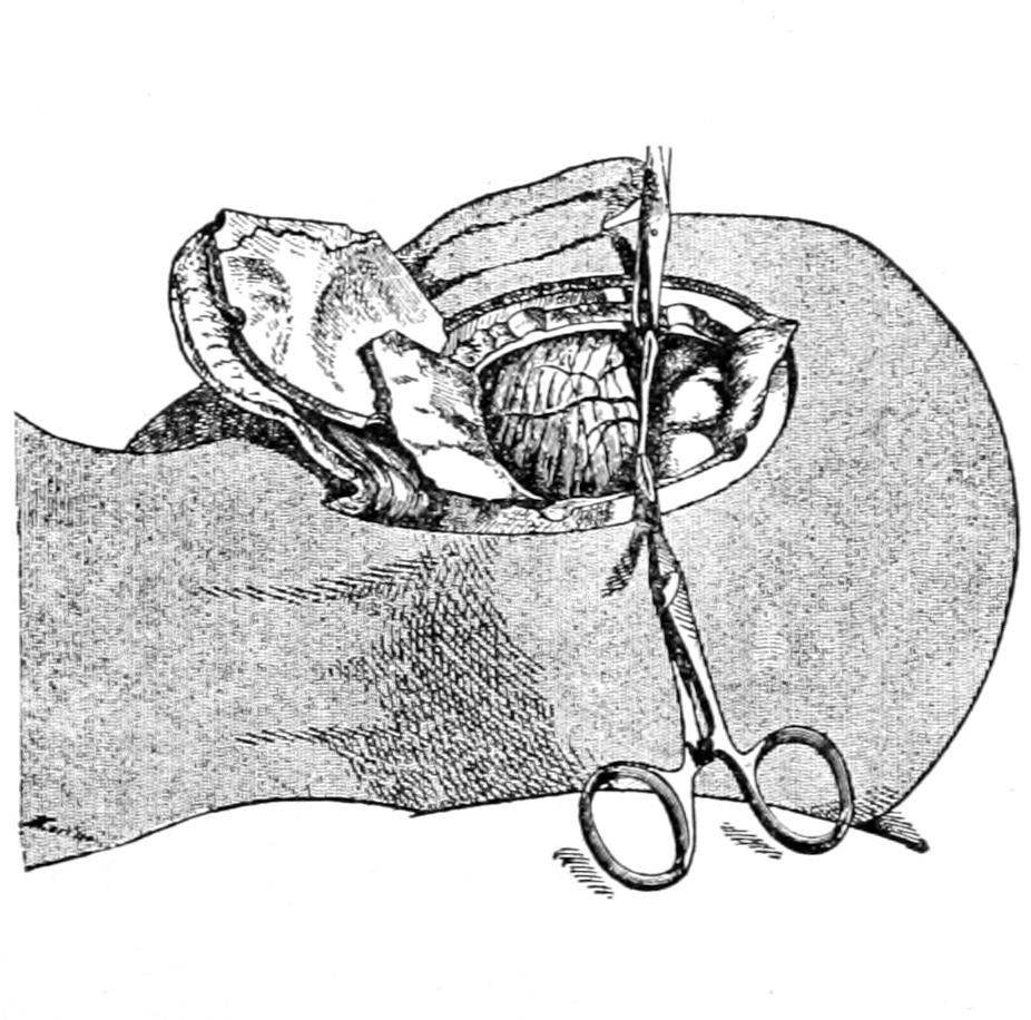
Exposure of cortex or of cerebellum after division of dura. (Marion.)
Fig. 389
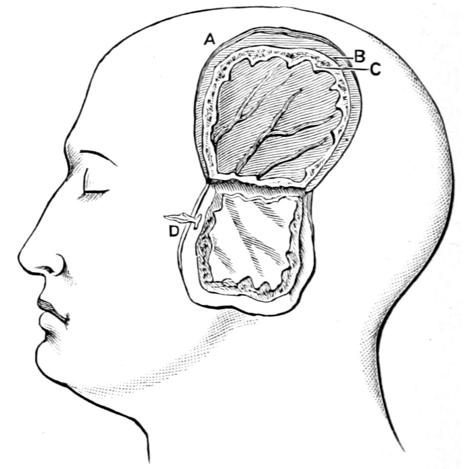
Osteoplastic resection after Wagner. (Chipault.)
Drains and drainage are to be avoided when possible, and should be removed early, except in cases of abscess. They may be made of catgut, horse-hair, gauze, rubber, or even of glass, like those short ones which Kocher inserts after extensive operations, their outer ends flanged to prevent their slipping beyond control.
Opening the skull, or, in general terms, trephining, is at present resorted to for the following purposes:
1. For relief of compression—
(a) By depressed bone, as in comminuted or gunshot fracture;
(b) By removal of clot or ligation of vessels;
(c) By evacuation of pus, either from the meningeal cavity or from a deeper abscess;
(d) By the removal of serous effusions, either extraventricular or intraventricular.
2. For removal of foreign bodies.
3. For relief of intracranial irritation—e. g., epilepsy, the psychoses, etc.
4. For removal of tumors.
5. To compensate for defective development.
6. For exploratory or purely empirical reasons, including the making of “relief openings” for relief of pain, etc.
Aside from the ordinary methods of trephining as applied for common conditions, modern surgery comprises the resort to essentially new methods for raising areas of skull of considerable size and then restoring them to their previous position. These are ordinarily spoken of as osteoplastic resections, and have added very materially to the art and resources of the surgeon. These consist, in a general way, of the formation of a window, as it were, in the vertex or lateral region of the skull by outlining a quadrangular or horseshoe flap of scalp, which is detached only for a slight distance around the incision, after which, by use of the revolving saw or by chisel and mallet, a groove is cut through the bone running parallel with the margin of the scalp-flap, but perhaps a centimeter within it. After this bone area is completely cut through on three sides it is then sprung up or elevated in such a way as to be broken across the base of the bone-flap. It is not at all detached nor separated from the scalp, and so when subsequently lowered into position retains its vitality by virtue of its vascular connections.
When some particular measure seems indicated in order to atone for a large defect in bone it has become quite customary to insert some artificial substitute, mainly either celluloid or a thin aluminum plate, previously absolutely sterilized and cut at the time into such shape as may be called for, but a trifle larger than the real defect, being let in or sprung in, as it were, either completely beneath the bone or into the bony opening, so as not to be easily detached or slip out of the way. By this heteroplastic method most admirable results have been achieved. I have used celluloid for this purpose in the spinal column also, closing with it the defect which remained after the extirpation of the sac of a spina bifida. It is rarely necessary to resort to this practice in the skull, as dense fibrous tissue in due time firmly protects the endocranial contents from external harm (Figs. 386, 387, 388 and 389).